Embed Size (px)
Citation preview
Care of the Patient with
Primary Angle Closure Glaucoma
OPTOMETRIC CLINICAL PRACTICE GUIDELINE
OPTOMETRY: THE PRIMARY EYE CARE PROFESSION
Doctors of optometry are independent primary health care providers who examine, diagnose, treat, and manage diseases and disorders of the visual system, the eye, and associated structures as well as diagnose related systemic conditions. Optometrists provide more than two-thirds of the primary eye care services in the United States. They are more widely distributed geographically than other eye care providers and are readily accessible for the delivery of eye and vision care services. There are approximately 32,000 full-time equivalent doctors of optometry currently in practice in the United States. Optometrists practice in more than 7,000 communities across the United States, serving as the sole primary eye care provider in more than 4,300 communities. The mission of the profession of optometry is to fulfill the vision and eye care needs of the public through clinical care, research, and education, all of which enhance the quality of life.
OPTOMETRIC CLINICAL PRACTICE GUIDELINE
CARE OF THE PATIENT WITH PRIMARY ANGLE CLOSURE GLAUCOMA
Reference Guide for Clinicians Prepared by the American Optometric Association Consensus Panel on Care of the Patient with Primary Angle Closure Glaucoma: Jimmy Jackson, O.D., M.S., Principal Author Leland W. Carr, III, O.D. Barry M. Fisch, O.D. Victor E. Malinovsky, O.D. David K. Talley, O.D. Reviewed by the AOA Clinical Guidelines Coordinating Committee: John F. Amos, O.D., M.S., Chair Barry Barresi, O.D., Ph.D. Kerry L. Beebe, O.D. Jerry Cavallerano, O.D., Ph.D. John Lahr, O.D. David Mills, O.D. Approved by the AOA Board of Trustees June 23, 1994; Revised October, 1998; Reviewed 2001 © American Optometric Association, 1994 243 N. Lindbergh Blvd., St. Louis, MO 63141-7881
Printed in U.S.A.
NOTE: Clinicians should not rely on the Clinical Guideline alone for patient care and management. Refer to the listed references and other sources for a more detailed analysis and discussion of research and patient care information. The information in the Guideline is current as of the date of publication. It will be reviewed periodically and revised as needed.

Primary Angle Closure Glaucoma iii
TABLE OF CONTENTS INTRODUCTION...................................................................................1 I. STATEMENT OF THE PROBLEM..................................................... 3
A. Description and Classification of Primary Angle Closure Glaucoma........................................................................... 3
1. Angle Closure Glaucoma with Pupillary Block................. 4 2. Angle Closure Glaucoma without Pupillary Block ........... 4 B. Epidemiology of Primary Angle Closure Glaucoma .................... 6 1. Prevalence and Incidence .................................................. 6 2. Risk Factors ....................................................................... 6 a. Race ....................................................................... 6 b. Family History ....................................................... 7 c. Age ....................................................................... 8 d. Gender.................................................................... 8 e. Refractive Error...................................................... 8 C. Clinical Background of Primary Angle Closure Glaucoma .......... 8 1. Natural History .................................................................. 8 2. Common Signs, Symptoms, and Complications................ 9 a. Subacute ACG........................................................ 9 b. Acute ACG........................................................... 10 c. Chronic ACG ....................................................... 10 d. Plateau Iris Configuration .................................... 11 3. Early Detection and Prevention ....................................... 11
II. CARE PROCESS ................................................................................. 13 A. Diagnosis of Primary Angle Closure Glaucoma ......................... 13 1. Patient History ................................................................. 13 2. Ocular Examination......................................................... 13 3. Provocative Testing ......................................................... 16 4. Assessment and Diagnosis............................................... 17 B. Management of Acute Primary Angle Closure Glaucoma.......... 18 1. Basis for Treatment ......................................................... 18 2. Available Treatment Options........................................... 18 a. Medical (Pharmaceutical) .................................... 18 b. Corneal Indentation.............................................. 21 c. Laser Treatment ................................................... 21 d. Surgery ................................................................. 23 3. Recommended Management Protocol ............................. 24 4. Patient Education............................................................. 25 5. Prognosis and Followup .................................................. 25
iv Primary Angle Closure Glaucoma
6. Management of Patients with Severe, Irreversible Vision Loss.................................................................................. 26
CONCLUSION ................................................................................................ 29 III. REFERENCES ..................................................................................... 30 IV. APPENDIX ........................................................................................... 43 Figure 1: Optometric Management of the Patient with Acute Primary
ACG: A Brief Flowchart................................................. 43 Figure 2: Frequency and Composition of Evaluation and
Management Visits for Primary ACG.............................. 44 Figure 3: ICD-9-CM Classification of Angle Closure Glaucoma ... 46 Abbreviations of Commonly Used Terms.............................................. 47 Glossary.................................................................................................. 48
Introduction 1
INTRODUCTION Optometrists, through their clinical education, training, experience, and broad geographic distribution, have the means to provide effective primary eye and vision care for a significant portion of the American public and are often the first health care practitioners to examine a patient with, or at risk for developing, primary angle closure glaucoma (ACG). This Optometric Clinical Practice Guideline for the Care of the Patient with Primary Angle Closure Glaucoma describes appropriate examination and treatment procedures to reduce the risk of visual disability from primary ACG. It contains recommendations for timely diagnosis, treatment, and when necessary, referral for consultation with or treatment by another health care provider. This Guideline will assist optometrists in achieving the following goals: • Identify patients in whom primary ACG is present or who are at risk
of developing primary ACG • Accurately diagnose primary ACG • Manage a patient who has an acute attack of primary ACG • Monitor and manage, as indicated, patients with intermittent or
chronic forms of primary ACG • Develop criteria for referral to the patient's primary care physician or
other health care practitioner when management options dictate • Improve the quality of care rendered to patients with primary ACG • Minimize the adverse effects of primary ACG and its management • Inform and educate patients and other health care practitioners about
the visual complications of primary ACG and the availability of treatment.
Statement of the Problem 3
I. STATEMENT OF THE PROBLEM Primary angle closure glaucoma is a relatively uncommon condition in the United States, accounting for less than 10 percent of all diagnosed cases of glaucoma.1-3 However, its importance as a health care issue is far greater than the relatively small number of cases would suggest. In contrast to open angle glaucoma, in which vision loss is slow and gradual, an acute attack of angle closure glaucoma can lead to blindness within hours or days. Prompt diagnosis and correct treatment are, therefore, critical. Accurate and timely diagnosis of the intermittent and chronic forms of primary ACG is also important because prophylactic treatment (peripheral iridotomy) can protect the eye against acute episodes and prevent damage from repeated intermittent attacks or chronic angle closure. A. Description and Classification of Primary Angle Closure
Glaucoma Glaucoma represents not one single clinical entity but a group of ocular diseases with various causes that ultimately are associated with progressive optic neuropathy leading to loss of vision. The glaucomas can be separated by etiology: those not related to another underlying condition, which are classified as primary, and those that are secondary to ocular or systemic disease. The glaucomas are generally classified as angle closure, open angle, mixed or combined mechanism, and developmental. In primary angle closure glaucoma intraocular pressure becomes elevated because the peripheral iris prevents aqueous from reaching the anterior chamber drainage tissue, the trabecular meshwork. The meshwork itself is presumed to function normally. In open angle glaucoma (OAG) aqueous has ready access to the trabecular meshwork, but drainage is impaired due to other mechanisms. Mixed mechanism glaucoma exists when both forms of glaucoma are present, a combination of ACG and OAG. Developmental glaucoma is caused by some anomaly of the anterior chamber that is present at birth and is associated with other ocular or systemic anomalies.4,5
4 Primary Angle Closure Glaucoma
1. Angle Closure Glaucoma with Pupillary Block The classification of angle closure glaucoma is based upon the presence or absence of pupillary block and whether the angle closure mechanism is primary or secondary (Table 1). With pupillary block, the normal flow of aqueous through the pupil from the posterior chamber to the anterior chamber is restricted. This block leads to increased pressure in the posterior chamber which pushes the peripheral iris forward (iris bombe) until it blocks the trabecular meshwork. Primary ACG with pupillary block exists when there is a predisposing anatomical basis, such as a narrow angle. Primary ACG may be further classified as suspect, subacute (intermittent angle closure with spontaneous resolution), acute (sudden blockage of the outflow of aqueous by the iris), or chronic (occlusion of the angle caused by the development of peripheral anterior synechiae or apposition of the iris). Secondary ACG with pupillary block is associated with some other primary disease process, such as anterior chamber inflammation. 2. Angle Closure Glaucoma without Pupillary Block Primary ACG without pupillary block, or plateau iris, occurs in two distinct forms. Plateau iris configuration is a clinical entity in which the central anterior chamber depth is normal, the iris plane is flat, and the anterior chamber angle is extremely narrow. Gonioscopy, which is required to make the diagnosis, reveals that the peripheral iris takes a sharp turn posteriorly before inserting into the ciliary body. Plateau iris syndrome occurs when the anterior chamber remains capable of closure in the presence of a patent iridotomy.6,7 Secondary ACG without pupillary block can be classified as either an anterior pulling mechanism or a posterior pushing mechanism. In the anterior form (e.g., neovascular glaucoma) the peripheral iris is "pulled" against the meshwork by the contraction of fibrovascular membranes on the iris and in the angle. Posterior pushing mechanisms (e.g., malignant glaucoma and related conditions such as choroidal detachment) "push" the peripheral iris against the meshwork.5,8 These conditions are invariably unilateral with little risk to the fellow eye.
Statement of the Problem 5
The scope of this Guideline includes the diagnosis, treatment, and management of primary angle closure glaucoma. See Appendix Figure 3 for ICD-9-CM classification of primary angle closure glaucoma. _________________________________________________________
Table 1
Classification of Angle Closure Glaucomas *
I. Angle closure glaucoma with pupillary block A. Primary angle closure with pupillary block 1. Suspect 2. Subacute (intermittent) 3. Acute 4. Chronic (creeping angle closure) B. Secondary angle closure with pupillary block 1. Posterior synechiae to lens, vitreous, or IOL 2. Ectopia lentis 3. Miotic induced 4. Spherophakia 5. Phacomorphic 6. Nanophthalmos II. Angle closure glaucoma without pupillary block A. Primary angle closure without pupillary block 1. Plateau iris
• Plateau iris configuration • Plateau iris syndrome
B. Secondary angle closure without pupillary block 1. Anterior pulling mechanism
• Neovascular glaucoma • ICE syndrome • Epithelial downgrowth • Inflammatory induced (PAS)
2. Posterior pushing mechanism (Malignant glaucomas and related causes)
* Modified from Hoskins and Kass.4
6 Primary Angle Closure Glaucoma
• Choroidal detachment • Ciliary body detachment • Intraocular tumors • Following scleral buckling procedure • Intravitreal air injection • Inflammatory induced • Retinopathy of prematurity • Following panretinal photocoagulation • Central retinal vein occlusion • Lens induced
_________________________________________________________ B. Epidemiology of Primary Angle Closure Glaucoma 1. Prevalence and Incidence Primary ACG accounts for less than 10 percent of all diagnosed cases of glaucoma in the United States. 1-3 An estimated 2-8 percent of the U.S. population have anterior chamber angles narrow enough to close. Of those cases, 5 percent will actually progress to primary ACG.1-3 In other populations, primary ACG occurs more frequently and may exceed the incidence of primary OAG. The prevalence of primary ACG within a particular population depends on a number of variables, including race, family history, age, gender, and refractive error. 2. Risk Factors a. Race In Caucasian populations, the prevalence of primary ACG is one-twelfth to one-sixth that of primary OAG.5,9-13 Primary ACG is thought to be exceedingly rare among African Americans14-18 and when it does occur in these individuals, it is usually as the chronic form of the disease.14,18-20 In addition, African Americans tend to have fewer symptoms than Caucasians during acute primary ACG attacks, which may lead to under reporting of cases.14,19
Statement of the Problem 7
Some Mongoloid populations, such as Eskimos,21-25 East and Southeast Asians,26-29 and Southern Asians,30-32 have a very high rate of primary ACG. On the other hand, Pacific Islanders, while of similar ancestry, have a very low rate of primary ACG,33,34 and Australian aborigines, whose ancestry is Southeast Asian, have no reported primary ACG.35 South American Amazon Indians have a higher prevalence of primary ACG than American Indians (in whom it is virtually nonexistent), although they have similar Asian ancestry.36,37 The dissimilar rates of primary ACG among groups of similar ancestry make it apparent that no clear genetic pattern exists. Nevertheless, numerous studies have shown that certain racial groups are at increased risk for primary ACG, as are those with small, crowded anterior segments. The tendency toward shallower anterior chamber depths in individuals with primary ACG has been reported consistently among Caucasians,38-40 Eskimos,21 and Asians.41,42 Asians also have a greater tendency toward plateau iris,26,43 which would increase the rate of primary ACG. Other factors associated with the high prevalence of primary ACG in Asia are intumescent cataract in India32 and trachoma in Burma.44 b. Family History A positive family history of primary ACG is an additional risk factor. The frequency of occludable angles is 3.5-6 times higher in first-degree relatives of patients with primary ACG.26,45-49 The inheritance pattern of primary ACG is believed to be polygenic,50-53 although pedigrees with a high prevalence of primary ACG have been reported with both autosomal dominant and recessive inheritance patterns.26,45,54 The configuration of the anterior chamber may be inherited under polygenic influence. This, rather than a specific gene linked to the disease, may explain the familial occurrence of primary ACG.39,55 However, most cases of primary ACG occur in patients with no known family history of the disease.
8 Primary Angle Closure Glaucoma
c. Age The prevalence of primary ACG increases with age, with a peak frequency in the sixth and seventh decades of life.11,13,23,25,56,57 ACG is considered rare below age 40,26,39 although cases involving children have been reported.58 Age-related factors that contribute to primary ACG include increasing lens thickness, increasing anterior lens surface curvature, slight anterior displacement of the lens, and pupillary miosis.3,39,41,59-63 d. Gender Women are considered more susceptible than men to primary ACG; reported female:male ratios vary from 2:1 to 4:1.13,19,21,26,64,65 In the African American population, however, some studies have found the rates of primary ACG to be equal,14 although this finding is disputed.19 An explanation for gender-based differences is that women generally have shallower anterior chamber depths and narrower angles.26,64,65 e. Refractive Error Numerous studies have reported that narrow angles and primary ACG occur more frequently in hyperopic eyes than in emmetropic or myopic eyes.1,61,64,66 Hyperopic eyes are generally smaller in globe volume, which results in a crowding of the anterior chamber when the lens size is normal. C. Clinical Background of Primary Angle Closure Glaucoma 1. Natural History Most cases of primary ACG involve some form of pupillary block and occur primarily in eyes with narrow angles.1,59,60,67-69 There is always a small amount of relative pupillary block in phakic individuals because the iris rests against the anterior surface of the lens. This relative pupillary block is usually of little importance; however, some circumstances can increase the force of contact between the iris and the lens. This contact increases resistance to aqueous flow through the pupil,
Statement of the Problem 9
leading to an increase in intraocular pressure (IOP) within the posterior chamber. Eventually, if sufficient force is generated on the posterior surface of the iris, it is displaced forward. Especially when the peripheral iris is lax and distensible as a result of pupillary dilation, it may balloon forward (iris bombe) and occlude the trabecular meshwork. Aqueous production continues, resulting in rapid, marked elevation in IOP. Only certain eyes have small enough anterior chambers and narrow enough angles for primary angle closure. Such susceptible eyes may undergo spontaneous pupillary block. More commonly, pupillary block is precipitated by a triggering mechanism such as pupillary dilation. In at-risk individuals dilation, with resultant pupillary block, may occur naturally following emotional upset or in dim illumination as in a restaurant or theater. It may also be induced pharmacologically by a variety of systemic and topical medications. During dilation the greatest iris-to-lens contact occurs when the pupil is in the mid-dilated state (3.5-4.0 mm).67,70 In contrast, when the pupil is widely dilated, there is little or no contact between the lens and the iris, therefore minimum pupillary block.59 The "high-risk" mid-dilation state occurs after the pupil has reached maximal dilation and is returning to its normal size. With pharmacologic dilation this typically occurs from one to several hours after administration of the dilating agent depending upon the agent used. 2. Common Signs, Symptoms, and Complications The signs and symptoms of primary ACG vary with the nature of the condition. Persons at risk for primary ACG are generally free of symptoms. A narrow anterior chamber angle is evident when viewed with a gonioscopic lens in patients at risk for a future primary ACG attack. a. Subacute ACG In the subacute stage of primary ACG patients undergo incomplete angle closure that resolves spontaneously. Symptoms vary widely on the basis
10 Primary Angle Closure Glaucoma
of IOP, the patient's pain threshold and level of awareness, and perhaps race. Subacute attacks tend to increase over time and the patient may progress to chronic primary ACG or have an acute angle closure attack. b. Acute ACG An acute angle closure attack is a true ophthalmic emergency and appropriate therapy must be instituted immediately to prevent vision loss. The diagnosis of acute angle closure glaucoma is not difficult; the signs and symptoms are fairly classic. An acute attack is almost always unilateral with the population most at risk consisting of elderly, hyperopic individuals.12,26 Typical signs and symptoms are: • Redness • Pain (mild to severe) • Blurred vision • Halos around lights • Tearing • Photophobia • Nausea and vomiting • Headache. In acute ACG, the development and progression of symptoms are typically rapid. The level of pain seems to be related more to the rapid rise in pressure than to the absolute level of the IOP increase. Because African Americans seem to experience less pain during an acute primary ACG attack, a relative lack of pain should not deter the clinician from a thorough evaluation. c. Chronic ACG Chronic primary ACG is defined as permanent closure of parts of the anterior chamber angle by peripheral anterior synechiae (PAS). Closure of the entire angle may progress slowly. Symptoms may be mild or absent until very late in the disease. The diagnosis of chronic ACG may therefore be made only on the basis of optic nerve and visual field changes and gonioscopic evidence of a narrow angle.
Statement of the Problem 11
d. Plateau Iris Configuration Patients with plateau iris configuration typically have no symptoms until they develop an acute or subacute attack of primary ACG. The diagnosis of plateau iris requires biomicroscopic evaluation and gonioscopy which reveals a flat central iris and that the peripheral iris takes a sharp turn posteriorly before inserting into the ciliary body. In most cases peripheral iridotomy can cure the patient with plateau iris syndrome by preventing future attacks of primary ACG, suggesting that pupillary block plays a considerable role in the development of acute glaucoma in these patients.56,71 However, others maintain that pupillary block contributes little to IOP rise and that peripheral iridotomy is of no benefit.7 Despite a patent iridotomy, plateau iris syndrome patients remain at risk for primary ACG. Treatment of this condition includes the use of miotic agents and peripheral gonioplasty.71 Failure to diagnose and appropriately manage an attack of acute angle closure can result in permanent optic nerve damage and vision loss. Repeated episodes of subacute or chronic primary ACG that are not properly diagnosed and managed can produce PAS and permanent elevation of IOP.72 3. Early Detection and Prevention Evaluation of the anterior chamber angle depth performed as part of a comprehensive eye and vision examination serves to prevent inadvertent pupillary dilation in a patient at risk for angle closure and identifies those patients in need of further management. There are three main methods for determining anterior chamber depth: • The penlight shadow test is a screening method for assessing anterior
chamber depth and iris convexity.73,74 The result is an estimation of the anterior chamber depth.
• The van Herick angle estimation technique is an excellent screening
procedure for assessing anterior chamber depth prior to dilation. This technique may be a part of the biomicroscopic evaluation.75
12 Primary Angle Closure Glaucoma
• Gonioscopy is the definitive test for determining anterior chamber depth. It allows the clinician actual visualization of angle structures and permits the detection of anomalies such as angle recession, plateau iris, PAS, and neovascularization.
The Care Process 13
II. CARE PROCESS This Guideline describes the optometric care provided a patient with primary angle closure glaucoma. The components of patient care described are not intended to be all inclusive. Professional judgement and individual patient symptoms and findings may have significant impact on the nature, extent, and course of the services provided. Some components of care may be delegated. A. Diagnosis of Primary Angle Closure Glaucoma Evaluation of a patient for ACG should begin with the assumption that any of the four types of primary ACG could be present. Although the identification of acute primary ACG rarely constitutes a diagnostic dilemma, other types of ACG may escape detection if a thorough evaluation is not done. 1. Patient History A thorough patient history is needed for diagnosis. Particular attention should be paid to eliciting symptoms suggestive of prior angle closure attacks. These symptoms include blurred vision, transient loss of vision, colored halos around lights, headaches, mild to severe ocular pain, photophobia, and congestion of the eye.6 These "attacks" are often relieved by sleep, exposure to bright light, or induced miosis. It is also important to determine whether there is a family history of primary ACG. 2. Ocular Examination The evaluation of a primary ACG suspect may include, but is not limited to, the following procedures: • Refraction (unless the patient is in acute angle closure) • Biomicroscopic evaluation of the anterior segment • Tonometry • Gonioscopy • Stereoscopic evaluation of the optic nerve
14 Primary Angle Closure Glaucoma
• Baseline photographs of the optic nerve • Baseline visual fields. The optometrist should look for signs of prior angle closure attacks: peripheral anterior synechiae, posterior synechiae, glaukomflecken (anterior subcapsular lens opacities), iris atrophy, pigment anterior to Schwalbe's line, and possibly glaucomatous optic nerve and visual field changes. With intermittent attacks the optic nerve may appear more pale than cupped. Close examination of the depth of the anterior chamber angle and central anterior chamber is needed. The van Herick angle estimation technique1 is commonly used to screen for the depth of the anterior chamber angle prior to dilation. The width of the black space formed by the anterior chamber angle interval is subjectively compared to the width of the corneal optic section. Angles are graded 1 to 4. Grades 3 and 4 are thought to be incapable of closure, while grades 1 or 2 should have gonioscopy performed before dilation (Table 2).1,73,76 Estimation of the anterior chamber axial depth may also be helpful because central chamber depth of less than 2.5 cm is the threshold for pupillary block primary ACG.64 When the angle appears narrow, gonioscopy should be performed. The two methods of gonioscopy are direct and indirect. In the more commonly used indirect method, a mirrored goniolens and biomicroscope enable examination of the anterior chamber angle opposite the direction of view. Many indirect gonioscopy lenses are available and each requires a slightly different technique. Among the multiple methods of grading the anterior chamber angle via gonioscopy, the Becker-Shaffer system is most widely used (Table 2). The amount of pigment in the angle is an important finding that is also usually graded (Table 2). To describe the angle fully, the examiner should note other gonioscopic findings such as PAS, angle recession, and neovascularization. It is important to ascertain the type and amount of refractive error because hyperopia is a definite risk factor for primary ACG. The examination should include measurement of intraocular pressure and stereoscopic evaluation of the optic nerve head. Baseline optic nerve
The Care Process 15
photos may be taken (stereophotography, if available). A detailed description and drawing is an appropriate alternative if photography is not available or feasible. Baseline central visual fields utilizing threshold or kinetic perimetry may be performed.
Table 2
Anterior Chamber Angle Grading Systems
van Herick Becker-Shaffer Angle Pigmentation Grade Width of Chamber Posterior-Most Amount of Interval/Width of Structure Visible Pigmentation Corneal Section 4 Width of Chamber Ciliary Body Dense Interval is Equal to or Greater Than the Width of the Corneal Optic Section 3 1/4 – 1/2 Scleral Spur Moderate 2 1/4 Anterior 1/2 -1/3 Mild of the Trabecular Meshwork 1 <1/4 Anterior-most Trace aspect of Meshwork; Schwalbe's Line 0 ------ No Structures None Visible _________________________________________________________ In chronic primary ACG, there is closure of only part of the angle. The patient may have an IOP that is normal or just slightly elevated at the time of examination. Such patients tend to develop optic nerve head and visual field changes identical to those of patients with primary OAG. The appropriate diagnostic procedure includes careful gonioscopy to reveal evidence of PAS in those patients who have primary ACG.
16 Primary Angle Closure Glaucoma
3. Provocative Testing Provocative testing to mirror "physiologic" conditions that induce dilation may help to determine which primary ACG suspects are at high risk for progression to an acute attack. These "high-risk" patients may benefit from a prophylactic iridotomy. The most commonly used provocative tests are:6,59,73,77,78 • Dark room test. The patient is placed in a dark room for 60-90
minutes after measurement of baseline IOP and gonioscopy. Though problematic, it is important not to allow the patient to fall asleep because the miosis of sleep counteracts the mydriasis of dim illumination. At the end of the prescribed time, the IOP is remeasured. The examiner must be careful not to expose the patient to bright light, which would constrict the pupil. A rise in IOP that equals or exceeds 8 mm mercury (Hg) is a positive finding. Gonioscopy should be repeated to confirm angle closure.
• Prone test. The patient is placed in the prone position for 60-90
minutes and is instructed to remain awake and to avoid direct pressure on the globe or orbit. Criteria for determining a positive test are the same as for the dark room test.
• Prone dark room test. The patient is placed in a dark room in the
prone position for 60-90 minutes. Instructions to the patient and criteria for a positive test are the same as for the previous two tests.
• Mydriatic test. After measurement of baseline IOP, the patient's
pupils are dilated with a weak cycloplegic agent such as 0.5% or 1% tropicamide. When IOP is remeasured 60-90 minutes later, a rise of 8 mm Hg is considered positive.
Unfortunately, none of these provocative tests has demonstrated adequate specificity and sensitivity in clinical trials. There is no consensus among glaucoma specialists regarding the use of provocative testing. Most clinicians rely on gonioscopy and clinical judgement to determine which patients can benefit from iridotomy. Prophylactic
The Care Process 17
iridotomy is recommended when the angle is narrow, the chamber is shallow, and any one of the following exists:6,79 • Evidence of appositional closure • Evidence of previous closure • Symptoms associated with past closure • Positive provocative test with evidence of angle closure. 4. Assessment and Diagnosis The examination of the patient with ACG classically reveals circumlimbal injection, a mid-dilated nonreactive pupil, corneal edema, anterior chamber inflammation, and an IOP in the range of 40-90 mm Hg. It is crucial to determine whether the patient has primary acute ACG with pupillary block rather than one of the secondary ACGs or some other cause of acute rise in IOP. Conditions to be considered in the differential diagnosis include: • Open angle glaucoma with unusually high IOP • Glaucomatocyclitic crisis • Early neovascular glaucoma • Malignant glaucoma • Angle mass • Plateau iris syndrome • Iridocorneal endothelial syndrome (ICE). Appropriate management of these conditions differs dramatically from that of acute primary ACG because pupillary block plays little or no role in their development. Gonioscopy and biomicroscopic evaluation are crucial in diagnosing the etiology of a rise in IOP. If the cornea is edematous, the use of topical glycerin may temporarily clear it enough to permit an adequate view. Alternatively, gonioscopy and biomicroscopic evaluation of the fellow eye may prove helpful in that anterior chamber anatomy is usually similar for both eyes. A review of the patient's medical history is needed for management. Particular emphasis should be directed toward the patient's cardiac, renal, and pulmonary status to rule out contraindications to the medical treatment of primary ACG.
18 Primary Angle Closure Glaucoma
B. Management of Acute Primary Angle Closure Glaucoma The extent to which an optometrist can provide treatment for angle closure glaucoma may vary depending on the state's scope of practice laws and regulations and the individual optometrist's certification. Care of the patient with primary ACG may require referral for consultation with or treatment by the patient's primary care physician or an ophthalmologist for services outside the optometrist's scope of practice. The optometrist may participate in the comanagement of the patient, including preoperative and postoperative care when appropriate. 1. Basis for Treatment The treatment of acute primary ACG with pupillary block is directed toward three main goals: • Rapid breaking of the attack using medical therapy, laser therapy, or
surgery • Performance of laser peripheral iridotomy or surgical iridectomy
(usually after the attack has been broken medically) • Evaluation for treatment of the fellow eye. 2. Available Treatment Options a. Medical (Pharmaceutical) Pharmaceuticals* used in the management of an acute primary angle closure attack include topical, oral, and intravenous agents. Topical agents include miotics (pilocarpine), beta- adrenergic blockers, an alpha-adrenergic agonist (apraclonidine), and steroids. Oral agents that may be used are carbonic anhydrase inhibitors (CAIs) and hyperosmotics. Hyperosmotics and CAIs may also be administered intravenously.
* Every effort has been made to ensure the drug dosage recommendations are accurate at the time of publication of this Guideline. However, as treatment recommendations change due to continuing research and clinical experience, clinicians should verify drug dosage schedules with product information sheets.
The Care Process 19
• Miotics. Pilocarpine firms the peripheral iris and pulls it away from the trabecular meshwork. Concentrations stronger than 2% are generally not used because they may produce ciliary body thickening, excess miosis, and vascular congestion, which can cause the anterior chamber to become shallow, increase pupillary block, and aggravate rather than relieve primary ACG.59,80
There is some controversy as to when pilocarpine should be administered. When the IOP is above 40-50 mm Hg, the pupillary sphincter muscle is ischemic and unresponsive to topical miotic agents.81-83 Once IOP has been reduced, normal blood flow returns to the iris sphincter and it becomes responsive to pilocarpine.83 Some clinicians recommend that pilocarpine not be administered until the pressure has been reduced to approximately 40 mm Hg.73,81,84 However, most experts still recommend giving pilocarpine at the first diagnosis of acute primary ACG to ensure its availability when sphincter muscle receptors regain function.59,68,77,85,86 The recommended dosage of pilocarpine is one drop of 2% solution every 15-60 minutes up to a total of two to four doses.57,86 Care should be taken to avoid over treatment which may produce a cholinergic crisis (nausea, vomiting, diarrhea, sweating, bradycardia, and hypotension), especially in elderly patients.57 A predisposed fellow eye should be maintained on pilocarpine, 2%, four times daily until laser peripheral iridotomy (LPI) is performed.59,87
• Beta blockers. If the patient has no pulmonary or cardiac
contraindications, any of the nonselective beta blockers may be used, with Timolol, 0.5% (Timoptic) probably the most commonly utilized. Betaxolol, 0.25% (Betoptic S) should be used for patients with pulmonary contraindications. The recommended dose of any beta blocker is one drop initially, repeated in 1 hour if necessary, and continued as one drop every 12 hours until LPI is performed.69
• Alpha-adrenergic agonists. Approved for use in anterior segment
laser procedures to prevent IOP spikes, Apraclonidine 1% (Iopidine) is an adjunct therapy in angle closure.77,88,89 It is an alpha-adrenergic agonist that lowers IOP by decreasing aqueous production. The

20 Primary Angle Closure Glaucoma
usual dose, one to two drops in the affected eye at the time of diagnosis, may be repeated once in 1 hour if necessary.
Dapiprazole hydrochloride 0.5% (Rev-Eyes) is an alpha-adrenergic blocking agent used to reverse pharmacologically induced mydriasis. There is no benefit from adding dapiprazole to the therapeutic regimen in cases of acute angle closure, but it is recommended to reverse mydriasis in ACG suspects whose pupils are dilated.90 Dapiprazole is superior to pilocarpine for reversal of dilation because dapiprazole does not increase pupillary block or cause shallowing of the anterior chamber.90-92
• Topical Steroids. Although topical steroids are not efficacious
during an acute angle closure attack, they are useful in managing inflammation once the attack has been broken medically. The usual dose is one drop of 1% prednisolone acetate four times a day until a LPI is performed.69,73
• Oral carbonic anhydrase inhibitors. An oral CAI should be given
immediately upon diagnosis when the patient is not nauseated. A 500 mg dose (two 250 mg tablets) of acetazolamide (Diamox) is most commonly used. The 500 mg Diamox Sequel should be avoided because it is a timed-release formulation, and, therefore, it has a slower onset of action. Acetazolamide should be avoided in patients with kidney problems for whom 100 mg of methazolamide (Neptazane) becomes the CAI treatment of choice. CAIs are sulfa-based drugs and should be avoided in allergic patients. When the patient is nauseous, 500 mg of intravenous acetazolamide should be administered. An antiemetic suppository may be used with oral medication to reduce nausea and avert the need for intravenous CAIs.
• Oral hyperosmotic agents. The most effective means of lowering
IOP during acute angle closure attacks are oral hyperosmotic agents. If the patient is not nauseated or vomiting, 50% glycerin (Osmoglyn) may be administered in a dose of 1.5 ml/kg body weight. Because it is not metabolized, 45% isosorbide (Ismotic) can be substituted in equal doses for glycerin in patients with diabetes mellitus.59,68,69,77
The Care Process 21
These agents are best tolerated if given chilled (serve over crushed ice), with the entire dose consumed in 5 minutes.69 Caution should be used when administering hyperosmotic agents in patients susceptible to dehydration. Older patients, who may be particularly vulnerable, may suffer disorientation, confusion, diarrhea, or seizures. Hyperosmotic drugs place stress upon the cardiovascular system because of the increased load created by higher fluid volumes within the vessels. As fluid is drawn from the tissues, intravascular fluid volume increases. This additional stress on an elderly individual with a decompensating heart or kidney disease could be fatal.77 When the patient is nauseated, an intravenous hyperosmotic agent such as urea or mannitol should be used. Most authorities consider mannitol the drug of choice; the recommended dose is 2.5-10 ml/kg of a 20% solution.77 Intravenous hyperosmotics should be used with caution due to their systemic complications. Although they are the same as those of the oral hyperosmotics, the systemic complications have a more rapid onset.
b. Corneal Indentation Corneal indentation is an adjunct procedure in which a cotton-tipped applicator or gonioscopy lens is used to indent the central cornea.93 Repeated indentation, each time lasting approximately 30 seconds, followed by 30 second rest, over 10-15 minutes displaces aqueous peripherally into the angle and opens the angle mechanically. Although this procedure may be successful, many patients suffering acute ACG attacks are already in acute pain and unable to tolerate corneal indentation. c. Laser Treatment In recent years, the use of lasers has largely replaced surgical iridectomy as the procedure of choice in most cases of ACG.94 • Laser peripheral iridotomy. Primary ACG, in which pupillary
block is the presumed cause, is an indication for LPI.95,96 Prophylactic LPIs are indicated for all fellow eyes after an acute angle closure attack.87,95-100 Intermittent and chronic pupillary blocks
22 Primary Angle Closure Glaucoma
are also considered to be indications for LPI.96,99,100 Patients with angle closure due to mechanisms other than pupillary block (such as neovascularization, inflammatory synechiae, or swelling of the ciliary body) are not candidates for LPI.100
Corneal edema may preclude LPI in the patient suffering an acute angle closure attack. It is generally preferable to manage the patient medically until the cornea clears and then proceed with the LPI. If an attack cannot be broken medically, the use of topical glycerin may clear the cornea enough to permit LPI. A flat anterior chamber is also a contraindication for LPI because it is very difficult to avoid corneal laser burns in these patients. Complications following LPI are not uncommon; however, they are not usually sight-threatening.101 A rise in postoperative IOP may occur88,98-100, 102-111 but can be minimized by prophylaxis.88,102-104,111 Transient anterior uveitis is very common,106,112 but typically resolves with topical steroid treatment. Blurred vision occurs frequently after LPI secondary to released pigment, cell and flare, or microhyphema. The resolution of blurred vision is usually rapid and spontaneous. Hemorrhaging, which can occur when the Nd:YAG laser is used for iridotomy, can usually be controlled by the application of slight digital pressure to the globe. There are no reports of serious complications related to the hemorrhaging.98,106-
109,112 Retinal damage is a rare but potentially severe complication of LPI. Use of an iridotomy lens, careful focusing of the laser, and direction of the beam away from the fovea minimize this risk.101 The most common "serious" complication is closure of the iridotomy. Closure rates following argon LPI's of up to 40 percent have been reported.105,108,110,113 Larger peripheral iridotomies with the argon laser and control of inflammation can minimize closure.101 Closure is very rare with the Nd:YAG laser,108,110,113 a leading reason many practitioners choose the Nd:YAG over the argon laser for LPIs.
• Laser peripheral gonioplasty. Laser peripheral gonioplasty
(iridoplasty) is a procedure in which the peripheral iris is contracted or flattened to pull it away from the angle. Thermal lasers can
The Care Process 23
produce significant contour changes in the iris because of their heat and coagulative effects. This technique, which can be used to "open" sections of the angle, may be effective in treating cases of acute angle closure that do not respond to medical management. Such unresponsive cases are not appropriate for LPI due to extreme corneal edema which renders precise focusing impossible.
Gonioplasty uses a larger spot size (300-500 microns vs. 25-50 microns for LPI), which makes precise focusing less critical. Although this procedure may be used to break an attack of acute angle closure secondary to pupillary block, it is not a cure. LPI will still be needed when the corneal edema resolves. Most patients are placed on a short course of topical steroids after gonioplasty.101 Complications are uncommon but, when they occur, are similar to those of other anterior segment laser procedures.
d. Surgery When the acute ACG attack cannot be broken within 3-6 hours of initiating treatment, and laser gonioplasty (and perhaps LPI utilizing glycerin) has been unsuccessful, the patient requires surgical iridectomy. Other situations in which surgical iridectomy may be required are: • When the laser fails to produce a patent iridotomy • When LPIs close repeatedly • When a laser is unavailable • When the patient is uncooperative or has severe nystagmus.59,101 Some eyes that develop acute primary ACG with pupillary block eventually require filtering surgery for IOP control.114-116 Consequently, primary filtering surgery is recommended for eyes that have had severe, prolonged, or recurrent attacks of angle closure glaucoma in the presence of significant PAS.117-119 Several studies have demonstrated that iridectomy combined with medical treatment provides results equal to those obtained by primary filtering surgery, but with fewer complications.115,116,120 When only 50 percent or less of the angle is closed, there is a good chance of controlling IOP with iridectomy. If
24 Primary Angle Closure Glaucoma
PAS exceeds 70 percent, the patient may have greater success with a trabeculectomy, or with goniosynechialysis followed by gonioplasty. In general, iridotomy is less likely to succeed when the attack is of long duration, when the eye is congested, or when there is optic nerve damage and visual field loss.6,121 These findings, plus the difficulty of predicting which eyes will ultimately require filtering surgery, have made iridectomy the surgical technique of choice. 3. Recommended Management Protocol Immediately after the diagnosis of acute primary angle closure, the patient should receive the following medications, providing no contraindications exist: • 500 mg acetazolamide orally • One drop of 0.5% timolol • One drop of 2% pilocarpine • One drop of 1% apraclonidine. While attempting to break an angle closure attack, the clinician should check IOP readings every 15-30 minutes. If the attack is not broken 1 hour after institution of treatment, oral hyperosmotics may be administered along with repeating all topical medications. When an attack is unbroken after 2 hours, the patient should have argon (or diode) laser gonioplasty. If the patient is still in angle closure 4-6 hours after initiation of treatment, emergency LPI or surgical iridectomy should be attempted. When the IOP falls to 20 mm Hg or below, gonioscopy should be performed to confirm that the angle is open. An acute attack of angle closure glaucoma should not be considered broken until the IOP has returned to normal levels, the pupil is miotic, and the angle is open. Low pressure is not, by itself, indicative of a broken attack. When the angle is not open, IOP will again rise to very high levels in hours to days. When the attack can be broken medically, the patient should be maintained on 2% pilocarpine four times a day bilaterally, and 1% prednisolone acetate four times daily in the affected eye until a LPI is performed. Most clinicians also keep the patient on a
The Care Process 25
topical beta blocker twice a day in the affected eye. Miosis helps guard against reclosure; topical steroids reduce the inflammation associated with angle closure; and the beta blocker decreases aqueous production. It is customary to wait 2-7 days after breaking the attack before performing the LPI to allow resolution of the iris congestion and the anterior chamber response.59,101 Appendix Figure 1 summarizes the recommended management of an acute angle closure attack. 4. Patient Education The optometrist should review signs and symptoms of an acute angle closure in detail with patients suspected of having ACG and those who have undergone an iridotomy. Patients should be instructed to seek care immediately if any of these signs or symptoms are noted. Because of the increased risk associated with a positive family history, all first-degree relatives of the patient should be encouraged to have a comprehensive eye and vision examination. 5. Prognosis and Followup Patients with primary ACG should not be considered cured even after successful LPI. Such patients should be considered glaucoma suspects for life and receive appropriate followup care. Elevated pressure in the immediate postiridotomy period can occur secondary to incomplete or closed iridotomy, inflammation or extensive PAS, or in response to steroid therapy. Late-stage IOP rise may be due to trabecular meshwork damage that occurred during the period of appositional closure, or to nonpupillary block components of angle closure, such as plateau iris and malignant glaucoma. The development of open angle glaucoma is also possible in these patients. Patients who have undergone LPI should be evaluated in the immediate postprocedure period (1-7 days). The examination should be directed toward establishing patency of the iridotomy, IOP measurement and control, and gonioscopy to reaffirm that the anterior chamber angle remains open. These patients should also be examined at 1, 2, and 6 months following LPI. Most iridotomy closures occur within the first 2 months, almost never past 6 months;105 therefore, evaluating patency of
26 Primary Angle Closure Glaucoma
the iridotomy is critical in these examinations. The 1-month visit should include dilation with stereoscopic evaluation of the optic nerve head. Baseline photos of the optic nerve head and baseline threshold visual fields may be conducted, if not previously obtained. Long-term followup of patients who have undergone LPI should be every 6-12 months. Primary ACG suspects should undergo baseline gonioscopy with standard classification and drawing, careful biomicroscopic evaluation, stereoscopic evaluation of the optic nerve head with baseline photos, and baseline threshold visual fields. Clinicians should educate these patients regarding the signs and symptoms of an acute angle closure attack and instruct them to seek care immediately under those circumstances. Long-term monitoring of these patients should be every 3-4 months for the first year and every 6-12 months thereafter. Appendix Figure 2 provides a summary of the frequency and composition of evaluations for patients with primary ACG. 6. Management of Patients with Severe, Irreversible Vision Loss Patients with primary ACG may suffer permanent vision loss. Consultation with an optometrist who has advanced training or clinical experience in low vision is advisable because patients may benefit from low vision rehabilitation including the use of specialized optical devices and training. Patients should be evaluated to determine the potential benefits from comprehensive low vision rehabilitation which reduces the debilitating effects of vision loss from primary ACG. This task-oriented evaluation may include, but is not limited to: • Expanded patient history and needs assessment • Evaluation of ocular health • Low vision assessment of visual acuity (including eccentric viewing) • Low vision refraction • Binocular function assessment • Supplemental testing, including visual fields, contrast sensitivity, and
color vision • Response to optical and electro-optical magnification
The Care Process 27
• Response to selective absorption filters. Once appropriate optical requirements have been determined, the clinician should educate and train the patient in methods of improving visual function with and without optical devices. The patient should be encouraged to use prescription optical devices for work, home, and social activities. The goal of low vision rehabilitation is to reduce ocular morbidity and enhance the quality of life. In addition to optical intervention, the evaluation should include the need for nonoptical devices, special lighting, posture aids, contrast enhancement, enlarged print, and nonvisual methods or devices when appropriate. These devices, which significantly enhance the rehabilitative process, are necessary to complement the use of optical devices. When indicated, the optometrist should recommend blind rehabilitation, occupational, vocational and independent living counseling services and psychosocial consultation. Patients should be informed of other resources including agencies that register and provide services and advocacy to individuals with legal blindness or visual impairment. These agencies can provide information regarding large-print and talking books, independent travel aids, and other devices geared to improve quality of life and functional ability within the patient's household. The optometrist should provide the patient written documentation of his or her status relating to legal blindness for state and federal (Internal Revenue Service) tax requirements. Local and national support groups for the visually impaired assist many patients in coping with the anxiety and concerns of vision loss. Such groups also provide information regarding resources to help patients function safely and productively in their environment.

Conclusion 29
CONCLUSION The presentation of primary ACG varies greatly; therefore, the optometrist needs a broad understanding of the epidemiology, pathophysiology, and clinical manifestations of this challenging group of conditions. Prompt, appropriate diagnosis and aggressive treatment and management are necessary to prevent, or minimize, significant ocular morbidity in patients with primary angle closure glaucoma.
30 Primary Angle Closure Glaucoma
III. REFERENCES 1. van Herick W, Schaffer RN, Schwartz A. Estimation of width of
angle of anterior chamber. Incidence and significance of the narrow angle. Am J Ophthalmol 1969; 68:626-9.
2. Cockburn DM. Slit lamp estimate of anterior chamber depth as a
predictor of the gonioscopic visibility of the angle structures. Am J Optom Physiol Opt 1982; 59:904-8.
3. Spaeth GL. The normal development of the human anterior
chamber angle: a new system of descriptive grading. Trans Ophthalmol Soc UK 1971; 91:709-39.
4. Hoskins HD Jr, Kass MA. Becker-Shaffer's diagnosis and
therapy of the glaucomas, 6th ed. St. Louis: CV Mosby, 1989:2-9.
5. Lewis TL. Definition and classification of glaucomas. In:
Lewis TL, Fingeret M, eds. Primary care of the glaucomas. Norwalk: Appleton & Lange, 1993:3-5.
6. Fisch BM. Primary angle closure glaucoma. In: Fisch BM, ed.
Gonioscopy and the glaucomas. Boston: Butterworth-Heinemann, 1993:59-76.
7. Tornquist R. Angle closure glaucoma in an eye with a plateau
type of iris. Acta Ophthalmol 1958; 36:413-20. 8. Luntz MH, Rosenblatt M. Malignant glaucoma. Surv
Ophthalmol 1987; 32(2):73-93. 9. Bankes JLK, Perkins ES, Tsolakis S, Wright JE. Bedford
Glaucoma Study. Br Med J 1968; 1:791-6. 10. Barkan O. Primary glaucoma: pathogenesis and classification.
Am J Ophthalmol 1954; 37(5):724-44.
References 31
11. Lehrfeld L, Reber J. Glaucoma at the Wills Hospital. Arch Ophthalmol 1937; 18(5):712-38.
12. Bengtsson B. The prevalence of glaucoma. Br J Ophthalmol
1981; 65:46-9. 13. Graham P, Hollows F. Intra-ocular pressure, glaucoma and
glaucoma suspects in a defined population. Br J Ophthalmol 1966; 50:570-86.
14. Alper MG, Laubach JL. Primary ACG in the American Negro.
Arch Ophthalmol 1968; 79:663-8. 15. Au Shalom A. Israeli ophthalmologists in Africa. J Israeli Med
Assoc 1966; 70:250-4. 16. Newman E, Zauberman H. Glaucoma survey in Liberia. Am J
Ophthalmol 1965; 59(1):8-12. 17. Olvrin O. Anterior chamber depth in Nigerians. Ann
Ophthalmol 1977; 9:315-26. 18. Venable HP. Glaucoma in the Negro. J Natl Med Assoc 1952;
44:7-14. 19. Luntz MH. Primary ACG in urbanized South African causacoid
and negroid communities. Br J Ophthalmol 1973; 57:445-56. 20. Venable HP. Recent advances in the diagnosis and therapy of
glaucoma. J Natl Med Assoc 1958; 50:79-96. 21. Alsbirk PH. Primary angle closure glaucoma: oculometry,
epidemiology and genetics in a high-risk population. Acta Ophthalmol 1976; 54(127):5-31.
22. Clemmesen V, Alsbirk PH. Primary angle closure glaucoma in
Greenland. Acta Ophthalmol 1971; 49:47-58.
32 Primary Angle Closure Glaucoma
23. Cox JE. Angle closure glaucoma among the Alaskan Eskimos. Glaucoma 1984; 6:135-7.
24. Van Rens GH, Arkell SM, Charlton W, Doesburg W. Primary
angle-closure glaucoma among Alaskan Eskimos. Doc Ophthalmol 1988; 70:265-76.
25. Drance SM. Angle closure glaucoma among Canadian Eskimos.
Arctic Ophthalmology Symposium 1973. Can J Ophthalmol 1973: 8:252-5.
26. Congdon N, Wang F, Tielsch JM. Issues in the epidemiology
and population-based screening of primary angle closure glaucoma. Surv Ophthalmol 1992; 36:411-23.
27. Shiose Y, Kitazawa Y, Tsukuhara S, et al. Epidemiology of
glaucoma in Japan: a nationwide glaucoma survey. Jpn J Ophthalmol 1991; 35:133-55.
28. Hung PT. Aetiology and mechanism of primary ACG. Asia Pac
J Ophthalmol 1990; 2:82-4. 29. Lim ASM. Primary ACG in Singapore. Aust J Ophthalmol
1979; 7:23-30. 30. Pararajasegaram R. Glaucoma pattern in Ceylon. Trans Asia
Pac Acad Ophthalmol 1968; 3:274-8. 31. Alsbirk PH. Prevention and control of visual impairment and
blindness (with special reference to glaucoma) in India. Consultant Report. World Health Organization, Southeast Asia Region/Ophthalmology, 1984.
32. Linner E. Assessment of glaucoma as a cause of blindness,
India. World Health Organization, Southeast Asia Region/Ophthalmology, 1982.
References 33
33. Holmes WJ. Glaucoma in the Central and South Pacific. Am J Ophthalmol 1961; 51(2):253-61.
34. Genio CA, Gavino BC. Glaucoma profiles at the Philippine
General Hospital. Philippine J Ophthalmol 1983; 15:1-2. 35. Murchland J. Anterior chamber depth of eyes of full blood
aborigines at a reserve in South Australia. Aust J Ophthalmol 1975; 3(1):56-8.
36. Allen H. Amazonian ophthalmology. Am J Ophthalmol 1971;
71(1):426-30. 37. Wilensky J. Glaucoma. In: Peyman G, Sanders D, Goldberg
M., eds. Principles and practice of ophthalmology, vol 1. Philadelphia: WB Saunders, 1980:671-737.
38. Lowe RF. Causes of shallow anterior chamber in primary angle-
closure glaucoma. Am J Ophthalmol 1969; 67:87-93. 39. Rosengren B. Studies in the depth of the anterior chamber of the
eye in primary glaucoma. Arch Ophthalmol 1950; 44(4):523-38. 40. Tornquist R. Shallow anterior chambers in acute glaucoma.
Acta Ophthalmol 1953; 31:1-74. 41. Aizawa K. Studies in the depth of the anterior chamber. Jpn J
Ophthalmol 1960; 4:272-86. 42. Sood NN, Jain RC, Agarwal HC. Ocular biometry in primary
angle closure glaucoma in Indians. Indian J Med Res 1988; 88:190-1.
34 Primary Angle Closure Glaucoma
43. Alsbirk PH. Oculometry in angle closure glaucoma. In: National symposium on recent advances in the diagnosis and management of glaucoma. New Delhi: All India Inst Med Sci, 1987.
44. Alsbirk PH. Prevention and control of visual impairment and
blindness (with special reference to glaucoma) in Burma. Consultant Report. World Health Organization, Southeast Asia Region/Ophthalmology, 1984.
45. Spaeth GL. Gonioscopy: Uses old and new: The inheritance of
occludable angles. Ophthalmology 1978; 85:222-32. 46. Leighton DA. Survey of the first-degree relatives of glaucoma
patients. Trans Ophthalmol Soc UK 1976; 96:28-32. 47. Lowe RF. Clinical types of primary angle closure glaucoma.
Aust N Z J Ophthalmol 1988; 16:245-50. 48. Paterson G. Studies on siblings of patients with both angle
closure and chronic simple glaucoma. Trans Ophthalmol Soc UK 1961; 81:561-76.
49. Perkins ES. Family studies in glaucoma. Br J Ophthalmol
1974; 58:529-35. 50. Alsbirk PH. Anterior chamber depth, genes and environment.
Acta Ophthalmol 1982; 60:223-34. 51. Francois J. Multifactorial or polygenic inheritance in
ophthalmology. In: Henkind P, ed. Acta XXIV Proceedings of the International Congress of Ophthalmology, vol 1, 1982. Philadelphia: JB Lippincott, 1983:1-24.
52. Lowe RF. Primary angle closure glaucoma: inheritance and
environment. Br J Ophthalmol 1972; 56:13-20.
References 35
53. Wang RR, Guo BK, Chen SC . Genetic rules of primary angle closure glaucoma. Chin Med J 1986; 99:535-43.
54. Phelps CD, Podes SM. Glaucoma. In: Goldberg MF, ed.
Genetic and metabolic eye disease. Boston: Little, Brown, & Co, 1974:237-59.
55. Tornquist R. Chamber depth in primary acute glaucoma. Br J
Ophthalmol 1956; 40(7):421-9. 56. Lowe RF, Ritch R. Angle closure glaucoma. Mechanisms and
epidemiology. In: Ritch R, Shields MB, Krupin T, eds. The glaucomas. St. Louis: CV Mosby, 1989:825-37.
57. Hillman JS. Acute angle closure glaucoma: an investigation
into the effect of delay of treatment. Br J Ophthalmol 1979; 63(12):817-21.
58. Appleby RS Jr, Kinder RSL. Bilateral angle closure glaucoma in
a 14 year old boy. Arch Ophthalmol 1971; 86(4):449-50. 59. Hoskins HD Jr, Kass MA. Becker-Shaffer's diagnosis and
therapy of the glaucomas, 6th ed. St. Louis: CV Mosby, 1989:208-37.
60. Tomlinson A, Leighton DA. Ocular dimensions in the heredity
of angle closure glaucoma. Br J Ophthalmol 1973; 57:475-86. 61. Fontana SC, Brubaker RF. Volume and depth of the anterior
chamber in the normal aging human eye. Arch Ophthalmol 1980; 98(10):1803-8.
62. Lowe RF, Clark BAJ. Radius of curvature of the anterior lens
surface. Correlations in normal eyes and in eyes involved with primary angle closure glaucoma. Br J Ophthalmol 1973; 57(7):471-4.
36 Primary Angle Closure Glaucoma
63. Brown N. The change in lens curvature with age. Exp Eye Res 1974; 19(2):175-83.
64. Lowe RF. Aetiology of the anatomical basis for primary angle-
closure glaucoma. Br J Ophthalmol 1970; 54:161-9. 65. Leighton DA, Phillips CI, Tsukahara S. Profile of presenting
states of eyes in angle closure glaucoma. Br J Ophthalmol 1971; 55:577-84.
66. Drance SM, Morgan RW, Bryett J, Fairclough M. Anterior
chamber depths and gonioscopic findings among Eskimos and Indians in the Canadian Arctic. Arctic Ophthalmology Symposium 1973. Can J Ophthalmol 1973; 8:255-9.
67. Chandler PA. Narrow angle glaucoma. Arch Ophthalmol 1952;
47:695-716. 68. Wollensak J, Zeisberg B. Pathophysiology, treatment, and
prophylaxis of angle closure glaucoma. Glaucoma 1986; 8:3-11. 69. Simmons RJ, Belcher CD, Dallow RL. Primary angle closure
glaucoma. In: Tasman W, Jaeger EA, eds. Clinical ophthalmology, vol 3. Philadelphia: JB Lippincott, 1985; 53:1-32.
70. Mapstone R. Mechanics of pupil block. Br J Ophthalmol 1968;
52:19-25. 71. Hoskins HD Jr, Kass MA. Becker-Shaffer's diagnosis and
therapy of the glaucomas, 6th ed. St. Louis: CV Mosby, 1989; 238-76.
72. Watson PG. Surgery of the glaucomas. Br J Ophthalmol 1972;
56:299-312.
References 37
73. Fingeret M, Kowal D. Acute glaucomas: diagnosis and treatment. In: Classe JG, ed. Optometry clinics, vol 1. Norwalk: Appleton & Lange, 1991:165-91.
74. Bresler MJ, Hoffman RS. Prevention of iatrogenic acute
narrow-angle glaucoma. Ann Emerg Med 1981; 10:535-7. 75. van Herick W, Shaffer RN, Schwartz A. Estimation of width of
angle of anterior chamber. Incidence and significance of the narrow angle. Am J Ophthalmol 1969; 68:626-9.
76. Fingeret M, Casser L, Woodcome HT. Atlas of primary eyecare
procedures. Norwalk: Appleton & Lange, 1990; 26-7. 77. Stelmack T. Angle closure glaucoma. In: Lewis TL, Fingeret
M, eds. Primary care of the glaucomas. Norwalk: Appleton & Lange, 1993; 347-62.
78. Novak JM. Provocative testing. In: Onofrey BE, ed. Clinical
optometric pharmacology and therapeutics. Philadelphia: JB Lippincott, 1993; 1-6.
79. Slamovits T, Dutton J. Should patients with anatomically
narrow angles have prophylactic iridectomy? Surv Ophthalmol 1996; 41:31-6.
80. Wollensak J. Prophylaxis and treatment of narrow angle
glaucoma. Glaucoma 1979; 1:91-5. 81. Garnias F, Mapstone R. Miotics in closed angle glaucoma. Br J
Ophthalmol 1975; 59(4):205-6. 82. Anderson DR, Davis EB. Sensitivities of ocular tissues to acute
pressure-induced ischemia. Arch Ophthalmol 1975; 93:267-74. 83. Zimmerman TJ. Pilocarpine. Ophthalmology 1981; 88:85-8.
38 Primary Angle Closure Glaucoma
84. Kramer P, Ritch R. The treatment of acute angle closure glaucoma revisited. Ann Ophthalmol 1984; 16:1101-3.
85. Hillman JS. Management of acute glaucoma with pilocarpine-
soaked hydrophilic lens. Br J Ophthalmol 1974; 58:674-9. 86. Greco JJ, Kelman CD. Systemic pilocarpine toxicity in the
treatment of angle closure glaucoma. Ann Ophthalmol 1973; 5:57-9.
87. Davidorf JM, Baker ND, Derick R. Treatment of the fellow eye
in acute angle-closure glaucoma: A case report and survey of members of the American Glaucoma Society. J Glaucoma 1996; 5:228-32.
88. Robin AL, Pollack IP, deFaller JM. Effects of topical ALO 2145
(p-aminoclonidine hydrochloride) on acute intraocular pressure rise after argon laser iridectomy. Arch Ophthalmol 1987; 105:1208-11.
89. American Academy of Ophthalmology. Preferred practice
pattern primary angle-closure glaucoma. San Francisco: AAO, 1992.
90. Johnson ME, Molinari JF. Efficacy of dapiprazole. Optom Vis
Sci 1993; 70:818-21. 91. Doughty MJ, Lyle WM. A review of the clinical
pharmacokinetics of pilocarpine, moxisylyte (thymoxamine), and dapiprazole in the reversal of diagnostic pupillary dilation. Optom Vis Sci 1992; 69:358-68.
92. Bonomi L, Marchini G, De Gregorio M. Ultrasonographic study
of the ocular effects of topical dapiprazole. Glaucoma 1986; 8:30-1.
93. Anderson DR. Corneal indentation to relieve acute angle closure
glaucoma. Am J Ophthalmol 1979; 88:1091-3.
References 39
94. American Academy of Ophthalmology. Ophthalmic procedure assessment: Laser peripheral iridotomy for pupillary-block glaucoma. Ophthalmol 1994; 101:1749-58.
95. Ritch R, Solomon IS. Laser treatment of glaucoma. In:
L'Esperance F, ed. Ophthalmic lasers. St. Louis: CV Mosby, 1989; 650-74.
96. Alward WLM. Laser iridotomy. In: Weingeist TA, Sneed SR,
eds. Laser surgery in ophthalmology. Norwalk: Appleton & Lange, 1992:139-47.
97. Rostron CK. Acute angle-closure glaucoma: surgery or laser?
Glaucoma 1985; 7:268-74. 98. Fleck BW, Dhillon B, Khanna V, et al. A randomized,
prospective comparison of Nd:YAG laser iridotomy and operative peripheral iridectomy in fellow eyes. Eye 1991; 5:315-21.
99. Hoskins HD Jr, Kass MA. Becker-Shaffer's diagnosis and
therapy of the glaucomas, 6th ed. St. Louis: CV Mosby, 1989:499-510.
100. Iwata K, Abe H, Sugiyama J. Argon laser iridotomy in primary
angle-closure glaucoma. Glaucoma 1985:7:103-6. 101. Jackson J. Clinical applications of lasers. In: Pitts D, Kleinstein
R, eds. Environmental vision - interactions of the eye, vision and the environment. Stoneham: Butterworths, 1993:239-56.
102. Krupin T, Stone RA, Cohen BH, et al. Acute intraocular
pressure response to argon laser iridotomy. Ophthalmol 1985; 92:922-6.
103. Schrems W, Eichelbronner O, Krieghtein GK. The immediate
IOP response of Nd:YAG laser iridotomy and its prophylactic treatability. Acta Ophthalmol 1984; 62:673-80.
40 Primary Angle Closure Glaucoma
104. Liu PF, Hung PT. Effect of timolol on intraocular pressure elevation following argon laser iridotomy. J Ocul Pharmacol 1987; 3:249-55.
105. Assaf AA. Argon laser iridectomies. Glaucoma 1985; 7:75-83. 106. Robin AL, Pollack IP. A comparison of neodymium:YAG and
argon laser iridotomies. Ophthalmology 1984; 91:1011-6. 107. Wise JB. Low-energy linear-incision neodymium:YAG laser
iridotomy versus linear-incision argon laser iridotomy. A prospective clinical investigation. Ophthalmology 1987; 94:1531-7.
108. Del Prioro LV, Robin AL, Pollack IP. Neodymium:YAG and
argon laser iridotomy. Long-term follow-up in a prospective, randomized clinical trial. Ophthalmology 1988; 95:1207-11.
109. Gray RH, Honre Nairn J, Ayliffe WHR. Efficacy of Nd:YAG
laser iridotomies in acute angle-closure glaucoma. Br J Ophthalmol 1989; 73:182-5.
110. Moster MR, Schwartz LW, Spaeth GL, et al. Laser iridectomy:
a controlled study comparing argon and neodymium:YAG. Ophthalmology 1986; 93:20-4.
111. King MH, Richards DW. Near syncope and chest tightness after
administration of apraclonidine before argon laser iridotomy. Letter. Am J Ophthalmol 1990; 110:308-9.
112. Schwartz LW, Moster MR, Spaeth GL, et al. Neodymium:
YAG laser iridectomies in glaucoma associated with closed or occludable angles. Am J Ophthalmol 1986; 102:41-4.
113. Prum BE, Shields SR, Shields MB, et al. In vitro videographic
comparison of argon and Nd:YAG. Am J Ophthalmol 1991; 111:589-94.
References 41
114. Bobrow JC, Drews RC. Long-term results of peripheral iridectomy. Glaucoma 1981; 3:319-22.
115. Hyams SW, Friedman Z, Keroub C. Mixed glaucoma. Br J
Ophthalmol 1977; 61:105-6. 116. Krupin T, Mitchell KB, Johnson MF, Becker B. The long-term
effects of iridectomy for primary acute angle-closure glaucoma. Am J Ophthalmol 1978; 86:506-9.
117. Forbes M. Indentation gonioscopy and efficacy of iridectomy in
angle-closure glaucoma. Trans Am Ophthalmol Soc 1974; 72:488-92.
118. Gelber EC, Anderson DR. Surgical decision in chronic angle-
closure glaucoma. Arch Ophthalmol 1976; 94:1481-4. 119. Playfair TJ, Watson PG. Management of acute primary angle-
closure glaucoma: a long-term follow-up of the results of peripheral iridectomy used as an initial procedure. Br J Ophthalmol 1979; 63:17-22.
120. Murphy MB, Spaeth GL. Iridectomy in primary angle-closure
glaucoma: classification and differential diagnosis of glaucoma associated with narrowness of the angle. Arch Ophthalmol 1974; 91:114-22.
121. Tanihara H, Makoto N. Argon-laser gonioplasty following
goniosynechialysis. Graefes Arch Clin Exp Ophthalmol 1991; 229:505-7.
Appendix 43
IV. APPENDIX
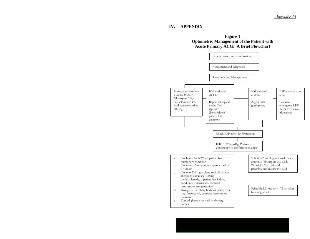
Figure 1 Optometric Management of the Patient with
Acute Primary ACG: A Brief Flowchart
Patient history and examination
Assessment and Diagnosis
Treatment and Management
Immediate treatment Timolol 0.5%, a Pilocarpine 2%,b Apraclonidine 1%, And Acetazolamide 500 mgc
IOP evaluated At 1 hr. Repeat all topical meds; Oral glycerind (Isosorbide if patient has diabetes)
IOP elevated at 2 hr. Argon laser gonioplasty
IOP elevated at 4-6 hr. Consider emergency LPI3 Refer for surgical iridectomy
Check IOP every 15-30 minutes
If IOP <20mmHg, Perform gonioscopy to confirm open angle
a. Use betaxolol 0.25% if patient has pulmonary condition
b. Use every 15-60 minutes up to a total of 2-4 doses
c. Use two 250 mg tablets; avoid if patient allergic to sulfa; use 100 mg methazolamide, if patient has kidney condition; if nauseated, consider intravenous acetazolamide
d. Dosage is 1.5 ml/kg body wt (serve over ice) if nauseated, consider intravenous mannitol
e. Topical glycerin may aid in clearing cornea
If IOP <20mmHg and angle open continue Pilocarpine 2% q.i.d., Timolol 0.5% b.i.d. and prednisolone acetate 1% q.i.d.
Schedule LPI usually < 72 hrs after breaking attack.
44 Primary Angle Closure Glaucoma
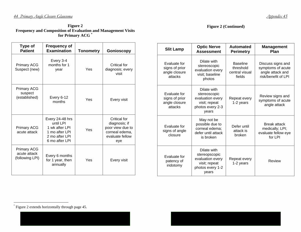
Figure 2 Frequency and Composition of Evaluation and Management Visits
for Primary ACG *
Type of Patient
Frequency of Examination
Tonometry
Gonioscopy
Primary ACG Suspect (new)
Every 3-4 months for 1
year
Yes Critical for
diagnosis; every visit
Primary ACG suspect
(established)
Every 6-12 months Yes Every visit
Primary ACG acute attack
Every 24-48 hrs until LPI
1 wk after LPI 1 mo after LPI 2 mo after LPI 6 mo after LPI
Yes
Critical for diagnosis; if
poor view due to corneal edema, evaluate fellow
eye
Primary ACG acute attack
(following LPI)
Every 6 months for 1 year, then
annually Yes Every visit
* Figure 2 extends horizontally through page 45.
Appendix 45
Figure 2 (Continued)
Slit Lamp Optic Nerve Assessment
Automated Perimetry
Management Plan
Evaluate for signs of prior angle closure
attacks
Dilate with stereoscopic
evaluation every visit; baseline
photos
Baseline threshold
central visual fields
Discuss signs and symptoms of acute angle attack and risk/benefit of LPI
Evaluate for signs of prior angle closure
attacks
Dilate with stereoscopic
evaluation every visit; repeat
photos every 2-3 years
Repeat every 1-2 years
Review signs and symptoms of acute
angle attack
Evaluate for signs of angle
closure
May not be possible due to corneal edema; defer until attack
is broken
Defer until attack is broken
Break attack medically; LPI;
evaluate fellow eye for LPI
Evaluate for patency of iridotomy
Dilate with stereopscopic
evaluation every visit; repeat
photos every 1-2 years
Repeat every 1-2 years Review
46 Primary Angle Closure Glaucoma
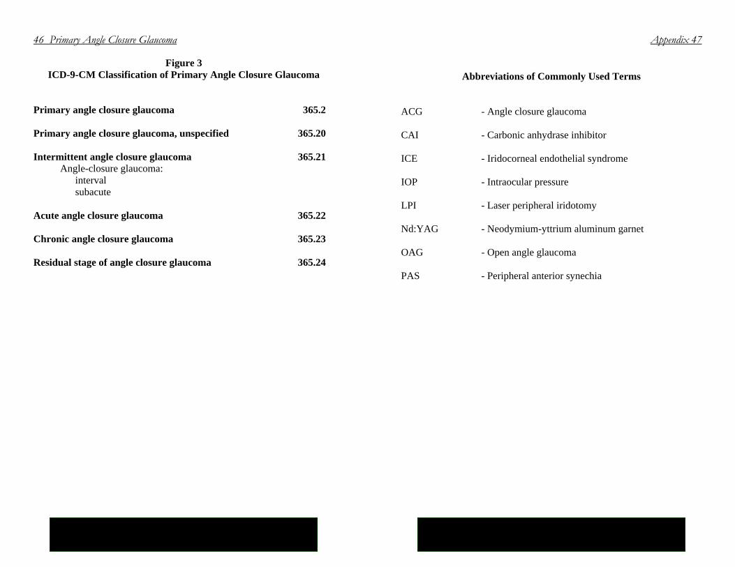
Figure 3 ICD-9-CM Classification of Primary Angle Closure Glaucoma
Primary angle closure glaucoma 365.2 Primary angle closure glaucoma, unspecified 365.20 Intermittent angle closure glaucoma 365.21 Angle-closure glaucoma: interval subacute Acute angle closure glaucoma 365.22 Chronic angle closure glaucoma 365.23 Residual stage of angle closure glaucoma 365.24
Appendix 47
Abbreviations of Commonly Used Terms
ACG - Angle closure glaucoma CAI - Carbonic anhydrase inhibitor ICE - Iridocorneal endothelial syndrome IOP - Intraocular pressure LPI - Laser peripheral iridotomy Nd:YAG - Neodymium-yttrium aluminum garnet OAG - Open angle glaucoma PAS - Peripheral anterior synechia
48 Primary Angle Closure Glaucoma
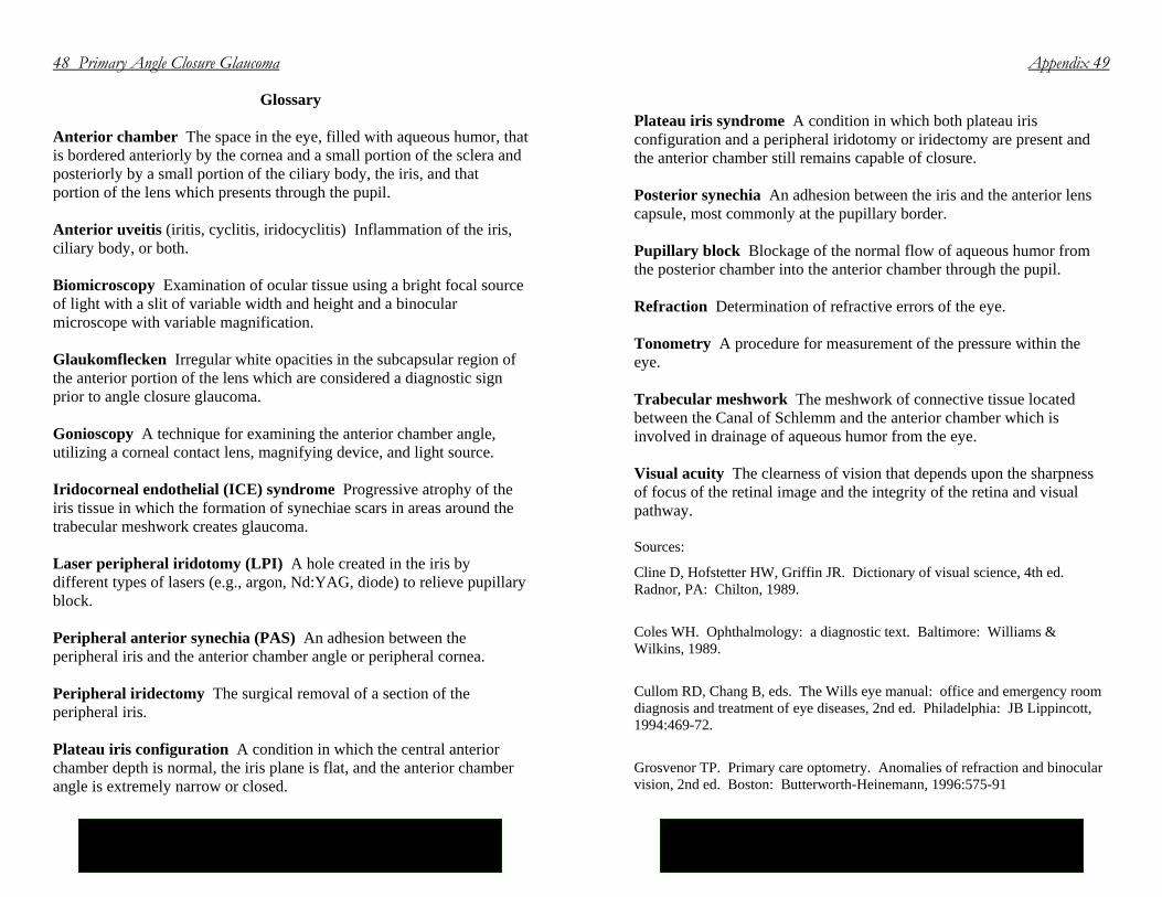
Glossary Anterior chamber The space in the eye, filled with aqueous humor, that is bordered anteriorly by the cornea and a small portion of the sclera and posteriorly by a small portion of the ciliary body, the iris, and that portion of the lens which presents through the pupil. Anterior uveitis (iritis, cyclitis, iridocyclitis) Inflammation of the iris, ciliary body, or both. Biomicroscopy Examination of ocular tissue using a bright focal source of light with a slit of variable width and height and a binocular microscope with variable magnification. Glaukomflecken Irregular white opacities in the subcapsular region of the anterior portion of the lens which are considered a diagnostic sign prior to angle closure glaucoma. Gonioscopy A technique for examining the anterior chamber angle, utilizing a corneal contact lens, magnifying device, and light source. Iridocorneal endothelial (ICE) syndrome Progressive atrophy of the iris tissue in which the formation of synechiae scars in areas around the trabecular meshwork creates glaucoma. Laser peripheral iridotomy (LPI) A hole created in the iris by different types of lasers (e.g., argon, Nd:YAG, diode) to relieve pupillary block. Peripheral anterior synechia (PAS) An adhesion between the peripheral iris and the anterior chamber angle or peripheral cornea. Peripheral iridectomy The surgical removal of a section of the peripheral iris. Plateau iris configuration A condition in which the central anterior chamber depth is normal, the iris plane is flat, and the anterior chamber angle is extremely narrow or closed.
Appendix 49
Plateau iris syndrome A condition in which both plateau iris configuration and a peripheral iridotomy or iridectomy are present and the anterior chamber still remains capable of closure. Posterior synechia An adhesion between the iris and the anterior lens capsule, most commonly at the pupillary border. Pupillary block Blockage of the normal flow of aqueous humor from the posterior chamber into the anterior chamber through the pupil. Refraction Determination of refractive errors of the eye. Tonometry A procedure for measurement of the pressure within the eye. Trabecular meshwork The meshwork of connective tissue located between the Canal of Schlemm and the anterior chamber which is involved in drainage of aqueous humor from the eye. Visual acuity The clearness of vision that depends upon the sharpness of focus of the retinal image and the integrity of the retina and visual pathway. Sources:
Cline D, Hofstetter HW, Griffin JR. Dictionary of visual science, 4th ed. Radnor, PA: Chilton, 1989.
Coles WH. Ophthalmology: a diagnostic text. Baltimore: Williams & Wilkins, 1989.
Cullom RD, Chang B, eds. The Wills eye manual: office and emergency room diagnosis and treatment of eye diseases, 2nd ed. Philadelphia: JB Lippincott, 1994:469-72.
Grosvenor TP. Primary care optometry. Anomalies of refraction and binocular vision, 2nd ed. Boston: Butterworth-Heinemann, 1996:575-91

50 Primary Angle Closure Glaucoma
Stedman’s medical dictionary, 26th ed. Baltimore: Williams and Wilkins, 1995. Vaughan D, Asbury T, Riordan-Eva P. General ophthalmology, 14th ed. Norwalk, CT: Appleton and Lange, 1995:419-22.