Embed Size (px)
Citation preview

International Journal of Food Microbiology 148 (2011) 99–106
Contents lists available at ScienceDirect
International Journal of Food Microbiology
j ourna l homepage: www.e lsev ie r.com/ locate / i j foodmicro
Prevalence of staphylococcal enterotoxins, toxin genes and genetic-relatedness offoodborne Staphylococcus aureus strains isolated in the Marmara Region of Turkey
Ali Aydin a,⁎, Mert Sudagidan b, Karlo Muratoglu a
a Department of Food Hygiene and Technology, Faculty of Veterinary Medicine, Istanbul University, Avcilar 34320, Istanbul, Turkeyb Biotechnology and Bioengineering Central Research Laboratory, Izmir Institute of Technology, Gulbahce Campus, Urla 35430, Izmir, Turkey
⁎ Corresponding author. Tel.: +90 2124737070x1718E-mail address: [email protected] (A. Aydin).
0168-1605/$ – see front matter © 2011 Elsevier B.V. Aldoi:10.1016/j.ijfoodmicro.2011.05.007
a b s t r a c t
a r t i c l e i n f oArticle history:Received 14 January 2011Received in revised form 3 April 2011Accepted 6 May 2011Available online 14 May 2011
Keywords:Staphylococcus aureusEnterotoxin genesEnterotoxinsPCRELISAPFGE
Staphylococcus aureus is a major foodborne pathogen and it has the ability to produce a number ofextracellular toxins. We analyzed 1070 food samples obtained from retail markets and dairy farms in theMarmara Region of Turkey for the presence of S. aureus. Out of 147 isolates, 92 (62.6%) were enterotoxigenic.PCR was used to investigate the presence of staphylococcal enterotoxin genes (sea, seb, sec, sed, see, seg, seh,sei, sej, sek, sel, sem, sen, seo, sep, seq and seu), exfoliative toxin genes (eta and etb) and the toxic−shocksyndrome toxin gene (tst). The PCR results showed that 53.3% of the isolates contained staphylococcalenterotoxin-like (SEl) toxin genes (seg, seh, sei, sej, sek, sel, sem, sen, seo, sep, seq and seu) which were morefrequent than classical enterotoxin genes (sea to see). Furthermore, seo, sei, sem, seg, seu and secwere found in37.0, 32.7, 30.4, 29.3, 29.3 and 27.2% of the isolates, respectively. The tst gene was detected and confirmed byDNA sequencing in 9 isolates. The presence of eta and etb were not found in the isolates. Enterotoxigeniccapabilities of isolates with SEA–SEE were investigated by ELISA. Enterotoxigenic S. aureus isolates producedone to three enterotoxins, with the most frequently produced types being enterotoxin A and C. There was acorrelation of 72.1% between production of a specific toxin and the presence of the respective genes. PFGEanalysis was used to identify genetic-relatedness of enterotoxigenic S. aureus isolates and the results revealedthat 13 groups of isolates from different or the same origin that contained the same genes showed 100%homology with indistinguishable band patterns. The other enterotoxigenic isolates showed related bandpatterns with 72–86% homology in sea-, 61–90% homology in sec-, 80–96% homology in seh-, and 69–96%homology in sep-positive isolates. To our knowledge, this is the first study to examine enterotoxins andrelated gene contents of S. aureus food isolates in the Marmara Region of Turkey.
2; fax: +90 2124737241.
l rights reserved.
© 2011 Elsevier B.V. All rights reserved.
1. Introduction
Staphylococcus aureus is a pathogen associated with seriouscommunity and hospital-acquired diseases. Although the number ofoutbreaks reported annually has decreased in the last few decades,staphylococcal food poisoning is still reported as the third mostprevalent cause of foodborne illness worldwide (Zhang et al., 1998).Illness is caused by enterotoxin-producing S. aureus (Cha et al., 2006;Sudagidan and Aydin, 2009; Wieneke et al., 1993). Raw meat (Pereiraet al., 2009), meat products, including fermented products such assucuk (Guven et al., 2010), raw milk (Rall et al., 2008), dairy products(Normanno et al., 2007) and ready-to-eat foods, including bakeryproducts (Oh et al., 2007) are among the foods reported to beassociated with S. aureus enterotoxin-induced food poisoning.
S. aureus produces more than 30 different extracellular by-products (Rogolsky, 1979) and staphylococcal toxins can be catego-
rized into groups: pyrogenic toxin superantigens (PTSAgs), exfoliativetoxins, leukocidins and other toxins. The family of PTSAgs includesstaphylococcal enterotoxins (SEs), SE-like (SEl) toxins and toxic-shock syndrome toxin-1 (TSST-1) (Lina et al., 2004). Generally, fiveclassical antigenic SE types (SEA to SEE) are recognized. Recently, theexistence of new SEs including SEl toxins (i.e., SElG to SElQ, SElR andSElU) has been reported (Bania et al., 2006; Jarraud et al., 2002;Nashev et al., 2007). However, their potential role in staphylococcalfood poisoning has not yet been clarified (Lina et al., 2004). TSST-1 isassociatedwith staphylococcal toxic-shock syndromeand is considered tobe the cause of nearly all cases of menstrual toxic-shock syndrome and atleast 50% of nonmenstrual cases (Bergdoll and Schlievert, 1984). Thepresence of toxic-shock syndrome toxin gene (tst) in S. aureus isolatedfrom foods has been reported (Cha et al., 2006). Moreover, clinical strainsof S. aureus are known to produce immunologically distinct exfoliativetoxin A and/or B (Bailey et al., 1980). The production of exfoliative toxin Aby S. aureus isolated from foods and animals has been reported (Adesiyunet al., 1991; Hayakawa et al., 1998).
Various typing methods have been used to characterize S. aureusisolates. PCR has been used as a simple technique for detecting

100 A. Aydin et al. / International Journal of Food Microbiology 148 (2011) 99–106
enterotoxigenic strains (Asperger and Zangerl, 2003). Although thePCR-based approach is specific, highly sensitive and rapid, it can onlydetect the presence of enterotoxigenic genes, not the production ofthe SE proteins (Boerema et al., 2006). Immunological methods arepreferred for the detection of enterotoxins (Chiang et al., 2008). ELISAis the method of choice, because reagents are commercially availablein polyvalent and monovalent formats for both toxin screening andserotype specific identification assays (Bennett, 2001). Numeroustechniques [amplified fragment length polymorphism, multi locussequence typing and pulsed-field gel electrophoresis (PFGE)] havebeen described for S. aureus genotyping (Melles et al., 2007), of whichPFGE is the “gold standard” technique for determining genetic-relatedness, especially in outbreaks, due to its high discriminatorypower (Weller, 2000).
The aim of this study was to determine the frequency of PTSAgs,including SE genes (sea, seb, sec, sed and see), SEl genes (seg, seh, sei,sej, sek, sel, sem, sen, seo, sep, seq and seu), tst and the coexistence ofexfoliative genes (eta and etb) in S. aureus food isolates, to determinethe ability of the isolates to produce staphylococcal enterotoxins A–Eand to investigate the genetic-relatedness of enterotoxigenic S. aureusisolates by PFGE.
2. Materials and methods
2.1. Sample collection and bacterial isolates
During the period of July 2007 and December 2008, 1070 foodsamples were collected from supermarkets, conventional markets,bazaars and dairy farms in and near large cities, including Balikesir,Bursa, Canakkale, Edirne, Istanbul, Kirklareli and Tekirdag, in theMarmara Region of Turkey. These samples included 115 meats (beef,mutton, chicken and turkey meat), 15 meat products [Turkish typefermented sausage (sucuk)], salami and sausage), 303 raw milk, 452dairy products (cheese, butter, yoghurt and cream), 141 bakeryproducts [pasta, thin sheets of dough (yufka) and cake] and 44 ready-to-eat foods. Raw milk samples were obtained from dairy farms
Fig. 1. Distribution of enterotoxigenic S. aureus isolates/total number of S. aureus isol
located near these cities. Isolation of S. aureus from food samples wasperformed using EN ISO 6881-1 standard procedures described by theInternational Organization for Standardization (Anonymous, 1999).Procedures for identification of the isolates were previously described(Aydin et al., 2011).
2.2. Detection of toxin genes
Bacterial genomic DNA isolation was carried out as describedpreviously (Sudagidan et al., 2008). The presence of staphylococcalenterotoxin genes sea, seb, sec and see (Johnson et al., 1991), sed(Jarraud et al., 2002; Johnson et al., 1991), seg, seh, sei, sej, sek, sel, sepand seq (Bania et al., 2006), sem, sen and seo (Jarraud et al., 2002) andseu (Nashev et al., 2007), and genes for exfoliative toxins (eta and etb)(Johnson et al., 1991) and toxic-shock syndrome toxin (tst) (Booth etal., 2001) was determined by either monoplex or multiplex PCR. ThePCR products were resolved in 1.5% (w/v) agarose gel electrophoresisin 1× TAE buffer (0.04 M Tris-acetate and 0.001 M EDTA). PCRexperiments were done twice for each isolate. Positive controls wereS. aureus subsp. aureus NCTC (National Collection of Type Cultures)10652 for sea, S. aureus subsp. aureus NCTC 10654 for seb, S. aureussubsp. aureus NCTC 10655 for sec and S. aureus subsp. aureus NCTC10656 for sed. S. aureusN315, positive for tst, sem, sen, seo and seu, waskindly supplied by Dr. Teruyo Ito and Prof. Keiichi Hiramatsu fromJuntendo University, Japan. S. aureus strains positive for seg, sei, sej, sehand seo genes were obtained from Department for Veterinary PublicHealth, Institute for Milk Hygiene, Milk Technology and Food Science,University of Veterinary Medicine, Wien, Austria.
2.3. DNA sequencing of tst gene
The DNA sequences of tst positive PCR products were determinedusing PCR primers described by Booth et al. (2001). The followingconditions were used for cycle sequencing of tst genes: 1 min at 96 °C,30 cycles of 10 s at 96 °C, 5 s at 56 °C (tst-forward and reverse) and
ates which were isolated from foods collected in the Marmara Region of Turkey.

Table 1The presence of enterotoxin genes in S. aureus isolates.
Enterotoxin genes Meat(n=13) (%)
Meat products(%) (n=6)
Raw milk(n=31) (%)
Dairy products(n=36) (%)
Bakery products(n=5) (%)
Ready-to-eat food(n=1) (%)
Total(n=92) (%)
sea 1 (7.7) 3 (9.7) 4 (11.1) 8 (8.6)sec 7 (22.6) 3 (8.3) 10 (10.9)seh 4 (30.7) 1 (3.2) 3 (8.3) 8 (8.6)sei 1 (3.2) 1 (1.1)sel 1 (7.7) 1 (1.1)sen 1 (2.8) 1 (1.1)seo 1 (3.2) 2 (5.5) 3 (3.2)sep 1 (7.7) 4 (13.0) 6 (16.6) 1 (20.0) 12 (13.0)seu 1 (2.8) 1 (1.1)sea, sec 1 (3.2) 1 (2.8) 1 (100) 3 (3.2)sea, seh 1 (3.2) 1 (1.1)sea, seq 1 (2.8) 1 (1.1)seb, sec 1 (2.8) 1 (1.1)seb, sep 1 (7.7) 1 (2.8) 2 (2.2)sec, seo 1 (20.0) 1 (1.1)seg, sei 1 (2.8) 1 (1.1)sel, sep 2 (6.5) 2 (2.2)sem, seo 1 (3.2) 1 (1.1)sec, sel, sep 1 (2.8) 1 (1.1)seh, sek, seq 1 (20.0) 1 (1.1)sem, seo, sep 1 (3.2) 1 (1.1)sea, seh, sek, seq 1 (3.2) 1 (20.0) 2 (2.2)sec, sei, sel, sep 1 (2.8) 1 (1.1)sea, seg, sei, seo, seu 1 (16.7) 1 (1.1)seb, sek, sen, seo, seq 1 (7.7) 1 (1.1)seg, sei, sem, sen, seo 1 (2.8) 1 (1.1)seg, sei, sem, seo, seu 1 (16.7) 4 (11.1) 5 (5.4)seb, seg, sei, sem, seo, seu 1 (2.8) 1 (1.1)seg, sei, sem, sen, seo, seu 1 (7.7) 3 (9.7) 2 (5.5) 1 (20.0) 7 (7.6)seg, sei, sem, seo, seq, seu 1 (3.2) 1 (2.8) 2 (2.2)sea, seg, sei, sem, sen, seo, seu 1 (16.7) 1 (1.1)sec, seg, sei, sel, sem, seo, seu 1 (7.7) 1 (1.1)sec, seg, sei, sem, sen, seo, seu 1 (3.2) 1 (1.1)sec, seh, sei, sem, sen, seo, seu 1 (16.7) 1 (1.1)seg, sei, sem, sen, seo, seq, seu 1 (7.7) 1 (1.1)sec, seg, seh, sei, sem, sen, seo, seu 2 (33.2) 2 (2.2)sec, seg, sei, sel, sem, sen, seo, seu 1 (7.7) 2 (6.5) 3 (3.2)
101A. Aydin et al. / International Journal of Food Microbiology 148 (2011) 99–106
4 min at 60 °C. The DNA sequencing was carried out using AppliedBiosystems 3130xl Genetic Analyzer (Foster City, California, USA).
2.4. Determination of enterotoxin production
Enterotoxin production was determined by ELISA and enterotoxinswere detected using a Ridascreen SET A, B, C, D, E assay kit (R-BiopharmAG, Germany). Briefly, the supernatant of 24 h cultures of S. aureus (9log CFU/ml) grown at 37 °C in Brain Hearth Infusion Broth (BHI, Oxoid)was separated from cells by centrifugation at 4500×g for 5 min at 4 °C.The supernatant was passed through 0.20 μm filter (Millipore) and100 μl of the filtrate were transferred to an ELISA plate. The solutionsweremixed gently by rocking of the plate for 1 h at 23 °C in the dark. Toeach well, 250 μl of washing buffer (0.55 g NaH2PO4⋅H2O, 2.85 gNa2HPO4⋅2H2O, 8.7 g NaCl; pH 7.2) was added, followed by aspiration.This procedure was repeated three times. Subsequently, 100 μl ofperoxidase conjugated anti-SET antibodieswere added to eachwell andincubated for 60 min at 23 °C in the dark. The liquid was then aspiratedfrom the wells and the wells were rinsed three times with 250 μl ofwash buffer. Urea peroxide (50 μl) and tetramethyl-benzidine (50 μg)were added to eachwell, mixed thoroughly and incubated for 30 min at23 °C in the dark. Stop reagent (100 μl of 1 N H2SO4) was added to eachwell and the absorbance was measured at 450 nm in an ELISA reader(ELX 800, Bio-tek Inst., Winooski, Vermont, USA) (Anonymous, 2003;Bennett, 2001).
2.5. PFGE analysis
Genetic-relatedness of 92 exterotoxigenic S. aureus food isolateswasdetermined byPFGE analysis. Agarose plugswere prepared as described
previously (Aydin et al., 2011; Sudagidan and Aydin, 2010). BacterialDNA in plugs was digested with 30U SmaI (Fermentas) for 18 h,followed by applying to 1% (w/v) pulsed-field certified agarose (Bio-Rad)with a 5–40 s pulse time, 6 V/cm, 120° angle, at 14 °C for 22 husingthe CHEF-Mapper PFGE system (Bio-Rad). After electrophoresis, the gelwas stained with ethidium bromide (5 μg/ml) and visualized withVersaDoc 4000MP image analyzer system (Bio-Rad). The obtained bandpatterns were analyzed using BIO-PROFIL Bio-1D++ software (VilberLourmat, France) at 13% homology coefficient. The similarity betweenthe isolates was determined automatically by specifying the formula ofNei and Li (1979). The clusteringwasperformedby theunweighted pairgroup method with arithmetic mean (UPGMA) (Vilber Lourmat).
3. Results and discussion
3.1. Prevalence of enterotoxin producing S. aureus in food samples
Enterotoxin-producing S. aureus is one of the causative agents offoodborne intoxication and for this reason determination of itsprevalence in foods is important with respect to assessing publichealth risks. In this study, 147 (13.8%) S. aureus strains, which wereisolated from 1070 food samples, were analyzed for toxigeniccapabilities. Ninety-two strains (62.6%) isolated from meat (13/13),meat products (6/6), raw milk (31/63), dairy products (36/54),bakery products (5/9) and ready-to-eat foods (1/2) were enterotoxi-genic. These strains were isolated mainly from samples collected inIstanbul (n=42), Edirne (n=15), Balikesir (n=13), Tekirdag(n=7), Kirklareli (n=7), Bursa (n=6) and Canakkale (n=2)(Fig. 1). Similar results were reported by Guven et al. (2010). Theyisolated 138 S. aureus strains from 413 food samples collected in the

Table 2Presence of the genes and the produced enterotoxins in S. aureus isolates.
Isolates(fooda, originb)
SEs detected by ELISA The genes detected by PCR
SEA SEB SEC SED sea seb sec Other SEl genes
SE21A, M, Buc + + + −HE4D, M, E + + sek, sen, seo, seqHE25A, M,E + + seg, sei, sel, sem, sen, seo,
seuHE25B, M, E + + seg, sei, sel, sem, seo, seuTE2, M, I + + + sepEU6B, MP, I + + seg, seh, sei, sem, sen, seo,
seuEU6C, MP, I + + seg, seh, sei, sem, sen, seo,
seuEU7A, MP, I + + + + seg, sei, seo, seuEU7B, MP, I + + seg, sei, sem, sen, seo, seuS35A, RM, K + + seg, sei, sem, sen, seo, seuS133A, RM, T + + seh, sek, seqS158B, RM, T + + + seg, sei, sel, sem, sen, seo,
seuS235, RM, I + + + + + −S255, RM, I + + −S269, RM, I + + + sehS272, RM, I + + + + −S273, RM, I + + + −PY6, DP, I + + sepPY92BY2, DP, I + + seqPY100C, DP, I + + + −PY104A, DP, I + + −PY104B, DP, I + + −PY178A, DP, K + + sei, sel, sepPY178B, DP, K + + sel, sepPY186A, DP, E + + + seg, sei, sem, seo, seuPY276, DP, B + + + −PY311B, DP, B + + −PY351A, DP, B + + −PY368A, DP, C + + + −YF62A, BP, B + + −YE15A, RTE, I + + + + −a M: Meat; MP: Meat Products; RM: Raw Milk; DP: Dairy Products; BP: Bakery
Products and RTE: Ready-To-Eat Food.b B: Balikesir; Bu: Bursa; C: Canakkale; E: Edirne; I: Istanbul; K: Kirklareli; and T: Tekirdag.c In 13 isolates, there was not a correlation between enterotoxin production and the
related genes are shown in boldface type.
102 A. Aydin et al. / International Journal of Food Microbiology 148 (2011) 99–106
central Anatolia region of Turkey and 83 (60.1%) of the strainssynthesized one or two enterotoxins. In Portugal, Pereira et al. (2009)observed that 101 (68.2%) out of 148 S. aureus isolates from variousfoods were positive for the presence of genes coding for one or moreenterotoxins. In another study, Normanno et al. (2007) found asimilar prevalence in Italy, in which 59.8% of the S. aureus strainsisolated from milk, dairy and meat products produced enterotoxins.
3.2. Detection of genes encoding staphylococcal enterotoxins
Studies on S. aureus isolated from foods have shown that thepercentage of enterotoxigenic strains is considerably higher if thenewly described SEl genes are considered together with genesencoding for the so-called classical enterotoxins (Bania et al., 2006;Le Loir et al., 2003). Enterotoxigenic S. aureus strains have different SEgene contents and they can harbor several of the genes. SE genes arelocated on plasmids (sed and sej), phages (sea, see and sep) andchromosomes (seb, sec, seg, seh, sei, sek, sel, sem, sen, seo and seq)(Kuroda et al., 2001). In this study, 17 SE and SEl genes wereinvestigated. It was found that 53.3% of the isolates negative for sea tosee carried SEl genes. This is in agreement with other studies showinghigher percentages of SEl genes relative to classical SE genes (Bania etal., 2006; Nashev et al., 2007; Rosec and Gigaud, 2002).
In the present study, 8.6% of the enterotoxigenic isolates werefound to encode only sea. In other studies, the sea was reported to bepresent in 15.4% (Cha et al., 2006) and 18.8% (Oh et al., 2007) ofS. aureus strains isolated from foods. In our study, the highestpercentage of single enterotoxigenic gene was sep (13%) (Table 1).The sep gene is another potential superantigen andwas detected in 19enterotoxigenic S. aureus isolates (20.7%), most commonly in dairyproducts (n=8) and rawmilk (n=7). The seb gene was detected in 5S. aureus isolates (5.4%) isolated from meat and dairy products(Table 1). However, in one isolate (HE4D), seb was accompanied bythe sek gene. Another study reported all seb-positive strains (n=3) tobe sek-negative (Bania et al., 2006). SEK has many of the biologicalactivities associated with the SEs, including superantigenicity,pyrogenicity and the ability to enhance the lethality of endotoxin(Orwin et al., 2001). Enterotoxigenic S. aureus strains containing seq,known to occur jointly with sek (Bania et al., 2006). In this study, itwas found that all sek-positive isolates (HE4D, S133A, YF62A andYF62B) were also positive for seq, whereas, 4 seq-positive isolates(PY38BY/1, PY92BY/2, S137AY and TE15A) did not contain sek.Furthermore, our results showed that none of the enterotoxigenicisolates contained sed, see and sej genes. Similar observations havebeen reported for the absence of see (Bania et al., 2006; Nashev et al.,2007; Pereira et al., 2009).
Kuroda et al. (2001) suggested a correlation among sec, sel and sepgenes. We found that only two isolates, both from dairy products(PY178A and PY178B), contained sec, sel, and sep and only twoisolates (HE25A and HE25B) contained sec and sel genes. Bania et al.(2006) reported that sec/sel-positive strains were sep-negative; thus,any relationship between these genes is not obvious. In this study, secwas detected in 27.2% of enterotoxigenic S. aureus isolates and amongthese isolates, 8% were found to carry sel. These results were inagreement with those reported by Bania et al. (2006) whichdemonstrated that 7.1% of S. aureus isolates contained sec and sel.
The coexistence of seg and sei genes on a common genetic elementwas presented by Jarraud et al. (2001) and, in our study, all seg-positive isolates (n=28) were also positive for sei. Interestingly, 3isolates (EU6A, PY178A and S226) containing sei did not contain seg.Similarly, MacLauchlin et al. (2000) detected 20 S. aureus isolateswhich were positive for seg and sei (n=19) or for only sei (n=1).Moreover, the SEU enterotoxin, encoded by seu, was homologous, butnot identical to any known enterotoxins (Letertre et al., 2003) and themajority of our strains (29.4%) contained seu gene and combinationswith other enterotoxin genes (Table 1).
SElH has been shown to have emetic activity and is considered as apotential causative agent for food poisoning (Su and Wong, 1995). Inthe present study, seh was detected the only enterotoxin genes in8 (8.6%) of the isolates and totally in 15 (16.3%) of the isolates seh andseh combinations with other enterotoxin genes were detected(Table 1). This prevalence was relatively lower than that (52%)reported by Bania et al. (2006), but comparable to that found byNashev et al. (2004), who reported a prevalence of 13.6% in clinicalS. aureus isolates.
3.3. Determination of enterotoxin production
Enterotoxins that cause human illness are referred to as classicalstaphylococcal enterotoxins (SEA to SEE) (Cha et al., 2006). Inapproximately 5% of cases, classical enterotoxins are not detected andthese cases have been attributed to the production of new enterotoxins(Kokan andBergdoll, 1987; Su andWong, 1995). The specificity of ELISA isvery high and the detection of enterotoxins A, B, C, D and E is highlysensitive (Bennett, 2001). In the present study, 46.7% S. aureus isolatespossessed the targeted classical SEs genes (sea, seb and sec). In addition,72.1%of theenterotoxigenicS. aureus isolatesproducedenterotoxinsSEA–SED, as detected by ELISA (Table 2). The most frequently encounteredenterotoxins in foods are SEA (Oh et al., 2007; Tsen et al., 1998) and SEC(Soriano et al., 2002; Tamarapu et al., 2001). Our observations were inagreement with other studies. We found that 2, 8 and 10 of the isolatesproduced only SEB, SEA and SEC, respectively, however, in 11 of the

103A. Aydin et al. / International Journal of Food Microbiology 148 (2011) 99–106
isolates produced more than one enterotoxin (Table 2). There was 72.1%correlation between enterotoxin types and presence of the respectivegenes. Similarly, Pereira et al. (2009) demonstrated 80% correlation inS. aureus isolates using the VIDAS assay and PCR.
In 12 S. aureus isolates (EU6A, S35B, S182, S215, S217, S242, S258,S266, S267, P33A, PY376A and PY406B) positive for SE genes, thepresence of enterotoxins were not detected by ELISA. The differencebetween PCR and ELISA results can also be explained by the low-levelof staphylococcal enterotoxin production, i.e. below the threshold ofimmunoassay detection (Bystron et al., 2006). The concentration oftoxin should be above 1 ng/ml, which is the lowest detection limit ofthe test used in this study. Another explanation might be theincomplete expression of enterotoxigenic genes. Furthermore, SEproduction can be affected by environmental conditions such astemperature, pH and water activity (Nájera-Sánchez et al., 2003). Theenvironment of enterotoxigenic S. aureus in food is important for bothgrowth and production of enterotoxins.
According to the ELISA results, 8 of the enterotoxigenic isolatesproduced SED and 4 isolates (TE2, EU7A, S272 and PY186A) producedSEC; however, sed and sec were not detected in these isolates by PCR(Table 2). Five isolates were positive for SED, whereas 3 were positivefor SEC and 3 were positive for SEC and SED. It is possible that thesepairs of PCR primers (SEC1–SEC2 and SED1–SED2) had low specificityowing to small sequence differences between strains (Nájera-Sánchezet al., 2003). Moreover, one isolate (S158B) that produced SEB andSEC tested negative for the presence of seb, but positive for sec and SEl
Fig. 2. Genetic-relatedness of sea (A), sec (B), seh
genes. According to the other reports, SEB and SEC share 62 to 64%amino acid identity (Bohach and Schlievert, 1987; Munson et al.,1998) and SEG is similar to SEB and SEC (Munson et al., 1998). Inaddition, 2 isolates (EU7A and PY186A) were positive SEC and SEDand these isolates contained also SEl genes (seg, sei, sem, seo and seu).These differences might have resulted from the use of preparedpolyclonal antibodies against each SE, which sometimes exhibitantigenic similarities among SEs and SEl toxins, thereby causingcross-reactions in the tests (Edwin et al., 1986). Because enterotoxinsSEA–SEE and SEH also share nucleotide sequence identity (≥32%), it ispossible that anewenterotoxingenecouldalso sharenucleotide sequenceidentity with the characterized enterotoxins (Munson et al., 1998).
3.4. Prevalence of tst gene
TSST-1 has been detected in S. aureus isolates most commonlyisolated from clinical samples with important clinical symptoms(Lappin and Ferguson, 2009), moreover, tst-positive isolates havebeen also isolated from various foods (Cha et al., 2006), poultry (Evanset al., 1983) and milk (Valle et al., 1991). In our study, 9.8% of theenterotoxigenic S. aureus isolates were positive for tst. Moreover, PCRproducts of tst were also confirmed by DNA sequencing. This is inagreement with Cha et al. (2006), who reported a 12% prevalence oftst in foodborne S. aureus isolates in Korea. Conversly, Tsen et al.(1998) found tst in 4.8% of clinical S. aureus isolates, but not in food
(C) and sep-positive (D) S. aureus isolates.
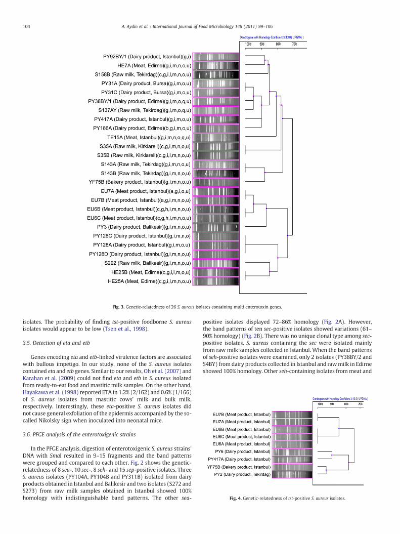
Fig. 3. Genetic-relatedness of 26 S. aureus isolates containing multi enterotoxin genes.
Fig. 4. Genetic-relatedness of tst-positive S. aureus isolates.
104 A. Aydin et al. / International Journal of Food Microbiology 148 (2011) 99–106
isolates. The probability of finding tst-positive foodborne S. aureusisolates would appear to be low (Tsen et al., 1998).
3.5. Detection of eta and etb
Genes encoding eta and etb-linked virulence factors are associatedwith bullous impetigo. In our study, none of the S. aureus isolatescontained eta and etb genes. Similar to our results, Oh et al. (2007) andKarahan et al. (2009) could not find eta and etb in S. aureus isolatedfrom ready-to-eat food and mastitic milk samples. On the other hand,Hayakawa et al. (1998) reported ETA in 1.2% (2/162) and 0.6% (1/166)of S. aureus isolates from mastitic cows’ milk and bulk milk,respectively. Interestingly, these eta-positive S. aureus isolates didnot cause general exfoliation of the epidermis accompanied by the so-called Nikolsky sign when inoculated into neonatal mice.
3.6. PFGE analysis of the enterotoxigenic strains
In the PFGE analysis, digestion of enterotoxigenic S. aureus strains’DNA with SmaI resulted in 9–15 fragments and the band patternswere grouped and compared to each other. Fig. 2 shows the genetic-relatedness of 8 sea-, 10 sec-, 8 seh- and 15 sep-positive isolates. ThreeS. aureus isolates (PY104A, PY104B and PY311B) isolated from dairyproducts obtained in Istanbul and Balikesir and two isolates (S272 andS273) from raw milk samples obtained in Istanbul showed 100%homology with indistinguishable band patterns. The other sea-
positive isolates displayed 72–86% homology (Fig. 2A). However,the band patterns of ten sec-positive isolates showed variations (61–90% homology) (Fig. 2B). There was no unique clonal type among sec-positive isolates. S. aureus containing the sec were isolated mainlyfrom raw milk samples collected in Istanbul. When the band patternsof seh-positive isolates were examined, only 2 isolates (PY38BY/2 andS4BY) from dairy products collected in Istanbul and rawmilk in Edirneshowed 100% homology. Other seh-containing isolates frommeat and

105A. Aydin et al. / International Journal of Food Microbiology 148 (2011) 99–106
dairy product samples were closely related (80–96% homology)(Fig. 2C). Fifteen sep-positive isolates displayed 69–100% homology.Four isolates (S15A, S15B, S15C and S15D) from raw milk samplescollected in Edirne and 2 isolates (PY330A and PY330B) from dairyproducts in Balikesir had the same band patterns. Interestingly, 2isolates (PY192A and PY245B) from dairy products collected indifferent cities (Kirklareli and Bursa) showed 100% homology(Fig. 2D). S. aureus isolates obtained from dairy products and rawmilk samples frequently contained the sep-gene.
The genetic-relatedness of S. aureus isolates containing multienterotoxin genes is shown in Fig. 3. Aside from the identical strains,11 S. aureus isolates displayed 71–95% homology. Seven groups of theisolates showed 100% homology. In these groups, 3 isolates (PY128A,PY128C and PY128D) and 6 groups of isolates (EU6B and EU6C, EU7Aand EU7B, HE25A and HE25B, PY31A and PY31C, S35A and S35B andS143A and S143B) displayed 100% homology. These isolates wereobtained from meat products, dairy products and raw milk samples.Isolates containing the same genes were closely related with morethan 80% homology (Fig. 3).
The presence of tst in our S. aureus isolates was low. The geneticrelatedness of 9 tst-positive isolates is shown in Fig. 4. Thedendrogram of tst-positive isolates is composed of two main groupswith 74% homology. One group is composed of 3 isolates (EU6A, EU6Band EU6C) and 2 isolates (EU7A and EU7B) all of which isolated frommeat products and showed 100% homology. Another group contains 4isolates (Fig. 4). S. aureus YF75B (from a bakery product) and PY2(from a dairy product) showed 94% homology and they were isolatedfrom samples collected in different cities. Moreover, 2 S. aureusisolates, which were isolated from dairy products in Istanbul, showed90% homology. All tst-positive S. aureus isolates were closely related,with more than 74% homology.
4. Conclusions
The recent discovery and characterization of new SEs resulted inan increased frequency of potentially enterotoxigenic S. aureusisolates from foods, suggesting that the prevalence pathogenic S.aureus may be higher than the previously recognized. The highprevalence of newly discovered enterotoxin genes, including thoseencoding emetic toxins, is evident in foodborne isolates. In light ofthese observations, additional work is needed to better understandingthe role of S. aureus in food poisoning and also tomonitor the presenceof these genes in foodborne isolates. The results demonstrated thehigh prevalence of newly discovered genes in foodborne S. aureus,especially in the isolates positive for classical enterotoxins, thuscontributing to the prevalence of isolates potentially capable ofcausing food poisoning. The distribution of other genes suggests thepossibility of the presence of yet unknown genetic elements encodingSEls. PFGE analysis showed a high genetic diversity among strains,indicating that contamination of various food products with S. aureuscould originate from numerous sources, preprocessing environments,processing areas and the market place.
Acknowledgements
The authors thank Prof. Dr. Larry Beuchat (University of Georgia,Center for Food Safety) for critical reading of the manuscript.Furthermore, the authors thank Biotechnology and BioengineeringCentral Research Laboratory (Izmir Institute of Technology) and Res.Assist. Funda Yilmaz (Istanbul University, Department of FoodHygiene and Technology). This work was supported by The Scientificand Technological Research Council of Turkey (TUBITAK) (Project No:107T266) and by the Research Fund of Istanbul University, Project No:UDP-3645/13042009. A part of this study was presented in the 3rdNational Veterinary Food Hygiene Congress (May 14–16, 2009) inBursa, Turkey.
References
Adesiyun, A.A., Lenz, W., Schaal, K.P., 1991. Exfoliative toxin production byStaphylococcus aureus strains isolated from animals and human beings in Nigeria.Microbiologica 14, 357–362.
Anonymous, 1999. International Organization for Standardization. EN ISO 6881-1Microbiology of food and animal feeding stuffs: horizontal method for theenumeration of coagulase positive staphylococci (Staphylococcus aureus and otherspecies). Part 1: technique using Baird Parker agarmedium. ISO, Geneva, Switzerland.
Anonymous, 2003. Enzyme immunoassay for the detection of Staphylococcus aureusenterotoxins. RIDASCREEN® SET A, B, C, D and E Art. No.: R1401. R-Biopharm AG,Darmstadt, Germany.
Asperger, H., Zangerl, P., 2003. Staphylococcus aureus. In: Roginski, H., Fuquay, J.W., Fox,P.F. (Eds.), Encyclopedia of Dairy Sciences. Academic Press and Elsevier Science,Amsterdam, pp. 2563–2569.
Aydin, A., Muratoglu, K., Sudagidan, M., Bostan, K., Okuklu, B., Harsa, S., 2011.Prevalance and antibiotic resistance of foodborne Staphylococcus aureus isolates inTurkey. Foodborne Pathogens and Disease 8, 63–69.
Bailey, C.J., de Azavedo, J., Arbuthnott, J.P., 1980. A comparative study of two serotypesof epidermolytic toxin from Staphylococcus aureus. Biochimica et Biophysica Acta624, 111–120.
Bania, J., Dabrowska, A., Bystron, J., Korzekwa, K., Chrzanowska, J., Molenda, J., 2006.Distribution of newly described enterotoxin-like genes in Staphylococcus aureusfrom food. International Journal of Food Microbiology 108, 36–41.
Bennett, R.W., 2001. Staphylococcal enterotoxins. Micro-slide double diffusion andELISA based methods. FDA/Bacteriological Analytical Manual. Chapter 13A.Available online: http://www.fda.gov/Food/ScienceResearch/LaboratoryMethods/BacteriologicalAnalyticalManualBAM/UCM073674#authors.
Bergdoll, M.S., Schlievert, P.M., 1984. Toxin shock syndrome toxin. Lancet 324, 691.Boerema, J.A., Clemens, R., Brightwell, G., 2006. Evaluation of molecular methods to
determine enterotoxigenic status and molecular genotype of bovine, ovine, humanand food isolates of Staphylococcus aureus. International Journal of FoodMicrobiology 107, 192–201.
Bohach, G.A., Schlievert, P.M., 1987. Nucleotide sequence of the staphylococcalenterotoxin C1 gene and relatedness to other pyrogenic toxins. Molecular andGeneral Genetics 209, 15–20.
Booth, M.C., Pence, L.M., Mahasreshti, P., Callegan, M.C., Gilmore, M.S., 2001. Clonalassociations among Staphylococcus aureus isolates from various sites of infection.Infection and Immunity 69, 345–352.
Bystron, J., Bania, J., Zarczynska, A., Korzekwa, K., Molenda, J., Kosek-Paszkowska, K.,2006. Detection of enterotoxigenic Staphylococcus aures strains using a commercialELISA and multiplex-PCR. Bulletin of Veterinary Institute Pulawy 50, 329–333.
Cha, J.O., Lee, J.K., Jung, Y.H., Yoo, J.I., Park, Y.K., Kim, B.S., Lee, Y.S., 2006. Molecularanalysis of Staphylococcus aureus isolates associated with staphylococcal foodpoisoning in South Korea. Journal of Applied Microbiology 101, 864–871.
Chiang, Y.C., Liao, W.W., Fan, C.M., Pai, W.Y., Chiou, C.S., Tsen, H.Y., 2008. PCR detectionof Staphylococcal enterotoxins (SEs) N, O, P, Q, R, U, and survey of SE types inStaphylococcus aureus isolates from food-poisoning cases in Taiwan. InternationalJournal of Food Microbiology 121, 66–73.
Edwin, C., Tatini, S.R., Maheswaran, S.K., 1986. Specificity and cross-reactivity ofstaphylococcal enterotoxin A monoclonal antibodies with enterotoxins B, C, D andE. Applied and Environmental Microbiology 52, 1253–1257.
Evans, J.B., Ananaba, G.A., Pate, C.A., Bergdoll, M.S., 1983. Enterotoxin production byatypical Staphylococcus aureus from poultry. Journal of Applied Bacteriology 54,257–261.
Guven, K., Mutlu, B.M., Gulbandilar, A., Cakir, P., 2010. Occurence and characterizationof Staphylococcus aureus isolated from meat and dairy products consumed inTurkey. Journal of Food Safety 30, 196–212.
Hayakawa, Y., Hayashi, M., Shimano, T., Komae, H., Takeuchi, K., Endou, M., Igarashi, H.,Hashimoto, N., Takeuchi, S., 1998. Production of exfoliative toxin A by Staphylo-coccus aureus isolated from mastitic cow's milk and farm bulk milk. The Journal ofVeterinary Medical Science 60, 1281–1283.
Jarraud, S., Peyrat, M.A., Lim, A., Tristan, A., Bes, M., Mougel, C., Etienne, J., Vandenesch,F., Bonneville, M., Lina, G., 2001. egc, A highly prevalent operon prevalent operon ofenterotoxin gene, forms a putative nursery of superantigens in Staphylococcusaureus. The Journal of Immunology 166, 669–677.
Jarraud, S., Mougel, C., Thioulouse, J., Lina, G., Meugnier, H., Forey, F., Nesme, X., Etienne,J., Vandenesch, F., 2002. Relationships between Staphylococcus aureus geneticbackground, virulence factors, agr groups (alleles) and human disease. Infectionand Immunity 70, 631–641.
Johnson, W.M., Tyler, S.D., Ewan, E.P., Ashton, F.E., Pollard, D.R., Rozee, K.R., 1991.Detection of genes for enterotoxins, exfoliative toxins and toxic shock syndrometoxin 1 in Staphylococcus aureus by the polymerase chain reaction. Journal ofClinical Microbiology 29, 426–430.
Karahan, M., Acik, M.N., Cetinkaya, B., 2009. Investigation of toxin genes by polymerasechain reaction in Staphylococcus aureus strains isolated from bovine mastitis inTurkey. Foodborne Pathogens and Disease 6, 1029–1035.
Kokan, N.P., Bergdoll, M.S., 1987. Detection of low-enterotoxin producing Staphylococ-cus aureus strains. Applied and Environmental Microbiology 53, 2675–2676.
Kuroda, M., Ohta, T., Uchiyama, I., Baba, T., Yuzawa, H., Kobayashi, I., Cui, L., Oguchi, A.,Aoki, K., Nagai, Y., Lian, J., Ito, T., Kanamori, M., Matsumaru, H., Maruyama, A.,Murakami, H., Hosoyama, A., Mizutani-Ui, Y., Takahashi, N.K., Sawano, T., Inoue, R.,Kaito, C., Sekimizu, K., Hirakawa, H., Kuhara, S., Goto, S., Yabuzaki, J., Kanehisa, M.,Yamashita, A., Oshima, K., Furuya, K., Yoshino, C., Shiba, T., Hattori, M., Ogasawara,N., Hayashi, H., Hiramatsu, K., 2001. Whole genome sequencing of meticillin-resistant Staphylococcus aureus. Lancet 357, 1225–1240.
106 A. Aydin et al. / International Journal of Food Microbiology 148 (2011) 99–106
Lappin, E., Ferguson, A.J., 2009. Gram-positive toxic shock syndromes. The LancetInfectious Diseases 9, 281–290.
Le Loir, Y., Baron, F., Gautier, M., 2003. Staphylococcus aureus and food poisoning.Genetics and Molecular Research 31, 63–76.
Letertre, C., Perelle, S., Dilasser, F., Fach, P., 2003. Identification of a new putativeenterotoxin SEU encoded by the egc cluster of Staphylococcus aureus. Journal ofApplied Microbiology 95, 38–43.
Lina, G., Bohach, G.A., Nair, S.P., Hiramatsu, K., Jouvin-Marche, E., Mariuzza, R., 2004.Standard nomenclature for superantigens expressed by Staphylococcus. The Journalof Infectious Disease 189, 2334–2336.
MacLauchlin, J., Narayanan, G.L., Mithani, V., O'Neill, G., 2000. The detection ofenterotoxins and toxic shock syndrome toxin genes in Staphylococcus aureus bypolymerase chain reaction. Journal of Food Protection 63, 479–488.
Melles, D.C., Van Leeuwen,W.B., Snijders, S.V., Horst-Kreft, D., Peeters, J.K., Verbrugh, H.A.,Van Belkum, A., 2007. Comparison of multilocus sequence typing (MLST), pulsed-field gel electrophoresis (PFGE), and amplified fragment length polymorphism(AFLP) for genetic typing of Staphylococcus aureus. Journal of MicrobiologicalMethods 69, 371–375.
Munson, S.H., Tremaine, M.T., Betley, M.J., Welch, R.A., 1998. Identification andcharacterization of staphylococcal enterotoxin types G and I from Staphylococcusaureus. Infection and Immunity 66, 3337–3348.
Nájera-Sánchez, G., Maldonado-Rodríguez, R., Olvera, P.R., de la Garza, L.M., 2003.Development of two multiplex polymerase chain reactions for the detection ofenterotoxigenic strains of Staphylococcus aureus isolated from foods. Journal ofFood Protection 66, 1055–1062.
Nashev, D., Toshkova, K., Salasia, S.I.O., Hassan, A.A., Lämmler, C., Zschöck, M., 2004.Distribution of virulence genes of Staphylococcus aureus isolated from stable nasalcarriers. FEMS Microbiology Letters 233, 45–52.
Nashev, D., Toshkova, K., Bizeva, L., Akineden, Ö., Lämmler, C., Zschöck, M., 2007.Distribution of enterotoxin genes among carriage- and infection-associated isolatesof Staphylococcus aureus. Letters in Applied Microbiology 45, 681–685.
Nei, M., Li, W.-H., 1979. Mathematical model for studying genetic variation in terms ofrestriction endonucleases. Proceedings of the National Academy of Sciences USA76, 5269–5273.
Normanno, G., La Salandra, G., Dambrosio, A., Quaglia, N.C., Corrente, M., Parisi, A.,Santagada, G., Firinu, A., Crisetti, E., Celano, G.V., 2007. Occurrence, character-ization and antimicrobial resistance of enterotoxigenic Staphylococcus aureusisolated from meat and dairy products. International Journal of FoodMicrobiology 115, 290–296.
Oh, S.K., Lee, N., Cho, Y.S., Shin, D.B., Choi, S.Y., Koo, M., 2007. Occurrence of toxigenicStaphylococcus aureus in ready-to-eat food in Korea. Journal of Food Protection 70,1153–1158.
Orwin, P.M., Leung, D.Y., Donahue, H.L., Novick, R.P., Schlievert, P.M., 2001. Biochemicaland biological properties of Staphylococcal enterotoxin K. Infection and Immunity69, 360–366.
Pereira, V., Lopes, C., Castro, A., Silva, J., Gibbs, P., Teixeira, P., 2009. Characterization forenterotoxin production, virulence factors, and antibiotic susceptibility of Staphy-lococcus aureus isolates from various foods in Portugal. Food Microbiology 26,278–282.
Rall,V.L.M.,Vieira, F.P., Rall, R., Vieitis, R.L., Fernandes Jr., A., Candeias, J.M.G., Cardoso, K.F.G.,Araújo Jr., J.P., 2008. PCR detection of staphylococcal enterotoxin genes inStaphylococcus aureus strains isolated from raw and pasteurized milk. VeterinaryMicrobiology 132, 408–413.
Rogolsky, M., 1979. Nonenteric toxins of Staphylococcus aureus. MicrobiologicalReviews 43, 320–360.
Rosec, J.P., Gigaud, O., 2002. Staphylococcal enterotoxin genes of classical and newtypes detected by PCR in France. International Journal of Food Microbiology 77,61–70.
Soriano, J.M., Font, G., Rico, H., Molto, J.C., Manes, J., 2002. Incidence of enterotoxigenicStaphylococci and their toxins in food. Journal of Food Protection 65, 857–860.
Su, Y.C., Wong, A.C., 1995. Identification and purification of a new staphylococcalenterotoxin H. Applied and Environmental Microbiology 61, 1438–1443.
Sudagidan, M., Aydin, A., 2009. Screening virulence properties of staphylococci isolatedfrom meat and meat products. Wiener Tierärztliche Monatsschrift 96, 128–134.
Sudagidan, M., Aydin, A., 2010. Virulence properties of methicillin-susceptibleStaphylococcus aureus food isolates encoding Panton–Valentine Leukocidin gene.International Journal of Food Microbiology 138, 287–291.
Sudagidan, M., Çavuşoğlu, C., Bacakoğlu, F., 2008. Investigation of the virulence genes inmethicillin-resistant Staphylococcus aureus strains isolated from biomaterialsurfaces. Mikrobiyoloji Bülteni 42, 29–39.
Tamarapu, S., Mckillip, J.L., Drake, M., 2001. Development of a multiplex polymerasechain reaction assay for detection and differentiation of Staphylococcus aureus indairy products. Journal of Food Protection 64, 664–668.
Tsen, H.Y., Yu, G.K., Wang, K.C., Wang, S.J., Chang, M.Y., Lin, L.Y., 1998. Comparison of theenterotoxigenic types, toxic shock syndrome toxin 1 (TSST-1) strains and antibioticsusceptibilities for enterotoxigenic Staphylococcus aureus strains isolated from foodand clinical samples. Food Microbiology 15, 33–41.
Valle, J., Vadillo, S., Piriz, S., Gomez-Lucia, E., 1991. Toxic shock syndrome toxin 1 (TSST-1)production by staphylococci isolated fromgoats and presence of specific antibodies toTSST-1 in serum and milk. Applied and Environmental Microbiology 57, 889–891.
Weller, T.M.A., 2000. Methicillin-resistant Staphylococcus aureus typing methods:which should be the international standard? Journal of Hospital Infection 44,160–172.
Wieneke, A.A., Roberts, D., Gilbert, R.J., 1993. Staphylococcal food poisoning in theUnited Kingdom, 1969–1990. Epidemiology and Infection 110, 519–531.
Zhang, S., Iandolo, J., Stewart, C., 1998. The enterotoxin D plasmid of Staphylococcusaureus encodes a second enterotoxin determinant (sej). FEMS Microbiology Letters168, 227–233.