Embed Size (px)
Citation preview
PREVALENCE OF CARDIOMYOPATHY IN APPARENTLY HEALTHY CATS
Christopher Francis Paige MS, DVM
Thesis submitted to the faculty of the Virginia Polytechnic Institute and State University in partial fulfillment of the requirements for the degree of
Master of Science
In Biomedical and Veterinary Sciences
Dr. Jonathan A. Abbott, Chair Dr. R. Lee Pyle
Dr. François Elvinger Dr. Colin Carrig
June 29, 2007 Blacksburg, Virginia
Keywords: Prevalence, Feline, Cardiomyopathy, Hypertrophic cardiomyopathy,
Murmur, Echocardiography.
Copyright 2007, Christopher F. Paige
PREVALENCE OF CARDIOMYOPATHY IN APPARENTLY HEALTHY CATS
Christopher Francis Paige
Committee Chair: Dr. Jonathan Abbott
ABSTRACT
Subclinical cardiomyopathy (CM) sometimes is identified after abnormalities are
detected during auscultation of apparently healthy cats. Little is known regarding the
prevalence of CM in this population. Furthermore, the clinical importance of auscultatory
abnormalities in apparently healthy cats is unclear. In order to estimate the prevalence of
murmurs and CM, we prospectively evaluated a sample of apparently healthy cats. Cats
with systemic hypertension or hyperthyroidism were excluded. 103 cats were subject to
physical and echocardiographic examinations which were performed by two different
investigators; the echocardiographer was unaware of the physical findings. Left
ventricular wall thickness was determined by two-dimensional echocardiography in
short- and long-axis planes. Left ventricular hypertrophy (LVH) was defined as an end-
diastolic wall thickness ≥ 6 mm. Cats with LVH but without left ventricular dilation were
considered to have hypertrophic CM (HCM). Cardiomyopathy was identified in 16 cats
(15.5%; 95% CI: [9.2, 24.0]); 15 had HCM and one had arrythmogenic right ventricular
cardiomyopathy. Murmurs were detected in 16 cats (15.5%; 95% CI: [9.2; 24.0]); of
these cats, 5 had CM. Of 15 cats with HCM, 11 had segmental LVH, three cats had
diffuse LVH, and one cat had borderline LVH and marked systolic anterior motion of the
mitral valve. The sensitivity and specificity of murmurs for detection of a CM was 31%
and 87%, respectively. The prevalence of feline subclinical CM in Southwest Virginia is
near 16%; approximately a third of these cats had murmurs. In apparently healthy cats, a
cardiac murmur is an insensitive marker of the presence of CM.
This investigation was supported by a grant from the Virginia Veterinary Medical
Association Veterinary Memorial Fund.
iii
DEDICATION
This thesis is dedicated to my wife, Christina R. Paige, for the many days she
sacrificed to help support this project. I also dedicate this work to our pets for their
unconditional friendship. Finally, to our unborn child, Sophia, we will meet you soon.
Sincerely,
Christopher F. Paige
iv
ACKNOWLEDGMENTS
This thesis would not have been possible without the collaboration, guidance, and
endless support of my major advisor, Dr. Jonathan Abbott. I would also like to thank R.
Lee Pyle for his assistance in data collection and many contributions to this project. Drs.
François Elvinger and Colin Carrig, committee members, provided valuable input during
the committee meetings. I also wish to acknowledge the contributions of Drs. Stephen
Werre and Daniel L. Ward who provided statistical consultation. Finally and most
importantly, I would like to gratefully acknowledge the students, staff, house officers and
faculty of the Virginia-Maryland Regional College of Veterinary Medicine who provided
the subjects for this study and also provided technical assistance.
v
ATTRIBUTION Dr. Jonathan Abbott is my graduate advisor. He contributed to the study design and data
analysis. He is the investigator who performed the echocardiographic examinations.
Dr. R. Lee Pyle is a committee member. He is the investigator who performed the
physical examinations.
Dr. François Elvinger is a committee member. He provided expertise in epidemiology
and contributed to study design and data analysis.
vi
TABLE OF CONTENTS
PREVALENCE OF CARDIOMYOPATHY IN APPARENTLY HEALTHY CATS ...... ii
DEDICATION................................................................................................................... iv
ACKNOWLEDGMENTS .................................................................................................. v
ATTRIBUTION................................................................................................................. vi
INTRODUCTION .............................................................................................................. 1
CHAPTER 1 ....................................................................................................................... 3
ABSTRACT.................................................................................................................... 3
INTRODUCTION .......................................................................................................... 4
MATERIAL AND METHODS...................................................................................... 4
RESULTS ..................................................................................................................... 11
DISCUSSION............................................................................................................... 16
CONCLUSIONS .............................................................................................................. 20
FUTURE INVESTIGATIONS......................................................................................... 20
LITERATURE CITED..................................................................................................... 21
VITA................................................................................................................................. 25
vii

LIST OF TABLES Table 1. A 2X2 table and calculated odds ratios as a measure of the association between
cardiac murmurs and echocardiographic findings in apparently healthy cats. ......... 14 Table 2. This 2X2 table provides raw prevalence data for unprovoked cardiac murmurs
and cardiomyopathy (CM) in apparently healthy cats.............................................. 15
viii
LIST OF FIGURES Figure 1. A schematic drawing showing regions measured at end-diastole. ...................... 8
Figure 2. A 2-D short-axis image of the left ventricle of an echocardiographically normal
cat.. .............................................................................................................................. 9 Figure 3. Right parasternal long-axis image of the left ventricle of an
echocardiographically normal cat. ............................................................................ 10
ix
INTRODUCTION
Hypertrophic cardiomyopathy (HCM) is characterized by hypertrophy of a non-
dilated ventricle in the absence of systemic disorders or structural cardiac diseases known
to induce hypertrophy.1 HCM in humans is sometimes inherited as a Mendelian
autosomal dominant trait and numerous mutations in genes that encode sarcomeric
proteins have been associated with this disease.2 Sudden death, left heart failure and
stroke are potential complications of this condition.2 In people, HCM is the most
common genetic cardiovascular disease; a prevalence of 0.2% has been reported.2 Feline
HCM, which is clinically, echocardiographically and histologically similar to the disorder
observed in humans, has been proposed as a model for the human disease.3 Studies have
demonstrated that HCM in the Maine Coon cat is heritable and genomic clarification has
recently been reported.4,5 To date, feline HCM is defined only by echocardiographic or
post-mortem criteria.6 Feline HCM may be subclinical or result in clinical signs
associated with left heart failure or aortic thromboembolism.3 The spectrum of severity is
wide and the rate of disease progression is variable, which makes it difficult to determine
risk factors that predict outcome.
HCM is the most common disorder in cats with clinically evident cardiac disease.
However, other forms of myocardial disease including dilated cardiomyopathy (DCM),
arrhythmogenic right ventricular (ARVCM), restrictive (RCM) and unclassified
cardiomyopathy (UCM) are also observed.1, 6, 7 The obstructive form of HCM is typically
associated with a cardiac murmur.6 Because a cardiac murmur may prompt
echocardiographic examination in the absence of clinical signs, patients with subclinical
feline HCM are commonly identified. However, murmurs are inconsistently present in
cats with all forms of cardiomyopathy. Therefore, the prevalence of feline
cardiomyopathy including less common forms such as RCM and ARVCM might be
greater than is generally thought.
The epidemiological characteristics of feline cardiomyopathy have been
addressed, but those studies were limited by referral bias, misclassification bias and the
shortcomings of retrospective analysis.6-10 The earliest study to address occurrence of
HCM was based on 4,933 necropsies, where 421 cats had acquired heart disease, and half
1
of those had characteristics of hypertrophic cardiomyopathy.11 However, it is relevant
that this study was carried out prior to the widespread recognition of systemic
hypertension and also prior to the first description of feline hyperthyroidism. Both of
these conditions can result in gross cardiac abnormalities that are indistinguishable from
those of HCM, and therefore, misclassification may have biased the prevalence estimate.
Other studies that followed were limited to retrospective analysis and restricted to referral
populations, which may have inflated the prevalence measure.6,7,9,10 Cote et al described
a 21% (23 /103 cats) prevalence of heart murmurs among overtly healthy cats.12 Because
the cats were enrolled in a blood donor program, cats with a heart murmur or history of
cardiac disease had been excluded prior to initiation of the study. A recent report
detected 9 % (8 /94) prevalence of subclinical HCM among cats with a normal physical
exam.13 However, cats with murmurs were excluded from the study. In both studies, the
exclusion of cats could have underestimated the prevalence estimates, because these cats
may have had subclinical cardiac disease.
Our study is the first to provide unbiased estimates of the prevalence of both
cardiomyopathy and murmurs in a sample of apparently healthy cats. To limit bias,
physical and echocardiographic examinations were performed by independent
investigators. Therefore, the echocardiographer was unaware of the physical findings.
Unlike the previous studies, we minimized sampling time to more accurately represent
the instantaneous occurrence of disease that is implicit in the definition of prevalence.14
We also sought to clarify the clinical relevance of cardiac murmurs in apparently healthy
cats by evaluating the diagnostic accuracy of this finding for detection of feline
cardiomyopathy. To achieve these objectives we conducted a community-based
population survey in which apparently healthy cats were subject to physical and
echocardiographic examinations. We hope that our estimate of population prevalence and
evaluation of cardiac murmurs will form the basis of future epidemiological investigation
of feline cardiomyopathies.
2
CHAPTER 1 PREVALENCE OF CARDIOMYOPATHY IN APPARENTLY HEALTHY CATS.
Christopher F. Paige, MS, DVM; Jonathan A. Abbott, DVM, DACVIM; R. Lee Pyle, VMD, MS,
DACVIM; François Elvinger, Dr. Med Vet, PhD, ACVPM, ECVPH
ABSTRACT Objective—To determine the prevalence of murmurs and cardiomyopathy in apparently
healthy cats, and to clarify the diagnostic utility of murmurs within this population.
Study Design—Cross-Sectional Study
Procedures—An electronic survey was used to identify apparently healthy cats.
Enrolled cats were subject to physical and echocardiographic examinations which were
performed by two independent investigators. Left ventricular wall thickness was
determined by two-dimensional echocardiography in short- and long-axis planes. Left
ventricular hypertrophy (LVH) was defined by an end-diastolic wall thickness ≥ 6 mm.
Cats with LVH but without left ventricular dilation were considered to have hypertrophic
cardiomyopathy (HCM). Murmurs and abnormal Doppler outflow tract velocities were
compared.
Results— Cardiomyopathy (CM) was identified in 16 cats (15.5%; 95% CI: [9.2, 24.0]);
15 had HCM and one had arrythmogenic right ventricular cardiomyopathy. Murmurs
were detected in 16 cats (15.5%; 95% CI: [9.2; 24.0]); of these cats, 5 had CM. Of 15
cats with HCM, 11 had segmental LVH, three cats had diffuse LVH, and one cat had
borderline LVH with marked systolic anterior motion of the mitral valve. The sensitivity
and specificity of murmurs for detecting a CM were 31% and 87%, respectively.
Conclusions— Prevalence of feline subclinical CM in a sample of apparently healthy
cats in Southwest Virginia is near 16%. In apparently healthy cats, a cardiac murmur has
low sensitivity as a marker of the presence of CM. Doppler echocardiographic evidence
of dynamic right or left ventricular outflow tract obstruction was associated with the
presence of a cardiac murmur. Cats in which Doppler echocardiographic evaluation
disclosed abnormalities of ventricular ejection were more likely to have a cardiac murmur
than those without this finding.
3
INTRODUCTION
Subclinical CM sometimes is identified after abnormalities are detected during
auscultation of apparently healthy cats. However, little is known regarding the prevalence
of CM in this population. Previous studies have addressed the prevalence of CM and
murmurs in healthy cats, but these studies had limitations that relate to referral bias,
retrospective analysis, and inclusion criteria.7, 9,10,12,13 While many feline patients are
referred for echocardiographic evaluation after detection of a murmur, the relationship
between murmurs and cardiomyopathy in healthy cats remains unclear. We attempted to
estimate the prevalence of cardiomyopathy and murmurs in apparently healthy cats and in
so doing, clarify the clinical relevance and diagnostic utility of cardiac murmurs in this
population. To achieve this objective we conducted a community-based population
survey in which apparently healthy cats were subject to physical and echocardiographic
examinations.
MATERIAL AND METHODS
We prospectively examined apparently healthy cats owned by veterinary students,
staff and faculty at the Virginia-Maryland Regional College of Veterinary Medicine
(VMRCVM), Blacksburg, VA. This investigation was approved by the Animal Care and
Use Committee and Institutional Review Board of Virginia Tech.
Enrollment— An email was sent to a distribution list that included students, technical
staff and faculty of the VMRCVM. Pet-owners that were willing to enroll their cats were
asked to complete an electronic, web-based survey which was used to identify cats that
met inclusion criteria and also provide zoographic data. Apparently healthy cats were
included if they had not previously been subject to echocardiographic examination, were
not receiving treatment for cardiovascular disease and did not have a history of chronic
illnesses such as inflammatory bowel disease, hyperthyroidism, renal disease, systemic
hypertension or diabetes mellitus. Cats were excluded if the pet-owner had sought
veterinary care for a systemic illness in the three months prior to recruitment. Cats that
4
had a history of a murmur, but had not been examined echocardiographically, were
included.
Procedure—Physical examination, Doppler blood pressure estimation followed by
electrocardiography, and then echocardiography were performed in that order in three
different rooms. The physical and echocardiographic examinations were performed by
two different board-certified veterinary cardiologists (RLP and JAA); the
echocardiographer (JAA) was unaware of the physical findings. Echocardiographic
images were digitally recorded for later quantitative and qualitative analysis. After
digital echocardiographic records were randomized and patient identifiers concealed,
echocardiographic measurements were obtained by a third investigator (CFP). The final
echocardiographic diagnosis was the consensus opinion of the echocardiographer and the
investigator (CFP) that performed the echocardiographic measurements and was
determined without knowledge of the physical findings.
Physical Examination—All cats were subject to systematic, dynamic auscultatory
examination. Auscultation was first performed when cats were at rest and then after
provocation, defined for the purpose of this study as a maneuver in which the examiner
quickly lifts the cat in the air at least two times. When identified, murmurs were
described in terms of: intensity, which was graded on a six interval scale according to the
recommendations of Levine, point of maximal intensity, and timing.15 The presence or
absence of a gallop sound was noted as was a description of the cardiac rhythm. Heart
rate and respiratory rate were recorded for all cats.
Echocardiography—Examinations were performed without chemical restraint. Utilizing
a Vingmed System FiVe sonographa with a 7.5 MHz transducer, transthoracic
echocardiography was performed as previously described.16 Two-dimensional (2-D)
short-axis and long-axis right parasternal images of the left ventricle were used to
measure wall thickness. In the short-axis plane, end-diastole was defined as the maximal
diastolic excursion of the ventricle or onset of the QRS. In the long-axis plane, end-
a General Electric Medical Systems, Waukesha, WI.
5
diastole was the first frame during which mitral valve closure was visible. The following
dimensions were obtained from the short-axis image: end-diastolic left ventricular
internal diameter (LVIDd), end-diastolic thickness of the interventricular septum (IVSd)
and end-diastolic thickness of the left ventricular posterior wall (LVPWd). The end-
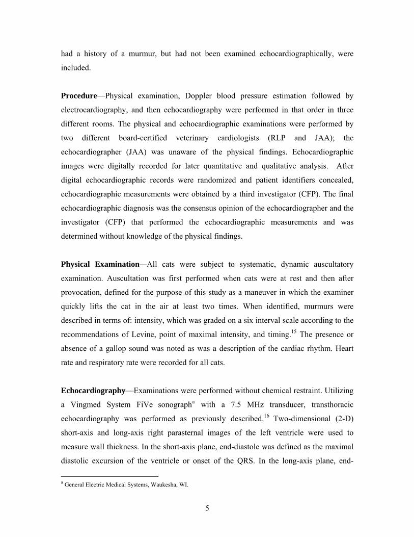
diastolic thickness of the IVS was also measured in two additional sites. Specifically, the
maximal thickness of the septum was measured in the two septal segments which extend
from the papillary muscles to the central point of the septum (Figure 1). The line of
measurement was parallel to a cord that extended through the centroid of the ventricular
lumen (Figure 2). End-diastolic basilar septal thickness was measured in the long-axis
plane and was the maximal dimension at a site between the aortic root and the point at
which the anterior mitral valve leaflet most closely approaches the IVS during diastole
(Figure 3). Septal measurements included left ventricular endocardial echoes but
excluded echoes arising from the right ventricular endocardium.17 Measurements of the
LVPW included endocardial echoes but did not include the pericardium. Aortic (Ao): left
atrial (LA) ratio was determined by M-mode. Left atrial enlargement was defined by a
left atrial-aortic ratio in excess of 1.54. The subjects were also subject to conventional
Doppler examination. Right and left ventricular outflow tract velocities were recorded.
Color Doppler mapping and pulsed-wave spectral Doppler were used to screen for
dynamic outflow tract obstruction. Echocardiographic dimensions and spectral Doppler
measurements were the average of three, usually consecutive cardiac cycles.
Hypertrophic cardiomyopathy (HCM) was defined by an end-diastolic wall
thickness ≥ 6 mm for more than 50% of any region of the interventricular septum or left
ventricular posterior wall and the presence or absence of systolic anterior motion of the
mitral valve was also noted.3 Other forms of feline cardiomyopathy were classified as
previously described.7,18 Dynamic right ventricular outflow obstruction (DRVOT) was
defined by a systolic jet that originated proximal to the infundibulum and had spectral
Doppler characteristics that indicated late-systolic acceleration.19 Dynamic left ventricular
outflow tract obstruction was similarly defined by late-systolic acceleration.
Blood Pressure / Electrocardiography—Systemic blood pressure was estimated for all
cats using the Doppler cuff-flowmeter method.20 All estimates were obtained utilizing
6
either the left or right front limb. Cats were considered hypertensive if the average of
three consecutive measurements was ≥ 180 mm Hg. After systemic blood pressure
measurements, the subjects were restrained while in right lateral recumbence and a six-
lead electrocardiogram was recorded. Average, electrocardiographic heart rate was
reported only from the six-lead electrocardiogram. Arrhythmias were documented by
either the six-lead electrocardiogram or the electrocardiogram that was recorded during
echocardiography.
Thyroid Function—After completion of the cardiovascular examination, all cats that
were ≥ 6 years old underwent jugular venipuncture, whole blood was obtained, and
centrifuged. The serum supernatant was refrigerated, and the DRI Thyroxine (T4) assayb
was performed utilizing an Olympus AV 400, Automated Chemistry Analyzer.c Those
cats in which the T4 determination exceeded the upper limit of our laboratory reference
range were considered hyperthyroid and excluded from further analysis.
Data analysis—Prevalence of cardiomyopathy and murmurs as well as the
corresponding 95% confidence intervals (CI) were computed using the frequency
procedure of SAS.d Associations between murmurs and the presence of dynamic outflow
tract obstruction were assessed using the chi-square test. When statistically significant,
the association was further assessed using the prevalence odds ratio. To determine the
diagnostic utility of unprovoked murmurs for cardiomyopathy detection, the sensitivity,
specificity, positive predictive value (PPV), negative predictive value (NPV), positive
and negative likelihood ratios (LR) and respective 95% CI were derived using SISA.e
Results associated with p-values that were less than 0.05 were considered statistically
significant.
b Microgenics Corporation, 46360 Fremont Blvd., Fremont, CA c Olympus America Inc., Two Corporate Center Drive, Melville, NY d SAS, Version 8.02, SAS Institute Inc. Cary, NC e Uitenbroek, Daan G."SISA-Binomial." 1997. <http://home.clara.net/sisa/binomial.htm>
7
Figure 1. A schematic drawing showing regions measured during peak end-diastole. Segment 1 and 2 = a single linear dimension was obtained from each segment; this measurement represents the maximal thickness of each segment. IVSd=end-diastolic thickness of the interventricular septum, LVIDd=left ventricular internal diameter during diastole, LVPWd=end-diastolic thickness of the left ventricular posterior wall.
8
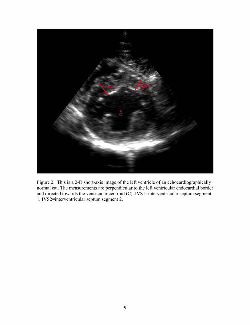
Figure 2. This is a 2-D short-axis image of the left ventricle of an echocardiographically normal cat. The measurements are perpendicular to the left ventricular endocardial border and directed towards the ventricular centroid (C). IVS1=interventricular septum segment 1, IVS2=interventricular septum segment 2.
9
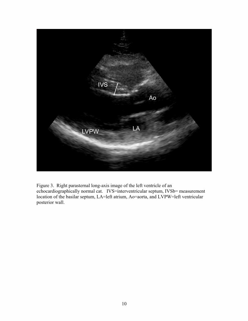
Figure 3. Right parasternal long-axis image of the left ventricle of an echocardiographically normal cat. IVS=interventricular septum, IVSb= measurement location of the basilar septum, LA=left atrium, Ao=aorta, and LVPW=left ventricular posterior wall.
10
RESULTS
Enrollment—There were 165 responses to our electronic survey and 145 cats met the
initial inclusion criteria. We examined 132 cats over six non-consecutive days from late
August 2005 to early November 2005. Twenty-nine of these cats were later excluded
because they resisted manual restraint (22), were hyperthyroid (4), or had incomplete
echocardiographic examinations (3). A total of a 103 apparently healthy cats were used
for prevalence estimates. Forty-three cats were females. Mean body-weight ± standard
deviation (SD) was 5.01 ± 1.13 kg. Precise ages were not always available so discrete age
categories were constructed: < 1 yr (n=6), 1-5 yrs (n=62), 6-10 yrs (n=27), 11 to 15 yrs
(n=7), and ≥ 16 yrs (n=1). The majority of cats were mixed breeds (domestic short hair
n=75, domestic medium hair n=9, and domestic long hair n=10). Pure breeds included
Himalayan (n=3), Siamese (n=3), Ocicat (n=2), and Maine Coon cat (n=1).
Physical Examination—Murmurs were detected in 16 cats (15.5%; 95% CI: [9.2; 24.0]).
Murmur intensities were classified as follows: grade 1/6 (n=5), grade 2/6 (n=9), and
grade 3/6 (n=2). A total of 28 cats had murmurs after the provocative maneuver; 13 out
of the 28 did not have the murmur at rest. One cat had a murmur at rest, but not after the
provocative maneuver. Based on auscultation, the mean heart rate of all cats was 173.7
beats per minute (bpm) ± 17.2 SD. The following additional abnormalities were
identified through auscultation: gallop rhythm (n=2), bradycardia (n=1), and
compensatory pause (n=1).
Doppler Blood Pressure / Electrocardiography—All cats were normotensive; average
systolic blood pressure in 87 echocardiographically normal cats was 131.1 mm Hg ± 17.8
SD. In 15 cats with HCM, average systolic blood pressure was 136.4 mm Hg ± 19.6 SD.
Based on electrocardiography, mean heart rate (HR) of 80 cats without CM was 189.4
bpm ± 23.8 SD; for cats with HCM, average HR was 188.7 bpm ± 27.2 SD. The
following electrocardiographic abnormalities were identified during electrocardiography
or echocardiography: two cats that did not have CM had ventricular pre-excitation, four
cats without CM had ventricular premature complexes (VPC) and one cat without CM
11
had ventricular tachycardia (VT). Of the cats with CM, two with HCM had VPCs.
Ventricular tachycardia was recorded from the cat with ARVC.
Echocardiography—Cardiomyopathy was identified in 16 cats (15.5%; 95% confidence
interval (CI): [9.2, 24.0]); 15 had HCM and one had arrythmogenic right ventricular
cardiomyopathy (ARVC). Of 15 cats with HCM, 11 had segmental LVH and in three of
these cats, hypertrophy was localized to the basilar septum. In the cats with basilar septal
hypertrophy, one cat was between 1 to 5 years of age while the remaining two were
greater than 10 years of age. Three cats had diffuse LVH. One cat with marked systolic
anterior motion of the mitral valve and maximal ventricular wall thickness of 5.9 mm was
classified as HCM. The cat classified as ARVC had a prominent right ventricle, abnormal
septal motion, tricuspid regurgitation, ventricular tachycardia, and an intermittent gallop
sound.
Dynamic right ventricular outflow tract obstruction (DRVOT) with peak
velocities ≥1.7 m/s was identified in 5 cats, and three other cats had lower velocities with
evidence of late systolic acceleration. Left ventricular outflow tract obstruction (LVOT)
was identified in 8 cats; two cats with HCM had SAM with supraphysiologic LVOT
velocities (4.2 m/s and 2.1 m/s) and mitral regurgitation, 2 had mid-left ventricular
obstruction and supraphysiologic velocities (1.8 m/s, 1.7 m/s), while the remaining 4 had
late systolic acceleration with lower velocities (1.0 m/s (n=2), 1.1 m/s, 1.6 m/s). Of the 6
cats with dynamic LVOT obstruction in which SAM was not detected, two had HCM and
four did not. Two cats, one with HCM and one without, had both right and left
ventricular dynamic outflow tract obstruction.
Association of Murmurs and Dynamic Outflow Tract Obstruction—Of the 16 cats
with murmurs, 5 had HCM. Six of 16 cats with unprovoked murmurs had a dynamic
outflow tract obstruction: RVOT only (n=3; HCM=0), RVOT/LVOT (n=1; HCM=1), and
LVOT/MR (n=2; HCM=2). Two cats with murmurs and HCM did not have an abnormal
Doppler spectrogram. Ten of 28 cats with murmurs during the provocative maneuver had
an Doppler evidence of dynamic outflow tract obstruction: RVOT only (n=5; HCM=0),
LVOT only (n=2; HCM=1), RVOT/LVOT (n=1; HCM=1), and LVOT/MR (n=2;
12
HCM=2). The six cats which had a murmur at rest and dynamic outflow tract obstruction
also developed a murmur during the provocative maneuver. A 2x2 table and calculated
odds ratios defining the association between physical and echocardiographic findings are
shown in Table 1. Statistically significant prevalence odds ratios describe the relationship
between the echocardiographic finding of dynamic outflow tract obstruction and the
physical finding of a murmur heard during rest and after provocation; cats in which
Doppler characteristics of ventricular outflow were abnormal were more likely to have a
cardiac murmur than those that did not.
Diagnostic Utility of Murmurs—To assess the diagnostic value of murmurs for
detection of a cardiomyopathy, the sensitivity, specificity, positive predictive value,
negative predictive value, and likelihood ratios (positive and negative) were calculated
and are shown in Table 2.
13
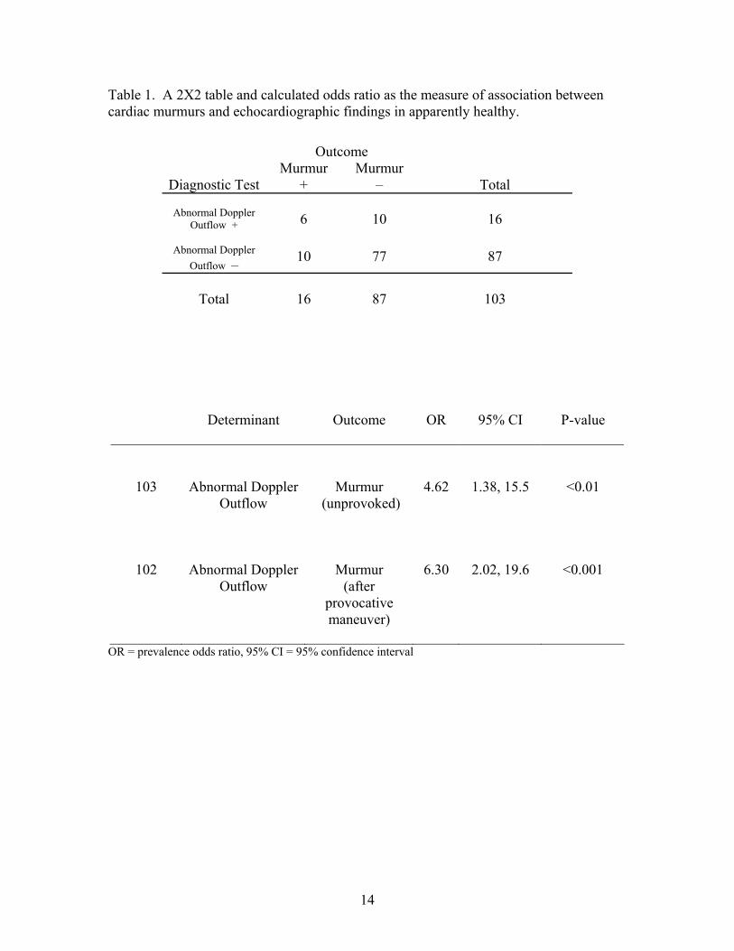
Table 1. A 2X2 table and calculated odds ratio as the measure of association between cardiac murmurs and echocardiographic findings in apparently healthy.
OR = prevalence odds ratio, 95% CI = 95% confidence interval
Determinant
Outcome
OR
95% CI
P-value
103
Abnormal Doppler Outflow
Murmur (unprovoked)
4.62
1.38, 15.5
<0.01
102
Abnormal Doppler Outflow
Murmur (after
provocative maneuver)
6.30
2.02, 19.6
<0.001
Outcome
Diagnostic Test Murmur
+ Murmur
–
Total
Abnormal Doppler Outflow +
6
10
16
Abnormal Doppler Outflow –
10
77
87
Total
16
87
103
14
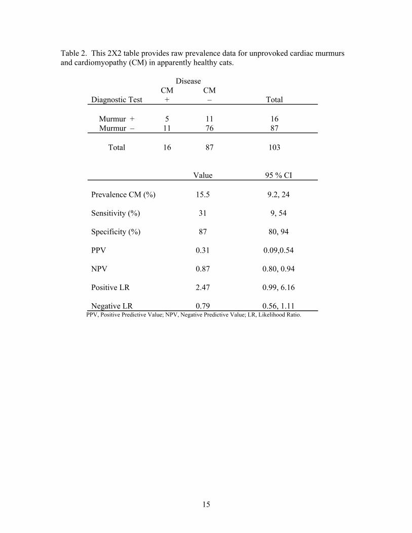
Table 2. This 2X2 table provides raw prevalence data for unprovoked cardiac murmurs and cardiomyopathy (CM) in apparently healthy cats.
Disease
Diagnostic Test CM +
CM –
Total
Murmur +
5
11
16
Murmur – 11 76 87
Total
16
87
103
Value 95 % CI Prevalence CM (%)
15.5
9.2, 24
Sensitivity (%)
31
9, 54
Specificity (%)
87
80, 94
PPV
0.31
0.09,0.54
NPV
0.87
0.80, 0.94
Positive LR
2.47
0.99, 6.16
Negative LR
0.79
0.56, 1.11
PPV, Positive Predictive Value; NPV, Negative Predictive Value; LR, Likelihood Ratio.
15
DISCUSSION
This study was the first prospective investigation of the relationship between
auscultatory abnormalities and echocardiographic findings in a community-based
population of apparently healthy cats. The prevalence of feline subclinical CM in
Southwest Virginia is near 16%; approximately a third of these cats had murmurs. The
majority of affected cats had HCM. Interestingly, only 5 out of the 16 cats with murmurs
had a CM.
Previous veterinary investigations have addressed epidemiological characteristics
of feline cardiomyopathy, but many of these studies evaluated referral-based populations
or excluded cats with murmurs from the study population. Cote et al described a 21%
(23 /103 cats) prevalence of heart murmurs among overtly healthy cats.12 Because cats
that had a heart murmur or history of cardiac disease had been excluded prior to initiation
of that study, their prevalence estimate can not be directly compared to our findings.
Because we included cats based on apparent health status, we believe we have achieved a
better estimate of the true prevalence within our population. Importantly, our study
design removed the echocardiographer’s inherent bias to identify the source of a cardiac
murmur. Each cat was examined systematically without knowledge of the physical
findings which should further increase the validity of our prevalence estimate.
Based on our findings, feline HCM is more prevalent than is HCM in human
beings.21 The prevalence of HCM in our population sample seemingly was high but there
are no similar published data with which to compare. However, it should be recognized
that, relative to M-mode echocardiography which has been used in many studies of feline
HCM, 2-D echocardiography is a sensitive method for detection of hypertrophy.22 The
majority (11/15) of cats with HCM had segmental left ventricular hypertrophy. The
subjects with HCM that we identified were apparently mildly affected; hypertrophy was
not marked, generally was segmental and none of the cats had echocardiographic
evidence of atrial enlargement. The high prevalence of HCM in apparently healthy cats is
consistent with the current understanding of the diversity of this disorder in human
beings. In the past, the clinical impact of HCM in humans and perhaps in cats has been
exaggerated because data typically have been obtained from referral populations. It is
16
currently accepted that the HCM in people is a genetic disorder that is associated with
diverse phenotypic expression and a variable clinical course.21,23 HCM in the Maine
Coon cat is heritable and the genetic mutation associated with this disorder has recently
been reported.4,5 Familial HCM has been observed in other breeds of cats and based on
this, it is possible that feline HCM generally is a genetic disorder.
Abnormal auscultatory findings often prompt an echocardiographic examination;
it is the next logical diagnostic step when feline CM is suspected. However, previous
retrospective studies have clearly demonstrated that cats without murmurs can also have a
CM.13,18 In this study, 11/16 cats with CM did not have murmurs. Within this study
population, sensitivity and specificity describe the proportion of cats with CM and
without CM that are identified by the presence or absence a murmur, respectively.23
Interestingly, only 31% of cats with CM had murmurs, and 87% of cats without CM did
not have murmurs. Because sensitivity is apparently low, the use of cardiac murmur as a
diagnostic, screening test would yield a high number of false negative results. 24 The 95%
confidence intervals (CI) for sensitivity and specificity were 0.085, 0.54 and 0.804,
0.943, respectively. The CI for sensitivity is wide because the sample size, though larger
than previous investigations, was, in relative terms, small (Table 2).
To further explore the diagnostic accuracy of CM detection by murmurs, the
positive predictive value (PPV) and negative predictive value (NPV) were calculated
(Table 2). Because the number of cats with murmurs and CM were equal, PPV and NPV
were numerically identical to our sensitivity and specificity estimates (Table 2). The PPV
is the probability that a cat with a murmur has CM, while the NPV is the probability that
a cat that does not have a murmur does not have CM.25 Predictive values depend on
population prevalence; our estimates are only relevant to the population of apparently
healthy cats in our geographic region. Within this study population, our findings suggest
that a murmur detects only 31 % of the cats with a murmur have CM, which means that
69% of cats with a murmur do not have CM. These predictive values suggest that
auscultation does not reliably identify feline cardiomyopathy.
The calculation of likelihood ratios (LR) is another approach to the evaluation of
diagnostic utility.26 The positive LR is the proportional relationship between the
probability of a positive test result in a subject that has disease and the probability of a
17
positive test result in a subject that does not have disease. For our data, the positive LR
relates the probability that a subject with CM has a murmur with the probability that a
subject that does not have CM has a murmur.26 Based on our data, a cat with CM is 2.5
times more likely to have a murmur than a cat without CM. However, this was only a
tendency of the data as the 95% CI included one; given our a priori specification of
alpha=0.05, the result was not statistically significant. The negative likelihood ratio was
0.79; that is, cats with CM are less likely to be free of a murmur than are cats without
CM. Again however, this result was not statistically significant. Taken together, these
measures of diagnostic accuracy suggest that the presence or absence of cardiac murmurs
in healthy cats does not usefully discriminate those with CM from those without.
In general, diagnostic tests that effectively screen populations for disease have
high sensitivity. In this regard, the presence or absence of a cardiac murmur certainly has
limitations. However, at present, physical examination likely remains the only practical
means to select apparently healthy cats that may have CM. Furthermore, the specificity of
auscultation is moderate.
The association between abnormal Doppler outflow tract velocities and murmurs
was evaluated through the calculation of prevalence odds ratios. The odds ratio is an
indirect measure of risk that is appropriate for use in the analysis of cross-sectional
studies.27 When statistically significant, it can be interpreted as the multiplicative
relationship between an explanatory variable and the binary outcome. In our study,
subjects with Doppler evidence of dynamic outflow tract obstruction were 5 times more
likely to have a murmur at rest than did cats that did not have this finding, and 6.3 times
more likely to have murmur after provocation (Table 1). The 95% CI of the odds ratio
provides a measure of the precision of the estimate.28
Because this was a cross-sectional study, we cannot infer a causal association
between echocardiographic findings and the presence of a murmur. However, we can
conclude that cats with Doppler evidence of abnormal ventricular outflow are nearly 5
times more likely to have a murmur at rest, and those cats with Doppler evidence of
abnormal ventricular outflow tract velocity are 6 times more likely to have murmur after
provocation. Previous studies have addressed the labile nature of murmurs in cats, which
can vary in intensity with heart rate.19 Because auscultation was not performed during the
18

echocardiogram, it is plausible that some, if not all, of these cats with dynamic outflow
tract obstruction, could develop a heart murmur during some circumstances.
These data must be interpreted in the context of the study limitations. The
confidence intervals of our descriptive statistics were relatively broad and this of course
is a reflection of sample size. It would have been better to examine larger number of cats
but the sample size was determined by issues of practicality. The subjects enrolled in this
investigation were pets and therefore it was not possible to confirm the diagnosis of HCM
by post-mortem examination. We recognize that 22 of the cats resisted manual restraint,
and inclusion of these cats may have influenced our prevalence results. A few cats had
isolated basilar septal hypertrophy. It is recognized that a change in aorticoseptal angle –
the development of a “sigmoid septum” – could possibly result in an artifactual
appearance of septal hypertrophy.29
In summary, our epidemiologic study of feline cardiomyopathy has provided an
unbiased prevalence estimate of murmurs and cardiomyopathy in apparently healthy cats.
We have also clarified the clinical relevance of murmurs and the diagnostic role of
auscultation for detection of CM. Our estimate of population prevalence and
understanding of cardiac murmurs may form the basis of future epidemiological
investigation of feline cardiomyopathies.
19
CONCLUSIONS
The prevalence of feline subclinical CM in Southwest Virginia is near 16%;
approximately a third of these cats had murmurs. Hypertrophic CM is the most prevalent
form of CM within our study population. Auscultation is not a sensitive diagnostic test
for detection of feline CM. The presence of a cardiac murmur was associated with
echocardiographic evidence of dynamic ventricular outflow tract obstruction.
FUTURE INVESTIGATIONS Now we have identified the unbiased prevalence of cardiomyopathy and murmurs
within this population of apparently healthy cats, future epidemiological studies can be
directed towards longitudinal follow-up of our normal cats. This would allow the
identification of incident cases, which can ultimately be used to calculate the incidence
density, an epidemiological measure of disease occurrence.24 Utilizing a cohort study
design and the incidence density, a relative risk for various factors (i.e. identification of a
murmur) could be determined between exposed and non-exposed groups of apparently
healthy cats.24 Any causal associations of these risk factors leading to an outcome of
cardiomyopathy could then be determined. Based on this new understanding of disease
progression and risk, therapeutic trials can be implemented, which currently remains the
most clinically important, yet least understood, aspect of feline cardiomyopathy.
While hypertrophic cardiomyopathy remains the most common genetic
cardiovascular disease in humans, this genetic association in Maine Coon cats has only
recently been revealed.2,5 Therefore, future investigations of this or other gene mutation
in pure and mixed breed cats should be further pursued. Once identified, long-term
follow-up of these cats would clarify the relationship of their genetic findings with the
phenotypic expression of left ventricular hypertrophy.
20
LITERATURE CITED
1. Richardson P, McKenna W, Bristow M, et al. Report of the 1995 World Health
Organization / International Society and Federation of Cardiology Task Force on
the definition and classification of cardiomyopathies. Circulation 93:841, 1996
2. Maron BJ. Hypertrophic cardiomyopathy: an important global disease. Am J Med
2004: 116: 63-65.
3. Fox PR, Liu SK, and Maron BJ. Echocardiographic assessment of spontaneously
occurring feline hypertrophic cardiomyopathy: an animal model of human
disease. Circulation 1995; 92:2645-2651.
4. Kittleson MD, Meurs KM, Munro BA, et al. Familial hypertrophic
cardiomyopathy in Maine Coon cats: an animal model of human disease.
Circulation 1999; 99:3172-3180.
5. Meurs KM, Sanchez X, David RM, et al. A cardiac myosin binding protein C
mutation in the Maine Coon cat with familial hypertrophic cardiomyopathy. Hum
Mol Genet 2005;14:3587-3593.
6. Fox PR. Feline Cardiomyopathies. In. Fox PR, Sisson D, Moïse SN, 2nd ed.
Textbook of Canine and Feline Cardiology: Principles and clinical practice.
Philadelphia, PA: WB Saunders; 1999:621-678.
7. Ferasin L, Sturgess CP, Cannon MJ, et al. Feline idiopathic cardiomyopathy: a
retrospective study of 106 cats (1994-2001). J Fel Med Surg: 2003:5:151-159.
8. Harpster, NK. Feline arrhythmias: diagnosis and management. In: Kirk RW, 11th
ed. Current Veterinary Therapy. Philadelphia, PA: WB Saunders; 1992: 732-733.
21
9. Rush JE, Freeman LM, Fenollosa NK, et al. Population and survival
characteristics of cats with hypertrophic cardiomyopathy: 260 cases (1990-1999).
JAVMA 2002: 220(2): 201-207.
10. Atkins CE, Gallo AM, Kurzman ID, et al. Risk factors, clinical signs, and survival
in cats with a clinical diagnosis of idiopathic hypertrophic cardiomyopathy: 74
cases (1985-1989). JAVMA 1992; 201:613-618.
11. Liu SK. Pathology of feline heart disease. Vet Clin North Am 1977; 7:323
12. Côté E, Manning AM, Emerson D, et al. Assessment of the prevalence of heart
murmurs in overtly healthy cats. JAVMA 2004:223 (3): 384-388.
13. Grover SL and Olson JK. Cardiac cats. Veterinary Forum 2005:37-41.
14. Ahlbom A and Norell S. Introduction to modern epidemiology. 2nd ed. Chestnut
Hill, MA: Epidemiology Resources Inc.; 1990: 4-10.
15. Braunwald E and Perloff JK. Physical examination of the heart and circulation.
In: Zipes DP, Libby P, et al. (ed). Braunwald’s Heart Disease: A Textbook of
Cardiovascular Medicine, 7th edition. Philadelphia, PA: Elsevier Saunders; 2005:
77-106.
16. Thomas WP, Gaber CE, Jacobs GJ, et al. Recommendations for standards in
transthoracic two-dimensional echocardiography in the dog and cat.
Echocardiography Committee of the Specialty of Cardiology, American College
of Veterinary Internal Medicine. J Vet Intern Med 1993;7:247-252.
17. Weyman AE. Principles and Practice of Echocardiography, 2nd Edition.
Philadelphia, PA: Lea & Febiger; 1994: 600.
22
18. Fox PR, Maron BJ, Basso C, et al. Spontaneously occurring arrhythmogenic right
ventricular cardiomyopathy in the domestic cat: A new animal model similar to
the human disease. Circulation 2000; 102:1863-1870.
19. Rishniw M, Thomas WP. Dynamic right ventricular outflow obstruction: a new
cause of systolic murmurs in cats. J Vet Intern Med 2002;16:547-552.
20. Grandy JL, Dunlop CI, Hodgson DS, et al. Evaluation of the Doppler ultrasonic
method of measuring systolic arterial blood pressure in cats. Am J Vet Res
1992;53:1166-1169.
21. Maron BJ. Hypertrophic cardiomyopathy: a systematic review. Jama
2002;287:1308-1320.
22. Maron BJ, McKenna WJ, Danielson GK, et al. American College of
Cardiology/European Society of Cardiology clinical expert consensus document
on hypertrophic cardiomyopathy. A report of the American College of Cardiology
Foundation Task Force on Clinical Expert Consensus Documents and the
European Society of Cardiology Committee for Practice Guidelines. J Am Coll
Cardiol 2003;42:1687-1713.
23. Altman DG, Bland JM. Diagnostic tests. 1: Sensitivity and specificity. BMJ
1994;308:1552.
24. Hennekens CHH, Buring JE. Epidemiology in medicine. Boston, MA: Little,
Brown and Company; 1987; 54-98, 327-347.
25. Altman DG, Bland JM. Diagnostic tests 2: Predictive values. BMJ 1994; 309:102.
26. Deeks JJ, Altman DG. Diagnostic tests 4: likelihood ratios. BMJ 2004;329:168-
169.
23
27. Zocchetti C, Consonni D, Bertazzi PA. Relationship between prevalence rate
ratios and odds ratios in cross-sectional studies. Int J Epidemiol 1997; 26:220-
223.
28. Rothman KJ, Greenland S. Modern Epidemiology, 2nd edition. Publishers
Philadelphia, PA: Lippincott-Raven; 183-199.
29. Binder J, Ommen SR, Gersh BJ, et al. Echocardiography-guided genetic testing
in hypertrophic cardiomyopathy: septal morphological features predict the
presence of myofilament mutations. Mayo Clin Proc 2006; 81(4): 459-467.
24
VITA
Christopher F. Paige was born in Tolland, Connecticut. He graduated with a
Bachelor of Science in Animal Sciences from the University of Connecticut, Storrs,
Connecticut. In August 1996, he started his master’s program in the Department of
Epidemiology and Community Health, and worked in collaboration with the Laboratory
Research Branch, G.W. Long Hansen’s Disease Center, United States Public Health
Service, Louisiana State University, Baton Rouge, Louisiana. He completed the degree of
Master of Science at Louisiana State University, School of Veterinary Medicine, in
August 1998. The following year, he worked as a research associate for Louisiana State
University Embryo and Biotechnology Laboratory in St. Gabriel, Louisiana. In 1999, he
was accepted and enrolled in the veterinary medicine program at Louisiana State
University, School of Veterinary Medicine. He obtained a Doctor of Veterinary Medicine
degree in May 2003. Following graduation, he did a rotating internship in small animal
medicine and surgery at Angell Animal Medical Center-Western New England,
Springfield, Massachusetts. In July 2004, Christopher began a 3-year cardiology
residency and combined Master’s of Science at Virginia-Maryland Regional College of
Veterinary Medicine, Virginia Polytechnic Institute & University, Blacksburg, Virginia.
He completed the Masters of Science at Virginia-Maryland College of Veterinary
Medicine in summer 2007.
25