Embed Size (px)
Citation preview
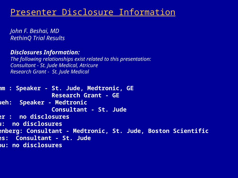
Presenter Disclosure Information
John F. Beshai, MDRethinQ Trial Results
Disclosures Information:The following relationships exist related to this presentation:Consultant - St. Jude Medical, AtricureResearch Grant - St. Jude Medical
R Grimm : Speaker - St. Jude, Medtronic, GE Research Grant - GES Nagueh: Speaker - Medtronic Consultant - St. JudeJ Baker : no disclosuresS Beau: no disclosuresS Greenberg: Consultant - Medtronic, St. Jude, Boston ScientificL Pires: Consultant - St. JudeP Tchou: no disclosures
The Resynchronization Therapy in Normal QRS (RethinQ) Study
John F. Beshai, MD, Richard A. Grimm, DO, Sherif F. Nagueh, MD, James H. Baker II, MD, Scott L. Beau, MD, Steven M. Greenberg, MD,
Luis A. Pires, MD, Patrick J. Tchou, MD for the RethinQ study investigators
Study Sponsored by St. Jude Medical
Background
• Currently, indications for cardiac resynchronization therapy (CRT) include QRS duration > 120ms, LVEF < 35% and NYHA
Class III-IV. • 20-30% of patients do not respond to CRT despite application of
established selection criteria.
• Patients with normal conduction or a slightly prolonged QRS duration also exhibit mechanical abnormalities due to intraventricular dyssynchrony.
• Myocardial Tissue Doppler Imaging (TDI) allows both the velocity and timing of regional longitudinal motion to be measured.
• LV dyssynchrony may also be useful in predicting the benefit of
CRT before implantation of the pulse generator.
Hypothesis
•We hypothesized that patients with NYHA class III, left ventricular ejection fraction less than or equal to 35%, narrow QRS duration < 130 ms, and evidence of mechanical dyssynchrony on echocardiography may benefit from cardiac resynchronization therapy.
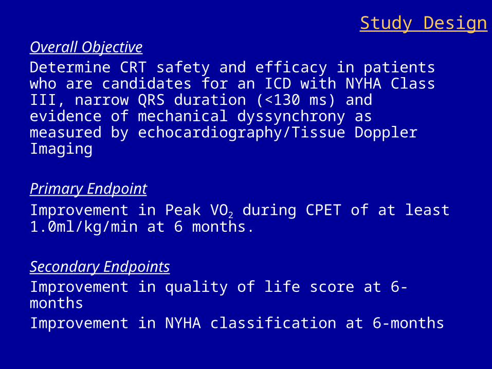
Overall ObjectiveDetermine CRT safety and efficacy in patients who are candidates for an ICD with NYHA Class III, narrow QRS duration (<130 ms) and evidence of mechanical dyssynchrony as measured by echocardiography/Tissue Doppler Imaging
Primary Endpoint
Improvement in Peak VO2 during CPET of at least 1.0ml/kg/min at 6 months.
Secondary EndpointsImprovement in quality of life score at 6-monthsImprovement in NYHA classification at 6-months
Study Design

Inclusion and Exclusion Criteria
Inclusion Criteria
• NYHA class III HF
• LVEF ≤ 35%
• Evidence of mechanical dyssynchrony
• QRS duration < 130ms
Exclusion Criteria
• NYHA class I, II, or IV
• Permanent Atrial Fibrillation
• Recent MI, unstable angina or cardiac revascularization
• Prior cardiac resynchronization therapy

Mechanical dyssynchrony considered present if either
•M-Mode - Septal posterior wall mechanical delay (SPWMD) ≥ 130 ms
OR
•Tissue Doppler Imaging (TDI) of the basal ventricular segments in apical 4/2/3 chamber views - Septal to lateral delay ≥ 65ms OR - Antero-septal to posterior delay ≥ 65ms
Echo Criteria for LV Dyssynchrony

172 patients randomized in 34
centers
Stratified by QRS < 120 ms or ≥ 120 ms
AndIschemic or Non-ischemic CM
Randomized to CONTROL or CRT
CONTROLFollowed for 6-months
CRTFollowed for 6-months
Study Design

Results
Baseline Characteristics Control
(n = 85)
CRT
(n = 87)
Age (yr), Mean ± SD 58 ± 14 60 ± 12
Male sex, n(%) 49 (58%) 62 (71%)
NYHA III, n(%) 84 (99%) 87 (100%)
QRS Duration (ms), Mean ± SD 106 ± 13 107 ± 12
Ischemic Cardiomyopathy, n(%) 43 (51%) 47 (54%)
Left ventricular ejection fraction (%) 26 ± 6 25 ± 5
Medications, n(%)
ACE inhibitor or substitute§
Beta-Blocker
Diuretic
Antiarrhythmic
77 (91%)
79 (93%)
74 (87%)
10 (12%)
77 (89%)
84 (97%)
73 (84%)
7 (8%)§ Includes ARBs and other ACE substitutes, including Hydralazine
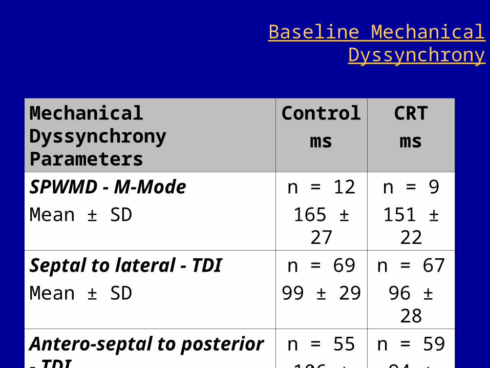
Baseline Mechanical Dyssynchrony
Mechanical Dyssynchrony Parameters
Control
ms
CRT
ms
SPWMD - M-Mode
Mean ± SD
n = 12
165 ± 27
n = 9
151 ± 22
Septal to lateral - TDI
Mean ± SD
n = 69
99 ± 29
n = 67
96 ± 28
Antero-septal to posterior - TDI
Mean ± SD
n = 55
106 ± 29
n = 59
94 ± 23

Baseline Echocardiographic Data0
20
40
60
80
Control
n = 85
CRT
n = 87
LV end diastolic diameter (mm)
Mean +/- 1 SD
p = 0.29
LV end systolic diameter (mm)
Mean +/- 1 SD
02
04
06
08
0
p = 0.06
CRT
n = 87
Control
n = 85
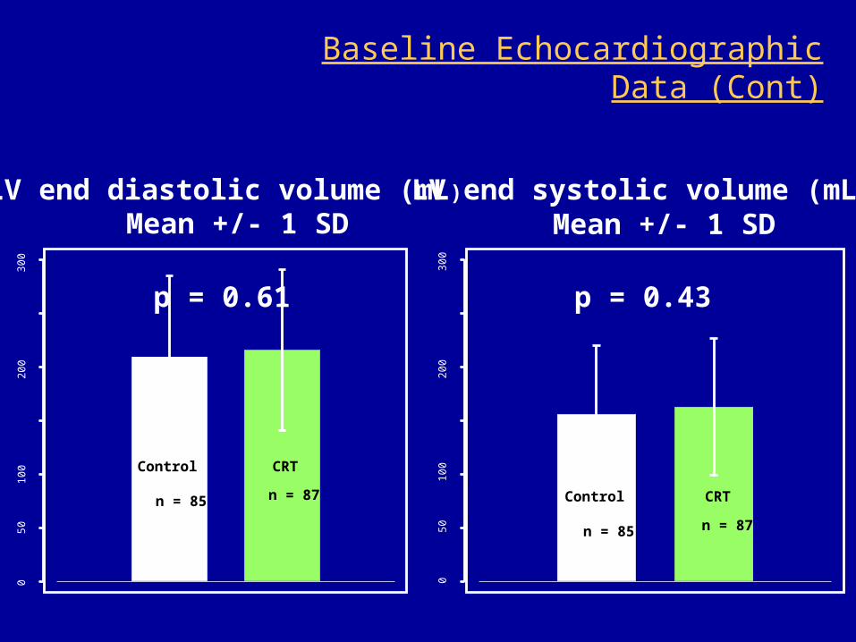
Baseline Echocardiographic Data (Cont)
05
01
00
20
03
00
LV end diastolic volume (mL)
Mean +/- 1 SD
p = 0.61
CRT
n = 87
Control
n = 85
05
01
00
20
03
00
LV end systolic volume (mL)
Mean +/- 1 SD
p = 0.43
CRT
n = 87
Control
n = 85

Baseline Evaluations0
51
01
52
02
50
51
01
52
02
5
Peak VO2(ml/kg/min)
Mean +/- 1 SD
p = 0.85
Control
n = 85
CRT
n = 87
05
10
15
05
10
15
Exercise Duration (min) Mean +/- 1 SD
p = 0.85
Control
n = 85
CRT
n = 87
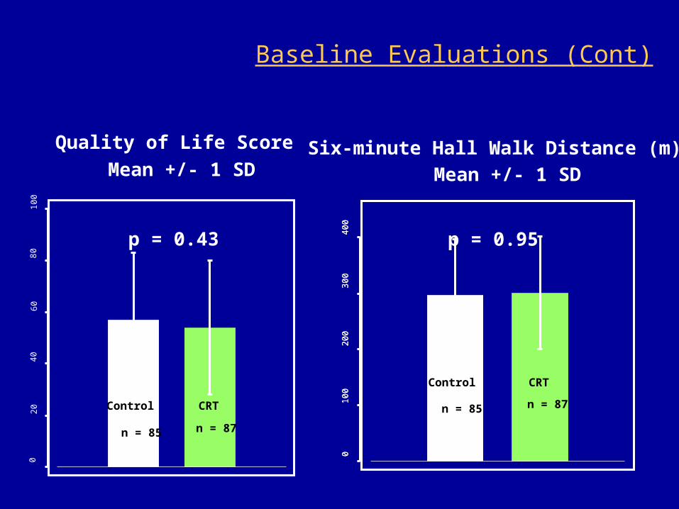
Baseline Evaluations (Cont)0
20
40
60
80
10
0
Quality of Life Score
Mean +/- 1 SD
p = 0.43
Control
n = 85
CRT
n = 87
01
00
20
03
00
40
00
10
02
00
30
04
00
Six-minute Hall Walk Distance (m)
Mean +/- 1 SD
p = 0.95
Control
n = 85
CRT
n = 87
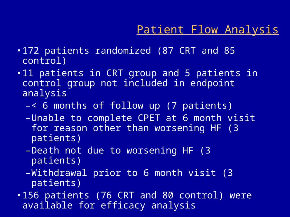
Patient Flow Analysis
•172 patients randomized (87 CRT and 85 control)
•11 patients in CRT group and 5 patients in control group not included in endpoint analysis–< 6 months of follow up (7 patients)–Unable to complete CPET at 6 month visit for
reason other than worsening HF (3 patients)–Death not due to worsening HF (3 patients)–Withdrawal prior to 6 month visit (3 patients)
•156 patients (76 CRT and 80 control) were available for efficacy analysis

Results - Primary Endpoint8
1012
14
Baseline 6-months
Peak VO2 (ml/kg/min) Median & 95% CI
p = 0.75
Control (n = 80)CRT (n = 76)
CRTn = 76
020
4060
8010
0
p = 0.63
41% 46%
Controln = 80
% Improved in Peak VO2
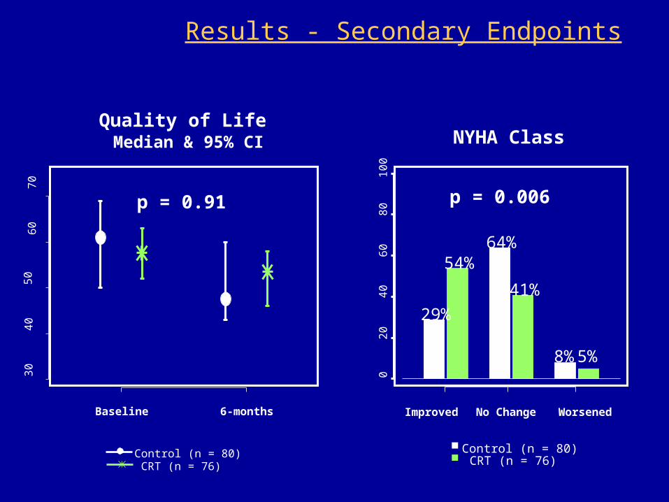
Results - Secondary Endpoints
Improved No Change Worsened
02
04
06
08
01
00
29%
54%64%
41%
8%5%
NYHA Class
p = 0.006
30
40
50
60
70
Baseline 6-months
Quality of Life
Median & 95% CI
p = 0.91
Control (n = 80)CRT (n = 76)
Control (n = 80)CRT (n = 76)
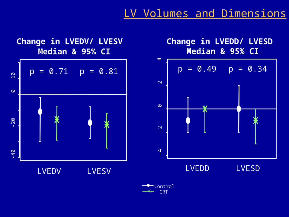
Change in LVEDV/ LVESV
Median & 95% CI
-40
-20
010
p = 0.71
LVEDV
p = 0.81
LVESV
Change in LVEDD/ LVESD
Median & 95% CI
-4-2
02
4
p = 0.49
LVEDD
p = 0.34
LVESD
ControlCRT
LV Volumes and Dimensions

Peak VO2 by sub-group
Peak VO2
(% improved from baseline)
020
4060
8010
0
QRS ≥ 120 ms
p = 0.02
QRS < 120 ms
p = 0.45
Control
n = 25
CRT
n = 17
Control
n = 55CRT
n = 59
Peak VO2
(% improved from baseline)
020
4060
8010
0
Ischemic
p = 0.82
Non-ischemic
p = 0.25
Control
n = 41
Control
n = 39
CRT
n = 40
CRT
n = 36
Summary
•This prospective, multi-center, randomized trial was designed to evaluate the effectiveness of CRT therapy in a HF population with narrow QRS duration and evidence of mechanical dyssynchrony.
•There was no statistical significant difference in the change in Peak VO2 between the treatment and control group during cardiopulmonary exercise testing.
•No improvement in other objective parameters including 6-minute walk test, LV reverse remodeling, and secondary endpoint - quality of life score .
Summary
•NYHA class improved to a greater extent in the treatment group than in the control group.
•Although numbers were small in the sub-group analysis, there was no statistically significant difference in the primary endpoint between ischemic and non-ischemic patients.
•Patients with QRS duration 120 - 130 ms demonstrated an improvement in peak VO2.

Conclusion
• CRT did not improve Peak VO2 during exercise in patients with NYHA Class III heart failure, QRS duration <130ms, EF ≤ 35% and mechanical dyssynchrony as specified in this trial.
• While there was a statistically significant improvement of NYHA class, a secondary endpoint, there was no improvement in quality-of-life, 6-minute walking test, or echocardiographic measures of reverse LV remodeling
• A subgroup of patients with QRS duration between 120 ms and 130 ms demonstrated an improvement from CRT, however
patients with QRS duration < 120 ms did not demonstrate improvement
• The subgroup of patients stratified on the basis of cardiomyopathy etiology did not demonstrate an improvement in peak VO2.