Embed Size (px)
Citation preview

7/30/2019 Presentation Sam
http://slidepdf.com/reader/full/presentation-sam 1/17
PET/CTBy Sam McNulty
7/30/2019 Presentation Sam
http://slidepdf.com/reader/full/presentation-sam 2/17
What is PET/CT
An imaging modality that allows theclinician to better differentiate benignvs malignant structural abnormalities
seen on CT as well as see possiblemalignancies where no structuralabnormalities are seen.
7/30/2019 Presentation Sam
http://slidepdf.com/reader/full/presentation-sam 3/17
PET
Stands for positron emissiontomography
Fluorine-18-deoxyglucose (FDG), aradionuclide labeled glucoseanalogue is injected and the pt isimaged
7/30/2019 Presentation Sam
http://slidepdf.com/reader/full/presentation-sam 4/17
Indications
Breast cancer: staging of distant metastasis, restaging,and monitoring response to treatment (when a change intherapy is anticipated based on results)
Cervical cancer: staging as adjunct to conventionalimaging
Colorectal cancer: diagnosis, staging, and restaging Esophageal cancer: diagnosis, staging, and restaging Head and neck cancer (non-thyroid, non-CNS): diagnosis,
staging, and restaging Lymphoma: diagnosis, staging, and restaging
7/30/2019 Presentation Sam
http://slidepdf.com/reader/full/presentation-sam 5/17
Indications con’t
Melanoma: diagnosis, staging, and restaging Non small cell lung cancer: diagnosis, staging, and
restaging Solitary pulmonary nodules: characterization Follicular cell thyroid cancer: restaging of recurrent or
residual disease previously treated by thyroidectomy andradioiodine ablation in the setting of serum thyroglobulin> 10 ng/ml and a negative I-131 whole body scan
Myocardial viability: primary or initial diagnosis orfollowing an inconclusive SPECT prior to revascularization
Refractory seizures (brain): pre-surgical evaluation only
7/30/2019 Presentation Sam
http://slidepdf.com/reader/full/presentation-sam 6/17
How does it work?
In the FDG decay process, positrons are emitted. When apositron is emitted it travels for a short distance from thesite of origin (on the order of 1-3 millimeters) graduallylosing energy to the tissue through which it passes. Whenmost of the positron’s kinetic energy has been lost, itundergoes a process called annihilation. In annihilation, the
positron reacts with an electron in the immediate area andthe result is the emission of two very high energy (511 keVeach) photons. The two 511 keV photons are emitted inopposite directions at approximately 180 degrees from eachother. These two photons interact with the PET detectorring at near opposite sites which define a line within thebody along which the annihilation occurred. With computerprocessing, this line between the two emitted photonspermits fairly precise localization of the annihilationreaction and thus defines a tissue site in the body wherethe positron emission occurred (i.e. an area of FDGactivity).
7/30/2019 Presentation Sam
http://slidepdf.com/reader/full/presentation-sam 7/17
Huh?
Malignant cells take inherently have ahigher metabolism than non-malignantcells. They have a higher mitotic rate as
well as more ineffecient aerobicmetabolism leading to more anaerobicmetabolism
Through these mechanisms they will take
up the FDG at a faster rate and this willcan be seen on the scan as the FDGdecays.
7/30/2019 Presentation Sam
http://slidepdf.com/reader/full/presentation-sam 8/17
PET/CT
PET and CT scans are now often done atthe same time.
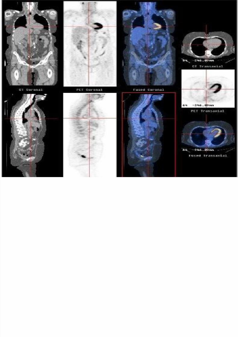
The CT and PET data sets are fused or “coregistered” electronically by the
scanner’s computer system and presentedto the interpreter on a work station. Thedata can then be simultaneously andinteractively viewed as CT data, PET data,and superimposed CT and PET data in anypercentage combination of these data setsdesired (e.g. 100% PET data, 100% CTdata, 50% CT / 50% PET data).
7/30/2019 Presentation Sam
http://slidepdf.com/reader/full/presentation-sam 9/17
PET/CT
Doing both scans at the same timeoffers the advantage over just thePET scan alone because the CT gives
exquisite anatomic detail as well asphysiologic information.
7/30/2019 Presentation Sam
http://slidepdf.com/reader/full/presentation-sam 10/17
7/30/2019 Presentation Sam
http://slidepdf.com/reader/full/presentation-sam 11/17
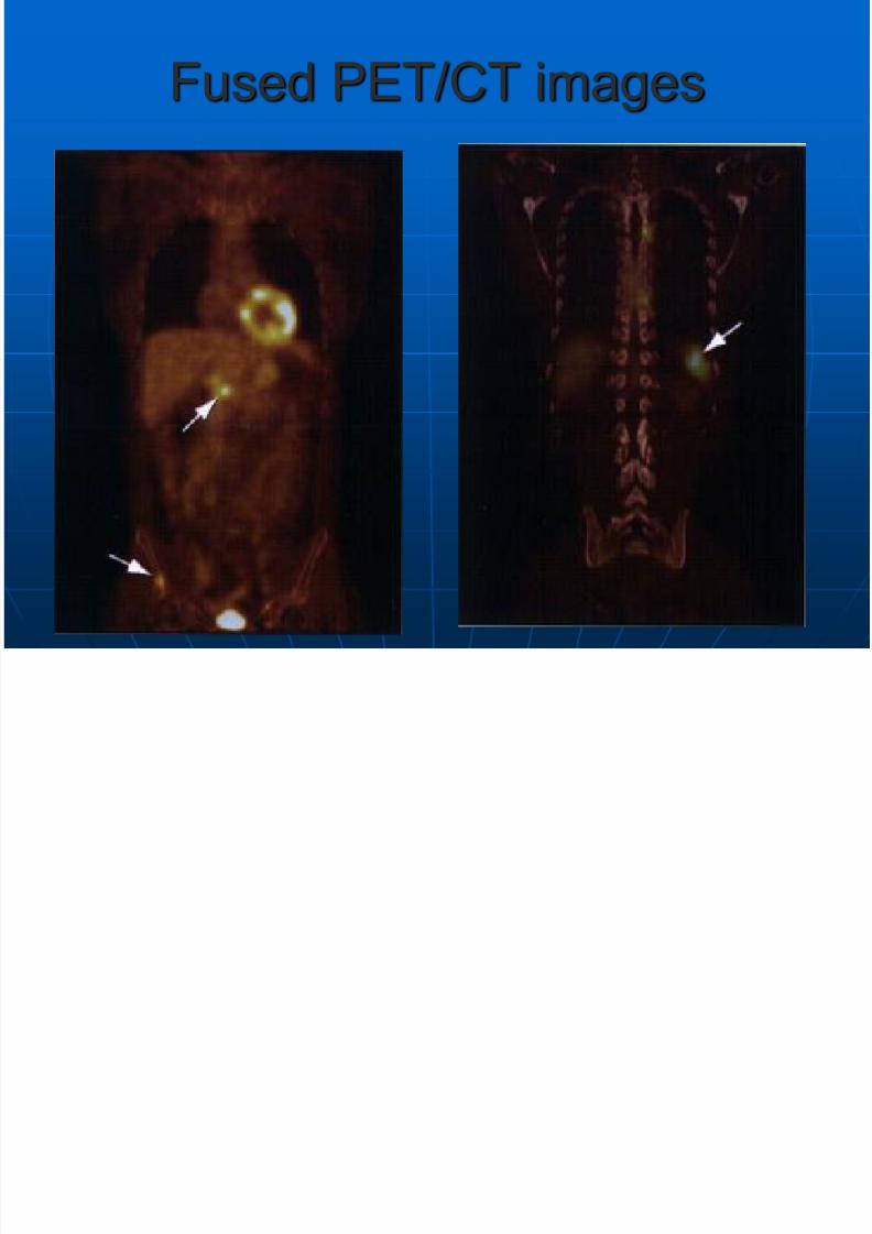
Fused PET/CT images
7/30/2019 Presentation Sam
http://slidepdf.com/reader/full/presentation-sam 12/17
SUV
When a PET camera is appropriatelycalibrated, it is able to assess the amountof FDG activity per volume(millicuries/millileter or mCi/mL) of tissueand this can be expressed in “SUV” units
SUV=Mean region of interest activity(mCi/mL) =administered activity(mCi)/body weight (g) expressed in g/mL
SUV over 2.5 is considered suspicious formalignancy
7/30/2019 Presentation Sam
http://slidepdf.com/reader/full/presentation-sam 13/17
Limitations of PET/CT
FDG is not cancer specific and willaccumulate in any areas of high rates of metabolism and glycolysis.
Therefore, increased uptake can be
expected in all sites of hyperactivity at thetime of FDG administration (e.g. musclesand nervous system tissues); at sites of active inflammation or infection (e.g.sarcoidosis, arthritis, infection etc.); andat sites of active tissue repair (e.g.surgical or traumatic wounds, healingfractures, etc.).
7/30/2019 Presentation Sam
http://slidepdf.com/reader/full/presentation-sam 14/17
Brain/CNS
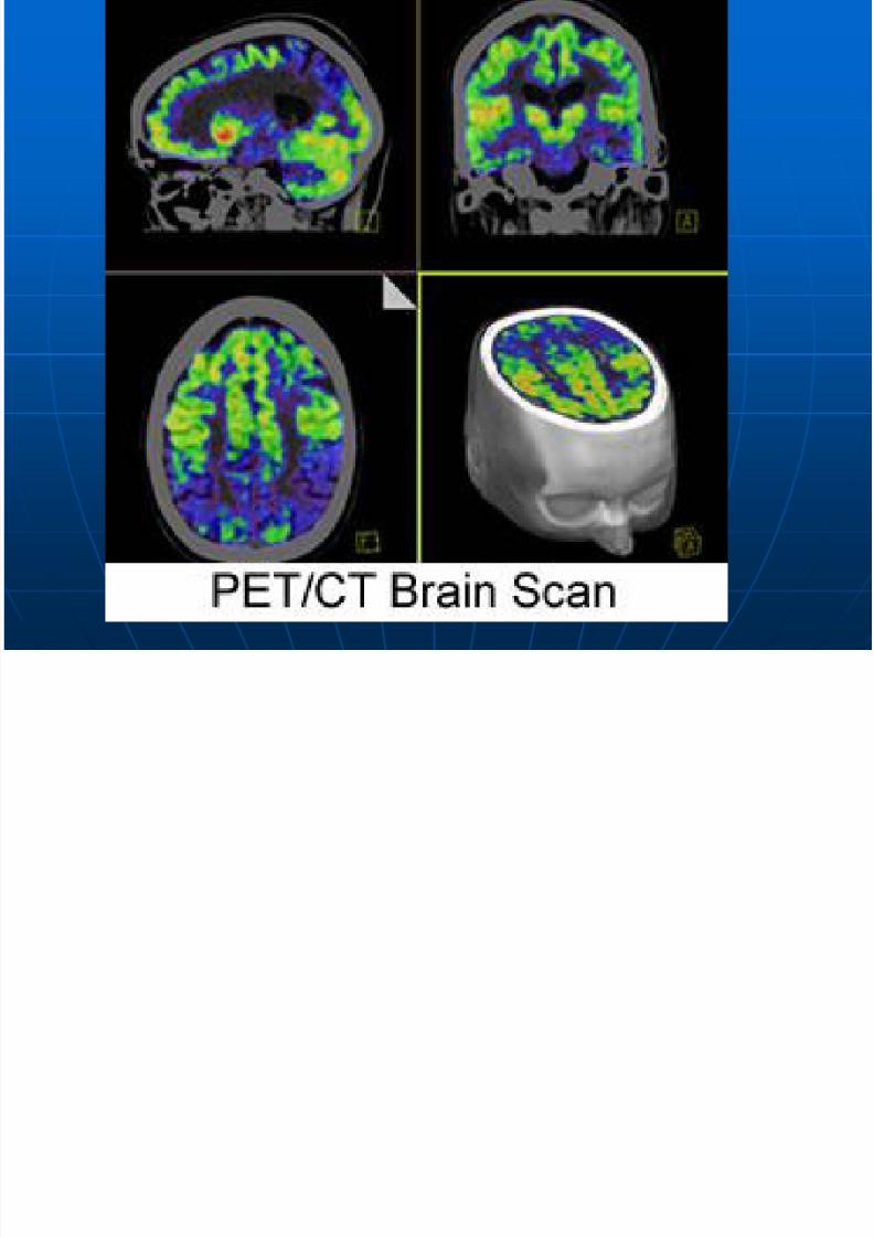
Brain tissue is always highly metabolicallyactive so detection of brain malignancies,particularly in gray matter, is limited withFGD PET.
Hypometabolism is also typical inepileptogenic foci during the interictalperiod. FDG activity may also bedecreased in one or bothfrontotemporoparietal associationcomplexes in Alzheimer’s dementia.
7/30/2019 Presentation Sam
http://slidepdf.com/reader/full/presentation-sam 15/17
7/30/2019 Presentation Sam
http://slidepdf.com/reader/full/presentation-sam 16/17
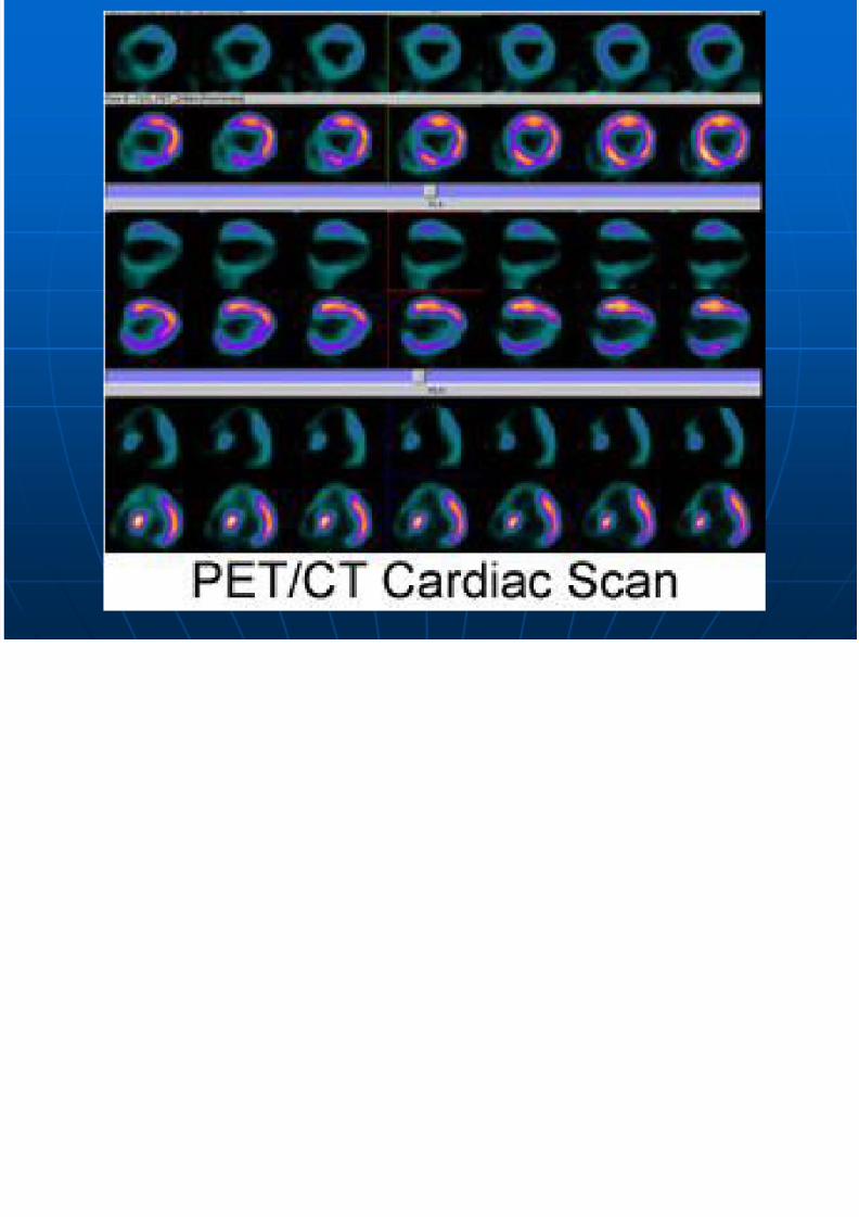
Cardiac
When compared to conventionalSPECT imaging, PET/CT usingRubidium- 82 coupled with a 64-slice
CT scanner is a faster, easier, andmore accurate method to measuremyocardial blood flow.
7/30/2019 Presentation Sam
http://slidepdf.com/reader/full/presentation-sam 17/17