Embed Size (px)
Citation preview
VTT PU
BLICATIO
NS 563
Preparation of nanoparticles consisting of methacrylic polym
ers and drugs by...H
annele Eerikäinen
Tätä julkaisua myy Denna publikation säljs av This publication is available from
VTT TIETOPALVELU VTT INFORMATIONSTJÄNST VTT INFORMATION SERVICEPL 2000 PB 2000 P.O.Box 2000
02044 VTT 02044 VTT FI–02044 VTT, FinlandPuh. 020 722 4404 Tel. 020 722 4404 Phone internat. +358 20 722 4404Faksi 020 722 4374 Fax 020 722 4374 Fax +358 20 722 4374
ISBN 951–38–6443–X (soft back ed.) ISBN 951–38–6444–8 (URL: http://www.vtt.fi/inf/pdf/)ISSN 1235–0621 (soft back ed.) ISSN 1455–0849 (URL: http://www.vtt.fi/inf/pdf/)
ESPOO 2005ESPOO 2005ESPOO 2005ESPOO 2005ESPOO 2005 VTT PUBLICATIONS 563
Hannele Eerikäinen
Preparation of nanoparticlesconsisting of methacrylic polymersand drugs by an aerosol flowreactor method
Drug-containing polymer nanoparticles are submicron-sized particlesconsisting of drug and stabilising or functional polymer. In thisexperimental study, methacrylic polymer nanoparticles with and withoutincorporated model drug were prepared using a novel method, namely,aerosol flow reactor method. Various properties of the preparednanoparticles were studied, such as particle size and size distribution,morphology, crystallinity, thermal properties, drug content, and drugrelease. It was found that this method could be used to produce amorphous,spherical, homogeneous matrix-type drug-polymer nanoparticles directly asdry powder.


VTT PUBLICATIONS 563
Preparation of nanoparticles consisting of methacrylic polymers
and drugs by an aerosol flow reactor method
Hannele Eerikäinen VTT Processes
Laboratory of Polymer Chemistry Department of Chemistry
Faculty of Science University of Helsinki
ACADEMIC DISSERTATION
To be presented, with the permission of the Faculty of Science of the University of Helsinki, for public criticism in Auditorium A129 of the Department of Chemistry
on June 3rd, 2005, at 12 o�clock.
ISBN 951�38�6443�X (soft back ed.) ISSN 1235�0621 (soft back ed.)
ISBN 951�38�6444-8 (URL: http://www.vtt.fi/inf/pdf/) ISSN 1455�0849 (URL: http://www.vtt.fi/inf/pdf/)
Copyright © VTT Technical Research Centre of Finland 2005
JULKAISIJA � UTGIVARE � PUBLISHER
VTT, Vuorimiehentie 5, PL 2000, 02044 VTT puh. vaihde 020 722 111, faksi 020 722 4374
VTT, Bergsmansvägen 5, PB 2000, 02044 VTT tel. växel 020 722 111, fax 020 722 4374
VTT Technical Research Centre of Finland, Vuorimiehentie 5, P.O.Box 2000, FI�02044 VTT, Finland phone internat. +358 20 722 111, fax + 358 20 722 4374
VTT Prosessit, Biologinkuja 7, PL 1602, 02044 VTT puh. vaihde 020 722 111, faksi 020 722 7021
VTT Processer, Biologgränden 7, PB 1602, 02044 VTT tel. växel 020 722 111, fax 020 722 7021
VTT Processes, Biologinkuja 7, P.O.Box 1602, FI�02044 VTT, Finland phone internat. +358 20 722 111, fax +358 20 722 7021
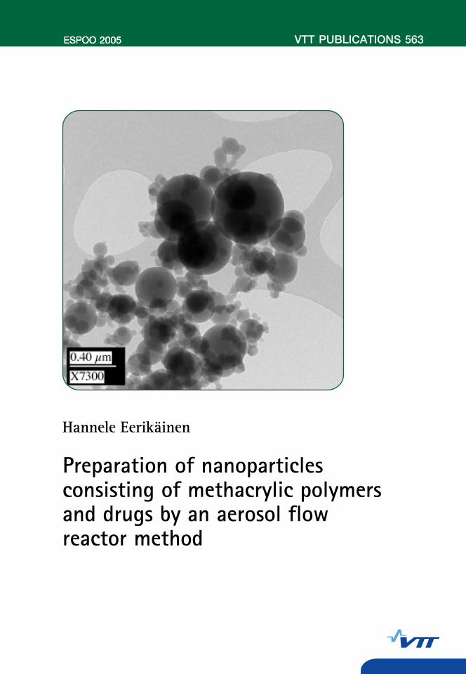
Cover picture: TEM image of nanoparticles containing 60% (w/w) BDP and 40% (w/w) Eudragit L (III). Otamedia Oy, Espoo 2005
3
Eerikäinen, Hannele. Preparation of nanoparticles consisting of methacrylic polymers and drugs byan aerosol flow reactor method. Espoo 2005. VTT Publications 563. 112 p. + app. 55 p.
Keywords methacrylic polymer nanoparticles, preparation of drug nanoparticles, aerosolflow reactor method, drug release, solubility properties, particle size,morphology, crystallinity, thermal properties, drug content
Abstract Drug-containing polymer nanoparticles are submicron-sized particles consisting of drug and stabilising or functional polymer. In this experimental study, methacrylic polymer nanoparticles with and without incorporated model drug were prepared using a novel method, namely, aerosol flow reactor method. This method involves first preparing a solution containing the drug and the polymer, followed by spraying the solution as nanosized droplets into a carrier gas stream, then drying the nanoparticles in a tubular laminar flow reactor tube, and finally collecting the nanoparticles. Model polymers used in this study were Eudragit L, Eudragit E, and Eudragit RS, which are commonly used methacrylic polymers in the pharmaceutical industry. Model drugs studied were beclomethasone dipropionate, ketoprofen, and naproxen.
Various properties of the prepared nanoparticles were studied, such as particle size and size distribution, morphology, crystallinity, thermal properties, drug content, and drug release. It was found that this method could be used to produce amorphous, spherical, homogeneous matrix-type drug-polymer nanoparticles. The size of the particles was adjusted between 90 and 200 nm by the concentration of the solution. The morphology of the particles varied as a function of the properties and composition of the starting solution.
The nanoparticles were collected as dry powders, but the stability of the powders in an amorphous form was found to be dependent on the interactions between the drug and the polymer. When the amount of the drug in the nanoparticles was below the solubility limit of the drug in the polymer, the amorphous nanoparticles were found to be stable and no crystallisation of the drug took place. When the amount of the drug was larger than the solubility limit, large crystalline structures were formed due to crystallisation of the drug. The crystallisation was also dependent on the thermal properties of the drug, as amorphous nanoparticles consisting of a drug having a high glass transition

4
temperature could be collected at room temperature. A low glass transition temperature of the drug led to crystallisation of the drug at ambient conditions, when the drug amount in the nanoparticles was larger than the solubility limit. Drug release from the nanoparticles could be modified by using polymers having specific solubility properties.
5
Hannele Eerikäinen
Center for New Materials Helsinki University of Technology
Finland &
Aerosol Technology Group VTT Processes
Technical Research Centre of Finland Finland
& Laboratory of Polymer Chemistry
Department of Chemistry University of Helsinki
Finland
Present address: Orion Corporation ORION PHARMA Pharmaceutical Product Development
Espoo Finland

6
Supervisors:
Professor Esko I. Kauppinen Aerosol Technology Group
VTT Processes Technical Research Centre of Finland
Finland &
Center for New Materials Helsinki University of Technology
Finland
Professor Heikki Tenhu Laboratory of Polymer Chemistry
Department of Chemistry University of Helsinki
Finland
Reviewers:
Professor Jukka Seppälä Laboratory of Polymer Technology
Department of Chemical Technology Helsinki University of Technology
Finland
Professor Arto Urtti Biopharmacy
Department of Pharmaceutics University of Kuopio
Finland
Opponent:
Professor Etienne Schacht Polymer Materials Research Group Department of Organic Chemistry
Ghent University Belgium
7
Preface The research work for this thesis was performed mainly in the Aerosol Technology Group, VTT Processes, Technical Research Centre of Finland and in Nanoparticle Technology Group, Center for New Materials, Helsinki University of Technology during the years 2002 and 2003. This thesis was finalised at Orion Pharma during the year 2004.
I would like to express my deepest gratitude to my supervisor, Professor Esko Kauppinen, for providing me the possibility to join his group for two years. In addition, I owe him my sincere appreciation for his guidance and encouragement during this work. I would also like to thank him for allowing me to finalise this thesis while working at Orion.
Professor Heikki Tenhu, Head of the Laboratory of Polymer Chemistry, University of Helsinki, is sincerely acknowledged for the supervision of my Ph.D. studies as well as for his advice and guidance during this work. With gratitude I acknowledge the reviewers of this thesis, Professor Jukka Seppälä and Professor Arto Urtti, for their constructive criticism and comments on the text.
Orion Pharma Product Development management, Head of the Analytical Development Department Tuula Romppanen, M.Sc., Laboratory Manager Eerika Skarp, M.Sc., and Laboratory Manager Tero Närvänen, Lic.Pharm. are gratefully acknowledged for allowing me to continue my studies by pursuing this work for two years. I express my sincere thanks to the personnel of the Laboratory of Physics at Orion Pharma for their friendship and encouragement during these years. All my former and present co-workers at Orion Pharma are acknowledged for their support. Special thanks go to Saara Tiittanen, M.Sc. for her friendship.
I would like to thank all my colleagues at the Aerosol Technology Group and Nanoparticle Technology Group for the innovative and pleasant working environment. Especially I owe my thanks to Dr. Petri Ahonen and Dr. Wiwik Watanabe for their help. I am most grateful to Dr. Janne Raula for his encouragement, help, friendship, and for the numerous discussions not only on the scientific work but also everything else but the science.

8
Dr. Leena Peltonen is acknowledged for her help and support with the spectrophotometry and dissolution analyses. Hanna Kortejärvi, M.Sc. is acknowledged for her guidance with the biopharmaceutical modelling. The contributions and efforts of all of my co-authors are appreciated.
Finally, I owe warm thanks to my parents, my sister, and my brothers for their love and encouragement during these years.
My sincerest thanks belong to my dear husband and my best friend, Jussi Hirvelä, for all his love, patience, and encouragement. Thank you for standing by me during both good and bad times.
Helsinki, April 2005
Hannele Eerikäinen
9
List of original publications This thesis is based on the following five publications, hereafter referred to in the text by their Roman numerals I�V.
I Eerikäinen, H., Watanabe, W., Kauppinen, E.I., and Ahonen, P.P. 2003. Aerosol flow reactor method for synthesis of drug nanoparticles. European Journal of Pharmaceutics and Biopharmaceutics, 55, 3, 357�360.
II Raula, J., Eerikäinen, H., and Kauppinen, E.I. 2004. Influence of the solvent composition on the aerosol synthesis of pharmaceutical polymer nanoparticles. International Journal of Pharmaceutics, 284, 1, 13�21.
III Eerikäinen, H., and Kauppinen, E.I. 2003. Preparation of polymeric nanoparticles containing corticosteroid by a novel aerosol flow reactor method. International Journal of Pharmaceutics, 263, 1�2, 69�83.
IV Eerikäinen, H., Kauppinen E.I., and Kansikas, J. 2004. Polymeric drug nanoparticles prepared by an aerosol flow reactor method. Pharmaceutical Research, 21, 1, 136�143.
V Eerikäinen, H., Peltonen, L., Raula, J., Hirvonen, J., and Kauppinen, E.I. 2004. Nanoparticles containing ketoprofen and acrylic polymers prepared by an aerosol flow reactor method. AAPS PharmSciTech, 5, 4, Article 68.

10
Contents
Abstract ................................................................................................................. 3
Preface .................................................................................................................. 7
List of original publications .................................................................................. 9
List of symbols and abbreviations ...................................................................... 12
1. Introduction................................................................................................... 13
2. Review .......................................................................................................... 15 2.1 Structures of drug nanoparticles .......................................................... 15 2.2 Applications of drug nanoparticles...................................................... 16
2.2.1 Biodegradable and non-biodegradable materials .................... 17 2.2.2 Oral administration.................................................................. 20
2.2.2.1 Effect of pH on the solubility of the drug................ 21 2.3 Theory of dissolution........................................................................... 23 2.4 Amorphous drug materials and amorphous solid solutions
consisting of drug and polymer ........................................................ 27 2.5 Polymer materials of the study ............................................................ 29 2.6 Methods of preparation of drug nanoparticles..................................... 30
3. Objective of the study ................................................................................... 34
4. Experimental................................................................................................. 35 4.1 Aerosol flow reactor method for the preparation of nanoparticles...... 35
4.1.1 Starting solution and atomisation............................................ 37 4.1.1.1 Preparation of the starting solution.......................... 37 4.1.1.2 Atomisation of the solution...................................... 37
4.1.2 Solvent evaporation................................................................. 37 4.1.2.1 Temperature and flow in the tubular reactor tube.... 38
4.1.3 Particle sampling and collection ............................................. 38 4.2 Materials .............................................................................................. 39
4.2.1 Drug materials......................................................................... 39 4.2.2 Polymer materials.................................................................... 41
4.3 Instrumentation and characterisation................................................... 42
11
5. Results........................................................................................................... 47 5.1 Particle size, particle size distribution, and particle morphology
(I, II, III, IV) ...................................................................................... 47 5.1.1 Particle size and particle size distribution as a function of
solution concentration (IV) ..................................................... 47 5.1.2 Particle size and particle morphology (I, II, III)...................... 48
5.1.2.1 Effect of temperature (I, III) .................................... 48 5.1.2.2 Effect of solvent system (II, III) .............................. 51
5.2 Collection and properties of bulk nanoparticle powder (III, IV, V) .... 53 5.2.1 Collection of the nanoparticles (III, IV, V) ............................. 53
5.2.1.1 Nanoparticles containing ketoprofen or naproxen (IV) ........................................................ 53
5.2.1.2 Interactions between ketoprofen and methacrylic polymers (V)............................................................ 62
5.2.1.3 Nanoparticles containing beclomethasone dipropionate (III) ..................................................... 68
5.2.2 Drug release from nanoparticles containing ketoprofen (IV, V) ... 70 5.2.2.1 Effects of dissolution on drug absorption ................ 75
5.2.3 Drug release from nanoparticles containing beclomethasone dipropionate ............................................................................ 79
5.2.4 Stability of the nanoparticles................................................... 80
6. Summary and conclusions ............................................................................ 81
References........................................................................................................... 83
Appendices
Publications I–V
Appendices of this publication are not included in the PDF version. Please order the printed version to get the complete publication (http://www.vtt.fi/inf/pdf/)

12
List of symbols and abbreviations BCS Biopharmaceutical classification system BDP Beclomethasone dipropionate BLPI Berner-type low pressure impactor CAB Cellulose-acetate-butyrate DMA Differential mobility analyser DNA Deoxyribonucleic acid DSC Differential scanning calorimetry EC Ethylcellulose GI Gastrointestinal IR Infra-red (spectroscopy) i.m. Intramuscular i.v. Intravenous Mw Weight average molar mass Mn Number average molar mass PAA Poly(acrylamide) PBCA Poly(butylcyanoacrylate) PCL Poly(ε-caprolactone) pDNA Plasmid DNA PECA Poly(ethylcyanoacrylate) PEI Poly(ethyleneimine) pKa Acid dissociation constant PLA Poly(d,l-lactic acid) PLGA Poly(d,l-lactide-co-glycolide) PMCA Poly(methylcyanoacrylate) PMMA Poly(methylmethacrylate) SEM Scanning electron microscope / Scanning electron microscopy TEM Transmission electron microscope / Transmission electron microscopy Tg Glass transition temperature Tm Melting temperature XRD X-ray diffraction

13
1. Introduction Drug nanoparticles can be specified as drug-containing particles having size smaller than 1 µm. Several applications have been proposed for these discrete submicron-sized particles, such as targeted drug delivery, controlled release, and increase in bioavailability of poorly water soluble drugs [1�4]. Due to the small size of the nanoparticles, drug targeting into, for example, cancerous tumours has been shown to be possible [5]. Moreover, nanoparticles can prove to be effective in stabilising and shielding labile drug molecules, such as proteins, peptides, or DNA molecules from degradation [2�4, 6, 7], thus opening new possibilities for protein drug delivery and gene therapy.
Drug nanoparticles consist of the drug material and, usually, a stabilising polymer. In addition to physically stabilising the drug nanoparticles, the polymers can also act as functional agents, leading to sustained release of the drug or drug release triggered by changes in environmental conditions, for example in pH level. A variety of materials have been used for the preparation of drug nanoparticles, such as biodegradable polymers and non-biodegradable, but pharmaceutically acceptable polymers. Polymers of the latter type were used as model materials for the purposes of this thesis.
First polymer nanoparticles for pharmaceutical applications were prepared in the late 1960�s and early 1970�s and were based on acrylamide micelle polymerisation [8�10]. Since then, various polymerization methods as well as methods involving the use of preformed polymers have been developed and studied [1, 9, 11]. Commonly, these methods result in a nanoparticle suspension in water stabilised with surfactants. However, the physical and chemical stability of such suspensions is often not adequate for practical applications. Some of the problems related to the instability, in addition to facilitating further processing, might be solved by having the nanoparticles as a dry powder [12�15]. This research was aimed to study the possibilities to use a new method for the preparation of drug-containing polymer nanoparticles. In this novel method, namely, aerosol flow reactor method, the particles are formed by the evaporation of the solvent from nanosized droplets containing the drug and the polymer. The nanoparticles are collected directly as dry powder. As several of the previous preparation methods are dependent on the formation of an oil-in-water emulsion,
14
the partitioning of the drug between the immiscible phases of the emulsion affects the drug loading in the particles. In the aerosol flow reactor method, each of the droplets forms one particle during solvent evaporation and, therefore, drug leakage cannot take place. The amount of drug in the starting solution determines the drug content of the resulting particles.
15
2. Review
2.1 Structures of drug nanoparticles
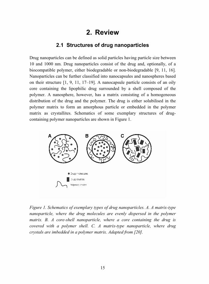
Drug nanoparticles can be defined as solid particles having particle size between 10 and 1000 nm. Drug nanoparticles consist of the drug and, optionally, of a biocompatible polymer, either biodegradable or non-biodegradable [9, 11, 16]. Nanoparticles can be further classified into nanocapsules and nanospheres based on their structure [1, 9, 11, 17�19]. A nanocapsule particle consists of an oily core containing the lipophilic drug surrounded by a shell composed of the polymer. A nanosphere, however, has a matrix consisting of a homogeneous distribution of the drug and the polymer. The drug is either solubilised in the polymer matrix to form an amorphous particle or embedded in the polymer matrix as crystallites. Schematics of some exemplary structures of drug-containing polymer nanoparticles are shown in Figure 1.
Figure 1. Schematics of exemplary types of drug nanoparticles. A. A matrix-type nanoparticle, where the drug molecules are evenly dispersed in the polymer matrix. B. A core-shell nanoparticle, where a core containing the drug is covered with a polymer shell. C. A matrix-type nanoparticle, where drug crystals are imbedded in a polymer matrix. Adapted from [20].

16
2.2 Applications of drug nanoparticles
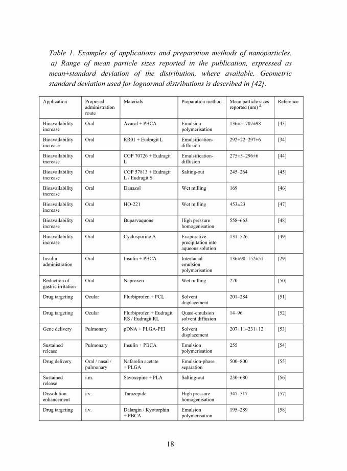
Both biodegradable nanoparticles for systemic drug delivery and non-biodegradable nanoparticles for drug dissolution modification have been studied [3, 11, 21, 22]. Proposed applications for drug nanoparticles vary from drug targeting and delivery [3, 5, 11, 23�26] to even gene [4, 7, 27] and protein [28, 29] therapies. Administration of nanoparticles by, for example, parenteral [21], ocular [30�32], transdermal [33], and oral routes have been studied. Nonetheless, the oral route is still the most convenient, preferred, and in many cases, also the most cost-effective method of drug administration [18, 28, 34�37]. Some of the proposed applications and administration routes of nanoparticles are summarised in Table 1.
17
2.2.1 Biodegradable and non-biodegradable materials
The route of administration determines to some extent whether biodegradable or non-biodegradable materials can be used in the nanoparticles. For parenteral administration the particles need to be biodegradable [22, 38] to avoid the risk of polymer accumulation in the body and to ensure complete elimination. Intravenously administered drug-containing nanoparticles can be used to target drugs or even DNA [4, 6, 7, 27] into specific organs or sites, for example into cancerous tumours [5, 18, 23, 26]. Targeting the drug in a specific site of action increases the effectiveness of the drug while reducing side effects [3]. Due to their small size, nanoparticles can be taken up by cells, for example by endocytosis [4, 5]. Inside the cell, the pH levels of different compartments vary, so pH-dependent particles could be used to target drugs in specific organelles of the cell [4, 23, 39]. Biodegradable nanoparticles can also be used intravenously for sustained and constant systemic drug release [1, 24]. For this application, the surface of the particles has to be suitably modified to avoid the uptake by mononuclear phagocyte system [18, 40, 41]. Such nanoparticles, after injection, circulate in the body acting as drug reservoirs [41], and slow release of the drug from the nanoparticles leads to constant drug levels.
18
Table 1. Examples of applications and preparation methods of nanoparticles. a) Range of mean particle sizes reported in the publication, expressed as mean±standard deviation of the distribution, where available. Geometric standard deviation used for lognormal distributions is described in [42].
Application Proposed administration route
Materials Preparation method Mean particle sizes reported (nm) a
Reference
Bioavailability increase
Oral Avarol + PBCA Emulsion polymerisation
136±5�707±98 [43]
Bioavailability increase
Oral RR01 + Eudragit L Emulsification-diffusion
292±22�297±6 [34]
Bioavailability increase
Oral CGP 70726 + Eudragit L
Emulsification-diffusion
275±5�296±6 [44]
Bioavailability increase
Oral CGP 57813 + Eudragit L / Eudragit S
Salting-out 245�264 [45]
Bioavailability increase
Oral Danazol Wet milling 169 [46]
Bioavailability increase
Oral HO-221 Wet milling 453±23 [47]
Bioavailability increase
Oral Buparvaquone High pressure homogenisation
558�663 [48]
Bioavailability increase
Oral Cyclosporine A Evaporative precipitation into aqueous solution
131�526 [49]
Insulin administration
Oral Insulin + PBCA Interfacial emulsion polymerisation
136±90�152±51 [29]
Reduction of gastric irritation
Oral Naproxen Wet milling 270 [50]
Drug targeting Ocular Flurbiprofen + PCL Solvent displacement
201�284 [51]
Drug targeting Ocular Flurbiprofen + Eudragit RS / Eudragit RL
Quasi-emulsion solvent diffusion
14�96 [52]
Gene delivery Pulmonary pDNA + PLGA-PEI Solvent displacement
207±11�231±12 [53]
Sustained release
Pulmonary Insulin + PBCA Emulsion polymerisation
255 [54]
Drug delivery Oral / nasal / pulmonary
Nafarelin acetate + PLGA
Emulsion-phase separation
500�800 [55]
Sustained release
i.m. Savoxepine + PLA Salting-out 230�680 [56]
Dissolution enhancement
i.v. Tarazepide High pressure homogenisation
347�517 [57]
Drug targeting i.v. Dalargin / Kyotorphin + PBCA
Emulsion polymerisation
195�289 [58]
19
Drug targeting i.v. Indomethacin / 5-fluorouracil + PLGA
Spontaneous emulsification solvent diffusion
338±67�637±40 [59]
Cancer therapy i.v. Piposulfan / Etoposide / Camptothecin / Paclitaxel
Wet milling 202±31�279±30 [60]
Toxicity reduction
i.v. Primaquine + PLA Solvent displacement
153�169 [61]
Drug targeting No routes proposed
Amoxicillin Supercritical antisolvent precipitation
250�1200 [62]
Drug targeting No routes proposed
Insulin Electrospray 88�117 [63]
Drug targeting No routes proposed
Triamcinolone acetonide + PLA
Emulsification-evaporation
476±410�710±406 [64]
Drug targeting No routes proposed
Atovaquone + PCL / PLA / PLGA
Solvent displacement
228±16�242±33 [65]
Drug targeting No routes proposed
Tamoxifen + PCL Solvent displacement
200�300 [23]
Gene delivery No routes proposed
pDNA + PLGA Double emulsion-evaporation
589±190�640±64 [7]
Sustained release
No routes proposed
Isradipine + PCL / PLA / PLGA
Solvent displacement
110�208 [66]
Technical studies on preparation method
Oral / ocular / topical
Indomethacin + EC / CAB / PMMA / Eudragit RS / Eudragit RL
Emulsification-evaporation
100�125 [12]
Technical studies on preparation method
No routes proposed
Chlorambucil + PLA / PLGA / PCL / Eudragit S
Emulsification-diffusion
246�591 [67]
Technical studies on preparation method
No routes proposed
PLA Emulsification-diffusion
100�450 [68]
Technical studies on preparation method
No routes proposed
PAA / PMMA / PBCA / PECA / PMCA
Polymerisation 51�145 [16]
Technical studies on preparation method
No routes proposed
PLA / Eudragit S / Eudragit E / ethyl cellulose
Salting-out 172�1117 [69]
However, for oral administration, a wider range of materials is available. For oral administration, there is no requirement that the particles need to be biodegradable. Instead, in oral administration, the nanoparticles can be used to
20
modify drug release [25, 36, 44, 45, 70]. In addition, non-biodegradable nanoparticles can be used, for example, for ocular [30, 32, 52] drug delivery.
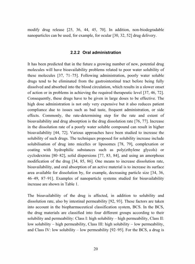
2.2.2 Oral administration
It has been predicted that in the future a growing number of new, potential drug molecules will have bioavailability problems related to poor water solubility of these molecules [37, 71�75]. Following administration, poorly water soluble drugs tend to be eliminated from the gastrointestinal tract before being fully dissolved and absorbed into the blood circulation, which results in a slower onset of action or in problems in achieving the required therapeutic level [37, 46, 72]. Consequently, these drugs have to be given in large doses to be effective. The high dose administration is not only very expensive but it also reduces patient compliance due to issues such as bad taste, frequent administration, or side effects. Commonly, the rate-determining step for the rate and extent of bioavailability and drug absorption is the drug dissolution rate [76, 77]. Increase in the dissolution rate of a poorly water soluble compound can result in higher bioavailability [44, 72]. Various approaches have been studied to increase the solubility of such drugs. The techniques proposed for solubility increase include solubilisation of drug into micelles or liposomes [78, 79], complexation or coating with hydrophilic substances such as poly(ethylene glycols) or cyclodextrins [80�82], solid dispersions [77, 83, 84], and using an amorphous modification of the drug [34, 85, 86]. One means to increase dissolution rate, bioavailability, and oral absorption of an active material is to increase its surface area available for dissolution by, for example, decreasing particle size [34, 36, 46�49, 87�91]. Examples of nanoparticle systems studied for bioavailability increase are shown in Table 1.
The bioavailability of the drug is affected, in addition to solubility and dissolution rate, also by intestinal permeability [92, 93]. These factors are taken into account in the biopharmaceutical classification system, BCS. In the BCS, the drug materials are classified into four different groups according to their solubility and permeability: Class I: high solubility � high permeability, Class II: low solubility � high permeability, Class III: high solubility � low permeability, and Class IV: low solubility � low permeability [92�95]. For the BCS, a drug is
21
considered to be highly soluble when the highest dose is soluble in 250 ml water over the pH range of 1.0�7.5. Similarly, a drug is considered to be highly permeable when the intestinal absorption is over 90% of the orally administered dose [93, 96]. When the bioavailability of a drug substance is limited by its poor solubility, enhancing the dissolution can improve bioavailability. However, when the bioavailability is limited by the intestinal permeability, faster dissolution is not expected to increase bioavailability. Therefore, of the BCS classes, class II drug molecules are the most promising to show increase in bioavailability as a result of enhanced solubility.
2.2.2.1 Effect of pH on the solubility of the drug
The gastrointestinal tract consists of several parts having different conditions with respect to pH, and fluid and enzyme compositions [97]. The pH of stomach is acidic, ranging from 1.3 to 5 depending on the fed-fasted conditions [90, 97, 98]. The small intestine, however, has a significantly higher pH level ranging from 6.5 to 7.5 [90, 98].
The environmental pH affects the dissolution of drug molecules, which contain ionisable groups [90]. Commonly, the neutral acid or basic form is insoluble or less soluble in water than the ionised form of the drug. The ionisation pH of the molecule can be described by the pKa value of the compound. This value characterises the pH at which half of the molecules carry a charge whereas half of the molecules are in neutral form. Weakly basic molecules are protonated in the acidic stomach, and are thus soluble to gastric fluid. In comparison, weakly acidic drugs containing e.g. carboxylic acid groups are poorly soluble in acidic conditions. When these compounds enter the region of higher pH, they are ionised and soluble. However, as several commonly used drug compounds have pKa values smaller than the pH of the absorption site, these compounds will be soluble early in the upper instestine [90] causing irritation and other side-effects.
The drug release in the GI tract can be controlled using pH-sensitive materials, in which the drug is released specifically in the small intestine near the absorption site. pH-dependent materials, which are insoluble in the acidic environment of the stomach but dissolve in the intestinal fluid are called enteric materials [98, 99]. Such materials have been traditionally used to coat tablets
22
and capsules to allow the drug dosage form to pass intact in the acidic environment of the stomach. These coatings have been used to prevent the degradation of labile drugs caused by the acidic environment or gastric enzymes, to reduce irritiation of gastric mucosa, and to deliver drugs selectively to the site of absorption [98, 99]. Enteric coating materials are polymers, which contain acid groups. In the acidic environment of the stomach the acid groups are nonionised, and the coating material is insoluble. Rapid dissolution and drug release is achieved in the upper intestine as a function of pH change in the environment. The polymer acid groups are ionised at higher pH and the material dissolves [98].
Side effects such as irritation, mucosal damage, and ulceration are often encountered in conjunction with the administration of, for example, non-steroidal anti-inflammatory drugs [50, 100�102]. These side effects have been shown to be at least partly of local instead of systemic origin [50, 103]. The protection of the mucosa can be accomplished by encapsulation of the drug by a protective layer surrounding the drug molecules, thus avoiding direct contact of the drug with the mucosal tissue [13, 14, 104, 105]. To maximise absorption and to improve bioavailability, the drug should be released selectively close to the site of absorption [44]. For instance, by using pH-dependent encapsulation materials, the drug release can be triggered by the change in pH level of the environment [34, 45, 97].
Oral administration of drugs incorporated in pH-sensitive particles has been shown to be effective for poorly water-soluble compounds in increasing drug bioavailability and giving fast absorption [34, 45, 70]. It has been speculated, that the good bioavailability of pH-controlled nanoparticulate drug systems relies in several properties, including the high specific surface area of the system, drug in an amorphous form or molecularly dispersed in the polymer matrix, and the release of drug close to the absorption site [34, 44, 70]. The dissolution of the particles should not be too rapid, however, as this might lead to drug precipitation [45] or crystallisation, [106] but should not be too slow resulting in elimination of the particles before absorption, either [45].
Drug nanoparticles containing a protective material can also be used to shield labile drugs such as peptides and proteins against degradation in the biological
23
environment caused by either acidic conditions of the stomach or by gastrointestinal enzymes [1, 28, 29, 107, 108]. Commonly, peptides and proteins have to be administered by injection to avoid degradation, and repeated injections are required [108]. Oral administration of a protein has been shown to be possible by formulating the drug as by using suitable excipients, which are resistant to gastric enzymes [28, 29, 109].
2.3 Theory of dissolution
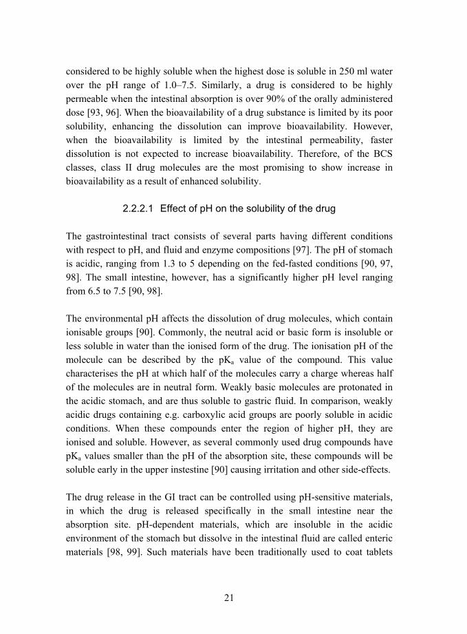
The dissolution of a solid material can be described by a diffusion layer model. This model was first suggested by Bruner and Tolloczko [110] and further developed into its classical form by Nernst and Brunner [111, 112]. When a particle of a solid material is immersed in a solvent, a stagnant diffusion layer is formed around it (see Figure 2).
Figure 2. Schematic representation of a model depicting the dissolution process [113].

24
In this diffusion layer, the velocity of the medium can be taken as zero and the transport of the dissolved molecules from the surface of the particle into the bulk liquid takes place by the diffusion of the molecules through the diffusion layer. The thickness of the diffusion layer is dependent on the experimental conditions such as the stirring rate [112]. The diffusion layer model assumes that the dissolution is very rapid and that saturation is achieved at the particle surface. At the surface of the particles, the concentration of the dissolving material is the saturation concentration, Cs, and the concentration is linearly decreasing in the diffusion layer until it reaches the concentration of the bulk medium, C (see Figure 2). Therefore, the diffusion of the molecules through the stagnant diffusion layer is the rate-limiting step for dissolution. The driving force for the diffusion of the molecules is a negative concentration gradient, which is consistent with the Fick´s first law of diffusion:
xCDJ∂∂
−= ,
where J is the diffusion current, the amount of material that passes perpendicularly through a surface per unit time, D is the diffusion coefficient of the dissolving species, and ∂C/∂x is the linear concentration gradient [114].
The chemical properties of the dissolving material affect the stagnant diffusion layer [115�119]. Especially, for the weakly acidic molecules, the microenvironment of the diffusion layer can show pH levels different from the bulk medium. Weakly acidic molecules are poorly soluble in the nonionised state below their pKa. Above their pKa, the acid groups dissociate and the molecules are soluble. The molecules dissolve in the diffusion layer in the nonionised state, but deprotonation can then take place in the diffusion layer. Consequently, the pH of the microenvironment is lower than of the bulk solution, as the acidic molecules self-buffer the diffusion layer. The lower pH of the diffusion layer decreases the solubility of the solid acidic material. Such self-buffering is relevant when the pH of the solution is higher than the pKa value of the dissolving material [115, 116].
When the bulk solution consists of a buffer solution, the diffusion layer pH of a solid acidic material can be affected. The buffer solution is able to consume the

25
released hydrogen ions, and the surface pH of the dissolving material approaches that of the bulk, leading to an increase in the dissolution rate [116]. Increasing buffer pH, buffer ionic strength, or buffer basicity increase the pH of the diffusion layer [116], resulting in faster dissolution. A schematic representation of the concentration of various ionic species in as a function of distance from the surface of a particle consisting of a carboxylic acid in buffer solution is shown in Figure 3 [116].
Figure 3. Diagrammatic representation of a solid carboxylic acid, HA, dissolving into a reactive medium containing hydroxide ion and buffer components B and BH+ with a Nernst diffusion layer existing between the solid and the bulk solution. Sink conditions exist in the bulk solution, and the products, BH+, A-, and H+, diffuse out of the diffusion layer at a rate determined by their chemical reactivity and diffusivity [116].
According to the Noyes-Whitney equation [112�114, 120, 121], the dissolution of a material is dependent on surface area of the dissolving species:
( )CCh
DAdtdm
s −= ,
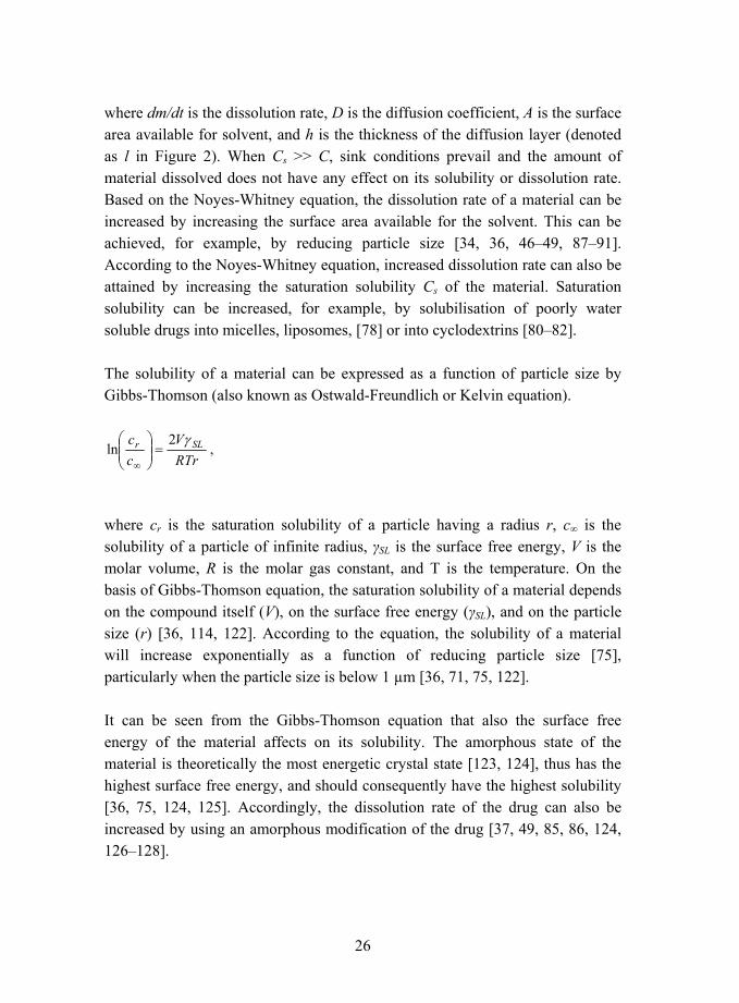
26
where dm/dt is the dissolution rate, D is the diffusion coefficient, A is the surface area available for solvent, and h is the thickness of the diffusion layer (denoted as l in Figure 2). When Cs >> C, sink conditions prevail and the amount of material dissolved does not have any effect on its solubility or dissolution rate. Based on the Noyes-Whitney equation, the dissolution rate of a material can be increased by increasing the surface area available for the solvent. This can be achieved, for example, by reducing particle size [34, 36, 46�49, 87�91]. According to the Noyes-Whitney equation, increased dissolution rate can also be attained by increasing the saturation solubility Cs of the material. Saturation solubility can be increased, for example, by solubilisation of poorly water soluble drugs into micelles, liposomes, [78] or into cyclodextrins [80�82].
The solubility of a material can be expressed as a function of particle size by Gibbs-Thomson (also known as Ostwald-Freundlich or Kelvin equation).
RTrV
cc SLr γ2
ln =⎟⎟⎠
⎞⎜⎜⎝
⎛
∞,
where cr is the saturation solubility of a particle having a radius r, c∞ is the solubility of a particle of infinite radius, γSL is the surface free energy, V is the molar volume, R is the molar gas constant, and T is the temperature. On the basis of Gibbs-Thomson equation, the saturation solubility of a material depends on the compound itself (V), on the surface free energy (γSL), and on the particle size (r) [36, 114, 122]. According to the equation, the solubility of a material will increase exponentially as a function of reducing particle size [75], particularly when the particle size is below 1 µm [36, 71, 75, 122].
It can be seen from the Gibbs-Thomson equation that also the surface free energy of the material affects on its solubility. The amorphous state of the material is theoretically the most energetic crystal state [123, 124], thus has the highest surface free energy, and should consequently have the highest solubility [36, 75, 124, 125]. Accordingly, the dissolution rate of the drug can also be increased by using an amorphous modification of the drug [37, 49, 85, 86, 124, 126�128].

27
2.4 Amorphous drug materials and amorphous solid solutions consisting of drug and polymer
Increase in the dissolution rate of a drug can be achieved by using an amorphous form of the drug [49, 85, 86, 124, 128]. Amorphous materials do not have any long-range order such as crystal lattice in comparison to crystalline materials [128]. Commonly, small drug molecules can easily crystallise, and consequently both the amorphous state and the crystalline state are possible. The amorphous state has higher internal energy, larger free volume, and greater molecular mobility in comparison to crystalline state [129]. These properties of the amorphous state lead to greater solubility, but as a drawback the amorphous structure is metastable with respect to crystalline state, and has a tendency to be spontaneously converted into a crystalline state of lower energy [128�133]. The differences in the energies of the crystalline and the amorphous (glassy) state are shown schematically in Figure 4 for an exemplary molecule, which can exist both in an amorphous and in a crystalline state. The crystalline material shows a small increase in enthalpy and volume as a function of temperature corresponding to a constant heat capacity and thermal expansion coefficient [129]. At the melting temperature, Tm, a first-order phase transition is observed as a change in the slope of enthalpy and volume versus temperature. When the liquid is cooled rapidly, it is possible for the system to follow the equilibrium line to the supercooled liquid region instead of converting back to the crystalline state. At the glass transition temperature, Tg, the supercooled material forms a glassy nonequilibrium state having high energy [129]. The Kauzmann temperature, TK, represents the lower limit for glass transition temperature [129].
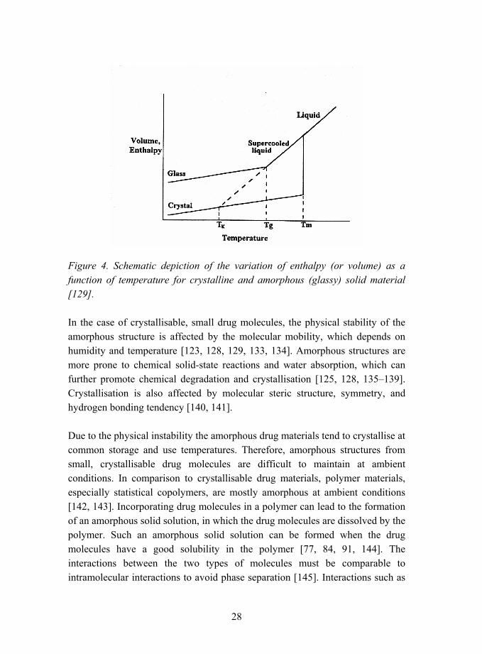
28
Figure 4. Schematic depiction of the variation of enthalpy (or volume) as a function of temperature for crystalline and amorphous (glassy) solid material [129].
In the case of crystallisable, small drug molecules, the physical stability of the amorphous structure is affected by the molecular mobility, which depends on humidity and temperature [123, 128, 129, 133, 134]. Amorphous structures are more prone to chemical solid-state reactions and water absorption, which can further promote chemical degradation and crystallisation [125, 128, 135�139]. Crystallisation is also affected by molecular steric structure, symmetry, and hydrogen bonding tendency [140, 141].
Due to the physical instability the amorphous drug materials tend to crystallise at common storage and use temperatures. Therefore, amorphous structures from small, crystallisable drug molecules are difficult to maintain at ambient conditions. In comparison to crystallisable drug materials, polymer materials, especially statistical copolymers, are mostly amorphous at ambient conditions [142, 143]. Incorporating drug molecules in a polymer can lead to the formation of an amorphous solid solution, in which the drug molecules are dissolved by the polymer. Such an amorphous solid solution can be formed when the drug molecules have a good solubility in the polymer [77, 84, 91, 144]. The interactions between the two types of molecules must be comparable to intramolecular interactions to avoid phase separation [145]. Interactions such as
29
electrostatic forces and hydrogen bonds can occur between the functional groups of the polymer and drug molecule [146�156]. A small amount of drug in the polymer matrix can be soluble, whereas higher amounts of drug can exceed the solubility limit of the drug in the polymer, leading to the formation of separate crystals [157�159].
Amorphous solid solutions have been used to achieve faster dissolution rates of drugs [77, 144, 160] and to modify drug release [84]. The role of the polymeric component in the amorphous solid solution is to provide a means to solubilise drug materials in the amorphous matrix and, in some cases, to control the dissolution of the drug. When the glass transition temperature of the polymer is higher than the ambient temperature, the polymer is in a glassy state and the material is brittle and hard [143]. Considering the preparation of nanoparticles having an amorphous solid solution structure, when the polymer is in the glassy state, it also provides mechanical strength to the particles. Under these conditions, the mechanical hardness and integrity of the particles can be maintained and coalescence of the particles can be avoided. Consequently, the production, collection, and possible subsequent processing of the nanoparticles are facilitated [107].
2.5 Polymer materials of the study
Model polymers were chosen for this study to investigate the preparation of drug-containing nanoparticles by the aerosol flow reactor method. The nanoparticles prepared in this study were aimed to be applicable to oral administration and, therefore, the polymers used had to be suitable for this application. Particularly, the requirements for the polymers were that they should be chemically stable with the drugs studied, non-toxic, accepted for oral administration, amorphous, and have glass transition temperatures higher than ambient temperature.
The model polymers chosen for this study have been used in the pharmaceutical industry for various film-coating applications [8, 148, 161, 162]. These methacrylic copolymers, namely Eudragit L, Eudragit E, and Eudragit RS, are accepted for oral formulations. Eudragit L has commonly been used as an enteric

30
coating material, as it is not soluble in the acidic environment of the stomach. Above pH 6, it is rapidly dissolved, releasing the encapsulated material [161, 162]. Eudragit E, however, is soluble at the acidic conditions due to its basic units. Therefore, it has been used as a rapidly dissolving coating, for example, to mask bad taste or bad smell of the encapsulated material, or to protect the material from moisture [161, 162]. Eudragit RS has been used as a sustained-release coating material. Water can penetrate in the Eudragit RS material and dissolve the encapsulated material, which then diffuses in the aqueous phase and finally into bulk solution [161, 162]. Eudragit materials have also previously been used as excipients for drug-containing micro- and nanoparticles [52, 70, 163�167].
The copolymers chosen are amorphous, and therefore, the formation of amorphous solid solutions with the drugs was assumed possible. The glass transition temperatures of the polymers studied are above ambient, and at room temperature the polymers are mechanically rigid and hard. Therefore, adequate mechanical hardness and rigidity of the nanoparticles was also expected. The polymers consist of different functional groups, which was presumed to lead to different interactions between the polymers and the model drugs. The polymers chosen are functional materials, which can be used for controlled release of drugs. Particularly, the three polymers chosen have different pH-dependent solubilities, which was assumed to affect on the drug release from the particles.
2.6 Methods of preparation of drug nanoparticles
Current methods used in the preparation of drug nanoparticles can be divided into two groups, namely, those based on polymerisation and those taking advantage of preformed polymers. Examples of nanoparticles produced using the various methods are listed in Table 1. In polymerisation methods, the monomers are polymerised to form the encapsulating polymer of the nanospheres or nanocapsules during the process [1, 10, 19, 20, 168, 169]. Small particle sizes, ranging from 50 to 300 nanometers have been achieved by emulsion polymerisation [16, 58]. Drug amount in the nanoparticles has been reported to vary from less than 1% (w/w) [43] to more than 10% (w/w), depending on the solubility of the drug [1, 10, 19, 20, 168, 169]. Drawbacks
31
which have limited the use of polymerisation methods for the synthesis of drug nanoparticles include the need to use surfactants to stabilise the emulsion during polymerisation, toxic or reactive residues of organic solvents, residues of initiators or unreacted monomers, risk of a chemical reaction between the drug molecule and the reactive monomer, and the formation of oligomers [1, 170, 171]. The residues from the synthesis require extensive purification work to result in a pharmaceutically acceptable product [1, 11]. By using preformed polymers for the preparation of nanoparticles many of the problems involved in the polymerisation methods can be avoided [1].
Probably the most common method to manufacture drug-containing polymer particles from preformed materials involves first dissolving the drug and the polymer into a water-immiscible solvent, such as dichloromethane or chloroform and forming a submicronic oil-in-water emulsion, for example by sonication. The organic solvent is evaporated using elevated temperature or reduced pressure [1, 11, 12, 40, 172, 173]. The resulting particle size and size distribution are determined by the emulsion droplet size and size distribution [172]. Generally, particle sizes from 100 to 800 nanometers have been reported [12, 34, 44, 64]. Various modifications of the emulsification methods have been also described [7, 59, 67, 68, 174�176]. Other examples of drug nanoparticle preparation methods include salting-out method [1, 56, 69], solvent displacement (nanoprecipitation or interfacial precipitation) method [1, 61, 170, 177], phase separation method [55], evaporative precipitation into aqueous solution [49, 85], and electrospray technique [63]. Particle sizes from 100 to 300 nanometers have been achieved with, for example, solvent displacement method [23, 51, 53, 61, 65, 66]. Reported drug loadings vary up to 50% (w/w) by emulsion method [12, 34, 44, 64] and up to 15% (w/w) by solvent displacement method [51]. The amount of the active ingredient entrapped in the nanoparticles manufactured by methods based on emulsification depends on the solubilities of the drug and the polymer. Drug partitioning between immiscible inner and outer phases of the emulsion reduces the amount of the drug that can be encapsulated in the nanoparticles [1, 12, 178�181]. In addition, many of the synthetic polymers are soluble only in organic solvents, so chlorinated and/or toxic solvents must often be used, which increases the risk of pharmaceutically unacceptable solvent residues [67, 173, 178].
32
Size-reduction techniques, such as wet milling and high-pressure homogenisation have also been used in attempts to create nanosized drug particles [46, 57, 60, 71, 72]. Long processing times are often required in order to reach submicron size, leading to an increased risk of microbiological contamination [36, 46, 60]. Moreover, the achievable particle size, shape, and morphology depend on the physicochemical properties of the drug, such as hardness and thermal stability [47, 48, 71, 75], and on the physicochemical properties of the stabiliser used [60] as well as on experimental parameters [36]. Particle sizes reported vary from less than 200 [46] to 700 nanometers [47, 48, 50, 57, 60]. The size-reduction method is best suitable for poorly water soluble materials, as slightly water-soluble materials dissolve in water and show physical instability [72].
Most of the current methods described above used to manufacture drug nanoparticles result in an aqueous suspension of nanoparticles [22]. Suspensions are, however, physically unstable and common problems of suspensions are drug leakage from the particles into water phase, drug degradation, microbiological problems, and physical changes, such as aggregate formation in the course of time [1, 14, 52, 182]. To prevent particle aggregation and coalescence, electrostatic and steric surfactants are included in suspensions [15, 22, 46, 57, 71, 72, 172, 173]. To increase the physical and chemical stability of the nanoparticles, dry powders would be desirable [14, 15, 47, 171, 173]. Freeze-drying (lyophilisation) and spray-drying of the aqueous suspensions to produce nanoparticle dry powders have been studied [14, 15, 182, 183]. However, these drying methods can induce physical changes in the nanoparticles during processing, such as aggregation or particle coalescence, or require the use of cryoprotectors to ensure particle stability during and after the process [1, 13, 47, 171, 184].
Drug-containing micro- and nanoparticles have also been prepared using supercritical fluids as either solvents or antisolvents for the drug and the polymer [178, 185�194]. Commonly, such processes can lead to a solvent-free dry product, without the need for further drying steps while avoiding the use of surfactants. Nonetheless, the complex ternary system consisting of a supercritical fluid, a solvent, and solutes in the supercritical region can lead to the formation of multiple fluid and liquid phases, the prediction and control of
33
which is difficult [62, 195]. The particle size and the particle morphology are determined by the operating conditions [62, 178, 190]. The preparation of uniform multicomponent particles consisting of a drug and a polymer has been shown to be difficult due to different crystallisation and precipitation kinetics of the drug and the polymer molecules [190] and due to partitioning of the drug into supercritical fluid [178, 196]. Supercritical carbon dioxide, the most commonly used supercritical fluid, can swell some polymers and act as a plasticiser to the polymer, which leads to lowering of the glass transition temperature and to particle aggregation [178, 190, 195]. Moreover, these methods need special equipment able to withstand high pressures while simultaneously allowing accurate control of pressure and temperature.
Spray-drying has been widely used for the production of micron-sized particles. Spray-drying involves the conversion of a solution droplet into a dry particle by evaporation of the solvent in a one-step process [126, 197, 198]. It has been shown that particles consisting of various polymers and drugs, both water-soluble and water-insoluble, can be prepared without problems of drug leakage to another phase [165, 199, 200], and thus, the recovery of drug in the particles is almost quantitative [201]. Also temperature-labile compounds such as proteins and enzymes have been successfully spray-dried [126, 197, 202]. Most examples of spray-drying a mixture of a polymer and a drug have been shown to result in an amorphous product, where the drug is dispersed in the amorphous polymer matrix [126, 203, 204]. The particle properties, especially morphology, can be controlled by the solvent properties and the spray-drying variables [126, 164, 200]. The structure of the particles can be further controlled by the choice of the starting solution system [197, 198, 205�210].
34
3. Objective of the study The aim of this thesis was to develop an aerosol flow reactor system for the preparation of nanoparticles consisting of various polymers and drugs. Aerosol flow reactor method has previously been used for the manufacture of micron-sized drug particles, and extending this approach to nanosized particles was studied. Polymer materials acceptable for oral administration of drugs were chosen for testing the suitability of this nanoparticle preparation method. The main purposes of this study were to specify the main factors affecting on the successful manufacture of the nanoparticles and to analyse the properties of the nanoparticles.
The specific aims of the study were:
o To evaluate the effects of various parameters, such as concentration of the starting solution (IV), solvent properties and solvent composition (II, III), and reactor temperature (I, III) on the nanoparticle size, size distribution, and morphology. The influences of drug and polymer on the formation of nanoparticles were studied (I, II, III).
o To evaluate the physical properties, such as crystallinity, thermal properties, and drug release for the nanoparticles consisting of various combinations of methacrylic polymers and drugs ketoprofen (IV, V), naproxen (IV), or beclomethasone dipropionate (III).
o To study the effects of the thermal properties of the drug and the interactions between the drug and the polymer on the collection of drug-polymer nanoparticle dry powders (III, IV, V).
Special interest was devoted to nanoparticles containing ketoprofen, as during the studies it was observed that these nanoparticles were amorphous with higher drug loadings than the nanoparticles containing naproxen (IV).

35
4. Experimental
4.1 Aerosol flow reactor method for the preparation of nanoparticles
The aerosol flow reactor method consists of three steps, i) atomisation of the solution containing the drug and the polymer to nanosized droplets, ii) drying the droplets in a heated tubular laminar flow reactor by solvent evaporation, and iii) collection of the solid particles. In this continuous particle preparation method, each of the generated droplets converts into one particle on drying. Schematics of particle formation in the aerosol flow reactor method are shown in Figure 5.
Figure 5. Schematics of particle formation in the aerosol flow reactor method.
The aerosol flow reactor method has previously been used for the manufacture of micronsized drug particles [211]. For the purposes of this thesis, the experimental set-up was modified with an atomiser producing nanosized droplets and a collection device capable of separating nanosized particles from the carrier gas. The experimental set-up used in this study is shown in Figure 6.

36
Good reproducibility of the aerosol flow reactor method was found by performing several experiments at the same conditions. The results were repeatable, and significant variation was not observed.
Reactor tube:SteelInner diam 30 mmHeated length 800 mm
p
Pressurizednitrogen
ElectrostaticSEM/TEM sampler
Vac. 0.3 l/min
Exhaust
Freshstartingsolution
Excesssolution
Poroustube
aerosoldiluter
p
Heater
p
Vac. 25 l/min
Thermal insulationand/or heating tape
p
p
N2, 3.0 l/min
DMA
CPCVac. 0.3 l/min
Pre-impactor Kr-85
Filter Vac. 3.0 l/min
T
Computer
BLPI Impactor
Aerosol generator
Heater
Critical orifice
Pressure meter
Valve
N2, 1.5 l/minor 3.5 l/min
Dilution gasN2, 25 l/min
T Temperature meter
Figure 6. Experimental set-up used in the preparation of nanoparticles (N2 = clean, dry pressurized nitrogen, Vac. = vacuum, l/min = standard litres per minute, Kr-85 aerosol neutraliser using 85Kr β-source, DMA = differential mobility analyser, CPC = condensation particle counter) (II, IV, V).

37
4.1.1 Starting solution and atomisation
4.1.1.1 Preparation of the starting solution
The solution containing the drug and the polymer was prepared as the first step of the particle preparation. Usually, the drug-polymer solution was prepared by separately dissolving the polymer and the drug into a single solvent and then mixing the solutions at various ratios (III, IV, V). To ensure the homogeneity of the droplets in the atomisation, the starting solution should also be homogeneous. Ethanol was used as the solvent (III, IV, V), as ethanol is a good solvent for the polymers and the drugs studied, as well as non-toxic and pharmaceutically acceptable. Also, the solvent and the solvent composition were varied to analyse the effects of solvent properties on particle morphology (II).
4.1.1.2 Atomisation of the solution
Aerosol was generated by the atomisation of the solution using a collision-type air jet atomiser as the aerosol generator (TSI 3076, TSI Inc. Particle Instruments, St. Paul, USA). This type of an atomiser is capable of producing droplets within the suitable submicron size range, having a geometric number mean diameter of approximately 300 nm [212].
4.1.2 Solvent evaporation
The nanosized droplets generated by the atomiser were carried with an inert carrier gas, dry nitrogen, into a heated tubular vertical laminar flow reactor. The solvent was evaporated from the droplets and solid particles were formed. The reactor tube was made of stainless steel with an inner diameter and a heated length of 30 mm and 800 mm, respetively. The temperature of the tube could be accurately controlled with four heaters. The temperatures used in the experiments were varied between 40 °C and 200 °C, each heater controlled to same temperature.

38
4.1.2.1 Temperature and flow in the tubular reactor tube
Computer fluid dynamics calculations of the reactor tube were performed for carrier gas flow rates of 1.5 and 3.5 l/min with temperatures covering the temperature range used in the experiments. The calculations showed that in the heated zone a fully developed laminar flow was achieved, and the wall temperature was reached by all particles [213, 214]. Figure 7 shows an example of the temperature distribution in the reactor tube. The calculation was axisymmetric with gravity pointing towards the left and aerosol flow in the opposite direction. The upper portion of the figure shows temperature contours and the lower shows axial velocity contours.
Figure 7. Temperature (t (K), upper part) and velocity contours (u (m/s), lower part) in the aerosol flow reactor, 80 °C temperature, 1.5 l/min carrier gas flow rate. Courtesy of David P. Brown (published with permission) [213, 214].
4.1.3 Particle sampling and collection
The nanoparticle aerosol was diluted in a porous tube aerosol diluter before sampling or collection (dilution ratio 1:17, dilution gas nitrogen at 50 °C (I, III) or 20 °C (II, V)).

39
Particle sampling was done directly from the diluted aerosol for particle size measurements using a differential mobility analyser (I, II, III, IV, V) and for particle morphology analyses using an electrostatic precipitator (I, II).
A Berner-type low-pressure cascade impactor was chosen for bulk collection of the particles (III, IV, V), as this impactor has cut-off sizes in the suitable size range from 30 nm to 15 µm [215]. The impactor collected and separated the particles according to their aerodynamic particle sizes to 11 stages. The nanoparticles were collected with the impactor onto aluminium foils and combined to form the dry powder samples (III, IV, V).
4.2 Materials
4.2.1 Drug materials
Drug materials used in this study were chosen from two different groups of materials. Firstly, poorly water soluble drug of corticosteroid-type chemical structure, beclomethasone dipropionate, was studied (I, III). Two other drug materials studied, ketoprofen (IV, V) and naproxen (IV), belong to a wide group of non-steroidal anti-inflammatory drugs. These chosen drugs have been previously studied in various micro- and nanoparticle systems [102, 122, 163, 216�222]. The physicochemical properties and the chemical structures of the studied drug materials are given in Table 2 and Scheme 1, respectively.
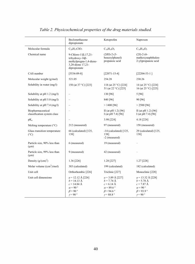
40
Table 2. Physicochemical properties of the drug materials studied.
Beclomethasone dipropionate
Ketoprofen Naproxen
Molecular formula C28H37ClO7 C16H14O3 C14H14O3
Chemical name 9-Chloro-11β,17,21-trihydroxy-16β-methylpregna-1,4-diene-3,20-dione 17,21-dipropionate
(2RS)-2-(3-benzoylphenol) propanoic acid
(2S)-2-(6-mathoxynaphthalen- 2-yl)propanoic acid
CAS number [5534-09-8] [22071-15-4] [22204-53-1 ]
Molecular weight (g/mol) 521.05 254.28 230.26
Solubility in water (mg/l) 150 (at 37 °C) [223] 118 (at 25 °C) [224] 51 (at 22 °C) [225]
14 (at 25 °C) [224] 16 (at 25 °C) [225]
Solubility at pH 1.2 (mg/l) � 130 [96] 5 [96]
Solubility at pH 5.0 (mg/l) � 840 [96] 90 [96]
Solubility at pH 7.4 (mg/l) � > 1400 [96] > 2500 [96]
Biopharmaceutical classification system class
� II (at pH 1.2) [96] I (at pH 7.4) [96]
II (at pH 1.2) [96] I (at pH 7.4) [96]
pKa � 3.98 [224] 4.18 [224]
Melting temperature (°C) 212 (measured) 97 (measured) 158 (measured)
Glass transition temperature (°C)
66 (calculated) [125, 138]
-14 (calculated) [125, 138] -2 (measured)
29 (calculated) [125, 138]
Particle size, 90% less than (µm)
6 (measured) 19 (measured) �
Particle size, 99% less than (µm)
9 (measured) 42 (measured) �
Density (g/(cm)3) 1.36 [226] 1.28 [227] 1.27 [228]
Molar volume ((cm3)/mol) 383 (calculated) 199 (calculated) 182 (calculated)
Unit cell Orthorhombic [226] Triclinic [227] Monoclinic [228]
Unit cell dimensions a = 12.12 Å [226] b = 14.13 Å c = 14.84 Å α = 90 ° β = 90 ° γ = 90 °
a = 3.89 Å [227] b = 7.74 Å c = 6.14 Å α = 89.6 ° β = 94.6 ° γ = 88.8 °
a = 13.32 Å [228] b = 5.78 Å c = 7.87 Å α = 90 ° β = 93.9 ° γ = 90 °
41
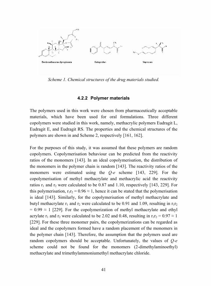
Scheme 1. Chemical structures of the drug materials studied.
4.2.2 Polymer materials
The polymers used in this work were chosen from pharmaceutically acceptable materials, which have been used for oral formulations. Three different copolymers were studied in this work, namely, methacrylic polymers Eudragit L, Eudragit E, and Eudragit RS. The properties and the chemical structures of the polymers are shown in and Scheme 2, respectively [161, 162].
For the purposes of this study, it was assumed that these polymers are random copolymers. Copolymerisation behaviour can be predicted from the reactivity ratios of the monomers [143]. In an ideal copolymerisation, the distribution of the monomers in the polymer chain is random [143]. The reactivity ratios of the monomers were estimated using the Q-e scheme [143, 229]. For the copolymerisation of methyl methacrylate and methacrylic acid the reactivity ratios r1 and r2 were calculated to be 0.87 and 1.10, respectively [143, 229]. For this polymerisation, r1r2 = 0.96 ≈ 1, hence it can be stated that the polymerisation is ideal [143]. Similarly, for the copolymerisation of methyl methacrylate and butyl methacrylate r1 and r2 were calculated to be 0.91 and 1.09, resulting in r1r2 = 0.99 ≈ 1 [229]. For the copolymerization of methyl methacrylate and ethyl acrylate r1 and r2 were calculated to be 2.02 and 0.48, resulting in r1r2 = 0.97 ≈ 1 [229]. For these three monomer pairs, the copolymerizations can be regarded as ideal and the copolymers formed have a random placement of the monomers in the polymer chain [143]. Therefore, the assumption that the polymers used are random copolymers should be acceptable. Unfortunately, the values of Q-e scheme could not be found for the monomers (2-dimethylaminoethyl) methacrylate and trimethylammoniumethyl methacrylate chloride.
42
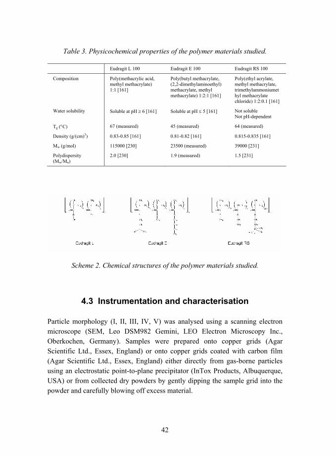
Table 3. Physicochemical properties of the polymer materials studied.
Eudragit L 100 Eudragit E 100 Eudragit RS 100
Composition Poly(methacrylic acid, methyl methacrylate) 1:1 [161]
Poly(butyl methacrylate, (2,2-dimethylaminoethyl) methacrylate, methyl methacrylate) 1:2:1 [161]
Poly(ethyl acrylate, methyl methacrylate, trimethylammoniumethyl methacrylate chloride) 1:2:0.1 [161]
Water solubility Soluble at pH ≥ 6 [161] Soluble at pH ≤ 5 [161] Not soluble Not pH-dependent
Tg (°C) 67 (measured) 45 (measured) 64 (measured)
Density (g/(cm)3) 0.83-0.85 [161] 0.81-0.82 [161] 0.815-0.835 [161]
Mw (g/mol) 115000 [230] 23500 (measured) 39000 [231]
Polydispersity (Mw/Mn)
2.0 [230] 1.9 (measured) 1.5 [231]
Scheme 2. Chemical structures of the polymer materials studied.
4.3 Instrumentation and characterisation
Particle morphology (I, II, III, IV, V) was analysed using a scanning electron microscope (SEM, Leo DSM982 Gemini, LEO Electron Microscopy Inc., Oberkochen, Germany). Samples were prepared onto copper grids (Agar Scientific Ltd., Essex, England) or onto copper grids coated with carbon film (Agar Scientific Ltd., Essex, England) either directly from gas-borne particles using an electrostatic point-to-plane precipitator (InTox Products, Albuquerque, USA) or from collected dry powders by gently dipping the sample grid into the powder and carefully blowing off excess material.

43
Internal structure of the particles (I, III, IV, V) was studied using a transmission electron microscope (TEM, Philips CM200 FEG, FEI Company, Eindhoven, the Netherlands). The samples were prepared onto copper grids coated with carbon film (Agar Scientific Ltd., Essex, England) as described for scanning electron microscopy.
Particle size distributions of the nanoparticles (I, II, III, IV, V) were analysed directly from the aerosol using a scanning mobility particle sizer (SMPS) equipped with a long differential mobility analyser (DMA, model 3071 or model 3081, TSI Inc. Particle Instruments, St. Paul, USA) and a condensation particle counter (CPC, model 3022 or model 3027, TSI Inc. Particle Instruments, St. Paul, USA).
Particle sizes of beclomethasone dipropionate and ketoprofen starting powder materials were determined using an optical microscope (Olympus AH-2, Olympus Optical Co. (Europa) GmbH, Hamburg, Germany) equipped with a digital camera (Colorview 12, Soft Imaging System GmbH, Münster, Germany) and an image analysis program (AnalySIS Pro, v. 3.1, Soft Imaging System GmbH, Münster, Germany). A small amount of powder was dispersed in liquid paraffin and the resulting suspension was spread onto microscope slide. The particle size of beclomethasone dipropionate was analysed using interactive analysis and magnification of 600x, while the particle size of ketoprofen was determined using automatic analysis and magnification of 200x.
The thermal properties of the prepared particles (III, IV, V) were studied using a differential scanning calorimeter (Mettler Toledo DSC 822e, Mettler Toledo AG, Greifensee, Switzerland) equipped with a Stare computer program. Approximately 3 mg of sample was accurately weighed into a 40 µl aluminium pan and sealed with a punched lid. The samples were heated from 25 °C to 300 °C (III) or from -50 °C to 200 °C (IV, V) using a heating rate of 10 °C/min. A nitrogen purge of 50 ml/min was used in the oven.
Glass transition temperature was determined as the midpoint temperature of the glass transition, as shown in Figure 8 for exemplary curves [127]. The transitions observed were verified as glass transitions by heating-cooling-heating experiments of pure polymers.

44
mW 2
min °C -40 -20 0 20 40 60 80 100 120 140 160 180
0 2 4 6 8 10 12 14 16 18 20 22 24
^exo
Midpoint temperature (marked with a cross)
Midpoint temperature (marked with a cross)A
B
Figure 8. Determination of glass transition temperatures for exemplary curves. A) Nanoparticles containing 25% (w/w) ketoprofen / 75% (w/w) Eudragit E, B) Nanoparticles containing 0% (w/w) ketoprofen / 100% (w/w) Eudragit L.
The thermal properties of the materials were also studied using an optical microscope (III) (Zeiss Axioskop, Oberkochen, Germany) equipped with a heating stage (Linkam THMS 600, Surrey, England) and a temperature controller (Linkam TMS 92, Surrey, England). The heating rate used was 20 °C/min.
The crystallinity of the particles was studied using X-ray diffraction (III, IV) (control unit Philips PW 1710, Philips, Eindhoven and Almelo, The Netherlands) with Cu Kα radiation (generator PW 1830, 40 kV, 50 mA, α1 wavelength 0.154060 nm, α2 wavelength 0.154439 nm with an α1/α2 ratio of 0.5). Diffraction angles (2θ) (goniometer PW 1820) used in the recording of the XRD patterns were 3�40°.
The amount of the drug incorporated in the nanoparticles (III, IV) was analysed using a spectrophotometer (Pharmacia LKB Ultrospec III, Pharmacia LKB Biochrom Ltd., Cambridge, England (III, IV)). A suitable amount of nanoparticles was dissolved, the absorbance of the drug at a specific wavelength was measured, and the concentration of the solution was assessed from a
45
calibration line based on Beer-Lambert law [232]. Beclomethasone dipropionate concentration was measured using wavelength 239 nm [223]. It was observed that Eudragits also show a small absorption at the same wavelength, so for every sample a background measurement was done with an ethanolic solution containing the same amount and type of Eudragit as the sample. Ketoprofen and naproxen concentrations were analysed using wavelengths 255 nm [233] and 271 nm [234], respectively. Eudragit materials did not interfere with the measurements at these wavelengths.
Drug release tests of nanoparticles containing ketoprofen (IV, V) were performed using a method based on the general drug release standard of US Pharmacopeia for delayed-release (enteric-coated) articles, method A [235]. An amount of nanoparticles corresponding to approximately 2 mg of drug was weighed. Erlenmeyer flasks (IV) or round-bottomed cylindrical glass vessels (V) were used as release chambers. The solutions were stirred using a magnetic stirrer at a speed of 100 rpm (IV) or 50 rpm (V). The temperature was either ambient (IV) or controlled to 37.0 ± 0.5 °C (V). In the acid stage, 75 ml of 0.1 N hydrochloric acid was used as the release medium. Aliquots were withdrawn at predetermined time intervals and immediately replaced with fresh medium equilibrated at 37 °C. After two hours, 25 ml of 0.2 M tribasic sodium phosphate was added to change the pH of the test medium to 6.8. The test was continued for further three (IV) or four (V) hours. The amount of the drug released was determined after filtration using a spectrophotometer (Philips PU 8620 Series UV/VIS/NIR, Pye Unicam Ltd., Cambridge, England (IV) or Pharmacia LKB Ultrospec III, Pharmacia LKB Biochrom Ltd., Cambridge, England (V)) using wavelength 260 nm for ketoprofen [233] and 270 nm for naproxen [234]. The concentration of drug in the acid stage was 0.027 mg/ml, whereas the ketoprofen solubility in acid conditions is stated to be 0.13 mg/ml [96]. Therefore, sink conditions were achieved. The tests were performed with two parallel runs, the values reported are mean values of the two runs. The repeatability of the method was evaluated by analysing six parallel samples, and it was found that the results are repeatable. The measured dissolution values have a standard deviation of 6% on average, while the maximum standard deviation was less than 10%. The highest standard deviation values were observed immediately after the pH change, probably due to incomplete mixing and equilibration of the pH in the dissolution vessel.

46
Drug release tests of nanoparticles containing beclomethasone dipropionate were performed in an aqueous medium containing 0.1 % (w/w) sodium lauryl sulphate. An amount of nanoparticles corresponding to approximately 0.5 mg of drug was weighed. Round-bottomed cylindrical glass vessels were used as release chambers. The solutions were stirred using a magnetic stirrer at a speed of 100 rpm. The temperature was controlled to 37.0 ± 0.5 °C. Aliquots were withdrawn at predetermined time intervals and immediately replaced with fresh medium equilibrated at 37 °C. The amount of the drug released was determined using a spectrophotometer (Pharmacia LKB Ultrospec III, Pharmacia LKB Biochrom Ltd., Cambridge, England) using wavelength 240 nm [223]. The tests were performed with two parallel runs, the values reported are mean values of the two runs.
47
5. Results
5.1 Particle size, particle size distribution, and particle morphology (I, II, III, IV)
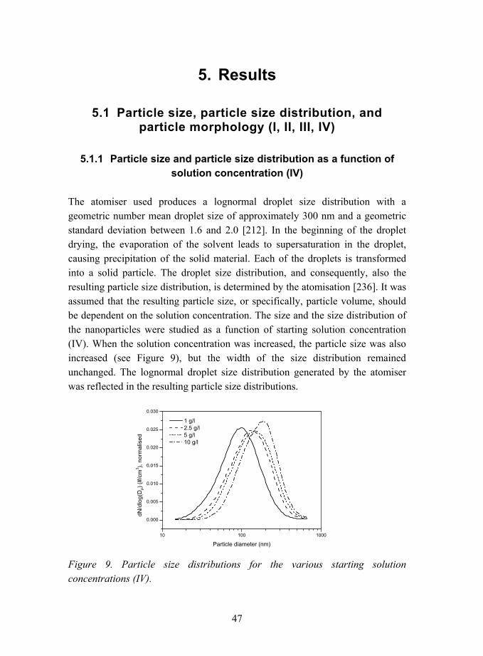
5.1.1 Particle size and particle size distribution as a function of solution concentration (IV)
The atomiser used produces a lognormal droplet size distribution with a geometric number mean droplet size of approximately 300 nm and a geometric standard deviation between 1.6 and 2.0 [212]. In the beginning of the droplet drying, the evaporation of the solvent leads to supersaturation in the droplet, causing precipitation of the solid material. Each of the droplets is transformed into a solid particle. The droplet size distribution, and consequently, also the resulting particle size distribution, is determined by the atomisation [236]. It was assumed that the resulting particle size, or specifically, particle volume, should be dependent on the solution concentration. The size and the size distribution of the nanoparticles were studied as a function of starting solution concentration (IV). When the solution concentration was increased, the particle size was also increased (see Figure 9), but the width of the size distribution remained unchanged. The lognormal droplet size distribution generated by the atomiser was reflected in the resulting particle size distributions.
Figure 9. Particle size distributions for the various starting solution concentrations (IV).

48
The geometric number mean particle diameter is shown as a function of starting solution concentration in Figure 10. In addition, the mean particle volume was calculated from the geometric mean particle diameter using the formula
3
61 dV π= ,
where V denotes the particle volume and d the particle diameter. It can be seen that the particle volume was linearly dependent on the concentration of the solution (see Figure 10). Deviations from a perfectly linear behaviour are likely to be caused by changes in solution viscosity due to changing concentration, which affects the atomisation [237].
Figure 10. Geometric number mean diameter and calculated particle volume as a function of starting solution concentration. The dashed line represents linear least squares fit of particle volume data points (r2 =0.99070) (IV).
5.1.2 Particle size and particle morphology (I, II, III)
5.1.2.1 Effect of temperature (I, III)
The effect of drying temperature on particle size and particle morphology was studied for the nanoparticles containing only drug (I, III) or both drug and
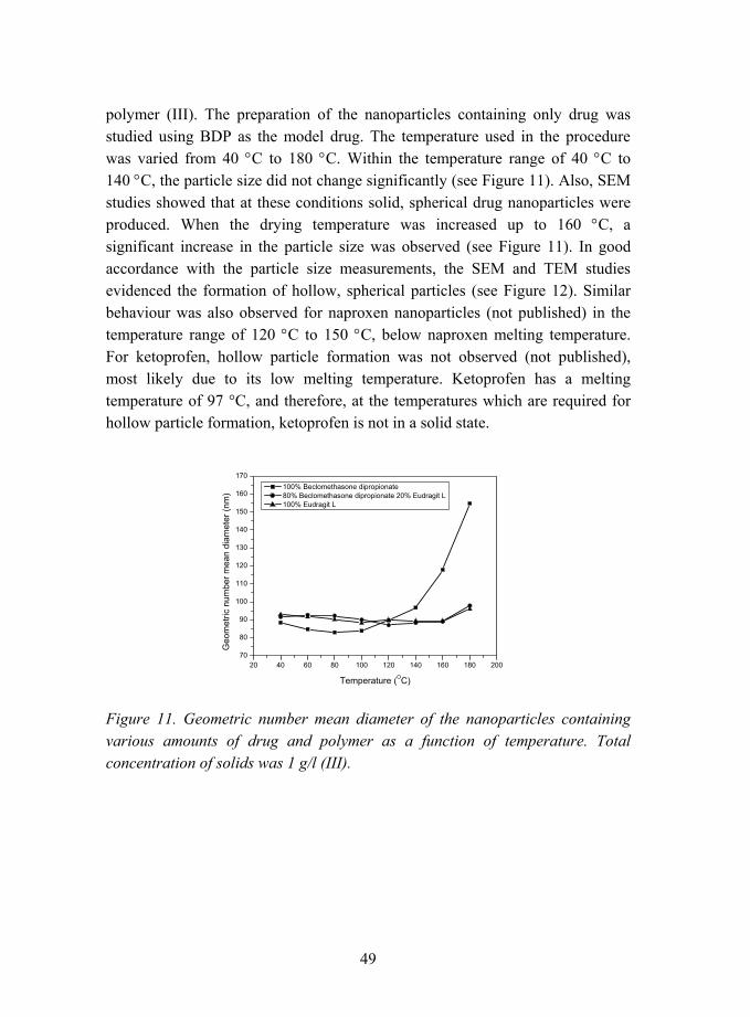
49
polymer (III). The preparation of the nanoparticles containing only drug was studied using BDP as the model drug. The temperature used in the procedure was varied from 40 °C to 180 °C. Within the temperature range of 40 °C to 140 °C, the particle size did not change significantly (see Figure 11). Also, SEM studies showed that at these conditions solid, spherical drug nanoparticles were produced. When the drying temperature was increased up to 160 °C, a significant increase in the particle size was observed (see Figure 11). In good accordance with the particle size measurements, the SEM and TEM studies evidenced the formation of hollow, spherical particles (see Figure 12). Similar behaviour was also observed for naproxen nanoparticles (not published) in the temperature range of 120 °C to 150 °C, below naproxen melting temperature. For ketoprofen, hollow particle formation was not observed (not published), most likely due to its low melting temperature. Ketoprofen has a melting temperature of 97 °C, and therefore, at the temperatures which are required for hollow particle formation, ketoprofen is not in a solid state.
Figure 11. Geometric number mean diameter of the nanoparticles containing various amounts of drug and polymer as a function of temperature. Total concentration of solids was 1 g/l (III).

50
Figure 12. TEM image of a hollow BDP nanoparticle produced (I).
Hollow particle formation is commonly achieved using fast heating rates, i.e. high drying temperatures and rapid solvent evaporation, whereas slower heating rates favour the formation of dense particles [238, 239]. Fast evaporation of the solvent creates a high supersaturation of the solute at the surface of the droplets. If the diffusion of the solvent to the surface is slow, the supersaturation causes precipitation of the material to create a solid crust [200, 236, 239�242]. If the crust formed is permeable to the solvent, diffusion of the solvent and the solute in the droplet can lead to spherical, solid particles [164]. If the crust is impermeable to solvent, the particle will form a hollow interior due to pressure build-up on further solvent evaporation, which expands the particle [200, 236]. Hollow spherical particles can also collapse to raisin- or lens-shaped particles [164, 202, 242, 243]. The formation of hollow particles is, in addition to fast heating rate, also promoted by using concentrated solutions and large droplet size according to the literature [240, 244]. Mechanisms of formation of solid and hollow particles are summarised in Figure 13.
When the nanoparticles were prepared from polymer and drug, the particles were observed to be spherical, solid, and amorphous when sampled with a point-to-plane electrostatic precipitator from nanoparticle aerosol and studied with TEM (not published). The drying temperature did not have an effect on the size
51
or the morphology of the nanoparticles containing polymer (see Figure 11) as opposed to the particles containing only drug without any polymer. The nanoparticles containing only 20% (w/w) polymer showed similar behaviour to the pure polymer nanoparticles (see Figure 11). The polymeric component, even in low amounts, determined the structure of the particles.
5.1.2.2 Effect of solvent system (II, III)
The influence of the solvent properties and the solvent composition on the morphology of the polymer nanoparticles was studied. When the particles were prepared from a good solvent for the polymer, the temperature used and thereby also the evaporation rate of the solvent determined the morphology of the particles. In this case, the interactions between the solvent and the polymer are strong, the polymer is well soluble into the solvent even in the later stages of evaporation, and spherical particles are obtained (see Figure 13). Moreover, the evaporation rate can be also controlled by the vapour pressure of the solvent, the solvent having a higher vapour pressure leading to faster evaporation.
When a poor solvent is added into the solution, the interactions between the polymer and the solvent are weakened. During the evaporation stage, the more volatile component of the solvent mixture is evaporated first, so the composition of the solvent mixture is constantly changing. The good solvents for the polymer used (II) are more volatile than the poor solvent, water, so the solvent composition changes towards poorer during evaporation. The solute has a tendency to precipitate at an early stage during solvent evaporation as the polymer-polymer interactions become more favourable than the polymer-solvent interactions. Due to sudden precipitation of the polymer, the particle appearance is wrinkled or shriveled instead of spherical. Similar results have been found previously in spray-drying experiments [164, 239]. The effect of solvent quality on the particle formation is shown schematically in Figure 13. Exemplary images of particles prepared from a good solvent and a solvent mixture consisting of a good and a poor solvent are shown in Figure 14. It was further verified that the polymer controlled the particle morphology, as the particle morphology did not change on addition of drug in the nanoparticles.
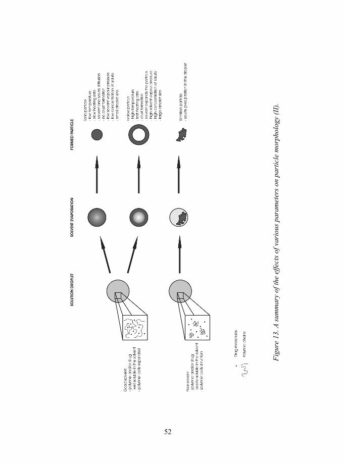
52
Figu
re 1
3. A
sum
mar
y of
the
effe
cts o
f var
ious
par
amet
ers o
n pa
rtic
le m
orph
olog
y (I
I).
52

53
Figure 14. SEM images of polymer nanoparticles containing 50% (w/w) Eudragit L and 50% (w/w) BDP. The nanoparticles were prepared from A) a good solvent (ethanol) or B) a mixture of a good solvent (ethanol) and a poor solvent (water). Nominal magnification 50000x.
5.2 Collection and properties of bulk nanoparticle powder (III, IV, V)
5.2.1 Collection of the nanoparticles (III, IV, V)
5.2.1.1 Nanoparticles containing ketoprofen or naproxen (IV)
The nanoparticles produced are amorphous due to rapid evaporation of the solvent from the droplets. Nonetheless, the stability of the nanoparticles in the amorphous form as dry powder during collection and storage is not necessarily achieved. The amorphous form of the drug incorporated has a tendency to spontaneously convert to a crystalline form [128, 129]. This was studied for various drug-polymer systems, with particular interest on the nanoparticles containing ketoprofen. Firstly, small drug molecules such as naproxen and ketoprofen were observed to be very unstable showing sintering of the nanoparticles and subsequent drug crystal growth in collection when polymer was not incorporated in the nanoparticles. Therefore, the use of polymers in the nanoparticles was found necessary to stabilise the amorphous particle structure against drug crystal growth.

54
It was found that when the amount of ketoprofen drug in the polymer nanoparticles was small, the particles produced and collected with the BLPI were amorphous (IV, V), see the XRD patterns (Figure 15), DSC measurements (Figure 16), and TEM image (Figure 17). In Figure 15, diffraction patterns of nanoparticles with two compositions are shown in addition to a diffraction pattern of untreated drug powder. When the amount of the drug was small, the XRD showed an amorphous pattern and no crystallinity (curve C of Figure 15). Figure 16 shows DSC scans for untreated drug powder and various compositions of nanoparticles. In good accordance with the XRD studies, crystallinity could not be detected by DSC when the amount of the drug was small, as peaks corresponding to the melting of the drug crystals were not observed. Amorphous structure was obtained when the amount of the drug was equal to or less than 33 % (w/w) (curve C of Figure 16). The amorphous nanoparticles prepared were also studied using TEM, and a typical image is shown in Figure 17. Crystallinity or grain boundaries were not found, instead, the nanoparticles were smooth with a uniform interior. Furthermore, typical SEM image of the prepared nanoparticles is shown in Figure 18 as image A. The nanoparticles were spherical and smooth, and major necking was not observed. The formation of an amorphous polymer-drug structure has previously been observed for spray-dried particles containing methacrylic polymers and ketoprofen drug [218, 219].
When the amount of ketoprofen in the nanoparticles was increased up to 50 % (w/w) an endothermic transition corresponding to melting of the drug crystals was detected at 93.1 °C in DSC (curve B of Figure 16). Moreover, in the XRD analyses, the broad background diffraction pattern of the amorphous structure became overlapped by peaks corresponding to the diffraction from the drug crystal lattice. The strongest diffraction peaks of crystalline ketoprofen at 6.4° (2θ) and 23.0° (2θ) can be identified in the diffraction curve of the nanoparticles containing 50% (w/w) ketoprofen (curve B of Figure 15). The TEM imaging further confirmed the formation of large crystalline structures. The collected nanoparticles coalesced, which was followed by drug diffusion in the polymer matrix and crystal growth between and out of the composite nanoparticles, as shown in the SEM image C in Figure 18.
Similar results were found also for the nanoparticles containing naproxen (see Figure 18, images B and D). However, for naproxen, only nanoparticles

55
containing 10% (w/w) naproxen were found to be amorphous, and crystals were observed to appear when the nanoparticles contained 25% (w/w) naproxen (IV).
Figure 15. X-ray diffraction patterns of the nanoparticles. A) Untreated ketoprofen. B) 50% (w/w) ketoprofen / 50% (w/w) Eudragit L nanoparticles. C) 25% (w/w) ketoprofen / 75% (w/w) Eudragit L nanoparticles. Curve A was reduced by a factor of 4 to fit in the same image. The curves are shifted along y-axis for clarification (IV).
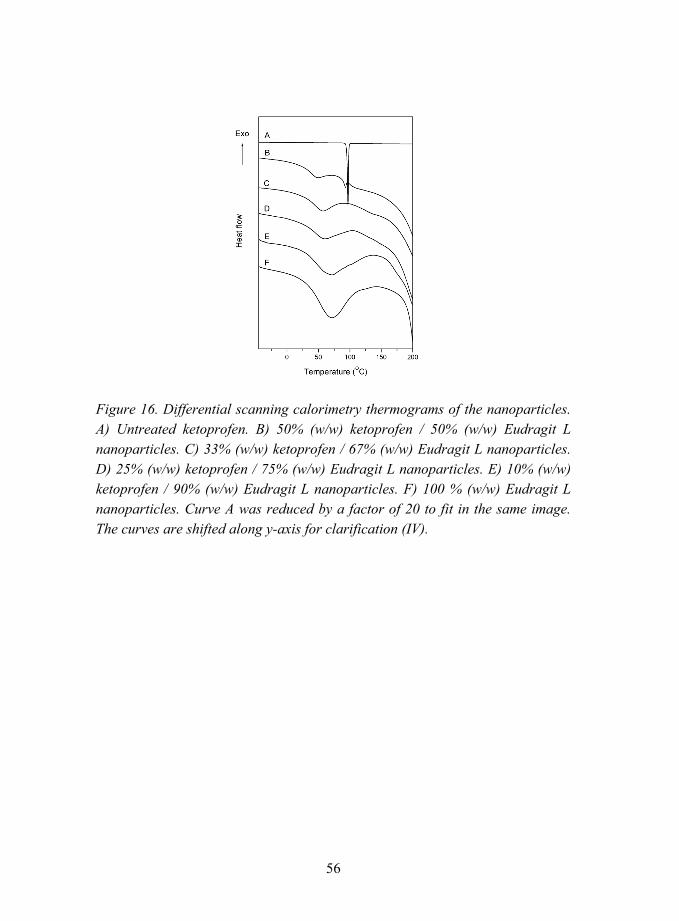
56
Figure 16. Differential scanning calorimetry thermograms of the nanoparticles. A) Untreated ketoprofen. B) 50% (w/w) ketoprofen / 50% (w/w) Eudragit L nanoparticles. C) 33% (w/w) ketoprofen / 67% (w/w) Eudragit L nanoparticles. D) 25% (w/w) ketoprofen / 75% (w/w) Eudragit L nanoparticles. E) 10% (w/w) ketoprofen / 90% (w/w) Eudragit L nanoparticles. F) 100 % (w/w) Eudragit L nanoparticles. Curve A was reduced by a factor of 20 to fit in the same image. The curves are shifted along y-axis for clarification (IV).
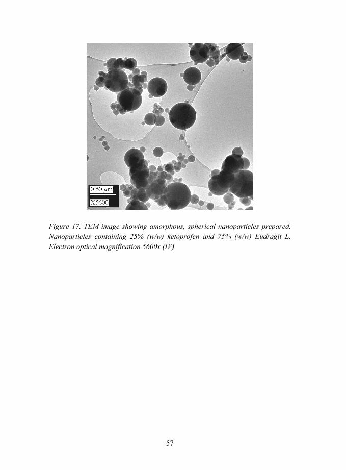
57
Figure 17. TEM image showing amorphous, spherical nanoparticles prepared. Nanoparticles containing 25% (w/w) ketoprofen and 75% (w/w) Eudragit L. Electron optical magnification 5600x (IV).
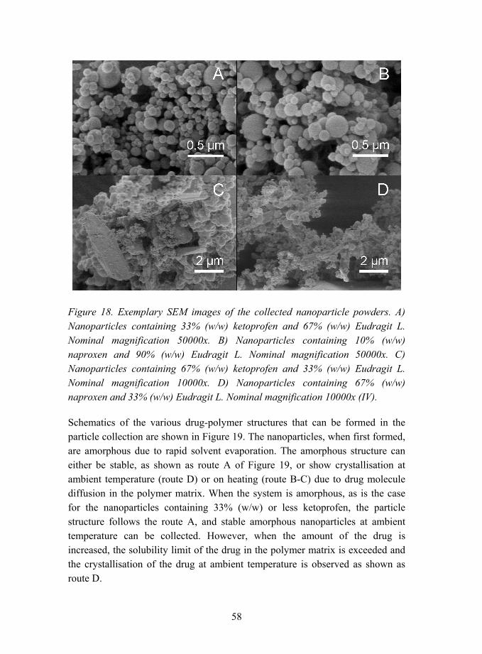
58
Figure 18. Exemplary SEM images of the collected nanoparticle powders. A) Nanoparticles containing 33% (w/w) ketoprofen and 67% (w/w) Eudragit L. Nominal magnification 50000x. B) Nanoparticles containing 10% (w/w) naproxen and 90% (w/w) Eudragit L. Nominal magnification 50000x. C) Nanoparticles containing 67% (w/w) ketoprofen and 33% (w/w) Eudragit L. Nominal magnification 10000x. D) Nanoparticles containing 67% (w/w) naproxen and 33% (w/w) Eudragit L. Nominal magnification 10000x (IV).
Schematics of the various drug-polymer structures that can be formed in the particle collection are shown in Figure 19. The nanoparticles, when first formed, are amorphous due to rapid solvent evaporation. The amorphous structure can either be stable, as shown as route A of Figure 19, or show crystallisation at ambient temperature (route D) or on heating (route B-C) due to drug molecule diffusion in the polymer matrix. When the system is amorphous, as is the case for the nanoparticles containing 33% (w/w) or less ketoprofen, the particle structure follows the route A, and stable amorphous nanoparticles at ambient temperature can be collected. However, when the amount of the drug is increased, the solubility limit of the drug in the polymer matrix is exceeded and the crystallisation of the drug at ambient temperature is observed as shown as route D.

59
Figure 19. Schematics of the drug-polymer structures formed in particle collection.
In the amorphous structure, the drug is solubilised by the polymer and the two components form an amorphous solid solution [144]. The interactions between the drug and the polymer molecules determine the solubility of these materials in each other [245]. When the drug-polymer molecular interactions are comparable
60
to the drug-drug and the polymer-polymer interactions, the drug is well solubilised by the polymer and large amounts of drug can be incorporated in the polymer matrix without drug crystallisation [145]. However, when the drug-polymer interactions are weaker than the drug-drug or the polymer-polymer interactions, the drug and the polymer have a preference to interact with the molecules of their own kind, leading to a potential for drug crystallisation. A small amount of drug, below the solubility limit of the drug in the polymer, can be solubilised by the polymer matrix [147, 246]. On increasing the amount of the drug over this limit, the drug is no longer soluble in the amorphous polymer, but can form separate crystallites [159].
The polymer nanoparticles consisting of Eudragit L were found to be able to contain 33% (w/w) ketoprofen in an amorphous form, but only 10% (w/w) naproxen. The solubilities of the drug molecules ketoprofen and naproxen in Eudragit L were estimated based on DSC measurements. The enthalpy of melting was determined for untreated, pure drug, which was taken to be 100% crystalline. The enthalpy of melting of unsolubilised drug was analysed for the nanoparticles, in which first crystals were found to appear, namely, the nanoparticles containing 50% (w/w) ketoprofen and 25% (w/w) naproxen. It was further assumed that the drug crystals formed in the nanoparticles were 100% crystalline. The amount of crystallised drug wc can then be calculated as a ratio
%100m
mc H
Hw
∆
∆= ,
where ∆Hm is the enthalpy of melting of the sample studied, and ∆Hm100% is the
enthalpy of melting of 100% crystalline material [247]. For ketoprofen, the enthalpies of melting of pure drug and 50% (w/w) nanoparticles were measured to be 114.9 J/g and 1.1 J/g, respectively. For 50% (w/w) nanoparticles, the amount of crystallised drug was thus determined to be 1%. Therefore, the solubility of ketoprofen in Eudragit L can be estimated as
( ) ( ) ( )w/w 49.5%0.011w/w 50% =−⋅ ,
which corresponds to
61
( ) g/g 0.98%49.5100
49.5%=
−,
meaning that 1 gram of polymer can solubilise 0.98 g ketoprofen. Similarly, for naproxen, the enthalpies of melting of pure drug and 25% (w/w) nanoparticles were measured to be 125.8 J/g and 4.5 J/g, respectively. The solubility of naproxen in Eudragit L can then be estimated to be 24.1% (w/w), corresponding to 0.32 g/g.
The differences in the behaviours of these materials are likely to be caused by interactions between the drug and the polymer as well as different molecular structures of the drugs studied [140, 141]. Firstly, crystallisation of the drug requires the diffusion of drug molecules and their arrangement into a crystal lattice. Diffusion of the drug in the polymer matrix has been shown to be a function of the molar volume of the drug, larger molecules having smaller diffusion coefficients and slower diffusion [248, 249]. Also, the interactions between the drug and the polymer affect the diffusion of drug in the polymer matrix, the stronger the interactions, the slower the drug diffusion [248]. It can be speculated that the drug molecules ketoprofen and naproxen should have similar diffusion coefficients, as the molar volumes of the drugs are fairly similar, namely 199 (cm)3/mol for ketoprofen and 182 (cm)3/mol for naproxen (calculated from density of crystalline material, see Table 2). Moreover, both drug molecules can interact with the polymer by the same functional group, carboxylic acid, so the interactions caused by this group should not differ in magnitude. The difference of these two molecules lies in the molecular structure, naproxen having a stiff naphthyl ring system in comparison to more flexible ketoprofen drug molecule. This difference is also reflected in the melting points of the drugs, as the more mobile and flexible molecule ketoprofen has a lower melting point than the stiffer naproxen molecule.
An analogy to the solubility of the drug in the polymer can be found from plasticisation, where flexible small molecules are mixed with the polymer to lower the Tg [146, 159, 245, 250]. Also in plasticisation, it is essential that the plasticising agent forms a uniform mixture with the polymer, and is solubilised by the polymer. Consequently, from this point of view, ketoprofen was a more effective plasticising agent for the Eudragit L polymer than naproxen [159].

62
5.2.1.2 Interactions between ketoprofen and methacrylic polymers (V)
The plasticising effect of ketoprofen was verified in the differential scanning calorimetry analyses showing depression in the Tg of polymers Eudragit L, Eudragit RS, and Eudragit E as a function of increasing drug amount in the nanoparticles (see Table 4 and Figure 20) (V). Crystallisation of the drug was not found for the nanoparticles prepared from the cationic polymer Eudragit RS or the basic polymer Eudragit E, in comparison to the acidic polymer Eudragit L. However, as the Tg was lowered close to the room temperature for the former two polymers, separate nanoparticles having more than 10% (w/w) of drug could not be collected with BLPI. SEM observations evidenced coalesced particles (see Figure 21). The nanoparticles prepared from the polymers Eudragit E and Eudragit RS followed the route A of Figure 19, but the glass transition was so close to the room temperature that the mechanical integrity of the nanoparticles was not maintained. The solubility of ketoprofen in the polymers Eudragit E and Eudragit RS is larger than the solubility in the polymer Eudragit L, as drug crystallites were not observed with the drug amounts studied. The values of solubility in the former two polymers could not be determined based on these experiments.
Table 4. Glass transition temperatures of the nanoparticles prepared from ketoprofen and various polymers. a) Also an endothermic transition corresponding to the melting of ketoprofen crystals was observed at 94 °C (V).
Amount of ketoprofen (w/w)
Eudragit L Eudragit E Eudragit RS
0 % 54 °C 49 °C 53 °C
5 % 52 °C 45 °C 50 °C
10 % 50 °C 41 °C 50 °C
25 % 50 °C 24 °C 28 °C
33 % 48 °C 23 °C 20 °C
50 % 40 °C a) � �
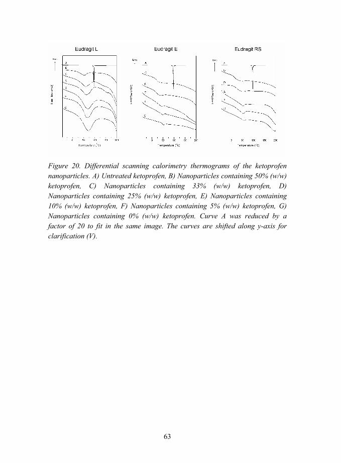
63
Figure 20. Differential scanning calorimetry thermograms of the ketoprofen nanoparticles. A) Untreated ketoprofen, B) Nanoparticles containing 50% (w/w) ketoprofen, C) Nanoparticles containing 33% (w/w) ketoprofen, D) Nanoparticles containing 25% (w/w) ketoprofen, E) Nanoparticles containing 10% (w/w) ketoprofen, F) Nanoparticles containing 5% (w/w) ketoprofen, G) Nanoparticles containing 0% (w/w) ketoprofen. Curve A was reduced by a factor of 20 to fit in the same image. The curves are shifted along y-axis for clarification (V).

64
Figure 21. Exemplary scanning electron microscopy images of the ketoprofen nanoparticles. A) Nanoparticles containing 25% (w/w) ketoprofen and 75% (w/w) Eudragit L. Nominal magnification 50000x. B) Nanoparticles containing 10% (w/w) ketoprofen and 90% (w/w) Eudragit RS. Nominal magnification 50000x. C) Nanoparticles containing 25% (w/w) ketoprofen and 75% (w/w) Eudragit E. Nominal magnification 5000x. D) Nanoparticles containing 25% (w/w) ketoprofen and 75% (w/w) Eudragit RS. Nominal magnification 10000x (V).
The glass transition temperature of an ideal two-component system can be calculated theoretically using the Gordon-Taylor equation:
21
)2(2)1(1)( Kww
TKwTwT gg
mixg +
+= ,
where Tg(mix) is the glass transition temperature of the two-component mixture, w1 and w2 are the weight fractions and Tg(1) and Tg(2) are the glass transition temperatures of the components 1 and 2, respectively [129, 251]. K can be estimated as a ratio:

65
2)2(
1)1(
ρρ
g
g
TT
K = ,
where ρ1 and ρ2 are the densities of the components 1 and 2 [129, 251].
The theoretical glass transitions of the composite nanoparticles over the whole composition range were calculated using the Gordon-Taylor equation, and are plotted together with the measured experimental values in Figure 22. It was found that the composite nanoparticles prepared from Eudragit E or Eudragit RS show a glass transition lower than predicted by the Gordon-Taylor equation. This can be interpreted as a deviation from ideality, caused by the interactions between ketoprofen and these polymers [251]. Ketoprofen interacts with the polymers. As a flexible molecule, it can effectively plasticise the polymers by separating the chains, thereby lowering the glass transition temperature of the polymers [152, 252].
Figure 22. Glass transition temperatures of the nanoparticles containing ketoprofen and polymer. For the calculated values, the glass transition temperature of the nanoparticles consisting of only polymer was used as the polymer glass transition temperature.
The interactions between ketoprofen and the various methacrylic polymers were further evaluated using IR spectroscopy (V). Ketoprofen has a carboxylic acid group, which can interact with the functional groups of the polymers. The carbonyl peaks in the IR spectra of ketoprofen were recorded at 1694 cm-1 and 1654 cm-1, and have previously been assigned to dimeric carboxylic acid
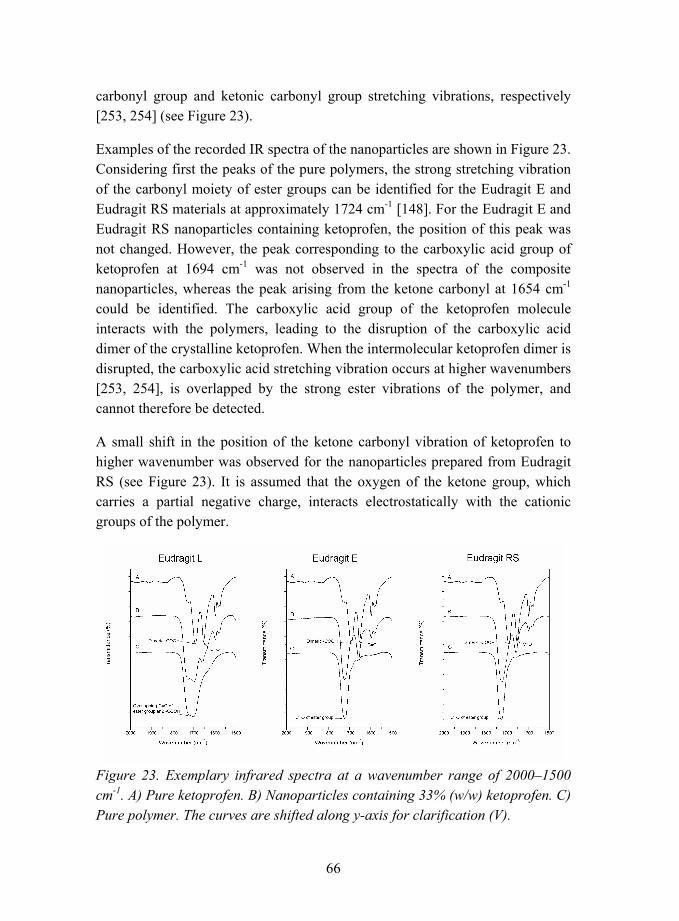
66
carbonyl group and ketonic carbonyl group stretching vibrations, respectively [253, 254] (see Figure 23).
Examples of the recorded IR spectra of the nanoparticles are shown in Figure 23. Considering first the peaks of the pure polymers, the strong stretching vibration of the carbonyl moiety of ester groups can be identified for the Eudragit E and Eudragit RS materials at approximately 1724 cm-1 [148]. For the Eudragit E and Eudragit RS nanoparticles containing ketoprofen, the position of this peak was not changed. However, the peak corresponding to the carboxylic acid group of ketoprofen at 1694 cm-1 was not observed in the spectra of the composite nanoparticles, whereas the peak arising from the ketone carbonyl at 1654 cm-1 could be identified. The carboxylic acid group of the ketoprofen molecule interacts with the polymers, leading to the disruption of the carboxylic acid dimer of the crystalline ketoprofen. When the intermolecular ketoprofen dimer is disrupted, the carboxylic acid stretching vibration occurs at higher wavenumbers [253, 254], is overlapped by the strong ester vibrations of the polymer, and cannot therefore be detected.
A small shift in the position of the ketone carbonyl vibration of ketoprofen to higher wavenumber was observed for the nanoparticles prepared from Eudragit RS (see Figure 23). It is assumed that the oxygen of the ketone group, which carries a partial negative charge, interacts electrostatically with the cationic groups of the polymer.
Figure 23. Exemplary infrared spectra at a wavenumber range of 2000�1500 cm-1. A) Pure ketoprofen. B) Nanoparticles containing 33% (w/w) ketoprofen. C) Pure polymer. The curves are shifted along y-axis for clarification (V).

67
Eudragit E is a polymer containing secondary amino groups capable of accepting a proton from an acidic substance. It was initially assumed that at least a fraction of the amino groups of the polymer would be protonated by the acidic drug. The peaks corresponding to the amino groups of the polymer have been identified previously [255, 256] at 2820 cm-1 and 2770 cm-1. However, any change in the position of these peaks was not observed when ketoprofen was incorporated in the nanoparticles (see Figure 24). Therefore, it can be concluded that ketoprofen drug does not protonate the amino groups, but mainly interacts with the ester groups of the Eudragit E polymer, similarly to Eudragit RS.
Figure 24. Exemplary infrared spectra at a wavenumber range of 3500�2500 cm-1. A) Pure ketoprofen. B) Nanoparticles containing 33% (w/w) ketoprofen. C) Pure polymer. The curves are shifted along y-axis for clarification (V).
For Eudragit L nanoparticles, glass transition values higher than predicted by Gordon-Taylor equation were observed (see Figure 22). The drug was able to plasticise the polymer, but not as much as predicted by the theory. When the amount of ketoprofen in the polymer matrix was larger than the solubility limit of drug in the polymer, excess amount of drug formed separate crystallites.
Eudragit L polymer contains both carboxylic acid and ester groups and, therefore, the IR spectra showed overlapping carbonyl vibrations of the ester

68
group at 1724 cm-1 and the carboxylic acid at 1710 cm-1 [150] (see Figure 23). However, when ketoprofen is included in the nanoparticles, this peak is splitted into two peaks as a function of increasing amount of the drug. The peak at approximately 1724 cm-1 can be attributed to the stretching vibrations of the polymer ester group carbonyl, similarly to Eudragit E and Eudragit RS polymers. However, the peak at the lower wavenumber is recorded at 1710 cm-1, 1705 cm-1, 1703 cm-1, 1700 cm-1, 1700 cm-1, and 1699 cm-1 for the nanoparticles containing 0% (w/w), 5% (w/w), 10% (w/w), 25% (w/w), 33% (w/w), and 50% (w/w) ketoprofen, respectively. This peak can be interpreted as arising due to the formation of a dimer from the carboxylic acid groups of the polymer and the drug. Due to the formation of the dimer, the vibration of the polymer carboxylic acid carbonyl was shifted to lower wavenumbers.
5.2.1.3 Nanoparticles containing beclomethasone dipropionate (III)
Nanoparticles were also prepared from a drug material having a glass transition temperature higher than ambient temperature (III). In this case, also the drug material itself can form a glassy, amorphous state without polymer. Below the glass transition temperature, the molecular mobility is slow, and the amorphous state is kinetically preserved even though it is not thermodynamically stable. In this study, the nanoparticles containing BDP and polymer showed single, separate nanoparticles over the whole concentration range even when pure drug nanoparticles were collected. The stability of these nanoparticles at room temperature was not affected by the amounts of the drug or the polymer on the contrary to ketoprofen and naproxen nanoparticles.
The glass transition temperature of ketoprofen was determined using DSC to be -2°C. For BDP and naproxen, however, the determinations were not successful due to degradation of the material during melting and rapid crystallisation on cooling, respectively. Glass transition temperature of a material can be estimated from its melting temperature by applying the rule Tg = 0.7·Tm, where Tg and Tm are expressed in Kelvins [125, 138]. The calculated glass transition temperatures of the drugs are 66 °C for BDP and 29 °C for naproxen. The calculated value of Tg of ketoprofen is -14°C. For ketoprofen, applying the rule Tg = 0.7·Tm gives actually quite an accurate estimation for the glass transition temperature. The coeffient 0.7 can be specified according to the experimental value as 0.73.

69
The Tg of BDP is higher than the collection temperature and, therefore, these drug nanoparticles did not crystallise or coalesce in the collection, corresponding to route B of Figure 19. The large size of the BDP molecule contributes to slow diffusion in the polymer matrix, further hindering the formation of crystals [248, 249]. When a system consisting of the amorphous drug material is heated, the molecular mobility is increased and crystallisation into a thermodynamically more favourable crystalline state can take place as shown in Figure 19 as route C. This was verified in DSC measurements. The drug nanoparticles consisting of only BDP showed a crystallisation exotherm at 141 °C (see Figure 25). Subsequent melting peak of the drug was recorded at 210 °C, similarly to the untreated drug powder. For the polymer-drug nanoparticles, the crystallisation of the drug was retarded to 155 °C and 164 °C for the nanoparticles containing 80% (w/w) and 60 % (w/w) of BDP, respectively. The viscous polymer hindered the diffusion of the drug molecules, retarding the crystallisation of the drug.
Figure 25. DSC scans of nanoparticles containing various amounts of BDP and Eudragit L. A) 100% (w/w) BDP, B) 80% (w/w) BDP / 20% (w/w) Eudragit L, C) 60% (w/w) BDP / 40% (w/w) Eudragit L, D) 50% (w/w) BDP / 50% (w/w) Eudragit L, E) 100% (w/w) Eudragit L. Observed crystallisation and melting of the crystals are marked with cr and m, respectively . The curves are shifted along y-axis for clarification (III).
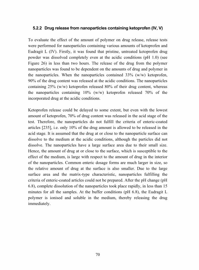
70
5.2.2 Drug release from nanoparticles containing ketoprofen (IV, V)
To evaluate the effect of the amount of polymer on drug release, release tests were performed for nanoparticles containing various amounts of ketoprofen and Eudragit L (IV). Firstly, it was found that pristine, untreated ketoprofen drug powder was dissolved completely even at the acidic conditions (pH 1.0) (see Figure 26) in less than two hours. The release of the drug from the polymer nanoparticles was found to be dependent on the amounts of drug and polymer in the nanoparticles. When the nanoparticles contained 33% (w/w) ketoprofen, 90% of the drug content was released at the acidic conditions. The nanoparticles containing 25% (w/w) ketoprofen released 80% of their drug content, whereas the nanoparticles containing 10% (w/w) ketoprofen released 70% of the incorporated drug at the acidic conditions.
Ketoprofen release could be delayed to some extent, but even with the lowest amount of ketoprofen, 70% of drug content was released in the acid stage of the test. Therefore, the nanoparticles do not fulfill the criteria of enteric-coated articles [235], i.e. only 10% of the drug amount is allowed to be released in the acid stage. It is assumed that the drug at or close to the nanoparticle surface can dissolve to the medium at the acidic conditions, although the particles did not dissolve. The nanoparticles have a large surface area due to their small size. Hence, the amount of drug at or close to the surface, which is susceptible to the effect of the medium, is large with respect to the amount of drug in the interior of the nanoparticles. Common enteric dosage forms are much larger in size, so the relative amount of drug at the surface is also smaller. Due to the large surface area and the matrix-type characteristic, nanoparticles fulfilling the criteria of enteric-coated articles could not be prepared. After the pH change (pH 6.8), complete dissolution of the nanoparticles took place rapidly, in less than 15 minutes for all the samples. At the buffer conditions (pH 6.8), the Eudragit L polymer is ionised and soluble in the medium, thereby releasing the drug immediately.

71
Figure 26. Ketoprofen release for nanoparticles containing various amounts of drug as a function of time (IV). Pure ketoprofen denotes untreated, commercial, crystalline ketoprofen powder (particle size 90% less than 19 µm).
Drug release was also studied for nanoparticles prepared from different polymers and ketoprofen (see Figure 27) (V). Nanoparticles prepared from Eudragit L showed an initial, instant release of approximately 30 % of the drug in the acidic stage of the test. However, the drug release was slightly slower than of pure ketoprofen. After the pH change, complete dissolution of the nanoparticles took place rapidly. At the buffer conditions (pH 6.8), the Eudragit L copolymer was ionised and soluble in the medium, thereby releasing the drug immediately. The nanoparticles studied contained 10% (w/w) ketoprofen, similarly to the nanoparticles studied in Figure 26. The difference in the drug release curves of Figure 26 and Figure 27 for the same nanoparticles is most likely caused by different conditions used in the two measurements, as the results were repeatable in both cases.
Eudragit E polymer, however, is soluble at the acidic conditions. Complete release of the drug was obtained for Eudragit E nanoparticles in the acidic medium in less than 15 minutes as a result of polymer dissolution. As the polymer dissolved, also the drug contained in the nanoparticles was forced into the solution. Drug release from the nanoparticles was much faster than of pure ketoprofen (see Figure 27).
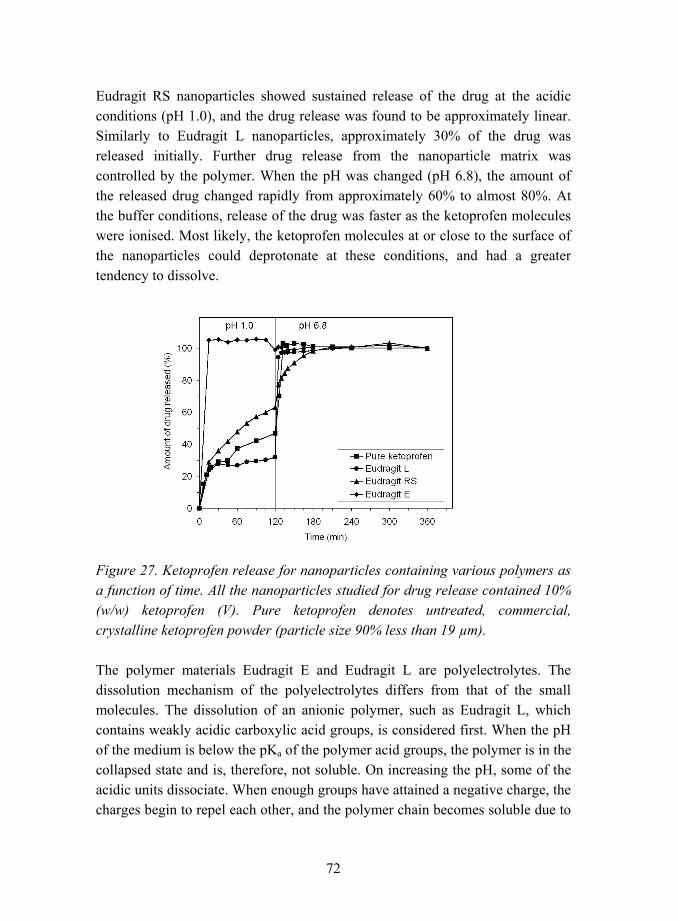
72
Eudragit RS nanoparticles showed sustained release of the drug at the acidic conditions (pH 1.0), and the drug release was found to be approximately linear. Similarly to Eudragit L nanoparticles, approximately 30% of the drug was released initially. Further drug release from the nanoparticle matrix was controlled by the polymer. When the pH was changed (pH 6.8), the amount of the released drug changed rapidly from approximately 60% to almost 80%. At the buffer conditions, release of the drug was faster as the ketoprofen molecules were ionised. Most likely, the ketoprofen molecules at or close to the surface of the nanoparticles could deprotonate at these conditions, and had a greater tendency to dissolve.
Figure 27. Ketoprofen release for nanoparticles containing various polymers as a function of time. All the nanoparticles studied for drug release contained 10% (w/w) ketoprofen (V). Pure ketoprofen denotes untreated, commercial, crystalline ketoprofen powder (particle size 90% less than 19 µm).
The polymer materials Eudragit E and Eudragit L are polyelectrolytes. The dissolution mechanism of the polyelectrolytes differs from that of the small molecules. The dissolution of an anionic polymer, such as Eudragit L, which contains weakly acidic carboxylic acid groups, is considered first. When the pH of the medium is below the pKa of the polymer acid groups, the polymer is in the collapsed state and is, therefore, not soluble. On increasing the pH, some of the acidic units dissociate. When enough groups have attained a negative charge, the charges begin to repel each other, and the polymer chain becomes soluble due to
73
ionized carboxylic acid groups and their mutual electrostatic repulsions [257, 258]. A cloud consisting of counter-ions and co-ions is formed around the dissolved polyelectrolyte [259]. The dissolution is represented schematically in Figure 28.
HOOC
Co-ions
COOH
COOH
COOH
COOH
HOOC
COO¯
¯OOC
COO¯
COO¯
COO¯
¯OOC
pH > 6
+
++
+
+
+
+−
+
−
−
−
−
Counter-ions
−
−
Figure 28. Schematic representation of dissolution of a polyelectrolyte containing carboxylic acid groups (Eudragit L).
Ketoprofen drug molecules will also attain a negative charge at pH levels higher than pKa of ketoprofen, 3.98 [224]. Considering the situation where both the polymer and the drug have negative charges, electrostatic repulsion between the negatively charged groups separates the drug molecules from the polymer. On the contrary, for the basic Eudragit E material, protonation and charge of the dimethyl amino groups takes place in the acidic environment. Therefore, the polymer chains are rapidly dissolved at the acidic conditions. It is assumed that the rapid release of the drug from the particles is achieved due to dissolution of the polymer matrix. Provided that the total concentration of the drug in the solution is lower that the saturation concentration, the released drug does not crystallise, but is maintained in the solution.
The drug molecule studied is a weak acid, so self-buffering could take place at the buffer stage of the test, when the pH of the medium is 6.8. Considering first only particles consisting only ketoprofen, most likely the pH at the particle surface remains close to the pKa value of ketoprofen (3.98) [115, 116]. Based on the results of Figure 26 and Figure 27, the dissolution behavior of the polymers is the determining factor for the release of the drug from the nanoparticles

74
containing both drug and polymer. Three different polymers were investigated in this study. Polymer Eudragit E is a basic polymer. Therefore, at acidic pH, this polymer protonates and attains a positive electrostatic charge. As the hydrogen ions are consumed by the protonating polymer, the pH of the dissolving particle surface can be higher than the bulk pH. However, when this polymer in the nanoparticles is dissolved, the ketoprofen in the particles is forced into the solution state. In this case, also the dissolved ketoprofen molecules can protonate the polymer. Consequently, an electrostatic interaction takes place between the positively charged polymer and negatively charged ketoprofen ions. At pH 6.8, the buffer stage, the particles are already dissolved and surface pH can no longer be considered relevant.
Eudragit L is an anionic polymer, similarly to ketoprofen drug molecule. The acidic pH used in the study is clearly below the pKa values of the molecules. Both the drug molecules and the polymer Eudragit L are almost completely in their protonated, neutral forms, and self-buffering can be considered as non-relevant [115]. When the pH of the bulk is increased to 6.8, self-buffering both by the polymer and by the drug molecule can take place. In this case, due to the formation of acidic microenvironment around the particles, the release of the drug from the particles would be similar to the release at the low pH. At pH 6.8 used in the study, the drug molecules are almost completely in the ionised form according to their pKa value, and are well soluble in the aqueous medium. Also, at this pH, the carboxylic acid groups of Eudragit L are deprotonated and the polymer is soluble. If self-buffering was to take place, the release rate of the drug would be slow. From Figure 27 it can be seen that the drug release rate from Eudragit L and Eudragit RS nanoparticles actually increases at pH 6.8. Therefore, if self-buffering takes place, its effect on drug release is negligible. Eudragit RS is not a water soluble polymer, and it does not show pH-dependency. At pH 1, self-buffering does not take place. However, at the buffer stage of the test, self-buffering by the anionic ketoprofen molecule can take place, when the drug molecules slowly diffuse into the water phase.
From Figure 27, it can be observed that for Eudragit L and Eudragit RS nanoparticles, 30% of drug content of the particles is released immediately at the acidic conditions. Two possible mechanisms are proposed for the initial 30% drug release from Eudragit L and Eudragit RS nanoparticles. Firstly, the nanoparticles can contain a larger a proportion of drug at the surface of the

75
particles in comparison to the interior of the particles. Such an uneven distribution could arise from diffusion of the small molecules to the particle surface during particle preparation and drying process. When such particles are immersed in dissolution medium, the drug at the surface is immediately released, and only further control of drug release is due to the polymers. Secondly, if the dissolution medium can penetrate to some extent to the polymer matrix, the drug molecules at and close to the surface will dissolve. Theoretically calculated for a 100 nm nanoparticle, which has a uniform distribution of drug (10% (w/w)) and polymer (90% (w/w)), medium should be able to initially penetrate to 5 nm depth to allow 30% drug release. As in the previous case, further control of drug release is controlled by the properties of the polymer.
Dissolution of the drug from an inert matrix can take place by two different processes: diffusion of the drug through the matrix into the solution or penetration of the solvent into the matrix and subsequent dissolution of drug into the penetrated solvent [113, 114, 260]. For the Eudragit RS nanoparticles, the drug release takes place by the solvent penetration process. Water slowly penetrates in the polymer matrix due to charged quaternary groups, and drug is released at a constant rate from the polymer matrix. In comparison, the drug release from Eudragit L nanoparticles pauses after the initial burst release of 30%. Dissolution medium cannot penetrate further into the hydrophobic nanoparticles, and additional drug release would have to take place by the drug diffusion mechanism. This case can be approximated as consisting of an insoluble and non-interacting matrix and a weakly soluble drug dissolved in the matrix [261]. However, diffusion of drug through the polymer matrix is a very slow process, and it is assumed that the time scale of drug diffusion in the polymer matrix is much longer than the time scale of the experiment.
5.2.2.1 Effects of dissolution on drug absorption
Differences were observed in the dissolution properties of the nanoparticles. However, such differences in dissolution are not necessarily clinically relevant. Firstly, the concentration of drug used in the dissolution studies (0.02 mg/ml) is much lower than the concentration considered in the biopharmaceutical classification system. In BCS, for example, for a standard dose of 100 mg ketoprofen, the concentration in 250 ml of fluid is 0.4 mg/ml. For each composition
76
of nanoparticles, the limiting factor for drug absorption in the gastrointestinal tract was identified, assuming a standard dose of 100 mg of ketoprofen.
Ketoprofen is a molecule having a high permeability [96, 262]. Therefore, it is almost completely absorbed in the gastrointestinal tract. For a drug molecule having a high permeability, the solubility can become the limiting factor for drug absorption. The classification of solubility depends on the dose of the drug. Considering a standard dose of 100 mg ketoprofen [263], ketoprofen can be classified as having a low solubility in acidic stomach fluid [96]. At pH 5, approximating the pH level of the intestine, a 100 mg dose of ketoprofen can be classified as having a high solubility [96]. Similarly, at pH 7.4, the approximate pH level of the colon, ketoprofen is highly soluble [96]. For pure ketoprofen, when the drug is in the gastrointestinal tract in a solution form, it is also absorbed as a result of its high permeability. Consequently, for pure ketoprofen of 100 mg dose, the gastric emptying time can be considered as the limiting step for drug absorption.
Nanoparticles prepared from ketoprofen and the polymer Eudragit E can be regarded as an immediate release preparation. The nanoparticles dissolve in the acidic stomach fluid. However, all ketoprofen cannot be dissolved due to the fact the the total solubility of ketoprofen is lower than the amount of ketoprofen in a 100 mg dose. Therefore, also for nanoparticles prepared from Eudragit E, the gastric emptying time is the limiting step for drug absorption. A pH-controlled preparation, consisting of ketoprofen and Eudragit L, is dissolved when the pH is higher than 5. Therefore, similarly to pure ketoprofen, for these nanoparticles the gastric emptying time is the limiting step for drug absorption. Neither drug nor the polymer is dissolved in the acidic stomach. Nanoparticles containing polymer Eudragit RS can also be denoted as a controlled-release preparation. In these nanoparticles, however, the polymer Eudragit RS controls the dissolution of ketoprofen from the particles at all pH levels. Therefore, for these nanoparticles, the product is the controlling factor for drug absorption. It can be concluded that only for this preparation, the nanoparticle formulation is likely to have an impact on drug absorption.
The blood plasma levels of ketoprofen were modeled using a compartmental absorption and transit (CAT) model [264, 265]. The structure of the model having seven compartments in the intestine is shown in Figure 29.
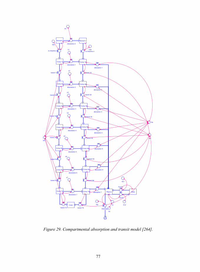
77
Stomach s
Comp 1d
Comp 2d
Comp 1s
Comp 2s
dissolution 1
dissolution 2
transit 1s transit 1d
to intestine s
k2
k1
Vd
Flux12
Flux21
Comp 3s Comp 3d
transit 2s
dissolution 3
transit 2dk3
K21
K12
Comp 4s Comp 4d
dissolution 4
transit 3s transit 3d
k4
Ct
Comp 5s Comp 5d
transit 4s transit 4d
dissolution 5
Kt2k5
Comp 6s Comp 6d
dissolution 6
transit 5s transit 5d
k6
Comp 7s Comp 7d
transit 6s transit 6d
dissolution 7
Eliminaatio
k7
KelColon
transit 7s transit 7d
Plasma
absorption 1
absorption 2
absorption 3
absorption 4
absorption 5
absorption 6
absorption 7
Ka
Kt1
k0s
Stomach d
Body
dissolution 0
to intestine dk0d
k0
Figure 29. Compartmental absorption and transit model [264].
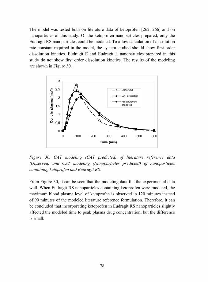
78
The model was tested both on literature data of ketoprofen [262, 266] and on nanoparticles of this study. Of the ketoprofen nanoparticles prepared, only the Eudragit RS nanoparticles could be modeled. To allow calculation of dissolution rate constant required in the model, the system studied should show first order dissolution kinetics. Eudragit E and Eudragit L nanoparticles prepared in this study do not show first order dissolution kinetics. The results of the modeling are shown in Figure 30.
0
0,5
1
1,5
2
2,5
3
0 100 200 300 400 500 600
Time (min)
Con
c in
pla
sma
(mg/
l) Observed
CAT predicted
Nanoparticlespredicted
Figure 30. CAT modeling (CAT predicted) of literature reference data (Observed) and CAT modeling (Nanoparticles predicted) of nanoparticles containing ketoprofen and Eudragit RS.
From Figure 30, it can be seen that the modeling data fits the experimental data well. When Eudragit RS nanoparticles containing ketoprofen were modeled, the maximum blood plasma level of ketoprofen is observed in 120 minutes instead of 90 minutes of the modeled literature reference formulation. Therefore, it can be concluded that incorporating ketoprofen in Eudragit RS nanoparticles slightly affected the modeled time to peak plasma drug concentration, but the difference is small.

79
5.2.3 Drug release from nanoparticles containing beclomethasone dipropionate
The drug release from nanoparticles containing 100% (w/w) beclomethasone dipropionate was analysed, and the results are shown in Figure 31. It can be observed that BDP nanoparticles dissolved faster than micronised BDP. Almost 90% of drug amount is released from the nanoparticles in five minutes, whereas only 45% of drug amount is released at the same time from the micron-sized particles. However, in fifteen minutes, the released amount from the micron-sized particles equals the amount released from the nanoparticles. Therefore, it can be concluded that the dissolution of drug from the nanoparticles was slightly faster than from the micron-sized particles, but the difference in the dissolution rates is very small.
Figure 31. BDP release for pure BDP (particle size 90% less than 6 µm) and BDP nanoparticles.

80
5.2.4 Stability of the nanoparticles
Physical stability of the nanoparticle dry powder was studied at three different conditions to evaluate the effects of temperature and relative humidity. The nanoparticles containing 33% (w/w) ketoprofen and 67% (w/w) Eudragit L were stored for three months in a refrigerator, at 25 °C at 0% relative humidity, and at 25 °C at 75% relative humidity. It was observed that the particles did not show any changes in their morphology in the course of time, but remained spherical and smooth regardless of the storage conditions of the study. The nanoparticle powder as-prepared was amorphous, as peaks corresponding to diffraction from drug crystal lattice were not detected. After three months storage time, crystallinity was not detected in the XRD patterns (see Figure 32). Therefore, it was concluded that the nanoparticles are physically stable for at least a time period of three months. However, chemical stability was not analysed during this time period. The chemical purity during storage time should be evaluated using liquid chromatography to identify degradation products.
Figure 32. X-ray diffraction patterns of nanoparticle dry powder containing 33% (w/w) ketoprofen and 67% (w/w) Eudragit L after 3 months storage time. A) Nanoparticle dry powder stored in a refrigerator. B) Nanoparticle dry powder stored at 25 °C at 0% relative humidity. C) Nanoparticle dry powder stored at 25 °C at 75% relative humidity. The curves are shifted along y-axis for clarification.

81
6. Summary and conclusions Polymer nanoparticles were prepared using a novel method, namely, aerosol flow reactor method, and the properties of the nanoparticles were studied. The nanoparticles were prepared from various drugs and pharmaceutically acceptable methacrylic polymers. Such nanosized particles could be used, for example, for oral, ocular, or parenteral administration of drug materials.
This method could be used to prepare spherical, amorphous, homogeneous matrix-type drug-polymer nanoparticles. The size distributions of the nanoparticles prepared were found to be unimodal and lognormal, reflecting the droplet size distribution produced by the atomiser. The concentration of the starting solution determined the resulting particle size.
The morphology of the nanoparticles was affected by the solvent properties and the solvent composition. Particularly, the morphology of the particles was influenced by the solute solubility and the solvent vapour pressure. It was concluded that the polymeric component determined the morphology of the nanoparticles.
The nanoparticles, when prepared, were amorphous, but depending on the thermal properties of the drug and on the interactions between the drug and the polymer, crystallisation of the drug could take place in collection. The nanoparticles containing beclomethasone dipropionate were found to be amorphous at room temperature regardless of the amounts of drug and polymer in the nanoparticles. Crystallisation of the amorphous beclomethasone dipropionate was observed when the nanoparticles were heated above glass transition temperatures of the drug and the polymer, provided that the amount of beclomethasone dipropionate was above the solubility limit of the drug in the polymer.
In comparison, when the nanoparticles contained small drug molecules, such as ketoprofen and naproxen, it was found that the use of polymeric excipients was necessary to prevent crystallisation of the drugs at room temperature. For the nanoparticles prepared from a methacrylic acid - methyl methacrylate copolymer, the solubilities of drugs ketoprofen and naproxen in the nanoparticles were estimated to be 49.5% (w/w) and 24.1% (w/w), respectively.
82
A drug amount smaller than this remained in the amorphous solid solution structure of the nanoparticles, whereas a higher amount of drug crystallised. In general, for the nanoparticles prepared from ketoprofen and methacrylic polymers, a significant lowering in the glass transition temperature of the polymer was observed when ketoprofen amount was increased.
As a conclusion it can be stated that, firstly, the interactions between the drug and the polymer determined the maximum amount of drug that could be solubilised in the amorphous polymer matrix. Large amounts of drug could be solubilised in the polymer, when the interactions between the drug and the polymer were strong. Below the solubility limit of the drug in the polymer, nanoparticles having amorphous solid solution structures were obtained. Above the solubility limit, the drug was observed to form crystallites, and amorphous nanoparticles could not be collected. Secondly, the crystallisation was dependent on the thermal properties of the materials. When the glass transition temperature of the drug was low, the drug had a tendency to crystallise already at room temperature. In comparison, when the drug had a high glass transition temperature, the crystallisation was observed to take place only when heat was applied to the nanoparticles.
Further studies on extending this approach to other pharmaceutical materials are suggested. A wider range of both active drug molecules and polymer materials should be covered to see if the trends observed in this study apply also to other drug-polymer systems in a more general level. Stability studies of the amorphous particles at various conditions over long periods of time should give valuable insight on the feasibility of these nanoparticles. Furthermore, the incorporation of the nanoparticles into dosage forms, such as tablets or oral suspensions should be surveyed to allow further pharmaceutical product development. To increase the manufacturing and collection capacity considering, for example, scale-up issues, the particle collection should be modified. The collection device used in this study, Berner-type low pressure impactor, separates the particles according to their particle size onto 11 separate stages, which is not feasible and does not allow further scale-up. Adaptation of the collection to lower temperatures is likely to increase the stability of nanoparticles during collection, and warrants further studies. Chemical purity of the drugs as a function of preparation procedure temperature should be evaluated more closely, for example with chromatography.
83
References [1] Allémann, E., Gurny, R., and Doelker, E. Drug-loaded nanoparticles - preparation methods and drug targeting issues. European Journal of Pharmaceutics and Biopharmaceutics, 1993. Vol. 39, pp. 173�191.
[2] Kawashima, Y. Nanoparticulate systems for improved drug delivery. Advanced Drug Delivery Reviews, 2001. Vol. 47, pp. 1�2.
[3] Soppimath, K. S., Aminabhavi, T. M., Kulkarni, A. R., and Rudzinski, W. E. Biodegradable polymeric nanoparticles as drug delivery devices. Journal of Controlled Release, 2001. Vol. 70, pp. 1�20.
[4] Panyam, J. and Labhasetwar, V. Biodegradable nanoparticle from drug and gene delivery to cells and tissue. Advanced Drug Delivery Reviews, 2003. Vol. 55, pp. 329�347.
[5]´Brigger, I., Dubernet, C., and Couvreur, P. Nanoparticles in cancer therapy and diagnosis. Advanced Drug Delivery Reviews, 2002. Vol. 54, pp. 631�651.
[6] Cui, Z. and Mumper, R. J. Plasmid DNA-entrapped nanoparticles engineered from microemulsion precursors: In vitro and in vivo evaluation. Bioconjugate Chemistry, 2002. Vol. 13, pp. 1319�1327.
[7] Cohen, H., Levy, R. J., Gao, J., Fishbein, I., Kousaev, V., Sosnowski, S., Slomkowski, S., and Golomb, G. Sustained delivery and expression of DNA encapsulated in polymeric nanoparticles. Gene Therapy, 2000. Vol. 7, pp. 1896�1905.
[8] Kreuter, J. Nanoparticles and Nanocapsules � New Dosage Forms in the Nanometer Range. Pharmaceutica Acta Helvetiae, 1978. Vol. 53, pp. 33�39.
[9] Kreuter, J. Nanoparticles. In: Swarbrick, J. and Boylan, J. C. (Eds.) Encyclopedia of Pharmaceutical Technology. pp. 165�190. New York, USA: Marcel Dekker, Inc., 1994. ISBN 0-8247-2809-2

84
[10] Birrenbach, G. and Speiser, P. P. Polymerized micelles and their use as adjuvants in immunology. Journal of Pharmaceutical Sciences, 1976. Vol. 65, pp. 1763�1766.
[11] Couvreur, P., Dubernet, C., and Puisieux, F. Controlled drug delivery with nanoparticles: current possibilities and future trends. European Journal of Pharmaceutics and Biopharmaceutics, 1995. Vol. 41, pp. 2�13.
[12] Bodmeier, R. and Chen, H. Indomethacin polymeric nanosuspensions prepared by microfluidization. Journal of Controlled Release, 1990. Vol. 12, pp. 223�233.
[13] de Chasteigner, S., Fessi, H., Cavé, G., Devissaguet, J. P., and Puisieux, F. Gastro-intestinal tolerance study of a freeze-dried oral dosage form of indomethacin-loaded nanocapsules. STP Pharma Sciences, 1995. Vol. 5, pp. 242�246.
[14] Pohlmann, A. R., Weiss, V., Mertins, O., Pesce da Silveira, N., and Guterres, S. S. Spray-dried indomethacin-loaded polyester nanocapsules and nanospheres: development, stability evaluation and nanostructure models. European Journal of Pharmaceutical Sciences, 2002. Vol. 16, pp. 305�312.
[15] Saez, A., Guzman, M., Molpeceres, J., and Aberturas, M. R. Freeze-drying of polycaprolactone and poly(-lactic-glycolic) nanoparticles induce minor particle size changes affecting the oral pharmacokinetics of loaded drugs. European Journal of Pharmaceutics and Biopharmaceutics, 2000. Vol. 50, pp. 379�387.
[16] Kreuter, J. Physicochemical characterization of polyacrylic nanoparticles. International Journal of Pharmaceutics, 1983. Vol. 14, pp. 43�58.
[17] Legrand, P., Barratt, G., Mosqueira, V., Fessi, H., and Devissaguet, J. P. Polymeric nanocapsules as drug delivery systems. A review. STP Pharma Sciences, 1999. Vol. 9, pp. 411�418.
[18] Barratt, G. M. Therapeutic applications of colloidal drug carriers. Pharmaceutical Science and Technology Today, 2000. Vol. 3, pp. 163�171.
85
[19] Fresta, M., Cavallaro, G., Giammona, G., Wehrli, E., and Puglisi, G. Preparation and characterization of polyethyl-2-cyanoacrylate nanocapsules containing antiepileptic drugs. Biomaterials, 1996. Vol. 17, pp. 751�758.
[20] Vauthier-Holtzscherer, C., Benabbou, S., Spenlehauer, G., Veillard, M., and Couvreur, P. Methodology for the preparation of ultra-dispersed polymer systems. STP Pharma Sciences, 1991. Vol. 1, pp. 109�116.
[21] Brannon-Peppas, L. Recent advances on the use of biodegradable microparticles and nanoparticles in controlled drug delivery. International Journal of Pharmaceutics, 1995. Vol. 116, pp. 1�9.
[22] Schmidt, C. and Bodmeier, R. Incorporation of polymeric nanoparticles into solid dosage forms. Journal of Controlled Release, 1999. Vol. 57, pp. 115�125.
[23] Chawla, J. S. and Amiji, M. M. Biodegradable poly(ε-caprolactone) nanoparticles for tumor-targeted delivery of tamoxifen. International Journal of Pharmaceutics, 2002. Vol. 249, pp. 127�138.
[24] Labhasetwar, V., Song, C., and Levy, R. J. Nanoparticle drug delivery system for restenosis. Advanced Drug Delivery Reviews, 1997. Vol. 24, pp. 63�85.
[25] Yang, L. and Alexandritis, P. Physicochemical aspects of drug delivery and release from polymer-based colloids. Current Opinion in Colloid & Interface Science, 2000. Vol. 5, pp. 132�143.
[26] Vauthier, C., Dubernet, C., Fattal, E., Pinto-Alphandary, H., and Couvreur, P. Poly(alkylcyanoacrylates) as biodegradable materials for biomedical applications. Advanced Drug Delivery Reviews, 2003. Vol. 55, pp. 519�548.
[27] Lambert, G., Fattal, E., and Couvreur, P. Nanoparticulate systems for the delivery of antisense oligonucleotides. Advanced Drug Delivery Reviews, 2001. Vol. 47, pp. 99�112.
86
[28] Damgé, C., Michel, C., Aprahamian, M., Couvreur, P., and Devissaguet, J. P. Nanocapsules as carriers for oral peptide delivery. Journal of Controlled Release, 1990. Vol. 13, pp. 233�239.
[29] Damgé, C., Vranckx, H., Balschmidt, P., and Couvreur, P. Poly(alkyl cyanoacrylate) nanospheres for oral administration of insulin. Journal of Pharmaceutical Sciences, 1997. Vol. 86, pp. 1403�1409.
[30] Gurny, R., Boye, T., and Ibrahim, H. Ocular therapy with nanoparticulate systems for controlled drug delivery. Journal of Controlled Release, 1985. Vol. 2, pp. 353�361.
[31] Khopade, A. J. and Jain, N. K. Self assembling nanostructures for sustained ophthalmic drug delivery. Pharmazie, 1995. Vol. 50, pp. 812�814.
[32] Pignatello, R., Bucolo, C., Ferrara, P., Maltese, A., Puleo, A., and Puglisi, G. Eudragit RS100 nanosuspensions for the ophthalmic controlled delivery of ibuprofen. European Journal of Pharmaceutical Sciences, 2002. Vol. 16, pp. 53�61.
[33] de Vringer, T. and de Ronde, H. A. G. Preparation and structure of a water-in-oil cream containing lipid nanoparticles. Journal of Pharmaceutical Sciences, 1995. Vol. 84, pp. 466�472.
[34] De Jaeghere, F., Allémann, E., Doelker, E., and Gurny, R. pH-dependent dissolving nano- and microparticles for improved peroral delivery of a highly lipophilic compound in dogs. AAPS Pharmsci, 2001. Vol. 3, pp. Article 8.
[35] Kreuter, J. Peroral administration of nanoparticles. Advanced Drug Delivery Reviews, 1991. Vol. 7, pp. 71�86.
[36] Müller, R. H., Jacobs, C., and Kayser, O. Nanosuspensions as particulate drug formulations in therapy Rationale for development and what can we expect for the future. Advanced Drug Delivery Reviews, 2001. Vol. 47, pp. 3�19.

87
[37] Shekunov, B. Y. and York, P. Crystallization processes in pharmaceutical technology and drug delivery design. Journal of Crystal Growth, 2000. Vol. 211, pp. 122�136.
[38] Mehnert, W. and Mäder, K. Solid lipid nanoparticles Production, characterization and applications. Advanced Drug Delivery Reviews, 2001. Vol. 47, pp. 165�196.
[39] Miller, K. J. and Das, S. K. Antisense oligonucleotides: strategies for delivery. Pharmaceutical Science and Technology Today, 1998. Vol. 1, pp. 377�386.
[40] Hans, M. L. and Lowman, A. M. Biodegradable nanoparticles for drug delivery and targeting. Current Opinion in Solid State and Materials Science, 2002. Vol. 6, pp. 319�327.
[41] Moghimi, S. M., Hunter, A. C., and Murray, J. C. Long-circulating and target-specific nanoparticles: theory to practice. Pharmacological Reviews, 2001. Vol. 53, pp. 283�318.
[42] Hinds, W. C. Aerosol technology : properties, behavior, and measurement of airborne particles. New York, USA: John Wiley & Sons, Inc., 1999. ISBN 0-471-19410-7.
[43] Beck, P. H., Kreuter, J., Müller, W. E. G., and Schatton, W. Improved peroral delivery of avarol with polybutylcyanoacrylate nanoparticles. European Journal of Pharmaceutics and Biopharmaceutics, 1994. Vol. 40, pp. 134�137.
[44] De Jaeghere, F., Allémann, E., Kubel, F., Galli, B., Cozens, R., Doelker, E., and Gurny, R. Oral bioavailability of a poorly water soluble HIV-1 protease inhibitor incorporated into pH-sensitive particles: effect of the particle size and nutritional state. Journal of Controlled Release, 2000. Vol. 68, pp. 291�298.
[45] Leroux, J.-C., Cozens, R. M., Roesel, J. L., Galli, B., Doelker, E., and Gurny, R. pH-sensitive nanoparticles: an effective means to improve the oral delivery of HIV-1 protease inhibitors in dogs. Pharmaceutical Research, 1996. Vol. 13, pp. 485�487.
88
[46] Liversidge, G. G. and Cundy, K. C. Particle size reduction for improvement of oral bioavailability of hydrophobic drugs: I. Absolute oral bioavailability of nanocrystalline danazol in beagle dogs. International Journal of Pharmaceutics, 1995. Vol. 125, pp. 91�97.
[47] Kondo, N., Iwao, T., Masuda, H., Yamanouchi, K., Ishihara, Y., Yamada, N., Haga, T., Ogawa, Y., and Yokoyama, K. Improved oral absorption of a poorly water-soluble drug, HO-221, by wet-bead milling producing particles in a submicron region. Chemical and Pharmaceutical Bulletin, 1993. Vol. 41, pp. 737�740.
[48] Müller, R. H. and Jacobs, C. Buparvaquone mucoadhesive nanosuspension: preparation, optimisation and long-term stability. International Journal of Pharmaceutics, 2002. Vol. 237, pp. 151�161.
[49] Chen, X., Young, T. J., Sarkari, M., Williams III, R. O., and Johnston, K. P. Preparation of cyclosporine A nanoparticles by evaporative precipitation into aqueous solution. International Journal of Pharmaceutics, 2002. Vol. 242, pp. 3�14.
[50] Liversidge, G. G. and Conzentino, P. Drug particle size reduction for decreasing gastric irritancy and enhancing absorption of naproxen in rates. International Journal of Pharmaceutics, 1995. Vol. 125, pp. 309�313.
[51] Gamisans, F., Lacoulonche, F., Chauvet, A., Espina, M., Garcia, M. L., and Egea, M. A. Flurbiprofen-loaded nanospheres: analysis of the matrix structure by thermal methods. International Journal of Pharmaceutics, 1999. Vol. 179, pp. 37�48.
[52] Pignatello, R., Bucolo, C., Spedalieri, G., Maltese, A., and Puglisi, G. Flurbiprofen-loaded acrylate polymer nanosuspensions for ophthalmic application. Biomaterials, 2002. Vol. 23, pp. 3247�3255.
[53] Bival-Benita, M., Romeijn, S., Juninger, H. E., and Borchard, G. PLGA-PEI nanoparticles for gene delivery to pulmonary epithelium. European Journal of Pharmaceutics and Biopharmaceutics, 2004. Vol. 58, pp. 1�6.

89
[54] Zhang, Q., Shen, Z., and Nagai, T. Prolonged hypoglycemic effect of insulin-loaded polybutylcyanoacrylate nanoparticle after pulmonary administration to normal rats. International Journal of Pharmaceutics, 2001. Vol. 218, pp. 75�80.
[55] Niwa, T., Takeuchi, H., Hino, T., Nohara, M., and Kawashima, Y. Biodegradable submicron carriers for peptide drugs: Preparation of DL-lactide/glycolide copolymer (PLGA) nanospheres with nafarelin acetate by a novel emulsion-phase separation method in an oil system. International Journal of Pharmaceutics, 1995. Vol. 121, pp. 45�54.
[56] Allémann, E., Doelker, E., and Gurny, R. Drug loaded poly(lactic acid) nanoparticles produced by a reversible salting-out process: purification of an injectable dosage form. European Journal of Pharmaceutics and Biopharmaceutics, 1993. Vol. 39, pp. 13�18.
[57] Jacobs, C., Kayser, O., and Müller, R. H. Nanosuspensions as a new approach for the formulation for the poorly soluble drug tarazepide. International Journal of Pharmaceutics, 2000. Vol. 196, pp. 161�164.
[58] Schroeder, U., Sommerfeld, P., Ulrich, S., and Sabel, B. A. Nanoparticle technology for delivery of drugs across the Blood-Brain Barrier. Journal of Pharmaceutical Sciences, 1998. Vol. 87, pp. 1305�1307.
[59] Niwa, T., Takeuchi, H., Hino, T., Kunou, N., and Kawashima, Y. Preparations of biodegradable nanospheres of water-soluble and insoluble drugs with D,L-lactide/glycolide copolymer by a novel spontaneous emulsification solvent diffusion method, and the drug release behaviour. Journal of Controlled Release, 1993. Vol. 25, pp. 89�98.
[60] Merisko-Liversidge, E., Sarpotdar, P., Bruno, J., Hajj, S., Wei, L., Peltier, N., Rake, J., Shaw, J. M., Pugh, S., Polin, L., Jones, J., Corbett, T., Cooper, E., and Liversidge, G. G. Formulation and antitumor activity evaluation of nanocrystalline suspensions of poorly soluble anticancer drugs. Pharmaceutical Research, 1996. Vol. 13, pp. 272�278.

90
[61] Rodrigues Jr., J. M., Fessi, H., Bories, C., Puisieux, F., and Devissaguet, J.-P. Primaquine-loaded poly(lactide) nanoparticles: physicochemical study and acute tolerance in mice. International Journal of Pharmaceutics, 1995. Vol. 126, pp. 253�260.
[62] Reverchon, E., Della Porta, G., and Falivene, M. G. Process parameters and morphology in amoxicillin micro and submicro particles generation by supercritical antisolvent precipitation. Journal of Supercritical Fluids, 2000. Vol. 17, pp. 239�248.
[63] Gomez, A., Bingham, D., de Juan, L., and Tang, K. Production of protein nanoparticles by electrospray drying. Journal of Aerosol Science, 1998. Vol. 29, pp. 561�574.
[64] Krause, H.-J., Schwarz, A., and Rohdewald, P. Polylactic acid nanoparticles, a colloidal delivery system for lipophilic drugs. International Journal of Pharmaceutics, 1985. Vol. 27, pp. 145�155.
[65] Cauchetier, E., Deniau, M., Fessi, H., Astier, A., and Paul, M. Atovaquone-loaded nanocapsules: influence of the nature of the polymer on their in vitro characteristics. International Journal of Pharmaceutics, 2003. Vol. 250, pp. 273�281.
[66] Leroueil-Le Verger, M., Fluckiger, L., Kim, Y.-I., Hoffman, M., and Maincent, P. Preparation and characterization of nanoparticles containing an antihypertensive agent. European Journal of Pharmaceutics and Biopharmaceutics, 1998. Vol. 46, pp. 137�143.
[67] Leroux, J.-C., Allémann, E., Doelker, E., and Gurny, R. New approach for the preparation of nanoparticles by an emulsification-diffusion method. European Journal of Pharmaceutics and Biopharmaceutics, 1995. Vol. 41, pp. 14�18.
[68] Quintanar-Guerrero, D., Fessi, H., Allémann, E., and Doelker, E. Influence of stabilizing agents and preparative variables on the formation of poly(D,L-lactic acid) nanoparticles by an emulsification-diffusion technique. International Journal of Pharmaceutics, 1996. Vol. 143, pp. 133�141.

91
[69] Allémann, E., Gurny, R., and Doelker, E. Preparation of aqueous polymeric nanodispersions by a reversible salting-out process: influence of process parameters on particle size. International Journal of Pharmaceutics, 1992. Vol. 87, pp. 247�253.
[70] Leroux, J.-C., Cozens, R., Roesel, J. L., Galli, B., Kubel, F., Doelker, E., and Gurny, R. Pharmacokinetics of a novel HIV-1 protease inhibitor incorporated into biodegradable or enteric nanoparticles following intravenous and oral administration to mice. Journal of Pharmaceutical Sciences, 1995. Vol. 84, pp. 1387�1391.
[71] Jacobs, C., Kayser, O., and Müller, R. H. Production and characterisation of mucoadhesive nanosuspensions for the formulation of bupravaquone. International Journal of Pharmaceutics, 2001. Vol. 214, pp. 3�7.
[72] Merisko-Liversidge, E., Liversidge, G. G., and Cooper, E. R. Nanosizing: a formulation approach for poorly-water-soluble compounds. European Journal of Pharmaceutical Sciences, 2003. Vol. 18, pp. 113�120.
[73] Mitchell, S. A., Reynolds, T. D., and Dasbach, T. P. A compaction process to enhance dissolution of poorly water-soluble drugs using hydroxypropyl methylcellulose. International Journal of Pharmaceutics, 2003. Vol. 250, pp. 3�11.
[74] Müller, R. H., Schmidt, S., Buttle, I., Akkar, A., Schmitt, J., and Brömer, S. SolEmuls-novel technology for the formulation of i.v. emulsions with poorly soluble drugs. International Journal of Pharmaceutics, 2004. Vol. 269, pp. 293�302.
[75] Müller, R. H. and Peters, K. Nanosuspensions for the formulation of poorly soluble drugs I. Preparation by a size-reduction technique. International Journal of Pharmaceutics, 1998. Vol. 160, pp. 229�237.
[76] Pillay, V. and Fassihi, R. Unconventional dissolution methologies. Journal of Pharmaceutical Sciences, 1999. Vol. 88, pp. 843�851.
[77] Vadnere, M. K. Coprecipitates and melts. In: Swarbrick, J. and Boylan, J. C. (Eds.) Encyclopedia of Pharmaceutical Technology. Pp. 337�352. New York, USA: Marcel Dekker, Inc., 1990. ISBN 0-8247-2802-5.

92
[78] Humberstone, A. J. and Charman, W. N. Lipid-based vehicles of the oral delivery of poorly water-soluble drugs. Advanced Drug Delivery Reviews, 1997. Vol. 25, pp. 103�128.
[79] Vemuri, S. and Rhodes, C. T. Preparation and characterization of liposomes as therapeutic delivery systems: a review. Pharmaceutica Acta Helvetiae, 1995. Vol. 70, pp. 95�111.
[80] Duchêne, D., Wouessidjewe, D., and Ponchel, G. Cyclodextrins and carrier systems. Journal of Controlled Release, 1999. Vol. 62, pp. 263�268.
[81] Hirayama, F. and Uekama, K. Cyclodextrin-based controlled drug release system. Advanced Drug Delivery Reviews, 1999. Vol. 36, pp. 125�141.
[82] Loftsson, T. and Brewster, M. E. Pharmaceutical applications of cyclodextrins. 1. Drug solubilization and stabilization. Journal of Pharmaceutical Sciences, 1996. Vol. 85, pp. 1017�1025.
[83] Beten, D. B. and Moës, A. J. Controlled-release coevaporates of dipyridamole prepared wth acrylic polymers. International Journal of Pharmaceutics, 1994. Vol. 103, pp. 243�251.
[84] Ford, J. L. The current status of solid dispersions. Pharmaceutica Acta Helvetiae, 1986. Vol. 61, pp. 69�88.
[85] Sarkari, M., Brown, J., Chen, X., Swinnea, S., Williams III, R. O., and Johnston, K. P. Enhanced drug dissolution using evaporative precipitation into aqueous solution. International Journal of Pharmaceutics, 2002. Vol. 243, pp. 17�31.
[86] Corrigan, O. I., Holohan, E. M., and Sabra, K. Amorphous forms of thiazide diuretics prepared by spray-drying. International Journal of Pharmaceutics, 1984. Vol. 18, pp. 195�200.
[87] Kondo, N., Iwao, T., Kikuchi, M., Shu, H., Yamanouchi, K., Yokoyama, K., Obyama, K., and Ogyu, S. Pharmacokinetics of a micronized, poorly water-

93
soluble drug, HO-221, in experimental animals. Biological and Pharmaceutical Bulletin, 1993. Vol. 16, pp. 796�800.
[88] Nimmerfall, F. and Rosenthaler, J. Dependence of area under the curve on proquazone particle size and in vitro dissolution rate. Journal of Pharmaceutical Sciences, 1980. Vol. 69, pp. 605�607.
[89] Mosharraf, M. and Nyström, C. The effect of particle size and shape on the surface specific dissolution rate of microsized practically insoluble drugs. International Journal of Pharmaceutics, 1995. Vol. 122, pp. 35�47.
[90] Hörter, D. and Dressman, J. B. Influence of physicochemical properties on dissolution of drugs in the gastrointestinal tract. Advanced Drug Delivery Reviews, 1997. Vol. 25, pp. 3�14.
[91] Florence, A. T. and Attwood, D. Physicochemical Principles of Pharmacy. Hampshire and London, UK: The Macmillan Press Ltd., 1988. ISBN 0-333-44996-7
[92] Hussain, A. S., Lesko, L. J., Lo, K. Y., Shah, V. P., Volpe, D., and Williams, B. L. The biopharmaceutics classification system: highlights of the FDA's draft guidance. Dissolution Technologies, 1999. Vol. 6, pp. 5�9.
[93] Yu, L. X., Amidon, G. L., Polli, J. E., Zhao, H., Mehta, M. U., Conner, D. P., Shah, V. P., Lesko, L. J., Chen, M.-L., Lee, V. H. L., and Hussain, A. S. Biopharmaceutics classification system: the scientific basis for biowaiver extensions. Pharmaceutical Research, 2002. Vol. 19, pp. 921�925.
[94] Dressman, J., Butler, J., Hempenstall, J., and Reppas, C. The BCS: where do we from here. Pharmaceutical Technology, 2001. Vol. July, pp. 68�76.
[95] Amidon, G. L., Lennernäs, H., Shah, V. P., and Crison, J. R. A theoretical basis for a biopharmaceutic drug classification: the correlation of in vitro drug product dissolution and in vivo bioavailability. Pharmaceutical Research, 1995. Vol. 12, pp. 413�420.
94
[96] Yazdanian, M., Briggs, K., Jankovsky, C., and Hawi, A. The "high solubility" definition of the current FDA guidance on biopharmaceutical classification system may be too strict for acidic drugs. Pharmaceutical Research, 2004. Vol. 21, pp. 293�299.
[97] Vandamme, T. F., Lenourry, A., Charrueau, C., and Chaumeil, J.-C. The use of polysaccharides to target drugs to the colon. Carbohydrate polymers, 2002. Vol. 48, pp. 219�231.
[98] Chambliss, W. G. Enteric Coatings. In: Swarbrick J. and Boylan, J. C. (Eds.) Encyclopedia of Pharmaceutical Technology. Pp. 189�200. New York, USA: Marcel Dekker, Inc., 1992. ISBN 0-8247-2804-1.
[99] Seitz, J. A., Mehta, S. P., and Yeager, J. L. Tablet Coating. In: Lachman, L., Lieberman H. A. and Kanig, J. L. (Eds.) The Theory and Practice of Industrial Pharmacy. Pp. 346�373. Philadelphia, USA: Lea & Febiger, 1986. ISBN 0-8121-0977-5.
[100] Earnest, D. L. NSAID-induced gastric injury: Its pathogenesis and management. Seminars in Arthritis and Rheumatism, 1990. Vol. 19, pp. 6�10.
[101] Fara, J. W. and Myrback, R. E. Formulation and dosage form design in drug-induced topical irritation of the gastrointestinal tract. Pharmaceutical Research, 1990. Vol. 7, pp. 616�620.
[102] Palmieri, G. F., Bonacucina, G., Di Martino, P., and Martelli, S. Microencapsulation of semisolid ketoprofen/polymer microspheres. International Journal of Pharmaceutics, 2002. Vol. 242, pp. 175�178.
[103] Cioli, V., Putzolu, S., Rossi, V., Scorza Barcellona, P., and Corradino, C. The role of direct tissue contact in the production of gastrointestinal ulcers by anti-inflammatory drugs in rates. Toxicology and Applied Pharmacology, 1979. Vol. 50, pp. 283�289.
[104] Ammoury, N., Fessi, H., Devissaguet, J.-P., Dubrasquet, M., and Benita, S. Jejunal absorption, pharmacological activity, and pharmacokinetic evaluation

95
of indomethacin-loaded poly(D,L-lactide) and poly(isobutyl-cyanoacrylate) nanocapsules in rats. Pharmaceutical Research, 1991. Vol. 8, pp. 101�105.
[105] Guterres, S. S., Fessi, H., Barratt, G., Puisieux, F., and Devissaguet, J.-P. Poly(D,L-lactide) nanocapsules containing non-steroidal anti-inflammatory drugs: gastrointestinal tolerance following intravenous and oral administration. Pharmaceutical Research, 1995. Vol. 12, pp. 1545�1547.
[106] Kondo, N., Iwao, T., Hirai, K.-I., Fukuda, M., Yamanouchi, K., Yokoyama, K., Miyaji, M., Ishihara, Y., Kon, K., Ogawa, Y., and Mayumi, T. Improved oral absorption of enteric coprecipitates of a poorly soluble drug. Journal of Pharmaceutical Sciences, 1994. Vol. 83, pp. 566�570.
[107] Andrianov, A. K. and Payne, L. G. Polymeric carriers for oral uptake of microparticulates. Advanced Drug Delivery Reviews, 1998. Vol. 34, pp. 155�170.
[108] Couvreur, P. and Puisieux, F. Nano- and microparticles for the delivery of polypeptides and proteins. Advanced Drug Delivery Reviews, 1993. Vol. 10, pp. 141�162.
[109] Morishita, M., Morishita, I., Takayama, K., Machida, Y., and Nagai, T. Novel oral microspheres of insulin with protease inhibitor protecting from enzymatic degradation. International Journal of Pharmaceutics, 1992. Vol. 78, pp. 1�7.
[110] Bruner, L. and Tolloczko, S. Über die Auflösungsgeschwindigkeit Fester Körper. Zeitscrift für Physikalische Chemie, 1900. Vol. 35, pp. 283�290.
[111] Nernst, W. Theorie der Reaktionsgeschwindigkeit in Heterogenen Systemen. Zeitscrift für Physikalische Chemie, 1904. Vol. 47, pp. 52�55.
[112] Brunner, E. Reaktionsgeschwindigkeit in Heterogenen Systemen. Zeitscrift für Physikalische Chemie, 1904. Vol. 47, pp. 56�102.
[113] Banakar, U. V. Pharmaceutical Dissolution Testing. New York, USA: Marcel Dekker, Inc., 1992. ISBN 0-8247-8567-3.
96
[114] Abdou, H. M. Dissolution, Bioavailability & Bioequivalence. Easton, USA: Mack Printing Company, 1989. ISBN 0-912-734-20-5
[115] Mooney, K. G., Mintun, M. A., Himmelstein, K. J., and Stella, V. J. Dissolution kinetics of carboxylic acids I: Effect of pH under unbuffered conditions. Journal of Pharmaceutical Sciences, 1981. Vol. 70, pp. 13�22.
[116] Mooney, K. G., Mintun, M. A., Himmelstein, K. J., and Stella, V. J. Dissolution kinetics of carboxylic acids II: Effect of buffers. Journal of Pharmaceutical Sciences, 1981. Vol. 70, pp. 22�32.
[117] Serajuddin, A. T. M. and Jarowski, C. I. Effect of diffusion layer pH and solubility on the dissolution rate of pharmaceutical bases and their hydrochloride salts I: Phenazopyridine. Journal of Pharmaceutical Sciences, 1985. Vol. 74, pp. 142�147.
[118] Serajuddin, A. T. M. and Jarowski, C. I. Effect of diffusion layer pH and solubility on the dissolution rate of pharmaceutical acids and their sodium salts II: Salicylic acid, theophylline, and benzoic acid. Journal of Pharmaceutical Sciences, 1985. Vol. 74, pp. 148�154.
[119] Higuchi, W. I., Parrott, E. L., Wurster, D. E., and Higuchi, T. Investigation of drug release from solids II. Theoretical and experimental study of influences of bases and buffers on rates of dissolution of acidic solids. Journal of the American Pharmaceutical Association, 1958. Vol. 47, pp. 376�383.
[120] Buckton, G. Interfacial Phenomena in Drug Delivery and Targeting. In: Florence, A. T. and Gregoriadis, G. (Eds.) Drug Delivery and Targeting. Chur, Switzerland: Harwood Academic Publishers, 1995. ISBN 3-7186-5633-7.
[121] Noyes, A. A. and Whitney, W. R. Ueber die Auflösungsgeschwindigkeit von Festen Stoffen in Ihren Eigenen Lösungen. Zeitscrift für Physikalische Chemie, 1897. Vol. 23, pp. 689�692.
[122] Vergote, G. J., Vervaet, C., Van Driessche, I., Hoste, S., De Smedt, S., Demeester, J., Jain, R. A., Ruddy, S., and Remon, J. P. In vivo evaluation of

97
matrix pellets containing nanocrystalline ketoprofen. International Journal of Pharmaceutics, 2002. Vol. 240, pp. 79�84.
[123] Buckton, G. and Darcy, P. Assessment of disorder in crystalline powders � a review of analytical techniques and their application. International Journal of Pharmaceutics, 1999. Vol. 179, pp. 141�158.
[124] Hancock, B. C. and Parks, M. What is the true solubility advantage for amorphous pharmaceuticals? Pharmaceutical Reseach, 2000. Vol. 17, pp. 397�404.
[125] Byrn, S., Pfeiffer, R., Ganey, M., Hoiberg, C., and Poochikian, G. Pharmaceutical solids: a strategic approach to regulatory considerations. Pharmaceutical Research, 1995. Vol. 12, pp. 945�954.
[126] Broadhead, J., Rouan, S. K. E., and Rhodes, C. T. The spray drying of pharmaceuticals. Drug Development and Industrial Pharmacy, 1992. Vol. 18, pp. 1169�1206.
[127] Kerc, J. and Srcic, S. Thermal analysis of glassy pharmaceuticals. Thermochimica Acta, 1995. Vol. 248, pp. 81�95.
[128] Yu, L. Amorphous pharmaceutical solids: preparation, characterization and stabilization. Advanced Drug Delivery Reviews, 2001. Vol. 48, pp. 27�42.
[129] Hancock, B. C. and Zografi, G. Characteristics and significance of the amorphous state in pharmaceutical systems. Journal of Pharmaceutical Sciences, 1997. Vol. 86, pp. 1�12.
[130] Giron, D. Thermal Analysis, microcalorimetry and combined techniques for the study of pharmaceuticals. Journal of Thermal Analysis and Calorimetry, 1999. Vol. 56, pp. 1285�1304.
[131] Phipps, M. A. and Mackin, L. A. Application of isothermal microcalorimetry in solid state drug development. Pharmaceutical Science and Technology Today, 2000. Vol. 3, pp. 9�17.

98
[132] Vippagunta, S. R., Brittain, H. G., and Grant, D. J. W. Crystalline solids. Advanced Drug Delivery Reviews, 2001. Vol. 48, pp. 3�26.
[133] Ward, G. H. and Schultz, R. K. Process-induced crystallinity changes in albuterol sulfate and its effect on powder physical stability. Pharmaceutical Research, 1995. Vol. 12, pp. 773�779.
[134] Buckton, G. Characterisation of small changes in the physical properties of powders of significance for dry powder inhaler formulations. Advanced Drug Delivery Reviews, 1997. Vol. 26, pp. 17�27.
[135] Byrn, S. R., Xu, W., and Newman, A. W. Chemical reactivity in solid-state pharmaceuticals: formulation implications. Advanced Drug Delivery Reviews, 2001. Vol. 48, pp. 115�136.
[136] Ahlneck, C. and Zografi, G. The molecular basis of moisture effects on the physical and chemical stability of drugs in the solid state. International Journal of Pharmaceutics, 1990. Vol. 62, pp. 87�95.
[137] Shalaev, E. Y. and Zografi, G. How does residual water affect the solid-state degradation of drugs in the amorphous state? Journal of Pharmaceutical Sciences, 1996. Vol. 85, pp. 1137�1141.
[138] Hancock, B. C. and Zografi, G. The relationship between the glass transition temperature and the water content of amorphous pharmaceutical solids. Pharmaceutical Research, 1994. Vol. 11, pp. 471�477.
[139] Kontny, M. J., Grandolfi, G. P., and Zografi, G. Water vapor sorption of water soluble substances: Studies of crystalline solids below their critical relative humidities. Pharmaceutical Research, 1987. Vol. 4, pp. 104�112.
[140] Fukuoka, E., Makita, M., and Yamamura, S. Glassy state of pharmaceuticals. III. Thermal properties and stability of glassy pharmaceuticals and their binary glass systems. Chemical and Pharmaceutical Bulletin, 1989. Vol. 37, pp. 1047�1050.
99
[141] Timko, R. J. and Lordi, N. G. Thermal analysis studies of glass dispersion systems. Drug Development and Industrial Pharmacy, 1984. Vol. 10, pp. 425�451.
[142] Nicholson, J. W. The Chemistry of Polymers. Cambridge, UK: The Royal Society of Chemistry, 1991. ISBN 0-85186-413-9
[143] Odian, G. Principles of Polymerization. New York, USA: John Wiley & Sons, Inc., 1991. ISBN 0-471-61020-8
[144] Leuner, C. and Dressman, J. Improving drug solubility for oral delivery using solid dispersions. European Journal of Pharmaceutics and Biopharmaceutics, 2000. Vol. 50, pp. 47�60.
[145] Ford, J. L. The use of thermal analysis in the study of solid dispersions. Drug Development and Industrial Pharmacy, 1987. Vol. 13, pp. 1741�1777.
[146] Jenquin, M. R., Liebowitz, S. M., Sarabia, R. E., and McGinity, J. W. Physical and chemical factors influencing the release of drugs from acrylic resin films. Journal of Pharmaceutical Sciences, 1990. Vol. 79, pp. 811�816.
[147] Jenquin, M. R. and McGinity, J. W. Characterization of acrylic resin matrix films and mechanisms of drug-polymer interactions. International Journal of Pharmaceutics, 1994. Vol. 101, pp. 23�34.
[148] Lin, S. Y. and Perng, R. I. Solid-state interaction studies of drugs/polymers: I. Indomethacin/Eudragit E, RL or S resins. STP Pharma Sciences, 1993. Vol. 3, pp. 465�471.
[149] Lin, S.-Y., Cheng, C.-L., and Perng, R.-I. Solid-state interaction studies of drug-polymers (II): Warfarin-Eudragit E, RL, or S resins. European Journal of Pharmaceutical Sciences, 1994. Vol. 1, pp. 313�322.
[150] Lin, S.-Y., Liao, C.-M., Hsiue, G.-H., and Liang, R.-C. Study of a theophylline-Eudragit L mixture using a combined system of microscopic Fourier-transform infrared spectroscopy and differential scanning calorimetry. Thermochimica Acta, 1995. Vol. 245, pp. 153�166.
100
[151] Lovrecich, M., Nobile, F., Rubessa, F., and Zingone, G. Effect of ageing on the release of indomethacin from solid dispersions with Eudragits. International Journal of Pharmaceutics, 1996. Vol. 131, pp. 247�255.
[152] Wu, C. and McGinity, J. W. Influence of ibuprofen as a solid-state plasticizer in Eudragit RS 30 D on the physicochemical properties of coated beads. AAPS PharmSciTech, 2001. Vol. 2, pp. Article 24.
[153] Yüksel, N., Tincer, T., and Baykara, T. Interaction between nicardipine hydrochloride and polymeric microspheres for a controlled release system. International Journal of Pharmaceutics, 1996. Vol. 140, pp. 145�154.
[154] Crowley, K. J. and Zografi, G. The effect of low concentrations of molecularly dispersed poly(vinylpyrrolidone) on indomethacin crystallization from the amorphous state. Pharmaceutical Research, 2003. Vol. 20, pp. 1417�1422.
[155] Karjalainen, T., Rich, J., and Seppälä, J. Release of model compounds from modified lactone copolymers. Journal of Applied Polymer Science, 2001. Vol. 81, pp. 2118�2126.
[156] Rich, J., Karjalainen, T., Ahjopalo, L., and Seppälä, J. Model compound release from DL-lactide/e-caprolactone copolymers and evaluation of specific interactions by molecular modeling. Journal of Applied Polymer Science, 2002. Vol. 86, pp. 1�9.
[157] Pignatello, R., Amico, D., Chiechio, S., Spadaro, C., and Puglisi, G. Preparation and analgesic activity of Eudragit RS100 microparticles containing diflunisal. Drug Delivery, 2001. Vol. 8, pp. 35�45.
[158] Pignatello, R., Ferro, M., and Puglisi, G. Preparation of solid dispersions of nonsteroidal anti-inflammatory drugs with acrylic polymers and studies on mechanisms of drug-polymer interactions. AAPS PharmSciTech, 2002. Vol. 3, pp. Article 10.
[159] Wu, C. and McGinity, J. W. Non-traditional plasticization of polymeric films. International Journal of Pharmaceutics, 1999. Vol. 177, pp. 15�27.

101
[160] Mura, P., Manderioli, A., Bramanti, G., and Ceccarelli, L. Properties of solid dispersions of naproxen in various polyethylene glycols. Drug Development and Industrial Pharmacy, 1996. Vol. 22, pp. 909�916.
[161] Shukla, A. J. Polymethacrylates. In: Wade, A. and Weller, P. J. (Eds.) Handbook of Pharmaceutical Excipients. Washington DC, USA: American Pharmaceutical Association, Pharmaceutical Press, 1994. ISBN 0-91730-66-8.
[162] Dittgen, M., Durrani, M., and Lehmann, K. Acrylic polymers A review of pharmaceutical applications. STP Pharma Sciences, 1997. Vol. 7, pp. 403�437.
[163] El-Kamel, A. H., Sokar, M. S., Al Gamal, S. S., and Naggar, V. F. Preparation and evaluation of ketoprofen floating oral delivery system. International Journal of Pharmaceutics, 2001. Vol. 220, pp. 13�21.
[164] Esposito, E., Roncarati, R., Cortesi, R., Cervellati, F., and Nastruzzi, C. Production of Eudragit microparticles by spray-drying technique: influence of experimental parameters on morphological and dimensional characteristics. Pharmaceutical Development and Technology, 2000. Vol. 5, pp. 267�278.
[165] Esposito, E., Cervellati, F., Menegatti, E., Nastruzzi, C., and Cortesi, R. Spray dried Eudragit microparticles as encapsulation devices for vitamin C. International Journal of Pharmaceutics, 2002. Vol. 242, pp. 329�334.
[166] Palmieri, G. F., Wehrlé, P., and Stamm, A. Evaluation of spray-drying as a method to prepare microparticles for controlled drug release. Drug Development and Industrial Pharmacy, 1994. Vol. 20, pp. 2859�2879.
[167] Takeuchi, H., Handa, T., and Kawashima, Y. Controlled release teophylline tablet with acrylic polymers prepared by spray-drying technique in aqueous system. Drug Development and Industrial Pharmacy, 1989. Vol. 15, pp. 1999�2016.
[168] Al Khouri Fallouh, N., Roblot-Treupel, L., Fessi, H., Devissaguet, J. P., and Puisieux, F. Development of a new process for the manufacture of polyisobutylcyanoacrylate nanocapsules. International Journal of Pharmaceutics, 1986. Vol. 28, pp. 125�132.

102
[169] Rollot, J. M., Couvreur, P., Roblot-Treubel, L., and Puisieux, F. Physicochemical and morphological characterization of polyisobutyl cyanoacrylate nanocapsules. Journal of Pharmaceutical Sciences, 1986. Vol. 75, pp. 361�364.
[170] Fessi, H., Puisieux, F., Devissaguet, J. P., Ammoury, N., and Benita, S. Nanocapsule formation by interfacial polymer deposition following solvent displacement. International Journal of Pharmaceutics, 1989. Vol. 55, pp. R1�R4.
[171] Magenheim, B. and Benita, S. Nanoparticle characterization: a comprehensive physicochemical approach. STP Pharma Sciences, 1991. Vol. 1, pp. 221�241.
[172] Arshady, R. Preparation of biodegradable microspheres and microcapsules: 2. Polylactides and related polyesters. Journal of Controlled Release, 1991. Vol. 17, pp. 1�22.
[173] Bodmeier, R., Chen, H., Tyle, P., and Jarosz, P. Spontaneous formation of drug-containing acrylic nanoparticles. Journal of Microencapsulation, 1991. Vol. 8, pp. 161�170.
[174] Kawashima, Y., Niwa, T., Handa, T., Takeuchi, H., Iwamoto, T., and Itoh, K. Preparation of controlled-release microspheres of ibuprofen with acrylic polymers by a novel quasi-emulsion solvent diffusion method. Journal of Pharmaceutical Sciences, 1989. Vol. 78, pp. 68�72.
[175] Kawashima, Y., Iwamoto, T., Niwa, T., Takeuchi, H., and Hino, T. Size control of ibuprofen microspheres with an acrylic polymer by changing the pH in an aqueous dispersion medium and its mechanism. Chemical and Pharmaceutical Bulletin, 1993. Vol. 41, pp. 191�195.
[176] Quintanar-Guerrero, D., Allémann, E., Doelker, E., and Fessi, H. Preparation and characterization of nanocapsules from preformed polymers by a new process based on emulsification-diffusion technique. Pharmaceutical Research, 1998. Vol. 15, pp. 1056�1062.
103
[177] Peltonen, L., Koistinen, P., Karjalainen, M., Häkkinen, A., and Hirvonen, J. The effect of cosolvents on the formulation of nanoparticles from low-molecular-weight poly(l)lactide. AAPS PharmSciTech, 2002. Vol. 3, pp. Article 32.
[178] Bodmeier, R., Wang, H., Dixon, D. J., Mawson, S., and Johnston, K. P. Polymeric microspheres prepared by spraying into compressed carbon dioxide. Pharmaceutical Research, 1995. Vol. 12, pp. 1211�1217.
[179] Bodmeier, R. and Chen, H. Preparation and characterization of microspheres containing the anti-inflammatory agents, indomethacin, ibuprofen, and ketoprofen. Journal of Controlled Release, 1989. Vol. 10, pp. 167�175.
[180] Bodmeier, R. and McGinity, J. W. Solvent selection in the preparation of poly(DL-lactide) microspheres prepared by the solvent evaporation method. International Journal of Pharmaceutics, 1988. Vol. 43, pp. 179�186.
[181] Jain, R. A. The manufacturing techniques of various drug loaded biodegradable poly(lactide-co-glycolide) (PLGA) devices. Biomaterials, 2000. Vol. 21, pp. 2475�2490.
[182] Müller, C. R., Schaffazick, S. R., Pohlmann, A. R., de Lucca Freitas, L., Pesce da Silveira, N., Dalla Costa, T., and Guterres, S. S. Spray-dried diclofenac-loaded poly(ε-caprolactone) nanocapsules and nanospheres. Preparation and physicochemical characterization. Pharmazie, 2001. Vol. 56, pp. 864�867.
[183] Vergote, G. J., Vervaet, C., Van Driessche, I., Hoste, S., De Smedt, S., Demeester, J., Jain, R. A., Ruddy, S., and Remon, J. P. An oral controlled release matrix pellet formulation containing nanocrystalline ketoprofen. International Journal of Pharmaceutics, 2001. Vol. 219, pp. 81�87.
[184] De Jaeghere, F., Allémann, E., Leroux, J.-C., Stevels, W., Feijen, J., Doelker, E., and Gurny, R. Formulation and lyoprotection of poly(lactic acid-co-ethylene oxide) nanoparticles: influence on physical stability and in vitro cell uptake. Pharmaceutical Research, 1999. Vol. 16, pp. 859�866.

104
[185] Reverchon, E. Supercritical antisolvent precipitation of micro- and nano-particles. Journal of Supercritical Fluids, 1999. Vol. 15, pp. 1�21.
[186] Reverchon, E. and Della Porta, G. Production of antibiotic micro- and nano-particles by supercritical antisolvent precipitation. Powder Technology, 1999. Vol. 106, pp. 23�29.
[187] Reverchon, E., De Marco, I., and Della Porta, G. Rifampicin micoparticles production by supercritical antisolvent precipitation. International Journal of Pharmaceutics, 2002. Vol. 243, pp. 83�91.
[188] Rogers, T. L., Johnston, K. P., and Williams III, R. O. Solution-based particle formation of pharmaceutical powders by supercritical or compressed fluid CO2 and cryogenic spray-freezing technologies. Drug Development and Industrial Pharmacy, 2001. Vol. 27, pp. 1003�1015.
[189] Tom, J. W. and Debenedetti, P. G. Particle formation with supercritical fluids � a review. Journal of Aerosol Science, 1991. Vol. 22, pp. 555�584.
[190] Tu, L. S., Dehghani, F., and Forster, N. R. Micronisation and microencapsulation of pharmaceuticals using a carbon dioxide antisolvent. Powder Technology, 2002. Vol. 126, pp. 134�149.
[191] Yeo, S.-D., Lim, G.-B., Debenedetti, P. G., and Bernstein, H. Formation of microparticulate protein powders using a supercritical fluid antisolvent. Biotechnology and Bioengineering, 1993. Vol. 41, pp. 341�346.
[192] York, P. Strategies for particle desing using supercritical fluid technologies. Pharmaceutical Science & Technology Today, 1999. Vol. 2, pp. 430�440.
[193] Palakodaty, S., York, P., and Pritchard, J. Supercritical fluid processing of materials from aqueous solutions: The application of SEDS to lactose as a model substance. Pharmaceutical Research, 1998. Vol. 15, pp. 1835�1843.
[194] Moshashaée, S., Bisrat, M., Forbes, R. T., Nyqvist, H., and York, P. Supercritical fluid processing of proteins I: Lysozyme precipitation from organic

105
solution. European Journal of Pharmaceutical Sciences, 2000. Vol. 11, pp. 239�245.
[195] Palakodaty, S. and York, P. Phase behavioural effects on particle formation processes using supercritical fluids. Pharmaceutical research, 1999. Vol. 16, pp. 976�985.
[196] Juppo, A. M., Boissier, C., and Khoo, C. Evaluation of solid dispersion particles prepared with SEDS. International Journal of Pharmaceutics, 2003. Vol. 250, pp. 385�401.
[197] Giunchedi, P. and Conte, U. Spray-drying as a preparation method of microparticulate drug delivery system: an overview. STP Pharma Sciences, 1995. Vol. 5, pp. 276�290.
[198] Nielsen, F. Spray drying pharmaceuticals. Manufacturing Chemist, 1982. Vol. 57, pp. 38�41.
[199] Bodmeier, R. and Chen, H. Preparation of biodegradable poly(+)lactide microparticles using a spray-drying technique. Journal of Pharmacy and Pharmacology, 1988. Vol. 40, pp. 754�757.
[200] Wang, F.-J. and Wang, C.-H. Sustained release of etanidazole from spray dried microspheres prepared by non-halogenated solvents. Journal of Controlled Release, 2002. Vol. 81, pp. 263�280.
[201] Pignatello, R., Vandelli, M. A., Giunchedi, P., and Puglisi, G. Properties of tolmetin-loaded Eudragit RL100 and Eudragit RS100 microparticles prepared by different techniques. STP Pharma Sciences, 1997. Vol. 7, pp. 148�157.
[202] Maa, Y.-F., Nguyen, P.-A. T., and Hsu, S. W. Spray-drying of air-liquid interface sensitive recombinant human growth hormone. Journal of Pharmaceutical Sciences, 1998. Vol. 87, pp. 152�159.
[203] Jung, J.-Y., Yoo, S. D., Lee, S.-H., Kim, K.-H., Yoon, D.-S., and Lee, K.-H. Enhanced solubility and dissolution rate of itraconazole by a solid dispersion technique. International Journal of Pharmaceutics, 1999. Vol. 187, pp. 209�218.
106
[204] Corrigan, O. I., Holohan, E. M., and Reilly, M. R. Physicochemical properties of indomethacin and related compounds co-spray dried with polyvinylpyrrolidone. Drug Development and Industrial Pharmacy, 1985. Vol. 11, pp. 677�695.
[205] Kristmundsdóttir, T., Gudmundsson, Ó. S., and Ingvarsdóttir, K. Release of diltiazem from Eudragit microparticles prepared by spray-drying. International Journal of Pharmaceutics, 1996. Vol. 137, pp. 159�165.
[206] Takenaka, H., Kawashima, Y., and Lin, S. Y. Polymorphism of spray-dried microencapsulated sulfamethoxazole with cellulose acetate phtalate and colloidal silica, montmorillonite, or talc. Journal of Pharmaceutical Sciences, 1981. Vol. 70, pp. 1256�1260.
[207] Tsapis, N., Bennett, D., Jackson, B., Weitz, D. A., and Edwards, D. A. Trojan particles: Large porous carriers of nanoparticles for drug delivery. Proceedings of the National Academy of Sciences of the United States of America, 2002. Vol. 99, pp. 12001�12005.
[208] Wan, L. S. C., Heng, P. W. S., and Chia, C. G. H. Preparation of coated particles using a spray drying process with an aqueous system. International Journal of Pharmaceutics, 1991. Vol. 77, pp. 183�191.
[209] Wan, L. S. C., Heng, P. W. S., and Chia, C. G. H. Plasticizers and their effects on microencapsulation process by spray-drying in an aqueous system. Journal of Microencapsulation, 1992. Vol. 9, pp. 53�62.
[210] Wan, L. S. C., Heng, P. W. S., and Chia, C. G. H. Spray drying as a process for microencapsulation and the effect of different coating polymers. Drug Development and Industrial Pharmacy, 1992. Vol. 18, pp. 997�1011.
[211] Watanabe, W., Ahonen, P., Kauppinen, E., Brown, D., Jokiniemi, J., and Muttonen, E. (2003). �Inhalation Particles.� Patent WO 03/002111, Orion Corporation.
[212] TSI Incorporated. Model 3075/3076 constant output atomizer instruction manual. St. Paul, USA: TSI Incorporated, 2000.
107
[213] Brown, D. P. Development of a three-dimensional coupled flow, species and aerosol model: Applications to particle deposition in gas turbines and aerosol formation and growth in jet engine exhausts. Ph.D. thesis. Cincinnati, USA: University of Cincinnati, Environmental Engineering, 1996.
[214] Brown, D. P., Jokiniemi, J. K., and Kauppinen, E. I. Heat and mass transfer in aerosol systems. Proceedings of Vapor Phase Synthesis of Materials III, Haikko, Finland, July 18�23, 1999.
[215] Hillamo, R. E. and Kauppinen, E. I. On the performance of the Berner low pressure impactor. Aerosol Science and Technology, 1991. Vol. 14, pp. 33�47.
[216] Giunchedi, P., Conti, B., Maggi, L., and Conte, U. Cellulose acetate butyrate and polycaprolactone for ketoprofen spray-dried microsphere preparation. Journal of Microencapsulation, 1994. Vol. 11, pp. 381�393.
[217] Moretti, M. D. L., Gavini, E., Juliano, C., Pirisino, G., and Giunchedi, P. Spray-dried microspheres containing ketoprofen formulated into capsules and tablets. Journal of Microencapsulation, 2001. Vol. 18, pp. 111�121.
[218] Palmieri, G. F., Bonacucina, G., Di Martino, P., and Martelli, S. Gastro-resistant microspheres containing ketoprofen. Journal of Microencapsulation, 2002. Vol. 19, pp. 111�119.
[219] Palmieri, G. F., Elisei, I., Di Martino, P., and Martelli, S. Formulation of microparticulate systems for modified release containing ketoprofen. 19th Pharmaceutical Technology Conference, April 11.�13., Italy, 2000.
[220] Sveinsson, S. J. and Kristmundsdóttir, T. Naproxen microcapsules: preparation and in vitro characterization. International Journal of Pharmaceutics, 1992. Vol. 82, pp. 129�133.
[221] Yamada, T., Onishi, H., and Machida, Y. Sustained release ketoprofen microparticles with ethylcellulose and carboxymethylethylcellulose. Journal of Controlled Release, 2001. Vol. 75, pp. 271�282.
108
[222] Kim, J.-H., Paxton, T. E., and Tomasko, D. L. Microencapsulation of naproxen using rapid expansion of supercritical solutions. Biotecnology Progress, 1996. Vol. 12, pp. 650�661.
[223] Rohdewald, P. Beclometasondipropionat. In: Hartke, K. (Ed.) Kommentar zum Europäischen Arzneibuch. Stuttgart, Eschborn, Germany: Wissenschaftliche Verlagsgesellschaft mbH, Govi-Verlag, 2001. ISBN 3-8047-1791-8.
[224] Avdeef, A., Berger, C. M., and Brownell, C. pH-metric solubility. 2: correlation between the acid-base titration and the saturation shake-flask solubility-pH methods. Pharmaceutical Research, 2000. Vol. 17, pp. 85�89.
[225] Yalkowsky, S. H. and Dannenfelser, R. M. The Aquasol Database of Aqueous Solubility. Tucson, USA: College of Pharmacy, University of Arizona, 1992.
[226] Millard, J. W. and Myrdal, P. B. Anhydrous beclomethasone dipropionate. Acta Crystallographica E, 2002. Vol. 58.
[227] Briard, P. P. and Rossi, J. C. Kétoprofène. Acta Crystallographica, 1990. Vol. C46, pp. 1036�1038.
[228] Ravikumar, K., Rajan, S. S., and Pattabhi, V. Structure of naproxen. Acta Crystallographica C, 1985. Vol. 41, pp. 280�282.
[229] Greenley, R. Z. Q and e values for Free Radical Copolymerization of Vinyl Monomers and Telogens. In: Brandrup, J. and Immergut, E. H. (Eds.) Polymer Handbook. pp. II/267�II/274. New York, USA: John Wiley & Sons, Inc., 1989. ISBN 0-471-81244-7.
[230] Porsch, B., Hillang, I., Karlsson, A., and Sundelöf, L.-O. Ion-exclusion controlled size-exclusion chromatography of methacryllic acid-methyl methacrylate copolymers. Journal of Chromatography A, 2000. Vol. 872, pp. 91�99.

109
[231] Wittgren, B., Welinder, A., and Porsch, B. Molar mass characterization of cationic methyl methacrylate � ethyl acrylate copolymers using size-exclusion chromatography with online multi-angle light scattering and refractometric detection. Journal of Chromatography A, 2003. Vol. 1002, pp. 101�109.
[232] Atkins, P. W. Physical Chemistry. Oxford, UK: Oxford University Press, 1990. ISBN 0-19-855283-1.
[233] Neubeck, M. Ketoprofen. In: Hartke, K. (Ed.) Kommentar zum Europäischen Arzneibuch. Stuttgart, Eschborn, Germany: Wissenschaftliche Verlagsgesellschaft mbH, Govi-Verlag, 2001. ISBN 3-8047-1791-8.
[234] Wirbizki, E. Naproxen. In: Hartke, K. (Ed.) Kommentar zum Europäischen Arzneibuch. Stuttgart, Eschborn, Germany: Wissenschaftliche Verlagsgesellschaft mbH, Govi-Verlag, 2001. ISBN 3-8047-1791-8.
[235] United States Pharmacopeia 25. <724> Drug Release. Rockville, MD, USA: United States Pharmacopeial Convention, 2001. ISBN 1-889788-10-4.
[236] Gurav, A., Kodas, T., Pluym, T., and Xiong, Y. Aerosol Processing of Materials. Aerosol Science and Technology, 1993. Vol. 19, pp. 411�452.
[237] Lefebvre, A. H. Atomization and Sprays. In: Chigier, N. (Ed.) Combustion: An International Series. New York, USA: Hemisphere Publishing Corporation, 1989. ISBN 0-89116-603-3.
[238] Che, S., Sakurai, O., Shinozaki, K., and Mizutami, N. Particle structure control through intraparticle reactions by spray pyrolysis. Journal of Aerosol Science, 1998. Vol. 29, pp. 271�278.
[239] Zhou, X. D., Zhang, S. C., Huebner, W., Ownby, P. D., and Gu, H. Effect of the solvent on the particle morphology of spray dried PMMA. Journal of Materials Science, 2001. Vol. 36, pp. 3759�3768.
[240] Jayanthi, G. V., Zhang, S. C., and Messing, G. L. Modeling of Solid Particle Formation During Solution Aerosol Thermolysis. The Evaporation Stage. Aerosol Science and Technology, 1993. Vol. 19, pp. 478�490.
110
[241] Xiong, Y. and Kodas, T. T. Droplet evaporation and solute precipitation during spray pyrolysis. Journal of Aerosol Science, 1993. Vol. 24, pp. 893�908.
[242] Leong, K. H. Morphological control of particles generated from the evaporation of solution droplets: theoretical considerations. Journal of Aerosol Science, 1987. Vol. 18, pp. 511�524.
[243] Giunchedi, P., Torre, M. L., Maggi, L., Conti, B., and Conte, U. Cellulose acetate trimetillate ethylcellulose blends for non-steroidal anti-inflammatory drug (NSAID) microspheres. Journal of Microencapsulation, 1996. Vol. 13, pp. 89�98.
[244] Leong, K. H. Morphological control of particles generated from the evaporation of solution droplets: experiment. Journal of Aerosol Science, 1987. Vol. 18, pp. 525�552.
[245] Dubernet, C. Thermoanalysis of microspheres. Thermochimica Acta, 1995. Vol. 248, pp. 259�269.
[246] Dubernet, C., Rouland, J. C., and Benoit, J. P. Ibuprofen-loaded ethylcellulose microspheres: analysis of the matrix structure by thermal analysis. Journal of Pharmaceutical Sciences, 1991. Vol. 80, pp. 1029�1033.
[247] Wunderlich, B. Thermal Analysis. San Diego, USA: Academic Press, Inc., 1990. ISBN 0-12-765605-7.
[248] Morimoto, Y., Kokubo, T., and Sugibayashi, K. Diffusion of drugs in acrylic-type pressure-sensitive adhesive matrix. II. Influence of interaction. Journal of Controlled Release, 1992. Vol. 18, pp. 113�122.
[249] Saleem, M., Asfour, A.-F. A., and De Kee, D. Diffusion of organic penetrants through low density polyethylene (LDPE) films: effect of size and shape of the penetrant molecules. Journal of Applied Polymer Science, 1989. Vol. 37, pp. 617�625.
[250] Lin, S.-Y., Chen, K.-S., and Run-Chu, L. Organic esters of plasticizers affecting the water absorption, adhesive property, glass transition temperature

111
and plasticizer permanence of Eudragit acrylic films. Journal of Controlled Release, 2000. Vol. 68, pp. 343�350.
[251] Cilurzo, F., Minghetti, P., Casiraghi, A., and Montanari, L. Characterization of nifedipine solid dispersions. International Journal of Pharmaceutics, 2002. Vol. 242, pp. 313�317.
[252] Wu, C. and McGinity, J. W. Influence of methylparaben as a solid-state plasticizer on the physicochemical properties of Eudragit RS PO hot-melt extrudates. European Journal of Pharmaceutics and Biopharmaceutics, 2003. Vol. 56, pp. 95�100.
[253] Mura, P., Faucci, M. T., Parrini, P. L., Furlanetto, S., and Pinzauti, S. Influence of the preparation method on the physicochemical properties of ketoprofen-cyclodextrin binary systems. International Journal of Pharmaceutics, 1999. Vol. 179, pp. 117�128.
[254] Sancin, P., Caputo, O., Cavallari, C., Passerini, N., Rodriguez, L., Cini, M., and Fini, A. Effects of ultrasound-assisted compaction on Ketoprofen/Eudragit S100 mixtures. European Journal of Pharmaceutical Sciences, 1999. Vol. 7, pp. 207�213.
[255] Röhm Pharma Polymers Degussa. Specifications and test methods for Eudragit. Darmstadt, Germany: Röhm GmbH & Co. KG, 2003.
[256] Lin, S.-Y., Yu, H.-L., and Li, M.-J. Formation of six-membered cyclic anhydrides by thermally induced intramolecular ester condensation in Eudragit E film. Polymer, 1999. Vol. 40, pp. 3589�3593.
[257] Morawetz, H. Theoretical aspects of chain configuration and counterion distribution in solutions of flexible chain polyelectrolytes. In: S. A. Rice and M. Nagasawa, Eds. Polyelectrolyte Solutions. pp. 207�248. New York: Academic Press, 1961.
[258] Tanford, C. Physical Chemistry of Macromolecules. New York: John Wiley & Sons, Inc., 1961. ISBN 0-471-84447-0.

112
[259] Meares, P. Transport in Ion-exchange Polymers. In: Crank, J. and Park, G. S. (Eds.) Diffusion in Polymers. pp. 373�428. London: John Wright & Sons, Inc., 1968.
[260] Higuchi, T. Mechanism of sustained-action medication. Journal of Pharmaceutical Sciences, 1963. Vol. 52, pp. 1145�1149.
[261] Corrigan, O. I. Mechanisms of dissolution of fast release solid dispersions. Drug Development and Industrial Pharmacy, 1985. Vol. 11, pp. 697�724.
[262] Lennernäs, H., Knutson, L., Knutson, T., Lesko, L., Salmonson, T., and Amidon, G. L. Human effective permeability data for furosemide, hydrochlortiazide, ketoprofen, and naproxen to be used in the proposed biopharmaceutical classification for IR-products. Pharmaceutical research, 1995. Vol. 12, pp. S-396.
[263] Pharmaca Fennica. Helsinki: Lääketietokeskus Oy, 2004. ISBN 0355-7472.
[264] Yu, L. X., Crison, J. R., and Amidon, G. L. Compartmental transit and dispersion model analysis of small intestinal transit flow in humans. International Journal of Pharmaceutics, 1996. Vol. 140, pp. 111�118.
[265] Yu, L. X. and Amidon, G. L. A compartmental absorption and transit model for estimating oral drug absorption. International Journal of Pharmaceutics, 1999. Vol. 186, pp. 199�125.
[266] Jamali, F. and Brocks, D. R. Clinical pharmacokinetics of ketoprofen and its enantiomers. Clinical Pharmacokinetics, 1990. Vol. 19, pp. 197�217.
Appendices of this publication are not included in the PDF version. Please order the printed version to get the complete publication (http://www.vtt.fi/inf/pdf/)

Published by
Series title, number and report code of publication
VTT Publications 563 VTT�PUBS�563
Author(s) Eerikäinen, Hannele Title
Preparation of nanoparticles consisting of methacrylic polymers and drugs by an aerosol flow reactor method
Abstract Drug-containing polymer nanoparticles are submicron-sized particles consisting of drug and stabilising or functional polymer. In this experimental study, methacrylic polymer nanoparticles with and without incorporated model drug were prepared using a novel method, namely, aerosol flow reactor method. This method involves first preparing a solution containing the drug and the polymer, followed by spraying the solution as nanosized droplets into a carrier gas stream, then drying the nanoparticles in a tubular laminar flow reactor tube, and finally collecting the nanoparticles. Model polymers used in this study were Eudragit L, Eudragit E, and Eudragit RS, which are commonly used methacrylic polymers in the pharmaceutical industry. Model drugs studied were beclomethasone dipropionate, ketoprofen, and naproxen.
Various properties of the prepared nanoparticles were studied, such as particle size and size distribution, morphology, crystallinity, thermal properties, drug content, and drug release. It was found that this method could be used to produce amorphous, spherical, homogeneous matrix-type drug-polymer nanoparticles. The size of the particles was adjusted between 90 and 200 nm by the concentration of the solution. The morphology of the particles varied as a function of the properties and composition of the starting solution.
The nanoparticles were collected as dry powders, but the stability of the powders in an amorphous form was found to be dependent on the interactions between the drug and the polymer. When the amount of the drug in the nanoparticles was below the solubility limit of the drug in the polymer, the amorphous nanoparticles were found to be stable and no crystallisation of the drug took place. When the amount of the drug was larger than the solubility limit, large crystalline structures were formed due to crystallisation of the drug. The crystallisation was also dependent on the thermal properties of the drug, as amorphous nanoparticles consisting of a drug having a high glass transition temperature could be collected at room temperature. A low glass transition temperature of the drug led to crystallisation of the drug at ambient conditions, when the drug amount in the nanoparticles was larger than the solubility limit. Drug release from the nanoparticles could be modified by using polymers having specific solubility properties.
Keywords methacrylic polymer nanoparticles, preparation of drug nanoparticles, aerosol flow reactor method, drug release, solubility properties, particle size, morphology, crystallinity, thermal properties, drug content
Activity unit VTT Processes, Biologinkuja 7, P.O.Box 1602, FI�02044 VTT, Finland
ISBN Project number 951�38�6443�X (soft back ed.) 951�38�6444�8 (URL:http://www.vtt.fi/inf/pdf/ )
Date Language Pages Price May 2005 English 112 p. + app. 55 p. D
Name of project Commissioned by
Series title and ISSN Sold by
VTT Publications 1235�0621 (soft back ed.) 1455�0849 (URL: http://www.vtt.fi/inf/pdf/)
VTT Information Service P.O.Box 2000, FI�02044 VTT, Finland Phone internat. +358 20 722 4404 Fax +358 20 722 4374

VTT PU
BLICATIO
NS 563
Preparation of nanoparticles consisting of methacrylic polym
ers and drugs by...H
annele Eerikäinen
Tätä julkaisua myy Denna publikation säljs av This publication is available from
VTT TIETOPALVELU VTT INFORMATIONSTJÄNST VTT INFORMATION SERVICEPL 2000 PB 2000 P.O.Box 2000
02044 VTT 02044 VTT FI–02044 VTT, FinlandPuh. 020 722 4404 Tel. 020 722 4404 Phone internat. +358 20 722 4404Faksi 020 722 4374 Fax 020 722 4374 Fax +358 20 722 4374
ISBN 951–38–6443–X (soft back ed.) ISBN 951–38–6444–8 (URL: http://www.vtt.fi/inf/pdf/)ISSN 1235–0621 (soft back ed.) ISSN 1455–0849 (URL: http://www.vtt.fi/inf/pdf/)
ESPOO 2005ESPOO 2005ESPOO 2005ESPOO 2005ESPOO 2005 VTT PUBLICATIONS 563
Hannele Eerikäinen
Preparation of nanoparticlesconsisting of methacrylic polymersand drugs by an aerosol flowreactor method
Drug-containing polymer nanoparticles are submicron-sized particlesconsisting of drug and stabilising or functional polymer. In thisexperimental study, methacrylic polymer nanoparticles with and withoutincorporated model drug were prepared using a novel method, namely,aerosol flow reactor method. Various properties of the preparednanoparticles were studied, such as particle size and size distribution,morphology, crystallinity, thermal properties, drug content, and drugrelease. It was found that this method could be used to produce amorphous,spherical, homogeneous matrix-type drug-polymer nanoparticles directly asdry powder.


![Original Article Acrylic and methacrylic hompolymers …urpjournals.com/tocjnls/1_1.pdf · Original Article Acrylic and methacrylic hompolymers based on pyramido [4, 5-d] pyrimidine](https://img.pdfslide.us/doc/110x75/5aa762187f8b9ac5648c0a41/original-article-acrylic-and-methacrylic-hompolymers-article-acrylic-and-methacrylic.jpg)