Embed Size (px)
Citation preview
610 Chiang Mai J. Sci. 2012; 39(4)
Chiang Mai J. Sci. 2012; 39(4) : 610-622http://it.science.cmu.ac.th/ejournal/Contributed Paper
Preparation and Performance Test of a SimplifiedScreen-Printed Carbon Electrode for Use as aChronoamperometric Transducer in GenosensorLow Kim Fatt [a], Kritsanaporn Chuenrangsikul [b], Patsamon Rijiravanich [b],Werasak Surareungchai [b] and Chan Yean Yean*[a][a] Department of Medical Microbiology and Parasitology, School of Medical Sciences, Universiti Sains
Malaysia, Kubang Kerian 16150, Kelantan, Malaysia.[b] School of Bioresources and Technology, King Mongkut’s University of Technology Thonburi,
Bangkhuntien, Bangkok 10150, Thailand.*Author for correspondence; e-mail: [email protected]
Received: 22 August 2011Accepted: 24 June 2012
ABSTRACTIn this work, a simplified two-electrode configuration metal-free screen-printed
carbon electrode (2-MFSPCE) was fabricated in-house and electrochemically evaluated.The electrode performance of the 2-MFSPCE was preliminary characterized withferricyanide by using cyclic voltammetry (CV). The electroactivity of TMB (3,3’,5,5’-tetramethylbenzidine) substrate on 2-MFSPCE was studied before the development of aHorseradish peroxidase (HRP) enzyme-based electrochemical genosensor detectionplatform for specific 40 mer-synthetic target DNA sequence from food-borne pathogen,Vibrio cholerae lolB gene. The CV results showed that the electron transfer/conductivity on2-MFSPCE was highly consistent and stable without any significant shifting. In addition,there was good electrochemical reproducibility that all individual 2-MFSPCE (withinand between batches of fabrication) showed relative low variation in electroanalysis.A detection potential (reduction potential) of -0.1 V was empirically determined on2-MFSPCE for enzymatic product from HRP/TMB catalytic reaction. By usingtwo-step sandwich-type hybridization strategy, the chronoamperometric analysis of theenzyme-based electrochemical genosensor with 2-MFSPCE as transducer bestowed adetection limit of 5 fM of target sequence. The encouraging results obtained from theelectrochemical characterization and the well response in electrochemical genosensor studyindicating the feasibility of this 2-MFSPCE to use as a low-cost disposable electrode inelectrochemical sensor application.
Keywords: two-electrode configuration, metal-free screen-printed carbon electrode,enzyme-based electrochemical genosensor, sandwich-typed hybridization
1. INTRODUCTIONBiosensors are self-contained integrated
devices that use a biological recognitionelement (e.g., nucleic acids, antibodies, cells,enzymes or aptamers) that is in direct spatial
Chiang Mai J. Sci. 2012; 39(4) 611
contact with a transduction element, tointeract with the compound of interest.The interaction or recognition event isthen converted into a measurable analyticalsignal.
Genosensors are DNA-based biosensorsthat exploit immobilized single-strandedDNA (ssDNA) or DNA probes as thebiological recognition elements to detect thepresence of complementary target DNAsequences through the formation of DNAprobe-target hybrids [1].
Although there are many genosensortransduction methods that are based onoptical [2], piezoelectric [3] or electrochemical[4, 5] principles, the latter method hasproperties that make it superior to others.Electrochemical biosensors are noted for theirrapid and sensitive detection (independentof sample turbidity), ease of manipulation,inexpensive cost, and their compatibilitywith mass fabrication and miniaturizationtechnologies. All these features make them apromising option for point-of-carediagnosis. The first electrochemical genosensorwas introduced at 1993 by Millan andMikkelsen [6].
There is currently intensive demandfor disposable screen-printed electrode(SPE) particularly in applications related todevelopment of the electrochemical biosensor[7]. Uneconomic use of commercializedSPE in biosensor research and the need inindustry for novel products has led manyresearches in this field of study to useSPE made in-house for the development ofprototype biosensors; such SPE is cheaperto produce and researchers have the abilityto manipulate the SPE configurationaccording to their own need. Therefore,the in-house production of SPE is anappropriate choice for research andcommercial purposes.
In contrast to conventional three-
electrode SPE that with metal elements(gold, silver, platinum, etc.) as the referenceand/or counter electrode, in this study asimplified two-electrode configurationmetal-free screen-printed carbon electrode(2-MFSPCE) consisting of a carbon workingelectrode and a carbon combined referenceand counter electrode was made in-house.The use of carbon ink has advantage ofhigh chemical inertness, which makes itcompatible with the use of strong chemicalreagents and means that a range of workingpotential can be applied on the electrodesurface. In addition, the simplified design of2-MFSPCE makes it simple and quick tomake, thus decreasing its production costs.The two-electrode configuration alsoenables small size electrodes to be made,which will significantly reduce the samplevolume required.
The aims of this current study were tocharacterize and explore electrochemicallythe possibility of using this simplified2-MFSPCE for the development of aDNA-based electrochemical detectionplatform. The lolB gene of the food-bornepathogen, Vibrio cholerae was used as modelof investigation. This sequence of gene ishighly conserved within Vibrio choleraespecies, which enables this organism to bediscriminated from other Vibrio speciesand enteric bacteria [8].
2. MATERIALS AND METHODS2.1 Materials
All DNA sequences used in thisstudy were synthesized by First BASELaboratories Sdn Bhd (Selangor, Malaysia)using phosphoramidite chemistry andprovided in lyophilized form. Reconstitutions,stock aliquots (20 μM) and working solutionswere made in type 1 ultrapure water (UPW)and stored at - 20°C. The DNA sequenceswere as follows:
612 Chiang Mai J. Sci. 2012; 39(4)
- Capture probe (biotinylated), 20 mer: • (Biotin) 5’ ATGAATGCTCTCGTA CGTCG 3’- Synthetic target, 40 mer: • 5’GAGTGACTTGGTGTGATTG CTGGCAGTAGGCGACGTACG A 3’- Non cognate, 40 mer:• 5’ACATTGACGGGTTGTAGCG GTGCGGTGGCACAACCTGC CA 3’- Detection probe (Digoxegenin-labeled), 20 mer: • 5’ GCAATCACACCAAGTCACTC 3’ (Dig)Potassium ferricyanide, K3Fe(CN)6
was purchased from MERCK (Darmstadt,Germany). Potassium chloride (KCl),ExtrAvidin®,1-ethyl-3-(3-dimethyl-aminopropyl) carbodiimide hydrochloride(EDC), N-hydroxysuccinimide (NHS),bovine serum albumin (BSA) andethanolamine chloride were purchasedfrom Sigma (Missouri, USA). Ready-to-use TMB (3,3’,5,5’-tetramethylbenzidine)substrates and Horseradish peroxidase-linkedanti-Digoxigenin antibody (anti-DIG-HRP)were purchased from Promega (Wisconsin,
USA) and Roche (Mannheim, Germany),respectively. The working solutions for allreagents mentioned above were preparedusing UPW that was obtained fromPURELAB Option Q-7BP MK 1 purificationsystem (ELGA, Lane End, UK).
2.2 ApparatusAll electroanalytical measurements
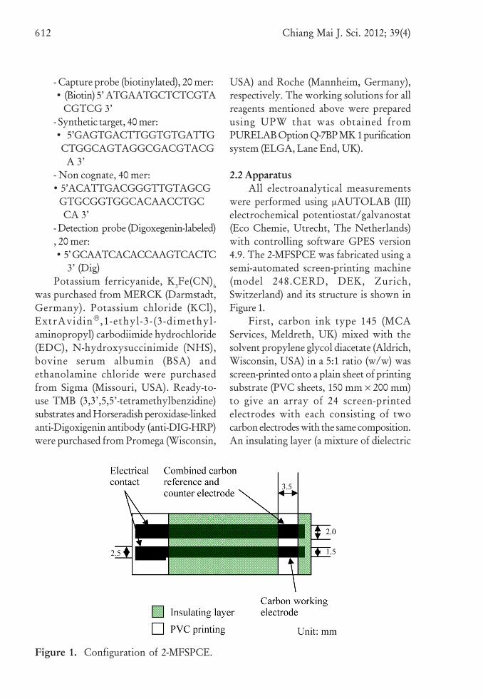
were performed using μAUTOLAB (III)electrochemical potentiostat/galvanostat(Eco Chemie, Utrecht, The Netherlands)with controlling software GPES version4.9. The 2-MFSPCE was fabricated using asemi-automated screen-printing machine(model 248.CERD, DEK, Zurich,Switzerland) and its structure is shown inFigure 1.
First, carbon ink type 145 (MCAServices, Meldreth, UK) mixed with thesolvent propylene glycol diacetate (Aldrich,Wisconsin, USA) in a 5:1 ratio (w/w) wasscreen-printed onto a plain sheet of printingsubstrate (PVC sheets, 150 mm × 200 mm)to give an array of 24 screen-printedelectrodes with each consisting of twocarbon electrodes with the same composition.An insulating layer (a mixture of dielectric
Figure 1. Configuration of 2-MFSPCE.
Chiang Mai J. Sci. 2012; 39(4) 613
ink and solvent) was then screen-printedover the sheet of printing substrate containingthe two carbon electrodes to define one areafor electrical contact and another as a sensingarea; the latter consisted of a workingelectrode (with area 1.5 mm × 3.5 mm) and acombined reference and counter electrode(with area 2 mm × 3.5 mm). There were 20-30 sheets of 2-MFSPCEs printed in eachbatch. After screen-printing, the 2-MFSPCEssheets were cured overnight in an oven at 55°Cand then allowed to cool at roomtemperature. Individual 2-MFSPCEs wereobtained by cutting them from the printingsubstrate.
2.3 Electrochemical Characterization of2-MFSPCE
The electrochemical performance of thefabricated 2-MFSPCE was assessed bycyclic voltammetry (CV) using K3Fe(CN)6
with KCl as supporting electrolyte. First,2-MFSPCE was pre-washed by applying100 ml of UPW onto the sensing area(covering both the electrode surfaces) andleaving for at least 2 min to make theelectrode surfaces wet and more hydrophilic.The UPW was then pipetted off andwithout allowing the electrode surfacesto dry, 50 ml of a freshly prepared 1 mMK3Fe(CN)6/0.1 M KCl solution was depositedonto the sensing area. The CV analysis wasbegun with the potential ramped (staircase,5mV step potential) from 0.6 to -0.6 V andthen reversed back to 0.6 V for a cycle; witha scan rate of 50 mV/s.
2.4 Electrochemical Study of TMB on2-MFSPCE2.4.1 Voltammetric study of TMB
The CV analysis of TMB (50 μl involume on sensing area of 2-MFSPCE)was performed at a scan rate of 50 mV/s.An initial pretreatment potential of -1.8 V
was applied for 30 sec; the CV was thenscanned (staircase, 5mV step potential)from potential of -1.8 V to 1.8 V andreversed back to -1.8 V.
2.4.2 Chronoamperometric Study ofHRP/TMB Reaction
One microliter of HRP solution(anti-DIG-HRP) was added to a 20 μl ofTMB on sensing area of 2-MFSPCE. Themixture was gently mixed using a pipette andsubjected to 5 sec incubation at 0 V standbypotential followed by chronoamperometrymeasurement at -0.1 V for 60 sec with aninterval time of 0.2 sec. The current value atthe end of the measurement period wasrecorded for data analysis.
2.5 Electrochemical Genosensor Studyon 2-MFSPCE
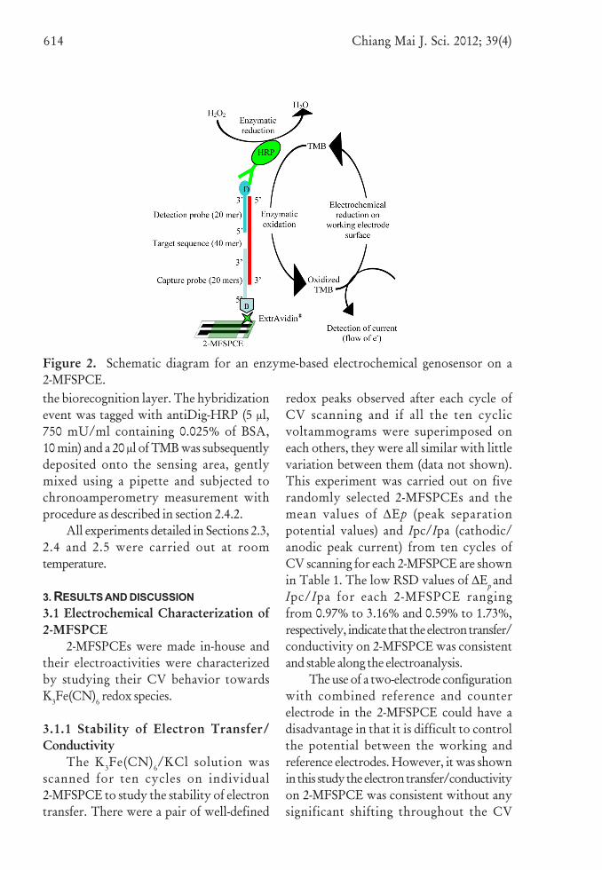
An enzyme-based electrochemicalgenosensor was set up using 2-MFSPCEwith schematic plan shown at Figure 2.Initially, ExtrAvidin® (10 μl, 0.05 mg/ml,10 min) was coated onto the sensing area of2-MFSPCE after surface activation usingEDC/NHS covalent agent (10 μl, a mixtureof 0.2 M EDC and 0.05 M NHS, 10 min),followed by treatment with ethanolaminechloride (50 μl, 1 M, pH 8.5, 10 min in dark)and BSA (50 μl, 3% w/v, 10 min). Asandwich-typed hybridization strategyusing a pair of DNA probes (1 μM ofcapture probe and 1 μM of detection probe)was employed for specific detection ofsynthetic target sequence. The sandwich-typehybridization of target sequence was atwo-step strategy in which the captureprobe (10 μl, 20 min) was firstly immobilizedonto the ExtrAvidin®-coated sensing area toform a biorecognition layer on 2-MFSPCEand subsequently, the target sequence-detectionprobe hybrid (10 μl, 20 min) that wasprepared in PCR tube was deposited onto
614 Chiang Mai J. Sci. 2012; 39(4)
the biorecognition layer. The hybridizationevent was tagged with antiDig-HRP (5 μl,750 mU/ml containing 0.025% of BSA,10 min) and a 20 μl of TMB was subsequentlydeposited onto the sensing area, gentlymixed using a pipette and subjected tochronoamperometry measurement withprocedure as described in section 2.4.2.
All experiments detailed in Sections 2.3,2.4 and 2.5 were carried out at roomtemperature.
3. RESULTS AND DISCUSSION3.1 Electrochemical Characterization of2-MFSPCE
2-MFSPCEs were made in-house andtheir electroactivities were characterizedby studying their CV behavior towardsK3Fe(CN)6 redox species.
3.1.1 Stability of Electron Transfer/Conductivity
The K3Fe(CN)6/KCl solution wasscanned for ten cycles on individual2-MFSPCE to study the stability of electrontransfer. There were a pair of well-defined
redox peaks observed after each cycle ofCV scanning and if all the ten cyclicvoltammograms were superimposed oneach others, they were all similar with littlevariation between them (data not shown).This experiment was carried out on fiverandomly selected 2-MFSPCEs and themean values of ∆Ep (peak separationpotential values) and Ipc/Ipa (cathodic/anodic peak current) from ten cycles ofCV scanning for each 2-MFSPCE are shownin Table 1. The low RSD values of ∆Ep andIpc/Ipa for each 2-MFSPCE rangingfrom 0.97% to 3.16% and 0.59% to 1.73%,respectively, indicate that the electron transfer/conductivity on 2-MFSPCE was consistentand stable along the electroanalysis.
The use of a two-electrode configurationwith combined reference and counterelectrode in the 2-MFSPCE could have adisadvantage in that it is difficult to controlthe potential between the working andreference electrodes. However, it was shownin this study the electron transfer/conductivityon 2-MFSPCE was consistent without anysignificant shifting throughout the CV
Figure 2. Schematic diagram for an enzyme-based electrochemical genosensor on a2-MFSPCE.
Chiang Mai J. Sci. 2012; 39(4) 615
scanning in cycles, indicating that there isa good, stable control of the potentialbetween both electrodes of the 2-MFSPCE.
3.1.2 Redox Behavior of Ferricyanide on2-MFSPCE
The CV analysis of the benchmarkredox species, ferricyanide, on the 2-MFSPCEproduced well-defined redox peaks (data notshown) with the Ipc/Ipa value remainingnear unity (~1) (Table 1) as expected forreaction of reversible species on electrodes.Nonetheless, the ∆Ep of the ferricyanideredox process on the 2-MFSPCE rangedfrom 229.50 ± 7.246 mV to 278.50 ± 3.689mV (Table 1), which are significantlydifferent from the ideal Nernstian value of57 mV for a single-electron transfer redoxprocess. This suggests that the redoxbehavior of ferricyanide on the 2-MFSPCEis quasi-reversible [9].
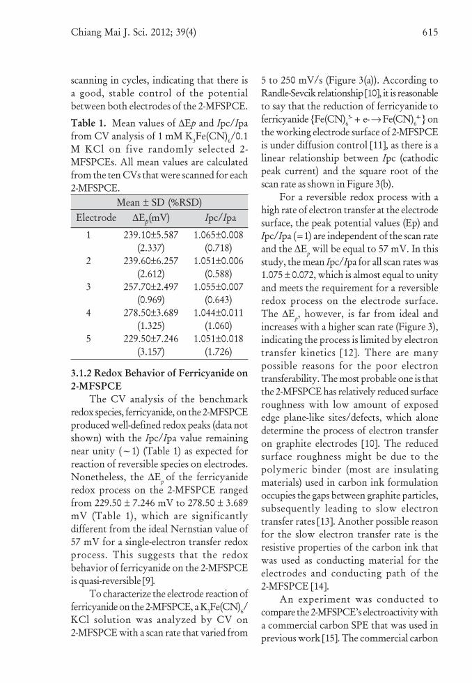
To characterize the electrode reaction offerricyanide on the 2-MFSPCE, a K3Fe(CN)6/KCl solution was analyzed by CV on2-MFSPCE with a scan rate that varied from
5 to 250 mV/s (Figure 3(a)). According toRandle-Sevcik relationship [10], it is reasonableto say that the reduction of ferricyanide toferricyanide {Fe(CN)6
3- + e- → Fe(CN)64- } on
the working electrode surface of 2-MFSPCEis under diffusion control [11], as there is alinear relationship between Ipc (cathodicpeak current) and the square root of thescan rate as shown in Figure 3(b).
For a reversible redox process with ahigh rate of electron transfer at the electrodesurface, the peak potential values (Ep) andIpc/Ipa (=1) are independent of the scan rateand the ∆Ep will be equal to 57 mV. In thisstudy, the mean Ipc/Ipa for all scan rates was1.075 ± 0.072, which is almost equal to unityand meets the requirement for a reversibleredox process on the electrode surface.The ∆Ep, however, is far from ideal andincreases with a higher scan rate (Figure 3),indicating the process is limited by electrontransfer kinetics [12]. There are manypossible reasons for the poor electrontransferability. The most probable one is thatthe 2-MFSPCE has relatively reduced surfaceroughness with low amount of exposededge plane-like sites/defects, which alonedetermine the process of electron transferon graphite electrodes [10]. The reducedsurface roughness might be due to thepolymeric binder (most are insulatingmaterials) used in carbon ink formulationoccupies the gaps between graphite particles,subsequently leading to slow electrontransfer rates [13]. Another possible reasonfor the slow electron transfer rate is theresistive properties of the carbon ink thatwas used as conducting material for theelectrodes and conducting path of the2-MFSPCE [14].
An experiment was conducted tocompare the 2-MFSPCE’s electroactivity witha commercial carbon SPE that was used inprevious work [15]. The commercial carbon
Table 1. Mean values of ∆Ep and Ipc/Ipafrom CV analysis of 1 mM K3Fe(CN)6/0.1M KCl on five randomly selected 2-MFSPCEs. All mean values are calculatedfrom the ten CVs that were scanned for each2-MFSPCE.
Mean ± SD (%RSD)Electrode ∆Ep(mV) Ipc/Ipa
1
2
3
4
5
239.10±5.587(2.337)
239.60±6.257(2.612)
257.70±2.497(0.969)
278.50±3.689(1.325)
229.50±7.246(3.157)
1.065±0.008(0.718)
1.051±0.006(0.588)
1.055±0.007(0.643)
1.044±0.011(1.060)
1.051±0.018(1.726)
616 Chiang Mai J. Sci. 2012; 39(4)
SPCE has a conventional three-electrodeconfiguration and with Ag/AgCl inkscreen-printed as the conducting paths forall the three carbon electrodes. The ∆Ep
values (n=5) for 2-MFSPCE and commercialcarbon SPE were 227.62 ± 10.356 mV(RSD 4.55%) and 248.60 ± 11.971 mV(RSD 4.82%), respectively; where as theIpc/Ipa values were 1.062 ± 0.024 (RSD2.27%) and 1.315 ± 0.234 (RSD 17.82%),respectively. The 2-MFSPCE exhibitedbetter electroactivity in which the ∆Ep andIpc/Ipa have a lower RSD and were closerto the values of an ideal reversible redoxprocess.
3.1.3 Reproducibility of 2-MFSPCEThe 2-MFSPCE in this study was
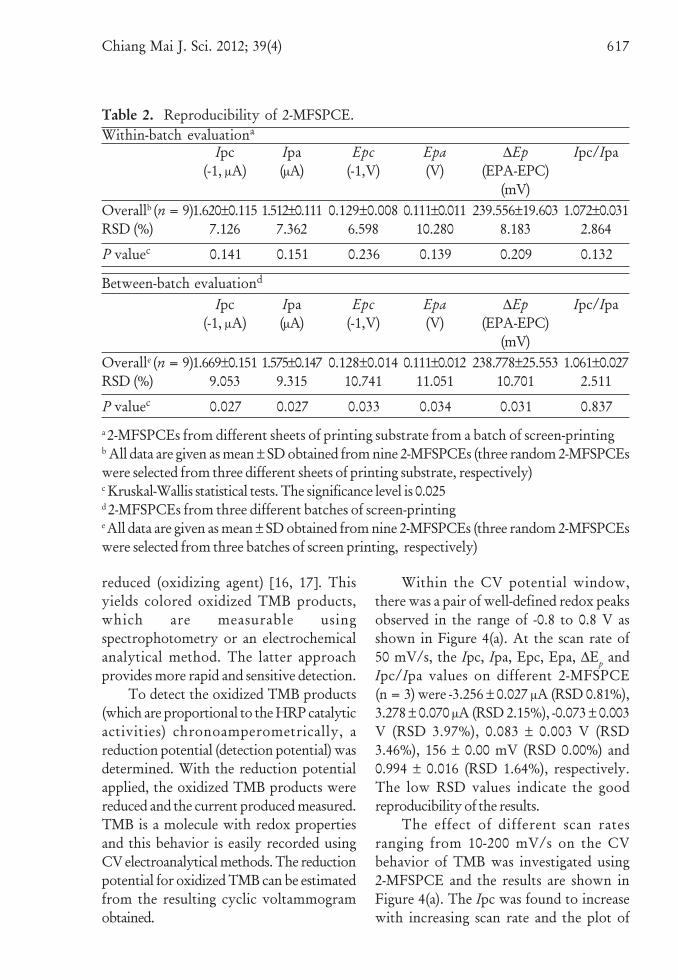
made in batches of 20-30 sheets of printingsubstrate, with each sheet comprising24 individual 2-MFSPCEs. In terms ofsensors development, reproducibility is oneof the main problems, especially whencomponents made in-house are used.Therefore, the reproducibility of 2-MFSPCEwithin and between batches was exploredby CV analysis of a K3Fe(CN)6/KCl solutionand the results obtained are shown in Table 2.The non-parametric Kruskal-Wallis statisticaltest (for both the within and between batch
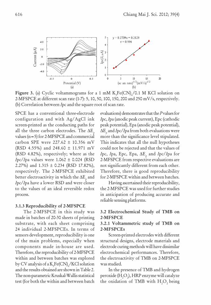
Figure 3. (a) Cyclic voltammograms for a 1 mM K3Fe(CN)6/0.1 M KCl solution on2-MFSPCE at different scan rate (1-7): 5, 10, 50, 100, 150, 200 and 250 mV/s, respectively.(b) Correlation between Ipc and the square root of scan rate.
evaluations) demonstrates that the P values forIpc, Ipa (anodic peak current), Epc (cathodicpeak potential), Epa (anodic peak potential),∆Ep and Ipc/Ipa from both evaluations weremore than the significance level stipulated.This indicates that all the null hypothesescould not be rejected and that the values ofIpc, Ipa, Epc, Epa, ∆Ep and Ipc/Ipa for2-MFSPCE from respective evaluations arenot significantly different from each other.Therefore, there is good reproducibilityfor 2-MFSPCE within and between batches.
Having ascertained their reproducibility,the 2-MFSPCE was used for further studiesin anticipation of producing accurate andreliable sensing platforms.
3.2 Electrochemical Study of TMB on2-MFSPCE3.2.1 Voltammetric study of TMB on2-MFSPCEs
Screen-printed electrodes with differentstructural designs, electrode materials andelectrode curing methods will have dissimilarelectrochemical performances. Therefore,the electroactivity of TMB on 2-MFSPCEwas studied.
In the presence of TMB and hydrogenperoxide (H2O2), HRP enzyme will catalyzethe oxidation of TMB with H2O2 being
Cur
rent
(μA
)
Potential (V)(a)
(sc an rate)1/2(mV/s)1/2
(b)
Ipc(
-l, μ
A)
Chiang Mai J. Sci. 2012; 39(4) 617
reduced (oxidizing agent) [16, 17]. Thisyields colored oxidized TMB products,which are measurable usingspectrophotometry or an electrochemicalanalytical method. The latter approachprovides more rapid and sensitive detection.
To detect the oxidized TMB products(which are proportional to the HRP catalyticactivities) chronoamperometrically, areduction potential (detection potential) wasdetermined. With the reduction potentialapplied, the oxidized TMB products werereduced and the current produced measured.TMB is a molecule with redox propertiesand this behavior is easily recorded usingCV electroanalytical methods. The reductionpotential for oxidized TMB can be estimatedfrom the resulting cyclic voltammogramobtained.
Within the CV potential window,there was a pair of well-defined redox peaksobserved in the range of -0.8 to 0.8 V asshown in Figure 4(a). At the scan rate of50 mV/s, the Ipc, Ipa, Epc, Epa, ∆Ep andIpc/Ipa values on different 2-MFSPCE(n = 3) were -3.256 ± 0.027 μA (RSD 0.81%),3.278 ± 0.070 μA (RSD 2.15%), -0.073 ± 0.003V (RSD 3.97%), 0.083 ± 0.003 V (RSD3.46%), 156 ± 0.00 mV (RSD 0.00%) and0.994 ± 0.016 (RSD 1.64%), respectively.The low RSD values indicate the goodreproducibility of the results.
The effect of different scan ratesranging from 10-200 mV/s on the CVbehavior of TMB was investigated using2-MFSPCE and the results are shown inFigure 4(a). The Ipc was found to increasewith increasing scan rate and the plot of
Table 2. Reproducibility of 2-MFSPCE.Within-batch evaluationa
Ipc(-1, μA)
Ipa(μA)
Epc(-1,V)
Epa(V)
∆Ep(EPA-EPC)
(mV)
Ipc/Ipa
Overallb (n = 9)RSD (%)
1.620±0.1157.126
1.512±0.1117.362
0.129±0.0086.598
0.111±0.01110.280
239.556±19.6038.183
1.072±0.0312.864
P valuec 0.141 0.151 0.236 0.139 0.209 0.132
Between-batch evaluationd
Ipc(-1, μA)
Ipa(μA)
Epc(-1,V)
Epa(V)
∆Ep(EPA-EPC)
(mV)
Ipc/Ipa
Overalle (n = 9)RSD (%)
1.669±0.1519.053
1.575±0.1479.315
0.128±0.01410.741
0.111±0.01211.051
238.778±25.55310.701
1.061±0.0272.511
P valuec 0.027 0.027 0.033 0.034 0.031 0.837
a 2-MFSPCEs from different sheets of printing substrate from a batch of screen-printingb All data are given as mean ± SD obtained from nine 2-MFSPCEs (three random 2-MFSPCEswere selected from three different sheets of printing substrate, respectively)c Kruskal-Wallis statistical tests. The significance level is 0.025d 2-MFSPCEs from three different batches of screen-printinge All data are given as mean ± SD obtained from nine 2-MFSPCEs (three random 2-MFSPCEswere selected from three batches of screen printing, respectively)
618 Chiang Mai J. Sci. 2012; 39(4)
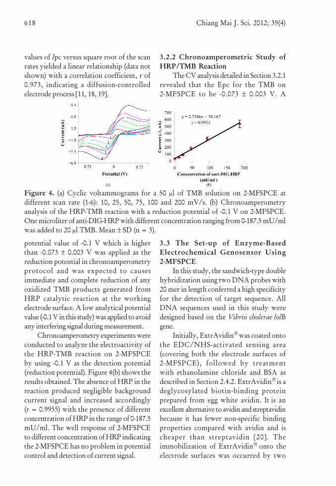
values of Ipc versus square root of the scanrates yielded a linear relationship (data notshown) with a correlation coefficient, r of0.973, indicating a diffusion-controlledelectrode process [11, 18, 19].
3.2.2 Chronoamperometric Study ofHRP/TMB Reaction
The CV analysis detailed in Section 3.2.1revealed that the Epc for the TMB on2-MFSPCE to be -0.073 ± 0.003 V. A
Figure 4. (a) Cyclic voltammograms for a 50 μl of TMB solution on 2-MFSPCE atdifferent scan rate (1-6): 10, 25, 50, 75, 100 and 200 mV/s. (b) Chronoamperometryanalysis of the HRP-TMB reaction with a reduction potential of -0.1 V on 2-MFSPCE.One microliter of anti-DIG-HRP with different concentration ranging from 0-187.5 mU/mlwas added to 20 μl TMB. Mean ± SD (n = 3).
potential value of -0.1 V which is higherthan -0.073 ± 0.003 V was applied as thereduction potential in chronoamperometryprotocol and was expected to causesimmediate and complete reduction of anyoxidized TMB products generated fromHRP catalytic reaction at the workingelectrode surface. A low analytical potentialvalue (-0.1 V in this study) was applied to avoidany interfering signal during measurement.
Chronoamperometry experiments wereconducted to analyze the electroactivity ofthe HRP-TMB reaction on 2-MFSPCEby using -0.1 V as the detection potential(reduction potential). Figure 4(b) shows theresults obtained. The absence of HRP in thereaction produced negligible backgroundcurrent signal and increased accordingly(r = 0.9955) with the presence of differentconcentration of HRP in the range of 0-187.5mU/ml. The well response of 2-MFSPCEto different concentration of HRP indicatingthe 2-MFSPCE has no problem in potentialcontrol and detection of current signal.
3.3 The Set-up of Enzyme-BasedElectrochemical Genosensor Using2-MFSPCE
In this study, the sandwich-type doublehybridization using two DNA probes with20 mer in length conferred a high specificityfor the detection of target sequence. AllDNA sequences used in this study weredesigned based on the Vibrio cholerae lolBgene.
Initially, ExtrAvidin® was coated ontothe EDC/NHS-activated sensing area(covering both the electrode surfaces of2-MFSPCE), followed by treatmentwith ethanolamine chloride and BSA asdescribed in Section 2.4.2. ExtrAvidin® is adeglycosylated biotin-binding proteinprepared from egg white avidin. It is anexcellent alternative to avidin and streptavidinbecause it has fewer non-specific bindingproperties compared with avidin and ischeaper than streptavidin [20]. Theimmobilization of ExtrAvidin® onto theelectrode surfaces was occurred by two
Chiang Mai J. Sci. 2012; 39(4) 619
mechanisms: (1) covalent binding throughthe EDC/NHS activated carboxyl groupson the electrode surfaces, and (2) passiveadsorption onto the electrode surfaces [21].In this study, the biorecognition layer wasformed on both the working and combinedreference and counter electrodes to increasethe number of recognition elements fordetection of target DNA sequence; thisconsequently improved the sensitivity of theexperiment.
Three different strategies of sandwich-type hybridization were investigated. In aone-step sandwich-type hybridizationstrategy, all three components [capture probe,synthetic target sequence and detectionprobe] were mixed together in a 0.5 ml PCRtube with a concentration of 1 μM, 0.5 μMand 1 μM, respectively. The reaction mixturewas incubated for 1 h at room temperature.The hybrid formed was then depositedonto the ExtrAvidin® coated layer on boththe electrode surfaces of 2-MFSPCE andincubated for 20 min at room temperature.Non-binding components were then washedoff the electrode surfaces using washingbuffer. The biotin from the hybrid interactedwith the ExtrAvidin® at the electrodesurfaces.
By contrast, in a two-step sandwich-typehybridization strategy, 1 μM of biotinylatedcapture probe was first immobilized byincubation for 20 min at room temperatureonto the ExtrAvidin®-coated electrodesurfaces to form a biorecognition layer.The synthetic target sequence-detectionprobe hybrid was then prepared in PCRtube with a concentration of 0.5 μM and1 μM, respectively; and incubated for 1 hat room temperature. The hybrid was thendeposited onto the biorecognition layer ofboth electrode surfaces of the 2-MFSPCEand incubated for 20 min at roomtemperature.
Lastly, in a three-step sandwich-typehybridization strategy, the capture probe,synthetic target sequence and detectionprobe were prepared individually in PCRtubes with a concentration of 1 μM, 0.5 μMand 1 μM, respectively, and depositedsequentially onto the ExtrAvidin®-coatedlayer on both electrode surfaces of the2-MFSPCE. Each DNA component on theelectrode surfaces was incubated for 20 minand the unbound sequence was removed bywashing buffer before proceeding to thenext stage.
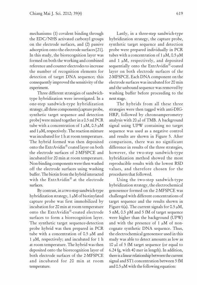
The hybrids from all these threestrategies were then tagged with anti-DIG-HRP, followed by chronoamperometryanalysis with 20 μl of TMB. A backgroundsignal using UPW containing no targetsequence was used as a negative controland results are shown in Figure 5. Aftercomparison, there was no significantdifference in results of the three strategies,however, the two-step sandwich-typehybridization method showed the mostreproducible results with the lowest RSDvalues, and therefore chosen for theprocedures that followed.
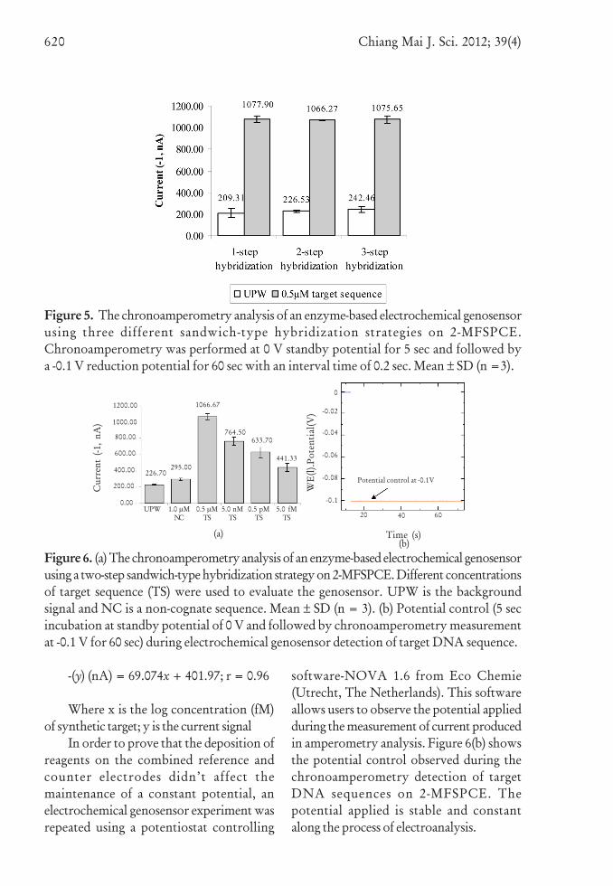
Using the two-step sandwich-typehybridization strategy, the electrochemicalgenosensor formed on the 2-MFSPCE waschallenged with different concentrations oftarget sequence and the results shown inFigure 6(a). The current signals for 0.5 μM,5 nM, 0.5 pM and 5 fM of target sequencewere higher than the background (UPW)and with the presence of 1 μM of non-cognate synthetic DNA sequence. Thus,the electrochemical genosensor used in thisstudy was able to detect amounts as low as10 μl of 5 fM target sequence (or equal to6.24 fg, with 40 mer in length). In addition,there is a linear relationship between the currentsignal and ST1 concentration between 5 fMand 0.5 μM with the following equation:
620 Chiang Mai J. Sci. 2012; 39(4)
Figure 5. The chronoamperometry analysis of an enzyme-based electrochemical genosensorusing three different sandwich-type hybridization strategies on 2-MFSPCE.Chronoamperometry was performed at 0 V standby potential for 5 sec and followed bya -0.1 V reduction potential for 60 sec with an interval time of 0.2 sec. Mean ± SD (n =3).
-(y) (nA) = 69.074x + 401.97; r = 0.96
Where x is the log concentration (fM)of synthetic target; y is the current signal
In order to prove that the deposition ofreagents on the combined reference andcounter electrodes didn’t affect themaintenance of a constant potential, anelectrochemical genosensor experiment wasrepeated using a potentiostat controlling
software-NOVA 1.6 from Eco Chemie(Utrecht, The Netherlands). This softwareallows users to observe the potential appliedduring the measurement of current producedin amperometry analysis. Figure 6(b) showsthe potential control observed during thechronoamperometry detection of targetDNA sequences on 2-MFSPCE. Thepotential applied is stable and constantalong the process of electroanalysis.
Figure 6. (a) The chronoamperometry analysis of an enzyme-based electrochemical genosensorusing a two-step sandwich-type hybridization strategy on 2-MFSPCE. Different concentrationsof target sequence (TS) were used to evaluate the genosensor. UPW is the backgroundsignal and NC is a non-cognate sequence. Mean ± SD (n = 3). (b) Potential control (5 secincubation at standby potential of 0 V and followed by chronoamperometry measurementat -0.1 V for 60 sec) during electrochemical genosensor detection of target DNA sequence.
(a)(b)
Time (s)
WE
(l).P
oten
tial
(V)
Potential control at -0.1V
0
-0.02
-0.04
-0.06
-0.08
-0.1
1200.00
1000.00
800.00
600.00
400.00
200.00
0.00
20 40 60
(b)
UPW 1.0 μMNC
0.5 μMTS
5.0 nMTS
0.5 pMTS
5.0 fMTS
Cur
rent
(-1
, nA
)
295.00226.70
1066.67
764.50633.70
441.33
Chiang Mai J. Sci. 2012; 39(4) 621
4. CONCLUSIONSThe current study showed that
2-MFSPCE with a simplified two-electrodeconfiguration (a carbon working electrodeand a carbon combined reference andcounter electrode), which is cheaper andeasier to be fabricated, can be usedefficiently as a transducer for genosensorapplications. All 2-MFSPCE tested in thestudy showed high consistency in terms oftheir electron transfer/conductivity,implying a stable electrode surfacestructure and a good stability in controllingof potential between both electrodes of the2-MFSPCE. In addition, all individual2-MFSPCE displayed good reproducibility,with no significant batch-to-batch variation.This is an important characteristic that isessential in electrochemical analyses usingscreen-printed electrodes.
The 2-MFSPCE used as HRP enzyme-based electrochemical genosensors andanalyzed using chronoamperometryrevealed the lowest detection limit on2-MFSPCE to be 10 μl of 5 fM synthetictarget sequence (or equal to 6.24 fg, with40 mer in length). The encouragingperformance of 2-MFSPCE indicates theirsuitability for use as a cost-effectivedisposable electrode in electrochemicalsensor applications; and with the inherentfavorable characteristics possessed;2-MFSPCE can be an alternative choice forresearchers with particular needs on SPE.
ACKNOWLEDGEMENTSThe authors thank the research
funding support in form of ResearchUniversity (RU) grant (grant no: 1001/PPSP/813020) from Universiti SainsMalaysia (USM), Malaysia. In addition,support from Institute of PostgraduateStudies (IPS), Universiti Sains Malaysia(USM) in the form of graduate
fellowship for Low Kim Fatt is gratefullyacknowledged.
REFERENCES
[1] Teles F.R.R. and Fonseca L.P., Trendsin DNA biosensors, Talanta, 2008; 77:606-623.
[2] Endo T., Kerman K., Nagatani N.,Takamura Y. and Tamiya E., Label-free detection of peptide nucleic acid-DNA hybridization using localizedsurface plasmon resonance basedoptical biosensor, Anal. Chem., 2005;77: 6976-6984.
[3] Lucarelli F., Tombelli S., MinunniM., Marrazza G. and Mascini M.,Electrochemical and piezoelectricDNA biosensors for hybridisationdetection, Analytica Chimica Acta,2008; 609: 139-159.
[4] Lai R.Y., Lagally E.T., Lee S.H., SohH.T., Plaxco K.W. and Heeger A.J.,Rapid, sequence-specific detection ofunpurified PCR amplicons via areusable, electrochemical sensor,Proc. Natl. Acad. Sci. USA., 2006; 103:4017-4021.
[5] Mix M., Reske T., Duwensee H. andFlechsig G.U., Electrochemicaldetection of asymmetric PCRproducts by labeling with osmiumtetroxide, Electroanalysis, 2009; 21:826-830.
[6] Kerman K., Kobayashi M. and TamiyaE., Recent trends in electrochemicalDNA biosensor technology, Meas. Sci.Technol., 2004; 15: R1-R11.
[7] Jubete E., Loaiza O.A., Ochoteco E.,Pomposo J.A., Grande H. andRodr -guez, Nanotechnology: A toolfor improved performance onelectrochemical screen-printed (Bio)sensors, J. Sens., 2009: 1-13.
622 Chiang Mai J. Sci. 2012; 39(4)
[8] Lalitha P., Siti Suraiya M.N., LimK.L., Lee S.Y., Nur HaslindawatyA.R., Chan Y.Y., Ismail A.,Zainuddin Z.F. and Ravichandran M.,Analysis of lol B gene sequence and itsuse in the development of a PCR assayfor the detection of Vibrio cholerae, J.Microbiol. Methods, 2008; 75: 142-144.
[9] Crew A., Alford C., Cowell D.C.C.and Hart J.P., Development of anovel electrochemical immuno-assayusing a screen printed electrodefor the determination of secretoryimmunoglobulin A in human sweat,Electrochim. Acta, 2007; 52: 5232-5237.
[10] Kadara R.O., Jenkinson N. andBanks C.E., Characterisation ofcommercially available electrochemicalsensing platforms, Sens. Actuator B-Chem., 2009; 138: 556-562.
[11] Liu C.C., Electrochemical Sensors, 2nd
Edn., The Biomedical EngineeringHandbook, CRC Press LLC, 2000.
[12] Sabzi R.E., Amperometric detectionof nitrite on glassy carbon electrodemodified with cobalt nitroprusside,Portugaliae Electrochim. Acta, 2007;25: 383-390.
[13] Grennan K., Killard A.J. and SmythM.R., Physical characterizations of ascreen-printed electrode for use in anamperometric biosensor system,Electroanalysis, 2001; 13: 745-750.
[14] Morrin A., Killard A.J. and SmythM.R., Electrochemical characteriza-tion of commercial and home-madescreen-printed carbon electrodes, Anal.Lett., 2003; 36: 2021-2039.
[15] Yean C.Y., Kamarudin B., OzkanD.A., Yin L.S., Lalitha P., Ismail A.,Ozsoz M. and RavichandranM., Enzyme-linked amperometricelectrochemical genosensor assay for
the detection of PCR amplicons on astreptavidin-treated screen-printedcarbon electrode, Anal. Chem., 2008;80: 2774-2779.
[16] Fanjul-Bolado P., Gonz lez-Garc -a M.B. and Costa-Garc -a A.,Amperometric detection in TMB/HRP-based assays, Anal. Bioanal.Chem., 2005; 382: 297-302.
[17] Shakeela S., Ram B.S. and SekaranC.B., Application of horseradishperoxidase mediated oxidativecoupling reaction for the determi-nation of mesalazine, Chiang Mai J.Sci., 2011; 38(4): 551-559.
[18] Nagappa L.T., Shankar N.P. andJaldappagari S., Electrochemicalstudies of nevirapine, an anti-HIVdrug, and its assay in tablets andbiological samples, J. Electrochem. Sci.Eng., 2012; doi: 10.5599/jese.2012.0008.
[19] Nadiah A.R., Nor Azah Y., NorAmirah M.M. and Siti M.M.N.,Development of electrochemicalsensor for simultaneous determinationof Cd(II) and Hg(II) ion by exploitingnewly synthesized cyclic dipeptide,Int. J. Electrochem. Sci., 2012; 7: 186-196.
[20] Kara P., Erdem A., Girousi S. andOzsoz M., Electrochemical detectionof enzyme labeled DNA based ondisposable pencil graphite electrode,J. Pharm. Biomed. Anal., 2005; 38:191-195.
[21] Wang Z.H., Viana A.S., Jin G. andAbrantes L.M., Immunosensorinterface based on physical andchemical immunoglubulin Gadsorption onto mixed self-assembledmonolayers, Bioelectrochem., 2006;69: 180-186.