Embed Size (px)
Citation preview
This content has been downloaded from IOPscience. Please scroll down to see the full text.
Download details:
IP Address: 59.174.243.198
This content was downloaded on 23/10/2014 at 16:11
Please note that terms and conditions apply.
Preparation and characterization of two types of separate collagen nanofibers with different
widths using aqueous counter collision as a gentle top-down process
View the table of contents for this issue, or go to the journal homepage for more
2014 Mater. Res. Express 1 045016
(http://iopscience.iop.org/2053-1591/1/4/045016)
Home Search Collections Journals About Contact us My IOPscience
Preparation and characterization of two types ofseparate collagen nanofibers with different widthsusing aqueous counter collision as a gentle top-downprocess
Tetsuo Kondo1, Daisuke Kumon1, Akiko Mieno1, Yutaro Tsujita1 andRyota Kose21Graduate School of Bioresource and Bioenvironmental Sciences, Kyushu University, 6-10-1Hakozaki, Higashi, Fukuoka 812-8581, Japan2 Faculty of Agriculture, Tokyo University of Agriculture and Technology, 3-5-8 Saiwai-cho,Fuchu, Tokyo 183-8509, JapanE-mail: [email protected]
Received 30 August 2014Accepted for publication 16 September 2014Published 22 October 2014
Materials Research Express 1 (2014) 045016
doi:10.1088/2053-1591/1/4/045016
AbstractTwo types of single collagen nanofibers with different widths were successfullyprepared from native collagen fibrils using aqueous counter collision (ACC) as atop-down process. A mild collision of an aqueous suspension at a 100MPaejection pressure yielded nanofibers, termed CNF100, which have an inherentaxial periodicity and are ∼100 nm in width and ∼10 μm in length. In contrast,ACC treatment at 200MPa provided a non-periodic, shorter and thinner nano-fiber, termed CNF10, that was ∼10 nm in width and ∼5 μm in length. Bothnanofibers exhibited the inherent triple helix conformation of native collagensupramolecules. Even a medial collision that exceeded the above ACC pressuresprovided solely a mixture of the two nanofiber products. The two nanofibertypes were well characterized, and their tensile strengths were estimated basedon their sonication-induced fragmentation behaviors that related to their indi-vidual fiber morphologies. As a result, CNF10, which was found to be a criticalminimum nanofibril unit, and CNF10 exhibited totally different features in sizes,morphology, tensile strength and viscoelastic properties. In particular, as themechanical strength of the molecular scaffold affects cell differentiation, the twocollagen nanofibers prepared here by ACC have the potential for controlling celldifferentiation in possibly different ways, as they have different mechanicalproperties. This encourages the consideration of the application of CNF100 andCNF10 in the fabrication of new functional materials with unique propertiessuch as a scaffold for tissue engineering.
Materials Research Express 1 (2014) 0450162053-1591/14/045016+16$33.00 © 2014 IOP Publishing Ltd
Keywords: collagen, nanofiber, tensile strength, morphology, structure-propertyrelationship, nano building block
1. Introduction
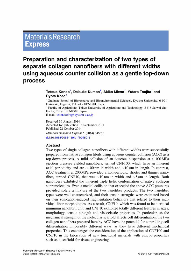
In nature, hierarchical structures within biomacromolecules, such as collagen, chitin andcellulose, are built up from the molecular level through the nanometer scale to nano/micrometerscales. Collagen molecules, which consist of three helical polypeptide chains and establish aunique hierarchical structure, are the most abundant biomacromolecules in animal tissues. Acollagen fibril in an extracellular matrix is synthesized as a long fibrous structure with thearrangement of the triple helical polypeptide units aligned in both longitudinal and obliquedirections and is further assembled with other fibers to produce a unique three-dimensional fiberstructure. A collagen fiber is comprised of a bundle of collagen fibrils 10–300 nm in width asbuilding blocks. The right-handed, triple helical structure of a collagen molecule, whichconsists of three left-handed helical polypeptide chains, is arranged regularly in both thelongitudinal and lateral directions in a collagen fibril. Such an arrangement of collagenmolecules forms the unique periodic-banding morphology of the collagen fibril (figure 1) [1–8].
Collagen supramolecules or tropocollagen consist of three helical polypeptide chains witha repeating Gly-X-Y amino acyl residue pattern, where the X and Y positions are often prolineand hydroxyproline, respectively [3, 4]. They are organized together to form a triple helicalstructure ∼1.5 nm in width and ∼300 nm in length. This triple helix is the minimum buildingblock in the hierarchical structure of a collagen fibril, and five supramolecules assemble to forma collagen microfibril [5, 6]. Microfibrils are finally arranged with a specific staggered pattern inboth the fibril’s longitudinal and lateral directions and are stabilized by intermolecular hydrogenbonds, electrostatic interactions and chemical crosslinks [7, 8]. Such a systematic arrangementof collagen molecules exhibit a specific typical periodic-banding morphology of the collagenfibril in transmission electron micrographs (TEM) [9, 10].
The relationships between the hierarchical structure of collagenous tissue and itsmechanical properties have been investigated at the micrometer scale. Namely, the strength of acollagen fiber has been reported to depend on the fiber diameter, which is in a relationshipbetween the hierarchical structure and mechanical properties [2, 11, 12]. In previous studies, theself-assembly of collagen molecules, which is water-solubilized collagen prepared by acid oralkali treatment, and gelatin obtained by heating the collagen molecules have been mainlyemployed for targeting products [13–15]. However, to our knowledge, there has been noresearch to date that has focused at the nanometer scale, presumably because there has been noestablished method to extract collagen nanofibers from native fibers in a top-down manner.Therefore, the production and study of a collagen nano-building block that includes collagennanofibers would provide valuable insight into important features or properties of collagen. Inparticular, it was expected here that the formation process and the strength of a collagenhydrogel formed from a collagen nanofiber network that includes large amounts of waterclusters would be very different from conventional results.
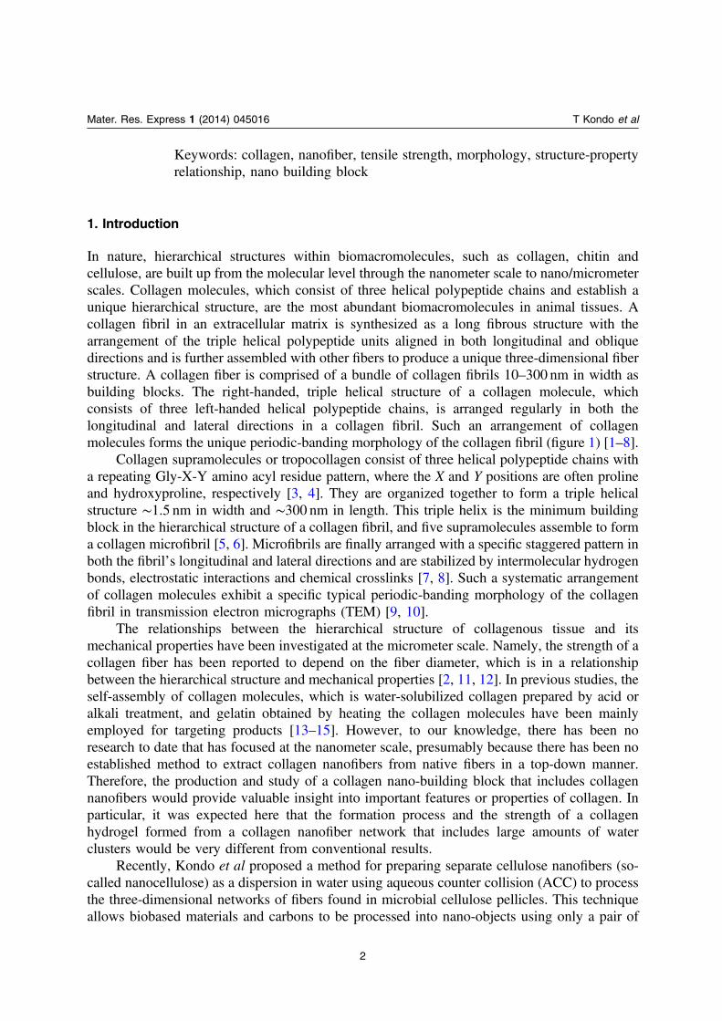
Recently, Kondo et al proposed a method for preparing separate cellulose nanofibers (so-called nanocellulose) as a dispersion in water using aqueous counter collision (ACC) to processthe three-dimensional networks of fibers found in microbial cellulose pellicles. This techniqueallows biobased materials and carbons to be processed into nano-objects using only a pair of
2
Mater. Res. Express 1 (2014) 045016 T Kondo et al
water jets, without the need for any chemical modifications [16–21]. Basically, ACC reducesbiomaterials to nanoscale objects using the collision energy of dual water jets. This strategy iscapable of overcoming van der Waals forces and hydrogen bonds in the absence of chemicalmodification. Thus, ACC is one of the gentlest, mildest methods available among the top-downreducing processes. This system involves an aqueous suspension of μm-sized sample particlesdivided between two nozzles that face each other (figure 2). The opposing ejected suspensionscollide at a high speed, resulting in nano-pulverization of the dispersed sample. The obtainedmaterial can be homogeneously reduced and further reduced in size by repeated collisions orincreased ejection pressure. In the current study, ACC was applied to hydrophobic carbons,such as fullerene (C60), multiwalled carbon nanotubes and graphite, to successfully prepareaqueous suspensions [21].
Furthermore, ACC can selectively cleave particular interactions in soft materials, such aspolysaccharides, proteins and nucleic acids, by controlling the number of collisions and/or theejection pressure. In this study, the goal was to use ACC to extract nano-building blocks fromnative collagen fibers. Considering the fiber's unique structure, three disassembly patterns wereassumed for reducing collagen fibers to nanoscale objects: 1) cleavage of longitudinalinterfacial interactions, 2) cleavage of both longitudinal and lateral interfacial interactions and3) cleavage of intramolecular interactions in the collagen triple helix. Thus, the goal of this
Figure 1. Schematic illustrations of a sequence of hierarchical assemblies of collagenmolecules to collagen fibril (upper image). The bottom images indicate (a) cross-sectional and (b) longitudinal schematic illustrations of collagen fibrils [1–8].
Figure 2. Aqueous counter collision system using a pair of water jets [16–21].
3
Mater. Res. Express 1 (2014) 045016 T Kondo et al
study was to understand how collagen fibers were reduced by ACC to nanofibers as well as toilluminate how the hierarchical structures relate to measured mechanical properties at each size-scale.
2. Materials and methods
2.1. ACC treatment of collagen fibers
Collagen fibers from bovine skin type I collagen fibrils were provided from Nippi Inc. (Tokyo,Japan). Collagen fibers (0.01% by wt) were dispersed in deionized water with stirring for 1 d atroom temperature prior to homogenization at 10 000 or 20 000 rpm for 3min using ahomogenizer (Physcotron NS-51, Microtec Co., Ltd, Chiba, Japan). In general, collagenmolecule self-assembly is performed in the 5–8 pH range [11], while an acid-solubilizedcollagen is prepared in 2–3 pH [11, 22]. Here, the initial pH value for the collagen fibersuspension before ACC treatment was 4, which was appropriate to maintain the morphologyand dispersion state of the collagen sample. Aqueous suspensions were subjected to ACCtreatment (figure 2, Sugino Co., Toyama, Japan) under 100, 120, 150, 180, 190 and 200MPa ofnozzle ejection pressure in combination with 10, 30, 60 and 90 cycle repetition times (or Pass)[16–21]. The number of collisions and the collision pressure were critical factors in tailoring theproperties of the resulting nanofibers. The nozzle diameter was 160 μm, with the jets’ collisionangle typically set at ∼170 degrees. A single collision of the jets simultaneously generates heatsuch that a 50 °C temperature increase is associated with a pressure of 200MPa [17], which isthe maximum ejection pressure employed in this study. Because of this heat generation, acooling system based on a flow of water was applied immediately downstream from the jet-collision zone in the chamber.
2.2. Measurements
2.2.1. TEM observation. An aqueous suspension containing 0.01% ACC-treated collagenfibers (by wt) was mounted on a copper grid. An aqueous 0.25% surfactant solution was nextapplied to the grid, followed by negative staining with aqueous 1% uranyl acetate. Thespecimen was then washed thoroughly with deionized water before a second application of thenegative stain; finally, the specimen was air-dried. A TEM observation was carried out with aJEM-1010 (JEOL Ltd, Tokyo, Japan) operated at 80 kV of accelerating voltage with a beamcurrent of <70 μA. The TEM images were acquired at magnifications from 300 k to 150 k in thenegative films. The images were scanned for digitization and for measurement of the widths andlengths of the >100 individual collagen nanofibers using Image-Pro Plus software version 4.1(Media Cybernetics, Inc., Rockville, MD, USA).
2.2.2. Circular dichroic (CD) spectroscopy. The specimens for the CD measurements wereprepared according to the following procedure: An aqueous suspension containing 0.15% ofcollagen fibers (by wt) was stirred for 1 d, followed by the addition of NaCl to a final 10%concentration (by wt). The dispersion was then centrifuged at 10 °C under 1.0 × 10⁴ g for 20minusing a high-speed refrigerated microcentrifuge (MX-301, Tomy Seiko Co., Ltd, Tokyo,Japan). After the precipitate was dialyzed in deionized water, 0.01% dispersions (by wt) weretreated by ACC using 100 or 200MPa ejection pressure with 10, 30, 60 or 90 Pass.
4
Mater. Res. Express 1 (2014) 045016 T Kondo et al
Dispersed samples in the water prepared as above were provided for CD spectroscopicmeasurements using a JASCO J-820 spectrometer (JASCO International Co., Ltd, Tokyo,Japan). The CD spectra were measured at wavelengths from 183 to 300 nm under a temperatureof 15 °C. The sample cell length for longer (210–300 nm) and shorter wavelengths(183–300 nm) was 10 and 1mm, respectively. The scanning rate was 50 nmmin−1 at a 1 nmresolution, and the response time was set at 2 s.
2.2.3. Sonication-induced fragmentation. Each aqueous dispersion of separated collagennanofibers was sonicated using a UD-200 sonicator (Tomy Seiko Co., Ltd) at 200W maximumpower and 20 kHz operating at 10% output power. Ultrasonic waves were applied in 20min on5min off cycles for up to 360min. The dispersions temperature was maintained at ∼8 °C in anice water bath.
2.2.4. Viscoelastic measurements. For dynamic viscoelastic measurements, specimens wereprepared in the following manner: collagen fibers (0.8% by wt) were dispersed in deionizedwater and stirring for 1 d prior to ACC treatment under either 100 or 200MPa ejection pressurewith 30 Pass. The resulting samples were held in a refrigerator for 2 d.
Dispersed samples in water prepared as above were analyzed with a cone-plate typerheometer (Rheosol-G2000, UBM Co., Ltd, Kyoto, Japan). The radius of both the cone andplate was 50mm, and the cone angle was 28 degrees. The rheometer was equipped with areservoir to prevent sample-drying during measurements. Dynamic viscoelastic measurementswere performed at 5 ± 0.1 °C and with measured frequencies ranging from 0.05 to 56 rad s−1.The dynamic strain amplitude (γ) was 0.208 (10%).
3. Results and discussion
3.1. Preparation of collagen nanofibers by ACC
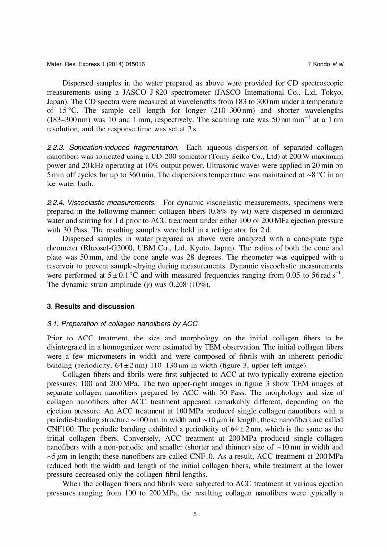
Prior to ACC treatment, the size and morphology on the initial collagen fibers to bedisintegrated in a homogenizer were estimated by TEM observation. The initial collagen fiberswere a few micrometers in width and were composed of fibrils with an inherent periodicbanding (periodicity, 64 ± 2 nm) 110–130 nm in width (figure 3, upper left image).
Collagen fibers and fibrils were first subjected to ACC at two typically extreme ejectionpressures: 100 and 200MPa. The two upper-right images in figure 3 show TEM images ofseparate collagen nanofibers prepared by ACC with 30 Pass. The morphology and size ofcollagen nanofibers after ACC treatment appeared remarkably different, depending on theejection pressure. An ACC treatment at 100MPa produced single collagen nanofibers with aperiodic-banding structure ∼100 nm in width and ∼10 μm in length; these nanofibers are calledCNF100. The periodic banding exhibited a periodicity of 64 ± 2 nm, which is the same as theinitial collagen fibers. Conversely, ACC treatment at 200MPa produced single collagennanofibers with a non-periodic and smaller (shorter and thinner) size of ∼10 nm in width and∼5 μm in length; these nanofibers are called CNF10. As a result, ACC treatment at 200MPareduced both the width and length of the initial collagen fibers, while treatment at the lowerpressure decreased only the collagen fibril lengths.
When the collagen fibers and fibrils were subjected to ACC treatment at various ejectionpressures ranging from 100 to 200MPa, the resulting collagen nanofibers were typically a
5
Mater. Res. Express 1 (2014) 045016 T Kondo et al
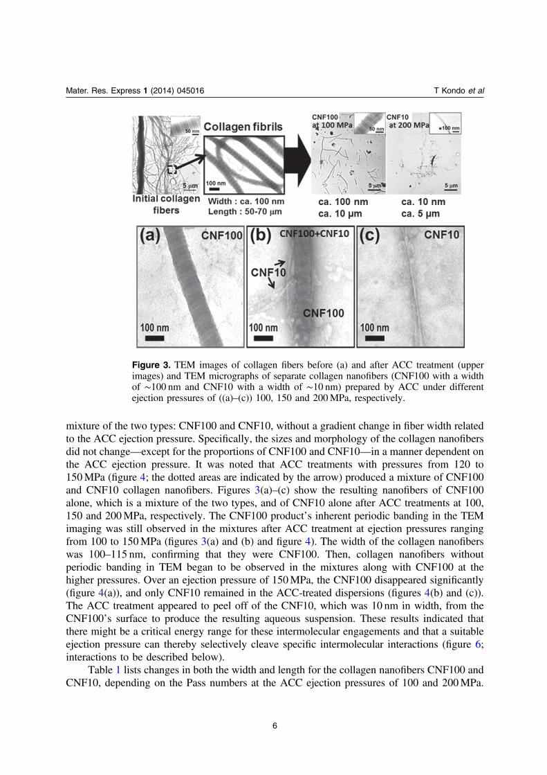
mixture of the two types: CNF100 and CNF10, without a gradient change in fiber width relatedto the ACC ejection pressure. Specifically, the sizes and morphology of the collagen nanofibersdid not change—except for the proportions of CNF100 and CNF10—in a manner dependent onthe ACC ejection pressure. It was noted that ACC treatments with pressures from 120 to150MPa (figure 4; the dotted areas are indicated by the arrow) produced a mixture of CNF100and CNF10 collagen nanofibers. Figures 3(a)–(c) show the resulting nanofibers of CNF100alone, which is a mixture of the two types, and of CNF10 alone after ACC treatments at 100,150 and 200MPa, respectively. The CNF100 product’s inherent periodic banding in the TEMimaging was still observed in the mixtures after ACC treatment at ejection pressures rangingfrom 100 to 150MPa (figures 3(a) and (b) and figure 4). The width of the collagen nanofiberswas 100–115 nm, confirming that they were CNF100. Then, collagen nanofibers withoutperiodic banding in TEM began to be observed in the mixtures along with CNF100 at thehigher pressures. Over an ejection pressure of 150MPa, the CNF100 disappeared significantly(figure 4(a)), and only CNF10 remained in the ACC-treated dispersions (figures 4(b) and (c)).The ACC treatment appeared to peel off of the CNF10, which was 10 nm in width, from theCNF100’s surface to produce the resulting aqueous suspension. These results indicated thatthere might be a critical energy range for these intermolecular engagements and that a suitableejection pressure can thereby selectively cleave specific intermolecular interactions (figure 6;interactions to be described below).
Table 1 lists changes in both the width and length for the collagen nanofibers CNF100 andCNF10, depending on the Pass numbers at the ACC ejection pressures of 100 and 200MPa.
Figure 3. TEM images of collagen fibers before (a) and after ACC treatment (upperimages) and TEM micrographs of separate collagen nanofibers (CNF100 with a widthof ∼100 nm and CNF10 with a width of ∼10 nm) prepared by ACC under differentejection pressures of ((a)–(c)) 100, 150 and 200MPa, respectively.
6
Mater. Res. Express 1 (2014) 045016 T Kondo et al
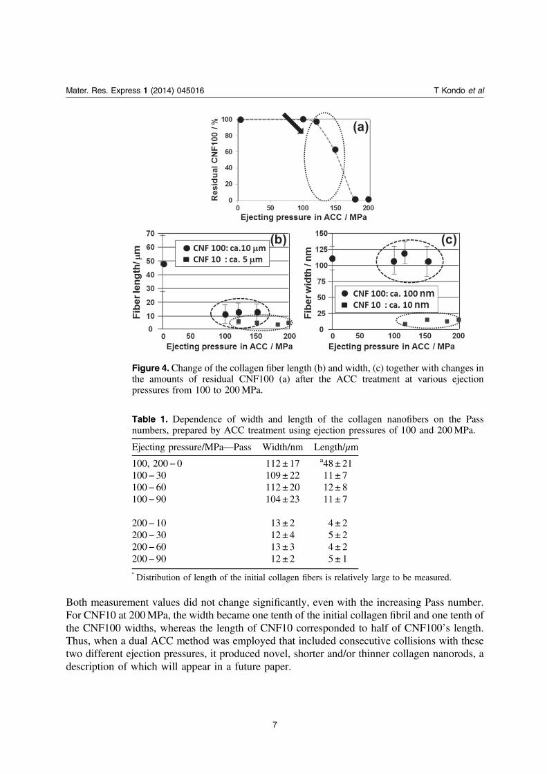
Both measurement values did not change significantly, even with the increasing Pass number.For CNF10 at 200MPa, the width became one tenth of the initial collagen fibril and one tenth ofthe CNF100 widths, whereas the length of CNF10 corresponded to half of CNF100’s length.Thus, when a dual ACC method was employed that included consecutive collisions with thesetwo different ejection pressures, it produced novel, shorter and/or thinner collagen nanorods, adescription of which will appear in a future paper.
Figure 4. Change of the collagen fiber length (b) and width, (c) together with changes inthe amounts of residual CNF100 (a) after the ACC treatment at various ejectionpressures from 100 to 200MPa.
Table 1. Dependence of width and length of the collagen nanofibers on the Passnumbers, prepared by ACC treatment using ejection pressures of 100 and 200MPa.
Ejecting pressure/MPa—Pass Width/nm Length/μm
100, 200− 0 112 ± 17 a48 ± 21100− 30 109 ± 22 11 ± 7100− 60 112 ± 20 12 ± 8100− 90 104 ± 23 11 ± 7
200− 10 13 ± 2 4 ± 2200− 30 12 ± 4 5 ± 2200− 60 13 ± 3 4 ± 2200− 90 12 ± 2 5 ± 1a
Distribution of length of the initial collagen fibers is relatively large to be measured.
7
Mater. Res. Express 1 (2014) 045016 T Kondo et al
3.2. Helicity of collagen molecules after ACC treatment
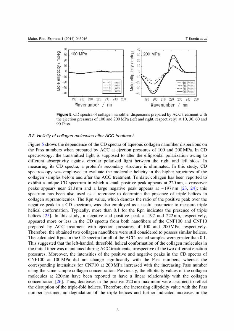
Figure 5 shows the dependence of the CD spectra of aqueous collagen nanofiber dispersions onthe Pass numbers when prepared by ACC at ejection pressures of 100 and 200MPa. In CDspectroscopy, the transmitted light is supposed to alter the ellipsoidal polarization owing todifferent absorptivity against circular polarized light between the right and left sides. Inmeasuring its CD spectra, a protein’s secondary structure is eliminated. In this study, CDspectroscopy was employed to evaluate the molecular helicity in the higher structures of thecollagen samples before and after the ACC treatment. To date, collagen has been reported toexhibit a unique CD spectrum in which a small positive peak appears at 220 nm, a crossoverpeaks appears near 213 nm and a large negative peak appears at ∼197 nm [23, 24]; thisspectrum has been also used as a reference to determine the presence of triple helices incollagen supramolecules. The Rpn value, which denotes the ratio of the positive peak over thenegative peak in a CD spectrum, was also employed as a useful parameter to measure triplehelical conformation. Typically, more than 0.1 for the Rpn indicates the presence of triplehelices [25]. In this study, a negative and positive peak at 197 and 222 nm, respectively,appeared more or less in the CD spectra from both nanofibers of the CNF100 and CNF10prepared by ACC treatment with ejection pressures of 100 and 200MPa, respectively.Therefore, the obtained two collagen nanofibers were still considered to possess similar helices.The calculated Rpns in the CD spectra for all of the ACC-treated samples were greater than 0.1.This suggested that the left-handed, threefold, helical conformation of the collagen molecules inthe initial fiber was maintained during ACC treatments, irrespective of the two different ejectionpressures. Moreover, the intensities of the positive and negative peaks in the CD spectra ofCNF100 at 100MPa did not change significantly with the Pass numbers, whereas thecorresponding intensities for CNF10 at 200MPa increased with the increasing Pass numberusing the same sample collagen concentration. Previously, the ellipticity values of the collagenmolecules at 220 nm have been reported to have a linear relationship with the collagenconcentration [26]. Thus, decreases in the positive 220 nm maximum were assumed to reflectthe disruption of the triple-fold helices. Therefore, the increasing ellipticity value with the Passnumber assumed no degradation of the triple helices and further indicated increases in the
Figure 5. CD spectra of collagen nanofiber dispersions prepared by ACC treatment withthe ejection pressures of 100 and 200MPa (left and right, respectively) at 10, 30, 60 and90 Pass.
8
Mater. Res. Express 1 (2014) 045016 T Kondo et al
collagen supramolecules or in similar molecular structures as a result of the ACC method. Thiswas suggested the conclusion that ACC treatment peeled off not only CNF10 having 10 nm inwidth from CNF100 surfaces but also collagen supramolecules, to some extent, into theresulting aqueous suspension with increasing in Pass number. In contrast, an ACC ejectionpressure of 100MPa was not sufficient to peel off the collagen supramolecules, but it couldcleave intermolecular interactions in the lateral direction of the fibrils.
3.3. Pulverizing behavior on a collagen fiber by ACC treatments
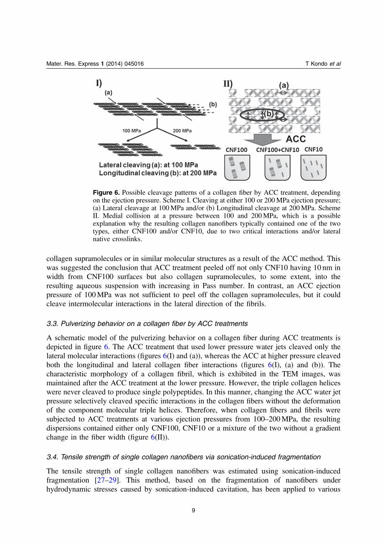
A schematic model of the pulverizing behavior on a collagen fiber during ACC treatments isdepicted in figure 6. The ACC treatment that used lower pressure water jets cleaved only thelateral molecular interactions (figures 6(I) and (a)), whereas the ACC at higher pressure cleavedboth the longitudinal and lateral collagen fiber interactions (figures 6(I), (a) and (b)). Thecharacteristic morphology of a collagen fibril, which is exhibited in the TEM images, wasmaintained after the ACC treatment at the lower pressure. However, the triple collagen heliceswere never cleaved to produce single polypeptides. In this manner, changing the ACC water jetpressure selectively cleaved specific interactions in the collagen fibers without the deformationof the component molecular triple helices. Therefore, when collagen fibers and fibrils weresubjected to ACC treatments at various ejection pressures from 100–200MPa, the resultingdispersions contained either only CNF100, CNF10 or a mixture of the two without a gradientchange in the fiber width (figure 6(II)).
3.4. Tensile strength of single collagen nanofibers via sonication-induced fragmentation
The tensile strength of single collagen nanofibers was estimated using sonication-inducedfragmentation [27–29]. This method, based on the fragmentation of nanofibers underhydrodynamic stresses caused by sonication-induced cavitation, has been applied to various
Figure 6. Possible cleavage patterns of a collagen fiber by ACC treatment, dependingon the ejection pressure. Scheme I. Cleaving at either 100 or 200MPa ejection pressure;(a) Lateral cleavage at 100MPa and/or (b) Longitudinal cleavage at 200MPa. SchemeII. Medial collision at a pressure between 100 and 200MPa, which is a possibleexplanation why the resulting collagen nanofibers typically contained one of the twotypes, either CNF100 and/or CNF10, due to two critical interactions and/or lateralnative crosslinks.
9
Mater. Res. Express 1 (2014) 045016 T Kondo et al
nanofibers, including carbon nanotubes, protein fibers and metal nanowires in aqueous systems[29]. In this method, the implosion dynamics of cavitation bubbles were induced by sonic wavepropagation followed by radial solvent flow into cavity bubble centers. Then, the fiber aroundthe cavity bubble is pulled into the bubble center, resulting in breakage from the tensile stress inthe fiber. Tensile stress decreases as filaments become shorter, such that the tensile stress is nolonger great enough to break the fiber. With prolonged sonication treatment, the fiber lengthswere found to approach an almost constant value, which was concluded to be the limiting length(Llim). Then, the tensile strength, which depends on the aspect ratio of the fragmentednanofibers, was estimated from the Llim, to be described later.
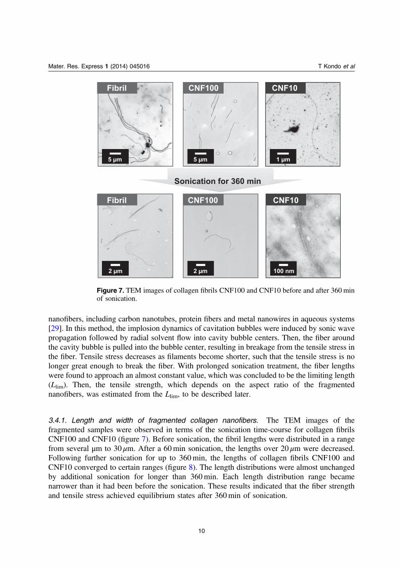
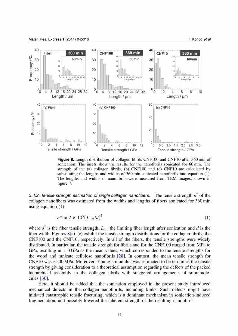
3.4.1. Length and width of fragmented collagen nanofibers. The TEM images of thefragmented samples were observed in terms of the sonication time-course for collagen fibrilsCNF100 and CNF10 (figure 7). Before sonication, the fibril lengths were distributed in a rangefrom several μm to 30 μm. After a 60min sonication, the lengths over 20 μm were decreased.Following further sonication for up to 360min, the lengths of collagen fibrils CNF100 andCNF10 converged to certain ranges (figure 8). The length distributions were almost unchangedby additional sonication for longer than 360min. Each length distribution range becamenarrower than it had been before the sonication. These results indicated that the fiber strengthand tensile stress achieved equilibrium states after 360min of sonication.
Sonication for 360 min
CNF10CNF100Fibril
CNF10CNF100Fibril
1 µm5 µm5 µm
100 nm2 µm2 µm
Figure 7. TEM images of collagen fibrils CNF100 and CNF10 before and after 360minof sonication.
10
Mater. Res. Express 1 (2014) 045016 T Kondo et al
3.4.2. Tensile strength estimation of single collagen nanofibers. The tensile strength σ* of thecollagen nanofibers was estimated from the widths and lengths of fibers sonicated for 360minusing equation (1)
( )L d* 2 10 / , (1)5lim
2σ ≈ ×
where σ* is the fiber tensile strength, Llim the limiting fiber length after sonication and d is thefiber width. Figures 8(a)–(c) exhibit the tensile strength distributions for the collagen fibrils, theCNF100 and the CNF10, respectively. In all of the fibers, the tensile strengths were widelydistributed. In particular, the tensile strength for fibrils and for the CNF100 ranged from MPa toGPa, resulting in 1–3GPa as the mean values, which corresponded to the tensile strengths forthe wood and tunicate cellulose nanofibrils [28]. In contrast, the mean tensile strength forCNF10 was ∼200MPa. Moreover, Young’s modulus was estimated to be ten times the tensilestrength by giving consideration to a theoretical assumption regarding the defects of the packedhierarchical assembly in the collagen fibrils with staggered arrangements of supramole-cules [30].
Here, it should be added that the sonication employed in the present study introducedmechanical defects in the collagen nanofibrils, including kinks. Such defects might haveinitiated catastrophic tensile fracturing, which is a dominant mechanism in sonication-inducedfragmentation, and possibly lowered the inherent strength of the resulting nanofibrils.
Figure 8. Length distribution of collagen fibrils CNF100 and CNF10 after 360min ofsonication. The insets show the results for the nanofibrils sonicated for 60min. Thestrength of the (a) collagen fibrils, (b) CNF100 and (c) CNF10 are calculated bysubstituting the lengths and widths of 360min-sonicated nanofibrils into equation (1).The lengths and widths of nanofibrils were measured from TEM images, shown infigure 7.
11
Mater. Res. Express 1 (2014) 045016 T Kondo et al
3.4.3. Hierarchical structures of collagen fiber versus mechanical properties. As describedabove, the two types of collagen nanofibers prepared using ACC exhibited different tensilestrengths; the tensile strength of CNF100 was about ten times higher than CNF10, indicatingthat greater numbers or amounts of lateral intermolecular interactions, including nativecrosslinks, could have contributed to the CNF100’s higher tensile strength. It might be that thepresence and amounts of the native crosslinks were critical factors for increasing tensilestrength. As collagen fibrils are stabilized by longitudinal and lateral intermolecular interactionsand crosslinks [31, 32], ACC treatment appeared capable of selectively cleaving intermolecularinteractions in collagen fibrils, depending on the water ejection pressures (figure 6). ACCtreatment at a 100MPa ejection pressure cleaved mostly longitudinal intermolecularinteractions in the collagen fibrils, whereas ACC treatment at 200MPa cleaved bothlongitudinal and lateral intermolecular interactions. The results obtained thus far in this studysuggested that the lateral intermolecular interactions, including the native crosslinks, could havebeen more strongly engaged than the intermolecular interactions in the longitudinal direction(figure 6(II)).
More specifically, the major intermolecular interactions and/or crosslinks that make upCNF10 were likely stronger since they are interactions that could not be cleaved by ACC.Namely, the collagen fibrils, the CNF100 and the CNF10 contained different intermolecularinteractions, including native crosslinks in the lateral direction, which resulted in differenttensile strengths. In a previous study, the reported tensile strength of the regenerated collagenfibrils from supramolecular solutions were lower than the results obtained in this study [33].This different result was presumably because such regenerated collagen fibrils possessed nocrosslinking, as they were already decomposed when the supramolecular solution was preparedfrom collagen fibrils. Conversely, the ACC method as a top-down process for native collagenfibers could not cleave the existing crosslinking, which thus persisted and resulted in tensilestrengths higher than those observed in the regenerated fibrils. The two types of collagennanofibers prepared using ACC exhibited different tensile strengths; the tensile strength ofCNF100 was about ten times higher than CNF10. This indicates that greater numbers oramounts of lateral intermolecular interactions, including native crosslinks, could havecontributed to the CNF100’s higher tensile strength. It might be that the presence and amountsof the native crosslinks were critical factors for increasing the tensile strength.
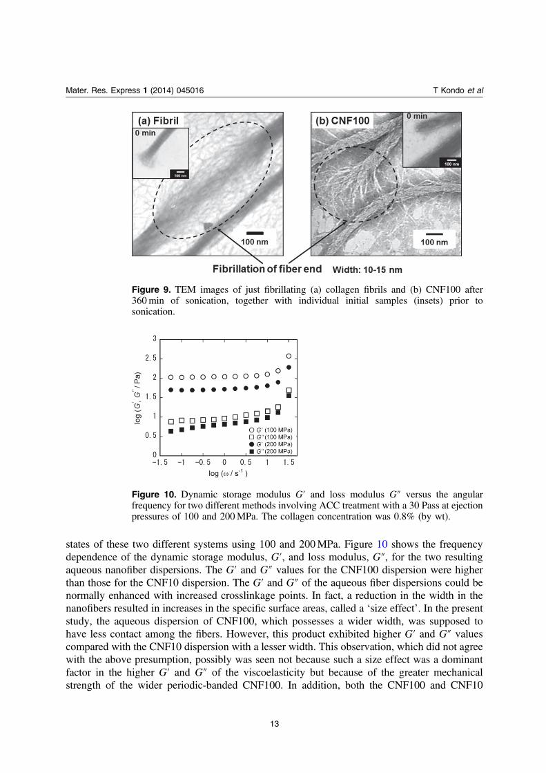
In fibrils and in CNF100 after 360min of sonication, the cleaving sites were found to befibrillated (figure 9). In the TEM images of these sites, thinner fibers that were ∼10 nm in widthand similar to CNF10 were observed. In other words, the fibrous morphology of CNF10remained after sonication. The result suggested the conclusion that some lateral intermolecularinteractions that make up the collagen fibrils and CNF100 were selectively cleaved bysonication. However, the lateral intermolecular interactions and/or native crosslinking that makeup CNF10 were strong enough to resist cleavage by sonication; thus, CNF10 might represent acritical minimum nanofibril unit.
3.5. Aqueous dispersion states of collagen nanofibers with different morphologies
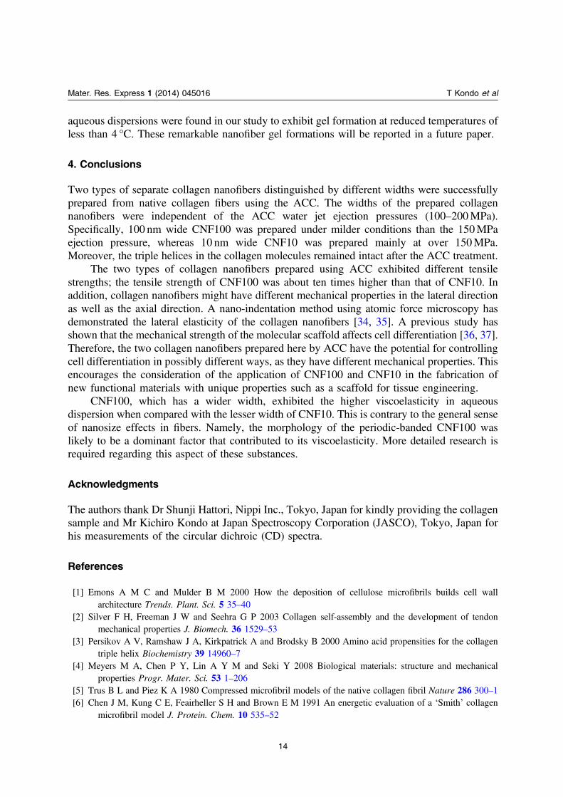
As described previously, the ACC treatment at 100MPa ejection pressure yielded shortnanofibers, called CNF100, that had initial widths of ∼100 nm as well as inherent periodicbanding; this was in contrast to CNF10, which had one tenth the width at 10 nm after the ACCtreatment at 200MPa. Viscoelastic measurements were performed to examine the dispersion
12
Mater. Res. Express 1 (2014) 045016 T Kondo et al
states of these two different systems using 100 and 200MPa. Figure 10 shows the frequencydependence of the dynamic storage modulus, G′, and loss modulus, G″, for the two resultingaqueous nanofiber dispersions. The G′ and G″ values for the CNF100 dispersion were higherthan those for the CNF10 dispersion. The G′ and G″ of the aqueous fiber dispersions could benormally enhanced with increased crosslinkage points. In fact, a reduction in the width in thenanofibers resulted in increases in the specific surface areas, called a ‘size effect’. In the presentstudy, the aqueous dispersion of CNF100, which possesses a wider width, was supposed tohave less contact among the fibers. However, this product exhibited higher G′ and G″ valuescompared with the CNF10 dispersion with a lesser width. This observation, which did not agreewith the above presumption, possibly was seen not because such a size effect was a dominantfactor in the higher G′ and G″ of the viscoelasticity but because of the greater mechanicalstrength of the wider periodic-banded CNF100. In addition, both the CNF100 and CNF10
Figure 9. TEM images of just fibrillating (a) collagen fibrils and (b) CNF100 after360min of sonication, together with individual initial samples (insets) prior tosonication.
Figure 10. Dynamic storage modulus G′ and loss modulus G″ versus the angularfrequency for two different methods involving ACC treatment with a 30 Pass at ejectionpressures of 100 and 200MPa. The collagen concentration was 0.8% (by wt).
13
Mater. Res. Express 1 (2014) 045016 T Kondo et al
aqueous dispersions were found in our study to exhibit gel formation at reduced temperatures ofless than 4 °C. These remarkable nanofiber gel formations will be reported in a future paper.
4. Conclusions
Two types of separate collagen nanofibers distinguished by different widths were successfullyprepared from native collagen fibers using the ACC. The widths of the prepared collagennanofibers were independent of the ACC water jet ejection pressures (100–200MPa).Specifically, 100 nm wide CNF100 was prepared under milder conditions than the 150MPaejection pressure, whereas 10 nm wide CNF10 was prepared mainly at over 150MPa.Moreover, the triple helices in the collagen molecules remained intact after the ACC treatment.
The two types of collagen nanofibers prepared using ACC exhibited different tensilestrengths; the tensile strength of CNF100 was about ten times higher than that of CNF10. Inaddition, collagen nanofibers might have different mechanical properties in the lateral directionas well as the axial direction. A nano-indentation method using atomic force microscopy hasdemonstrated the lateral elasticity of the collagen nanofibers [34, 35]. A previous study hasshown that the mechanical strength of the molecular scaffold affects cell differentiation [36, 37].Therefore, the two collagen nanofibers prepared here by ACC have the potential for controllingcell differentiation in possibly different ways, as they have different mechanical properties. Thisencourages the consideration of the application of CNF100 and CNF10 in the fabrication ofnew functional materials with unique properties such as a scaffold for tissue engineering.
CNF100, which has a wider width, exhibited the higher viscoelasticity in aqueousdispersion when compared with the lesser width of CNF10. This is contrary to the general senseof nanosize effects in fibers. Namely, the morphology of the periodic-banded CNF100 waslikely to be a dominant factor that contributed to its viscoelasticity. More detailed research isrequired regarding this aspect of these substances.
Acknowledgments
The authors thank Dr Shunji Hattori, Nippi Inc., Tokyo, Japan for kindly providing the collagensample and Mr Kichiro Kondo at Japan Spectroscopy Corporation (JASCO), Tokyo, Japan forhis measurements of the circular dichroic (CD) spectra.
References
[1] Emons A M C and Mulder B M 2000 How the deposition of cellulose microfibrils builds cell wallarchitecture Trends. Plant. Sci. 5 35–40
[2] Silver F H, Freeman J W and Seehra G P 2003 Collagen self-assembly and the development of tendonmechanical properties J. Biomech. 36 1529–53
[3] Persikov A V, Ramshaw J A, Kirkpatrick A and Brodsky B 2000 Amino acid propensities for the collagentriple helix Biochemistry 39 14960–7
[4] Meyers M A, Chen P Y, Lin A Y M and Seki Y 2008 Biological materials: structure and mechanicalproperties Progr. Mater. Sci. 53 1–206
[5] Trus B L and Piez K A 1980 Compressed microfibril models of the native collagen fibril Nature 286 300–1[6] Chen J M, Kung C E, Feairheller S H and Brown E M 1991 An energetic evaluation of a ‘Smith’ collagen
microfibril model J. Protein. Chem. 10 535–52
14
Mater. Res. Express 1 (2014) 045016 T Kondo et al
[7] Bailey A J, Robins S P and Balian G 1974 Biological significance of the intermolecular crosslinks of collagenNature 251 105–9
[8] Kadler K E, Holmes D F, Trotter J A and Chapman J A 1996 Collagen fibril formation Biochem. J. 316 1–11[9] Schmitt F O, Hall C E and Jakus M A 1942 Electron microscope investigation of the structure of collagen
J. Cell. Comp. Physiol. 20 11–33[10] Petruska J A and Hodge A J 1964 A subunit model for the tropocollagen macromolecule Proc. Natl. Acad.
Sci. USA 51 871–6[11] Christiansen D L, Huang E K and Silver F H 2000 Assembly of type I collagen: fusion of fibril subunits and
the influence of fibril diameter on mechanical properties Matrix. Biol. 19 409–20[12] Roeder B A, Kokini K, Sturgis J E, Robinson J P and Voytik-Harbin S L 2002 Tensile mechanical properties
of three-dimensional type I collagen extracellular matrices with varied microstructure J. Biomech. Eng. 124214–22
[13] Kessler A, Rosen H and Levenson S M 1960 Chromatographic fractionation of acetic acid-solubilized rat tailtendon collagen J. Biol. Chem. 235 989–94
[14] Yoshimura K, Terashima M, Hozan D and Shirai K 2000 Preparation and dynamic viscoelasticitycharacterization of alkali-solubilized collagen from shark skin J. Agric. Food Chem. 48 685–90
[15] Flory P J and Garrett R R 1958 Phase transitions in collagen and gelatin systems J. Am. Chem. Soc. 804836–45
[16] Kondo T, Morita M, Hayakawa K and Onda Y 2008 Wet pulverizing of polysaccharide US Patent 7,357,339[17] Kondo T, Kose R, Naito H and Kasai W 2014 Aqueous counter collision using paired water jets as a novel
means of preparing bio-nanofibers Carbohydr. Polym. 112 284–90[18] Kose R, Mitani I, Kasai W and Kondo T 2011 ‘Nanocellulose’ as a single nanofiber prepared from pellicle
secreted by Gluconacetobacter xylinus using aqueous counter collision Biomacromolecules 12 716–20[19] Tsuboi K, Yokota S and Kondo T 2014 Difference between bamboo- and wood-derived cellulose nanofibers
prepared by the aqueous counter collision method Nord. Pulp paper Res. J. 29 69–76[20] Kose R and Kondo T 2013 Size effects of cellulose nanofibers for enhancing the crystallization of poly(lactic
acid) J. Appl. Polym. Sci. 128 1200–5[21] Kawano Y and Kondo T 2014 Preparation of aqueous carbon material suspensions by aqueous counter
collision Chem. Lett. 43 483–5[22] Zeng S, Zhang C, Lin H, Yang P, Hong P and Jiang Z 2009 Isolation and characterisation of acid-solubilised
collagen from the skin of Nile tilapia (Oreochromis niloticus) Food Chem. 116 879–83[23] Brown F R III, Carver J P and Blout E R 1969 Low temperature circular dichroism of poly(glycyl-L-prolyl-L-
alanine) J. Mol. Biol. 39 307–13[24] Brown F R III, di Corate A, Lorenzi G P and Blout E R 1972 Synthesis and structural studies of two collagen
analogues: Poly(L-prolyl-L-seryl-glycyl) and poly(L-prolyl-L-alanyl-glycyl) J. Mol. Biol. 63 85–99[25] Kwak J, Capua A D, Locardi E and Goodman M 2002 TREN (Tris(2-aminoethyl)amine): an effective
scaffold for the assembly of triple helical collagen mimetic structures J. Am. Chem. Soc. 124 14085–91[26] Chu F H and Lukton A 1974 Collagenase induced changes in the circular dichroism spectrum of collagen
Biopolymers 13 1427–34[27] Ahir S V, Huang Y Y and Terentjev E M 2008 Polymers with aligned carbon nanotubes: active composite
materials Polymer 49 3841–54[28] Saito T, Kuramae R, Wohlert J, Berglund L A and Isogai A 2013 An ultrastrong nanofibrillar biomaterial: the
strength of single cellulose nanofibrils revealed via sonication-induced fragmentation Biomacromolecules14 248–53
[29] Huang Y Y, Knowles T P J and Terentjev E M 2009 Strength of nanotubes, filaments, and nanowires fromsonication-induced scission Adv. Mater. 21 3945–8
[30] Young R J and Lovell P A 1981 Introduction to polymers 2nd edn (Boca Raton FL: CRC Press) pp 319–21[31] Kang A H and Gross J 1970 Relationship between the intra and intermolecular cross-links of collagen Proc.
Natl. Acad. Sci. USA 67 1307–14
15
Mater. Res. Express 1 (2014) 045016 T Kondo et al
[32] Streeter I and de Leeuw N H 2011 A molecular dynamics study of the interprotein interactions in collagenfibrils Soft Matter 7 3373–82
[33] van der Rijt J A, van der Welf K O, Bennink M L, Dijkstra P J and Feijen J 2007 Micromechanical testing ofindividual collagen fibrils Macromol. Biosci. 18 697–702
[34] Wenger M P E, Bozec L, Horton M A and Mesquida P 2007 Mechanical properties of collagen fibrilsBiophys. J. 93 1255–63
[35] Strasser S, Zink A, Janko M, Heckl W M and Thalhammer S 2007 Structural investigations on nativecollagen type I fibrils using AFM Biochem. Biophys. Res. Commun. 354 27–32
[36] Engler A J, Sen S, Sweeney H L and Discher D E 2006 Matrix elasticity directs stem cell lineage specificationCell 126 677–89
[37] McDaniel D P, Shaw G A, Elliott J T, Bhadriraju K, Meuse C, Chung K H and Plant A L 2007 The stiffnessof collagen fibrils influences vascular smooth muscle cell phenotype Biophys. J. 92 1759–69
16
Mater. Res. Express 1 (2014) 045016 T Kondo et al