Embed Size (px)
Citation preview

THE JOURNAL OF EXPERIMENTAL ZOOLOGY 274~248-254 (1996)
Premature Death (p) Mutation of Ambystoma mexicanum Affects the Ability of Ectoderm To Respond to Neural Induction
ANN C. GRAVESON AND JOHN B. ARMSTRONG Department of Biology, Dalhousie University, Halifax, Nova Scotia, B3H 4J1 (A.C.G.) and Department of Biology, University of Ottawa, Ottawa, Ontario, K1N 6N5 (J.B.A.), Canada
ABSTRACT The premature death (p) mutation of the axolotl prevents the differentiation of cartilage from cranial neural crest, and likely affects other subpopulations of the neural crest as well. The mutant is, therefore? a useful model for understanding the process of specification of the neural crest. To test whether the defect can be attributed to a failure of neural induction, dorsal lip implants were performed between pIp and wild-type embryos. Dorsal lips from mutant em- bryos were found to induce secondary neural structures as frequently as wild-type tissue, and the secondary neural folds developed cartilage in explant culture. Wild-type dorsal lips were also able to induce secondary neural structures in pIp recipients, but these secondary folds were unable to form cartilage in culture. We therefore conclude that the p gene affects the ability of the ectoderm to respond to the inductive signal(s) provided by the mesoderm. 0 1996 Wiley-Liss, Inc.
The premature death ( p ) mutation of Am- bystoma mexicanum is one of a very limited num- ber of mutations that perturb the development of a subset of neural crest derivatives not including the pigment cells (Hall and Horstadius, '88). In affected embryos, the most obvious defects are the following: The primary gill filaments remain short and never develop secondary filaments; the mor- phology of the pharynx (particularly the branchial pouches) is abnormal; the anterior portions of the heart contain a plug of undifferentiated cells, and circulation is never established (Trottier and Armstrong, '77).
The branchial arch structures are derived from ectoderm, mesoderm, and endoderm, and also neu- ral crest. However, when the p mutation was first examined, the contribution of neural crest cells to the heart had not been discovered. Based on parabiosis and transplantation experiments? Armstrong and coworkers (Trottier and Arm- strong, '77; Mes-Hartree and Armstrong, '80) con- cluded that the pharyngeal endoderm was the most likely focus of the mutation. However, when we later tested some of these tissues in vitro, to isolate them from the deleterious influence of the degenerating mutant embryos, we were able to show that mutant pharyngeal endoderm can in- duce both heart-forming mesoderm and cranial neural crest-derived cartilage (Graveson and Armstrong, '90). We also demonstrated that mu- @ 1996 WILEY-LISS, INC,
tant mesoderm could form functional myocardial tissue in vitro. Thus, at lease some functions of the endoderm and mesoderm are unaffected by the p gene.
In 1983, Kirby and Stewart described the "car- diac neural crest," which contributes to portions of the heart. Our attention? therefore, turned to the neural crest as the possible focus of the mu- tation. We found that the migration patterns of mutant neural crest cells are normal, at least at the gross level (Graveson and Armstrong, '94). Pig- ment cell differentiation also appears to be nor- mal. However, the differentiative capacity of the chondrogenic neural crest is severely deficient (Graveson and Armstrong, '90). Transplantation and extirpation experiments suggest that the af- fected sub-population also includes the neural crest cells involved in the morphogenesis of the gills, and in the establishment of a functional cir- culatory system (Graveson and Armstrong, '94).
Although we have established that pIp cranial neural crest is unable to form cartilage in response to normal inductive signals from the endoderm (Graveson and Armstrong, 'go), we do not know why the mutant neural crest lacks this capacity
Received April 5 , 1995; revision accepted January 4, 1996. Address reprint requests to Dr. John B. Armstrong, Department of
Biolom, University of Ottawa, Ottawa, Ontario K1N 6N5, Canada.

NEURAL CREST SPECIFICATION IN p-MUTANT AXOLOTLS 249
The neural crest is, itself, induced early in devel- opment - although whether this is a part of neu- ral induction or a separate, subsequent, induction is still the subject of debate (see Discussion, and Graveson, ’93). However, even if we do not know the exact mechanism of neural crest induction, we can still ask whether the abnormal crest in the p mutant results from an abnormal inductive signal, or an abnormal response of the ectoderm to that signal.
A well-established procedure for inducing sec- ondary neural structures, including secondary folds, is the implantation of tissue from the dor- sal lip of the blastopore into the blastocoele of another embryo (the “Einsteck” method; see Hamburger, ’88, p. 5 5 ) . In this paper we show that secondary neural folds produced by im- planting wild-type dorsal lips in a pIp mutant em- bryo are still without chondrogenic potential. However, in the complementary experiment, where presumptive’ p/p dorsal lips were implanted in wild-type recipients, the secondary folds appear to have normal chondrogenic potential. We con- clude, therefore, that the defect in specification does not result from an inability of the mutant to produce a normal inductive signal.
MATERIALS AND METHODS Animals
Ambystoma mexicanum embryos were obtained from spawnings of animals raised and bred at the University of Ottawa Axolotl Colony. The condi- tions for maintenance and spawning of the ani- mals have been described previously (Armstrong and Duhon, ’89; Armstrong et al., ’89).
The embryos were kept in 25% Holtfreter’s so- lution at 18-20°C until they were used. (See Asashima et al., ’89, for the composition of all me- dia.) Developmental stages were determined ac- cording to the normal tables and descriptions of Bordzilovskaya et al. (’89).
Dorsal lip implantations Operations were performed when both donor
and host were at stage 9. The embryos were manu- ally dejelled using sharpened watchmakers’ for- ceps, passed through a series of sterile 100% Steinberg’s solution rinses, and placed in sterile 100% Steinberg‘s solution, supplemented with 50
‘Dorsal lips must be removed from donors long before their pheno- type can be determined. Therefore, we either have to resort to statis- tical arguments or remove only a portion of the dorsal lip and hope that the donor survives long enough to be identified.
pg/ml gentamicin sulphate, in plastic Petri dishes lined with 1% Noble agar. Removal of the vitelline membranes was facilitated by puncturing the cen- ter of the animal cap.
Dorsal lip material from the donor was im- planted into the host by sliding it into the blasto- coele, via the hole in the blastocoele roof, toward the side opposite the host’s dorsal lip. The size of the hole was then minimized by gently pushing the edges together with hair loops. Host embryos, and in some cases donor embryos, were left un- disturbed for at least 24 h. Secondary neural folds were removed from the host embryos for explant culture, after which the embryos were transferred to agar-lined 24-well tissue culture plates contain- ing 100% Steinberg’s solution. These embryos were maintained until after stage 37, when their phenotypes could be determined.
Explantation and culture The host’s secondary neural folds were removed
at about stage 17, which is before the primary neural plate forms a tube, and well before neural crest cell migration begins (at stg. 23; C. Nolte, personal communication). In this way, contami- nation of the secondary tissue by primary neural crest cells was avoided. The secondary neural folds were combined in explant culture with inductive pharyngeal endoderm from wild-type, stage 17 neurulae. The procedures for in vitro differentia- tion of cartilage from cranial neural crest have been described previously (Graveson and Arm- strong, ’87). Briefly, the desired tissues were dis- sected from the embryos using hair loops and electrolytically sharpened tungsten needles and were allowed to heal together before being trans- ferred to agar-lined BEEM capsules containing ap- proximately 150 pl of 100% Steinberg’s solution. Explants were cultured for 14 days prior to pro- cessing for light microscopy
Histology Explants and embryos were fixed with 2% glut-
araldehyde, 0.5% cetylpyridinium chloride, 0.5% polyvinylpyrrolidone in 0.1 M cacodylate buffer, pH 7.7, rinsed in cacodylate buffer, and dehy- drated in a graded ethanol series. Material to be sectioned was embedded in glycol methacrylate (JB-4; SorvallKIupont Instruments or J.B. EM Services), and 4-pm sections were prepared with a Sorvall JB-4 microtome. Sections were stained in 0.5% toluidine blue in 0.1 M benzoate buffer, pH 4.4, rinsed in benzoate buffer, dipped in ac- etone, and air dried.
250 A.C. GRAVESON AND J.B. ARMSTRONG
RESULTS We first tested the ability of mutant ectoderm
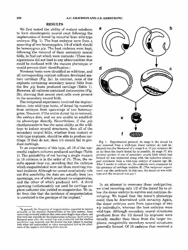
to form chondrogenic neural crest following the implantation of dorsal lip material from wild-type embryos (Fig. 1). The host embryos were from a spawning of two heterozygotes, 1/4 of which should be homozygous plp . The host embryos were kept, following the removal of their secondary neural folds, to find out which were mutants. (These ma- nipulations did not lead to any abnormalities that could be confused with the mutant phenotype or would prevent their identification.)
Nineteen hosts were identified as wild-type, and all corresponding explant cultures developed ma- ture cartilage (Fig. 2a). In contrast, none of the explants containing secondary neural folds from the five p/p hosts produced cartilage (Table 1). However, all cultures contained melanocytes (Fig. 2b), showing that neural crest cells were present in the secondary neural folds.
The reciprocal experiment involved the implan- tation, into wild-type hosts, of dorsal lip material from embryos from spawnings of two heterozy- gotes. However, if the entire dorsal lip is removed, the embryo dies, and we are unable to establish its phenotype directly. Nevertheless, if the p / p chordamesoderm has the same ability as the wild- type to induce neural structures, then all of the secondary neural folds, whether from mutant or wild-type implants, should be able to produce car- tilage. If they do not, then 1/4 should fail to pro- duce cartilage.
In an experiment of this type, all 16 of the suc- cessful explant cultures produced cartilage (Table 1). The probability of not having a single mutant in 16 embryos is in the order of 1%. Thus, the re- sults appear clear cut, providing that the embryos which exogastrulated were not those receiving mu- tant implants. Although we cannot conclusively rule out this possibility, the data are actually from two spawnings, one of which produced no exogastrulae, and the other of which produced 44%. A third spawning (unfortunately not used for cartilage ex- plant cultures) also yielded no exogastrulae. We in- fer from this that the incidence of exogastrulation is unrelated to the genotype of the implant.'
'In general, the frequency of exogastrulation appeared to depend primarily on the physical characteristics of the host embryo. Some spawnings produced embryos that were more fragile than others and that were less suitable for the implantation technique. Such embryos flattened more after the animal cap was punctured and the vitelline membrane was removed, and the animal cap was subject to tearing, both when left undisturbed, or when manipulated during the place- ment of the implant and closing of the wound.
f
Fig. 1. Experimental protocol. At stage 9, the dorsal lip was removed from a wild-type donor embryo (a) and im- planted into the blastocoel of a stage 9 or 10 pIp recipient (b) as far from the host's dorsal lip as possible. At stage 17, the anterior portion of one of secondary neural folds which had formed ( c ) was explanted along with the inductive pharyn- geal endoderm from a wild-type embryo of similar age (d). After 2 weeks in culture (el, the explants were examined for the presence of cartilage nodules (0. [The reciprocal experi- ment was also performed. In this case, the donor (a) was wild- type and the recipient was plp.1
In an attempt to overcome these ambiguities, we tried removing only 1/2 of the dorsal lip to al- low the donor embryo to survive and continue de- veloping. We hoped that the donor's phenotype could then be determined with certainty. Again, the donor embryos were from spawnings of two +/p individuals, whereas the host embryos were wild-type. Although secondary neural structures produced from the 1/2 dorsal lip implants were usually smaller than those from the larger im- plants, recognizable secondary neural structures generally formed. Of 18 embryos that received a

NEURAL CREST SPECIFICATION IN p-MUTANT AXOLOTLS
Fig. 2. Secondary neural folds cultured with inductive endoderm for 14 days. c, carti- lage nodule; m, melanophore. a: Culture containing secondary folds produced in a wild- type host by a 1/2-dorsal-lip implant from a p/p donor embryo. b: Culture containing secondary folds produced in ap/p host by a wild-type dorsal lip implant. Bar = 200 pm.
251

252 A.C. GRAVESON AND J.B. ARMSTRONG
TABLE 1. Cartilage formation from secondary neural folds
2” fold exdants vielding
Donor’ Host Transplants Gastrulated normally Produced 2” folds Cartilage No cartilage
Experiments in which the entire dorsal lip was transplanted wt wt
25 24 wt PIP P? wt 25 18
19
5 18
19 0
0 5 162 0
Experiments in which ?h the dorsal lip was transplanted W t wt 10 9 7 7 0 PIP wt 8 8 8 8 0 ‘wt: wild-type; p?: embryos from a +Jp x +lp spawning. 2Two cultures died.
112 dorsal lip, ten of the donors were identified as wild type and eight as plp. Although the mutant frequency is significantly greater than the ex- pected 114 (P = ,041, all of the characteristics ofpl p embryos were evident. None of the remaining ten donor embryos, nor any of an additional six wild-type embryos that had this part of the dor- sal lip removed, exhibited any abnormalities. Fur- thermore, even if some donors are not plp, the probability that none are is even lower, and the secondary folds from all the hosts produced ma- ture cartilage when explanted with inductive pha- ryngeal endoderm (Table 1).
DISCUSSION One view of regional specification during
early amphibian development is that the em- bryo is progressively partitioned by a hierar- chical series of inductions. One of the earliest is that of the dorsal lip region - the Spemann organizer. The progression continues with the involution of the “organizer,” followed by its in- duction of the neurectoderm (for review, see Gil- bert and Saxen, ’93).
The neural crest may arise as a direct result of neural induction, or, indirectly, as the result of sub- sequent processes (Graveson, ’93). The direct mod- els include that of Raven and Kloos (’45, ’46). They proposed that the inductive influence of the “or- ganizer” is rather broad, with quantitative differ- ences in the signal emanating from the medial and lateral portions. According to this model, the response of the ectoderm would depend on the quantity of the signal. High levels would give rise to mid-neural plate and low levels to neural crest.
A second “direct” model is that of Nieuwkoop and Albers (Nieuwkoop, ’85; Nieuwkoop et al., ’85; Albers, ’87). They proposed that the organizer in- duces only mid-neural-plate ectoderm, and that
the induction then spreads homiogenetically through the ectoderm on either side. At the same time, the competence of the ectoderm to respond is gradually lost. Neural crest is produced when a lower level of responsiveness is reached.
The indirect models suggest that the neural crest forms as the result of an interaction between the neural plate and the non-neural ectoderm. Moury and Jacobson’s (’89) model requires only the presence of ectoderm next to neurectoderm, but Rollhauser-ter-Horst (’77a, b; ’79) has sug- gested that neural crest formation may require the presence of additional (unidentified) tissues.
One avenue to understanding induction and re- gional specification is the characterization of mu- tants defective in some aspect of the process. However, most mutations involving neural crest cells are ones altering pigmentation patterns. The significance of the p mutation is that a large sub- set of other neural crest derivatives may be af- fected, and that the affected cells are manifestly different from their wild-type counterparts by early neurula stages (Graveson and Armstrong, ’90, ’94). This suggest that the defect occurs early in neural crest ontogeny.
Despite the normal appearance of the neural folds and the lack of obvious neural defects (Trottier and Armstrong, ’771, the phenotype could conceivably result from a general failure in neu- ral induction. Gordon and Brodland (’87; see also Clausi and Brodland, ’93) have suggested that neural plate formation is a mechanical event re- sulting from the resolution of inherent instabili- ties of the cytoskeleton. Thus, morphogenesis may be a poor indicator of the success, or failure, of specification resulting from “classic7’ neural induc- tion. In the present study, we tested this possibil- ity by implanting a wild-type organizer in a mutant embryo and asking whether the second-

NEURAL CREST SPECIFICATION INp-MUTANT AXOLOTLS 253
ary neural folds that formed contained neural crest cells with chondrogenic ability. They did not. This strongly suggests that the role played by the organizer in the induction of neural derivatives is unaffected, and that the deficiency lies in in- terpreting the signal(s), or further downstream in the specification process.
If this hypothesis is correct, then pIp dorsal lips should be able to induce normal secondary struc- tures in a wild-type host. This experiment was also performed, and every time secondary folds were obtained, these folds had chondrogenic po- tential. However, the difficulty in unequivocally identifying the donor genotype clouds the inter- pretation somewhat. In the first experiment with possible mutant donors, the entire dorsal lip was taken, precluding identification of the donor’s phe- notype. Even though all the successful implants had “normal” secondary folds, a significant num- ber of the recipients exogastrulated. Although on balance we consider it unlikely, we cannot rule out the possibility that these were the embryos that received the mutant dorsal lips.
We therefore attempted to remove only 1/2 the dorsal lip to preserve the donors for later identi- fication of their phenotype. In this experiment, unlike the previous one, exogastrulation was not a problem, but a significantly higher proportion of the donors were identified as mutants than the expected 1/4. This raises the question of whether the removal of 1/2 the organizer could lead to a p / p phenocopy in a wild-type embryo. However, we could not duplicate this effect with known wild- type (+/+) embryos. We therefore have no good rea- son, other than their frequency, to question their identification of the mutant donors. Furthermore, the secondary folds induced in aZE the recipients had cartilage-forming potential, no matter whether the implant donor was identified as mutant or wild type. Thus, we feel that the two experiments, considered together, provide convincing evidence that pIp dor- sal lip material is able both to induce neural plate formation from competent ectoderm and to fulfill any other roles it may have in the specification of neural crest with chondrogenic potential.
The possibility remains that the focus of the mutation does not lie in the presumptive neural crest. If, as proposed in the “indirect” models, the neural crest is formed by an interaction between the neural plate and the adjacent ectoderm, then the presumptive neural crest cells may not be af- fected by the mutation directly, but may simply never receive the proper signals from the ecto- derm. Nevertheless, the target tissue of the p gene
is clearly the gastrula ectoderm. Although we do not know the full extent of the ectodermal abnor- malities in the p mutant, Smith et al. (’94) have recently reported that the neuromasts of the lat- eral-line system, which are derived from ectoder- ma1 placodes immediately adjacent to the neural folds, do not differentiate normally Other ecto- dermal derivatives may also be affected. Experi- ments now in progress will not only address the possibilities outlined above, but may also help re- solve the sequence of early interactions between the neural plate, neural crest, placodes, and epi- dermal ectoderm.
ACKNOWLEDGMENTS This work was supported by a grant from the
Natural Sciences and Engineering Research Coun- cil, Canada to J.B.A.
LITERATURE CITED Albers, B. (1987) Competence as the main factor determin-
ing the size of the neural plate. Dev. Growth Differ., 29535- 545.
Armstrong, J.B., and S.T. Duhon (1989) Induced spawnings, artificial insemination, and other genetic manipulations. In: Developmental Biology of the Axolotl. J.B. Armstrong and G.M. Malacinski, eds. Oxford University Press, New York,
Armstrong, J.B., S.T. Duhon, and G.M. Malacinski (1989) Raising the axolotl in captivity In: Developmental Biology of the Axolotl. J.B. Armstrong and G.M. Malacinski, eds. Oxford University Press, New York, pp. 220-227.
Asashima, M., G.M. Malacinski, and S.C. Smith (1989) Sur- gical manipulation of embryos. In: Developmental Biology of the Axolotl. J.B. Armstrong and G.M. Malacinski, eds. Oxford University Press, New York, pp. 255-263.
Bordzilovskaya, N.P., Dettlaff, T.A., S.T. Duhon, and G.M. Malacinski (1989) Developmental-stage series of axolotl embryos. In: Developmental Biology of the Axolotl. J.B. Armstrong and G.M. Malacinski, eds. Oxford University Press, New York, pp. 201-219.
Clausi, D.A., and G.W. Brodland (1993) Mechanical evalua- tion of theories of neurulation using computer simulations. Development, 118:1013-1023.
Gilbert, S.F., and L. Saxen (1993) Spemann’s organizer: mod- els and molecules. Mech. Dev., 41:73-89.
Gordon, R., and G.W. Brodland (1987) The cytoskeletal me- chanics of brain morphogenesis: cell state splitters and pri- mary neural induction. Cell Biophys., 11:177-237.
Graveson, A.C. (1993) Neural crest: contributions to the de- velopment of the vertebrate head. Am. Zool., 33:424433.
Graveson, A.C., and J.B. Armstrong (1987) Differentiation of cartilage from cranial neural crest in the axolotl (Ambystoma mexicanum). Differentiation, 35: 16-20.
Graveson, A.C., and J.B. Armstrong (1990) The premature death mutation of Ambystoma mexicanum affects a subpopu- lation of neural crest cells. Differentiation, 45:71-75.
Graveson, A.C., and J.B. Armstrong (1994) In vivo evidence tha t the premature death ( p ) mutation of Ambystoma mexicanum affects an early segregating subpopulation of neural crest cells. J. Exp. Zool., 269:327-335.
pp. 228-235.
254 A.C. GRAVESON AND J.B. ARMSTRONG
Hall, B.K., and S. Horstadius (1988) The Neural Crest. Ox- ford University Press, London.
Hamburger, V (1988) The Heritage of Experimental Embry- ology: Hans Spemann and the Organizer. Oxford Univer- sity Press, New York.
Kirby, M.L., and D.E. Stewart (1983) Neural crest origin of cardiac ganglion cells in the chick embryo: identification and extirpation. Dev. Biol., 97:433-443.
Mes-Hartree, M., and J.B. Armstrong (1980) Evidence that the premature death mutation (p) in the Mexican axolotl (Ambystoma mexicanurn) is not an autonomous cell lethal. J. Embryol. Exp. Morphol., 60:295-302.
Moury, J.D., and A.G. Jacobson (1989) Neural fold formation at newly created boundaries between neural plate and epi- dermis in the axolotl. Dev. Biol., 133:44-57.
Nieuwkoop, PD. (1985) Inductive interactions in early am- phibian development and their general nature. J. Embryol. Exp. Morphol. [Suppl.], 89:333-347.
Nieuwkoop, PD., A.G. Johnen, and B. Albers (1985) The Epi- genetic Nature of Early Chordate Development: Inductive Interaction and Competence. Cambridge University Press, Cambridge.
Raven, C.P., and J.K. Kloos (1945) Induction by medial and
lateral pieces of the archenteron roof, with special refer- ence to the determination of the neural crest. Acta Neerl. Morphol., 5:348-362.
Raven, C.P., and J.K. moos (1946) Induction by medial and lateral parts of the archenteron roof: Determination of the neural crest. In: Experimental Embryology in the Nether- lands, 1940-1945. M.W. Woerdeman and C.P. Raven, eds. Elsevier Publishing Go., New York.
Rollhauser-ter-Horst, J. (1977a) Artificial neural induction in Amphibia. I. Sandwich explants. Anat. Embryol. (Berl.),
Rollhauser-ter-Horst, J. (197713) Artificial neural induction in Amphibia. 11. Host embryos. Anat. Embryol. (Bed.),
Rollhauser-ter-Horst, J. (1979) Artificial neural crest forma- tion in Amphibia. Anat. Embryol. (Berl.), 157:113-120.
Smith, S.C., A.C. Graveson, and B.K. Hall (1994) Evidence for a developmental and evolutionary link between placodal ectoderm and neural crest. J. Exp. Zool., 270:292-301.
Trottier, T.M., and J.B. Armstrong (1977) Experimental stud- ies on a mutant gene (p) causing premature death of Am- bystorna mexicanurn embryos. J. Embryol. Exp. Morphol., 39:139-149.
151~309-316.
151 :317-324.