Embed Size (px)
Citation preview

Journal of Academia and Industrial Research (JAIR) Volume 3, Issue 12 May 2015 613
©Youth Education and Research Trust (YERT) jairjp.com Jagtap, 2015
ISSN: 2278-5213
Preliminary Phytochemical Screening and Antioxidant Activity of Rhizome Extracts of Kaempferia scaposa (Nimmo) Benth
Sanjay Jagtap
Dept. of Botany, Elphinstone College, Mumbai, India [email protected]; +91 9969421282
______________________________________________________________________________________________
Abstract Kaempferia scaposa is a perennial herb belonging to family Zingiberaceae, an important medicinal plant widely used in several indigenous medicinal formulations. In the present study, phytochemical and antioxidant activity of the rhizome extracts of K. scaposa were evaluated. Phytochemical screening indicated that rhizomes are rich in a variety of primary and secondary metabolites such as carbohydrates, alkaloids, vitamin C and E, flavonoids, phenols, glycosides, saponanins and minerals. Present investigation shows DPPH anti-scavenging activity in organic extracts such as methanol (93.12%), water (63.5%), chloroform (33.28%) and acetone (26.92%). This study highlights the phytochemical and ethnopharmacological significance of K. scaposa.
Keywords: Kaempferia scaposa, phytochemicals, antioxidant, flavonoid, ethnopharmacology.
Introduction Kaempferia scaposa is an erect perennial herb about 0.5 to 1.5 m tall with fleshy sweet scanted rhizome belonging to family Zingiberaceae, an important medicinal plant widely used in several indigenous medicinal formulations. Distributed widely in peninsular and extrapeninsular parts of India; generally found on floors of moist deciduous forests from 600-750 m; has been reported from Anamalai Hills-Tamil Nadu; Hassan and Canada districts-Karnataka; Ranchi-Orissa; Palmaujungke-Bihar; Kashia Hills-Assam; Dehradun-Haryana; Punjab and Bengal and Lonawala in Pune district of Maharashtra (Hooker, 1882; Babu, 1977; Mathew, 1981-1983; Ugemuge, 1986; Cooke, 1903; Duthie, 1911; Bamer, 1916; Fischer, 1921; Yadav and Sardesai, 2010). Kaempferia galanga L. rhizome is stomachic and anti-inflammatory, in the form of powder or ointment it is applied to wounds and bruises to reduce swellings. They improve complexion and cure burning sensations, mental disorders and insomnia. Decoction of rhizome is used for dyspepsia, head ache and malaria. Roasted rhizomes are applied hot in rheumatism and for hastening the ripening of inflammatory tumors. Leaves are used in lotions and poultices for sore eyes, sore throat, swellings, rheumatism and fevers. The tuber powdered and mixed with honey is prescribed for coughs, asthma and pectoral affections. Kaempferia rotunda L. is an erect herb with tuberous rhizomes. The whole plant is useful in the form of powder or ointment as an application to wounds and bruises to reduce swellings. The powder extracted from Kaempferia rotunda is made into ointment and is used for healing fresh wounds. It is taken internally to remove coagulated blood or purulent matter and is used in many ayurvedic preparations.
In Ayurveda, the important formulations using this herb are Cyavanaprasam, Asokariṣṭam, Baladhatryaditailam, Kalyanakaghritham, etc. (Sivrajan and Indira, 1994). The drug “Hallakam” prepared from this is in popular use in the form of powder or as an ointment application to wounds and bruises to reduce swellings. The tubers are useful in vitiated conditions of vata and kapha, gastropathy, dropsy, inflammations, wound, ulcers, blood clots, tumors and cancerous swellings (Warrier et al., 1995). Keeping the above facts in view, in the present study, a high-throughput micro-scaled method which enables digestion of small quantities of plant samples for subsequent elemental profiling by ICP-spectrometry, DPPH anti-scavenging activity and flavonoid analysis was investigated. The findings led to the identification of phytochemical contents of rhizome extracts of Kaempferia scaposa of indigenous origin. Materials and methods Sampling: Fresh samples of rhizome of Kaempferia scaposa were collected from Lonavala, Dist., Pune region of Western Ghats of Maharashtra in September 2012 (Fig. 1 and 2). These plants were identified and authenticated using herbarium collection at Dept. of Botany, DST-FIST School of Life Science, SRTM University, Nanded (MS) and Dept. of Botany, Dr. Babasaheb Ambedkar Marathwada, University, Aurangabad (MS), India. Fresh rhizomes were washed thoroughly under running tap water followed by sterile distilled water and dried under shade. The shade dried material was ground into coarse powder using mechanical grinder and this coarse powder was sieved by 1 mm pore size sieve. The powder was stored in airtight containers at room temperature till further phytochemical screening of secondary metabolites.
RESEARCH ARTICLE

Journal of Academia and Industrial Research (JAIR) Volume 3, Issue 12 May 2015 614
©Youth Education and Research Trust (YERT) jairjp.com Jagtap, 2015
Fig. 1. Habit of Kaempferia scaposa.
Fig. 2. Rhizome of Kaempferia scaposa.
Soxhlet extraction: Exhaustive Soxhlet extraction was performed using a classical Soxhlet apparatus with accurately weighed 10 g of the crude powder of plant material for 18-40 h. Extraction was performed with water, methanol, chloroform and acetone as the extracting solvent. The extraction was conducted for 6-8 h/d and finally all the extracts were evaporated under vacuum. The water, methanol, chloroform and acetone extracts of rhizomes of the plant were prepared according to standard methods (Harbone, 1998). These extracts were sealed in airtight containers and stored at -4C. Phytochemical screening: Phytochemical screening of active plant extracts was done by following the standard method of Khandelwal (2000), for the qualitative analysis of various phytochemicals such as alkaloids, carbohydrate, glycosides, saponanins, flavonoids and phenols which could be responsible for antioxidant activity.
Mineral analysis Micro-scaled digestion: CEM-MARS 6 microwave oven was used for micro-scaled digestion. About 0.5 g of herbal samples were weighed and transferred to CEM-Xpress vessels, 8-10 mL of conc. HNO3 was added to the samples. The samples were pre-digested for 10-15 min prior to capping the vessels. The CEM-Xpress vessels were assembled for microwave irradiation. The microwave program was adjusted with respect to the number of vessels and reference to the guidelines of CEM at 1000 W with 100% level, 25 min ramping period was used to reach the digestion temperature of 180C which there upon was maintained for 15 min. The CEM-Xpress vessels were kept in fume hood for cooling and to release the pressure by uncapping. The contents were transferred to 50 mL volumetric flasks and volume was made with distilled water. The solutions were filtered prior to use. For calibration, Leeman and Thomas Baker standard sample were used as the reference for the calibration range. The spray chamber, nebulizer and torch assembly was completely cleaned to eliminate contamination. The plasma was stabilized for 15 min by flushing with distilled water. An instrument calibration was performed to check the wavelength shift and the same was successful with a minimum deviation of <10% with master scan. After calibration, the instrument was optimized with 10 ppm solution containing elements (Table 1) and the same as optimized with maximum intensity and best BEC at the parameters mentioned in operating condition above. Diluted samples were used for further analysis by using Teledyne Leeman, ICP (Induction Coupled Plasma). Table 1. Instrumental characteristics and setting for ICP-OES:
Spectrometer Leeman Lab’s simultaneous ICP-OES Prodigy XP dual system.
Parameters range Actual
parameters Min Max Power 0.1 2.0 1.1 Kw Coolant flow 5 20 18 L/Min Auxiliary flow 0.0 2.0 0.2 L/M Nebulizer flow 5 60 34 psi Plasma torch - - Dual Spray chamber - - Cyclonic Nebulizer - - Concentric Sample aspiration rate 0.5 2.0 1.4 mL/min
Replicate read time - - 40 sec per replicate for axial
Determination of Vitamin E by HPLC: Standard preparation: dl -tocopherolacetate (96%), (Vitamin E) manufactured by Merck was used for calibration of standard curves. About 1 mg of dl -tocopherolacetate was dissolved in 1 mL in HPLC grade methanol. Dilutions of 100, 50, 25 and 10 μg/mL was prepared and the pre-treated sample extracts and stock solutions were filtered through 0.45 μm syringe filters.

Journal of Academia and Industrial Research (JAIR) Volume 3, Issue 12 May 2015 615
©Youth Education and Research Trust (YERT) jairjp.com Jagtap, 2015
Reverse phase HPLC method: The concentration of -tocopherol (Vitamin E) in the extracts was determined by Agilent Technologies 1200 series Quaternary system, equipped with auto sampler, quaternary pump, degasser, column oven and a DAD detector. The spectral data was collected at UV detection at 220 nm. The solvent system of acetonitrile and water (95:5) was used a gradient mobile phase on Agilent ZORBAX 300 SB column (4.6 × 150 mm × 5 μm) at a flow rate of 1.0 mL/min, 10 μL injection volume and detection was optimized at 220 nm with 15 min separation time. Vitamin C: About 0.25% ethanolic solution of DCPI (2,6-dichlorophenol-indophenol sodium salt) was prepared for the detection of Vitamin C. To 0.5 mL of sample extracts, 2 drops of DCPI indicator was added. The blue coloration changed to red confirmed the presence of vitamin C. The test was carried out for all the extracts (British Nutrition Foundation, 2004). Anti-scavenging activity: DPPH solution (0.1 mM) was prepared in methanol by dissolving 0.0394 g DPPH in 1000 mL methanol. The solution was kept in darkness for 30 min to complete the reaction. The free radicals scavenging activity of the crude extracts was determined by the 1,1-diphenyl-2-picryl-hydrazil (DPPH). Antioxidant activity was measured by the standard method described by Brand-Willium et al. (1995) wherein, the bleaching rate of stable free radical DPPH was monitored at a characteristic wavelength in the presence of the sample. In its radical form, DPPH absorbed at 570 nm, but upon reduction by an antioxidant or radical species its absorption decreased. The capability to scavenge the DPPH radical was calculated using the following equation: DPPH scavenging effect (%) = (ABScontrol-ABSsample)/(ABScontrol) X 100), whereas ABScontrol is absorbance of negative control and ABSsample is the absorbance of the reaction mixture containing the sample extract. Flavonoids analysis by HPTLC: The standards Quercetin, Kaemferol, Catechin gallate, Rutin hydrate and Hesperdin were procured from Sigma Aldrich USA. All the standard solutions were prepared in ethanol whereas hesperdin in water. Chromatography was performed on silica gel 60F254 (10 cm X 10 cm; 25 mm layer thickness; Merk) with aqueous, methanolic, chloroform and acetone extracts of Kaempferia scaposa rhizome.
The fraction residues were collected and (10 µL) subjected for HPTLC (CAMAG, Switzerland) analysis. The fractions were impregnated on silica gel 60F254 TLC plate. The plate was air-dried and then inserted in CAMAG-twin through lass chamber containing solvent system of composition with ethyl acetate, acetic acid, formic acid and water (100:11:11:27) as a gradient mobile phase for 20 min. The well eluted TLC plate was then dried at 105C for 15 min and scanned using Scanner 3 (CAMAG, Switzerland) at 254 and 366 nm using Win Cat 4 software. Results and discussion Optimization of extraction method: In order to extract the phytochemicals efficiently, variables involved in this procedure were optimized, including extraction solvent (Water, Methanol, Chloroform, Acetone, 100%), extraction method (Soxhlet, reflux, percolation) and extraction time (18-40 h). The extraction time in water was 40 h. The biomass was refluxed for 40 h and dried naturally for 2-3 d. To the dried biomass, 100% methanol was added and the reaction was percolated to extract phytochemicals. The methanolic fraction was collected in amber colored bottle under nitrogen atmosphere. The material was dried for 5-6 h. The procedure was repeated for chloroform and acetone. The extraction time was optimized for all the samples. All the extracts were preserved under nitrogen atmosphere in amber coloured bottles at -4oC. Phytochemical screening of the rhizome extracts of Kaempferia scaposa revealed the presence of different phytochemicals. Indeed, phytochemical investigations of the plant K. scaposa have resulted in occurrence of carbohydrates, alkaloids, glycosides, saponanins, flavonoids, tannins, phenols, vitamin E and C. Table 2 illustrates the results of phytochemical screening of all the extracts of K. scaposa. The qualitative analysis of carbohydrates (Benedict’s reagent test) and glycosides (Borntranger’s reagent) were carried out in all extracts i.e. aqueous (WE), methanol (ME), acetone (AE) and chloroform (CE) extracts of K. scaposa. The solutions turned red and pink confirmed the presence of carbohydrates and glycosides respectively. The hydrophilic carbohydrates and glycosides were present in water (WE) whereas and hydrophobic carbohydrates and glycosides were detected in rest of the organic solvents (AE-ME). The Mayer’s test of extracts ME displayed appearance of white turbidity for alkaloids.
Table 2. Preliminary phytochemical screening of rhizome extracts of Kaempferia scaposa. Extract Car Sta Prot Glyc Alk Sap Tan Flav Phe Vit. C Vit. E
WE +++ - - ++ - ++ - ++ + +++ - ME ++ ++ ++ + ++++ + PT ++ +++ ++ - - CE ++ - - + - - - + - - ++ AE ++ - - + - - - + - - -
Car = Carbohydrate, Sta = Starch, Prot = Protien, Glyc = Glycoside, Alk = Alkaloids, Sap = Saponanins, Tan = Tannin, Flav = Flavonoids, Phe = Phenols, Vit. C = Vitamin C, Vit. E = Vitamin E, WE = Water Extract, ME = Methanol Extract, CE = Chloroform Extract, AE = Acetone Extract, GT = Gallotannins, PT = Pseudotannins. +=Significant, ++=Moderate, +++=Very good.

Journal of Academia and Industrial Research (JAIR) Volume 3, Issue 12 May 2015 616
©Youth Education and Research Trust (YERT) jairjp.com Jagtap, 2015
The alkaloids were absent in WE, AE and CE extracts of K. scaposa. The dark brown coloration test for phenols was observed in WE-ME. No traces of phenols are found in AE-CE extracts. The extracts were shaken with distilled water. The persistence of froth in WE and ME was observed, indicated the presence of saponanins. The hydrophilic flavonoids were detected in extract WE. The water soluble vitamin C was found in Kaempferia scaposa. Vitamin E was qualitatively and quantitatively analyzed by HPLC in rhizome extracts ME, AE and CE of Kaempferia scaposa. Microwave digestion and mineral analysis Optimization and calibration of K. scaposa rhizome extracts: Iron and copper are of great importance for life. As redox-active metal, they are involved in photosynthesis, mitochondrial respiration, nitrogen assimilation and hormone biosynthesis. Manganese is essential for plant metabolism and development and occurs in oxidation states II, III, and IV in approximately 35 enzymes of a plant cell. Zinc is important as a component of enzymes for protein synthesis and energy production and maintains the structural integrity of bio-membranes. Most of the zinc enzymes are involved in regulation of DNA-transcription, RNA-processing and translation. Although the essentiality of Se to plants has not been established yet, Se is considered as a beneficial element in promoting plant growth in some plant species. The focal point of our study was to develop effective digestion method for the preparation of mineral analysis by ICP. Microwave digestion is a common technique used by elemental scientists to dissolve heavy metals in the presence of organic molecules prior to analysis by inductively coupled plasma, atomic absorption, or atomic emission measurements (Kingston and Lois, 1988). Quantitative multi-elemental analysis by inductively coupled plasma (ICP) spectrometry depends on complete digestion of solid samples. However, fast and thorough sample digestion is a challenging analytical task which constitutes a bottleneck in modern multi-elemental analysis. Additional obstacles may be that sample quantities are limited with low elemental concentrations. In such cases, digestion in small volumes with minimum dilution and contamination is required in order to obtain high accuracy data. After optimization, a new calibration method was created for measuring these samples; the wavelengths used for calibration were Cu 324.754 nm, Mn 257.610 nm, Se 196.090 nm, Fe 259.940 nm and Zn 213.856 nm (Table 3). Calibrated STD solutions were measured 3 times one by one with an RSD <1%. Once all the calibration standards are finished, a necessary background correction was applied for each wavelength. The results are depicted in Table 3. We have developed a micro-scaled microwave digestion procedure and optimized it for accurate elemental profiling of plant materials. A commercially available 40-position rotor with 5 mL Polytetraflouro ethylene (PTFE) vials, originally designed for microwave-based
parallel organic synthesis were used as a platform for the digestion. The novel micro-scaled method was successfully validated by the use of various certified reference materials (CRM). We have determined 5 elements in coarse power given in Table 3. Thereby, the concentration of minerals in plant extracts had the different profiles and quantitative differences had been detected. The most abundant microelement was Mn in Kaempferia scaposa whereas, Cu was found at the lowest concentration. The content of Mn was especially high in comparison to Fe, Cu, Zn and in consequence, Se was not detected. The concentration of Zn content was less abundant. Vitamin E and C, carotenoids, Se and other trace minerals are important antioxidant components of animal diets and their roles in animal health and immune function are indispensable. In addition, several metallo enzymes which include glutathione peroxidase (Se), catalase (Fe), and superoxide dismutase (Cu, Zn, and Mn) are also critical in protecting the internal cellular constituents from oxidative damage.
Fig. 3. Accuracy of elemental concentrations in Kaempferia scaposa after micro-scaled digestion expressed in ppm.
Determination of Vitamin E by HPLC: Vitamins are a diverse group of organic compounds essential in trace amounts for the normal growth and maintenance of life. To ensure the adequate intake of vitamins, the human diet can be completed with a high range of multivitamin tablets and food products enriched with vitamins, in other words, these compounds are usually administered as nutraceutical or functional ingredient. They are classified as either water-soluble or fat soluble. Vitamin E is fat-soluble whereas Vitamin C is water-soluble. Vitamin E is a generic term for tocopherols and tocotrienols and it is fat-soluble antioxidant that blocks the production of reactive oxygen species formed when lipids undergoes oxidation. We employed reverse phase HPLC-analytical tool for qualitative estimation of vitamin E, in which HPLC has been coupled with UV detector. Optimization of HPLC method: To meet the requirements for quantitative analysis, various HPLC parameters were examined, including different columns (Agilent SB-C18 length 250 and 150 mm, width 4.6, particle size 5 µm), column temp. (25C) and UV wavelength (220 nm).
25.8271 1.4318
432.2496
0
325.5662
Zn Cu Mn Se Fe
Con
cent
ratio
n (p
pm)

Journal of Academia and Industrial Research (JAIR) Volume 3, Issue 12 May 2015 617
©Youth Education and Research Trust (YERT) jairjp.com Jagtap, 2015
Fig. 4. Calibration curve of dl α-tocopherol acetate (96%) (Vitamin E).
The best chromatographic resolution was obtained on Agilent SB-C18 length 4.6 X 150 mm, 5 µm column at 25C. The UV detector was monitored at 200-380 nm for fingerprinting analysis because the peaks were observed under this wavelength. The high intense peak was observed at 220 nm. Method validation and calibration: A calibration curve is simply a graph where concentration is plotted along the x-axis and area is plotted along the y-axis (Response, absorbance, intensity, peak height, etc.). The line represents the calibration curve. Figure 4 showed a calibration curve of vitamin E. We have constructed a calibration curve for vitamin E. It was created by running 4 different calibration standards (10, 25, 50 and 100 g/mL). Each concentration gave a peak area (287.717, 761.253, 1594, 3023.3) respectively. Peak area was then plotted against the concentrations. The linear trend line has been drawn and linear regression equation has been calculated as y = mx + C. Whereas, y = Area under the peak or Response, m = Slope of the linear line (Constant), x = Concentration in mL and C = intercept (Constant). HPLC is most widely used technique to analyze tocols, and both normal-phase (NP) and reversed-phase (RP) chromatography are applied (Kamal-Eldin et al., 2000; Abidi, 2000; Ruperez et al., 2001). Vitamin E functions as a chain-breaking antioxidant, neutralizing free radicals and preventing oxidation of lipids within membranes (McDowell, 2000). The lipophilic vitamin E has been detected in the chloroform extract of Kaempferia scaposa. The organic rhizome extracts of K. scaposa displayed significant antioxidant activity, proposed that the concentration of vitamin E might be higher along with the other natural antioxidants. Quantitative estimation of lipophilic vitamin E in rhizome extracts of Kaempferia scaposa is depicted in Table 3. The chloroform extracts showed (1.7289 µg/mL) concentration of vitamin E.
Fig. 5. Vitamin E standard peak at 10 µL.
Fig. 6. Vitamin E standard peak at 25 µL.
Fig. 7. Chloroform extract contains Vitamin E.
Fig. 8. Methonolic extract contains Vitamin E.
Vitamin C: The hydrophilic vitamin C (L-Ascorbic acid or L-ascorbate), an essential nutrient for humans and other animal species have been detected in aqueous aqueous rhizome extract of Kaempferia scaposa. The vitamin C and β-carotene, which is the precursor for vitamin A, are important for absorption of some nutrients and eye vision. Anti-scavenging activity: Many reports are available on the protective effects of natural antioxidants against oxidative stress related disorders like ageing, degenerative diseases and cancer (Cozzi et al., 1997). The phenolic compounds may have a direct contribution in antioxidant activity (Bidchol et al., 2011).
0287.717
761.253
1594
3023.3y = 30.45x + 6.514
R² = 0.999
0
500
1000
1500
2000
2500
3000
3500
0 10 20 30 40 50 60 70 80 90 100 110
Res
pons
e (A
rea
unde
r the
pea
k)
Concentration (µg/mL)
Journal of Academia and Industrial Research (JAIR) Volume 3, Issue 12 May 2015 618
©Youth Education and Research Trust (YERT) jairjp.com Jagtap, 2015
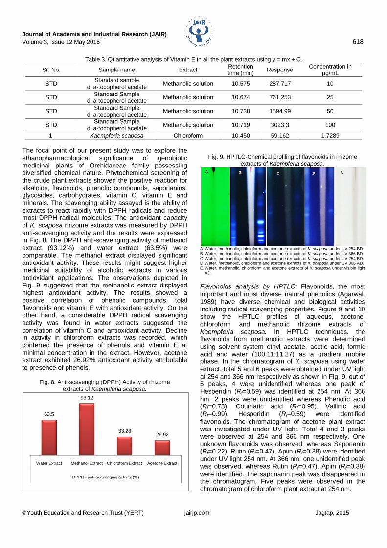
The focal point of our present study was to explore the ethanopharmacological significance of genobiotic medicinal plants of Orchidaceae family possessing diversified chemical nature. Phytochemical screening of the crude plant extracts showed the positive reaction for alkaloids, flavonoids, phenolic compounds, saponanins, glycosides, carbohydrates, vitamin C, vitamin E and minerals. The scavenging ability assayed is the ability of extracts to react rapidly with DPPH radicals and reduce most DPPH radical molecules. The antioxidant capacity of K. scaposa rhizome extracts was measured by DPPH anti-scavenging activity and the results were expressed in Fig. 8. The DPPH anti-scavenging activity of methanol extract (93.12%) and water extract (63.5%) were comparable. The methanol extract displayed significant antioxidant activity. These results might suggest higher medicinal suitability of alcoholic extracts in various antioxidant applications. The observations depicted in Fig. 9 suggested that the methanolic extract displayed highest antioxidant activity. The results showed a positive correlation of phenolic compounds, total flavonoids and vitamin E with antioxidant activity. On the other hand, a considerable DPPH radical scavenging activity was found in water extracts suggested the correlation of vitamin C and antioxidant activity. Decline in activity in chloroform extracts was recorded, which conferred the presence of phenols and vitamin E at minimal concentration in the extract. However, acetone extract exhibited 26.92% antioxidant activity attributable to presence of phenols.
Fig. 8. Anti-scavenging (DPPH) Activity of rhizome extracts of Kaempferia scaposa.
Fig. 9. HPTLC-Chemical profiling of flavonoids in rhizome extracts of Kaempferia scaposa.
A. Water, methanolic, chloroform and acetone extracts of K. scaposa under UV 254 BD. B. Water, methanolic, chloroform and acetone extracts of K. scaposa under UV 366 BD. C. Water, methanolic, chloroform and acetone extracts of K. scaposa under UV 254 BD. D. Water, methanolic, chloroform and acetone extracts of K. scaposa under UV 366 AD. E. Water, methanolic, chloroform and acetone extracts of K. scaposa under visible light
AD. Flavonoids analysis by HPTLC: Flavonoids, the most important and most diverse natural phenolics (Agarwal, 1989) have diverse chemical and biological activities including radical scavenging properties. Figure 9 and 10 show the HPTLC profiles of aqueous, acetone, chloroform and methanolic rhizome extracts of Kaempferia scaposa. In HPTLC techniques, the flavonoids from methanolic extracts were determined using solvent system ethyl acetate, acetic acid, formic acid and water (100:11:11:27) as a gradient mobile phase. In the chromatogram of K. scaposa using water extract, total 5 and 6 peaks were obtained under UV light at 254 and 366 nm respectively as shown in Fig. 9, out of 5 peaks, 4 were unidentified whereas one peak of Hesperidin (Rf=0.59) was identified at 254 nm. At 366 nm, 2 peaks were unidentified whereas Phenolic acid (Rf=0.73), Coumaric acid (Rf=0.95), Vallinic acid (Rf=0.99), Hesperidin (Rf=0.59) were identified flavonoids. The chromatogram of acetone plant extract was investigated under UV light. Total 4 and 3 peaks were observed at 254 and 366 nm respectively. One unknown flavonoids was observed, whereas Saponanin (Rf=0.22), Rutin (Rf=0.47), Apiin (Rf=0.38) were identified under UV light 254 nm. At 366 nm, one unidentified peak was observed, whereas Rutin (Rf=0.47), Apiin (Rf=0.38) were identified. The saponanin peak was disappeared in the chromatogram. Five peaks were observed in the chromatogram of chloroform plant extract at 254 nm.
63.5
93.12
33.2826.92
Water Extract Methanol Extract Chloroform Extract Acetone Extract
DPPH - anti-scavenging activity (%)
Table 3. Quantitative analysis of Vitamin E in all the plant extracts using y = mx + C.
Sr. No. Sample name Extract Retention time (min) Response Concentration in
µg/mL
STD Standard sample dl a-tocopherol acetate Methanolic solution 10.575 287.717 10
STD Standard Sample dl a-tocopherol acetate Methanolic solution 10.674 761.253 25
STD Standard Sample dl a-tocopherol acetate Methanolic solution 10.738 1594.99 50
STD Standard Sample dl a-tocopherol acetate Methanolic solution 10.719 3023.3 100
1 Kaempferia scaposa Chloroform 10.450 59.162 1.7289

Journal of Academia and Industrial Research (JAIR) Volume 3, Issue 12 May 2015 619
©Youth Education and Research Trust (YERT) jairjp.com Jagtap, 2015
Fig. 10. HPTLC peaks at 254 nm and 366 nm after
derivatization of rhizome extracts of K. scaposa.
After derivatization 254 nm After derivatization 366 nm
M=Methanol, C=Chloroform, A=Acetone, W=Water.
Fig. 11. HPTLC peaks of standard and rhizome
extracts of K. scaposa.
All peaks at 254 AD (Q-STD Quercetin, S1-Water, S2-Acetone, S3-Chloroform and S4-Methanol). Among 5 peaks, 3 peaks were unidentified and peaks of Kaempferol (Rf=0.82), Coumaric acid (Rf=0.93) were located in the chromatogram. At 366 nm, 6 peaks were identified, out of 6 peaks, 2 were unidentified and peaks of saponanin acid (Rf=0.21), Hesperidin (Rf=0.55), Kaempferol (Rf=0.82), Coumeric acid (Rf=0.92) were identified as flavonoids. In methanolic plant extract, the chromatogram displayed 3 peaks of unidentified flavonoids at 254 nm.
Table 4. Chemical profiling of rhizome extracts of K. scaposa at 254 nm and 366 nm after derivatization (AD).
Plant extract 254 nm AD 366 nm AD
Rf value Height (mm)
Area (AU)
Assigned substances
Rf value
Height (mm)
Area (AU)
Assigned substances
Water
0.17 0.5 22311.2 Unknown 0.19 0.2 29105.9 Unknown 0.31 0.6 346.2 Unknown 0.25 0.1 196.8 Unknown 0.43 4.0 841.2 Unknown 0.59 7.7 273.4 Hesperidin 0.59 7.2 315.4 Hesperidin 0.73 2.9 433.7 Phenolic acid 0.63 2.1 215.9 Unknown 0.95 11.7 374.5 Coumaric acid
0.99 1.5 178.8 Vallinic acid
Acetone
0.22 13.9 478.3 Saponanin 0.01 0.8 116.1 Unknown 0.38 21.7 466.7 Apiin 0.38 11.8 289.7 Apiin 0.43 19.3 380.9 Unknown 0.47 2.4 244.2 Rutin 0.47 2.2 407.5 Rutin
Chloroform
0.01 0.3 9158.5 Unknown 0.01 0.3 9833.4 Unknown 0.20 12.1 282.4 Unknown 0.06 5.7 282.2 Unknown 0.40 13.4 288.3 Unknown 0.21 10.2 307.6 Saponanin acid 0.82 2.6 930.3 Kaempferol 0.55 7.2 542.4 Hesperidin 0.93 13.2 737.5 Coumaric acid 0.82 0.3 363.3 Kaempferol
0.90 15.1 395.9 Coumaric acid
Methanol
0.04 3.7 259.3 Unknown -0.02 9.1 103.1 Unknown 0.22 23.2 934.9 Unknown 0.05 16.1 766.0 Unknown 0.28 13.0 492.3 Unknown 0.13 10.6 758.8 Unknown
0.22 41.8 1786.6 Saponanin acid 0.28 25.3 810.1 Diosmin 0.34 18.1 968.2 Luteolin 0.57 5.4 622.0 Hesperidin 0.94 11.5 398.0 Coumaric acid 0.97 3.5 198.9 Quercetin
S4
S3
S2
S1 STD-Q

Journal of Academia and Industrial Research (JAIR) Volume 3, Issue 12 May 2015 620
©Youth Education and Research Trust (YERT) jairjp.com Jagtap, 2015
At 366 nm there were 9 peaks, out of 9, 3 peaks were unidentified and peaks of saponanin (Rf=0.22), Diosmin (Rf=0.28), Luteolin (Rf=0.34), Hesperidin (Rf=0.57), Coumeric acid (Rf=0.94), Qurecetin (Rf=0.97) were identified as flavonoids. The results are depicted in Table 4 and Fig. 10 and 11. Conclusion Investigations of the phytochemicals and their biological activity have provided scientific support for many of its traditional uses. An improved RP-HPLC-UV-method has been successfully applied for determination of dl α-tocopherol acetate in organic extracts of Kaempferia scaposa. Similarly, the results obtained from preliminary phytochemical analysis illustrated the occurrences of various micronutrients i.e. carbohydrates, vitamin C and E, flavonoids, phenols, glycosides, saponanins and minerals i.e. Zn, Cu, Mn and Fe. The present findings for microelements suggested that their contents are responsible for significant antioxidant activity in all extracts. The quantitative estimation of vitamin E also showed its role as a natural antioxidant. The highest DPPH antioxidant activity in methanolic extract revels it’s medicinal as well as antioxidant potential. The detection of flavonoids by HPTLC also showed strong antioxidant activity in all the extracts. The structural characterisations (FTIR, NMR studies) of isolated flavonoids from various extracts of K. scaposa are in progress. Acknowledgements Authors sincerely acknowledge the valuable support provided by Institute of Forensic Science, Institute of Science, Mumbai, Nagpur and Aurangabad; Lab India, Mumbai for ICP mineral analysis, THINQ PharmaInc., Nilawar Laboratory, Wadhhamna, Nagpur for antioxidant activity. References 1. Abidi, S.L. 2000. Chromatographic analysis of
tocol-derived lipid antioxidants. J. Chromatogr. A. 881: 197-216.
2. Agarwal, P.K. 1989. Carbon 13 NMR of falvonoids. Elsevier Science, Amsterdam.
3. Babu, C.R. 1977. Herbaceous flora of Dheradun, C.S.I.R., New Delhi, India.
4. Bamer, C.J. 1916. Plants of Punjab, Superintendent, Government Printing, Lahore, Pakistan.
5. Banso, A. and Adeyemo, S.O. 2007. Evalution of antibacterial Properties of tannis isolated from Dichrostachyes cinerea. Afr. Biotechnol. 6: 1785-1787.
6. Bidchol, A.M., Wilfred, A., Abhijna, P. and Harish, R. 2011. Free radical scavenging activity of aqueous and ethanolic extract of Brassica oleracea L. var. italica. Food Bioproc. Tech. 4(7): 1137-1143.
7. Brand-Willium, W., Cuveliear, M.E. and Berset, C. 1995. Use of free radical method to evaluate antioxidant activity. Lebenson Wiss Technol. 28: 25-30.
8. Cooke, T. 1903. The Flora of the Presidency of Bombay, I-III, Sri Gouranga Press Pvt. Ltd. Calcutta, India.
9. Cozzi, R., Ricordy, R., Aglitti, T., Gatta, V., Petricone, P. and De Salvia, R. 1997. Ascorbic acid and b-carotene as modulators of oxidative damage. Carcinogen. 18: 223-228.
10. Dressler, R.L. 1993. Phylogeny and classification of the Orchid Family. Cambridge University Press, Cambridge, UK.
11. Duthie, J. F. 1911. Flora of the upper gangetic plain and of the adjacent Siwalik and Sub-Himalayan tracts. Government Printing Press, Calcutta, India
12. Fischer, C.E.C. 1921. Flora of Anamalai Hills: Records of the Botanical Survey of India, IX, (I), Superintendent, Government Printing Press, Calcutta, India.
13. Harbone, J.B. 1998. Methods of extraction and isolation. In: Phytochemical methods, Chapman and Hall, London, pp.60-66.
14. Hooker, J.D. 1882.The Flora of British India. J. A.O.A.C. 32: 291-294.
15. Kamal-Eldin, A., Gorgen, S., Pettersson, J. and Lampi, A.M. 2000. Normal-phase high-performance liquid chromatography of tocopherols and tocotrienols-Comparison of different chromatographic columns. J. Chromatogr. A. 881: 217-227.
16. Khandelwal, K.R. 2000. Practical pharmacognosy techniques and experiments. 2nd ed., Niraliprakashan, Pune.
17. Kingston, H.M. and Lois, B. 1988. Introduction to Microwave Sample Preparation: Theory and Practice, ACS Professional Reference Book Series, American Chemical Society.
18. Mathew, K.N. 1981-1983. The flora of Tamil nadu, Carnatic III, part III. Monocotyledons. The Ranipat Herberium, Tiruchirapalli, India.
19. McDowell, L.R. 2000. Vitamins in animal and human nutrition, 2nd ed., Iowa State University Press, Ames, IA.
20. Ruperez, F.J., Martin, D., Herrera, E. and Barbas, C. 2001. Chromatographic analysis of alpha-tocopherol and related compounds in various matrices. J. Chromatogr. A. 935: 45-69.
21. Sivarajan V.V. and Indira B. 1994. Oxford and IBH Publishing Co. Pvt. Ltd, New Delhi, India, Ayurvedic drugs and their plant sources, p.570.
22. Ugemuge, N.R. 1986. Flora of Nagpur District, Maharashtra. Shree Prakashan, Shankarnagar, Nagpur, India.
23. Warrier, P.K., Nambiar, V.P. and Ramankutty, C. 1995. Madras: Orient Longman Ltd., Ind. Med. Plants. pp.1-5.
24. Yadav, S.R. and Sardesai, M.M. 2010. Flora of Kolhapur district. Shivaji University, Kolhapur, India.