Embed Size (px)
Citation preview

Document dowDocument dow
Rev Port Cardiol. 2012;31(5):389---394
Revista Portuguesa de
CardiologiaPortuguese Journal of Cardiology
www.revportcardiol.org
CASE REPORT
Pregnancy and acute pulmonary embolism: A case report�
Luís Ferreira dos Santosa,∗,d, Cláudia Andradeb,d, Bruno Rodriguesa,Davide Moreiraa, Anne Delgadoa, Pedro Mansob, António Pipab, Pedro Gamaa,Luís Nunesa, Odete Dionísioa, Nuno Ribeiroc, Oliveira Santosa
a Servico de Cardiologia, Centro Hospitalar Tondela Viseu, E.P.E., Viseu, Portugalb Servico de Obstetrícia, Centro Hospitalar Tondela Viseu, E.P.E., Viseu, Portugalc Servico de Radiologia, Centro Hospitalar Tondela Viseu, E.P.E., Viseu, Portugal
Received 6 September 2011; accepted 2 November 2011Available online 20 April 2012
KEYWORDSPulmonary embolism;Pregnancy;Thrombolytictherapy;Tenecteplase
Abstract We describe the case of a 37-year-old pregnant woman who presented at 29 weeks ofgestation with syncope and shortness of breath caused by pulmonary embolism. Due to persis-tent hypotension thrombolytic therapy with tenecteplase was administered and the clinical andhemodynamic response was excellent, with no maternal or fetal hemorrhagic complications.
The clinical presentation of pulmonary embolism is sometimes camouflaged by the physiolog-ical changes that occur in pregnancy and diagnosis is often delayed by reluctance to expose thefetus to ionizing radiation. Systemic thrombolysis is considered a high-risk treatment in preg-nancy and very few women have received it. However, the complication rates of thrombolytictherapy are acceptable in the light of the underlying disease.© 2011 Sociedade Portuguesa de Cardiologia. Published by Elsevier España, S.L. All rightsreserved.
PALAVRAS-CHAVETromboembolismopulmonar;Gravidez;
Tromboembolismo pulmonar agudo na gravidez. A propósito de um caso clínico
Resumo Grávida de 37 anos de idade com 29 semanas de idade gestacional recorre ao servicode urgência por síncope e dispneia causadas por tromboembolismo pulmonar com repercussão
nloaded from http://www.elsevier.es, day 06/07/2017. This copy is for personal use. Any transmission of this document by any media or format is strictly prohibited.nloaded from http://www.elsevier.es, day 06/07/2017. This copy is for personal use. Any transmission of this document by any media or format is strictly prohibited.
Trombólise; hemodinâmica. Por hipotensão persistente foi-lhe administrada terapêutica trombolítica come resposta hemodinâmica e clínica, sem intercorrências hemorrági-
Tenecteplase tenecteplase com excelentcas maternas ou fetais.A apresentacão clínica do tromboembolismo pulmonar é por vezes camuflada pelas
transformacões fisiológicas que ocorrem na gravidez e o diagnóstico é muitas vezes atrasadopela relutância em expor o feto a radiacão ionizante. A trombólise é um tratamento de alto
� Please cite this article as: Santos, L, et al. Imagem Tromboembolismo pulmonar agudo na gravidez: A propósito de um caso clínico. RevPort Cardiol. 2012. doi:10.1016/j.repc.2011.11.007.
∗ Corresponding author.E-mail address: [email protected] (L. Ferreira dos Santos).
d Joint authors.
2174-2049/$ – see front matter © 2011 Sociedade Portuguesa de Cardiologia. Published by Elsevier España, S.L. All rights reserved.
390 L. Ferreira dos Santos et al.
risco na grávida e há poucos casos descritos da sua utilizacão; porém, as taxas de complicacõescom a terapia trombolítica são aceitáveis em relacão à doenca subjacente.© 2011 Sociedade Portuguesa de Cardiologia. Publicado por Elsevier España, S.L. Todos osdireitos reservados.
I
Iwfiawcbo
gdpaes
C
A(fstciiflst
(sot
Fo
Table 1 The Wells scoring system for diagnosis of PTE.
Findings Points
Clinical signs of DVT 3.0No alternate diagnosis likely or more likely
than PTE3.0
Heart rate >100 bpm 1.5Immobilization in the previous 3 days or
surgery in the previous 4 weeks1.5
Previous diagnosis of DVT/PTE 1.5Hemoptysis 1.0Cancer 1.0
Clinical probability:0---1, low;2---6, intermediate;
DvprHdvt
sic
aintermediate (Table 1). She was medicated with subcuta-
Document downloaded from http://www.elsevier.es, day 06/07/2017. This copy is for personal use. Any transmission of this document by any media or format is strictly prohibited.Document downloaded from http://www.elsevier.es, day 06/07/2017. This copy is for personal use. Any transmission of this document by any media or format is strictly prohibited.
ntroduction
t is estimated that 0.2---4% of pregnancies in the Westernorld are complicated by cardiovascular disease, and thisgure is increasing. There is frequently a need for diagnosticnd/or therapeutic cardiological intervention in pregnantomen, and this is always a challenge, both because mostardiologists lack experience with this patient group andecause few cardiological interventions have been thor-ughly validated in this population.
The recently published ESC guidelines on the mana-ement of cardiovascular diseases during pregnancy wereeveloped more by extrapolating the evidence for non-regnant patients than on the basis of the limited datavailable, and most of their recommendations are level ofvidence C (consensus of opinion of the experts and/or smalltudies, retrospective studies, registries).1
ase report
37-year-old pregnant woman at 29 weeks of gestationgravida 2 para 1), with a history of overweight, no relevantamily history and not taking any regular medication, pre-ented with fatigue and pain in the side and back of the lefthigh 24 hours after a short flight of about 3 hours. The clini-al setting was interpreted as a possible herniated disc withnflammation of the left sciatic nerve, and she was med-cated with analgesics and anti-inflammatory agents. Heratigue worsened, with shortness of breath on successivelyess exertion; on the ninth day she suffered brief loss of con-ciousness at home and oppressive chest pain, and went tohe emergency department.
On physical examination she was agitated, hypotensive73/35 mmHg), tachycardic (115 bpm), hypoxemic (oxygen
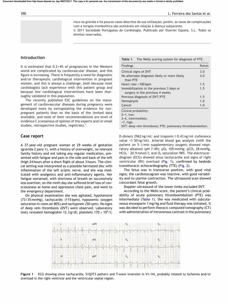
aturation in room air 80%) and tachypneic (50 cpm). No signsf deep vein thrombosis (DVT) were observed. Laboratoryests revealed hemoglobin 12.3 g/dl; platelets 170 × 109/l;igure 1 ECG showing sinus tachycardia, S1Q3T3 pattern and T-wverload in the right ventricle and the ventricular septal region.
nww
>7, high.DVT: deep vein thrombosis; PTE: pulmonary thromboembolism.
-dimers 2962 ng/ml; and troponin I 0.43 ng/ml (referencealue <1.50 ng/ml). Arterial blood gas analysis (with theatient on 5 l/min supplementary oxygen) showed respi-atory alkalosis (pH 7.49); pO2 105 mmHg; pCO2 28 mmHg,CO3
− 20.9 mmol/l; and O2 saturation 98%. The electrocar-iogram (ECG) showed sinus tachycardia and signs of rightentricular (RV) overload (Fig. 1), confirmed by bedsideransthoracic echocardiography (TTE) (Fig. 2).
The fetus was in transverse position, with good vitaligns; the cardiotocogram was reactive, with good variabil-ty and no uterine contraction. The ultrasound scan showedoncordant fetal growth.
Doppler ultrasound of the lower limbs excluded DVT.According to the Wells score, the patient’s clinical prob-
bility of acute pulmonary thromboembolism (PTE) was
ave inversion in V1---V4, probably related to ischemia and/or
eous enoxaparin 1 mg/kg and fluid therapy was initiated. Itas decided to perform thoracic computed tomography (CT)ith administration of intravenous contrast in the pulmonary
Pregnancy and acute pulmonary embolism: A case report 391
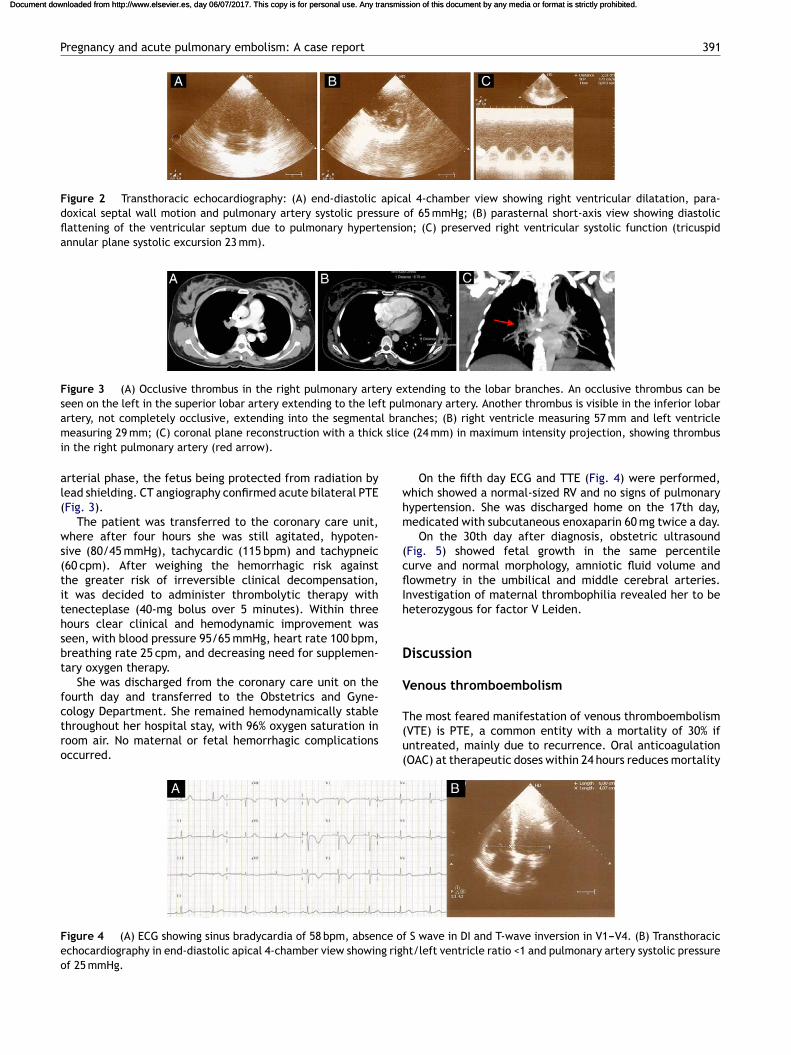
Figure 2 Transthoracic echocardiography: (A) end-diastolic apical 4-chamber view showing right ventricular dilatation, para-doxical septal wall motion and pulmonary artery systolic pressure of 65 mmHg; (B) parasternal short-axis view showing diastolicflattening of the ventricular septum due to pulmonary hypertension; (C) preserved right ventricular systolic function (tricuspidannular plane systolic excursion 23 mm).
Figure 3 (A) Occlusive thrombus in the right pulmonary artery extending to the lobar branches. An occlusive thrombus can beseen on the left in the superior lobar artery extending to the left pulmonary artery. Another thrombus is visible in the inferior lobar
l braslic
whm
(cflIh
D
V
Document downloaded from http://www.elsevier.es, day 06/07/2017. This copy is for personal use. Any transmission of this document by any media or format is strictly prohibited.Document downloaded from http://www.elsevier.es, day 06/07/2017. This copy is for personal use. Any transmission of this document by any media or format is strictly prohibited.
artery, not completely occlusive, extending into the segmentameasuring 29 mm; (C) coronal plane reconstruction with a thickin the right pulmonary artery (red arrow).
arterial phase, the fetus being protected from radiation bylead shielding. CT angiography confirmed acute bilateral PTE(Fig. 3).
The patient was transferred to the coronary care unit,where after four hours she was still agitated, hypoten-sive (80/45 mmHg), tachycardic (115 bpm) and tachypneic(60 cpm). After weighing the hemorrhagic risk againstthe greater risk of irreversible clinical decompensation,it was decided to administer thrombolytic therapy withtenecteplase (40-mg bolus over 5 minutes). Within threehours clear clinical and hemodynamic improvement wasseen, with blood pressure 95/65 mmHg, heart rate 100 bpm,breathing rate 25 cpm, and decreasing need for supplemen-tary oxygen therapy.
She was discharged from the coronary care unit on thefourth day and transferred to the Obstetrics and Gyne-
cology Department. She remained hemodynamically stablethroughout her hospital stay, with 96% oxygen saturation inroom air. No maternal or fetal hemorrhagic complicationsoccurred.T(u(
Figure 4 (A) ECG showing sinus bradycardia of 58 bpm, absence ofechocardiography in end-diastolic apical 4-chamber view showing righof 25 mmHg.
nches; (B) right ventricle measuring 57 mm and left ventriclee (24 mm) in maximum intensity projection, showing thrombus
On the fifth day ECG and TTE (Fig. 4) were performed,hich showed a normal-sized RV and no signs of pulmonaryypertension. She was discharged home on the 17th day,edicated with subcutaneous enoxaparin 60 mg twice a day.On the 30th day after diagnosis, obstetric ultrasound
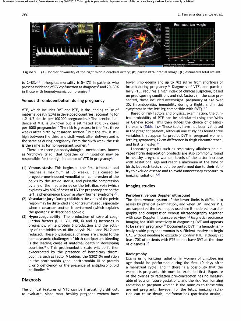
Fig. 5) showed fetal growth in the same percentileurve and normal morphology, amniotic fluid volume andowmetry in the umbilical and middle cerebral arteries.
nvestigation of maternal thrombophilia revealed her to beeterozygous for factor V Leiden.
iscussion
enous thromboembolism
he most feared manifestation of venous thromboembolismVTE) is PTE, a common entity with a mortality of 30% ifntreated, mainly due to recurrence. Oral anticoagulationOAC) at therapeutic doses within 24 hours reduces mortality
S wave in DI and T-wave inversion in V1---V4. (B) Transthoracict/left ventricle ratio <1 and pulmonary artery systolic pressure
392 L. Ferreira dos Santos et al.
arte
tpi
V
Vm1dpwhti
ar
(
(
(
D
Tt
lblos3s
iotivla
viwbii
I
PTaagwitiOlo
REaawo
Document downloaded from http://www.elsevier.es, day 06/07/2017. This copy is for personal use. Any transmission of this document by any media or format is strictly prohibited.Document downloaded from http://www.elsevier.es, day 06/07/2017. This copy is for personal use. Any transmission of this document by any media or format is strictly prohibited.
Figure 5 (A) Doppler flowmetry of the right middle cerebral
o 2---8%.2,3 In-hospital mortality is 5---17% in patients whoresent evidence of RV dysfunction at diagnosis4 and 20---30%n those with hemodynamic compromise.5
enous thromboembolism during pregnancy
TE, which includes DVT and PTE, is the leading cause ofaternal death (20%) in developed countries, accounting for
.2---4.7 deaths per 100 000 pregnancies.6 The precise inci-ence of VTE is unknown but is estimated at 0.5---2 caseser 1000 pregnancies.7 The risk is greatest in the first threeeeks after birth by cesarean section,2 but the risk is stilligh between the third and sixth week after delivery and ishe same as during pregnancy. From the sixth week the risks the same as for non-pregnant women.8
There are three pathophysiological mechanisms, knowns Virchow’s triad, that together or in isolation may beesponsible for the high incidence of VTE in pregnancy9:
1) Venous stasis: This begins in the first trimester andreaches a maximum at 36 weeks. It is caused byprogesterone-induced venodilation, compression of thepelvis by the gravid uterus, and pulsatile compressionby any of the iliac arteries on the left iliac vein (whichexplains why 80% of cases of DVT in pregnancy are on theleft, a phenomenon known as May-Thurner syndrome)10;
2) Vascular injury: During childbirth the veins of the pelvicregion may be distended and/or traumatized, especiallywhen a cesarean section is performed (which explainsthe greater risk described above);
3) Hypercoagulability: The production of several coag-ulation factors (I, II, VII, VIII, IX and X) increases inpregnancy, while protein S production and the activ-ity of the inhibitors of fibrinolysis PAI-1 and PAI-2 arereduced. These physiological changes are crucial to thehemodynamic challenges of birth (peripartum bleedingis the leading cause of maternal death in developingcountries11). This prothrombotic state will be furtherexacerbated by the presence of hereditary throm-bophilia such as factor V Leiden, the G20210A mutationin the prothrombin gene, antithrombin III or proteinC or S deficiency, or the presence of antiphospholipidantibodies.12
iagnosis
he clinical features of VTE can be frustratingly difficulto evaluate, since most healthy pregnant women have
arat
ry; (B) parasagittal cranial image; (C) estimated fetal weight.
ower limb edema and up to 70% suffer from shortness ofreath during pregnancy.13 Diagnosis of VTE, and particu-arly PTE, requires a high index of clinical suspicion, basedn predisposing conditions and risk factors (in the case pre-ented, these included overweight, pregnancy at age over5, thrombophilia, immobility during a flight, and initialymptoms in the left leg compatible with DVT).1,2
Based on risk factors and physical examination, the clin-cal probability of PTE can be calculated using the Wellsr Geneva score. This then guides the choice of diagnos-ic exams (Table 1).2 These tools have not been validatedn the pregnant patient, although one study has found threeariables that appear to predict DVT in pregnant women:eft leg symptoms, >2 cm difference in thigh circumference,nd first trimester.14
Laboratory results such as respiratory alkalosis or ele-ated fibrin degradation products are also commonly foundn healthy pregnant women; levels of the latter increaseith gestational age and reach a maximum at the time ofirth, but such tests should be performed due to their abil-ty to exclude disease and to avoid unnecessary exposure toonizing radiation.1,15
maging studies
eripheral venous Doppler ultrasoundhe deep venous system of the lower limbs is difficult tossess by physical examination, and when DVT and/or PTEre suspected the techniques used are B-mode echocardio-raphy and compression venous ultrasonography togetherith color Doppler in transverse view.1,2 Magnetic resonance
maging has 100% sensitivity in diagnosing DVT and appearso be safe in pregnancy.16 Documented DVT in a hemodynam-cally stable pregnant woman is sufficient motive to beginAC without needing to exclude or confirm PTE, although at
east 70% of patients with PTE do not have DVT at the timef diagnosis.17
adiographyxams using ionizing radiation in women of childbearingge should be performed during the first 10 days aftermenstrual cycle, and if there is a possibility that the
oman is pregnant, this must be excluded first. Exposuref the ovaries to radiation pre-conception has no measur-
ble effects on future gestations, and the risk from ionizingadiation to pregnant women is the same as to those whore not pregnant. However, for the fetus, ionizing radia-ion can cause death, malformations (particular ocular),
ampsi(fawesPRmptocurrhdinmrieaoTco
M
Wttfetfam
pwglTams
Document downloaded from http://www.elsevier.es, day 06/07/2017. This copy is for personal use. Any transmission of this document by any media or format is strictly prohibited.Document downloaded from http://www.elsevier.es, day 06/07/2017. This copy is for personal use. Any transmission of this document by any media or format is strictly prohibited.
Pregnancy and acute pulmonary embolism: A case report
growth retardation and mutagenic and carcinogenic effects,which depend mainly on gestational age (the most vulnera-ble period is between the second and eighth week) and theabsorbed radiation dose.
A major problem with diagnosis of PTE is clinicians’ reluc-tance to expose the fetus to ionizing radiation, often due tooverestimation of the risk of harm. When faced with the clin-ical probability of PTE, the primary diagnostic modalities arepulmonary ventilation-perfusion scintigraphy (VPS) and tho-racic CT. The estimated radiation dose from CT absorbed bythe fetus is 0.003---0.13 mGy, while from VPS it is 0.2 mGy.There is no evidence that doses of up to 50 mGy lead tofetal abnormalities, low IQ, growth restriction or miscar-riage. Less radiation is absorbed by the mother’s mammaryand pulmonary tissue with VPS than with CT.18,19 AlthoughVPS and CT appear to be safe for the fetus, it should be notedthat some studies suggest that exposure to low radiationdoses in utero can increase the risk of childhood leukemia(1 in 2000 compared to the baseline risk of 1 in 2800), whichdoes not compare with the risk of maternal death from undi-agnosed and untreated PTE (15%).20
In a pregnant woman with normal chest X-ray, VPS maybe more valuable in diagnosing PTE than CT, since in thelatter exam the contrast material can be interrupted byunopacified blood from the inferior vena cava. Conversely,CT should be used when the chest X-ray is abnormal, sinceit can diagnose other conditions such as pneumonia or otherlung disease. Pulmonary angiography should not be used inpregnancy.1,21
Iodinated contrast agents may lead to fetal thyroiddysfunction (although this has never been reported with iso-lated use), and this should be assessed in the first week afterbirth.
Treatment
Prevention
All women should be routinely assessed for risk of VTE beforeconception or in the first weeks of pregnancy. The mostimportant risk factors are history of unprovoked DVT, recur-rent VTE, PTE or thrombophilias (for which a family historyof VTE is an important factor). Half of the women with VTEduring pregnancy have either a thrombophilic disorder ora previous thrombotic event, and there are thought to beidentifiable risk factors in around 80% of cases of deathfrom PTE in pregnancy.1,22 Prevention of VTE in pregnancy iswithout doubt better than cure and prophylactic measures(OAC with enoxaparin 0.5 mg/kg and compression stockings)should be taken when the risk is considered to be high oreven moderate.1
Acute treatment
OAC, together with unfractionated or low molecular weightheparin, should be administered to achieve therapeuticdoses within 24 hours. This reduces mortality by pre-
venting recurrence of PTE and improving RV function.There is no evidence of differences in mortality betweenOAC alone or in combination with thrombolytics, althoughin patients with signs of RV dysfunction thrombolysis is‘ooi
393
ssociated with less clinical deterioration (10% vs. 25%),23
ore rapid resolution of hemodynamic alterations, androbable long-term improvement in pulmonary artery pres-ure and pulmonary vascular resistance.24 Thrombolysis isndicated in PTE when there is severe clinical instabilityi.e. with shock or systolic blood pressure <90 mmHg or aall of >40 mmHg in 15 minutes not caused by new-onsetrrhythmia, hypovolemia or sepsis), a situation associatedith high early mortality (>15%). Although it may be consid-red in other situations such as severe hypoxemia, severecintigraphic perfusion defects, RV dysfunction, massiveTE on CT, free-floating thrombus in the right atrium orV, and patent foramen ovale, there is general agree-ent on the use of thrombolysis only in the case ofersistent hypotension.2 Current thinking is to prescribehrombolysis not on the basis of the extent or severityf PTE but solely to counteract its hemodynamic reper-ussions. Pregnancy is a relative contraindication to these of thrombolytics2, but successful thrombolysis has beeneported in at least 200 pregnant women.1 The reportedisks are 1% for maternal death, 6% for fetal loss and 8% foremorrhage, mostly from the genital tract. At the time ofelivery, thrombolytic treatment should not be used exceptn extremely severe cases and if surgical embolectomy isot immediately available. The thrombolytics most com-only used in pregnancy are streptokinase, urokinase and
ecombinant tissue plasminogen activator (rt-PA). If OACs absolutely contraindicated, as in the immediate postop-rative or postpartum period, possible treatments includen inferior vena cava filter, thrombus fragmentation withr without local thrombolysis, or surgical embolectomy.he use of fluid challenge in PTE-induced hemodynamicompromise is controversial; it should not exceed correctionf 500---1000 cm3.2
aintenance treatment
arfarin should not be used in pregnancy, particularly inhe first trimester due to the risk of embryopathy and inhe third trimester due to the risk of placental abruption oretal and neonatal hemorrhage, but can be used after deliv-ry and during breastfeeding. Vaginal delivery is preferableo cesarean section, which should be reserved for specificetal or maternal indications. It is safe to begin OAC 12 hoursfter delivery; it should be continued for at least threeonths.1,2
Tenecteplase (TNKase) is a genetically engineered glyco-rotein derived from rt-PA by substituting three amino acids,hich confers slower plasma clearance, longer half-life,reater fibrin binding, less fibrinogenolysis and coagu-opathy, and greater resistance to inactivation by PAI-1.enecteplase does not cross the blood-placenta barrier,nd single-bolus administration results in more rapid plas-in formation and hence to resolution of the clinical
etting.25
A search in PubMed for the keywords ‘‘pregnancy’’ and
‘tenecteplase’’ reveals that this is the fifth report of the usef this thrombolytic in pregnancy and the first in the contextf PTE (of the previous four cases, two were of myocardialnfarction and two of mechanical valve thrombosis).
3
C
Pitsdscbt
C
T
R
1
1
1
1
1
1
1
1
1
1
2
2
2
2
2
Document downloaded from http://www.elsevier.es, day 06/07/2017. This copy is for personal use. Any transmission of this document by any media or format is strictly prohibited.Document downloaded from http://www.elsevier.es, day 06/07/2017. This copy is for personal use. Any transmission of this document by any media or format is strictly prohibited.
94
onclusions
ulmonary thromboembolism is common in pregnancy ands associated with significant maternal morbidity and mor-ality. It should always be considered in the presence ofuspicious symptoms and signs and confirmed by appropriateiagnostic exams, including VPS or CT. Oral anticoagulationhould be begun immediately, and thrombolysis should beonsidered in cases of hemodynamic instability as it haseen shown to be effective in the few cases described inhe literature and in the case presented here.
onflict of interest
he authors have no conflict of interest to declare.
eferences
1. The Task Force on the Management of Cardiovascular Diseasesduring Pregnancy of the European Society of Cardiology. ESCguidelines on the management of cardiovascular diseases duringpregnancy. Eur Heart J. 2011, doi:10.1093/eurheartj/ehr218.
2. The Task Force for the Diagnosis, Management of Acute Pul-monary Embolism of the European Society of Cardiology.Guidelines on diagnosis and management of acute pulmonaryembolism. Eur Heart J. 2008;29:2276---315.
3. Laack TA, Goyal DG. Pulmonary embolism: an unsuspectedkiller. Emerg Med Clin N Am. 2004;22:961---83.
4. Kasper W, Konstantinides S, Geibel A, et al. Prognostic sig-nificance of right ventricular afterload stress detected byechocardiography in patients with clinically suspected pul-monary embolism. Heart. 1997;77:346---9.
5. Fedullo PF, Tapson VF. The evaluation of suspected pulmonaryembolism. New Engl J Med. 2003;349:1247---56.
6. Chang J, Elam-Evans LD, Berg CJ, et al. Pregnancy-related mor-tality surveillance-United States, 1991---1999. MMWR SurveillSumm. 2003;52:1.
7. Liu S, Rouleau J, Joseph KS, et al. Epidemiology of pregnancy-associated venous thromboembolism: a population-based study
in Canada. J Obstet Gynaecol Can. 2009;31:611---20.8. Jacobsen AF, Skjeldestad FE, Sandset PM. Ante- and postnatalrisk factors of venous thrombosis: a hospital-based case---controlstudy. J Thromb Haemost. 2008;6:905---12.
2
L. Ferreira dos Santos et al.
9. Aird WC. Vascular bed-specific thrombosis. J Thromb Haemost.2007;5:283---91.
0. Macklon NS, Greer IA, Bowman AW. An ultrasound study of ges-tational and postural changes in the deep venous system of theleg in pregnancy. Br J Obstet Gynaecol. 1997;104:191---7.
1. Khan KS, Wojdyla D, Say L, et al. WHO analysis ofcauses of maternal death: a systematic review. Lancet.2006;367:1066---74.
2. Walker MC, Garner PR, Keely EJ. Thrombosis in pregnancy: areview. J Soc Obstet Gynaecol Can. 1998;20:943---52.
3. Weinberger SE, Weiss ST, Cohen WR, et al. Pregnancy and thelung. Am Rev Respir Dis. 1980;121:559.
4. Chan WS, Lee A, Spencer FA, et al. Predicting deep venousthrombosis in pregnancy: out in ‘‘LEFt’’ field? Ann Intern Med.2009;151:85.
5. Kline JA, Williams GW, Hernandez-Nino J. D-dimer concen-trations in normal pregnancy: new diagnostic thresholds areneeded. Clin Chem. 2005;51:825.
6. Spritzer CE, Evans AC, Kay HH. Magnetic resonance imaging ofdeep venous thrombosis in pregnant women with lower extrem-ity edema. Obstet Gynecol. 1995;85:603.
7. Turkstra F, Kuijer PM, van Beek EJ, et al. Diagnostic utility ofultrasonography of leg veins in patients suspected of havingpulmonary embolism. Ann Intern Med. 1997;126:775.
8. ACOG, Committee on Obstetric Practice. Guidelines for diagnos-tic imaging during pregnancy. Obstet Gynecol. 2004;104:647.
9. Ratnapalan S, Bona N, Chandra K, et al. Physicians’ perceptionsof teratogenic risk associated with radiography and CT duringearly pregnancy. AJR Am J Roentgenol. 2004;182:1107.
0. Harvey EB, Boice Jr JD, Honeyman M, et al. Prenatal X-ray expo-sure and childhood cancer in twins. N Engl J Med. 1985;312:541.
1. U-King-Im JM, Freeman SJ, Boylan T, et al. Quality of CTpulmonary angiography for suspected pulmonary embolus inpregnancy. Eur Radiol. 2008;18:2709.
2. Knight M. Antenatal pulmonary embolism: risk factors, mana-gement and outcomes. BJOG. 2008;115:453---61.
3. Konstantinides S, Geibel A, Heusel G, et al. Heparin plusalteplase compared with heparin alone in patients with sub-massive pulmonary embolism. N Engl J Med. 2002;347:1143.
4. Sharma K, Folland E, McIntyre M, et al. Longterm hemodynamicbenefit of thrombolytic therapy in pulmonary embolic disease
(abstract). J Am Coll Cardiol. 1990;15:65.5. Kline J, Hernandez-Nino J, Jones A. Tenecteplase to treat pul-monary embolism in the emergency department. J ThrombThrombolysis. 2007;23:101---5.