Embed Size (px)
Citation preview
PREGNANCY 3rd Trimester
PREGNANCY 3rd Trimester
I. Uterine ChangesA. Uterus experiences greatest period of
growthB. Reaches into epigastric region by 8th
monthC. Endometrium
1. Layer thickens2. Glands enlarge, may penetrate
myometrium
PREGNANCY 3rd Trimester, con’t…
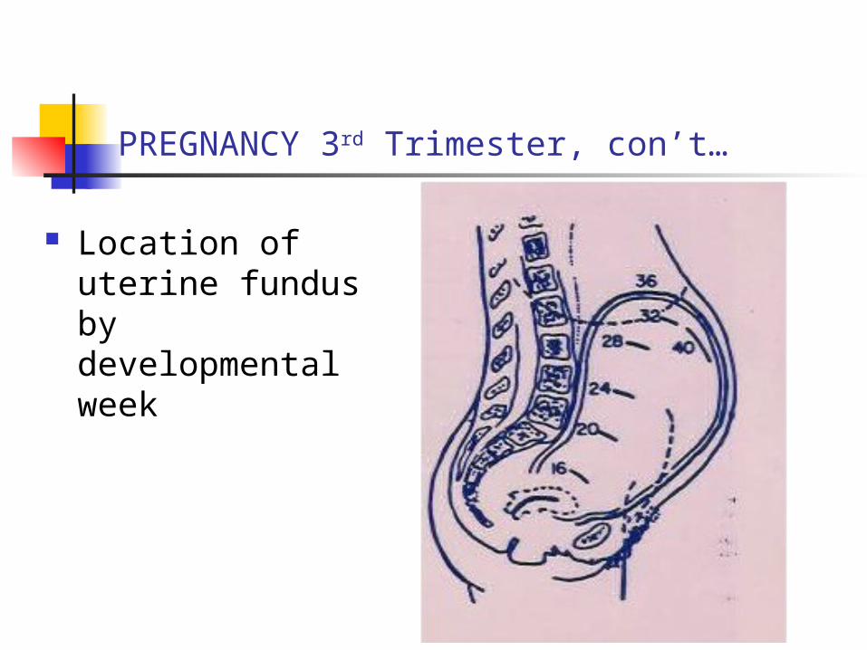
Location of uterine fundus by developmental week
Uterine changes, continued …
D. Myometrium: arranged into three distinct layers
1. External layer: beneath peritoneum
a. Fibers pass transversely across fundus
b. converge @ superior angles of uterus
Uterine changes, continued …
c. Fibers extend onto 1. fallopian tubes2. round ligaments 3. ovarian ligaments
2. Middle layer: a. thickest b. meshwork of random fibers c. many blood vessels
Uterine changes, continued … Myometrium
3. Inner layer:
a. fibers arranged circularly
b. form cones
1. Apices surround fallopian tubes
2. Fibers form sphincters around internal ossa of tubes
Uterine changes, continued … Myometrium
4. Smooth muscle fibers hypertrophy
a. >800% (8x) larger
b. muscle fibers undergo mitosis
1. only during pregnancy
2. Lose fibers post-partum
Uterine changes, continued …
D. Wall of uterus thins as pregnancy progresses
E. Placental “ascension” or “migration”
1. During the last part of the 3rd trimester
2. Differential growth of lower uterine segment
3. Artifact of development
PREGNANCY 3rd Trimester, con’t…
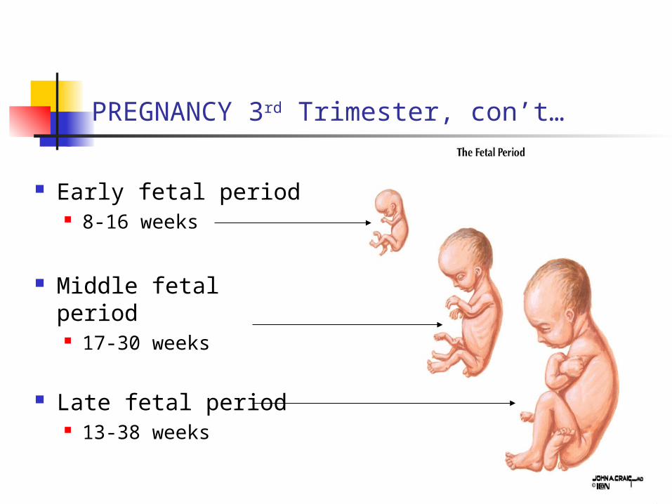
Early fetal period 8-16 weeks
Middle fetal period 17-30 weeks
Late fetal period 13-38 weeks
PREGNANCY 3rd Trimester, con’t…
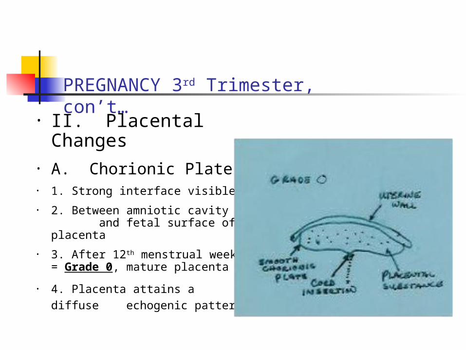
• II. Placental Changes
• A. Chorionic Plate• 1. Strong interface visible
• 2. Between amniotic cavity and fetal surface of placenta
• 3. After 12th menstrual week = Grade 0, mature placenta
• 4. Placenta attains a diffuse
echogenic pattern
PREGNANCY 3rd Trimester, con’t…
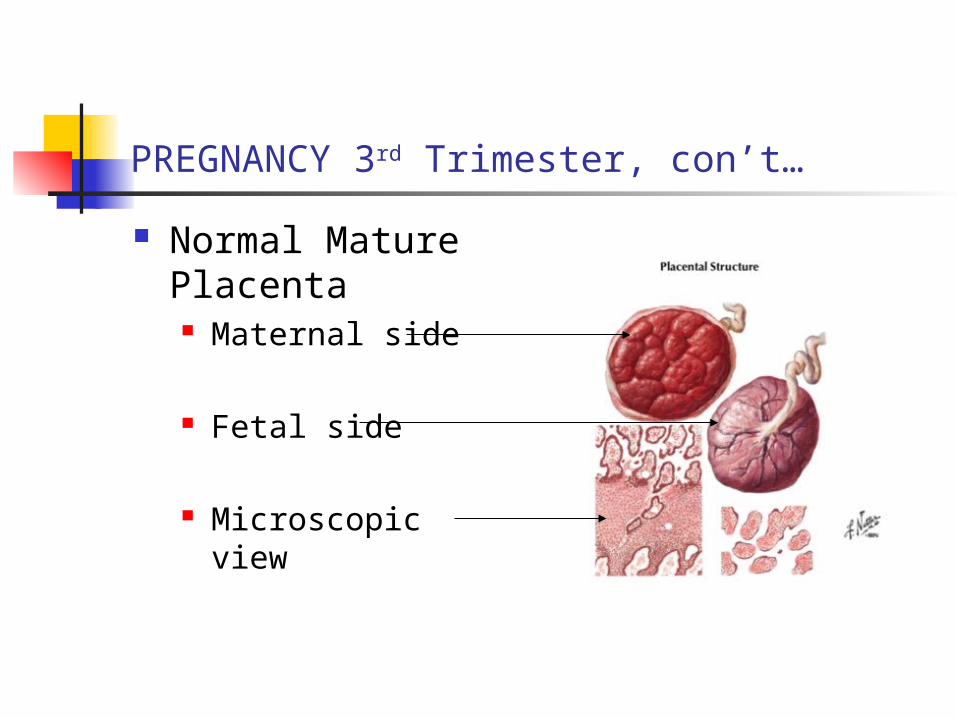
Normal Mature Placenta Maternal side
Fetal side
Microscopic view
Placental Changes, con’t….
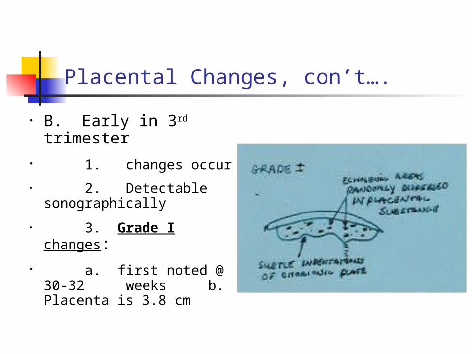
• B. Early in 3rd trimester
• 1. changes occur
• 2. Detectable sonographically
• 3. Grade I changes:
• a. first noted @ 30-32 weeks
b. Placenta is 3.8 cm
Placental Changes, continued …
d. Chorionic plate develops “wavy” appearance
e. Homogeneous look is lost
f. See increased glands, calcifications
g. see more scattered echogenic regions
Placental Changes, continued …
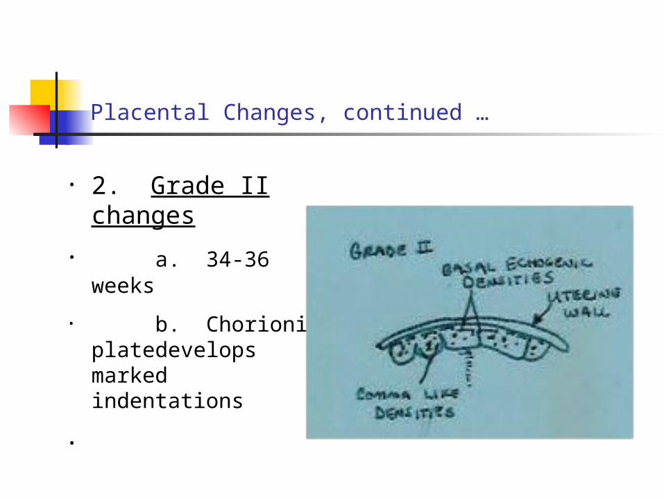
• 2. Grade II changes
• a. 34-36 weeks
• b. Chorionic plate develops
marked indentations
•
Placental changes, continued…
c. Comma-shaped septa extend from chorionic plate toward basalis
d. Placenta becomes more echogenic
e. Placental “mottling” appears
Placental Changes, continued …
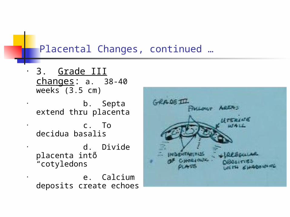
• 3. Grade III changes: a. 38-40 weeks (3.5 cm)
• b. Septa extend thru placenta
• c. To decidua basalis
• d. Divide placenta into “cotyledons”
• e. Calcium deposits create echoes
Placental changes, continued …
f. May find “cysts” between chorionic plate and placenta
g. Areas of excessive fibrin deposition
h. placenta starts deteriorating
i. Venous lakes develop
Placental changes, continued …
C. Thickness gradually decreases after 32 weeks
D. Growth ceases after 38 weeks
E. Changes normal for senescent placenta
1. At birth: weighs ~ 1 lb
2. Size: ~6-8” diameter
3. Surface area: ~ 16 m2
Pregnancy, 3rd Trimester, con’t…
F. Amount of amniotic fluid
1. Increases during pregnancy
2. May reach one gallon in late 3rd trimester
PREGNANCY 3rd Trimester, con’t…
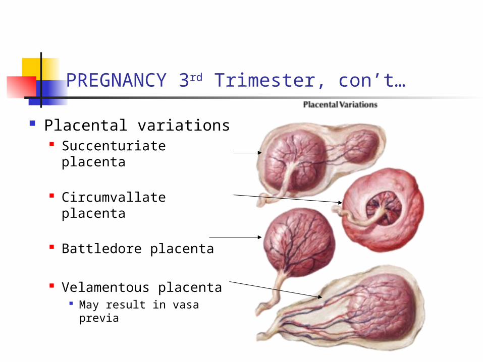
Placental variations Succenturiate placenta
Circumvallate placenta
Battledore placenta
Velamentous placenta May result in vasa
previa
III.Fetal Structures
A. Fetal Head
1. First visualized at 5-6 weeks
2. After 12 weeks, details can be seen:a. Falx cerebrib. Ventriclesc. Thalamid. Corpus callosum
PREGNANCY 3rd Trimester, con’t…
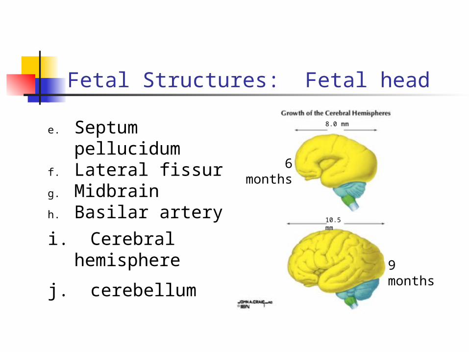
Fetal Structures: Fetal head
e. Septum pellucidumf. Lateral fissure g. Midbrainh. Basilar artery
i. Cerebral hemisphere
j. cerebellum
6 months
9 months
10.5 mm
8.0 mm
PREGNANCY 3rd Trimester, con’t…
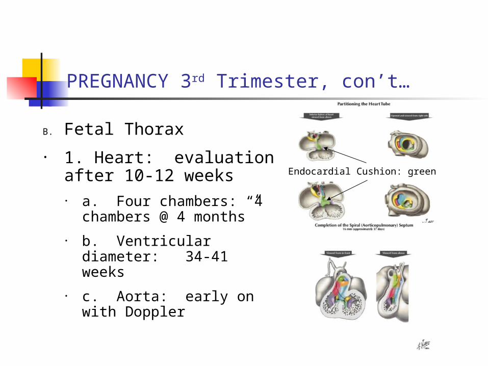
B. Fetal Thorax
• 1. Heart: evaluation after 10-12 weeks• a. Four chambers: “4
chambers @ 4 months”
• b. Ventricular diameter: 34-41 weeks
• c. Aorta: early on with Doppler
Endocardial Cushion: green
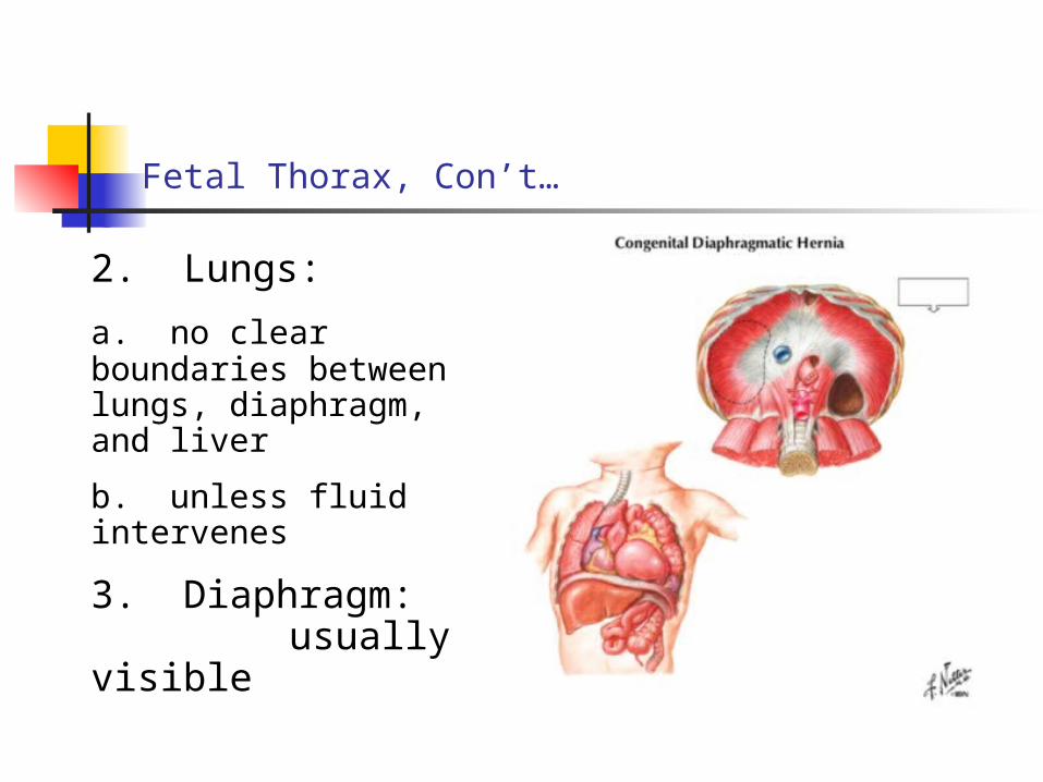
Fetal Thorax, Con’t…
2. Lungs:
a. no clear boundaries between lungs, diaphragm, and liver
b. unless fluid intervenes
3. Diaphragm: usually
visible
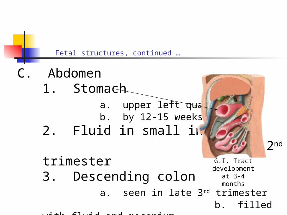
Fetal structures, continued …
C. Abdomen1. Stomach
a. upper left quadrant b. by 12-15 weeks
2. Fluid in small intestine by 2nd trimester
3. Descending colon a. seen in late 3rd trimester b. filled with fluid and meconium
G.I. Tract development at
3-4 months
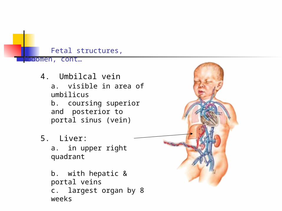
Fetal structures, Abdomen, cont…
4. Umbilcal vein a. visible in area of umbilicus
b. coursing superior and posterior to portal sinus (vein)
5. Liver: a. in upper right quadrant
b. with hepatic & portal veins
c. largest organ by 8 weeks
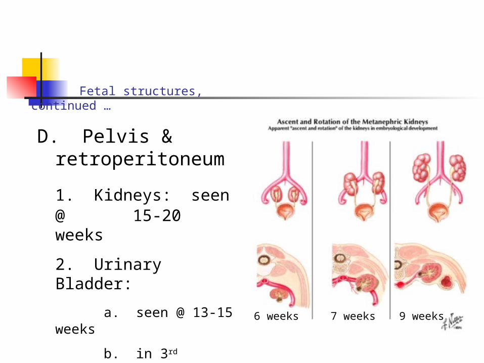
Fetal structures, continued …
D. Pelvis & retroperitoneum
1. Kidneys: seen @ 15-20 weeks
2. Urinary Bladder:
a. seen @ 13-15 weeks
b. in 3rd trimester, urine-filled
6 weeks 7 weeks 9 weeks
Fetal structures, continued …
E. Other structures
1. Skeletal components
a. Axial skeleton
b. Long bones
2. Extremities
3. Genitalia by 10 weeks (+/-)
IV. FETAL CIRCULATION
A. General
1. Ovum and yolk sac provide initial nutrients to embryo
2. Other means must develop early on
FETAL CIRCULATION, CON’T…
3. Blood vessel and blood formation
a. begin15-16 days after fertilization
b. in mesoderm of yolk sac, body stalk, chorion
4. Cardiovascular system first to develop
Fetal circulation, continued …
5. Blood flow begins end of the third week
6. Heart beat begins about 22 days
7. REMEMBER:
a. fetus is totally dependent on outside source for oxygen, nutrients, and waste disposal.
b. This source is the placenta
Fetal circulation, continued …
B. Course of blood through fetal circulation
1. Always begin at placenta
2. Vessels named with respect to fetus
3. Blood leaves placenta via umbilical or placental vein (+ O2 blood)
Fetal circulation, continued …
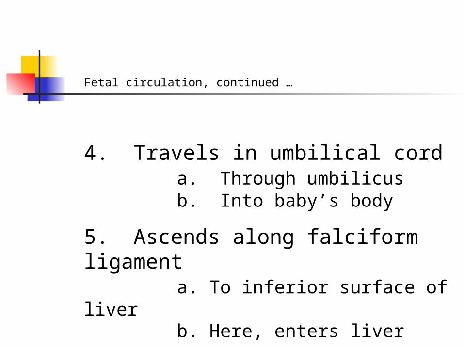
4. Travels in umbilical cord a. Through umbilicus b. Into baby’s body
5. Ascends along falciform ligament a. To inferior surface of liverb. Here, enters liver
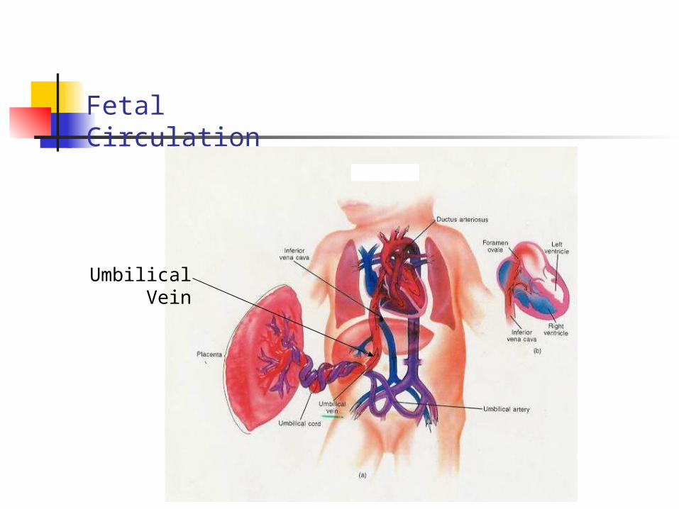
Fetal Circulation
Umbilical Vein
Fetal circulation, continued …
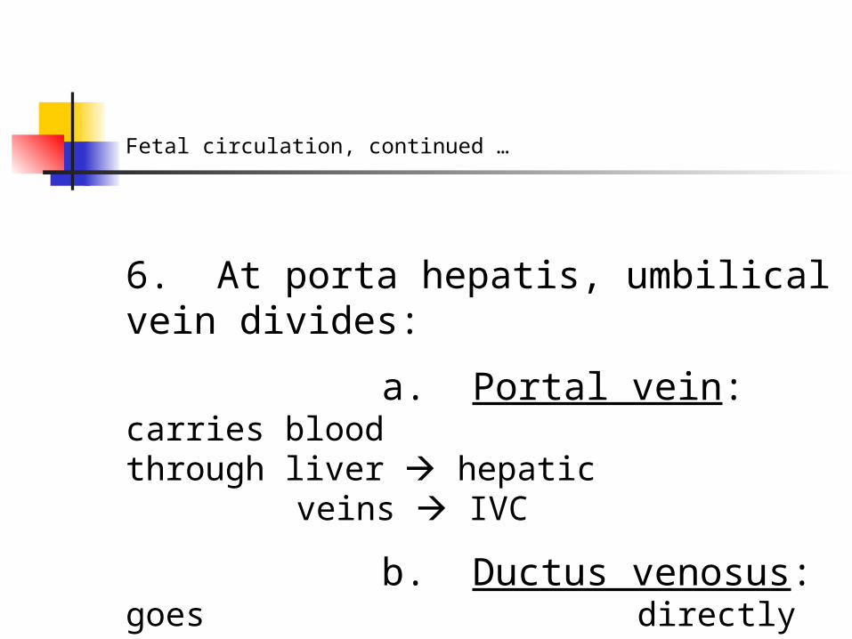
6. At porta hepatis, umbilical vein divides:
a. Portal vein: carries blood through liver
hepatic veins IVC
b. Ductus venosus: goes directly to IVC
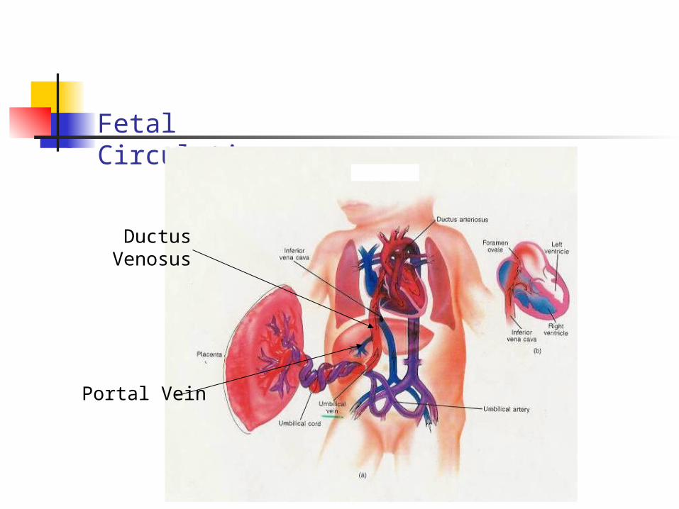
Fetal Circulation
Ductus Venosus
Portal Vein

Fetal Circulation, continued …
7. Blood in IVC is –O2 blood, and is
a. mixes with +O2 blood from hepatic veins, ductus venosus
b. is now “mixed” blood
8. IVC enters right atrium
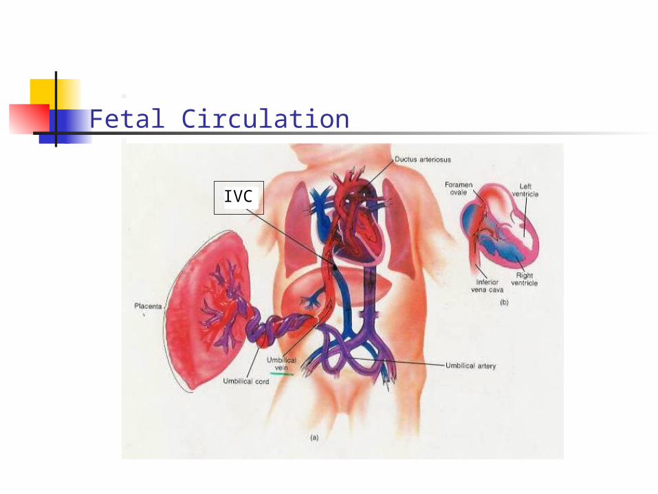
Fetal Circulation
IVC
Fetal Circulation, continued …
a. some blood follows post-partum path
b. Most blood is diverted
1. by valve of IVC (Eustachian valve)
2. through foramen ovale into left atrium
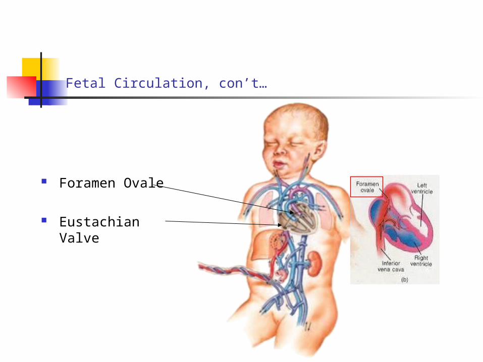
Fetal Circulation, con’t…
Foramen Ovale
Eustachian Valve
Fetal Circulation, continued …
9. From left atrium:
a. blood is pumped to left ventricle
b. out the aorta
c. throughout systemic circulation
Fetal Circulation, continued …
10. Blood from head and upper extremities
a. returns to right atrium via SVC b. mixes with blood from IVC
11. Blood to lower body
a. passes from aorta common iliac arteries internal iliac arteries
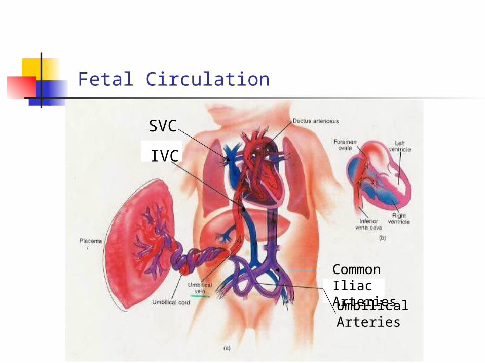
Fetal Circulation
SVC
IVC
Common Iliac Arteries
Umbilical Arteries
Fetal Circulation, continued …
12. Fetal lungs are non-functional
a. require minimal blood supply
b. shunt is present between pulmonary artery and aorta
c. ductus arteriosus: by-passes lungs
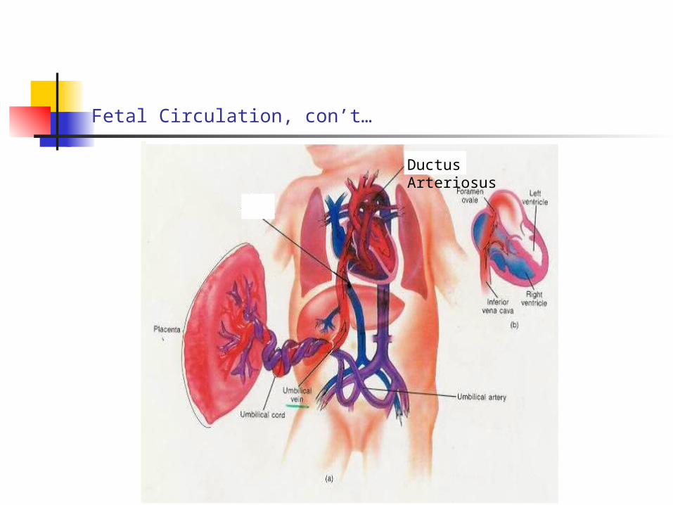
Fetal Circulation, con’t…
Ductus Arteriosus
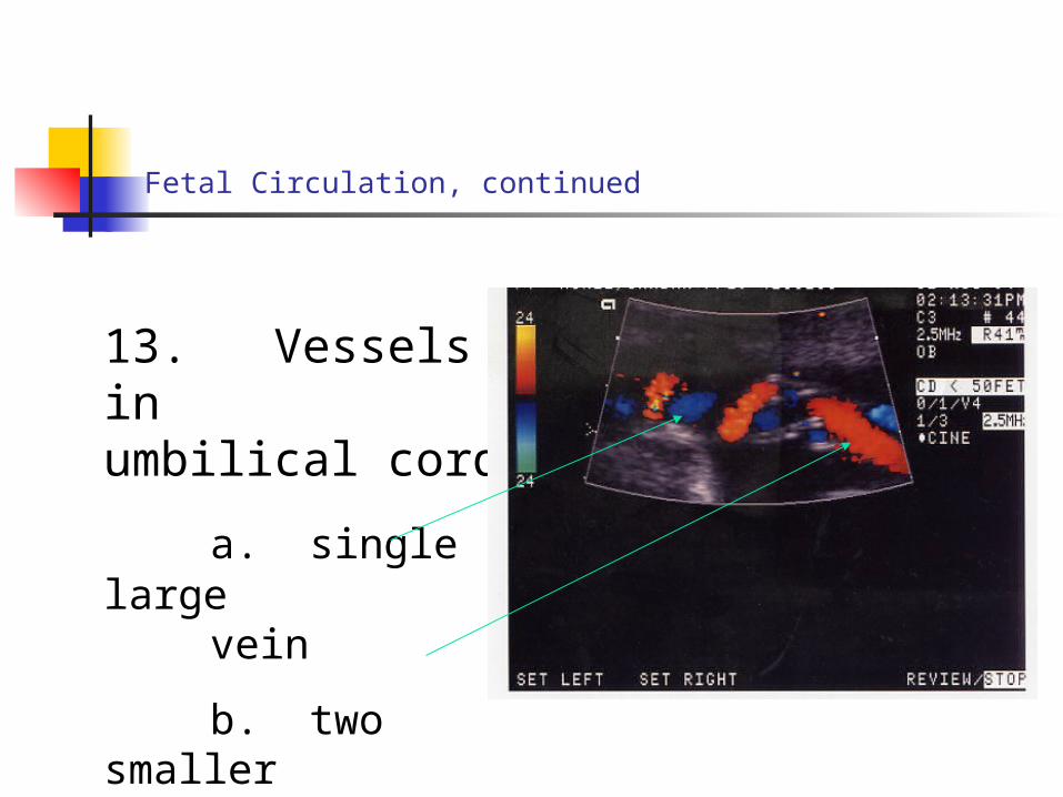
13. Vessels in umbilical
cord
a. single large vein
b. two smaller
arteries
Fetal Circulation, continued
Fetal circulation, continued …
14. Recap of fetal circulation:
a. Oxygenated blood carried by
1. umbilical vein
2. portal vein
3. ductus venosus
Fetal circulation, continued …
b. Placenta is:
1. Fetal organ of respiration, nutrition & excretion
2. Most blood from placenta a. goes thru liver b. before entering IVC
Fetal circulation, continued …
c. Eustachian Valve
1. directs most blood from IVC to left atrium
2. directs blood from SVC to right ventricle
Fetal circulation, continued …
d. Blood from umbilical vein, IVC 1. go from L.A. to L.V. to
aorta
2. then mostly to head, upper extremities
e. Descending aorta contains mixed blood
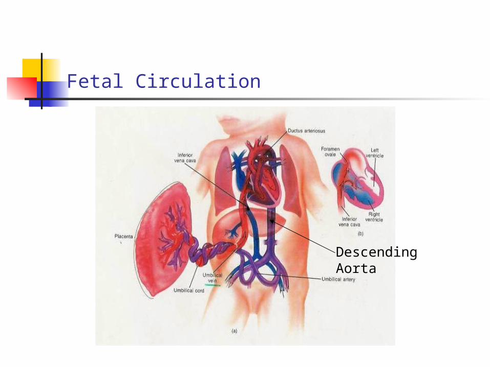
Fetal Circulation
Descending Aorta
Fetal circulation, con’t…
C. Changes in Cardiovascular System at Birth
1. After pulmonary respiration begins
a. the ductus arteriosus constricts
b. blood flows to lungs
Fetal circulation, con’t…
2. More blood returns to left atrium
a. flow thru foramen ovale stops
b. Seals to become fossa ovalis
3. Ductus arteriosus
a. fills with fibrous C.T.
b. becomes ligamentum arteriosum
Fetal circulation, con’t…
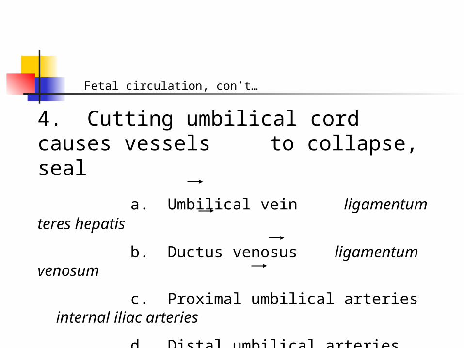
4. Cutting umbilical cord causes vessels to collapse, seal
a. Umbilical vein ligamentum teres hepatis
b. Ductus venosus ligamentum venosum
c. Proximal umbilical arteries internal iliac arteries
d. Distal umbilical arteries lateral umbilical ligaments
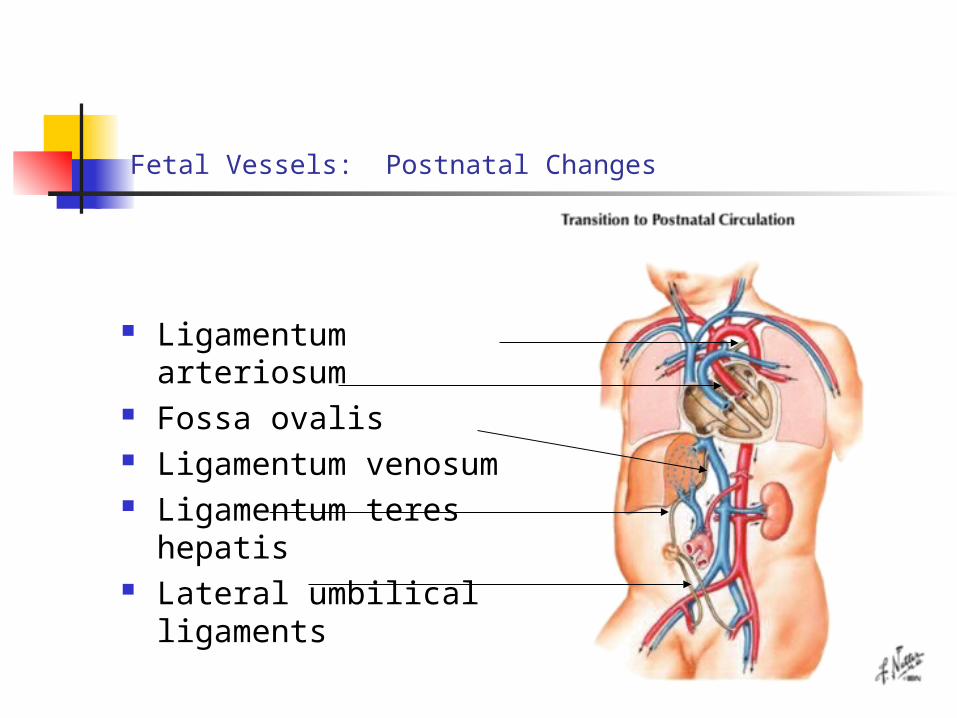
Fetal Vessels: Postnatal Changes
Ligamentum arteriosum Fossa ovalis Ligamentum venosum Ligamentum teres
hepatis Lateral umbilical
ligaments
Fetal circulation, con’t…
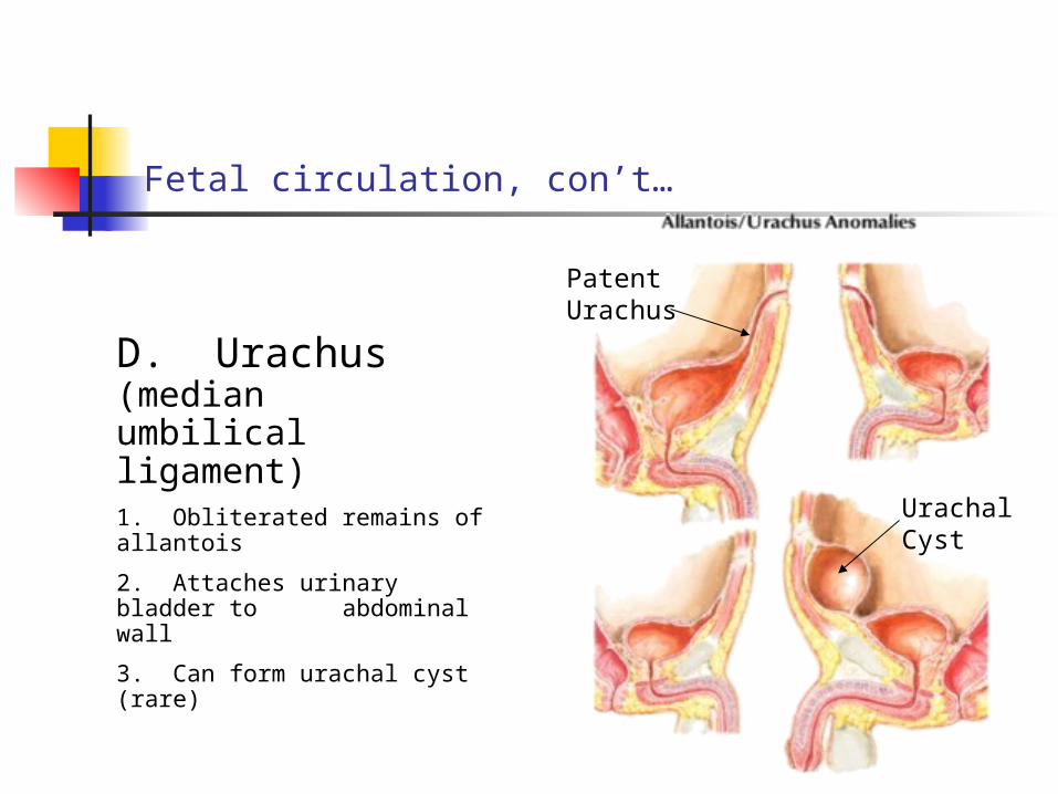
Patent Urachus
UrachalCyst
D. Urachus (median umbilical ligament)1. Obliterated remains of allantois
2. Attaches urinary bladder to abdominal wall
3. Can form urachal cyst (rare)
Fetal circulation, con’t…
3. Passes superiorly from apex of bladder to umbilicus
4. Composed of fibrous C.T. and smooth muscle
5. Lies between umbilical ligaments
V. THIRD TRIMESTER BLEEDING
A. Abruptio Placentae (Placental abruption):
1. Premature separation of a normally implanted placenta
2. May be mistaken for placenta previa
3. May be only a few mm separation or a complete detachment
Third trimester bleeding, con’t.
4. Retroplacental bleeding occurs
5. May be external or concealed
6. Signs and symptoms vary
a. depends on degree of separation
b. amount of blood loss
Third Trimester Bleeding, continued …
B. Placenta Previa:
1. Placenta implants over internal os of cervix
2. Occurs in 1:200 cases
3. False positive rate of 10%
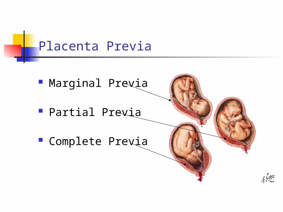
Placenta Previa
Marginal Previa
Partial Previa
Complete Previa
Third Trimester Bleeding, continued …
4. Symptoms:
a. sudden painless vaginal bleeding
b. in late third trimester
c. Followed by painless massive bright red bleeding
Third Trimester Bleeding, continued …
5. May be mistaken for abruptio placentae
6. Ultrasound is diagnostic modality
Third trimester bleeding, continued …
C. Rh Incompatibility: [erythroblastosis fetalis, hemolytic disease of the newborn (HDN)]
1. lysis of fetal RBC’s
2. due to Rh- isoimunization
3. Rh- multiparous female
4. Rh+ father and fetus
Third trimester bleeding, continued …
5. Leads to:
a. hemolysis of fetal RBC’s
b. anemia of fetus
c. congestive heart failure
d. may cause jaundice
Erythroblastosis Fetalis, continued …
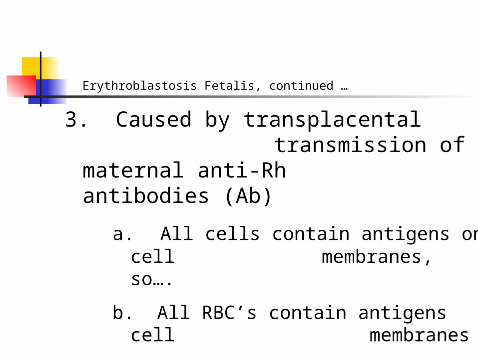
3. Caused by transplacental transmission of maternal anti-Rh
antibodies (Ab)
a. All cells contain antigens on cell membranes, so….
b. All RBC’s contain antigens cell membranes
Erythroblastosis Fetalis, continued …
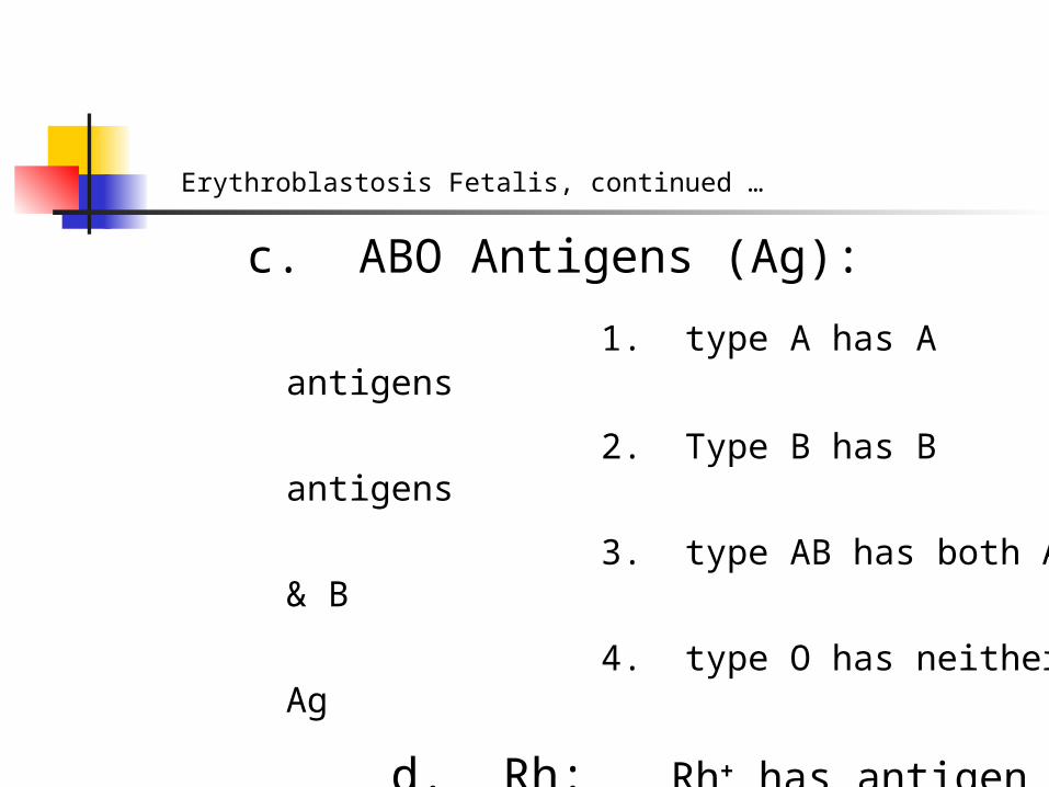
c. ABO Antigens (Ag):
1. type A has A antigens
2. Type B has B antigens
3. type AB has both A & B
4. type O has neither Ag
d. Rh: Rh+ has antigen Rh- lacks antigen(ABO & Rh: major antigens involved in antibody incompatibility)
Erythroblastosis Fetalis, continued ….
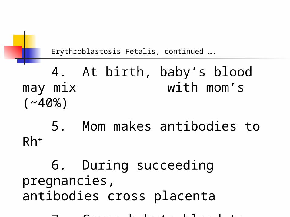
4. At birth, baby’s blood may mix with mom’s (~40%)
5. Mom makes antibodies to Rh+
6. During succeeding pregnancies, antibodies cross placenta
7. Cause baby’s blood to agglutinate

Erythroblastosis Fetalis, continued …
9. Rhogam
a. antibody formation may be prevented: “fools” mom’s immune system
b. Rhogam given within 72 hours after fetus “leaves”
c. Currently, Rhogam given to all Rh- moms every trimester
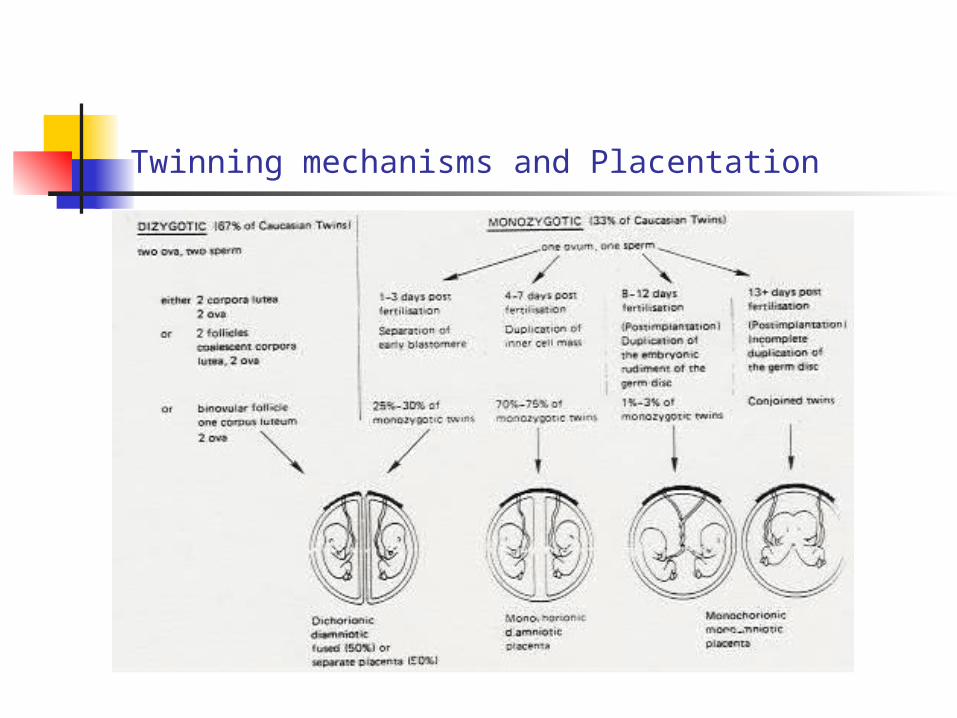
Twinning mechanisms and Placentation
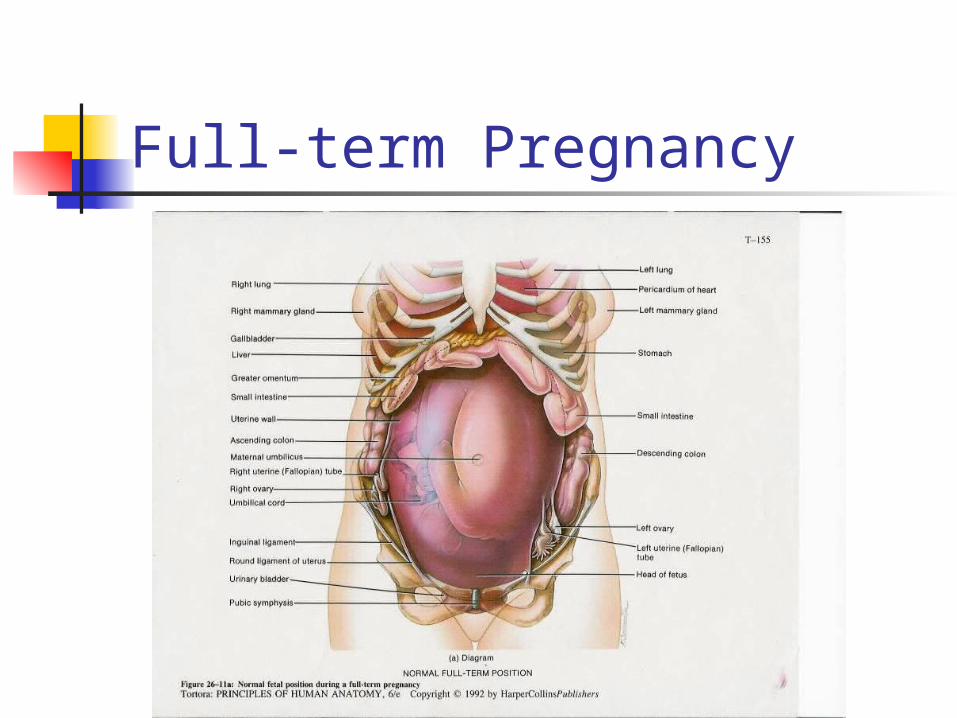
Full-term Pregnancy