Embed Size (px)
Citation preview

Case ReportPreeclampsia Induced Liver Dysfunction Complicated byDisseminated Intravascular Coagulopathy and PlacentalAbruption: A Case Report and Review of the Literature
JordanMyers,1 GaryWu,2 Robert E. Shapiro ,2 andManuel C. Vallejo 1,2,3
1Department of Anesthesiology, West Virginia University School of Medicine, Morgantown, WV 26506, USA2Department of Obstetrics & Gynecology, West Virginia University School of Medicine, Morgantown, WV 26506, USA3Department of Medical Education, West Virginia University School of Medicine, Morgantown, WV 26506, USA
Correspondence should be addressed to Manuel C. Vallejo; [email protected]
Received 23 December 2018; Revised 3 March 2019; Accepted 18 March 2019; Published 4 April 2019
Academic Editor: Maurizio Marandola
Copyright © 2019 Jordan Myers et al. This is an open access article distributed under the Creative Commons Attribution License,which permits unrestricted use, distribution, and reproduction in any medium, provided the original work is properly cited.
A 33-year-old primigravida at 32-week gestationwas admitted to labor and delivery complaining of severe right upper quadrant painand worsening coagulopathy. We report the anesthetic and obstetrical management of a complex case of a parturient with a mixedpicture of hemolysis, elevated liver enzymes and low platelets who was delivered under general anesthesia further complicated byDisseminated Intravascular Coagulopathy (DIC) and placental abruption.
1. Introduction
Preeclampsia in its severe form involves end organ damagerequiringmultidisciplinary treatment to avoid perinatalmor-tality [1]. AFLP is a rare disorder defined by microvesicularfatty infiltration of hepatocytes, occurring in the 3rd trimesterof pregnancy or early postpartum period [1]. Symptomsdevelop with accumulation of fatty acid break down productsresulting in damage to maternal hepatocytes [2]. Diagnosiscan be difficult due to clinical overlap with other commondisorders of pregnancy such as cholestasis, viral hepatitis,severe preeclampsia, and HELLP syndrome. AFLP presentswith nonspecific findings and varying degrees of severity,further increasing diagnostic difficulty. Prompt delivery ofthe fetus and patient support are the most important factorsfor maternal recovery. HELLP syndrome is a complicationof pregnancy characterized by hemolysis, elevated liverenzymes, and low platelets (HELLP) [3]. HELLP syndrome,as with any disorder of pregnancy causing liver dysfunction,can result in DIC.
We report the anesthetic and obstetrical managementand literature review of a complex case of a parturient witha mixed picture of preeclampsia induced liver dysfunction
complicated by Disseminated Intravascular Coagulopathy(DIC) and placental abruption.
2. Case Description
A 33-year-old 78 kg, 155 cm, primigravida at 32-week gesta-tion presented to labor and delivery complaining of severeepigastric chest pain. The patient had been seen in clinicprior to admission for lower extremity edema and headacheand was diagnosed with mild preeclampsia. Over the twodays prior to admission, she reported shortness of breathand worsening oliguria despite adequate oral intake. Initially,her epigastric pain was thought to be gastroesophageal refluxdisorder (GERD) which now worsened to a constant 10/10pain mostly confined to the right upper abdominal quadrant.Initial laboratory results at 03:52 were unremarkable. Morethan 3 hours later at 07:21, the platelet count went from195 K/uL to “unable to perform count, platelets clumpedon slide Aspartate aminotransferase (AST) increased from183 u/L to 3180 u/L, and Alanine aminotransferase (ALT)increased from 221 u/L to 3495 u/L (an increase more than15x admission values). Because of her rapidly worseningclinical picture, the decision was made to proceed with an
HindawiCase Reports in AnesthesiologyVolume 2019, Article ID 4305849, 5 pageshttps://doi.org/10.1155/2019/4305849

2 Case Reports in Anesthesiology
urgent cesarean delivery.The platelet count was repeated andbecause of a high clinical suspicion for thrombocytopenia, athromboelastogram (TEG) was performed, showing featuresof coagulopathy (Figure 1). Repeat platelet count at 09:28 wasagain “unable to perform count, platelets clumped on slide.”Alarmingly, the AST had increased to 4,059 u/L and ALT to4,000 u/L. Because of her severe coagulopathy, the patientwasnot determined to be a candidate for neuraxial anesthesia.Her initial blood pressure on admission was 199/100 mmHg. Oral labetalol and intravenous hydralazine were givenfor treatment of her high blood pressure. Intravenous mag-nesium was started for seizure prophylaxis and treatment ofHEELP syndrome.
The urgent cesarean section was delayed until FreshFrozen Plasma (FFP), cryoprecipitate, platelets, and packedred blood cells (pRBCs) could be available and in theoperating room so that blood component therapy could beinstituted at the time of the procedure. While waiting forthe blood products, a second 18-gauge peripheral intravenouscatheter was placed as well as a radial arterial line forassessment of her blood pressure on a beat-to-beat basis, fortreatment for hemodynamic instability, and to obtain bloodfor laboratory analysis.
After bloodproductsweremade available in the operatingroom, intravenous cefazolin 2 gm was given for antibioticprophylaxis. The patient had taken nothing by mouth sincemidnight. Induction medications included 200 mg of propo-fol, 100 mcg of fentanyl, and 160 mg of succinylcholineintravenously under rapid sequence intubation with cricoidpressure, and esmolol (30 mg) to attenuate the hypertensiveresponse to laryngoscopy and intubation. The trachea wasintubated on the first attempt with a 7.0 endotracheal tubeconfirmed by auscultation of bilateral breath sounds and byend tidal CO
2monitor detection. Sevoflurane (4%) and 100%
oxygen (4 liters) were used for maintenance of anesthesiawhich was changed to Sevoflurane (1.5%) with 60% nitrousoxide in oxygen after delivery of the baby for prevention ofuterine atony. After fetal delivery, 4 mg of midazolam and 2mg of hydromorphone were administered intravenously overdivided doses for additional amnesia and analgesia.Through-out the procedure, the patient’s blood pressure trended in the110-140 mm Hg systolic and 50-80 mm Hg diastolic range.
During hysterotomy, the placenta was noted to beabrupting. A female neonate was delivered weighing 1570gm with Apgar scores of 5 at 1 minute and 8 at fiveminutes. Uterine atony was noted with oozing and poorblood clotting. Standard oxytocin dose at West VirginiaUniversity Hospital is a 5-unit bolus of oxytocin and 35-unitinfusion of oxytocin in 1 liter of lactated ringer’s solution.The patient received 250-mcg carboprost intramuscularlyand 1000 mcg misoprostol placed intrauterine for treatmentof uterine atony. The placental abruption was noted to besmall and was able to be controlled upon removal of theplacenta. Tranexamic acid (1 gm) was considered but notadministered since uterine tone became adequate over severalminutes. Intraoperatively, the patient was transfused 2 unitsof FFP, 2 units of cryoprecipitate, and 1 pack of platelets.Estimated blood loss was over 2.2 liters. After the patientsatisfied extubation criteria (train of four > 0.9, sustained
head lift for 5-sec, and tidal volume > 5ml/kg), the patientwas extubated without complication and able to maintain herairway.The patient recovered in the obstetrical intensive careunit. Bilateral sequential compression devices were placed onboth legs and enoxaparin (40 mg SQ QD) was administeredfor deep venous thromboembolism prophylaxis.
A postoperative TEG was obtained, showing slightimprovement in her coagulopathy, consistent with multior-gan system dysfunction (Figure 1). The patient’s postoper-ative course was complicated by worsening coagulopathy,respiratory, and renal dysfunction. The patient required 5liters of supplemental oxygen via nasal cannula postop-eratively for shortness of breath and hypoxia. On post-operative day (POD) 1, arterial blood gas analysis wasconsistent with respiratory alkalosis (pH=7.42, PCO
2=30.0,
Bicarbonate=21.7, and Base Deficit=3.9). A chest x-rayrevealed a left pleural effusion. Pulmonology was consultedfor assistance managing her respiratory status and recom-mended discontinuing magnesium therapy and substitutingdilantin due to a concern for magnesium worsening herpulmonary edema. Nephrology was consulted for assis-tance managing her acute kidney injury as her creatinineincreased to 4.69 umol/L on POD-3 from a baseline of 0.78umol/L on admission.Nephrology recommended continuingtreatment for hypertension, avoiding Nonsteroidal Anti-inflammatory Drugs (NSAID’s), Angiotensin-Converting-Enzyme Inhibitor (ACEI), Angiotensin-Receptor Blocker(ARB), and contrast dye, and continuing fluid restriction, andhemodialysis was not indicated. Hepatic AST/ALT enzymeswhich peaked at 4059/4000 u/L at 09:28 the day of surgerycontinued to steadily improve and fell into the normal rangeupon hospital discharge. The patient’s multiorgan systemdysfunction steadily improved over several days, and she wasdischarged home along with her baby in stable condition onPOD-8 with a 1-week follow-up for a blood pressure checkand a 6-week obstetric postpartum follow-up.
3. Discussion
The diagnosis of preeclampsia, HELLP, and acute fatty liverof pregnancy (AFLP) in the setting of placental abruptionall appear to have overlapping criteria and can be difficultto distinguish. Preeclampsia is characterized by new onsethypertension and proteinuria after 20-week gestational age[5]. Without severe features, blood pressure are greater than140/90 mmHg with proteinuria greater than 300 mg/24-hourperiod or a point-of-care test of protein-creatinine ratio of0.3 [5]. Preeclampsia with severe features involves signs andsymptoms of end-organ damage: blood pressure greater than160/100 mmHg, serum creatinine greater than 1.1 mg/dL ortwice baseline, elevated liver function tests of twice normalwith persistent right upper quadrant pain, severe headacheswith or without vision changes, and thrombocytopenia withplatelet count less than 100,000/mL [5].
The incidence of AFLP is 1:7000 – 1:15000 pregnancies [2].In a report by Gregory et al. [6], they discussed 3 cases ofAFLP and how variable each case can present. One patientpresented with elevated liver function, malaise, and nausea,much like our patient.The two other cases they describe have

Case Reports in Anesthesiology 3
09:38 AM10 millimeters
SPmin7.5
Rmin9.8
5 --10
Kmin13.81--3
Angledeg18.7
53 -- 72
MAmm29.0
50--70
Gd/sc2.0K
0.0K -- 0.0K
EPL%0.0
0 -- 15
LY30%0.0
0 -- 8
A30mm29.0
PMA
1.0
SPmin7.0
Rmin9.8
5 --10
Kmin6.4
1--3
Angledeg40.4
53 -- 72
MAmm35.7
50--70
Gd/sc2.8K
0.0K -- 0.0K
EPL%0.0
0 -- 15
LY30%0.0
0 -- 8
A30mm35.7
PMA
1.0
SPmin7.8
Rmin8.6
5 --10
Kmin5.0
1--3
Angledeg43.6
53 -- 72
MAmm39.9
50--70
Gd/sc3.3K
0.0K -- 0.0K
EPL%0.0
0 -- 15
LY30%0.0
0 -- 8
A30mm39.9
PMA
1.0
10 millimeters
10 millimeters
13:57 PM
21.40 PM
Figure 1: Thromboelastograms (TEGs) before cesarean delivery, after blood replacement therapy, and approximately 12 hours after cesareandelivery. Legend: 09:38 TEG prior to cesarean delivery, note decreased maximum amplitude, elevated K value, and decreased alpha angle;13:57 TEG after blood replacement therapy, note K value, alpha angle, and maximum amplitude improving but still below normal; 21:40 TEGlater that evening, below normal values.

4 Case Reports in Anesthesiology
patients who presented with abdominal pain, pruritus, andnausea. Their patients did not present with elevated bloodpressure where our patient did. Stander and Cadden [7]first described AFLP in 1934. Sheehan [8] then described itin 1940 as acute yellow atrophy—where postmortem examswere performed on 400 obstetric patients and 6 were foundto have significant liver pathology. Mortality in the past wasrecorded as high as 75-85% [9]. The mortality has beensignificantly reduced to 18 to 23% [10]. Today, there is adecrease in mortality from AFLP due to prompt maternalsupportive care and delivery of the fetus. In the past, a liverbiopsywas indicated for diagnosis ofAFLP, leading to delayedmanagement and a risk for bleeding since these patientscan have severe coagulopathy. At that time, serology wasunavailable for viral hepatitis [11]; and clinically the patientspresent similarly. Now AFLP has become a diagnosis basedon detailed history, laboratory results, and imaging such asliver ultrasound (Table 1). AFLP has nonspecific findings,which are often mistaken for HELLP syndrome, cholestasisof pregnancy, or preeclampsia (Table 2).
HELLP syndrome is a complication of pregnancy char-acterized by H-hemolysis, EL-elevated liver enzymes, anda LP-low platelet count [3, 5, 12]. HELLP syndrome is avariant of severe pre-eclampsia, occurring in about 0.7% ofpregnancies and affects about 15% of women with eclampsiaor severe preeclampsia [3, 5, 12]. HELLP usually beginsduring the last three months of pregnancy or shortly afterchildbirth [3, 5, 12]. Symptoms may include feeling tired,retaining fluid, headache, nausea, upper right abdominalpain, blurry vision, nosebleeds, and seizures [3, 5, 12].Complications may include disseminated intravascular coag-ulation (DIC), placental abruption, acute kidney failure,pulmonary edema, cerebral edema, cerebral hemorrhage,eclampsia, liver hematoma, liver rupture, and death [3, 5, 12].Diagnostic criteria for HELLP syndrome include microan-giopathic hemolytic anemia with schistocytes on periph-eral blood smear, thrombocytopenia (platelets <100,000cells/mL), serumAST> 2 times normal (usually>70 units/L),LDH greater than 600 U/L, and total bilirubin > 1.2 mg/dL[3, 5, 12]. Treatment generally involves delivery of the baby assoon as possible, which is particularly true if the pregnancyis beyond 34 weeks of gestation [3, 5, 12]. Medications shouldbe used to decrease blood pressure and blood transfusionsmay be required [3, 5, 12]. Additionally, corticosteroids canbe given to speed the development of fetal lung maturity[3, 5, 12].
In normal pregnancy, the physiologic increase inglomerular filtration rate (GFR) results in a decrease inserum creatinine, which falls by an average of 0.4ml/dlto a pregnancy range of 0.4 to 0.8 mg/dl [13]. Our casedemonstrates the development of acute kidney injury (AKI)because of multisystem organ damage associated with AFLP.The pathogenesis of AKI is still somewhat unclear. In 1974,Finkelstein et al. [14] described “reversible postpartum renalfailure” in the setting of hypertension. The glomeruli onrenal biopsy showed widespread fibrin deposition of theperitubular capillaries. In 1976, Arias et al. [15] showedfibrin deposition outlining the hepatic sinusoids on needlebiopsy in patients with AFLP. This suggests a common
Table 1: Swansea Criteria for AFLP [4].
Abdominal PainVomitingLeukocytosis (> 11 x 109 K/uL)Polydipsia/PolyuriaRenal Impairment (Creatinine > 150 umol/L)Ascites or Bright Liver on Hepatic USCoagulopathy (PT > 14 seconds or PTT > 34 seconds)Elevated ammonia (> 47 umol/L)Elevated AST/ALT (> 42 IU/L)Elevated Bilirubin (> 14 umol/L)Elevated Urate Level (> 340 umol/L)EncephalopathyHypoglycemia (< 4 mmol/L)Microvesicular steatosis on liver biopsy
mechanism for organ damage, particularly in the kidneyand liver. The fact that fibrinogen and its derivatives may beabundant in AFLP patients infers that an abnormality in thecoagulation-fibrinolysis system may be an inciting factor.
Diagnosis of AFLP can be made using the SwanseaCriteria (Table 1). Six or greater symptoms are necessary fordiagnosing AFLP. The sensitivity and specificity of Swanseacriteria were 100% and 57%with positive and negative predic-tive values of 85% and 100% in a study by Goel et al. [4]. Ourpatient had the following criteria: abdominal pain, ascites(ascites noted on the operative report upon entrance intothe abdomen), coagulopathy (elevated PT/PTT), elevatedAST/ALT, elevated bilirubin, and encephalopathy.
In comparison to HELLP syndrome, evidence of hep-atic insufficiency such as encephalopathy, severely elevatedAST/ALT, hypoglycemia, and abnormalities in coagulationstudies is more consistent with acute fatty liver of pregnancy[16]. Both HELLP and acute fatty liver of pregnancy canbe associated with DIC. Fortunately, treatments of bothHELLP syndrome and AFLP are largely the same. Supportivecare, delivery of the fetus, and coagulopathy reversal are themainstays of treatment.
Point of care viscoelastic hemostatic assessment testing ismethods of testing the efficiency of blood coagulation and canbe beneficial in the correction of coagulation disorders [17].Both thromboelastography (TEG) and thromboelastometry(TEM) demonstrate the global interaction of platelets in thecoagulation cascade (aggregation, clot strengthening, fibrincross-linking, and fibrinolysis) and can guide transfusionstrategy [4]. Point of care viscoelastic hemostatic assessmentcan supplement the more common tests of blood coagulationincluding prothrombin time (PT), partial thromboplastintime (aPTT), and the international normalized ratio (INR),[17]. Based on TEG analysis, correction can be providedwith specific blood product administration (i.e., reactiontime > 5-10 mins [FFP indicated], kinetic time > 1-3 mins[cryoprecipitate indicated], 𝛼-angle < 53-72 degrees [cry-oprecipitate indicated], maximum amplitude < 50-70 mm[platelets and/or DDAVP indicated], and lysis at 30 min > 0-8% [tranexamic acid and/or aminocaproic acid indicated).
Case Reports in Anesthesiology 5
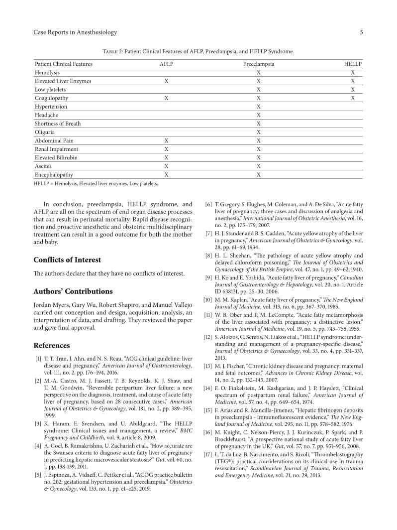
Table 2: Patient Clinical Features of AFLP, Preeclampsia, and HELLP Syndrome.
Patient Clinical Features AFLP Preeclampsia HELLPHemolysis X XElevated Liver Enzymes X X XLow platelets X XCoagulopathy X X XHypertension XHeadache XShortness of Breath XOliguria XAbdominal Pain X XRenal Impairment X XElevated Bilirubin X XAscites X XEncephalopathy X XHELLP = Hemolysis, Elevated liver enzymes, Low platelets.
In conclusion, preeclampsia, HELLP syndrome, andAFLP are all on the spectrum of end organ disease processesthat can result in perinatal mortality. Rapid disease recogni-tion and proactive anesthetic and obstetric multidisciplinarytreatment can result in a good outcome for both the motherand baby.
Conflicts of Interest
The authors declare that they have no conflicts of interest.
Authors’ Contributions
Jordan Myers, Gary Wu, Robert Shapiro, and Manuel Vallejocarried out conception and design, acquisition, analysis, aninterpretation of data, and drafting. They reviewed the paperand gave final approval.
References
[1] T. T. Tran, J. Ahn, and N. S. Reau, “ACG clinical guideline: liverdisease and pregnancy,” American Journal of Gastroenterology,vol. 111, no. 2, pp. 176–194, 2016.
[2] M.-A. Castro, M. J. Fassett, T. B. Reynolds, K. J. Shaw, andT. M. Goodwin, “Reversible peripartum liver failure: a newperspective on the diagnosis, treatment, and cause of acute fattyliver of pregnancy, based on 28 consecutive cases,” AmericanJournal of Obstetrics & Gynecology, vol. 181, no. 2, pp. 389–395,1999.
[3] K. Haram, E. Svendsen, and U. Abildgaard, “The HELLPsyndrome: Clinical issues and management. a review,” BMCPregnancy and Childbirth, vol. 9, article 8, 2009.
[4] A. Goel, B. Ramakrishna, U. Zachariah et al., “How accurate arethe Swansea criteria to diagnose acute fatty liver of pregnancyin predicting hepatic microvesicular steatosis?”Gut, vol. 60, no.1, pp. 138-139, 2011.
[5] J. Espinoza, A. Vidaeff, C. Pettker et al., “ACOGpractice bulletinno. 202: gestational hypertension and preeclampsia,” Obstetrics& Gynecology, vol. 133, no. 1, pp. e1–e25, 2019.
[6] T.Gregory, S.Hughes,M.Coleman, andA.De Silva, “Acute fattyliver of pregnancy; three cases and discussion of analgesia andanesthesia,” International Journal of Obstetric Anesthesia, vol. 16,no. 2, pp. 175–179, 2007.
[7] H. J. Stander and B. S. Cadden, “Acute yellow atrophy of the liverin pregnancy,”American Journal of Obstetrics &Gynecology, vol.28, pp. 61–69, 1934.
[8] H. L. Sheehan, “The pathology of acute yellow atrophy anddelayed chloroform poisoning,” The Journal of Obstetrics andGynaecology of the British Empire, vol. 47, no. 1, pp. 49–62, 1940.
[9] H. Ko and E. Yoshida, “Acute fatty liver of pregnancy,”CanadianJournal of Gastroenterology & Hepatology, vol. 20, no. 1, ArticleID 638131, pp. 25–30, 2006.
[10] M.M. Kaplan, “Acute fatty liver of pregnancy,”TheNew EnglandJournal of Medicine, vol. 313, no. 6, pp. 367–370, 1985.
[11] W. B. Ober and P. M. LeCompte, “Acute fatty metamorphosisof the liver associated with pregnancy; a distinctive lesion,”American Journal of Medicine, vol. 19, no. 5, pp. 743–758, 1955.
[12] S. Aloizos, C. Seretis,N. Liakos et al., “HELLP syndrome: under-standing and management of a pregnancy-specific disease,”Journal of Obstetrics & Gynaecology, vol. 33, no. 4, pp. 331–337,2013.
[13] M. J. Fischer, “Chronic kidney disease and pregnancy: maternaland fetal outcomes,” Advances in Chronic Kidney Disease, vol.14, no. 2, pp. 132–145, 2007.
[14] F. O. Finkelstein, M. Kashgarian, and J. P. Hayslett, “Clinicalspectrum of postpartum renal failure,” American Journal ofMedicine, vol. 57, no. 4, pp. 649–654, 1974.
[15] F. Arias and R. Mancilla-Jimenez, “Hepatic fibrinogen depositsin preeclampsia - immunofluorescent evidence,”The New Eng-land Journal of Medicine, vol. 295, no. 11, pp. 578–582, 1976.
[16] M. Knight, C. Nelson-Piercy, J. J. Kurinczuk, P. Spark, and P.Brocklehurst, “A prospective national study of acute fatty liverof pregnancy in the UK,” Gut, vol. 57, no. 7, pp. 951–956, 2008.
[17] L. T. da Luz, B. Nascimento, and S. Rizoli, “Thrombelastography(TEG�): practical considerations on its clinical use in traumaresuscitation,” Scandinavian Journal of Trauma, Resuscitationand Emergency Medicine, vol. 21, no. 29, 2013.

Stem Cells International
Hindawiwww.hindawi.com Volume 2018
Hindawiwww.hindawi.com Volume 2018
MEDIATORSINFLAMMATION
of
EndocrinologyInternational Journal of
Hindawiwww.hindawi.com Volume 2018
Hindawiwww.hindawi.com Volume 2018
Disease Markers
Hindawiwww.hindawi.com Volume 2018
BioMed Research International
OncologyJournal of
Hindawiwww.hindawi.com Volume 2013
Hindawiwww.hindawi.com Volume 2018
Oxidative Medicine and Cellular Longevity
Hindawiwww.hindawi.com Volume 2018
PPAR Research
Hindawi Publishing Corporation http://www.hindawi.com Volume 2013Hindawiwww.hindawi.com
The Scientific World Journal
Volume 2018
Immunology ResearchHindawiwww.hindawi.com Volume 2018
Journal of
ObesityJournal of
Hindawiwww.hindawi.com Volume 2018
Hindawiwww.hindawi.com Volume 2018
Computational and Mathematical Methods in Medicine
Hindawiwww.hindawi.com Volume 2018
Behavioural Neurology
OphthalmologyJournal of
Hindawiwww.hindawi.com Volume 2018
Diabetes ResearchJournal of
Hindawiwww.hindawi.com Volume 2018
Hindawiwww.hindawi.com Volume 2018
Research and TreatmentAIDS
Hindawiwww.hindawi.com Volume 2018
Gastroenterology Research and Practice
Hindawiwww.hindawi.com Volume 2018
Parkinson’s Disease
Evidence-Based Complementary andAlternative Medicine
Volume 2018Hindawiwww.hindawi.com
Submit your manuscripts atwww.hindawi.com