Embed Size (px)
Citation preview
Predictive therapeutic planning for irreversible electroporation treatment ofspontaneous malignant glioma
Paulo A. Garcia*,a)
School of Biomedical Engineering and Sciences, Virginia Tech –Wake Forest University, Blacksburg, VA 24061, USALaboratory for Energy and Microsystems Innovation, Department of Mechanical Engineering, Massachusetts Institute ofTechnology, Cambridge, MA 02142, USA
Bor Kos*Faculty of Electrical Engineering, University of Ljubljana, Trzaska 25, 1000 Ljubljana, Slovenia
John H. Rossmeisl Jr.School of Biomedical Engineering and Sciences, Virginia Tech –Wake Forest University, Blacksburg, VA 24061, USADepartment of Small Animal Clinical Sciences, Virginia-Maryland Regional College of Veterinary Medicine, Blacksburg, VA 24060,USAVeterinary and Comparative Neuro-oncology Laboratory, Virginia-Maryland Regional College of Veterinary Medicine, Blacksburg,VA 24060, USA
Denis Pavliha and Damijan Miklav�ci�cFaculty of Electrical Engineering, University of Ljubljana, Trzaska 25, 1000 Ljubljana, Slovenia
Rafael V. DavalosSchool of Biomedical Engineering and Sciences, Virginia Tech –Wake Forest University, Blacksburg, VA 24061, USA
(Received 16 August 2016; revised 14 April 2017; accepted for publication 7 May 2017;published 25 July 2017)
Purpose: Irreversible electroporation (IRE) has been developed as a promising minimally invasive
treatment to ablate spontaneous brain tumors with pulsed electric fields in canine patients. The pur-
pose of the study is to determine the Peleg-Fermi parameters needed to incorporate pulse number
and pulse duration into the therapeutic planning of IRE.
Methods: Seven canine patients were treated with IRE for spontaneous malignant glioma with MRI-
based treatment planning. The treatment planning method consists of building patient-specific finite
element models and using them to compute electric fields used in the IRE treatment. We evaluate the
predictive power of tumor coverage with electric field alone vs. cell kill probability using radiograph-
ically confirmed clinical outcomes.
Results: Results of post-treatment diagnostic imaging, tumor biopsies, and neurological examina-
tions indicated successful tumor ablation without significant direct neurotoxicity in six of the seven
dogs. Objective tumor responses were seen in four (80%) of five dogs with quantifiable target lesions
according to RANO criteria. Two dogs experienced survivals in excess of 1 yr, including one dog that
resulted in complete response to IRE treatment for 5+ years to date. Tumor fraction exposed to elec-
tric field over 600 V/cm was between 0.08 and 0.73, while tumor fraction exposed to electric field
over 300 V/cm was between 0.17 and 0.95. Probability of cell kill of ≥ 90% was found in tumor vol-
ume fractions between 0.21 and 0.99.
Conclusions: We conclude that IRE is a safe and effective minimally invasive treatment for malig-
nant glioma and can be predicted with the Peleg-Fermi cell kill probability function. A tumor cover-
age of ≥ 0.9 at a cell kill probability ≥ 90% can be used to guide IRE treatments of spontaneous
malignant glioma based on the radiographically confirmed clinical outcomes achieved. © 2017 The
Authors. Medical Physics published by Wiley Periodicals, Inc. on behalf of American Association of
Physicists in Medicine. [https://doi.org/10.1002/mp.12401]
Key words: brain tumor, minimally invasive, neurosurgery, pulsed electric fields, treatment planning
1. INTRODUCTION
Malignant gliomas, especially glioblastoma multiforme
(GBM), are among the most aggressive human malignancies.
Patients suffering from this disease have poor prognosis with
a 5-yr survival rate of about 10%.1 The median survival for
patients with GBM is only 15 months even with singular or
multimodal therapy consisting of surgical resection, radiation
therapy, and/or chemotherapy.1,2 Despite development of
novel therapies for treatment of malignant glioma in trans-
genic and genetically engineered rodent glioma models, few
therapeutic advances have emerged that drastically improve
survival for patients with aggressive, high-grade gliomas.3
An obstacle for the translation of therapeutic developments
into humans is the inability of the animal model to reliably
reproduce the phenotypically and genotypically diverse tumor
4968 Med. Phys. 44 (9), September 2017 0094-2405/2017/44(9)/4968/13
© 2017 The Authors. Medical Physics published by WileyPeriodicals, Inc. on behalf of American Association of Physicists in
Medicine. This is an open access article under the terms of theCreative Commons Attribution-NonCommercial License, which
permits use, distribution and reproduction in any medium,provided the original work is properly cited and is not used for
commercial purposes.
4968
population that is observed in humans. Therefore, developing
treatment technologies in a glioma model system of a mean-
ingful size that faithfully reproduces the human disease is
critical for improving the outcome of brain cancer patients.
Canine patients with spontaneous brain tumors have been
recognized for their promise as a large translational glioma
model for preclinical assessment and development of novel
therapeutics.4 In dogs with several types of brain tumors, sta-
tistics on median survivals of 0.2, 0.9, and 4.9 months Statis-
tics have been reported after receiving either symptomatic
therapy, cytoreductive surgery, or multimodal therapy (radio-
therapy and surgery or hyperthermia) respectively.5 These
statistics demonstrate the poor long-term prognosis for dogs
with brain tumors and the urgent need for the development of
more effective therapies.5 There are substantial clinical, epi-
demiological, cytogenetic, and pathophysiological similari-
ties between human and canine brain tumors that have been
identified.6 The role and value of dogs with spontaneous
brain tumors in the development of experimental therapeutics
has been demonstrated. Seminal work in dogs with sponta-
neous gliomas illustrated the feasibility and importance of
real-time imaging monitoring of convection-enhanced deliv-
ery (CED) for confirmation of target coverage, as well as pro-
viding an opportunity to detect and remedy any local adverse
effects of CED treatment, including reflux.7 A preclinical
study investigating dendritic cell vaccination of glioma-bear-
ing dogs with tumor cell lysates containing a toll-like recep-
tor ligand adjuvant in combination with in situ adenoviral
interferon-gamma gene transfer demonstrated sufficient
safety and promise to result in rapid translation of this
immunogenetic therapy to a human glioblastoma clinical
trial,8 and promising active immunotherapeutic approaches
using dogs with intracranial meningiomas have recently been
published. Canine patients with spontaneous disease are thus
an attractive preclinical model since the size and heterogene-
ity of their gliomas closely reproduce the characteristics of
brain tumors in humans.
Recently, we demonstrated the safety and feasibility of
irreversible electroporation (IRE) for the treatment of
spontaneous intracranial gliomas in canine patients using
patient-specific therapeutic planning.9 IRE is a new, safe, and
effective minimally invasive nonthermal ablation modality
with the potential to treat unresectable tumors such as many
gliomas in close proximity to critical structures.10–14 An IRE
treatment involves placing minimally invasive electrodes
within or around the tumor tissue and delivering a series of
short high voltage electric pulses to the targeted tumor tis-
sue.15–17 The exposure of the tumor to pulsed electric fields
destabilizes the membranes of the cancer cells, achieving
death in a precise and controllable manner with cell-scale
resolution.16,18–20
In order to guide the physicians and veterinarians perform-
ing electroporation-based therapies, anatomically accurate
numerical models have been developed for maximizing
tumor coverage while minimizing damage to surrounding
healthy tissue during pulse delivery.21–24 The pulse parame-
ters employed in the clinical study were determined from
patient-specific computational models of the electric field
distributions due to the sensitive nature of the intracranial
environment.9,25 In these models, as in many other studies of
electroporation technologies, the electric field distribution
was used to determine the tumor coverage based on electric
field threshold values.21,26–30 The imaging-based treatment
planning model developed for each canine patient dictated
the surgical approach for intracranial electrode placement.
The models also helped determine the voltages that needed to
be applied assuming a lethal electric field threshold of
500 V/cm that was determined from magnetic resonance
imaging (MRI) reconstructions of IRE ablation zones with
ninety 50-ls pulses in normal brain.9,28 The radiographically
confirmed clinical outcomes demonstrate that we achieved
two complete responses (CR), two stable diseases (SD), and
one partial response (PR) after IRE therapy of the malignant
gliomas in the five canine patients that were treated with
curative intent.
The goal of this study is to further refine the imaging-
based computational models to explicitly incorporate addi-
tional critical treatment parameters such as pulse number and
pulse duration to improve the predictive power of the cell kill
distribution during the therapeutic planning procedure which
can serve to improve treatment planning for brain tumors in
the future.31–33 Specifically, we correlate the radiographically
confirmed clinical outcomes with advanced modeling
approaches to provide the clinical community with predictive
planning tools for IRE treatment of intracranial glioma.
2. MATERIALS AND METHODS
2.A. Study design and canine patient population forclinical study
This was a single-center, prospective study conducted to
assess the safety and feasibility of using the NanoKnife Sys-
tem for ablation of spontaneous canine intracranial gliomas
with irreversible electroporation.9 Canine patients were trea-
ted according to the standards in the Guide for Care and Use
of Laboratory Animals, and were approved by the institu-
tional animal care and use committee and the Virginia-Mary-
land College of Veterinary Medicine clinical review board.
Candidates for this study consisted of client-owned dogs
with suspected spontaneous telencephalic gliomas based on
MRI evaluation and tumors were biopsied and histologically
confirmed prior to IRE treatment (Table I). For inclusion,
canine patients were required to undergo a diagnostic MRI
within 2 weeks of enrollment showing a single telencephalic
intra-axial mass lesion with imaging characteristics consis-
tent with glioma,34 have stable neurological and cardiopul-
monary functions, and have no evidence of substantial
hepatic or renal dysfunction or concurrent systemic malig-
nancy. Any type of radiotherapy treatment of the brain tumor
was prescribed as exclusion criteria. A passive approach was
used to enroll cases following identification from the routine
clinical population presented to the Veterinary Teaching
Hospital or its referral partners. The recruitment of each
Medical Physics, 44 (9), September 2017
4969 Garcia et al.: Predictive IRE planning for malignant glioma 4969
patient occurred after the principal investigator’s clinical
review of each potential candidate. Clients provided written,
informed consent for their canine patients to receive irre-
versible electroporation treatments. The population with
intent-to-treat was defined as those clinical cases in which
the NanoKnife electrode (www.angiodynamics.com) probes
were inserted into the brain tissue. The specific IRE treat-
ment protocols, electrode configuration, and tumor character-
istics are given in Table II.9
2.B. MRI sequence for tumor diagnosis,segmentation, and treatment planning
Each canine patient was scanned in a 1.5-T Phillips Intera
magnet according to the standardized clinical brain tumor
protocol sequences presented in Table I. Mimics 14.1 image
analysis software (Materialise, Leuven, Belgium) and the
Visifield (www.visifield.com) software were used to segment
the normal brain and tumor tissue in the T1-weighted post-
contrast MRI sequences. The tumors were traced in a semi-
automated manner in each of the two-dimensional (2-D) axial
planes based on the increased intensity due to Gadolinium
uptake. The treatment zones were identified by (a) the
changes of Gadolinium contrast agents in T1W + C scans or
(b) anatomical inspection of the tumor location in the two
canine patients with non-enhancing tumors. Specifically,
therapeutic response assessments were evaluated with
response assessment in neurooncology (RANO) criteria.35
For each patient individually, the segmented medical images
were used to construct the 3D model using the following
approach. The electrode tips and trajectories were identified
in the original medical images. The electrodes were then
inserted in the model as cylinders with 1-mm diameter and
length according to the case, while aligning the coordinate
system of the model with that of the original medical images.
The mesh was built in a rectangular cuboid domain with the
faces either 5 cm away from the electrodes or corresponding
to the edges of the original medical images, whichever was
closer. For the investigation of the effect of increasing or
reducing the tumor size, the binary segmentation image was
dilated or eroded with a circular structural element with a
radius of two pixels in the axial slices. The tumor was not
dilated or eroded in the z direction of the images, because the
distance between slices was much larger than the pixel size
and some tumors were visible only for a few sequential slices
(e.g., three slices in the case of patient 3). Although the elec-
trodes displaced a small volume of tissue during the treat-
ment, the model was not deformed in any way because the
tissue was soft and compressible.
2.C. Computational modeling of electric field forassessing tumor coverage
The electric field modeling was performed using the finite
element method. We used COMSOL Multiphysics (Comsol
AB, Stockholm, Sweden) and its AC/DC module to model
the electric field. The stationary Laplace equation for electric
potential was solved in the box-shaped computational
domain, which extended 5 cm from the outer dimensions of
the electrodes. The active electrodes were assigned the
Dirichlet boundary condition, while the outer boundaries
were defined as electrically insulating. The electric field was
solved with an iterative scheme, where COMSOL Multi-
physics was controlled from Matlab (Mathworks, Natick,
MA, USA). The material properties were assigned to the
mesh elements according to the segmented images, where
each tissue was assigned different electrical properties.36 The
electric properties of tumor and brain tissues were taken from
previous work,28,30 since the increase of conductivity during
electroporation is a well-documented phenomenon.37,38 The
values of tissue conductivities used are reported in
Table III.39,40 Each iteration consisted of a static simulation
TABLE I. Sequence parameters for MRI acquisition in canine patients.
Sequence
Image
weighting
Acquisition
plane
2D vs.
3D
TE
(ms)
TR
(ms)
TI
(ms)
Flip angle
(degrees) NEX
Echo
train
length
Slice
thickness
(mm)
Interslice
gap (mm)
FOV
(mm)
Matrix
dimensions b-value
TSE T2 Sag. 2D 100 3828 – 90 6 15 3 3.3 110 184 9 179 –
TSE T2 Trans. 2D 110 7048 – 90 6 22 3 3.3 110 184 9 158 –
GRE T2 Trans. 2D 23 1050 – 18 2 1 3 3.3 110 156 9 142 –
SE T1 Trans. 2D 11 400 – 80 2 1 2.6 2.9 110 184 9 201 –
SE T1 Sag. 2D 11 400 – 80 2 1 3 3.3 110 184 9 183 –
GRE T1 Trans. 3D 4.5 25 – 30 2 1 1.6 0.8 110 168 9 167 –
FLAIR T2 Trans. 2D 140 11000 2800 90 4 27 3 3.3 110 140 9 90 –
DWI T2 Trans. 2D 72 4216 – 90 3 63 2.6 2.9 110 72 9 57 1000
S/G/Da T2 Trans. 3D 49 35 – 7 1 19 3.5 – 110 64 9 60 –
SE T1 Post Trans. 2D 11 400 – 80 2 1 2.6 3.3 110 184 9 201 –
SE T1 Post Sag. 2D 11 400 – 80 2 1 3 3.3 110 184 9 183 –
SE T1 Post Dorsal 2D 11 400 – 80 2 1 3 3.3 110 216 9 185 –
GRE T1 Post Trans. 3D 4.5 25 – 30 2 1 1.6 0.8 110 168 9 167 –
aDenotes S-PRESTO/GRE/DCE-MR.
Medical Physics, 44 (9), September 2017
4970 Garcia et al.: Predictive IRE planning for malignant glioma 4970
of the electric field; however, between the iterations, the tis-
sue conductivities were increased as a function of the electric
field.30,41,42 This was repeated for every electrode pair used
in the treatment and the maximum electric field was
recorded.43 For the outcome analysis, the maximum electric
field from all electrode pairs was taken to compute the cover-
age of the treated volume.
2.D. Cell kill probability calculations for patient-specific brain tumors
The modeling of cell kill probability was performed using
the Peleg-Fermi model.44 This mathematical description was
first developed to describe microbial inactivation after treat-
ment with pulsed electric fields, and later adapted for use in
irreversible electroporation.31–33 The model uses the follow-
ing equation:
S ¼1
1þ expE�EcðnÞAðnÞ
h i ; (1)
where S is the probability of cell survival, E is the electric
field to which the cells are exposed, EcðnÞ is the critical elec-tric field as a function of the number of pulses over which
50% of cells are expected to die and AðnÞ is a shape factor
which determines the transition between unaffected and com-
pletely ablated tissue. Specifically, EcðnÞ and AðnÞ are
defined as follows:
EcðnÞ ¼ Ec0 � expð�k1nÞ; (2)
AðnÞ ¼ A0 � expð�k2nÞ; (3)
with Ec0 being the initial value of the critical electric field, A0
the initial value of the shape factor, k1 and k2 the exponential
decay coefficients, and n being the number of pulses used in
the treatment respectively. Although the model was initially
based on in vitro data, it has since been applied to in vivo
results for IRE in normal brain tissue of rats.45 The values of
the parameters used in the literature so far are listed in
Table IV. The exact values of parameters were adjusted from
Sharabi et al.45 based on the complete response (CR)
achieved in patient 4 with IRE treatment alone.
In the cases where more than one electrode pair was used,
Eq. (1) was computed for every electrode pair separately as a
scalar field and then multiplied:
Sðx; y; zÞ ¼Y
8i
Siðx; y; zÞ (4)
where Si is the probability of survival for i-th electrode pair.
The cell kill probability is then defined as 1 � S.
3. RESULTS
3.A. Electric field distribution within MRI-basedintracranial environment
One of the main advantages of IRE in comparison with
other focal ablation techniques is the dependency on the elec-
tric field exposure to achieve therapeutic effects. Sufficient
electric field exposure is thus required for tumor response to
the IRE treatment without damaging surrounding critical
structures such as major blood vessels. The steps to compute
the electric field distribution from MRI-based volumetric
reconstructions of the tissue and tumor components in the
brain are demonstrated in Fig. 1. First, individual tissues
(e.g., white matter, gray matter, and tumor) are segmented
from each of the MR images in the sequence [Fig. 1(a)]. The
tissue-specific segmentation allows for determining the tumor
dimension and formulating the insertion approach to use dur-
ing the surgery. The individual 2D segmentations are then
reconstructed to generate a single 3D volumetric representa-
tion of the tissue components [Fig. 1(b)]. The volumetric
TABLE II. Physical tumor characteristics, electrode configuration, and electrical parameters employed during irreversible electroporation treatment of sponta-
neous malignant glioma.
Patient Tumor type Tumor (cm3)
Electrode
number Pulses per electrode pair Total pulses Voltage (V) Post-IRE (months)
Best clinical
outcome
1 AOA 1.15 2 40/80 120 500/625 4 CR
2 GBM 2.85 2 80/80 160 625/500 n/a ND
3 AO 0.98 4 80/80 160 750/1000 3 PR
4 AA 2.17 6 90/90/90 270 900/1000/800 24a CR
5 GBM 4.98 6 50/50/50 150 500/1000/1000 2 PR
6 O 0.55 2 90 90 1000 12 SD
7 A 0.85 4 40/40/40/40 160 800/800/800/800 2 SD
Note: aThis patient has been in complete response for 5+ years post-IRE treatment. AA, anaplastic (Grade III) astrocytoma; AO, anaplastic (Grade III) oligodendroglioma;
AOA, anaplastic (Grade III) oligoastrocytoma; GBM, glioblastoma multiforme (Grade IV astrocytoma); O, Grade II oligodendroglioma (Grade II); A, astrocytoma (Grade
II).
TABLE III. Physical tissue properties employed in the treatment planning
models.
Tissue rinitial (S/m) rmaxium (S/m) References
Gray matter 0.285 0.7359 [39]
White matter 0.3621 0.7357 [39]
Brain tumor (glioma) 0.435 0.7373 [39]
Skull 0.02 n/a [40]
Medical Physics, 44 (9), September 2017
4971 Garcia et al.: Predictive IRE planning for malignant glioma 4971
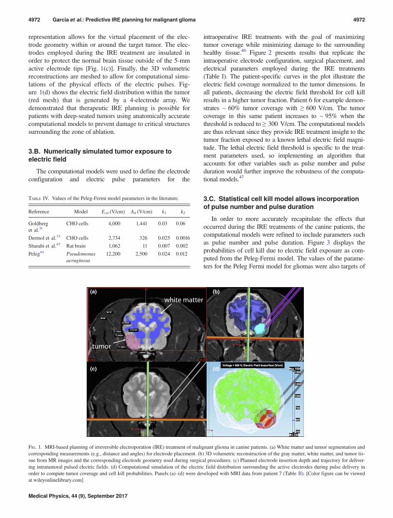
representation allows for the virtual placement of the elec-
trode geometry within or around the target tumor. The elec-
trodes employed during the IRE treatment are insulated in
order to protect the normal brain tissue outside of the 5-mm
active electrode tips [Fig. 1(c)]. Finally, the 3D volumetric
reconstructions are meshed to allow for computational simu-
lations of the physical effects of the electric pulses. Fig-
ure 1(d) shows the electric field distribution within the tumor
(red mesh) that is generated by a 4-electrode array. We
demonstrated that therapeutic IRE planning is possible for
patients with deep-seated tumors using anatomically accurate
computational models to prevent damage to critical structures
surrounding the zone of ablation.
3.B. Numerically simulated tumor exposure toelectric field
The computational models were used to define the electrode
configuration and electric pulse parameters for the
intraoperative IRE treatments with the goal of maximizing
tumor coverage while minimizing damage to the surrounding
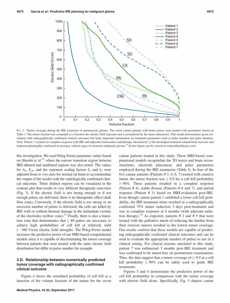
healthy tissue.46 Figure 2 presents results that replicate the
intraoperative electrode configuration, surgical placement, and
electrical parameters employed during the IRE treatments
(Table I). The patient-specific curves in the plot illustrate the
electric field coverage normalized to the tumor dimensions. In
all patients, decreasing the electric field threshold for cell kill
results in a higher tumor fraction. Patient 6 for example demon-
strates ~ 60% tumor coverage with ≥ 600 V/cm. The tumor
coverage in this same patient increases to ~ 95% when the
threshold is reduced to ≥ 300 V/cm. The computational models
are thus relevant since they provide IRE treatment insight to the
tumor fraction exposed to a known lethal electric field magni-
tude. The lethal electric field threshold is specific to the treat-
ment parameters used, so implementing an algorithm that
accounts for other variables such as pulse number and pulse
duration would further improve the robustness of the computa-
tional models.47
3.C. Statistical cell kill model allows incorporationof pulse number and pulse duration
In order to more accurately recapitulate the effects that
occurred during the IRE treatments of the canine patients, the
computational models were refined to include parameters such
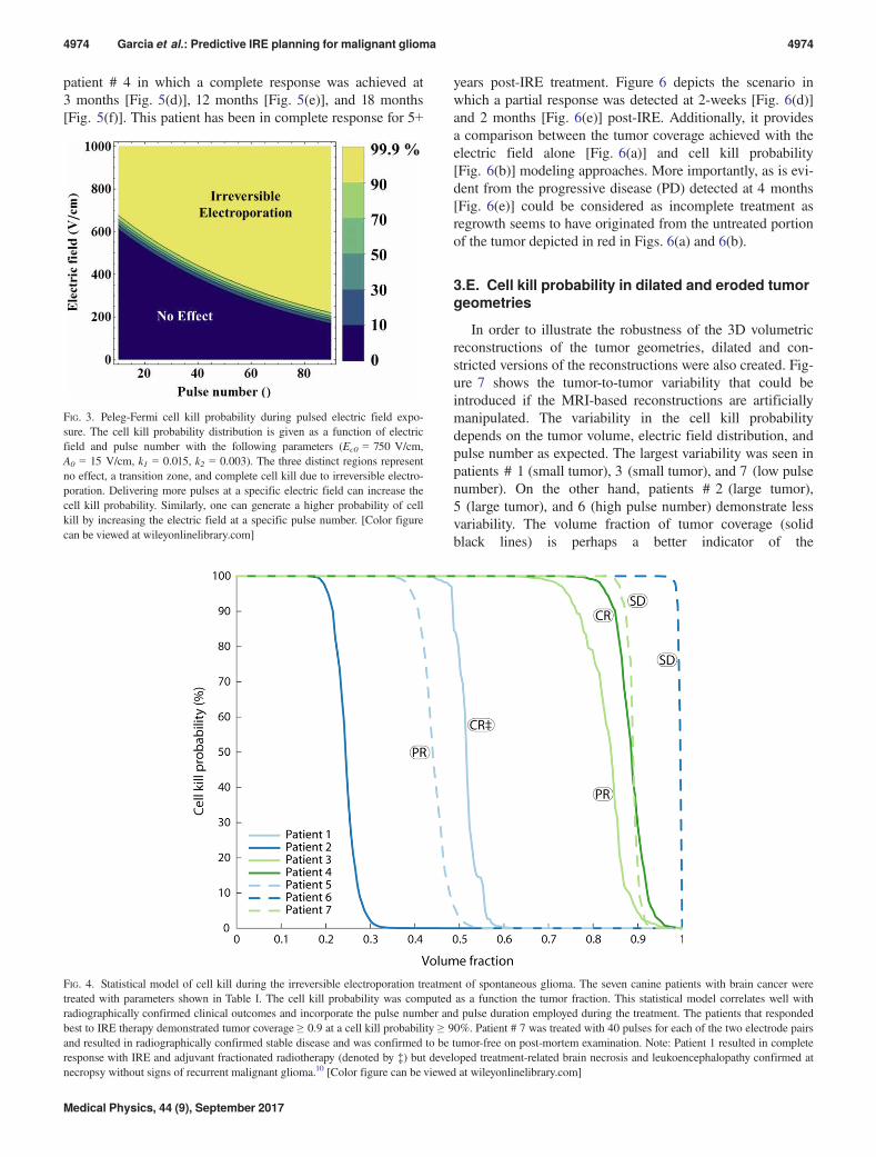
as pulse number and pulse duration. Figure 3 displays the
probabilities of cell kill due to electric field exposure as com-
puted from the Peleg-Fermi model. The values of the parame-
ters for the Peleg Fermi model for gliomas were also targets of
TABLE IV. Values of the Peleg-Fermi model parameters in the literature.
Reference Model Ec0 (V/cm) A0 (V/cm) k1 k2
Goldberg
et al.31CHO cells 4,000 1,441 0.03 0.06
Dermol et al.33 CHO cells 2,734 326 0.025 0.0016
Sharabi et al.45 Rat brain 1,062 11 0.007 0.002
Peleg44 Pseudomonas
aeruginosa
12,200 2,500 0.024 0.012
white matter
tumor
(a) (b)
(c) (d)
FIG. 1. MRI-based planning of irreversible electroporation (IRE) treatment of malignant glioma in canine patients. (a) White matter and tumor segmentation and
corresponding measurements (e.g., distance and angles) for electrode placement. (b) 3D volumetric reconstruction of the gray matter, white matter, and tumor tis-
sue from MR images and the corresponding electrode geometry used during surgical procedures. (c) Planned electrode insertion depth and trajectory for deliver-
ing intratumoral pulsed electric fields. (d) Computational simulation of the electric field distribution surrounding the active electrodes during pulse delivery in
order to compute tumor coverage and cell kill probabilities. Panels (a)–(d) were developed with MRI data from patient 7 (Table II). [Color figure can be viewed
at wileyonlinelibrary.com]
Medical Physics, 44 (9), September 2017
4972 Garcia et al.: Predictive IRE planning for malignant glioma 4972
this investigation. We used Peleg-Fermi parameter values based
on Sharabi et al.45 where the narrow transition region between
IRE-ablated and unablated regions was also noted. The values
for A0, Ec0, and the exponent scaling factors k1 and k2 were
adjusted from in vivo data for normal rat brain to accommodate
the output of the model with the radiologically confirmed clini-
cal outcomes. Three distinct regions can be visualized in the
contour plot that results in very different therapeutic outcomes
(Fig. 3). If the electric field is not strong enough or if not
enough pulses are delivered, there is no therapeutic effect (dark
blue zone). Conversely, if the electric field is too strong or an
excessive number of pulses is delivered, the cells are killed by
IRE with or without thermal damage in the immediate vicinity
of the electrodes (yellow zone).32 Finally, there is also a transi-
tion zone that demonstrates that ≥ 80 pulses are necessary to
achieve high probabilities of cell kill at relatively mild
(~ 300 V/cm) electric field strengths. The Peleg-Fermi model
increases the predictive power of our MRI-based computational
models since it is capable of discriminating the tumor coverage
between patients that were treated with the same electric field
distribution but differ in pulse number for example.
3.D. Relationship between numerically predictedtumor coverage with radiographically confirmedclinical outcome
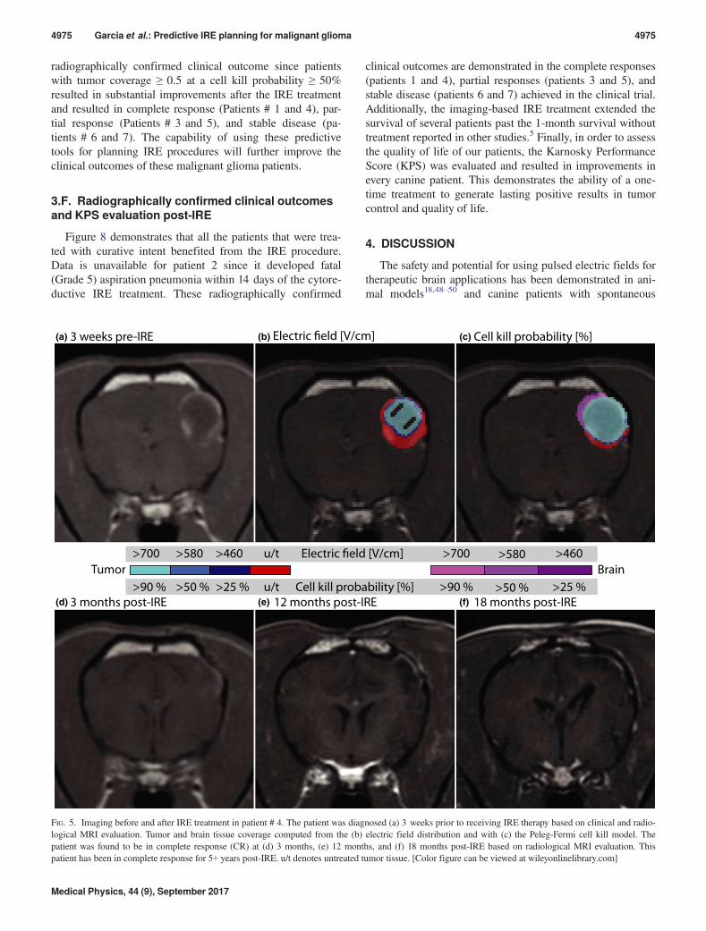
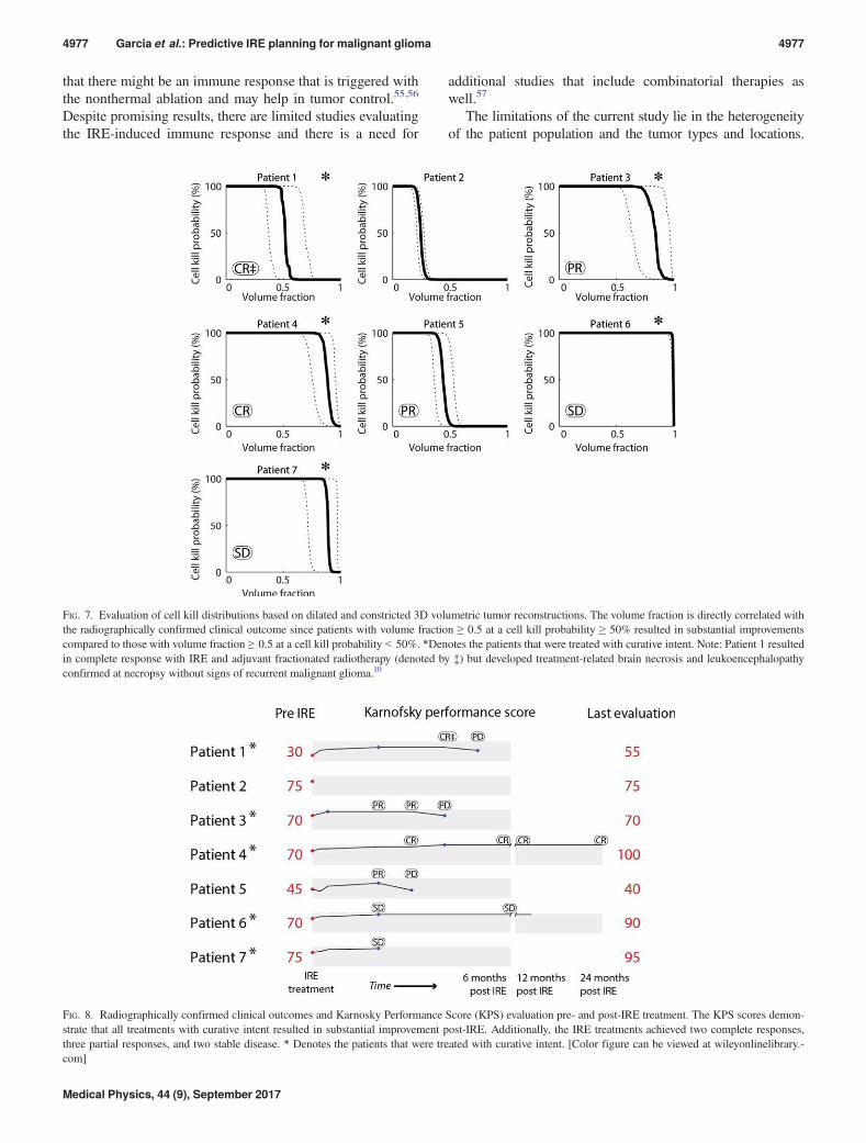
Figure 4 shows the simulated probability of cell kill as a
function of the volume fraction of the tumor for the seven
canine patients treated in this study. These MRI-based com-
putational models recapitulate the 3D tumor and brain recon-
structions, electrode placement, and pulse parameters
employed during the IRE treatments (Table I). In four of the
five canine patients (Patients # 3, 4, 6, 7) treated with curative
intent, the tumor fraction was ≥ 0.9 for a cell kill probability
≥ 90%. These patients resulted in a complete response
(Patient # 4), stable disease (Patients # 6 and 7), and partial
response (Patient # 3) based on MRI-evaluation post-IRE.
Even though canine patient 1 exhibited a lower cell kill prob-
ability, the IRE treatment alone resulted in a radiographically
confirmed 75% tumor reduction 3 days post-treatment and
was in complete response at 4 months (with adjuvant radia-
tion therapy).10 As expected, patients # 2 and # 5 that were
treated with the palliative intent of reducing the burden from
their oversize tumors resulted in the lowest tumor coverage.
Our results confirm that these models are capable of predict-
ing radiographically confirmed clinical outcomes and can be
used to evaluate the appropriate number of pulses to use in a
clinical setting. For clinical reasons unrelated to this study,
patient 7 was euthanized 3 months post-IRE treatment and
was confirmed to be tumor-free on postmortem examination.
Thus, the data suggest that a tumor coverage of ≥ 0.9 at a cell
kill probability ≥ 90% can be safely used to guide IRE
treatments.
Figures 5 and 6 demonstrate the predictive power of the
cell kill probability in comparison with the tumor coverage
with electric field alone. Specifically, Fig. 5 depicts canine
FIG. 2. Tumor coverage during the IRE treatment of spontaneous glioma. The seven canine patients with brain cancer were treated with parameters shown in
Table I. The tumor fraction was computed as a function the electric field exposure and is normalized by the tumor dimension. This model demonstrates good cor-
relation with radiographically confirmed clinical outcomes but lacks important information on treatment parameters such as pulse number and pulse duration.
Note: Patient 1 resulted in complete response with IRE and adjuvant fractionated radiotherapy (denoted by ‡) but developed treatment-related brain necrosis and
leukoencephalopathy confirmed at necropsy without signs of recurrent malignant glioma.10 [Color figure can be viewed at wileyonlinelibrary.com]
Medical Physics, 44 (9), September 2017
4973 Garcia et al.: Predictive IRE planning for malignant glioma 4973
patient # 4 in which a complete response was achieved at
3 months [Fig. 5(d)], 12 months [Fig. 5(e)], and 18 months
[Fig. 5(f)]. This patient has been in complete response for 5+
years post-IRE treatment. Figure 6 depicts the scenario in
which a partial response was detected at 2-weeks [Fig. 6(d)]
and 2 months [Fig. 6(e)] post-IRE. Additionally, it provides
a comparison between the tumor coverage achieved with the
electric field alone [Fig. 6(a)] and cell kill probability
[Fig. 6(b)] modeling approaches. More importantly, as is evi-
dent from the progressive disease (PD) detected at 4 months
[Fig. 6(e)] could be considered as incomplete treatment as
regrowth seems to have originated from the untreated portion
of the tumor depicted in red in Figs. 6(a) and 6(b).
3.E. Cell kill probability in dilated and eroded tumorgeometries
In order to illustrate the robustness of the 3D volumetric
reconstructions of the tumor geometries, dilated and con-
stricted versions of the reconstructions were also created. Fig-
ure 7 shows the tumor-to-tumor variability that could be
introduced if the MRI-based reconstructions are artificially
manipulated. The variability in the cell kill probability
depends on the tumor volume, electric field distribution, and
pulse number as expected. The largest variability was seen in
patients # 1 (small tumor), 3 (small tumor), and 7 (low pulse
number). On the other hand, patients # 2 (large tumor),
5 (large tumor), and 6 (high pulse number) demonstrate less
variability. The volume fraction of tumor coverage (solid
black lines) is perhaps a better indicator of the
FIG. 3. Peleg-Fermi cell kill probability during pulsed electric field expo-
sure. The cell kill probability distribution is given as a function of electric
field and pulse number with the following parameters (Ec0 = 750 V/cm,
A0 = 15 V/cm, k1 = 0.015, k2 = 0.003). The three distinct regions represent
no effect, a transition zone, and complete cell kill due to irreversible electro-
poration. Delivering more pulses at a specific electric field can increase the
cell kill probability. Similarly, one can generate a higher probability of cell
kill by increasing the electric field at a specific pulse number. [Color figure
can be viewed at wileyonlinelibrary.com]
FIG. 4. Statistical model of cell kill during the irreversible electroporation treatment of spontaneous glioma. The seven canine patients with brain cancer were
treated with parameters shown in Table I. The cell kill probability was computed as a function the tumor fraction. This statistical model correlates well with
radiographically confirmed clinical outcomes and incorporate the pulse number and pulse duration employed during the treatment. The patients that responded
best to IRE therapy demonstrated tumor coverage ≥ 0.9 at a cell kill probability ≥ 90%. Patient # 7 was treated with 40 pulses for each of the two electrode pairs
and resulted in radiographically confirmed stable disease and was confirmed to be tumor-free on post-mortem examination. Note: Patient 1 resulted in complete
response with IRE and adjuvant fractionated radiotherapy (denoted by ‡) but developed treatment-related brain necrosis and leukoencephalopathy confirmed at
necropsy without signs of recurrent malignant glioma.10 [Color figure can be viewed at wileyonlinelibrary.com]
Medical Physics, 44 (9), September 2017
4974 Garcia et al.: Predictive IRE planning for malignant glioma 4974
radiographically confirmed clinical outcome since patients
with tumor coverage ≥ 0.5 at a cell kill probability ≥ 50%
resulted in substantial improvements after the IRE treatment
and resulted in complete response (Patients # 1 and 4), par-
tial response (Patients # 3 and 5), and stable disease (pa-
tients # 6 and 7). The capability of using these predictive
tools for planning IRE procedures will further improve the
clinical outcomes of these malignant glioma patients.
3.F. Radiographically confirmed clinical outcomesand KPS evaluation post-IRE
Figure 8 demonstrates that all the patients that were trea-
ted with curative intent benefited from the IRE procedure.
Data is unavailable for patient 2 since it developed fatal
(Grade 5) aspiration pneumonia within 14 days of the cytore-
ductive IRE treatment. These radiographically confirmed
clinical outcomes are demonstrated in the complete responses
(patients 1 and 4), partial responses (patients 3 and 5), and
stable disease (patients 6 and 7) achieved in the clinical trial.
Additionally, the imaging-based IRE treatment extended the
survival of several patients past the 1-month survival without
treatment reported in other studies.5 Finally, in order to assess
the quality of life of our patients, the Karnosky Performance
Score (KPS) was evaluated and resulted in improvements in
every canine patient. This demonstrates the ability of a one-
time treatment to generate lasting positive results in tumor
control and quality of life.
4. DISCUSSION
The safety and potential for using pulsed electric fields for
therapeutic brain applications has been demonstrated in ani-
mal models18,48–50 and canine patients with spontaneous
Cell kill probability [%]3 weeks pre-IRE
Electric field [V/cm]
3 months post-IRE 12 months post-IRE 18 months post-IRE
>700 >580 >460 u/t
>90 % >50 % >25 %
>700 >580 >460
>90 % >50 % >25 %u/t
Tumor Brain
Cell kill probability [%]
(a) (b) Electric field [V/cm] (c)
(d) (e) (f)
FIG. 5. Imaging before and after IRE treatment in patient # 4. The patient was diagnosed (a) 3 weeks prior to receiving IRE therapy based on clinical and radio-
logical MRI evaluation. Tumor and brain tissue coverage computed from the (b) electric field distribution and with (c) the Peleg-Fermi cell kill model. The
patient was found to be in complete response (CR) at (d) 3 months, (e) 12 months, and (f) 18 months post-IRE based on radiological MRI evaluation. This
patient has been in complete response for 5+ years post-IRE. u/t denotes untreated tumor tissue. [Color figure can be viewed at wileyonlinelibrary.com]
Medical Physics, 44 (9), September 2017
4975 Garcia et al.: Predictive IRE planning for malignant glioma 4975
disease.9,10 These applications involve reversible disruption
of the blood-brain-barrier for delivery of therapeutic
agents48,49,51,52 or irreversible treatment for localized tissue/
tumor ablation.9,10,53 Recently, Tumor Treating Fields (TTFs)
received FDA approval for GBM treatment due to the demon-
strated ability to add 3 months to the progression-free and
overall survival of human patients when used in combination
with temozolomide chemotherapy in a phase III clinical
trial.54 Despite encouraging clinical outcomes, the median
survival for human GBM patients is only 15 months with sin-
gular or multimodal therapy consisting of surgical resection,
chemotherapy, and/or radiation therapy.1,2 In this study, all
the canine patients that were treated with curative intent
demonstrated partial-to-complete responses and improved
quality of life, including a patient (patient 4) that has been
tumor free for 5+ years and one that was tumor free based on
histological evaluation post-mortem 3 months after
treatment.
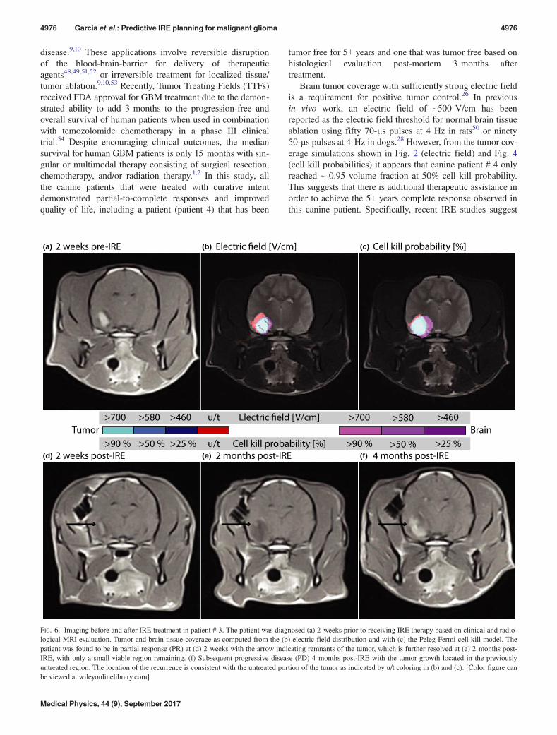
Brain tumor coverage with sufficiently strong electric field
is a requirement for positive tumor control.26 In previous
in vivo work, an electric field of ~500 V/cm has been
reported as the electric field threshold for normal brain tissue
ablation using fifty 70-ls pulses at 4 Hz in rats50 or ninety
50-ls pulses at 4 Hz in dogs.28 However, from the tumor cov-
erage simulations shown in Fig. 2 (electric field) and Fig. 4
(cell kill probabilities) it appears that canine patient # 4 only
reached ~ 0.95 volume fraction at 50% cell kill probability.
This suggests that there is additional therapeutic assistance in
order to achieve the 5+ years complete response observed in
this canine patient. Specifically, recent IRE studies suggest
Electric field [V/cm] Cell kill probability [%]2 weeks pre-IRE
2 weeks post-IRE 2 months post-IRE 4 months post-IRE
>700 >580 >460 u/t
>90 % >50 % >25 %
>700 >580 >460
>90 % >50 % >25 %u/t
Tumor Brain
Electric field [V/cm]
Cell kill probability [%]
(a) (b) (c)
(d) (e) (f)
FIG. 6. Imaging before and after IRE treatment in patient # 3. The patient was diagnosed (a) 2 weeks prior to receiving IRE therapy based on clinical and radio-
logical MRI evaluation. Tumor and brain tissue coverage as computed from the (b) electric field distribution and with (c) the Peleg-Fermi cell kill model. The
patient was found to be in partial response (PR) at (d) 2 weeks with the arrow indicating remnants of the tumor, which is further resolved at (e) 2 months post-
IRE, with only a small viable region remaining. (f) Subsequent progressive disease (PD) 4 months post-IRE with the tumor growth located in the previously
untreated region. The location of the recurrence is consistent with the untreated portion of the tumor as indicated by u/t coloring in (b) and (c). [Color figure can
be viewed at wileyonlinelibrary.com]
Medical Physics, 44 (9), September 2017
4976 Garcia et al.: Predictive IRE planning for malignant glioma 4976
that there might be an immune response that is triggered with
the nonthermal ablation and may help in tumor control.55,56
Despite promising results, there are limited studies evaluating
the IRE-induced immune response and there is a need for
additional studies that include combinatorial therapies as
well.57
The limitations of the current study lie in the heterogeneity
of the patient population and the tumor types and locations.
FIG. 7. Evaluation of cell kill distributions based on dilated and constricted 3D volumetric tumor reconstructions. The volume fraction is directly correlated with
the radiographically confirmed clinical outcome since patients with volume fraction ≥ 0.5 at a cell kill probability ≥ 50% resulted in substantial improvements
compared to those with volume fraction ≥ 0.5 at a cell kill probability < 50%. *Denotes the patients that were treated with curative intent. Note: Patient 1 resulted
in complete response with IRE and adjuvant fractionated radiotherapy (denoted by ‡) but developed treatment-related brain necrosis and leukoencephalopathy
confirmed at necropsy without signs of recurrent malignant glioma.10
FIG. 8. Radiographically confirmed clinical outcomes and Karnosky Performance Score (KPS) evaluation pre- and post-IRE treatment. The KPS scores demon-
strate that all treatments with curative intent resulted in substantial improvement post-IRE. Additionally, the IRE treatments achieved two complete responses,
three partial responses, and two stable disease. * Denotes the patients that were treated with curative intent. [Color figure can be viewed at wileyonlinelibrary.-
com]
Medical Physics, 44 (9), September 2017
4977 Garcia et al.: Predictive IRE planning for malignant glioma 4977
The seven patients are from different types of dog breeds
which means a large variability in the genetic factors and pos-
sibly differences in the capabilities of the immune system.
There were also histological differences between tumors in the
patient population. The tumor sizes were also very diverse,
with the smallest being 0.5 cm3 and the largest 5.0 cm3.
Another limitation is the transition from in vitro models that
were used to develop the statistical approach for cell survival.
Namely, the first proposal to use the Peleg-Fermi model was
based on in vitro studies of cell killing with up to 10 pulses.31
A more recent study concluded that the mathematical descrip-
tion used in this model is accurate, however, using a larger
number of pulses resulted in substantially different parameters
of the model.33 This model has also been used to evaluate cell
death of normal brain tissue,45 but not yet for brain tumors.
The parameters of the Peleg-Fermi model found in this study
can therefore be considered to be based on in vivo data and
glioma-specific, but additional experiments need to be per-
formed to determine if the tumor grade at treatment has an
effect on the model parameters. It is likely that specific param-
eters of the cell death model may need to be found for each dif-
ferent tumor histological type. Another limitation is that the
current model does not take into account the action of the
immune system, therefore it could lead to using overly aggres-
sive pulse protocols when attempting to ensure 99.99% cell
death in the entire radiologically confirmed tumor volume.
The clinical implementation of the presented study will be
possible in the near future. The required steps would be addi-
tional studies for determination of the safety of such a treat-
ment in human patients with malignant glioma. A treatment
planning framework similar to the approach used in radiother-
apy is already in development (Visifield.com), but was used in
this study only for segmentation of the brain and tumor tissue
for patients 1 and 2. Initially, treatment planning has been envi-
sioned as being performed in advance of the treatment, i.e., at
least a day or up to a week before. This can be performed on
contemporary personal workstation computer, since the deter-
mination of the electric field between one pair of electrodes
takes approximately 3 min. In order to perform treatment plan-
ning during the treatment itself – based on actual realized posi-
tioning of the electrodes, the time of computation however
would need to be reduced considerably. With image fusion
with intraoperative imaging, this kind of approach may also
allow for inclusion of tissue deformations due to electrode
insertion, which were not considered in this study. In the case
of treatment of brain tumors, careful positioning of the elec-
trodes and planning of the clinical target volume is critical.
The feasibility of using numerical treatment planning for brain
tumors has been studied previously25 and validated in this
study using real clinical cases and confirmed using radiologi-
cal evaluation post-treatment. The treatment planning approach
and optimization of the cases has been developed previ-
ously21,58 and is extended in the current study using statistical
modeling of the treatment outcome. The coupling of such a
treatment planning approach with existing solutions for optical
navigation has also been demonstrated.22,59 Therefore, each
step in the treatment workflow has been developed. The
translation into the clinic would therefore only require the
combination of the already developed steps into a finalized
multidisciplinary approach. Before implementation into clini-
cal practice, it is still necessary to obtain regulatory approval,
since any kind of treatment planning tool for delivery of energy
has to be considered as a medical device.
For direct and immediate prediction of the treatment out-
come, the monitoring of electric field during the treatment
using MRI has been demonstrated on a research magnet.60,61
Using this approach, the electric field and the expected out-
come of the treatment can be known immediately after the
end of electroporation pulse delivery, potentially identifying
any areas where the expected treatment efficacy could be sub-
optimal. To realize this monitoring, further steps are required:
the development of MRI compatible electrodes and pulse
generator, validation of the method in a clinical scanner, and
validation of the predicted outcomes with clinical data.
5. CONCLUSIONS
This study presents the first attempt to validate numerical
models of cell death in a spontaneous tumor tissue. The treat-
ment protocols used during the surgical procedures of the
canine patients allowed us to fine-tune the computational
models and establish their predictive power for use in thera-
peutic IRE planning. We conclude that for the chosen param-
eters of the Peleg-Fermi models, a coverage of more than 0.9
volume fraction of the tumor with more than 90% probability
of cell kill is a good predictor of therapeutic success. The
parameters of the Peleg-Fermi model are also such that they
require lower electric fields to achieve success than are typi-
cally used in IRE procedures. This will be very useful in
cases where sparing critical structures is essential for the
treatment safety and/or maintaining patients’ quality of life.
Finally, the electric field thresholds that produced good treat-
ment outcomes were also lower than commonly reported in
the IRE literature outside the brain.
ACKNOWLEDGMENTS
This work was supported by the Wallace H. Coulter Foun-
dation Early Career Translational Research Awards and the
National Science Foundation CBET-0933335 and CAREER
CBET-1055913. This research was supported in part by the
Slovenian Research Agency (Grants BI-US/14-15-016, P2-
0249 and Z3-7126). It has been performed within the scope
of LEA EBAM. The study was made possible due to net-
working efforts of the COST TD1104 action (www.electropo
ration.net). AngioDynamics, Inc. loaned the NanoKnife
System and manufactured the electrodes used in the study
(www.angiodynamics.com).
AUTHOR CONTRIBUTIONS
Conceived and designed the simulations: PAG BK DM
RVD. Performed the canine treatments: JHR PAG RVD.
Medical Physics, 44 (9), September 2017
4978 Garcia et al.: Predictive IRE planning for malignant glioma 4978
Analyzed the data: PAG BK JHR RVD DM. Contributed
materials/analysis tools: DP DM RVD. Wrote the paper: PAG
BK JHR DM RVD.
CONFLICTS OF INTEREST
BK and DP have no competing interests. PAG, RVD, and
JHR have pending and issued patents in the area of irre-
versible electroporation. RVD has also provided consulting
services to AngioDynamics and received travel reimburse-
ment for company meetings. DM holds patents on elec-
trochemotherapy that have been licensed to IGEA S.p.a. and
is also consultant to various companies with interest in elec-
troporation based technologies and treatments.
*These authors contributed equally to this work.a)Author to whom correspondence should be addressed. Electronic mail:
REFERENCES
1. Stupp R, Mason WP, van den Bent MJ, et al. Radiotherapy plus con-
comitant and adjuvant temozolomide for glioblastoma. N Engl J Med.
2005;352:987–996
2. La Rocca RV, Mehdorn HM. Localized BCNU chemotherapy and the
multimodal management of malignant glioma. Curr Med Res Opin.
2009;25:149–160
3. Chen L, Zhang Y, Yang J, Hagan JP, Li M. Vertebrate animal models of
glioma: understanding the mechanisms and developing new therapies.
Biochim Biophys Acta (BBA). 2013;1836:158–165.
4. Kimmelman J, Nalbantoglu J. Faithful companions: a proposal for neu-
rooncology trials in pet dogs. Cancer Res. 2007;67:4541–4544.
5. Heidner GL, Kornegay JN, Page RL, Dodge RK, Thrall DE. Analysis of
survival in a retrospective study of 86 dogs with brain tumors. J Vet
Intern Med. 1991;5:219–226
6. Stoica G, Kim HT, Hall DG, Coates JR. Morphology, immunohisto-
chemistry, and genetic alterations in dog astrocytomas. Vet Pathol.
2004;41:10–19
7. Dickinson PJ, LeCouteur RA, Higgins RJ, et al. Canine spontaneous
glioma: a translational model system for convection-enhanced delivery.
Neuro Oncol. 2010;12:928–940.
8. Pluhar GE, Grogan PG, Seiler C, et al. Anti-tumor immune response
correlates with neurological symptoms in a dog with spontaneous astro-
cytoma treated by gene and vaccine therapy. Vaccine. 2010;28:3371–
3378.
9. Rossmeisl JH, Garcia PA, Robertson JL, et al. Safety and feasibility of
the NanoKnife system for irreversible electroporation ablative treatment
of canine spontaneous intracranial gliomas. J Neurosurg.
2015;123:1008–1025.
10. Garcia PA, Pancotto T, Rossmeisl JH, et al. Non-thermal irreversible
electroporation (N-TIRE) and adjuvant fractionated radiotherapeutic
multimodal therapy for intracranial malignant glioma in a canine patient.
Technol Cancer Res Treat. 2011;10:73–83.
11. Cannon R, Ellis S, Hayes D, Narayanan G, Martin RC 2nd. Safety and
early efficacy of irreversible electroporation for hepatic tumors in prox-
imity to vital structures. J Surg Oncol. 2013;107:544–549.
12. Deipolyi AR, Golberg A, Yarmush ML, Arellano RS, Oklu R. Irre-
versible electroporation: the evolution of a laboratory technique to be
used in interventional oncology. Diagn Interv Radiol. 2014;20:147–154.
13. Martin RC 2nd, McFarland K, Ellis S, Velanovich V. Irreversible electro-
poration therapy in the management of locally advanced pancreatic ade-
nocarcinoma. J Am Coll Surg. 2012;215:361–369.
14. Scheffer HJ, Nielsen K, de Jong MC, et al. Irreversible electroporation
for nonthermal tumor ablation in the clinical setting: a systematic review
of safety and efficacy. J Vasc Interv Radiol. 2014;25:997–1011.
15. Al-Sakere B, Andre F, Bernat C, et al. Tumor ablation with irreversible
electroporation. PLoS One. 2007;2:e1135.
16. Yarmush ML, Golberg A, Ser�sa G, Kotnik T, Miklav�ci�c D. Electropora-
tion-based technologies for medicine: principles, applications, and chal-
lenges. Annu Rev Biomed Eng. 2014;16:295–320.
17. Jiang CL, Davalos RV, Bischof JC. A review of basic to clinical studies
of irreversible electroporation therapy. IEEE Trans Biomed Eng.
2015;62:4–20.
18. Ellis TL, Garcia PA, Rossmeisl JH Jr, Henao-Guerrero N, Robertson J,
Davalos RV. Nonthermal irreversible electroporation for intracranial surgi-
cal applications. Laboratory investigation. J Neurosurg. 2011;114:681–688.
19. Kotnik T, Kramar P, Pucihar G, Miklavcic D, Tarek M. Cell membrane
electroporation-part 1: the phenomenon. IEEE Electr Insul Mag.
2012;28:14–23.
20. Haberl S, Miklavcic D, Sersa G, Frey W, Rubinsky B. Cell membrane
electroporation-part 2: the applications. IEEE Electr Insul Mag.
2013;29:29–37.
21. Zupanic A, Kos B, Miklavcic D. Treatment planning of electroporation-
based medical interventions: electrochemotherapy, gene electrotransfer
and irreversible electroporation. Phys Med Biol. 2012;57:5425.
22. Groselj A, Kos B, Cemazar M, et al. Coupling treatment planning with
navigation system: a new technological approach in treatment of head and
neck tumors by electrochemotherapy. BioMed Eng Online. 2015;14:S2–S2.
23. Kos B, Voigt P, Miklavcic D, Moche M. Careful treatment planning
enables safe ablation of liver tumors adjacent to major blood vessels by
percutaneous irreversible electroporation (IRE). Radiol Oncol.
2015;49:234–241.
24. Edhemovic I, Gadzijev EM, Brecelj E, et al. Electrochemotherapy: a
new technological approach in treatment of metastases in the liver. Tech-
nol Cancer Res Treat. 2011;10:475–485.
25. Sel D, Lebar AM, Miklavcic D. Feasibility of employing model-based
optimization of pulse amplitude and electrode distance for effective tumor
electropermeabilization. IEEE Trans Biomed Eng. 2007;54:773–781.
26. Miklavcic D, Semrov D, Mekid H, Mir LM. A validated model of
in vivo electric field distribution in tissues for electrochemotherapy and
for DNA electrotransfer for gene therapy. Biochim Biophys Acta.
2000;1523:73–83.
27. Edd JF, Davalos RV. Mathematical modeling of irreversible electropo-
ration for treatment planning. Technol Cancer Res Treat. 2007;6:275–
286.
28. Garcia PA, Rossmeisl JH, Neal RE II, et al. Intracranial nonthermal irre-
versible electroporation: in vivo analysis. J Membr Biol. 2010;236:127–
136.
29. Miklavcic D, Snoj M, Zupanic A, et al. Towards treatment planning and
treatment of deep-seated solid tumors by electrochemotherapy. BioMed
Eng Online. 2010;9:10.
30. Garcia PA, Rossmeisl JH Jr, Neal RE 2nd, Ellis TL, Davalos RV. A para-
metric study delineating irreversible electroporation from thermal dam-
age based on a minimally invasive intracranial procedure. Biomed Eng
Online. 2011;10:34.
31. Golberg A, Rubinsky B. A statistical model for multidimensional irre-
versible electroporation cell death in tissue. Biomed Eng Online.
2010;9:13.
32. Garcia PA, Davalos RV, Miklavcic D. A numerical investigation of the
electric and thermal cell kill distributions in electroporation-based thera-
pies in tissue. PLoS One. 2014;9:e103083.
33. Dermol J, Miklav�ci�c D. Mathematical models describing chinese ham-
ster ovary cell death due to electroporation in vitro. J Membr Biol.
2015;248:865–881.
34. Young BD, Levine JM, Porter BF, et al. Magnetic resonance imaging
features of intracranial astrocytomas and oligodendrogliomas in dogs.
Vet Radiol Ultrasound. 2011;52:132–141.
35. Rossmeisl JH Jr, Garcia PA, Daniel GB, et al. Invited review–neu-
roimaging response assessment criteria for brain tumors in veterinary
patients. Vet Radiol Ultrasound. 2014;55:115–132.
36. �Astr€om M, Zrinzo LU, Tisch S, Tripoliti E, Hariz MI, W�ardell K.
Method for patient-specific finite element modeling and simulation of
deep brain stimulation. Med Biol Eng Comput. 2009;47:21–28.
37. Cukjati D, Batiuskaite D, Andre F, Miklavcic D, Mir LM. Real time
electroporation control for accurate and safe in vivo non-viral gene ther-
apy. Bioelectrochemistry. 2007;70:501–507.
Medical Physics, 44 (9), September 2017
4979 Garcia et al.: Predictive IRE planning for malignant glioma 4979
38. Corovic S, Lackovic I, Sustaric P, Sustar T, Rodic T, Miklavcic D.
Modeling of electric field distribution in tissues during electroporation.
BioMed Eng Online. 2013;12:1–27.
39. Latikka J, Kuurne T, Eskola H. Conductivity of living intracranial tis-
sues. Phys Med Biol. 2001;461611–1616.
40. Gabriel S, Lau RW, Gabriel C. The dielectric properties of biological tis-
sues: III. Parametric models for the dielectric spectrum of tissues. Phys
Med Biol. 1996;41:2271–2293.
41. Sel D, Cukjati D, Batiuskaite D, Slivnik T, Mir LM, Miklavcic D.
Sequential finite element model of tissue electropermeabilization. IEEE
Trans Biomed Eng. 2005;52:816–827.
42. Neal RE II, Garcia PA, Robertson JL, Davalos RV. Experimental charac-
terization and numerical modeling of tissue electrical conductivity dur-
ing pulsed electric fields for irreversible electroporation treatment
planning. IEEE Trans Biomed Eng. 2012;59:1076–1085.
43. Kos B, Zupanic A, Kotnik T, Snoj M, Sersa G, Miklavcic D. Robustness
of treatment planning for electrochemotherapy of deep-seated tumors. J
Membr Biol. 2010;236:147–153.
44. Peleg M. A model of microbial survival after exposure to pulsed electric
fields. J Sci Food Agric. 1995;67:93–99.
45. Sharabi S, Kos B, Last D, et al. A statistical model describing combined
irreversible electroporation and electroporation-induced blood-brain bar-
rier disruption. Radiol Oncol. 2016;50:28–38.
46. Mar�can M, Pavliha D, Kos B, Forjani�c T, Miklav�ci�c D. Web-based tool
for visualization of electric field distribution in deep-seated body struc-
tures and planning of electroporation-based treatments. BioMed Eng
Online. 2015;14:S4.
47. Pucihar G, Krmelj J, Rebers M, et al. Equivalent pulse parameters for
electroporation. IEEE Trans Biomed Eng. 2011;58:3279–3288.
48. Garcia PA, Rossmeisl JH Jr, Robertson JL, et al. 7.0-T magnetic reso-
nance imaging characterization of acute blood-brain-barrier disruption
achieved with intracranial irreversible electroporation. PLoS One.
2012;7:e50482.
49. Salford LG, Persson BR, Brun A, Ceberg CP, Kongstad PC, Mir LM. A
new brain tumour therapy combining bleomycin with in vivo electroper-
meabilization. Biochem Biophys Res Commun. 1993;194:938–943.
50. Hjouj M, Last D, Guez D, et al. MRI study on reversible and irreversible
electroporation induced blood brain barrier disruption. PLoS One.
2012;7:e42817.
51. Arena CB, Garcia PA, Sano MB, et al. Focal blood-brain-barrier disrup-
tion with high-frequency pulsed electric fields. Technology.
2014;02:206–213.
52. Bonakdar M, Wasson EM, Lee YW, Davalos RV. Electroporation of
brain endothelial cells on chip toward permeabilizing the blood-brain
barrier. Biophys J . 2016;110:503–513.
53. Arena CB, Sano MB, Rossmeisl JH, et al. High-frequency irreversible
electroporation (H-FIRE) for non-thermal ablation without muscle con-
traction. BioMedical Eng Online. 2011;10:102.
54. Stupp R, Taillibert S, Kanner AA, et al. Maintenance therapy with
tumor-treating fields plus temozolomide vs temozolomide alone for
glioblastoma: a randomized clinical trial. JAMA. 2015;314:2535–2543.
55. Li X, Xu K, Li W, et al. Immunologic response to tumor ablation with
irreversible electroporation. PLoS One. 2012;7:e48749.
56. Neal RE II, Rossmeisl JH Jr, Robertson JL, et al. Improved local
and systemic anti-tumor efficacy for irreversible electroporation in
immunocompetent versus immunodeficient mice. PLoS One. 2013;8:
e64559.
57. Bastianpillai C, Petrides N, Shah T, Guillaumier S, Ahmed HU, Arya M.
Harnessing the immunomodulatory effect of thermal and non-thermal
ablative therapies for cancer treatment. Tumor Biol. 2015;36:9137–9146.
58. Zupanic A, Corovic S, Miklavcic D. Optimization of electrode position
and electric pulse amplitude in electrochemotherapy. Radiol Oncol.
2008;42:93–101.
59. Beyer LP, Pregler B, Nießen C, et al. Stereotactically-navigated percuta-
neous Irreversible Electroporation (IRE) compared to conventional IRE:
a prospective trial. PeerJ. 2016;4:e2277.
60. Kranjc M, Markelc B, Bajd F, et al. In situ monitoring of electric field
distribution in mouse tumor during electroporation. Radiology.
2015;274:115–123.
61. Ser�sa I, Kranjc M, Miklav�ci�c D. Current density imaging sequence for
monitoring current distribution during delivery of electric pulses in irre-
versible electroporation. BioMed Eng Online. 2015;14:S6–S6.
Medical Physics, 44 (9), September 2017
4980 Garcia et al.: Predictive IRE planning for malignant glioma 4980