Embed Size (px)
Citation preview

Prediction of Linear Cationic Antimicrobial Peptides Based onCharacteristics Responsible for Their Interaction with theMembranesBoris Vishnepolsky* and Malak Pirtskhalava*
I. Beritashvili Center of Experimental Biomedicine, Laboratory of Bioinformatics, Tbilisi 0160, Georgia
ABSTRACT: Most available antimicrobial peptides (AMP) prediction methods use common approach for different classes ofAMP. Contrary to available approaches, we suggest that a strategy of prediction should be based on the fact that there are severalkinds of AMP that vary in mechanisms of action, structure, mode of interaction with membrane, etc. According to our suggestionfor each kind of AMP, a particular approach has to be developed in order to get high efficacy. Consequently, in this paper, aparticular but the biggest class of AMP, linear cationic antimicrobial peptides (LCAP), has been considered and a newlydeveloped simple method of LCAP prediction described. The aim of this study is the development of a simple method ofdiscrimination of AMP from non-AMP, the efficiency of which will be determined by efficiencies of selected descriptors only andcomparison the results of the discrimination procedure with the results obtained by more complicated discriminative methods.As descriptors the physicochemical characteristics responsible for capability of the peptide to interact with an anionic membranewere considered. The following characteristics such as hydrophobicity, amphiphaticity, location of the peptide in relation tomembrane, charge density, propensities to disordered structure and aggregation were studied. On the basis of thesecharacteristics, a new simple algorithm of prediction is developed and evaluation of efficacies of the characteristics as descriptorsperformed. The results show that three descriptors, hydrophobic moment, charge density and location of the peptide along themembranes, can be used as discriminators of LCAPs. For the training set, our method gives the same level of accuracy as morecomplicated machine learning approaches offered as CAMP database service tools. For the test set accuracy obtained by ourmethod gives even higher value than the one obtained by CAMP prediction tools. The AMP prediction tool based on theconsidered method is available at http://www.biomedicine.org.ge/dbaasp/.
■ INTRODUCTION
Antimicrobial peptides (AMP) are small peptides of low length,which interact with the bacterial cells and kill them. The greatinterest in these proteins is explained by their possible use forclinical purposes as a substitute for conventional antibioticswhen resistance takes place.1 Most of AMP act directly on thebacterial membrane, consequently it is difficult for bacteria todevelop immunity against antimicrobial peptides.2 Recently,there has been a large number of both theoretical andexperimental studies that were focused on the properties ofAMP, their mechanism of action and the design of novelpeptides (see, for example, ref 3). Of particular interest are insilico methods of research of AMP that allow the capability toboth predict the antimicrobial activity of the peptides based ontheir sequence and to serve as the first step to design newantimicrobial peptides. Methods for predicting AMP are basedon some general properties that distinguish AMP from similarpeptides that do not have antimicrobial activity.
Available prediction methods are generally based ondiscriminative analysis and essentially machine learningmethods.4−12 These methods, as a positive training set, haveused a full set of antimicrobial peptide sequences, not takinginto account variation in mechanisms of action, structure, modeof interaction with membrane and other differences. Contraryto available approaches, we think that strategy of predictionshould be based on the fact that there are at least four kinds ofAMPs for which four independent algorithms of predictionhave to be developed in order to get high efficacy. For thesefour types of AMPs, we can consider: linear cationicantimicrobial peptides (LCAP), cationic peptides stabilizingstructure by interchain covalent bond (CCP), peptides rich inproline and arginine (PRP) and anionic antimicrobial peptides(AAP).
Received: November 28, 2013Published: April 14, 2014
Article
pubs.acs.org/jcim
© 2014 American Chemical Society 1512 dx.doi.org/10.1021/ci4007003 | J. Chem. Inf. Model. 2014, 54, 1512−1523

Cationic antimicrobial peptides (CAP) of LCAP type, inaddition to positive charge and amphiphilicity, possess simplemechanism of structure stabilization in membrane, hydrogenbonding only.13,14 Absence of any other stabilization factorsgives possibilities to determine the forces governing peptide−lipid or peptide−peptide interactions and predict structure ofthe peptides in water and membrane environment on a base ofonly sequence information. Consequently, quantitative charac-teristics for prediction would be easily revealed on the basis ofsequence only. Structures of CAP of CCP type due tointerchain bonds are more stable and structurally complicatedboth in water and in membrane environments. But despite thefact that the forces governing CCP membrane or CCP−CCPinteractions are identical to the case of LCAP, complicated 3Dstructure and lack of information about 3D structure require aprincipally different approach for the development of CCPprediction algorithm. It is known that peptides of PRP type arepenetrating. In other words, they do not destabilize membranesand as a rule, have a target inside cell.15,16 It is clear that thedevelopment of the algorithm for the prediction of CAP of PRPtype requires a peculiar approach. For AMP of AAP type, themode of action principally differs from CAP and thedevelopment of the algorithm for prediction AAP indeedrequires its own approach. In this work is considered CAP ofLCAP type only. According to the available databases,17 this isthe biggest class of antimicrobial peptides.Prediction accuracy is largely determined by the set of
descriptors that can be used in prediction. Most currentmethods use a large number of characteristics for AMPprediction, using their optimization by machine learningmethods, such as artificial neuron networks (ANN) andsupport vector machines (SVM).4−12 Meanwhile, the influenceof the individual characteristics on the AMP prediction isstudied much less extensively. In this paper, we describe theinfluence of the characteristics that may be responsible for theprediction of LCAP on the basis of their basic function−interaction with the bacterial membrane.There are a large number of proteins that interact with the
membrane also and so resemble AMP in this regard. Forinstance, the so-called transmembrane proteins are generallyinserted into the membrane but without destroying it. It is clearthat a selection pressure on sequence random variation directsevolution of peptides with particular function (for instance,transmembrane protein fragments (TMP), LCAP, etc.). So, inorder to determine what characteristics efficiently distinguishLCAP from other peptides (other membrane-interactive ornonfunctional (random)), we think that it is reasonable tomake comparative analysis of sequences of the three sets ofpeptides: LCAP, TMP and randomly selected fragments fromthe soluble proteins (RFP). This work concerns just thecomparative analysis of LCAP, TMP and RFP sequences.Consequently, an attempt to reveal that characteristics that
can discriminate antimicrobial peptides from both solublenonmembrane proteins and transmembrane proteins (orfragments of membrane proteins) has been done. Taking intoaccount the structure of the bacterial membrane, which is ananionic lipid bilayer, amphiphatic in nature, it can be assumedthat, for discriminators, the following characteristics areconvenient: (1) hydrophobicity, (2) amphiphaticity, (3) chargedensity, (4) propensity to the aggregation and (5) propensityto disordering. We think that just the values of thesecharacteristics are responsible for: (a) capability of the peptide
to interact with an anionic membrane and (b) the results ofinteraction (mechanisms of action).Quantitative estimation of all the characteristics except
amphiphaticity requires information on amino acid sequencesof the peptides only. Amphiphaticity in addition needs three-dimensional structure information. The exact three-dimensionalstructure of most linier antimicrobial peptides is unknown. Butin the case of linier peptides, based on the theory of Wimly andWhite13,14 and the fact that all transmembrane domains ofmembrane proteins consist mainly of regular secondarystructure elements (α-helices or β-sheets saturated withhydrogen bonds), we can assume that the membraneenvironment will impel the peptide to regular conformation.So, we are motivated estimate in regular structure approx-imation and evaluate the hydrophobic moment of LCAP inorder to see whether the hydrophobic moment can be a gooddiscriminator and which regular structure is more suitable foreffective discrimination.There are various statistical approaches for the prediction of
AMP that take into account a number of different character-istics.4,5,11,18−23 In this paper, our goal is (a) to develop thesimplest method of discrimination (based on threshold valueonly) of AMP from non-AMP, efficiency of which will bedetermined by efficiencies of selected descriptors only and (b)to compare the results of the discrimination procedure with theresults obtained by more refined and complicated discrim-inative methods such as SVM, ANN, etc.
■ METHODSBenchmarks. Training Sets. For the analysis of the
characteristics, the following benchmarks were selected: setfor LCAP, set for randomly selected fragments from the solubleproteins and set of fragments from transmembrane proteins.The LCAP set was selected from APD2 database17 and consistsof 1083 peptides (positive set). To estimate the discriminativeefficiency of characteristics, a set of nonantimicrobial peptideshas been required. Because there is a small number of peptideswith experimentally verified no antimicrobial activity,5 we haveused a voluminous set of random sequences; in other words, aset of sequences with a great variety of functions. So, the last setcan be considered as a nonfunctional set on average, as well asnonantimicrobial (negative set). The set of random sequenceswas selected from an UniProt using the filters: non-AMP, non-membrane and non-secretory proteins. Three such sets wereused. The first set (RFP10000) was used for optimizingparameters for various descriptors and consists of randomlychosen fragments in the amount of 10 000 for each length ofpeptides from 4 to 50 amino acids. The other sets were used forthe estimation of the descriptors by receiver operatingcharacteristic (ROC) curves. 500 (for RFP500) and 10 (forRFP10) randomly selected fragments from globular proteinswith lengths corresponding to each peptide from LCAP sethave been included into these sets. The last set was used forcomparing our results with other available prediction tools.For membrane proteins, a full set of transmembrane
(helices) fragments of more than 11 residues from databaseof transmembrane proteins PDB-TM24,25 was chosen. This setcontains 1691 sequences (TMP set).
Test Sets. Two test sets were used for the evaluation of AMPdescriptors. The first test set, compiled on the basis of CAMP11
predicted data set, contained 1153 sequences identified asantimicrobial based on the evidence of similarity or annotationsin NCBI as “antimicrobial regions”, without experimental
Journal of Chemical Information and Modeling Article
dx.doi.org/10.1021/ci4007003 | J. Chem. Inf. Model. 2014, 54, 1512−15231513

evidence. After eliminating sequences: containing nonstandardamino acids; disulfide bonds; having full negative charges; withthe length of more than 50 amino acids and rich in Pro andArg, only 98 sequences were left (TPS1). TPS1 will serve as anindependent positive test data set. An additional test set wasobtained from DBAASP database (http://www.biomedicine.org.ge/dbaasp) (TPS2). Only experimentally validated peptideswith AMP activity have been included in this set. After peptidesthat were found in the training LCAP set were excluded, theabove-mentioned conditions proposed for TPS1 and peptideswith more than 80% homology, the TPS2 set contains 174peptides. As mentioned above, we could not use any additionalindependent sample as an independent negative test set. So, wehave used RFP10 as a negative data set for the evaluation of theaccuracy for the selected descriptors.Optimization of the Parameters Defining the Charac-
teristics of AMP. There is evidence, especially for disulfide-bounded AMP, that despite their short length, they are unionsof functional (structural) blocs.26 So, we can propose that linearpeptides are arranged in bloc principle also and not all theconsidered peptide, but part of it can participate in theinteraction with the membrane. Accordingly, for each peptide,the descriptors were calculated for all fragments (windows) of acertain length and peptides are characterized by the particularfragment selected on certain criteria.The values of different descriptors, in most cases, depend on
various parameters, such as length of the fragment for whichconsidered characteristic for the peptide is computed, hydro-phobicity scale (see below), etc. It is necessary to chooseoptimal parameters for descriptors on the base of the LCAP set.Optimization of the descriptors was made by the requirementof increasing the ratio (percent) of the peptides for which theprobability of appearance of their sequence as a result ofrandom normal process is less than P. The value of P wasdetermined by z-score. That is, for each peptide’s particulardescriptor, its own z-score is defined as zpd (where d ishydrophobicity, hydrophobic moment and other descriptors, pcorresponds to certain peptide defined by its sequence). Maincriterion of optimality (MOC) of descriptors was themaximality of the number of peptides from the LCAP sethaving z-score zpd >2. The exception was a location of thepeptide in relation to membrane, for which optimization hasbeen made differently (see below).Hydrophobicity. The AMP overall hydrophobicity, defined
as the sum of transfer (from water into the hydrophobicenvironment) energy of the residue (hydrophobicity), can beused as an AMP characteristic. In the literature, there is a largenumber of papers27−32 that define transfer energies of theamino acids (hydrophobicity scales). The values of the transferenergies in these scales depend on the method of determinationand differ from scale to scale. Therefore, the hydrophobicityscale can be used as an optimization parameter for assessing thesuitability of the hydrophobicity as AMP characteristics. Thefollowing hydrophobicity scales were considered: KD,27 WW,28
UHC,29 Hes,30 EG31 and MF.32
For each peptide, hydrophobicity was calculated for allfragments of a certain length and the peptide characteristic wasdefined by the fragment of the highest hydrophobicity.Therefore, peptide fragment length can be the otheroptimization parameter. The optimal length and hydro-phobicity scale were chosen by MOC. Fragment length wasvaried within the range of 4−50 residues. Moreover, if thepeptide length was less than the length of the considered
fragment, hydrophobicity was computed for the full peptide. Asimilar method for optimizing the fragment length was used forthe other descriptors.
Amphipathicity. One of the main features of antimicrobialpeptides is their amphipathicity.33 The separation of hydro-phobic and hydrophilic regions in these peptides can berealized in one of the two ways: due to the internal 3Dstructure and by the linear separation that is due to the unevendistribution of hydrophobic and hydrophilic residues along thepeptide chain. Accordingly, two characteristics were used forthe evaluation of amphipathicity: hydrophobic moment34 andlinear hydrophobic moment (see below).
Hydrophobic Moment. Hydrophobic moment was esti-mated by Eisenberg:34
∑ ∑μ = · ϑ· + · ϑ·= =
h n h n([ sin( )] [ cos( )] )n
N
nn
N
n1
2
1
2 1/2
where μ is hydrophobic moment of the peptide, contained Namino acids, hn is the numerical hydrophobicity of the nthresidue, and ϑ is turn of the residue along the helix axis.According to the formula the existence of regular
conformation is assumed. As mentioned above LCAP inmembrane environment is likely to have regular secondarystructure. So, ϑ is used as a parameter that determineshydrophobic moment. The last parameter was used asoptimization parameter and varied from 60 to 180°. Hydro-phobicity scale and fragment length were also used asoptimization parameters. Optimization of the parameters wascarried out by MOC.
Linear Hydrophobic Moment. As mentioned above,separation of the hydrophobic and hydrophilic parts may alsobe carried out due to an uneven distribution of hydrophobicand hydrophilic residues along the peptide chain. To estimatethe separation along the chain, we have introduced thecharacteristic “linear hydrophobic moment”, which is definedas follows:
∑ ∑= −+ −
M D h h( )k k
where
∑ ∑ ∑ ∑= | · − · |+ + − −
D h k h h k h/ /k k k k
Here D is the distance between the centers of hydrophobic andhydrophilic parts of the considered fragment of length N; k = 1,N, hk
+ and hk− are the transfer energies of the k-th residue from
water to the hydrophobic environment under the conditionsthat hk
+ > 0 corresponds to hydrophobic residue and hk− < 0
corresponds to hydrophilic residue.Hydrophobicity scale and fragment length were used as
optimization parameters. Optimization of the parameters wascarried out by MOC.
Charge Density. Cationic antimicrobial peptides at neutralpH have a positive charge due to the large percentage of Lysand Arg, which facilitates them to interact with the negativelycharged membrane. So, it is natural to assume that the chargeof the peptide can be considered as a characteristic of LCAP.Because electrostatic interaction is long-term, we think that thenet charge of the whole peptides determines the results ofinteraction with membrane. So the charge was calculated forthe entire peptide. Charge density determined as full charge
Journal of Chemical Information and Modeling Article
dx.doi.org/10.1021/ci4007003 | J. Chem. Inf. Model. 2014, 54, 1512−15231514

divided by the peptide molecular weight was used as the AMPdescriptor.Initially, for the charge descriptor, full net charge normalized
on the peptide length was used, but after suggestion from oneof the reviewers, we have found out that charge densitydetermined as full charge divided by the peptide molecularweight gives better discrimination AMP from non-AMP and sowe have used charge density as the AMP descriptor.Location of the Peptide in Relation to Membrane
(LPM). Mechanism of action of AMP largely depends on theirenergetically most favorable location within the membranebilayer. Taking into account the fact that the majority of theLCAP peptides has an α-helical conformation in the membraneenvironment (see above and the Results section), LPM wasdescribed by the penetration depth (d), i.e., distance of thegeometrical center of peptide helix from membrane surface andangle (θ) between peptide helix axis and perpendicular to themembrane surface. It would be interesting to explore thepossibility of using d and θ as discriminators to distinguishantimicrobial from nonantimicrobial peptides. In contrast tothe previous LCAP characteristics, location of the LCAP withinthe bilayer is an integrated feature that will largely depend onthe other previously considered characteristics (hydrophobicity,amphipathicity, charge density). To calculate d and θ, thehydrophobic potential designed by Senes et al.,35 whichrepresents the energy difference between the residue in waterand within the bilayer at a given depth, is used. All calculationsof LPM were performed for different fragment (window)lengths and the peptide characteristic was defined by thefragment with minimal energy.Another (different from MOC) approach was used for the
optimization of d and θ. The approach is based on receiveroperating characteristic (ROC) curve analysis. ROC curve,which represents the dependence of sensitivity (Sn) (y-axis)versus 1 − specificity (Sp) (x-axis) was used for quantifyingdifferences of LCAP from membrane proteins and solubleprotein fragments. RFP500 and TMP were used as the negativesets. For each LPM, the area under the ROC curve AUC wascalculated, defined as AUC_R, relative to the RFP500 negativeset and as AUC_T, relative to the TMP negative set. Asmentioned above, peptide fragment length varied and so it wasused as an optimization parameter. The maximization (AUC_R+ AUC_T) value was used to optimize LPM (d and θ). Duringoptimization, d and θ vary from 0 to 30 Å and 0−180°,respectively and were used as optimization parameters forLPM. Other variables δ(dk) and δ(θk) (for each kth d and θ)were used for plotting ROC curve also. For each of the values,dk and θk, δ(dk) and δ(θk) varied and for ith their, values δ(dk)iand δ(θk)i, intervals dk ± δ(dk)i, θk ± δ(θk)i, i.e., ith area on the(d, θ) plane is determined. The number of peptides from thepositive data sets with energetically most favorable depth andorientation lying within the interval dk ± δ(dk)i, θk ± δ(θk)idetermines sensitivity Sni and the number of peptides from thenegative data sets with energetically most favorable depth andorientation lying within the same interval (dk ± δ(dk)i, θk ±δ(θk)i) determines specificity Spi. Sni and Spi give ith point of theROC curve. For each length, dk and θk ROC curves andconsequently (AUC_R + AUC_T) values were calculated andmaxima among the calculated values correspond to optimumsof length, dk and θk.Disordering. It is reasonable to consider such short cationic
peptides as LCAP disordered in water environments. Indeed,there is experimentally proved data for many LCAP showing
disordered structure in water environments.36 It is interestingto mention, whether the disordering connected with the shortlength only, or other causes for structure destabilization exist(for example, total positive charge). Uversky37 investigateddisordered protein and concluded that disordered protein canbe predicted on the basis of the estimation of hydrophobic/charge (h/r) ratio. As the LCAPs are characterized by verypeculiar balance between hydrophobic and positively chargedresidues, we think that it will be interesting to estimate if the h/r ratio can be the cause of the disordered structure of LCAP inwater environment according to the Uversky’s rule. So, weassume that it is interesting to estimate efficiency of theUversky’s relations as discriminator.According to Uversky’s formula, the degree of disordering of
globular protein under physiological conditions is defined bythe relation
= − −S H R2.785 1.151
where ⟨H⟩ is the average hydrophobicity of the protein and ⟨R⟩its charge.Negative values of S correspond to the protein to be
disorded.Aggregation Propensity. We have used two descriptors
for aggregation propensity; aggregation in solution (in vitroaggregation) and aggregation in bacteria membrane (in vivoaggregation). In vitro aggregation propensity evaluation wasmade by employing the TANGO software.38 Tango counts thepartition function of the conformational phase space assumingthat every segment on the protein populates one state: randomcoil, β-turn, α-helix, α-helix aggregation and β-sheet aggrega-tion. Therefore, TANGO software can predict aggregation insolution, considering only structural parameters defined by thepeptide sequence.In vivo aggregation was propensity calculated using
AGGRESCAN, an algorithm based on an amino acidaggregation-propensity scale derived from in vivo experimentsand on the assumption that short and specific sequencestretches modulate protein aggregation. The algorithm canactually predict the aggregation propensity of peptides in thepresence of cell material.39
Evaluation of the Efficiency of Characteristics. Receiveroperating characteristic (ROC) curves were used to evaluatethe effectiveness of various characteristics. Each point, i, of theROC curve corresponds to values of sensitivity and specificity(Sni and Spi), which are calculated for the variable zi (where zichanges from min(zpd) to max(z
pd) with step 0.1). The number
of peptides from the positive data set (LCAP) with z-score zpd> zi (for hydrophobic moment, charge density and linearhydrophobic moment) and z-score zpd < zi for (hydrophobicityand disordering) determines Sni and the number of peptidesfrom the negative data sets (RFP500 for ROC_R and TMP forROC_T) with z-score zpd > zi (for hydrophobic moment,charge density and linear hydrophobic moment) and z-scorezpd < zi (for hydrophobicity and disordering) determines Spi.Quantitative evaluations of the effectiveness of the character-istics are made on the basis of area under the ROC curve.
Evaluation of the Prediction Quality. A threshold foreach characteristic was evaluated and the prediction of theexistence of antimicrobial activity of the peptide was done onthe basis of it. The threshold is determined by a point on theROC curve closest to the point (0,1). Sensitivity, specificity andaccuracy for the thresholds have been evaluated.The following equations were used for the prediction quality:
Journal of Chemical Information and Modeling Article
dx.doi.org/10.1021/ci4007003 | J. Chem. Inf. Model. 2014, 54, 1512−15231515

= +S TP/(TP FN)n
= +S TN/(TN FP)p
= +S SBAC ( )/2n p
= + + + +AC (TP TN)/(TP FN TN FP)
where Sn is the sensitivity, Sp the specificity, BAC the balancedaccuracy and AC the accuracy.The calculation of balanced accuracy is used for the
evaluation of the prediction quality because the negative setscontain more peptides than the positive ones and the balancedaccuracy reflects equal influence of positive and negative setsirrespective of the number of contained peptides in them.
■ RESULTS AND DISCUSSIONOptimization of the Descriptors. The following
descriptors were considered: hydrophobic moment, chargedensity, location of the peptide in relation to membrane(LPM), linear hydrophobic moment, disordering and propen-sities to in vitro and in vivo aggregation.The optimization of the most descriptors (except LPM) has
been made by MOC criterion (see the Methods section). Thecorresponding data are given in Table 1. For normal (random)distribution, the probability that z-score > 2 is equal to 0.02. So,on the basis of the obtained results, we can say that for alloptimized descriptors probabilities that z-score > 2 are higherthan expected from fully random processes. It means we canassume some kind of selection pressure on sequence randomvariation.For hydrophobic moment, for example, the optimal value of
the turn (ϑ) of residue (ϑ varied from 60° to 180°) in regularstructure approximation is 96°, which shows that the optimalsecondary structure for discrimination LCAP from Non-AMP isan α-helix. This result can be expected.For LPM, another criterion of optimality (different from
MOC) was used that was based on d and θ distributions in theconsiderable set (see the Methods section). Assuming thepeptide is α-helical (see above) for each peptide, we cancalculate the energetically most favorable location (d and θ) ofthe peptide fragments of the particular length in the membrane.Figure 1a shows a plot of relative density of the orientation andthe depth of energetically most favorable fragments of length 17on the basis of the three considered sets (LCAP, RFP500 andTMP).From Figure 1a, it is clear that the orientation distribution of
peptides in different sets varies from each other. For mostLCAP, the more energetically favorable depth is within 8−15 Å,which corresponds to the boundary between the interface siteand the hydrophobic core of the membrane (Figure 1a). It alsoshows that most of the peptides are located at a relatively smallangle to the membrane surface (θ ∼ 90°). These results areconsistent with experimental data, according to which most of
the CAP penetrate into the membrane at a shallow depthparallel to the membrane surface.40 Maximum density on the
Table 1. Optimal Parameters for Different Descriptorsa
hydrophobicity scale fragment length angle ϑ (deg) MOCb No. MOCb % d θ AUC_ R AUC_T
hydrophobic moment MF 24 96 679 62.70hydrophobicity KD 21 258 22.30linear hydrophobic moment EG 31 119 10.00LPM 17 12.9 81.0 0.76 0.78
aFor charge density, disordering, propensity to aggregation in vitro and in vivo parameters optimization was not carried out. bA number (No.) andpercent (%) of the peptides from the LCAP set, which satisfy MOC criterion.
Figure 1. Plots of relative density of the orientation (θ) and the depth(d) of energetically most favorable fragments of length 17 on the basisof the (A) LCAP, (B) TMP and (C) RFP500 set. The values of thedensity are given relative to the densities of uniform distribution.
Journal of Chemical Information and Modeling Article
dx.doi.org/10.1021/ci4007003 | J. Chem. Inf. Model. 2014, 54, 1512−15231516

(d, θ) plot for the LCAP set is higher than for the other peptidesets (RFP500 and TMP). From Figure 1b, it can be seen that itis energetically more favorable for the membrane proteins to
penetrate more deeply into the membrane (d = 2−10 Å). Atthe same time, peptides from the data set of random proteinfragments are located closer to the membrane surface (d = 12−
Figure 2. ROC curves for evaluation prediction quality of linear hydrophobic moment for training sets: ROC_R corresponds to positive LCAP andnegative RFP500 sets and ROC_T corresponds to positive LCAP and negative TMP sets.
Figure 3. ROC curves for evaluation prediction quality of propensity to aggregation in vitro for training sets: ROC_R corresponds to positive LCAPand negative RFP500 sets and ROC_T corresponds to positive LCAP and negative TMP sets.
Figure 4. ROC curves for evaluation prediction quality of disordering for training sets: ROC_R corresponds to positive LCAP and negative RFP500sets and ROC_T corresponds to positive LCAP and negative TMP sets.
Journal of Chemical Information and Modeling Article
dx.doi.org/10.1021/ci4007003 | J. Chem. Inf. Model. 2014, 54, 1512−15231517

30 Å; see Figure 1c). Peptides from the later (last) set aredistributed on the (d, θ) plot less densely than from the otherpeptide sets.On the basis of these data, we have decided to use the
receiver operating characteristic (ROC) curve to quantifydifferences of LCAP from membrane proteins and solubleproteins fragments (see the Methods section). Calculationshave shown that the optimal values of the penetration depthand angle (d and θ) are 12.9 Å and 81° at a fragment length of17 amino acids. The optimal AUC_R and AUC_T values (seethe Methods section) for the two data sets (RFP500 and TMP)at the same time are 0.76 and 0.78, respectively. Therefore, weconcluded that the location of the peptide in relation tomembrane can be used as a descriptor to distinguish linearcationic antimicrobial peptides from other peptides.Evaluation of the Efficiency of LCAP Prediction.
Receiver operating characteristic (ROC) curves were used toevaluate the effectiveness of various characteristics for LCAPprediction. ROC curves, plotted for each characteristic, are
shown in Figures 2−10. Quantitative evaluation of effectivenessof the characteristics is made on the basis of the followingquantities: (a) area under the ROC curve (defined as AUC_Rrelative to the RFP500 negative set and defined as AUC_Trelative to the TMP negative set) and (b) a threshold for eachcharacteristic by which prediction of peptide antimicrobialitywill be done. A threshold is determined by a point on the ROCcurve closest to the point (0,1). Sensitivity, specificity andbalanced accuracy for the thresholds have been evaluated.Varying zi from min(zpd) to max(zpd) and based on the
assumption that the values of the descriptors must be higher forLCAP than for non-AMP (as in the case of hydrophobicmoment, charge density, linear hydrophobic moment,propensities to aggregation in vitro and in vivo), condition ofzpd > zi was used to calculate sensitivity and specificity (Sni andSpi) that is ith point of the ROC curve. When we assumed thatthe values of the descriptors must be less for LCAP than fornon-AMP (as in the case of hydrophobicity and disordering),the condition zpd < zi was used to calculate sensitivity andspecificity (Sni and Spi) that is ith point of the ROC curve. If theassumption is true that the value of AUC for each descriptorwill be higher than 0.5. It can be noted that the higher the valueof AUC, the better the descriptor discriminates AMP from non-AMP. The value of AUC for good descriptors must be no lessthan 0.7. But as we can see, our results show that the values ofAUC_R for linear moment and in vitro aggregation are close to0.5 (see Table 2 and Figures 2 and 3) and for disordered evenless than 0.5 (see Table 2 and Figure 4). It means that the lastcharacteristics cannot distinguish antimicrobial from non-antimicrobial peptides. The low value of AUC_R = 0.56 forthe linear moment suggests that for the most antimicrobialpeptides, there is no significant linear separation of hydro-phobic and hydrophilic residues along the peptide chain. Onthe other hand, the ROC curve plotted for linear moment ofTMP set relative to the negative set RFP500 (ROC_RM(Figure 5)) gives the value 0.73 for the area under the ROCcurve (AUC_RM = 0.73). These differences between the valuesof AUC_R and AUC_RM can be explained by the fact that incontrast to antimicrobial peptides, in the transmembranepeptides, linear separation of hydrophobic and hydrophilicgroup of residues occurs. Such separation was revealed by otherauthors41,42 also, who supposed that amphyphilic residues are
Figure 5. ROC curve for evaluation prediction quality of thetransemembrane helixes of linear hydrophobic moment (ROC_RM)for TMP and RFP500 sets.
Figure 6. ROC curves for evaluation prediction quality of hydrophobic moment for training sets: ROC_R corresponds to positive LCAP andnegative RFP500 sets and ROC_T corresponds to positive LCAP and negative TMP sets.
Journal of Chemical Information and Modeling Article
dx.doi.org/10.1021/ci4007003 | J. Chem. Inf. Model. 2014, 54, 1512−15231518

concentrated at the ends of the transmembrane helix whilehydrophobic residues are located in the middle.
AUC_R value for disordered is 0.47, which is less than 0.5,and it can be said that according to the proposed by Uverskycriteria,37 antimicrobial peptides are more ordering than
Figure 7. ROC curves for the evaluation of prediction quality of charge density for training sets: ROC_R corresponds to positive LCAP and negativeRFP500 sets and ROC_T corresponds to positive LCAP and negative TMP sets.
Figure 8. ROC curves for evaluation prediction quality of location of the peptide along the membrane (LPM) for training sets: ROC_R correspondsto positive LCAP and negative RFP500 sets and ROC_T corresponds to positive LCAP and negative TMP sets.
Figure 9. ROC curves for evaluation prediction quality of hydrophobicity for training sets: ROC_R corresponds to positive LCAP and negativeRFP500 sets and ROC_T corresponds to positive LCAP and negative TMP sets.
Journal of Chemical Information and Modeling Article
dx.doi.org/10.1021/ci4007003 | J. Chem. Inf. Model. 2014, 54, 1512−15231519

fragments of random proteins. This may be due to the fact thatthe Uversky criterion, which determines the degree of thedisorder of globular proteins, is not suitable for the evaluationof the disorder of small peptides.In the case of hydrophobicity, hydrophobic moment, LPM
and charge density and propensity to aggregation in vivoAUC_R > 0.7 (see Table 2 and Figures 6−10), which indicatethat these characteristics can be used to distinguish
antimicrobial from soluble nonantimicrobial peptides. For thehydrophobic moment and charge density, AUC_T > AUC_R.On the basis of this, we can suggest that if the value of the lastcharacteristics can discriminate a peptide (potential LCAP)from the nonmembrane peptides, it should rather discriminatethe peptide from the transmembrane peptides also. So, forthese characteristics, only a single threshold defined fromROC_R can be used, because for this threshold, sensitivities arethe same for ROC_R and ROC_T, but specificity and thusaccuracy obtained from ROC_T is larger than from ROC_R.Consequently, LCAP peptides can be discriminated from themembrane peptides with accuracy obtained from the thresholddefined from ROC_R only.LPM was already optimized in such a way that the greater
difference from the RFP500 and TMP sets was reached (seeabove). AUC_R for this descriptor is 0.76, so it can be used as aLCAP characteristic (see Table 2).Though, for the hydrophobicity, AUC_R = 0.71, but
AUC_T = 0.11 < 0.5 (see Table 2), it means that the LCAPhas a lower average hydrophobicity than the transmembranehelices, but greater than random fragments from the solubleproteins. Therefore, we cannot use single threshold todiscriminate nonantimicrobial and antimicrobial peptides.Analogous results were obtained for the propensity to in vivo
aggregation (AUC_R = 0.75, but AUC_T = 0.08 < 0.5). Thequestion of AMP aggregation is difficult and unclear. There isspeculation that AMP greatly differ in the predisposition toaggregation.43 Our results confirm this speculation becausevarious AMP peptides from the considered benchmarks great
Figure 10. ROC curves for evaluation prediction quality of propensity to aggregation in vitro for training sets: ROC_R corresponds to positiveLCAP and negative RFP500 sets and ROC_T corresponds to positive LCAP and negative TMP sets.
Table 2. Comparison of the Different Descriptors for Training Set (LCAP and RFP500)
AUC_R*100 AUC_T *100 Rmina*100 Sn*100 Sp*100 BAC*100
hydrophobic moment 88.63 92.01 23.32 80.79 86.77 83.78charge 90.29 97.61 22.99 86.24 81.58 83.91LPM 76.12 78.32 37.38 76.36 68.12 72.24hydrophobicity 71.18 11.09 47.96 64.27 69.15 66.14linear hydrophobic moment 56.09 33.37 68.65 40.63 65.54 53.08disordering 46.59 93.75 74.96 50.97 43.30 47.13in vitro aggregationb 57.41 4.34 64.23 46.63 64.26 55.45in vivo aggregationc 75.38 7.87 40.57 75.81 67.43 71.62
aDistance from the point (0,1) to the point on the ROC curve closest to the point (0,1). bTANGO AGG index. cAGGERSCAN Na4vSS index.
Table 3. Prediction Quality of Combined Use of ThreeDescriptors for Training Set (LCAP and RFP500)
AUC_R*100 Sn*100 Sp*100 BAC*100
hydrophobic moment +charge
91.23 84.21 91.69 87.95
hydrophobic moment +charge + LPM
91.38 84.03 92.50 88.26
Table 4. Prediction Quality for Different Methods for SetsLCAP and RFP10
Sn*100 Sp*100 BAC*100 AC*100
hydrophobic moment 80.79 89.54 85.17 88.74charge 86.24 74.78 80.51 75.83hydrophobic moment + charge 84.21 92.36 88.23 89.61hydrophobic moment + charge +LPM
84.03 93.00 88.52 90.20
SVM 93.44 87.57 90.51 88.11RF 95.57 86.51 91.04 87.33ANN 90.21 86.20 88.21 86.56DA 92.89 86.72 89.81 87.28
Journal of Chemical Information and Modeling Article
dx.doi.org/10.1021/ci4007003 | J. Chem. Inf. Model. 2014, 54, 1512−15231520
differ by aggregation index, especially for in vitro aggregation. Awide range of proposed mechanisms of AMP action can beexplained by the fact that AMP behave differently in terms ofthe stability of their aggregates both in the membrane and inthe aqueous environment. Our results (Table 1) show thatpropensity to in vitro aggregation does not discriminate AMPfrom non-AMP and propensity to in vivo aggregationdiscriminates AMP from non membrane non-AMP but doesnot do it from transemembrane non-AMP. So, we have notused these descriptors as discriminated LCAP characteristics.The highest values of AUC_R correspond to hydrophobic
moment and charge density. Thus, we can suggest that thesecharacteristics are the best separators between nonantimicrobialand antimicrobial peptides.So, three descriptors: hydrophobic moment, charge density
and LPM were selected as the most effective LCAP descriptors.Given the above, it can be assumed that the combined use ofthese three characteristics can improve the prediction of LCAP.To combine these characteristics, we have taken into accountthe fact that for the separation of LCAP and non-AMP, specificset of threshold values that can be obtained from the analysis ofROC curves were used. Accordingly, by changing synchro-nously thresholds for different characteristics, we can simplyoptimize these thresholds to obtain the greatest accuracy. Thecorresponding balanced accuracy for the charge density alone,hydrophobic moment alone, hydrophobic moment and chargedensity together and for the three characteristics together incase of the training set (LCAP and RFP500) are 83.91, 83.78,87.95 and 88.26, respectively (Tables 2 and 3) .We have also tried to evaluate our results with other
prediction methods. As we have mentioned above, severalcomputational methods4,5,11,18−23 have been proposed for thepredicting AMPs. However, some methods4,5,18 did not containavailable web services for testing our data sets. BACTI-BASE19,20 and PhytAMP21 methods were specifically designedfor bacteriocin and plant, respectively. As for AntiBP22 andAntiBP2 methods,23 they were designed to identify the AMPsin a protein sequence, and hence could not be used to comparewith our method. So, to make the comparison meaningful, ourmethod was compared with CAMP method,11 which wasdeveloped based on the random forests (RF), SVM, ANN anddiscriminant analysis (DA). This method can be used for theevaluation of the sensitivity, specificity and accuracy for theconsidered training positive set. As a set of nonantimicrobialpeptides (negative set), we have used a set of 10 peptidefragments (instead of 500) for each peptide in the AMP set(RFP10). The corresponding balanced accuracy for threeconsidered characteristics together when using this set is 88.52.For the same positive and negative sets, CAMP method gives
the following values for the balanced accuracy: random forests(RF), 91.04; SVM, 90.51; discriminant analysis (DA), 89.81;ANN, 88.21 (Table 4).For testing purposes, two independent positive sets (TPS1
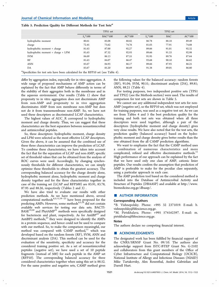
and TPS2) (see the Methods section) were used. The results ofcomparison for test sets are shown in Table 5.We cannot use any additional independent test sets for non-
AMP (negative set), so the RFP10 set, which was not employedfor training purposes, was used as a negative test set. As we cansee from Tables 4 and 5 the best prediction quality for thetraining and both test sets was obtained when all threedescriptors were used together, although a pair of thedescriptors (hydrophobic moment and charge density) givesvery close results. We have also noted that for the test sets, theprediction quality (balanced accuracy) based on the hydro-phobic moment and charge density gives better results than theone obtained from the all CAMP prediction algorithms.We want to emphasize the fact that the CAMP method uses
a combination of numerous characteristics and morecomplicated, refined and effective discriminative methods.11
High performance of our approach can be explained by the factthat we have used only one class of AMP, cationic linierpeptides. Our results confirm the assumption that prediction ofAMP is preferable to make for the peculiar class separately,using a particular approach in each case.The AMP prediction tool based on the considered method is
included into the Database of Antimicrobial Activity andStructure of Peptides (DBAASP) and available at http://www.biomedicine.org.ge/dbaasp/.
■ AUTHOR INFORMATIONCorresponding Authors*B. Vishnepolsky. Phone: +995 32 2371019. E-mail: [email protected].*M. Pirtskhalava. Phone: +995 574162397. E-mail: [email protected].
NotesThe authors declare no competing financial interest.
■ ACKNOWLEDGMENTSThe designated work has been fulfilled by financial support ofthe CNRS/SRNSF Grant No. 09/10. The authors alsoacknowledge support from ISTC-BTEP Grant No. G-2102and collaborators from this grant members of the Office ofCyber Infrastructure and Computational Biology (OCICB),National Institute of Allergy and Infectious Diseases (NIAID)Mike Tartakovsky, Alex Rosenthal, Andrei Gabrielian andDarrell Hurt.
Table 5. Prediction Quality for Different Methods for Test Setsa
TPS1 set TPS2 set
Sn*100 BAC*100 AC*100 Sn*100 BAC AC*100
hydrophobic moment 80.61 85.08 88.82 86.78 88.18 89.49charge 72.45 73.62 74.76 81.03 77.91 74.88hydrophobic moment + charge 81.63 87.00 92.27 89.66 91.01 92.32hydrophobic moment + charge + LPM 81.63 87.32 92.93 89.66 91.33 92.98SVM 84.69 86.13 87.55 91.95 89.76 87.64RF 81.63 84.07 86.47 93.68 90.10 86.62ANN 83.67 84.93 86.17 89.66 87.93 86.25DA 83.67 85.20 86.69 91.38 89.05 86.80
aSpecificities for test sets have been calculated for the RFP10 set (see Table 4).
Journal of Chemical Information and Modeling Article
dx.doi.org/10.1021/ci4007003 | J. Chem. Inf. Model. 2014, 54, 1512−15231521

■ REFERENCES(1) Sang, Y.; Blecha, F. Antimicrobial peptides and bacteriocins:alternatives to traditional antibiotics. Anim. Health Res. Rev. 2008, 9,227−235.(2) Hancock, R. E. W.; Rozek, A. Role of membranes in the activitiesof antimicrobial cationic peptides. FEMS Microbiol. Lett. 2002, 206 (2),143−149.(3) Fjell, C. D.; Hiss, J. A.; Hancock, R. E.; Schneider, G. Designingantimicrobial peptides: form follows function. Nat. Rev. Drug Discovery2011, 11 (1), 37−51.(4) Torrent, M.; Andreu, D.; Nogues, V. M.; Boix, E. ConnectingPeptide Physicochemical and Antimicrobial Properties by a RationalPrediction Model. PLoS One 2011, 6 (2), e16968.(5) Wang, P.; Hu, L.; Liu, G.; Jiang, N.; Chen, X.; Xu, J.; Zheng, W.;Li, L.; Tan, M.; Chen, Z.; Song, H.; Cai, Y.; Chouet, K. Prediction ofAntimicrobial Peptides Based on Sequence Alignment and FeatureSelection Methods. PLoS One 2011, 6 (4), e18476.(6) Frecer, V.; Ho, B.; Ding, J. L. De novo design of potentantimicrobial peptides. Antimicrob. Agents Chemother. 2004, 48, 3349−3357.(7) Jenssen, H.; Lejon, T.; Hilpert, K.; Fjell, C. D.; Cherkasov, A.;Hancock, R. E. W. Evaluating different descriptors for model design ofantimicrobial peptides with enhanced activity toward P. aeruginosa.Chem. Biol. Drug Des. 2007, 70, 134−142.(8) Jenssen, H.; Fjell, C. D.; Cherkasov, A.; Hancock, R. E. QSARmodeling and computer-aided design of antimicrobial peptides. J. Pept.Sci. 2008, 14, 110−114.(9) Frecer, V. QSAR analysis of antimicrobial and haemolytic effectsof cyclic cationic antimicrobial peptides derived from protegrin-1.Bioorg. Med. Chem. 2006, 14, 6065−6074.(10) Cherkasov, A.; Jankovic, B. Application of ‘inductive’ QSARdescriptors for quantification of antibacterial activity of cationicpolypeptides. Molecules 2004, 9, 1034−1052.(11) Waghu, F. H.; Gopi, L.; Barai, R. S.; Ramteke, P.; Nizami, B.;Idicula-Thomas, S. CAMP: Collection of sequences and structures ofantimicrobial peptides. Nucleic Acids Res. 2014, 42, D1154−D1158.(12) Taboureau, O.; Olsen, O. H.; Nielsen, J. D.; Raventos, D.;Mygind, P. H.; Kristensen, H. H. Design of novispirin antimicrobialpeptides by quantitative structure-activity relationship. Chem. Biol.Drug Des. 2006, 68, 48−57.(13) White, S. H.; Wimley, W. C. Membrane protein folding andstability: physical principles. Annu. Rev. Biophys. Biomol. Struct. 1999,28, 319−365.(14) Jayasinghe, S.; Hristova, K.; White, S. H. Energetics, stability,and prediction of transmembrane helices. J. Mol. Biol. 2001, 312, 927−934.(15) Scocchi, M.; Tossi, A.; Gennaro, R. Proline-rich antimicrobialpeptides: converging to a non-lytic mechanism of action. Cell. Mol. LifeSci. 2011, 68, 2317−30.(16) Kragol, G.; Hoffmann, R.; Chattergoon, M. A.; Lovas, S.; Cudic,M.; Bulet, P.; Condie, B. A.; Rosengren, K. J.; Montaner, L. J.; Otvos,L. Identification of crucial residues for the antibacterial activity of theproline-rich peptide, pyrrhocoricin. Eur. J. Biochem. 2002, 269, 4226−4237.(17) Wang, Z.; Wang, G. APD: the Antimicrobial Peptide Database.Nucleic Acids Res. 2004, 32, D590−D592.(18) Fjell, C. D.; Hancock, R. E.; Cherkasov, A. AMPer: a databaseand an automated discovery tool for antimicrobial peptides.Bioinformatics 2007, 23, 1148−1155.(19) Hammami, R.; Zouhir, A.; Ben Hamida, J.; Fliss, I.BACTIBASE: a new web-accessible database for bacteriocin character-ization. BMC Microbiol. 2007, 7, 89.(20) Hammami, R.; Zouhir, A.; Le Lay, C.; Ben Hamida, J.; Fliss, I.BACTIBASE second release: a database and tool platform forbacteriocin characterization. BMC Microbiol. 2010, 10, 22.(21) Hammami, R.; Ben Hamida, J.; Vergoten, G.; Fliss, I. PhytAMP:a database dedicated to antimicrobial plant peptides. Nucleic Acids Res.2009, 37, D963−968.
(22) Lata, S.; Sharma, B. K.; Raghava, G. P. Analysis and prediction ofantibacterial peptides. BMC Bioinf. 2007, 8, 263 (2007).(23) Lata, S.; Mishra, N. K.; Raghava, G. P. AntiBP2: improvedversion of antibacterial peptide prediction. BMC Bioinf. 2010, 11(Suppl1), S19; Bioinformatics 2005, 59, 252−265.(24) Tusnady, G. E.; Dosztanyi, Z.; Simon, I. PDB_TM: selectionand membrane localization of transmembrane proteins in the proteindata bank. Bioinformatics 2004, 20 (17), 2964−2972.(25) Kozma, D.; Simon, I.; Tusnady, G. E. PDBTM: Protein DataBank of transmembrane proteins after 8 years. Nucleic Acids Res. 2013,41 (D1), D524−D529.(26) Yeaman, M. R.; Yount, N. Y. Unifying themes in host defenceeffector polypeptides. Nat. Rev. Microbiol. 2007, 5, 727−740.(27) Kyte, J.; Doolittle, R. F. A simple method for displaying thehydropathic character of a protein. J. Mol. Biol. 1982, 157 (1), 105−132.(28) Wimley, W. C.; White, S. H. Experimentally determinedhydrophobicity scale for proteins at membrane interfaces. Nat. Struct.Biol. 1996, 3, 842−848.(29) Koehler, J.; Woetzel, N.; Staritzbichler, R.; Sanders, C. R.;Meiler, J. A unified hydrophobicity scale for multispan membraneproteins. Proteins 2009, 76, 13−29.(30) Hessa, T.; Meindl-Beinker, N. M.; Bernsel, A.; Kim, H.; Sato, Y.;Lerch-Bader, M.; Nilsson, I.; White, S. H.; von Heijne, G. Molecularcode for transmembrane-helix recognition by the Sec61 translocon.Nature 2007, 450, U1026−U1032.(31) Eisenberg, D.; Weiss, R. M.; Terwilliger, T. C. The hydrophobicmoment detects periodicity in protein hydrophobicity. Proc. Natl.Acad. Sci. U. S. A. 1984, 81 (1), 140−144.(32) Moon, C. P.; Fleming, K. G. Side-chain hydrophobicity scalederived from transmembrane protein folding into lipid bilayers. Proc.Natl. Acad. Sci. U. S. A. 2011, 108 (25), 10174−10177.(33) Yeaman, M. R.; Yount, N. Y. Mechanisms of antimicrobialpeptide action and resistance. Pharmacol. Rev. 2003, 55 (1), 27−55.(34) Eisenberg, D.; Weiss, R. M.; Terwilliger, T. C. The helicalhydrophobic moment: a measure of the amphiphilicity of a helix.Nature 1982, 299 (5881), 371−374.(35) Senes, A.; Chadi, D. C.; Law, P. B.; Walters, R. F. S.; Nanda, V.;DeGrado, W. F. Ez, a Depth-dependent Potential for Assessing theEnergies of Insertion of Amino Acid Side-chains into Membranes:Derivation and Applications to Determining the Orientation ofTransmembrane and Interfacial Helices. J. Mol. Biol. 2007, 366, 436−448.(36) Shai, Y. Mode of action of membrane active antimicrobialpeptides. Biopolymers 2002, 66, 236−248.(37) Uversky, V.; Gillespie, J.; Fink, A. Why are “natively unfolded”proteins unstructured under physiological conditions? Proteins: Struct.Funct. Gen. 2000, 41 (3), 415−427.(38) Fernandez-Escamilla, A. M.; Rousseau, F.; Schymkowitz, J.;Serrano, L. Prediction of sequence-dependent and mutational effectson the aggregation of peptides and proteins. Nat. Biotechnol. 2004, 22,1302−1306.(39) Conchillo-Sole, O.; de Groot, N. S.; Aviles, F. X.; Vendrell, J.;Daura, X.; Ventura, S. AGGRESCAN: a server for the prediction andevaluation of “hot spots” of aggregation in polypeptides. BMC Bioinf.2007, 8, 65.(40) Bechinger, B. The structure, dynamics and orientation ofantimicrobial peptides in membranes by multidimensional solid-stateNMR spectroscopy. Biochim. Biophys. Acta 1999, 1462 (1−2), 157−83.(41) Mitaku, S.; Hirokava, T.; Tsuji, T. Amphiphilicity index of polaramino acids as an aid in the characterization of amino acid preferenceat membrane-water interfaces. Bioinformatics 2002, 18, 608−16.(42) Ulmschneider, M. B.; Sansom, M. S. P.; Di Nola, A. Propertiesof Integral Membrane Protein Structures:Derivation of an ImplicitMembrane Potential. Proteins: Struct., Funct., Bioinf. 2005, 59, 252−262.
Journal of Chemical Information and Modeling Article
dx.doi.org/10.1021/ci4007003 | J. Chem. Inf. Model. 2014, 54, 1512−15231522

(43) Wimley, W. C. Describing the Mechanism of AntimicrobialPeptide Action with the Interfacial Activity Model. ACS Chem. Biol.2010, 5, 905−917.
Journal of Chemical Information and Modeling Article
dx.doi.org/10.1021/ci4007003 | J. Chem. Inf. Model. 2014, 54, 1512−15231523