Embed Size (px)
Citation preview
ORIGINAL RESEARCHINTERVENTIONAL
Preclinical Evaluation of the Thrombogenicity andEndothelialization of Bare Metal and Surface-Coated
Neurovascular StentsS. Krajewski, B. Neumann, J. Kurz, N. Perle, M. Avci-Adali, G. Cattaneo, and H.P. Wendel
ABSTRACT
BACKGROUND AND PURPOSE: Stent-assisted coiling is routinely used for the endovascular treatment of complex or wide-neck intra-cranial aneurysms. However, in-stent thrombosis, thromboembolic events, and ischemic complications remain a major concern associatedwith stent implants. Therefore, a novel low-profile neurovascular stent with a bare metal surface was investigated with regard tothrombogenicity and endothelialization and compared with the same stent coated with albumin or heparin.
MATERIALS AND METHODS: The bare metal and heparin- or albumin-coated stents were loaded in heparin-coated tubing, which wasthen filled with heparinized human blood (n � 5) and circulated at 150 mL/min and 37°C for 60 minutes. Before and after circulation,measurement of various inflammation and coagulation markers and scanning electron microscopy were performed. Endothelialization ofthe stents was investigated in an in vitro model including human umbilical vascular endothelial cells.
RESULTS: Our results showed that platelet loss and platelet activation and activation of the coagulation cascade, which are induced bythe bare metal stents, were significantly reduced by heparin or albumin coating. Adverse effects on erythrocytes, leukocytes, and thecomplement cascade were not induced by the bare metal or coated stents. Moreover, the bare metal and albumin-coated stents showedgood endothelialization properties.
CONCLUSIONS: Albumin and heparin coatings clearly improve the thrombogenicity of the stents and thus may represent fundamentalprogress in the treatment of intracranial aneurysms. Moreover, preclinical evaluation of neurovascular stents under physiologic conditionssupports and accelerates the development of more biocompatible neurovascular stents.
ABBREVIATIONS: HUVECs � human umbilical vascular endothelial cells; SEM � scanning electron microscopy; TAT � thrombin-antithrombin III
Endovascular treatment of complex or wide-neck intracranial
aneurysms has been proved feasible by using the stent-assisted
coiling technique, which was primarily described in 1997.1 In this
procedure, self-expandable, nitinol-made stents, braided or laser
cut, are delivered into the parent vessel by using microcatheters
with an inner lumen between 0.0165 and 0.027 inches to retain
coils within the aneurysm.1,2
Thus, the implanted stent prevents coil protrusion within the
parent vessel, reduces coil compaction and hence recanalization,
and provides a scaffold for endothelialization.2-5
However, the endoluminal presence of the implant can lead to
activation of platelets and leukocytes following adverse effects like
thromboembolic complications, hemorrhage, or proinflamma-
tory reactions. Platelet activation is associated with the release of
granules containing a plethora of molecules mainly promoting
platelet activation and blood coagulation. Therefore, antiplatelet
therapy is required, which can prevent intra-aneurysmal throm-
bosis and thus aneurysm occlusion but also increases the risk of
hemorrhagic complications.6 Moreover and despite antiplatelet
therapy, ischemic complications remain a major concern associ-
ated with stent implants. A retrospective review of stent-assisted
coiling procedures in 624 patients between 2000 and 2011 re-
vealed intra- and postprocedural in-stent thrombosis and throm-
boembolic events in 9.0% of the patients, whereas low or no re-
sponse to antiplatelet agents and early interruption of therapy are
possible causes for ischemic events.2
A retrospective study of 36 patients treated with stent-assisted
Received March 6, 2014; accepted after revision June 23.
From the Department of Thoracic, Cardiac and Vascular Surgery (S.K., B.N., J.K.,N.P., M.A.-A., H.P.W.), Clinical Research Laboratory, University Hospital Tuebingen,Tuebingen, Germany; and Acandis GmbH & Co KG (G.C.), Pforzheim, Germany.
The study was funded by the German Federal Ministry of Economic Affairs(Economics and Energy).
Please address correspondence to Stefanie Krajewski, PhD, Department of Tho-racic, Cardiac and Vascular Surgery, University Hospital Tuebingen, Clinical Re-search Laboratory, Tuebingen University, Calwerstr 7/1, 72076 Tuebingen,Germany; e-mail: [email protected]
Indicates open access to non-subscribers at www.ajnr.org
http://dx.doi.org/10.3174/ajnr.A4109
AJNR Am J Neuroradiol 36:133–39 Jan 2015 www.ajnr.org 133
coiling after aneurysm rupture showed that in 17%, stent-related
thromboembolic complications occurred. Intra- and postproce-
dural clopidogrel administration was related to an incidence of
thrombotic events of 20% and 29%, respectively. Even pretreat-
ment with clopidogrel could not prevent thrombotic events,
which were shown in 6% of cases.7
Therefore, preventing or strongly decreasing activation of
platelets and the coagulation cascade, while enhancing or at least
not impairing endothelialization, is a primary objective in the
treatment of endovascular diseases. Stent design, influence on
fluid dynamics, and particularly surface modifications are current
options to decrease activation of platelets and the coagulation
cascade, and they also address hemorrhagic stroke, because the
necessity for antiplatelet therapy can be reduced. Previous studies
show that surface coatings with the antithrombotic molecules
heparin and albumin, which is the most abundant protein in
blood, increase the biocompatibility of different biomaterials.8-10
In our study, an in vitro model was established, which allows
investigation of neurovascular stents with regard to thromboge-
nicity and coagulation activation.
Small-diameter tubing and low flow rates simulating intracra-
nial vessel conditions were used to investigate novel low-profile
neurovascular stents with bare metal surfaces or 2 coatings, in-
cluding albumin and heparin, with regard to thrombogenicity.
Furthermore, a predisposition toward endothelialization was in-
vestigated in an in vitro rotation model including human umbil-
ical vascular endothelial cells (HUVECs).
MATERIALS AND METHODSFor this study, the neurovascular stent Acclino (n � 24; Acan-
dis, Pforzheim, Germany) was used. Acclino is a low-profile,
laser-cut stent with an electropolished surface and strut di-
mensions ranging between �55 �m (wall thickness) and �35
�m (strut width).
The stent is deliverable within the 1.9F microcatheter Neuro-
Slider 17 (Acandis) with a lumen of 0.0165 inches (�0.42 mm).
The small catheter lumen and the size of the stent (3.5 and 4.5 mm
in expanded configurations) allow the application in distal intra-
cranial vessels with diameters between 2 and 4 mm, whereas the
closed-cell structure allows retrieval and repositioning up to 90%
of its deployment.
For the current study, specific Acclino stent samples with a
wall thickness of �75 �m were manufactured to reproduce a
worst-case scenario with regard to blood flow interruption caused
by the stent structure.
Blood SamplingBlood-sampling procedures were approved by the ethics commit-
tee of the University of Tuebingen, Tuebingen, Germany. Blood
was collected from healthy volunteers (n � 5), who gave signed
informed consent, and was anticoagulated with 1.5 IU/mL Hep-
arin-Natrium (Rathiopharm, Ulm, Germany). Exclusion criteria
for the blood donators were the following: smokers; drug intake in
the last 2 weeks before blood sampling, especially hemostasis-
affecting agents like acetylsalicylic acid; oral contraceptives; non-
steroidal antiphlogistics; and others, to guarantee optimal hemo-
static function.
Neurovascular Stent CoatingFor albumin coating, 8 neurovascular stents were first incubated
for 2 hours at room temperature in 1% human serum albumin
(HSA; CSL Behring, Marburg, Germany), followed by a washing
step in saline and a 30-minute incubation in a suspension con-
taining 0.5-mg of 1-ethyl-3-(3-dimethylaminopropyl)carbodi-
imide hydrochloride (Thermo Scientific, Karlsruhe, Germany)
for albumin cross-linking. Afterward, the stents were incubated
again in 1% HSA for 2 hours at room temperature followed by
another washing step in saline. After being coated, the stents were
air-dried and stored at 4°C until analysis in the in vitro thrombo-
genicity model. The heparin coating of the stents (n � 8) for the
thrombogenicity and endothelialization experiments was per-
formed according to standard methods.11 To guarantee sterile
conditions in the endothelialization model, we sterilized the
stents by using ethylene oxide.
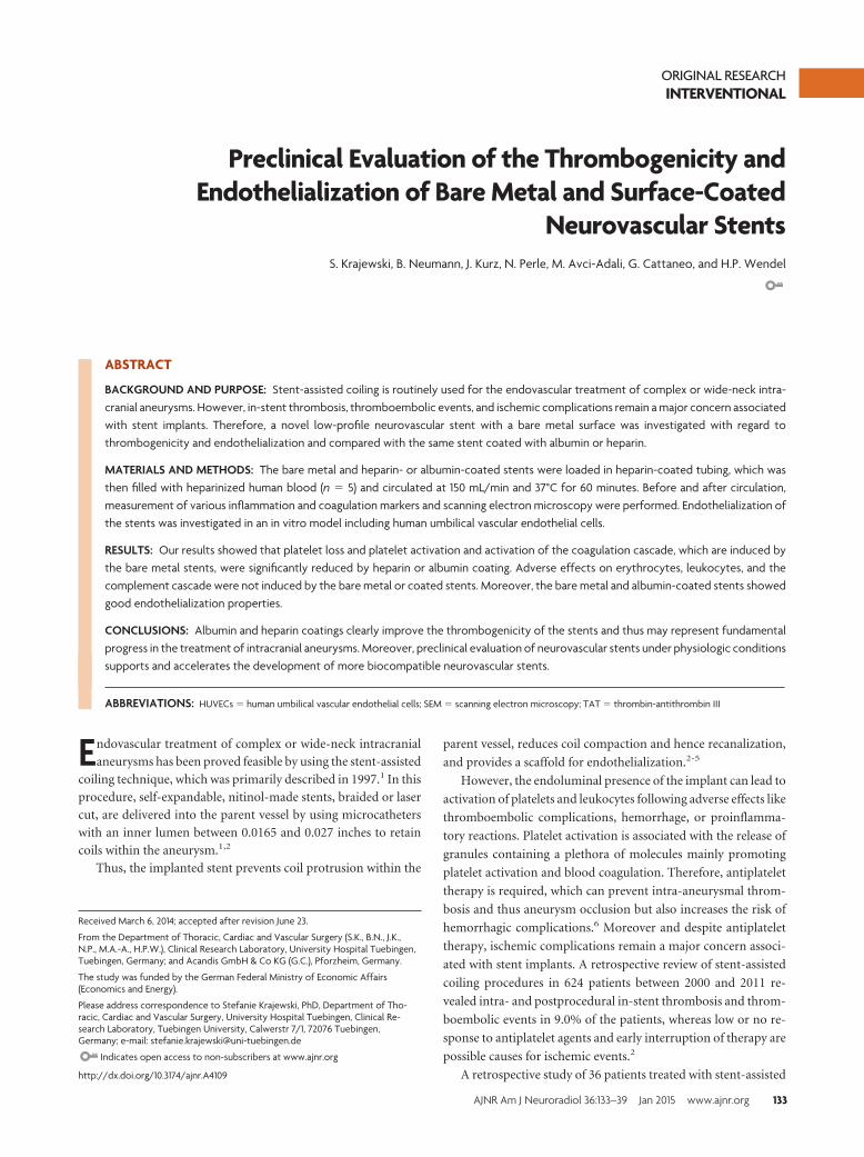
In Vitro Thrombogenicity ModelThis novel model was established to investigate the effect of neu-
rovascular stents on various cascade reactions of the human he-
mostatic system (coagulation cascade, cell alteration, comple-
ment system, and inflammation) during blood circulation (Fig
1A, -B).
For each blood donor (n � 5), 4 polyvinyl chloride tubes (in-
ner diameter � 3.2 mm, length � 75 cm; Tygon; Saint-Gobain
Performance Plastics, Chamy, France) were coated with heparin
by Ension (Ension, Cape Coral, Florida) and left empty or loaded
with either 1 bare metal, 1 albumin-, or 1 heparin-coated stent.
Subsequently, each tubing was filled with a total of 6-mL heparin-
ized human blood, closed by silicone connection tubing, and cir-
culated at 150 mL/min in a water bath at 37°C for 60 minutes. For
each donor, 6 mL of heparinized blood served for the measure-
ment of baseline values before circulation. Before and after circu-
lation, the blood was directly transferred to the corresponding
terminating media and was further processed for enzyme-linked
immunosorbent assays and whole blood analysis.
FIG 1. Closed-loop in vitro circulation model for neurovascular stenttesting by using fresh human whole blood. Schematic overview (A)and photograph (B) of the in vitro circulation model consisting of thefollowing: 1) heparin-coated tubing (inner diameter � 3.2 mm,length � 75 cm), 2) connection tubing, 3) peristaltic pump (flow rate �150 mL/min), 4) test device. C, Representative macroscopic images ofuncoated and albumin- and heparin-coated stents after circulationfor 60 minutes at 37°C (�20 magnification).
134 Krajewski Jan 2015 www.ajnr.org
Endothelialization ModelThe endothelialization experiments (n � 3) were performed as
previously described.12 Briefly, 2-mL bioreactor chambers were
loaded with either a sterilized bare metal, an albumin-, or a hep-
arin-coated stent. Isolation of HUVECs was adapted from a
previously described method for the isolation of endothelial
cells from residual saphenous vein biopsies.12 For each sample,
2.5 � 106 cells were resuspended in 2.5-mL VascuLife EnGS
medium (CellSystems, Troisdorf, Germany) containing a
VascuLife EnGS LifeFactors Kit, 50-�g/mL gentamicin, and 0.05-
�g/mL amphotericin B (PAA Laboratories, Colbe, Germany).
Following the addition of the cell suspension, the bioreactor
chambers were assembled in the rotation device and rotated at
37°C and 5% CO2. After 72 hours of incubation, the stents were
washed in phosphate buffered saline and subsequently prepared
for scanning electron microscopy analysis.
Measurement of Hemostatic MarkersThe analysis of markers indicating activation of platelets and
leukocytes and the coagulation cascade and complement
system was performed in plasma by enzyme-linked immu-
nosorbent assays, according to the manufacturer’s instruc-
tions. To investigate released, plasmatic markers indicating
platelet and leukocyte activation, we analyzed PF-4 (Abcam,
Cambridge, United Kingdom) and poly-
morphonuclear neutrophil elastase (De-
meditec Diagnostics, Kiel-Wellsee, Ger-
many) levels. As a marker for coagulation,
we determined thrombin-antithrombin
III (TAT) complexes (Siemens Health-
care, Marburg, Germany). Moreover, we
analyzed SC5b-9 (TECOmedical, Bunde,
Germany) to detect activation of the com-
plement system.
Whole Blood Count AnalysisThe number of erythrocytes, leukocytes,
and platelets and hematocrit and hemo-
globin values were measured by a cell
counter system (ABX Micros 60; Axon
Lab AG, Baden, Switzerland) in baseline
samples and after circulation in all
groups.
Scanning Electron MicroscopyScanning electron microscopy (SEM) was
performed to investigate the thromboge-
nicity and the endothelialization rate of
the different stent surfaces after contact
with human whole blood and HUVECs,
respectively. After each experiment, the
stents were rinsed in phosphate buffered
saline and fixed in glutaraldehyde solu-
tion (2% in phosphate buffered saline,
pH 7.4) overnight at 4°C. After another
washing step with phosphate buffered sa-
line, the stents were dehydrated by using
ascending concentrations of ethanol
(Emsure; Merck, Darmstadt, Germany) and critical point dried.
After sputtering with gold palladium, we performed SEM analysis
by using the EVO LS 10 scanning electron microscope (Carl Zeiss,
Jena, Germany).
StatisticsData are depicted as means with standard error of the mean. Data
were analyzed by using repeated-measures ANOVA with the Bon-
ferroni multiple comparison test to analyze differences among
groups. All analyses were performed by using the statistical soft-
ware package GraphPad Prism (Version 5; GraphPad Software,
San Diego, California). Statistical significance was defined as
P � .05.
RESULTSHemocompatibility Testing of Neurovascular StentsContact of whole blood with artificial surfaces like stents, cathe-
ters, implants, and so forth is associated with various alterations of
the hemostatic system. To evaluate the hemocompatibility of the
bare metal (n � 5), albumin- (n � 5), or heparin-coated (n � 5)
stents for neurovascular application, we developed an in vitro
model to guarantee a defined and continuous blood flow mim-
icking blood circulation in the brain (Fig 1A, -B). Directly after
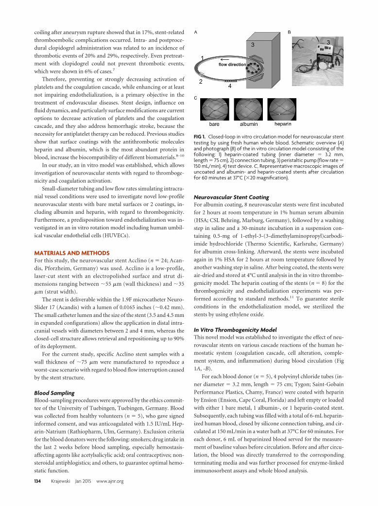
FIG 2. Neurovascular stents do not change hematocrit levels, erythrocyte number, or hemo-globin concentration. Heparin-coated tubing was loaded with bare metal, albumin-, or heparin-coated neurovascular stents and filled with 6 mL of fresh heparinized human blood. Tubingwithout a stent served as a negative control. Hematocrit level (A), erythrocyte numbers (B), andhemoglobin concentrations (C) were analyzed before and after 60 minutes of circulation at 150mL/min. Data are given as means and standard error of the mean (n � 5) and were analyzed byusing repeated-measures ANOVA with the Bonferroni multiple-comparison test. One asteriskindicates P � .05; two asterisks, P � .01; three asterisks, P � .001.
AJNR Am J Neuroradiol 36:133–39 Jan 2015 www.ajnr.org 135
circulation, macroscopic images, which are exemplarily shown in
Fig 1C from 1 blood donor, were obtained of each stent.
Whole Blood Count Analysis. Before and after circulation in the
in vitro model, the whole blood was analyzed in all groups. Cir-
culation induced a significant decrease of hematocrit (P � .05, Fig
2A), red blood cell counts (P � .01, Fig
2B), and hemoglobin levels (P � .001, Fig
2C) in the control group containing no
stent. No significant changes were ob-
served in the bare metal and albumin- and
heparin-coated stent groups compared
with the control group.
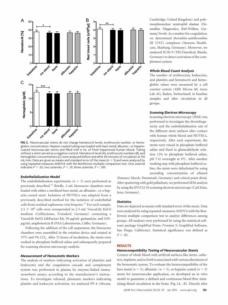
Platelet and Leukocyte Activation. The
concentration of PF-4, a small chemokine
released from platelet � granules, is signif-
icantly increased during circulation in
both control groups containing no stent
and a bare metal stent, respectively (P �
.05, Fig 3A). In the groups containing al-
bumin- or heparin-coated stents, the re-
lease of PF-4 was significantly reduced
(P � .05). Platelet activation was also
estimated by the analysis of platelet
counts. Compared with a platelet count of
217.200 � 9.300 �L before circulation, a
significant loss of platelets in the control
group containing no stent (P � .001) was
observed, which was further increased in
the bare metal stent group (P � .001, Fig
3B). Again, the decrease in platelet num-
bers was less severe in the coated stent
groups (P � .01).
Despite platelet activation, leukocyte
activation based on granule release and
leukocyte counts was investigated. The
plasma concentration of polymorphonu-
clear neutrophil-elastase released from
granulocytes significantly increased be-
cause of circulation in the in vitro model
(P � .01, Fig 3C). In the stent groups, no
significant differences compared with the
control group were detected. Similar re-
sults were obtained for the leukocyte
counts (Fig 3D), in which no significant
differences were detected among the
groups after circulation.
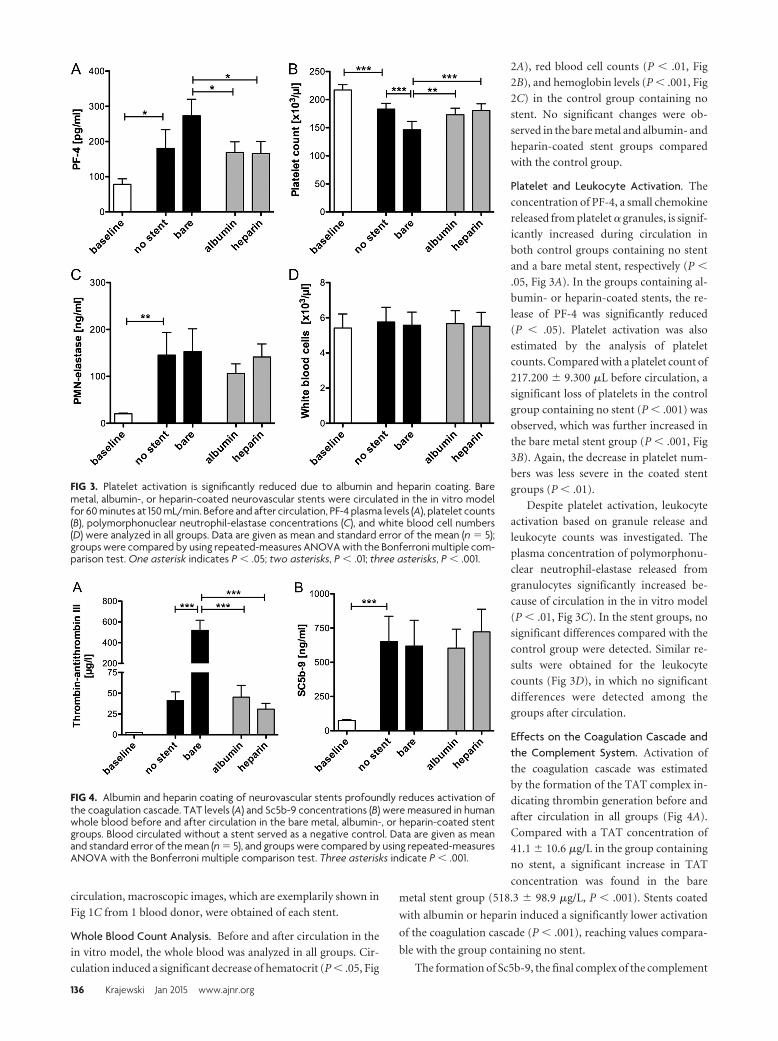
Effects on the Coagulation Cascade andthe Complement System. Activation of
the coagulation cascade was estimated
by the formation of the TAT complex in-
dicating thrombin generation before and
after circulation in all groups (Fig 4A).
Compared with a TAT concentration of
41.1 � 10.6 �g/L in the group containing
no stent, a significant increase in TAT
concentration was found in the bare
metal stent group (518.3 � 98.9 �g/L, P � .001). Stents coated
with albumin or heparin induced a significantly lower activation
of the coagulation cascade (P � .001), reaching values compara-
ble with the group containing no stent.
The formation of Sc5b-9, the final complex of the complement
FIG 3. Platelet activation is significantly reduced due to albumin and heparin coating. Baremetal, albumin-, or heparin-coated neurovascular stents were circulated in the in vitro modelfor 60 minutes at 150 mL/min. Before and after circulation, PF-4 plasma levels (A), platelet counts(B), polymorphonuclear neutrophil-elastase concentrations (C), and white blood cell numbers(D) were analyzed in all groups. Data are given as mean and standard error of the mean (n � 5);groups were compared by using repeated-measures ANOVA with the Bonferroni multiple com-parison test. One asterisk indicates P � .05; two asterisks, P � .01; three asterisks, P � .001.
FIG 4. Albumin and heparin coating of neurovascular stents profoundly reduces activation ofthe coagulation cascade. TAT levels (A) and Sc5b-9 concentrations (B) were measured in humanwhole blood before and after circulation in the bare metal, albumin-, or heparin-coated stentgroups. Blood circulated without a stent served as a negative control. Data are given as meanand standard error of the mean (n � 5), and groups were compared by using repeated-measuresANOVA with the Bonferroni multiple comparison test. Three asterisks indicate P � .001.
136 Krajewski Jan 2015 www.ajnr.org
system, significantly increased on circulation (P � .001, Fig 4B).
No significant differences in Sc5b-9 generation were observed be-
tween the control and the stent-containing groups.
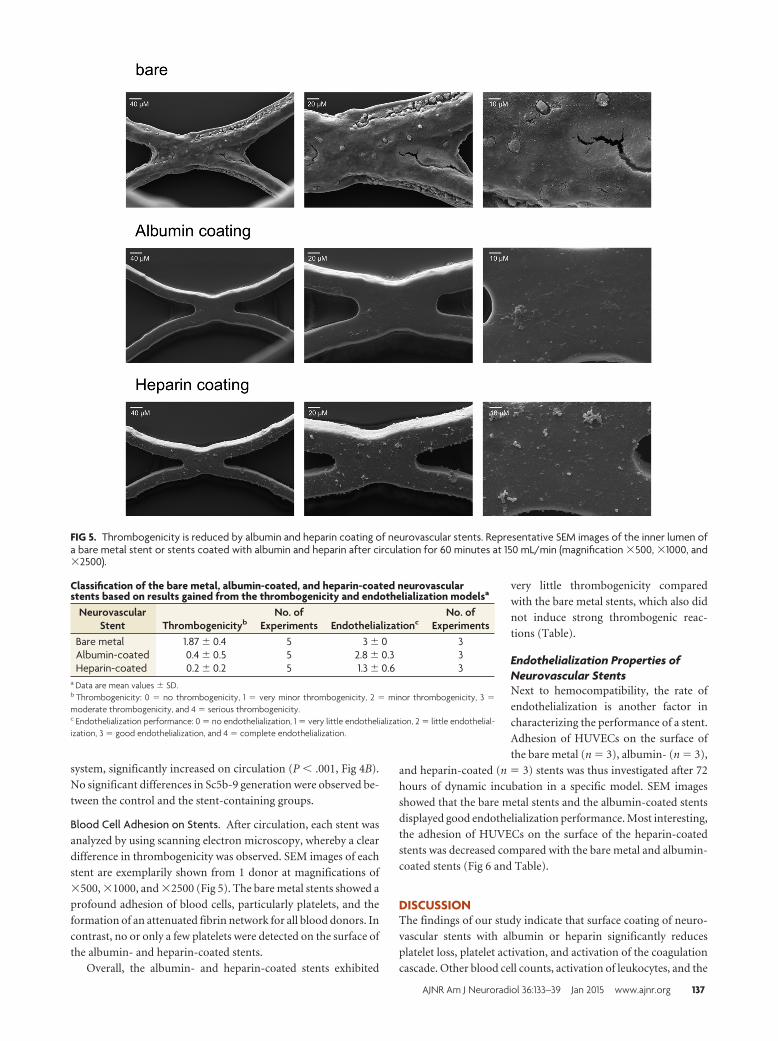
Blood Cell Adhesion on Stents. After circulation, each stent was
analyzed by using scanning electron microscopy, whereby a clear
difference in thrombogenicity was observed. SEM images of each
stent are exemplarily shown from 1 donor at magnifications of
�500, �1000, and �2500 (Fig 5). The bare metal stents showed a
profound adhesion of blood cells, particularly platelets, and the
formation of an attenuated fibrin network for all blood donors. In
contrast, no or only a few platelets were detected on the surface of
the albumin- and heparin-coated stents.
Overall, the albumin- and heparin-coated stents exhibited
very little thrombogenicity compared
with the bare metal stents, which also did
not induce strong thrombogenic reac-
tions (Table).
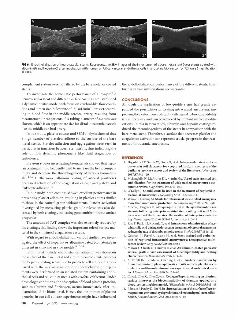
Endothelialization Properties ofNeurovascular StentsNext to hemocompatibility, the rate of
endothelialization is another factor in
characterizing the performance of a stent.
Adhesion of HUVECs on the surface of
the bare metal (n � 3), albumin- (n � 3),
and heparin-coated (n � 3) stents was thus investigated after 72
hours of dynamic incubation in a specific model. SEM images
showed that the bare metal stents and the albumin-coated stents
displayed good endothelialization performance. Most interesting,
the adhesion of HUVECs on the surface of the heparin-coated
stents was decreased compared with the bare metal and albumin-
coated stents (Fig 6 and Table).
DISCUSSIONThe findings of our study indicate that surface coating of neuro-
vascular stents with albumin or heparin significantly reduces
platelet loss, platelet activation, and activation of the coagulation
cascade. Other blood cell counts, activation of leukocytes, and the
FIG 5. Thrombogenicity is reduced by albumin and heparin coating of neurovascular stents. Representative SEM images of the inner lumen ofa bare metal stent or stents coated with albumin and heparin after circulation for 60 minutes at 150 mL/min (magnification �500, �1000, and�2500).
Classification of the bare metal, albumin-coated, and heparin-coated neurovascularstents based on results gained from the thrombogenicity and endothelialization modelsa
NeurovascularStent Thrombogenicityb
No. ofExperiments Endothelializationc
No. ofExperiments
Bare metal 1.87 � 0.4 5 3 � 0 3Albumin-coated 0.4 � 0.5 5 2.8 � 0.3 3Heparin-coated 0.2 � 0.2 5 1.3 � 0.6 3
a Data are mean values � SD.b Thrombogenicity: 0 � no thrombogenicity, 1 � very minor thrombogenicity, 2 � minor thrombogenicity, 3 �moderate thrombogenicity, and 4 � serious thrombogenicity.c Endothelialization performance: 0 � no endothelialization, 1 � very little endothelialization, 2 � little endothelial-ization, 3 � good endothelialization, and 4 � complete endothelialization.
AJNR Am J Neuroradiol 36:133–39 Jan 2015 www.ajnr.org 137
complement system were not altered by the bare metal or coated
stents.
To investigate the hemostatic performance of a low-profile
neurovascular stent and different surface coatings, we established
a dynamic in vitro model with focus on cerebral-like flow condi-
tions and lumen size. A flow rate of 150 mL/min�1 was set accord-
ing to blood flow in the middle cerebral artery, resulting from
measurement in 92 patients.13 A tubing diameter of 3.2 mm was
chosen, which is an appropriate size for distal intracranial vessels
like the middle cerebral artery.
In our study, platelet counts and SEM analysis showed that
a high number of platelets adhere to the surface of the bare
metal stents. Platelet adhesion and aggregation were seen in
particular at junctions between stent struts, thus indicating the
role of flow dynamic phenomena like fluid stagnation or
turbulence.
Previous studies investigating biomaterials showed that hepa-
rin coating is most frequently used to increase the hemocompat-
ibility and decrease the thrombogenicity of various biomateri-
als.14-16 Furthermore, albumin coating of arterial prostheses
decreased activation of the coagulation cascade and platelet and
leukocyte adhesion.17
In our study, both coatings showed excellent performance in
preventing platelet adhesion, resulting in platelet counts similar
to those in the control group without stents. Platelet activation
investigated by measuring platelet granule release was also de-
creased by both coatings, indicating good antithrombotic surface
properties.
The amount of TAT complex was also extremely reduced by
the coatings; this finding shows the important role of surface ma-
terial in the (intrinsic) coagulation cascade.
With regard to endothelialization, various studies have inves-
tigated the effect of heparin- or albumin-coated biomaterials in
different in vitro and in vivo models.8,18-21
In our in vitro study, endothelial cell adhesion was shown on
the surface of the bare metal and albumin-coated stents, whereas
the heparin coating seems not to promote cell adhesion. Com-
pared with the in vivo situation, our endothelialization experi-
ments were performed in an isolated system containing endo-
thelial cells and cell culture media with 2% fetal calf serum. Under
physiologic conditions, the adsorption of blood plasma proteins,
such as albumin and fibrinogen, occurs immediately after im-
plantation of the biomaterial. Hence, the low amount of plasma
proteins in our cell culture experiments might have influenced
the endothelialization performance of the different stents; thus,
further in vivo investigations are warranted.
CONCLUSIONSAlthough the application of low-profile stents has greatly ex-
panded the possibilities in treating intracranial aneurysms, im-
proving the performance of stents with regard to biocompatibility
is still necessary and can be achieved by implant surface modifi-
cations. In this in vitro study, albumin and heparin coatings re-
duced the thrombogenicity of the stents in comparison with the
bare metal stent. Therefore, a surface that decreases platelet and
coagulation activation can represent crucial progress in the treat-
ment of intracranial aneurysms.
REFERENCES1. Higashida RT, Smith W, Gress D, et al. Intravascular stent and en-
dovascular coil placement for a ruptured fusiform aneurysm of thebasilar artery: case report and review of the literature. J Neurosurg1997;87:944 – 49
2. McLaughlin N, McArthur DL, Martin NA. Use of stent-assisted coilembolization for the treatment of wide-necked aneurysms: a sys-tematic review. Surg Neurol Int 2013;4:43
3. O’Kelly CJ. Should stents be used in the treatment of ruptured in-tracranial aneurysms? J Neurosurg Sci 2011;55:27–33
4. Wanke I, Forsting M. Stents for intracranial wide-necked aneurysms:more than mechanical protection. Neuroradiology 2008;50:991–98
5. Mocco J, Fargen KM, Albuquerque FC, et al. Delayed thrombosis orstenosis following Enterprise-assisted stent-coiling: is it safe? Mid-term results of the interstate collaboration of Enterprise stent coil-ing. Neurosurgery 2011;69:908 –13, discussion 913–14
6. Ries T, Buhk JH, Kucinski T, et al. Intravenous administration of ace-tylsalicylic acid during endovascular treatment of cerebral aneurysmsreduces the rate of thromboembolic events. Stroke 2006;37:1816–21
7. Golshani K, Ferrel A, Lessne M, et al. Stent-assisted coil emboliza-tion of ruptured intracranial aneurysms: a retrospective multi-center review. Surg Neurol Int 2012;3:84
8. Marois Y, Chakfe N, Guidoin R, et al. An albumin-coated polyesterarterial graft: in vivo assessment of biocompatibility and healingcharacteristics. Biomaterials 1996;17:3–14
9. Mulvihill JN, Faradji A, Oberling F, et al. Surface passivation byhuman albumin of plasmapheresis circuits reduces platelet accu-mulation and thrombus formation: experimental and clinical stud-ies. J Biomed Mater Res 1990;24:155– 63
10. Chen J, Chen C, Chen Z, et al. Collagen/heparin coating on titaniumsurface improves the biocompatibility of titanium applied as ablood-contacting biomaterial. J Biomed Mater Res A 2010;95:341– 49
11. Johnson I, Perchy D, Liu H. In vitro evaluation of the surface effects onmagnesium-yttrium alloy degradation and mesenchymal stem cell ad-hesion. J Biomed Mater Res A 2012;100:477–85
FIG 6. Endothelialization of neurovascular stents. Representative SEM images of the inner lumen of a bare metal stent (A) or stents coated withalbumin (B) and heparin (C) after incubation with human umbilical vascular endothelial cells in a rotating bioreactor for 72 hours (magnification�1000).
138 Krajewski Jan 2015 www.ajnr.org
12. Avci-Adali M, Kobba J, Neumann B, et al. Application of a rotatingbioreactor consisting of low-cost and ready-to-use medical dispos-ables for in vitro evaluation of the endothelialization efficiency ofsmall-caliber vascular prostheses. J Biomed Mater Res B Appl Biom-ater 2013;101:1061– 68
13. Zhao M, Amin-Hanjani S, Ruland S, et al. Regional cerebral bloodflow using quantitative MR angiography. AJNR Am J Neuroradiol2007;28:1470 –73
14. Hirsh J, Siragusa S, Cosmi B, et al. Low-molecular-weight heparins(LMWH) in the treatment of patients with acute venous thrombo-embolism. Thromb Haemost 1995;74:360 – 63
15. Blezer R, Fouache B, Willems GM, et al. Activation of blood coagulationat heparin-coated surfaces. J Biomed Mater Res 1997;37:108–13
16. Klement P, Du YJ, Berry L, et al. Blood-compatible biomaterials bysurface coating with a novel antithrombin-heparin covalent com-plex. Biomaterials 2002;23:527–35
17. Kottke-Marchant K, Anderson JM, Umemura Y, et al. Effect of albu-
min coating on the in vitro blood compatibility of Dacron arterialprostheses. Biomaterials 1989;10:147–55
18. Bae IH, Park IK, Park DS, et al. Thromboresistant and endothelial-ization effects of dopamine-mediated heparin coating on a stentmaterial surface. J Mater Sci Mater Med 2012;23:1259 – 69
19. Moer R, Myreng Y, Mølstad P, et al. Stenting in small coronary ar-teries (SISCA) trial: a randomized comparison between balloon an-gioplasty and the heparin-coated beStent. J Am Coll Cardiol2001;38:1598 – 603
20. Serruys PW, Emanuelsson H, van der Giessen W, et al. Heparin-coated Palmaz-Schatz stents in human coronary arteries: earlyoutcome of the Benestent-II Pilot Study. Circulation 1996;93:412–22
21. Blindt R, Vogt F, Astafieva I, et al. A novel drug-eluting stent coatedwith an integrin-binding cyclic Arg-Gly-Asp peptide inhibits neo-intimal hyperplasia by recruiting endothelial progenitor cells. J AmColl Cardiol 2006;47:1786 –95
AJNR Am J Neuroradiol 36:133–39 Jan 2015 www.ajnr.org 139