Embed Size (px)
Citation preview

Innovation with IntegrityPreclinical Imaging
Preclinical in vivo Imaging Nine modalities - Unlimited research capabilities

The widest range of preclinical imaging modalities from asingle sourceDelivering greater insights and productivity
Bruker’s customers benefit from a wide range of possibilities for combining multiple
modalities from a single source for seamless workflow and higher throughput. All
our non-invasive in vivo imaging systems are designed to deliver greater scientific
insights based on animal centric solutions.
Bruker is the only provider that offers nine different imaging modalities:
• PET – Positron Emission Tomography
• SPECT – Single Photon Emission Computed Tomography
• MRI – Magnetic Resonance Imaging
• micro-CT – Micro Computed Tomography
• X-Ray
• Fluorescence
• Luminescence
• Radioisotopic
• MPI – Magnetic Particle Imaging
2

Table of contents
2 Nine different imaging modalities from one single trusted manufacturer
4 Bruker’s world of multi-modality imaging
8 A solution for a wide range of imaging application
18 Bruker‘s comprehensive service and support
22 Bruker - Continuous scientific innovation
3

Welcome to Bruker’s world of multi-modality imagingNine imaging modalities from a single source
For every imaging task Bruker has the optimum so-
lution. An unmatched portfolio of instrument fami-
lies and imaging modalities can be combined to ex-
pand and develop all manner of research programs
and capabilities. Our flexible solutions provide
customers with the versatility to set their own pace
and agenda at the performance and budget levels
they require. Whichever solution you choose, our
complete personalized service will always go that
extra mile for you, leaving you free to focus on what
matters - your core business.
Bruker is able to provide its customers with un-
paralleled technical and scientific expertise gai-
ned at the forefront of innovation in the scientific
communities for more than 50 years. This unique
heritage inspires design and development, ensuring
the specific needs of research communities are met.
Every year our customers go on to publish thousands
of scientific studies, driving research to new levels of
excellence.
We offer upgrade paths for all instruments and conti-
nually support our customers in extending the capa-
bilities and functionalities of their systems, helping
secure instrumentation funding and investment.
Customers who become part of our network benefit
from trusted service and support, as well as an exten-
sive portfolio of products that perfectly complement
each other.
Serving a wide spectrum of industries and research areas
MPI
m
icro-CT MRI
PET
Oncology, Neuroscience,Cardiovascular, Stem Cells,
Autoimmune Disease, Genetics,Endocrinology, Inflammation,
Infectious Disease, Bone Disease,Metabolism, Respiratory Disease,Drug Discovery, Ophthalmology,Nanotechnology, Plant Biology
•
X-R
ay Fluorescence Luminescence
Radioiso
topi
c
S
PE
CT
4

MPI
m
icro-CT MRI
PET
Oncology, Neuroscience,Cardiovascular, Stem Cells,
Autoimmune Disease, Genetics,Endocrinology, Inflammation,
Infectious Disease, Bone Disease,Metabolism, Respiratory Disease,Drug Discovery, Ophthalmology,Nanotechnology, Plant Biology
•
X-R
ay Fluorescence Luminescence
Radioiso
topi
c
S
PE
CT
Cance
r Res
earc
h
Funct
ional
Brain
Imag
ing
Neu
roan
atom
y
Car
dio
logy
Oncology
Perfusion
Imaging
Tracer Imaging
OncologyStudies
Agriculture
Studies
Tumor
Detection
Metabolomic
Disease Studies
Fluo
resc
ent
&
Lum
ines
cent
Im
agin
g
Inflammation
Pro
be
Dev
elo
pm
ent
Anatomical Studies
Dynamic contrast
Whole Body
Imaging
Orthopedics
5

6
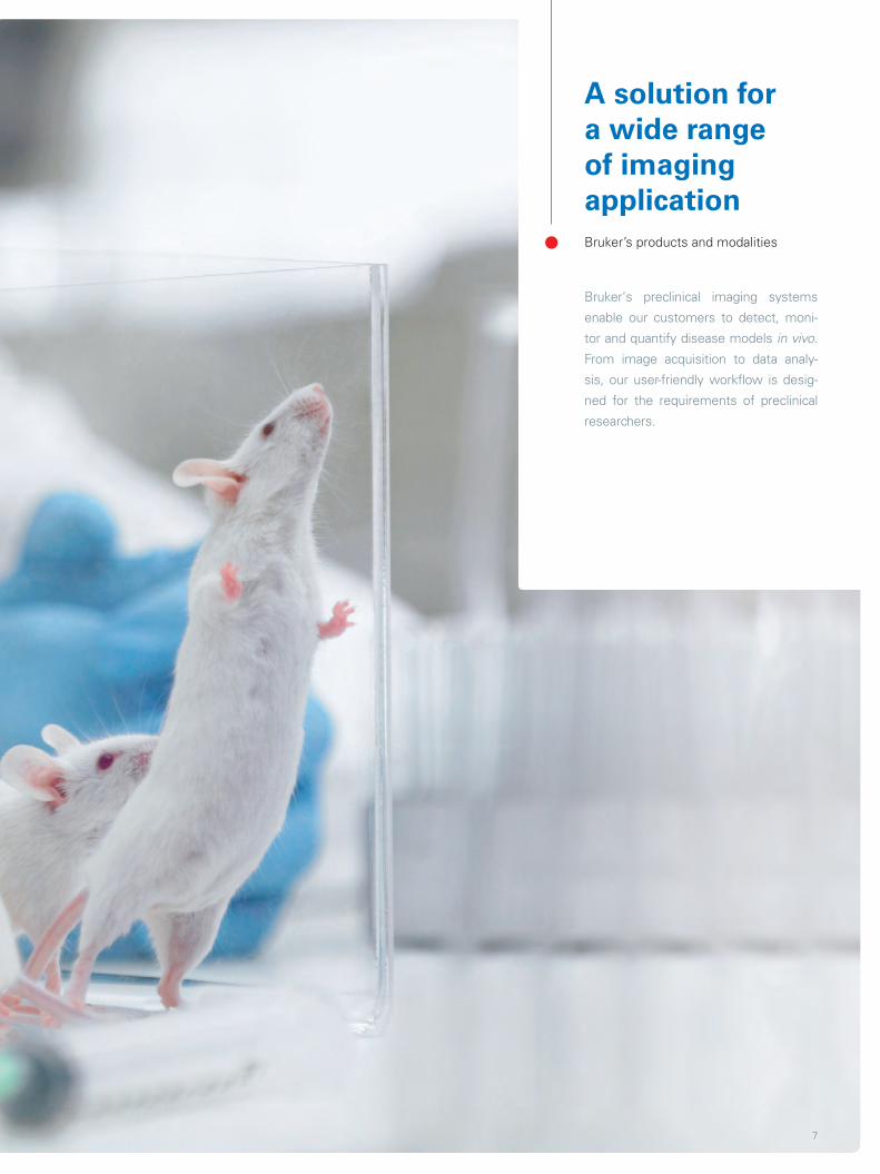
A solution for a wide range of imaging applicationBruker’s products and modalities
Bruker‘s preclinical imaging systems
enable our customers to detect, moni-
tor and quantify disease models in vivo.
From image acquisition to data analy-
sis, our user-friendly workflow is desig-
ned for the requirements of preclinical
researchers.
7

BioSpec/PhamaScan ClinScan
BioSpec® and PharmaScan® are es-sential components of any research program in the life sciences that utilizes MRI/MRS for disease and metabolism studies. BioSpec sys-tems are offered from 4.7 Tesla to 17.2 Tesla allowing for a wide range of research animal studies.
ClinScan is our clinically oriented MRI scanner offered at 7 Tesla. This sys-tem allows direct and rapid transfer of preclinical studies on animal models to clinical studies on humans. ClinScan uses Siemens clinical user interface syngo®MR with our advanced magnets, gradients, RF-coils, and animal handling solutions.
3D whole body anatomical, functional and molecular imagingHigh Field Magnetic Resonance Imaging
Bruker provides small animal MRI solutions for the emerging market of preclinical
and molecular MR imaging. By combining latest MRI CryoProbe and gradient tech-
nology with ultra-high field magnets, our systems deliver high spatial resolution
inside living organisms.
bruker.com/MRI8

OncologyCardiologyContrast / Molecular imagingDiabetes and obesityNeurobiology
• Diffusion Imaging Axial map of the major principle diffusion direction of a mouse brain enabling to investigate brain connectivity. Courtesy: University Hospital Freiburg, Germany
• AngiographyVisualization of the vascular structure in the mouse brain
• SpectroscopyLocalized spectroscopy for metabolite analysis andmetabolic imaging
• NeurologyHigh resolution brain imaging at the microscopic level
• Funtional ImagingBOLD imaging showing functional brain activation
9

Powerful MRI, SimplifiedCompact Magnetic Resonance Imaging
The new compact-shielded, cryogen-free magnet design with its very small foot-
print has resulted in an easy-to-install MRI system with very low running costs. The
ICON has a negligible magnetic fringe field – enabling safe siting of the system
in any facility and the use by any individual. This compact MRI system has been
designed to provide researchers with the most powerful MRI system in the most
convenient form possible.
Key Features
• Innovative permanent magnet from Aspect Imaging, with negligible fringe field
• Minimum operating and maintenance costs
• No dedicated facilities – all you need is 1.2 m² for the footprint of the sys-tem and a power socket
• Easy-to-use Paravision® 6 software enabling everyone to achieve maximum efficiency
• Integrated easy animal handling and monitoring
• Unique portfolio of MR imaging methods for a wide range of pre- clinical applications
ICON™ • Compact whole body MRI
bruker.com/ICON10

• InflammationVolume measurement of limb lesion in mouse demonstrating the change in intracel lular/extracel-lular water balance. Courtesy of J. Zheng, STTARR (UHN), Toronto, Canada
• Diet control Fat segmentation and volume measurement in high fat diet mouse, subcutanous adipose tissue in red, abdominal adipose tissue in green.Courtesy of S. Aime, the Molecular Imaging Center, University of Torino, Italy
• PET/MRI PET/MR imaging of flank tumor in mouse: Correlation observed between metabolic tumor hetero geneity in PET and Anatomical hete-rogeneity in MRI during sequential acquisition. Courtesy of U. Mahmood, P. Heidari and P . Habibollahi, MGH, Boston, Massachusetts, USA
Oncology NeurologyCardiologyInflammationDiabetes and obesityInfections
• MR/bioluminescence Imaging of ovarian cancer tumor model in mouse: Combining MR images with optical images enab-les tumors to be visualized in anatomical context, improving accuracy of the results. Courtesy of R. de Souza and J. Zheng, STTAR (UHN), Toronto, Canada.
11

3D images down to the sub-micron level micro-CT
Bruker’s SkyScan product line allows you to cut virtual sections or even fly through
samples non-destructively. No preparation, coating or vacuum treatment is nee-
ded. Microtomography is available in a range of easy to use desktop instruments,
which generate 3D images of your sample’s morphology and internal microstruc-
ture with resolution down to the sub-micron level. Software for visualization and
analysis in 3D is included with all SkyScan systems.
SkyScan1176 in vivo micro-CT
The SkyScan 1176 is a high performance in vivo microCT scanner for preclinical research. The large format 11 megapixel X-Ray camera gives an unrivalled com-bination of resolution, image field size and scan speed – everything that is re-quired
in a busy and demanding biomedical re-search laboratory. Image field width up to 68 mm allows full body mouse and rat scanning and distal limb scanning for big animals, such as rabbits.
SkyScan1176 • in vivo micro-CT
bruker.com/microCT12

• Cardiovascular Full body in vivo scan of a mouse using contrast agent
• Bone researchIn vivo scan of mouse hindlimb (knee) with 9 µm pixel size
Bone-OrthopedicsDental-OdontologyCardiovascularPulmonaryOncologyMetabolism-ObesityRenal-Hepatic-GIT
SkyScan1178 high-throughput micro-CT
The SkyScan 1178 is a fast micro-CT scanner with a scanning + reconstruc-tion cycle of less than one minute for the entire volume. Static object position facilitates in vivo scanning of laborato-ry animals and industrial applications, such as quality control and process mo-nitoring. Animal beds for rats and mice
made of carbon fiber are supplied on an interchangeable holder to combine with PET, SPECT, and optical imaging. A physiological monitoring subsystem can measure breathing and heartbeat in real time while also providing signals for gated acquisition.
13

Albira: Small footprint, huge possibilitiesPET/SPECT/CT
Albira combines PET, SPECT and CT imaging in a novel and extremely powerful way.
The system‘s highly compact, modular design gives you the freedom to purchase
what you need now and upgrade as your research needs evolve.
Albira‘s unique detector system uses an exclusive, patented combination of con-
tinuous crystal detectors, PSPMTs (position sensitive photo-multiplier tubes) and
advanced electronics to deliver exquisite sensitivity with rapid acquisition of extre-
mely high resolution, quantitative, precise and accurate PET and SPECT images.
Use Albira for precisely quantified in vivo studies including pharmacokinetics, phar-
macodynamics, ADME, protein expression, metabolic studies, gene expression,
toxicology, perfusion studies, cell tracking, receptor binding and more.
Albira • PET/SPECT/CT
bruker.com/Albira
Key Features
• Choice of seven configurations - Tri-modal - Bi-modal - Standalone• Field upgradeable and customizeable• Compact footprint
• Novel gamma detector technology with continuous crystals
• Automatic co-registration and MR compatibility
• Powerful quantification & dynamic analysis
• High throughput - 4 mice at a time• User friendly, state-of-the-art software
14

• Neurology: Functional brain imagingImaging brain hypoxia in rats using 18F-FMISO Courtesy of Prof. M.A.Pozo Instituto Pluridisciplinar Universidad Complutense de Madrid, Spain
• Perfusion imaging pulmonary studies 99mTc-MAA imaging of lung perfusion in a mouse.Courtesy of Dr. W. Matthew Leevy, NDIIF, University of Notre Dame, Indiana, USA
• Metabolic disease studiesSegmentation of Adipose Tissue in Obesity Mouse Model
OncologyNeurologyCardiologyMetabolic diseaseDrug DiscoveryBone Disease
• Anatomical imagingUse of barium sulfate contrast agent to image the GI tract with computed tomography
• CardiologyTransverse slice of a 18FDG PET/CT overlay of the mouse heart Image courtesy of Dr. W. Matthew Leevy, NDIIF, University of Notre Dame, Indiana, USA
• Oncology18F-FDG image of a prostate cancer tumor xenograft on a rat
15
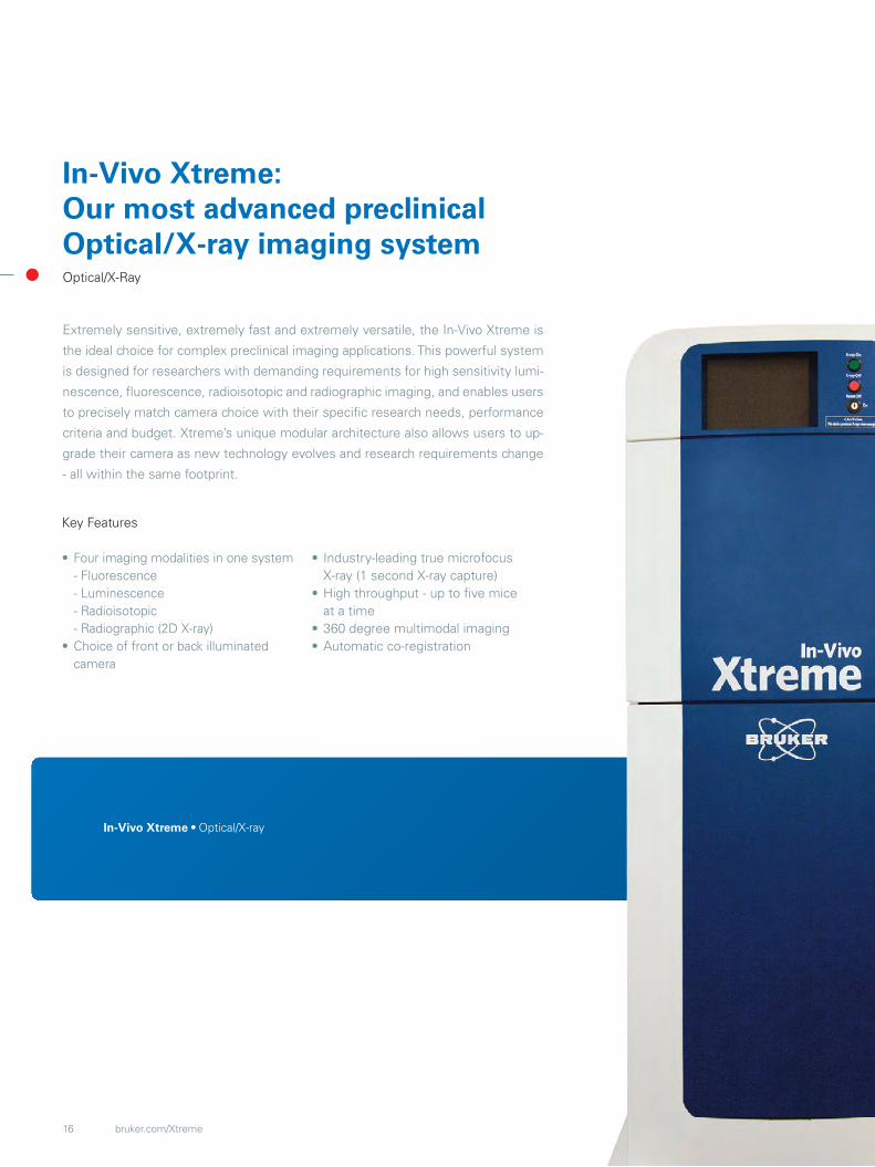
In-Vivo Xtreme: Our most advanced preclinicalOptical/X-ray imaging systemOptical/X-Ray
Extremely sensitive, extremely fast and extremely versatile, the In-Vivo Xtreme is
the ideal choice for complex preclinical imaging applications. This powerful system
is designed for researchers with demanding requirements for high sensitivity lumi-
nescence, fluorescence, radioisotopic and radiographic imaging, and enables users
to precisely match camera choice with their specific research needs, performance
criteria and budget. Xtreme’s unique modular architecture also allows users to up-
grade their camera as new technology evolves and research requirements change
- all within the same footprint.
Key Features
• Four imaging modalities in one system - Fluorescence - Luminescence - Radioisotopic - Radiographic (2D X-ray)• Choice of front or back illuminated camera
• Industry-leading true microfocus X-ray (1 second X-ray capture)• High throughput - up to five mice at a time• 360 degree multimodal imaging• Automatic co-registration
In-Vivo Xtreme • Optical/X-ray
bruker.com/Xtreme16
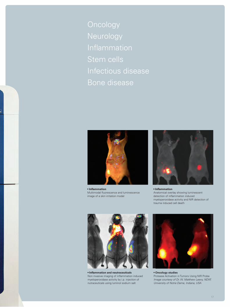
• InflammationAnatomical overlay showing l uminescent detection of inflammation induced myeloperoxidase activity and NIR detection of trauma induced cell death
• InflammationMultimodal fluorescence and luminescence image of a skin irritation model
• Inflammation and neutraceuticalsNon invasive imaging of inflammation induced myeloperoxidase activity by i.p. injection of nutraceuticals using luminol sodium salt
Oncology NeurologyInflammationStem cellsInfectious diseaseBone disease
• Oncology studiesProtease Activation in Tumors Using NIR ProbeImage courtesy of Dr. W. Matthew Leevy, NDIIF, University of Notre Dame, Indiana, USA
17
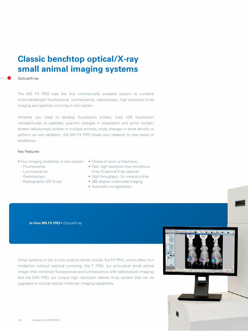
In-Vivo MS FX PRO • Optical/X-ray
Classic benchtop optical/X-ray small animal imaging systemsOptical/X-ray
The MS FX PRO was the first commercially available system to combine
multi-wavelength fluorescence, luminescence, radioisotopic, high resolution X-ray
imaging and spectral unmixing in one system.
Whether you need to develop fluorescent probes; track NIR fluorescent
nanoparticules or peptides; quantify changes in localization and tumor burden;
screen radioisotopic probes in multiple animals; study changes in bone density or
perform ex vivo validation, the MS FX PRO drives your research to new levels of
excellence.
Other systems in the In-Vivo product family include the FX PRO, which offers four
modalities without spectral unmixing; the F PRO, our entry-level small animal
imager that combines fluorescence and luminescence with radioisotopic imaging;
and the DXS PRO, our unique high resolution cabinet X-ray system that can be
upgraded to include optical molecular imaging capabilities.
bruker.com/MSFXPro
Key Features
• Four imaging modalities in one system - Fluorescence - Luminescence - Radioisotopic - Radiographic (2D X-ray)
• Choice of zoom or fixed lens• Fast, high resolution true microfocus X-ray (3 second X-ray capture)• High throughput - 3+ mice at a time• 360 degree multimodal imaging• Automatic co-registration
18

• Infection imagingDetection of Inflammation induced myeloperoxi-dase activity after Microbial Infection. Lumine-scent detection of Inflammation with X-Ray overlay
• Oncology: Interaction of tumors and inflammatory responseMulti-modal anatomical and molecular image overlay of NIR fluorescent tumor cells and luminescent detection of inflammation induced myeloperoxidase activity
• Probe development and validationSpectral unmixing of two NIR fluorescent probes (red and blue) and separation from gut autofluore-scence (green)
• Agriculture studiesPseudocolored X-Ray screening of seedsfor viability
Oncology NeurologyProbe & biomarker development and ValidationInfectious diseaseAgriculture
19

Delivering optimum customer experienceBruker‘s comprehensive service and support: reliability, expertise and performance
Bruker is strongly committed to provi-
de the highest quality of service and
support. Our comprehensive service
portfolio ensures that customers are al-
ways supported by a global corporation
with over 6,500 colleagues in over 100
offices worldwide.
Customers benefit from this wide-
spread service network, our experience
with installations and customer support
around the world, our market share in
preclinical imaging, our dedication to
service, and much more.
20

Bruker’s comprehensive services at a glance
• Site Planning - Customized Services: For some instruments, this is the first step in
ensuring optimum system performance. Bruker‘s technical departments can provide
space-planning and site preparation services tailored to your individual needs.
• Responsive Technical and Software Support: Bruker‘s Service & Support hotlines are
your first point of call. Support center engineers and scientists will quickly and efficiently
gather key information, suggest relevant diagnostics and provide a swift solution.
• Applications Support: Our trusted experts continue to develop innovative in vivo
imaging applications and solutions that meet a wide range of demanding needs in
pre-clinical imaging, molecular medicine, biomedical and pharmaceutical research.
• Education and Training: Bruker offers a variety of advanced professional trainings,
webinars, seminars and workshops. Our courses cover a wide range of applications
and include hands-on lab sessions in our dedicated application support centers. To
learn more about the training schedule and registration, please visit: www.bruker.com/
pci-training.html
For more information please contact us at:
Contact application support /Contact on service support
21

Bruker – Continuous scientific innovation
Bruker has been driven by the idea to
always provide the best technologi-
cal solution for each analytical task for
more than 50 years now. Today, world-
wide more than 6,500 employees are
working on this permanent challenge
at over 100 locations on all continents.
Bruker systems cover a broad spectrum
of applications in all fields of research
and development and are used in all
industrial production processes for the
purpose of ensuring quality and process
reliability.
22

A performance leader in preclinical
imaging instrumentation
Bruker offers advanced preclinical
imaging solutions for a broad spectrum
of application fields, such as cancer
research, functional and
anatomical neuroimaging, orthopedics,
cardiac imaging, stroke models and
many more. Our flexible instruments
can be combined for several different
research programs – thus enabling
higher versatility across all instrument
platforms and significantly expanding
research capabilities.
Our range of techniques includes
PET – Positron Emission Tomography,
SPECT – Single Photon Emission
Computed Tomography, microCT –
Micro Computed Tomography, X-Ray,
Fluorescence, Luminescence, Radio-
isotopic, MRI – Magnetic Resonance
Imaging, MPI – Magnetic Particle
Imaging.
23
© B
ruke
r B
ioS
pin
01/1
4 13
T13
7475
[email protected] more information on preclinical applications, solutions and instruments please visit http://www.bruker.com/preclinicalimaging
Enhancing research capabilities and accelerating time-to-market of drugs
• Knowledge and expertise of a global market leader in imaging technologies
• An unmatched portfolio of nine preclinical imaging modalities
• All modalities can be used singly or in combination with each other
• Advanced, proven imaging technologies from a single source
• Accelerated time-to-market of drugs and therapies
• A broad spectrum of industry, application and research tasks covered
• Service throughout the whole lifecycle of instruments and solutions
• Protection of investments in instruments and solutions
• A company managed by scientists, understanding the needs of scientists


![Imaging, Diagnosis, Prognosis Cancer Research Preclinical and … · Imaging, Diagnosis, Prognosis Preclinical and Clinical Evidence that Deoxy-2-[18F]fluoro-D-glucose Positron Emission](https://img.pdfslide.us/doc/110x75/5e9a6009a0a8a60ac52aaf27/imaging-diagnosis-prognosis-cancer-research-preclinical-and-imaging-diagnosis.jpg)