Embed Size (px)
Citation preview

NCCN®
Version 2.2012, 11/01/11 © National Comprehensive Cancer Network, Inc. 2011, All rights reserved. The NCCN Guidelines and this illustration may not be reproduced in any form without the express written permission of NCCN .® ®
NCCN Guidelines IndexRectal Cancer Table of Contents
Discussion
Continue
NCCN.org
NCCN Clinical Practice Guidelines in Oncology (NCCN Guidelines )®
Rectal Cancer
Version 2.2012
Continue

NCCN®
Version 2.2012, 11/01/11 © National Comprehensive Cancer Network, Inc. 2011, All rights reserved. The NCCN Guidelines and this illustration may not be reproduced in any form without the express written permission of NCCN .® ®
NCCN Guidelines IndexRectal Cancer Table of Contents
Discussion
Continue
NCCN Guidelines Panel Disclosures
NCCN Guidelines Version 2.2012 Panel MembersRectal Cancer
† Medical Oncology
¶ Surgery/Surgical oncologyPathology
‡ Hematology/Hematology OncologyÞ Internal medicine
§ Radiotherapy/Radiation oncology
*Writing Committee Member
�
ф Diagnostic/Interventional Radiology
¥ Patient advocate
David P. Ryan, MD †Massachusetts General Hospital CancerCenter
Leonard Saltz, MD † ‡ ÞMemorial Sloan-Kettering Cancer Center
Sunil Sharma, MD †Huntsman Cancer Institute at the Universityof Utah
David Shibata, MD ¶H. Lee Moffitt Cancer Center and ResearchInstitute
The University of Texas MD Anderson CancerCenter
William Small, Jr., MD §Robert H. Lurie Comprehensive CancerCenter of Northwestern University
Constantinos T. Sofocleous, MD, PhDMemorial Sloan-Kettering Cancer Center
UCSF Helen Diller Family ComprehensiveCancer Center
Christopher Willett, MD §Duke Cancer Institute
Deborah Freedman-Cass, PhDLauren Gallagher, PharmDKristina M. Gregory, RN, MSN, OCN
John M. Skibber, MD ¶
Alan P. Venook, MD † ‡
ф
NCCN
James W. Fleshman, Jr., MD ¶Siteman Cancer Center at Barnes-JewishHospital and Washington University Schoolof Medicine
Charles S. Fuchs, MD, MPH †Dana-Farber/Brigham and Women's CancerCenter
Jean L. Grem, MD †UNMC Eppley Cancer Center at The NebraskaMedical Center
James A. Knol, MD ¶University of Michigan ComprehensiveCancer Center
Lucille A. Leong, MD †City of Hope Comprehensive Cancer Center
Edward Lin, MD †Fred Hutchinson Cancer ResearchCenter/Seattle Cancer Care Alliance
Kilian Salerno May, MD §Roswell Park Cancer Institute
Mary F. Mulcahy, MD ‡Robert H. Lurie Comprehensive CancerCenter of Northwestern University
Kate Murphy, BA ¥Fight Colorectal Cancer
Eric Rohren, MD, PhD фThe University of Texas MD Anderson CancerCenter
Al B. Benson, III, MD/Chair †Robert H. Lurie Comprehensive CancerCenter of Northwestern University
¶
†
Emily Chan, MD, PhD †Vanderbilt-Ingram Cancer Center
Yi-Jen Chen, MD, PhD §City of Hope Comprehensive Cancer Center
¶Comprehensive Cancer
Center at
¶
Paul F. Engstrom, MD †Fox Chase Cancer Center
Peter C. Enzinger, MD †Dana-Farber/Brigham and Women’s CancerCenter
J. Pablo Arnoletti, MDUniversity of Alabama at BirminghamComprehensive Cancer Center
Tanios Bekaii-Saab, MDThe Ohio State University ComprehensiveCancer Center - James Cancer Hospital andSolove Research Institute
Michael A. Choti, MD, MBAThe Sidney Kimmel
Johns Hopkins
Harry S. Cooper, MDFox Chase Cancer Center
Raza A. Dilawari, MDSt. Jude Children's ResearchHospital/University of Tennessee CancerInstitute
�
*
*
*
*

NCCN®
Version 2.2012, 11/01/11 © National Comprehensive Cancer Network, Inc. 2011, All rights reserved. The NCCN Guidelines and this illustration may not be reproduced in any form without the express written permission of NCCN .® ®
NCCN Guidelines IndexRectal Cancer Table of Contents
Discussion
NCCN Rectal Cancer Panel Members
Summary of the Guidelines Updates
Surveillance (REC-7)Recurrence and Workup (REC-8)Postoperative CEA Elevation (REC-8)
Principles of Pathologic Review (REC-A)Principles of Surgery (REC-B)Principles of Adjuvant Therapy (REC-C)Principles of Radiation Therapy (REC-D)Chemotherapy for Advanced or Metastatic Disease (REC-E)Principles of Survivorship (REC-F)
Staging (ST-1)
�
�
�
Pedunculated polyp with invasive cancer (REC-1)
Sessile polyp with invasive cancer (REC-1)
Rectal cancer appropriate for resection (REC-2)T1-2, N0: Primary and Adjuvant Treatment (REC-3)T3, N0 or T any, N1-2: Primary and Adjuvant Treatment (REC-4)T4 and/or locally unresectable: Primary and Adjuvant Treatment (REC-4)T any, N any, M1: Resectable Metastases Treatment (REC-5)T any, N any, M1: Unresectable Metastases or Medically InoperableTreatment (REC-6)
�
�
�
�
�
Clinical Presentations and Primary Treatment:
Clinical Trials:
Categories of Evidence andConsensus:NCCN
Thebelieves that the best managementfor any cancer patient is in a clinicaltrial. Participation in clinical trials isespecially encouraged.
To find clinical trials online at NCCNmember institutions,
All recommendationsare Category 2A unless otherwisespecified.
See
NCCN
click here:nccn.org/clinical_trials/physician.html
NCCN Categories of Evidenceand Consensus
The NCCN Guidelines are a statement of evidence and consensus of the authors regarding their views of currently accepted approaches to treatment.
Any clinician seeking to apply or consult the NCCN Guidelines is expected to use independent medical judgment in the context of individual clinical
circumstances to determine any patient’s care or treatment. The National Comprehensive Cancer Network (NCCN ) makes no representations or
warranties of any kind regarding their content, use or application and disclaims any responsibility for their application or use in any way. The NCCN
Guidelines are copyrighted by National Comprehensive Cancer Network . All rights reserved. The NCCN Guidelines and the illustrations herein may not
be reproduced in any form without the express written permission of NCCN. ©2011.
®
® ®
®
NCCN Guidelines Version 2.2012 Table of ContentsRectal Cancer

NCCN®
Version 2.2012, 11/01/11 © National Comprehensive Cancer Network, Inc. 2011, All rights reserved. The NCCN Guidelines and this illustration may not be reproduced in any form without the express written permission of NCCN .® ®
NCCN Guidelines IndexRectal Cancer Table of Contents
Discussion
UPDATES
General: In regard to intravenous therapy, continuous changed toinfusional throughout the Guidelines.
Footnote “e” added to this page: Observation may beconsidered, with the understanding that there is significantlygreater incidence of adverse outcomes (residual disease,recurrent disease, mortality, hematogenous metastasis, butnot lymph node metastasis) than polypoid malignant polyps.
- Endoscopicallyremoved malignant polyp.
Endorectal MRI removed as a recommended test in theworkup of a patient with rectal cancer appropriate forresection.
Preferred designation added to continuous 5-FU/RT andcapecitabine/RT as compared to bolus 5-FU/RT.Footnote “k” modified by deleting the last sentence, “Trials arestill pending in rectal cancer.” (also applies to REC-4 and REC-5)Previous footnote “k” deleted: “Data regarding the use ofcapecitabine/RT are limited and no phase III randomized data areavailable. Trials are pending. Kim J-Sang, Kim J-Sung, Cho, M, etal Preoperative chemoradiation using oral capecitabine in locallyadvanced rectal cancer. Int J Radiation Oncology Biol Phys2002;54(2):403-408.” (also applies to REC-4, REC-5, and REC-6)
Preoperative capecitabine/RT changed from a category 2A to acategory 1 designation and preferred as compared to bolus 5-FU/RT.
Category 1 and preferred for infusional 5-FU/RT and capecitabine/RTapplies to stage II and stage III disease (previously only stage IIIdisease).
FOLFOX + cetuximab removed as a treatment option.Preferred designation added to continuous 5-FU/RT andcapecitabine/RT as compared to bolus 5-FU/RT.
Isolated pelvic/anastomotic recurrence, potentially resectable: “ifnot given previously” removed from preoperative 5-FU + RT andreferral to the Principles of Radiation Therapy added for furtherguidance.
Response changed to “No progression” and No response changedto “Progression”.Adjuvant therapy recommendations following resection and noprevious chemotherapy: Active chemotherapy regimen for advanceddisease (REC-E) changed to adjuvant therapy for stage III disease(REC-4).
Lymph node evaluation, bullet 1: early stage replaced stage II.
BRAF Testing, second bullet: The following sentence was added:Allele-specific PCR is another acceptable method for detectingBRAF V600E mutation.
�
�
�
�
�
�
�
�
�
�
�
�
�
�
�
Adjuvant therapy recommendations following staged orsynchronous resection of metastases + rectal lesion: Activechemotherapy regimen for advanced disease (REC-E) changed toadjuvant therapy for stage III disease (REC-4).
REC-1
REC-2
REC-3
REC-4
REC-5
REC-8
REC-9
REC-A 4 of 6
See Principles of Pathologic Review (REC-A)
REC-4
REC-A 5 of 6
NCCN Guidelines Version 2.2012 UpdatesRectal Cancer
Summary of changes in the 1.2012 version of the Rectal Cancer Guidelines from the 4.2011 version include:
Summary of changes in the 2.2012 version of the Rectal Cancer Guidelines from the 1.2012 version include:
The recommendation of cetuximab (KRAS WT only) + irinotecan after progression on first-line FOLFOX ± bevacizumab or CapeOx ±bevacizumab was changed from a category 2B to a category 2A.Panitumumab (KRAS WT only) + irinotecan (category 2A) added as an alternate treatment option to cetuximab + irinotecan in advanced ormetastatic disease after progression on first- or second-line therapy.
The discussion section updated to reflect the changes in the algorithm.
�
�
�
REC-E 1 of 7
MS-1

NCCN®
Version 2.2012, 11/01/11 © National Comprehensive Cancer Network, Inc. 2011, All rights reserved. The NCCN Guidelines and this illustration may not be reproduced in any form without the express written permission of NCCN .® ®
NCCN Guidelines IndexRectal Cancer Table of Contents
Discussion
REC-B 1 of 3
REC-C 1 of 2
REC-D
REC-E 1 of 7
REC-E 2 of 7
REC-E 4 of 7 REC-E 7 of 7
�
�
�
�
�
�
�
�
�
�
�
�
Transabdominal Resection, Management Principles:Sub-bullet 3 deleted “Laparoscopic surgery is not recommended outside the setting of a clinical trial” and replaced with“Laparoscopic surgery is preferred in the setting of a clinical trial” with the following footnote “Long term outcomes fromlaparoscopic surgery have not been reported. Current clinical trials are exploring open versus laparoscopic approach.”
First paragraph modifications: Adjuvant therapy for rectal cancer consists of regimens that include both concurrent chemotherapy/RTand adjuvant chemotherapy. . A total of 6months of perioperative treatment is preferred.Recommendations combined for postoperative adjuvant therapy (previously divided based on whether patient received preoperativechemotherapy/RT).Adjuvant postoperative chemotherapy: All regimens clarified “to a total of 6 mo perioperative therapy.”Adjuvant postoperative chemotherapy: FLOX regimen added with supporting reference: Kuebler JP, Wieand HS, O'Connell MJ, et al.Oxaliplatin combined with weekly bolus fluorouracil and leucovorin as surgical adjuvant chemotherapy for stage II and III coloncancer: results from NSABP C-07. J Clin Oncol 2007;25:2198-2204.Adjuvant postoperative chemotherapy: Mayo clinic regimen deleted: Jager E, Heike M, Bernhard H, et al. Weekly high-dose leucovorinversus low-dose leucovorin combined with fluorouracil in advanced colorectal cancer: results of a randomized multicenter trial. J ClinOncol 1996;14:2274-2279.Concurrent chemotherapy/RT: Capecitabine/RT changed from a category 2B recommendation to a category 2A recommendation.
Bullet 4 modified - Intensity modulated radiotherapy (IMRT) should only be used in the setting of a clinical trial.
Bullet 6 modified: Intraoperative radiotherapy (IORT), if available, should be considered for very close or positive margins afterresection, as an additional boost, especially for patients with T4 or recurrent cancers. If IORT is not available, 10-20 Gy external beamradiation to a limited volume could be considered soon after surgery, prior to adjuvant chemotherapy.Bullet 10 removed the second sentence: All male patients should be evaluated for erectile dysfunction and considered for earlytreatment intervention if necessary.
Patient appropriate for intensive therapy: FOLFOX + cetuximab removed as a treatment option for initial therapy of advanced or
metastatic disease.
Patient not appropriate for intensive therapy: Capecitabine ± bevacizumab added as a treatment option for initial therapy of advanced
or metastatic disease.through
The chemotherapy/RT may be administered either pre or postoperatively approximately
or in unique clinicalsituations including re-irradiation of recurrent disease after previous radiotherapy
and/or brachytherapy
� Chemotherapy regimen dosing and references expanded and updated.
UPDATES
NCCN Guidelines Version 2.2012 UpdatesRectal Cancer
Summary of changes in the 1.2012 version of the Rectal Cancer Guidelines from the 4.2011 version include:

NCCN®
Version 2.2012, 11/01/11 © National Comprehensive Cancer Network, Inc. 2011, All rights reserved. The NCCN Guidelines and this illustration may not be reproduced in any form without the express written permission of NCCN .® ®
NCCN Guidelines IndexRectal Cancer Table of Contents
Discussion
REC-F
REC-7
NCCN Breast Cancer Screening GuidelinesNCCN Cervical Cancer Screening GuidelinesNCCN Prostate Early Detection Guidelines
Cancer Surveillance. Previous bullets deleted and the following added:See .Long term surveillance should be carefully managed with routine good medical care and monitoring, including cancer screening, routine healthcare, and preventive care.Routine CEA monitoring and routine CT scanning is not recommended beyond 5 years.
Management of Late Sequelae of Disease or Treatment: The following bullets added:Urogenital Dysfunction after Resection and/or Pelvic Radiation
Screen for urinary incontinence, frequency, and urgencyConsider referral to urologist or gynecologist for persistent symptoms.
Cancer Screening Recommendations. Previous bullets deleted and the following added:These recommendations are for average risk patients. Recommendations for high risk individuals should be made on an individual basis.
Breast Cancer: See theCervical Cancer: See theProstate Cancer: See the
Counseling Regarding Healthy Lifestyle and Wellness. Previous bullets deleted and the following added:Maintain a healthy body weight throughout life.Adopt a physically active lifestyle (At least 30 minutes of moderate intensity activity on most days of the week). Activity recommendations mayrequire modification based on treatment sequelae (i.e. ostomy, neuropathy).Consume a healthy diet with emphasis on plant sources.Limit alcohol consumption.Smoking cessation counseling as appropriate.Additional health monitoring and immunizations should be performed as indicated under the care of a primary care physician. Survivors areencouraged to maintain a therapeutic relationship with a primary care physician throughout their lifetime.
�
�
�
�
�
�
�
�
�
�
�
�
6,7
�
�
�
Screen for sexual dysfunction, erectile dysfunction, dyspareunia, and vaginal dryness
UPDATES
NCCN Guidelines Version 2.2012 UpdatesRectal Cancer
Summary of changes in the 1.2012 version of the Rectal Cancer Guidelines from the 4.2011 version include:

NCCN®
Version 2.2012, 11/01/11 © National Comprehensive Cancer Network, Inc. 2011, All rights reserved. The NCCN Guidelines and this illustration may not be reproduced in any form without the express written permission of NCCN .® ®
NCCN Guidelines IndexRectal Cancer Table of Contents
Discussion
Single specimen, completely
removed with favorable
histological features and
clear margins (T1 only)
d
�
�
�
Pathology review
Colonoscopy
Marking of
cancerous polyp
site (at time of
colonoscopy or
within 2 wks)
b,c
CLINICAL
PRESENTATIONa
Pedunculated polyp
or Sessile polyp
(adenoma [tubular,
tubulovillous, or
villous]) with
invasive cancer
WORKUP FINDINGS
Fragmented specimen or
margin cannot be
assessed or unfavorable
histological featuresd
See Primary andAdjuvantTreatment (REC-3)
a
e
ll
Endoscopically removed malignant polyp
Observation may be considered, with the understanding that there is significantly greater incidence of adverse outcomes (residual disease, recurrent disease, mortality,hematogenous metastasis, but not lymph node metastasis) than polypoid malignant polyps. - Endoscopically removedmalignant polyp.
A patients with rectal cancer should be counseled for family history. Patients with suspected hereditary non-polyposis colon cancer (HNPCC), familial adenomatouspolyposis (FAP) and attenuated FAP, see the .
Confirm the presence of invasive cancer (pT1). pTis has no biological potential to metastasize.
It has not been established if molecular markers are useful in treatment determination (predictive markers) and prognosis. College of American Pathologists ConsensusStatement 1999. Prognostic factors in colorectal cancer. Arch Pathol Lab Med 2000;124:979-994.
- .
b
c
d
NCCN Colorectal Cancer Screening Guidelines
See Principles of Pathologic Review (REC-A)
See Principles of Pathologic Review (REC-A)
Note: All recommendations are category 2A unless otherwise indicated.
Clinical Trials: NCCN believes that the best management of any cancer patient is in a clinical trial. Participation in clinical trials is especially encouraged.
NCCN Guidelines Version 2.2012Rectal Cancer
ObserveorSee Primary Treatment
on page
e
REC-3
REC-1
Pedunculated
polyp with
invasive cancer
Sessile polyp
with invasive
cancer
Observe

NCCN®
Version 2.2012, 11/01/11 © National Comprehensive Cancer Network, Inc. 2011, All rights reserved. The NCCN Guidelines and this illustration may not be reproduced in any form without the express written permission of NCCN .® ®
NCCN Guidelines IndexRectal Cancer Table of Contents
Discussion
Note: All recommendations are category 2A unless otherwise indicated.
Clinical Trials: NCCN believes that the best management of any cancer patient is in a clinical trial. Participation in clinical trials is especially encouraged.
CLINICAL STAGE
T1-2, N0
�
�
�
�
�
�
�
�
�
Biopsy
Pathology review
Colonoscopy
Rigid proctoscopy
Chest/abdominal/pelvic CT
CEA
Endorectal ultrasound or pelvic MRI
Enterostomal therapist as indicated for
preoperative marking of site, teaching
PET-CT scan is not routinely indicated
T3, N0
or
T any, N1-2
aAll patients with rectal cancer should be counseled for family history. Patients with suspected hereditary non-polyposis colon cancer (HNPCC), familialadenomatous polyposis (FAP) and attenuated FAP, see the NCCN Colorectal Cancer Screening Guidelines.
Rectal cancer
appropriate
for resection
See Primary Treatment (REC-6)
See Primary Treatment (REC-4)
See Primary Treatment (REC-3)
T4 and/or locally
unresectableSee Primary Treatment (REC-4)
See Primary Treatment (REC-5)
T any, N any, M1Resectable
metastases
T any, N any, M1Unresectable
metastases or
medically inoperable
REC-2
CLINICAL
PRESENTATIONaWORKUP
NCCN Guidelines Version 2.2012Rectal Cancer

NCCN®
Version 2.2012, 11/01/11 © National Comprehensive Cancer Network, Inc. 2011, All rights reserved. The NCCN Guidelines and this illustration may not be reproduced in any form without the express written permission of NCCN .® ®
NCCN Guidelines IndexRectal Cancer Table of Contents
Discussion
CLINICAL
STAGE
PRIMARY TREATMENT
cT1-2,
N0f
Transanal
excision, if
appropriategT1, NX with
high risk
featuresh
or T2, NX
Trans-
abdominal
resectiong
T1, NX;
Margins
negative
Observe
fT1-2, N0 should be based on assessment of endorectal ultrasound or MRI.
High risk features include positive margins, lymphovascular invasion and poorlydifferentiated tumors.
The use of FOLFOX or capecitabine ± oxaliplatin are extrapolations from theavailable data in colon cancer.
g
h
j
k
i
See Principles of Surgery (REC-B). See Principles of Radiation Therapy (REC-D).
See Principles of Adjuvant Therapy (REC-C).
ADJUVANT TREATMENTi,j
(6 MO PERIOPERATIVE TREATMENT PREFERRED)
pT3, N0, M0
or
pT1-3, N1-2
pT1-2, N0, M0 Observe
pT3, N0, M0
or
pT1-3, N1-2
pT1-2,
N0, M0Observe
5-FU ± leucovorin
or
capecitabine/RT (preferred), then 5-FU ±
leucovorin or FOLFOX
or FOLFOX or capecitabine
± oxaliplatin, then infusional 5-FU/RT
(preferred) or bolus 5-FU/leucovorin/RT
or capecitabine ±
oxaliplatinorInfusional 5-FU/RT (preferred) or bolus 5-FU/
leucovorin/RT or capecitabine/RT (preferred)
followed by 5-FU ± leucovorin or FOLFOX or
capecitabine ± oxaliplatin
k
k
k k
k
k
k
k k
Surveillance(See REC-7)
REC-3
cT1, N0f
Note: All recommendations are category 2A unless otherwise indicated.
Clinical Trials: NCCN believes that the best management of any cancer patient is in a clinical trial. Participation in clinical trials is especially encouraged.
Trans-
abdominal
resectiong
5-FU ± leucovorin or FOLFOX or capecitabine ± oxaliplatin, then
infusional 5-FU/RT (preferred) or bolus 5-FU/leucovorin/RT or
capecitabine/RT (preferred), then 5-FU ± leucovorin or FOLFOX or
capecitabine ± oxaliplatinorInfusional 5-FU/RT (preferred) or bolus 5-FU/leucovorin/RT or
capecitabine/RT (preferred) followed by 5-FU ± leucovorin or FOLFOX or
capecitabine ± oxaliplatin
k k
k
k k
k
k
k k
NCCN Guidelines Version 2.2012Rectal Cancer

NCCN®
Version 2.2012, 11/01/11 © National Comprehensive Cancer Network, Inc. 2011, All rights reserved. The NCCN Guidelines and this illustration may not be reproduced in any form without the express written permission of NCCN .® ®
NCCN Guidelines IndexRectal Cancer Table of Contents
Discussion
Surveillance(See REC-7)
Note: All recommendations are category 2A unless otherwise indicated.
Clinical Trials: NCCN believes that the best management of any cancer patient is in a clinical trial. Participation in clinical trials is especially encouraged.
pT3, N0, M0
or pT1-3, N1-2
l,m
pT1–2, N0, M0 Observe
5-FU ± leucovorinorFOLFOXorCapecitabine ± oxaliplatin
k,o
kk
g
i
j
k
l
m
n
o
.
.
.
The use of FOLFOX or capecitabine ± oxaliplatin are extrapolations from the available data in colon cancer.
The use of agents other than fluoropyrimidines (eg, oxaliplatin) are not recommended concurrently with RT.
For patients with proximal T3, N0 disease with clear margins and favorable prognostic features, the incremental benefit of RT is likely to be small. Considerchemotherapy alone.
Postoperative therapy is indicated in all patients who receive preoperative therapy, regardless of the surgical pathology results.
An ongoing Intergroup trial compares 5-FU/leucovorin, FOLFOX, and FOLFIRI after surgery.
See Principles of Surgery (REC-B)
See Principles of Adjuvant Therapy (REC-C)
See Principles of Radiation Therapy (REC-D)
T3, N0
or
T any, N1-2
Preoperative infusional 5-FU/RT
or capecitabine/RT (category 1
and preferred for both) or bolus
5-FU/leucovorin/RT
Transabdominal
resectiong
Reconsider:5-FU ± leucovorin or FOLFOX or capecitabine ±
oxaliplatin, then infusional 5-FU/RT (preferred) or
bolus 5-FU/leucovorin/RT or capecitabine/RT
(preferred), then 5-FU ± leucovorin or FOLFOX or
capecitabine ± oxaliplatinorInfusional 5-FU/RT (preferred) or bolus 5-FU/
leucovorin/RT or capecitabine/RT (preferred)
followed by 5-FU ± leucovorin or FOLFOX or
capecitabine ± oxaliplatin
k
k
l k
k k
l
k
k
k k
Infusional IV 5-FU/RT
or capecitabine/RT
or
bolus 5-FU/leucovorin/RT Resection,
if possible
T4 and/or
locally
unresectableAny T
CLINICAL
STAGE
PRIMARY TREATMENT ADJUVANT TREATMENTh,i,n
(6 MO PERIOPERATIVE TREATMENT PREFERRED)
Transabdominal
resectiong
Patients with
medical
contraindication
to combined
modality therapy
5-FU ± leucovorinorFOLFOXorCapecitabine ± oxaliplatin
k,o
k k
REC-4
NCCN Guidelines Version 2.2012Rectal Cancer

NCCN®
Version 2.2012, 11/01/11 © National Comprehensive Cancer Network, Inc. 2011, All rights reserved. The NCCN Guidelines and this illustration may not be reproduced in any form without the express written permission of NCCN .® ®
NCCN Guidelines IndexRectal Cancer Table of Contents
Discussion
Note: All recommendations are category 2A unless otherwise indicated.
Clinical Trials: NCCN believes that the best management of any cancer patient is in a clinical trial. Participation in clinical trials is especially encouraged.
CLINICAL
STAGE
PRIMARY TREATMENT
Staged or synchronous
resection of metastases
+ rectal lesion
g
T Any,
N Any, M1
Resectable
synchronous
metastasesp
pT1-2, N0, M1
pT3-4, Any N,
orAny T, N1-2, M1
M1
Staged or
synchronous resection
of metastases and
rectal lesion
g
Consider infusional IV 5-FU/pelvic RT
(preferred) or bolus 5-FU/leucovorin/pelvic RT
or capecitabine/RT (preferred)
g
i
j
k
o
r
t
.
The use of FOLFOX or capecitabine ± oxaliplatin are extrapolations from theavailable data in colon cancer.
Determination of tumor K- KRAS and BRAF
Mutation Testing.
Patients with a V600E BRAF mutation appear to have a poorer prognosis.Retrospective subset analyses suggest potential benefit from anti-EGFR monoclonalantibodies in the first-line setting with active chemotherapy regardless of V600Emutation status.The safety of administering bevacizumab pre or postoperatively, in combination with5-FU-based regimens, has not been adequately evaluated. There should be at leasta 6 wk interval between the last dose of bevacizumab and elective surgery. There isan increased risk of stroke and other arterial events especially in age 65. The useof bevacizumab may interfere with wound healing.FOLFOXIRI is not recommended in this setting.RT only recommended for patients at increased risk for pelvic recurrence.
An ongoing Intergroup trial compares 5-FU/leucovorin, FOLFOX, and FOLFIRIafter surgery.
RAS (if KRAS non-mutated, consider BRAF testing).p
q
s
�
See Principles of Surgery (REC-B).See Principles of Adjuvant Therapy (REC-C)See Principles of Radiation Therapy (REC-D).
See Principles of Pathologic Review (REC-A 5 of 6)
5-FU ± leucovorin or
then infusional 5-FU/RT
(preferred) or bolus 5-FU/leucovorin/RT
then 5-FU ±
leucovorin
FOLFOX or capecitabine
± oxaliplatin,
or
capecitabine/RT (preferred),
or FOLFOX or capecitabine ±
oxaliplatin
k,o k
t t
t
k,o k
k
k
Staged or
synchronous resection
of metastases and
rectal lesion
g
Infusional IV 5-FU/
pelvic RT (preferred) or bolus
5-FU/leucovorin/pelvic RT or
capecitabine/RT (preferred)
or
Combination chemotherapy
(2-3 months)
(FOLFIRI or FOLFOX
or CapeOX) ± bevacizumab or
(FOLFIRI or FOLFOX) ±
panitumumab or FOLFIRI ±
cetuximab [KRAS wild-type
gene only]
r
p,q
or
Surveillance(See REC-7)
Staged or synchronous
resection of metastases
and rectal lesion
gInfusional IV 5-FU/pelvic RT (preferred)
or bolus 5-FU + leucovorin/pelvic RT or
capecitabine/RT (preferred)
or
ADJUVANT THERAPY (resected metastatic disease)
(6 MO PERIOPERATIVE TREATMENT PREFERRED)
i,j
Active chemotherapy regimen for advanced
disease ( ) (category 2B)s See REC-E
REC-5
See adjuvant therapy for
stage III disease on REC-4
NCCN Guidelines Version 2.2012Rectal Cancer

NCCN®
Version 2.2012, 11/01/11 © National Comprehensive Cancer Network, Inc. 2011, All rights reserved. The NCCN Guidelines and this illustration may not be reproduced in any form without the express written permission of NCCN .® ®
NCCN Guidelines IndexRectal Cancer Table of Contents
Discussion
Note: All recommendations are category 2A unless otherwise indicated.
Clinical Trials: NCCN believes that the best management of any cancer patient is in a clinical trial. Participation in clinical trials is especially encouraged.
Combination systemic chemotherapyor5-FU/RT or
Capecitabine/RT (category 2B)or
u
k
Resection of involved rectal segment
orLaser recanalizationorDiverting colostomyorStenting
See Chemotherapy for Advancedor Metastatic Disease (REC-E)
T Any, N Any, M1Unresectablesynchronousmetastasesor medicallyinoperable
p
CLINICAL STAGE PRIMARY TREATMENT
pDetermination of tumor K - KRAS and BRAF Mutation Testing.RAS (if KRAS non-mutated, consider BRAF testing).u
See Principles of Pathologic Review (REC-A 5 of 6)
See Chemotherapy for Advanced or Metastatic Disease (REC-E).
Symptomatic
Asymptomatic Reassess response todetermine resectability
See Chemotherapy forAdvanced or MetastaticDisease (REC-E)
REC-6
NCCN Guidelines Version 2.2012Rectal Cancer

NCCN®
Version 2.2012, 11/01/11 © National Comprehensive Cancer Network, Inc. 2011, All rights reserved. The NCCN Guidelines and this illustration may not be reproduced in any form without the express written permission of NCCN .® ®
NCCN Guidelines IndexRectal Cancer Table of Contents
Discussion
�
�
�
History and physical every 3-6 mo for 2 y,
then every 6 mo for a total of 5 y
CEA every 3-6 mo for 2 y, then every 6 mo
for a total of 5 y for T2 or greater lesions
Chest/abdominal/pelvic CT annually x 3-5 y
for patients at high risk for recurrence
Colonoscopy in 1 y except if no
preoperative colonoscopy due to
obstructing lesion, colonoscopy in 3-6 moIf advanced adenoma, repeat in 1 yIf no advanced adenoma, repeat in 3 y,
then every 5 y
Consider proctoscopy every 6 mo x 5 y for
patients status post LAR
PET-CT scan is not routinely recommended
See
v
x
y
z
aa
�
�
�
�
�
�
Principles of Survivorship (REC-F)
SURVEILLANCEw
Serial CEA elevation or
documented recurrenceSee Workup andTreatment (REC-8)
Note: All recommendations are category 2A unless otherwise indicated.
Clinical Trials: NCCN believes that the best management of any cancer patient is in a clinical trial. Participation in clinical trials is especially encouraged.
v
w
y
z
aa
If patient is a potential candidate for resection of isolated metastasis.
Desch CE, Benson III AB, Somerfield MR, et al. Colorectal cancer surveillance: 2005 update of the American Society of Clinical Oncology Practice Guideline. J ClinOncol 2005;23(33):8512-8519.
CT scan may be useful for patients at high risk for recurrence (eg, lymphatic or venous invasion by tumor, or poorly differentiated tumors).
Villous polyp, polyp > 1 cm, or high grade dysplasia.
Rex DK, Kahi CJ, Levin B, et al. Guidelines for colonoscopy surveillance after cancer resection: a consensus update by the American Cancer Society and the US Multi-Society Task Force on Colorectal Cancer. Gastroenterology 2006;130(6):1865-71.
x
Patients with rectal cancer should also undergo limited endoscopic evaluation of the rectal anastomosis to identify local recurrence. Optimal timing for surveillance isnot known. No specific data clearly support rigid versus flexible proctoscopy. The utility of routine endoscopic ultrasound for early surveillance is not defined.
REC-7
NCCN Guidelines Version 2.2012Rectal Cancer

NCCN®
Version 2.2012, 11/01/11 © National Comprehensive Cancer Network, Inc. 2011, All rights reserved. The NCCN Guidelines and this illustration may not be reproduced in any form without the express written permission of NCCN .® ®
NCCN Guidelines IndexRectal Cancer Table of Contents
Discussion
Note: All recommendations are category 2A unless otherwise indicated.
Clinical Trials: NCCN believes that the best management of any cancer patient is in a clinical trial. Participation in clinical trials is especially encouraged.
WORKUPRECURRENCE
g
p
j
Determination of tumor K - KRAS and BRAF Mutation Testing.
Patients should be evaluated by a multidisciplinary team including surgical consultation for potentially resectable patients.
RAS (if KRAS non-mutated,consider BRAF testing).bb
See Principles of Surgery (REC-B).
See Principles of Pathologic Review (REC-A 5 of 6)
See Principles of Radiation Therapy (REC-D).
Isolated pelvic/
anastomotic
recurrence
TREATMENT
REC-8
Documented
metachronous
metastases
by CT, MRI
and/or biopsy
p,bb
Resectableg
See Primary
Treatment REC-9
See Primary
Treatment REC-10
Resectable
Unresectable
Consider
PET-CT
scan
Unresectable
(potentially
convertible or
unconvertible)
f
Serial
CEA
elevation
Negative
findings
Positive
findings
�
�
�
�
Physical exam
Colonoscopy
Chest/abdominal/
pelvic CT
Consider PET-CT
scan
�
�
Consider PET-CT scan
Reevaluate chest/
abdominal/pelvic CT
in 3 mo
Negative
findings
Positive
findingsSee treatment for Isolated
pelvic/anastomotic recurrence
or Documented metachronous
metastases, below
Potentially
resectableg
Unresectable
ResectionorPreoperative 5-FU + RTj
Chemotherapy ± RTi
See treatment for Isolated
pelvic/anastomotic recurrence
or Documented metachronous
metastases, below
Resection ± IORTj
Chemotherapy + RTj
NCCN Guidelines Version 2.2012Rectal Cancer

NCCN®
Version 2.2012, 11/01/11 © National Comprehensive Cancer Network, Inc. 2011, All rights reserved. The NCCN Guidelines and this illustration may not be reproduced in any form without the express written permission of NCCN .® ®
NCCN Guidelines IndexRectal Cancer Table of Contents
Discussion
Note: All recommendations are category 2A unless otherwise indicated.
Clinical Trials: NCCN believes that the best management of any cancer patient is in a clinical trial. Participation in clinical trials is especially encouraged.
ccHepatic artery infusion ± systemic 5-FU/leucovorin (category 2B) is also an option at institutions with experience in both the surgical and medical oncologic aspects ofthis procedure.
Perioperative therapy should be considered for up to a total of 6 months.dd
REC-9
Neoadjuvant
chemotherapy
( )
(2-3 mo)
See REC-E
PRIMARY TREATMENT
Previous
chemotherapy
Resectioncc
No previous
chemotherapy
or
Resectioncc
Neoadjuvant
chemotherapy
( )
(2-3 mo)
See REC-E
Resectioncc
or
Resectioncc
Active chemotherapy
regimen ( )orObservation
dd See REC-E
Active chemotherapy
regimen ( )orObservation
dd See REC-E
Repeat neoadjuvant
therapyorFOLFOX
RESECTABLE
METACHRONOUS METASTASES
Active chemotherapy
regimen ( )orObservation
dd See REC-E
Repeat neoadjuvant
therapyorFOLFOX
No
progression
Progression
No
progression
Progression
See adjuvant therapy for stage III disease on REC-4
NCCN Guidelines Version 2.2012Rectal Cancer

NCCN®
Version 2.2012, 11/01/11 © National Comprehensive Cancer Network, Inc. 2011, All rights reserved. The NCCN Guidelines and this illustration may not be reproduced in any form without the express written permission of NCCN .® ®
NCCN Guidelines IndexRectal Cancer Table of Contents
Discussion
Note: All recommendations are category 2A unless otherwise indicated.
Clinical Trials: NCCN believes that the best management of any cancer patient is in a clinical trial. Participation in clinical trials is especially encouraged.
g
p
dd
ee
Determination of tumor K - KRAS and BRAF Mutation Testing.
Hepatic artery infusion ± systemic 5-FU/leucovorin (category 2B) is also an option at institutions with experience in both the surgical and medical oncologic aspects ofthis procedure.
Perioperative therapy should be considered for up to a total of 6 months.
Patients with a V600E BRAF mutation appear to have a poorer prognosis. Limited available data suggest lack of antitumor activity from anti-EGFR monoclonalantibodies in the presence of a V600E mutation when used after patient has progressed on first-line therapy.
RAS (if KRAS non-mutated, consider BRAF testing.cc
See Principles of Surgery (REC-B).
See Principles of Pathologic Review (REC-A 5 of 6)
REC-10
PRIMARY TREATMENT
�
�
�
Previous adjuvant FOLFOX
> 12 months
Previous 5-FU/LV or
capecitabine
No previous chemotherapy
Active chemotherapy
regimen ( )See REC-E
Converted to
resectable
Remains
unresectable
Active
chemotherapy
regimen
( )orObservation
dd
See REC-EResectioncc
Active chemotherapy
regimen ( )See REC-E
FOLFIRI ± bevacizumab
or FOLFIRI ± cetuximab
or panitumumab (KRAS
WT gene only)p,ee
Re-evaluate for
conversion to
resectable every
2 mo if conversion
to resectability is
a reasonable goal
g
UNRESECTABLE
METACHRONOUS METASTASES
� Previous adjuvant FOLFOX
within past 12 months
NCCN Guidelines Version 2.2012Rectal Cancer

NCCN®
Version 2.2012, 11/01/11 © National Comprehensive Cancer Network, Inc. 2011, All rights reserved. The NCCN Guidelines and this illustration may not be reproduced in any form without the express written permission of NCCN .® ®
NCCN Guidelines IndexRectal Cancer Table of Contents
Discussion
PRINCIPLES OF PATHOLOGIC REVIEW (1 of 6)
Note: All recommendations are category 2A unless otherwise indicated.
Clinical Trials: NCCN believes that the best management of any cancer patient is in a clinical trial. Participation in clinical trials is especially encouraged.
Endoscopically removed malignant polyps
A malignant polyp is defined as one with cancer invading through the muscularis mucosae and into the submucosa (pT1). pTis is notconsidered a “malignant polyp.”
Favorable histological features grade 1 or 2, no angiolymphatic invasion and negative margin of resection. There is no consensus as to thedefinition of what constitutes a positive margin of resection. A positive margin has been defined as 1) tumor < 1 mm from the transectedmargin, 2) tumor < 2 mm from the transected margin, 3) tumor cells present within the diathermy of the transected margin.
Unfavorable histological features grade 3 or 4, or angiolymphatic invasion, or a “positive margin.” See above for definition of a positivemargin.
There is controversy as to whether malignant colorectal polyps with a sessile configuration can be successfully treated by endoscopicremoval. The literature seems to indicate that endoscopically removed sessile malignant polyps have a significantly greater incidence ofadverse outcome (residual disease, recurrent disease, mortality, hematogenous metastasis, but not lymph node metastasis) than dopolypoid malignant polyps. However, when one closely looks at the data, configuration by itself is not a significant variable for adverseoutcome and endoscopically removed malignant sessile polyps with grade I or II histology, negative margin, and no lymphovascular invasioncan be successfully treated with endoscopic polypectomy.
Transanal excision
Favorable histopathological features: < 3 cm size, T1, grade I or II, no lymphatic or venous invasion, negative margins.
Unfavorable histopathological features: > 3 cm in size, T1, with grade III, or lymphovascular invasion, or positive margin.
Rectal cancer appropriate for resection
Histological confirmation of primary malignant rectal neoplasm.
�
�
�
�
�
�
�
1-4
3-7
8,9
8-10
See Lymph node evaluation on page 4 of 6 REC-A
See references on page 6 of 6 REC-ASee KRAS and BRAF Mutation Testing page 5 of 6 REC-A
REC-A1 of 6
See Pathological stage on page 2 of 6 REC-A
NCCN Guidelines Version 2.2012Rectal Cancer

NCCN®
Version 2.2012, 11/01/11 © National Comprehensive Cancer Network, Inc. 2011, All rights reserved. The NCCN Guidelines and this illustration may not be reproduced in any form without the express written permission of NCCN .® ®
NCCN Guidelines IndexRectal Cancer Table of Contents
Discussion
PRINCIPLES OF PATHOLOGIC REVIEW (2 of 6)
Note: All recommendations are category 2A unless otherwise indicated.
Clinical Trials: NCCN believes that the best management of any cancer patient is in a clinical trial. Participation in clinical trials is especially encouraged.
REC-A2 of 6
See references on page 6 of 6 REC-A
Pathological stage
The following parameters should be reported.Grade of the cancerDepth of penetration, (T) the T stage is based on viable tumor. Acellular mucin pools are not considered residual tumor in those casestreated with neoadjuvant therapy.Number of lymph nodes evaluated and number positive (N). Acellular mucin pools are not considered residual tumor in those casestreated with neoadjuvant therapy.Status of proximal, distal, and circumferential (radial) margins.
A positive circumferential resection margin (CRM) has been defined as 1 mmCircumferential resection marginNeoadjuvant treatment effectLymphovascular invasionPerineural invasionExtra nodal tumor deposits
Circumferential resection margin - A positive CRM is defined as tumor 1 mm from the margin. This assessment includes both tumor withina lymph node as well as direct tumor extension, however, if CRM positivity is based solely on intranodal tumor this should be so stated inthe pathology report. A positive CRM is a more powerful predictor of local recurrence in patients treated with neoadjuvant therapy. A positiveCRM secondary to lymph node metastasis in some studies has been associated with lower recurrence rates than by direct extension.
Neoadjuvant treatment effect - The most recent College of American Pathologists Guidelines on examination specimens of the rectum andthe 7th Edition of the AJCC Staging Manual require commenting on treatment effect after neoadjuvant therapy. The minimum requirement is:
Treatment effect present.No definitive response identified.
The system used to grade tumor response is modified from Ryan R, et al. Histopathology 2005;47:141-146.0 (complete response) - no viable cancer cells.1 (moderate response) - single cells or small groups of cancer cells.2 (minimal response) - residual cancer outgrown by fibrosis.3 (poor response) - minimal or no tumor kill; extensive residual cancer.According to the College of American Pathologists, it is optional to grade the tumor response to treatment. However, the NCCN RectalCancer Guidelines Panel recommends grading tumor response.
�
�
�
�
�
�
�
�
�
�
�
�
�
11-12
13-14
13-17
15,16,18,19
15,16,20
21-23
24-25
13-17
15,16,18,19
�
�
�
�
�
�
�
�
See Staging (ST-1)
See Lymph node evaluation on page 4 of 6 REC-A
See KRAS and BRAF Mutation Testing page 5 of 6 REC-A
See Endoscopically removed malignant polyp, rectal cancer appropriate for resection on page 1 of 6 REC-A
See Pathological stage continued on page 3 of 6 REC-A
NCCN Guidelines Version 2.2012Rectal Cancer

NCCN®
Version 2.2012, 11/01/11 © National Comprehensive Cancer Network, Inc. 2011, All rights reserved. The NCCN Guidelines and this illustration may not be reproduced in any form without the express written permission of NCCN .® ®
NCCN Guidelines IndexRectal Cancer Table of Contents
Discussion
PRINCIPLES OF PATHOLOGIC REVIEW (3 of 6)
Note: All recommendations are category 2A unless otherwise indicated.
Clinical Trials: NCCN believes that the best management of any cancer patient is in a clinical trial. Participation in clinical trials is especially encouraged.
REC-A3 of 6
See references on page 6 of 6 REC-A
See Lymph node evaluation on page 4 of 6 REC-A
See KRAS and BRAF Mutation Testing page 5 of 6 REC-A
See Endoscopically removed malignant polyp, rectal cancer appropriate for resection on page 1 of 6 REC-A
See Pathological stage on page 2 of 6 REC-A
Pathological stage (continued)
Perineural invasion - The presence of perineural invasion is associated with a significantly worse prognosis. In multivariate analysis, PNI has
been shown to be an independent prognostic factor for cancer specific and overall disease-free survival. For stage II rectal cancer, those with
PNI have a significantly worse 5 year disease-free survival compared to those without PNI 29% vs 82% (p=0.0005). In stage III rectal cancer,
those with PNI have a significantly worse prognosis.
�
�
21-23
Extra nodal tumor deposits - Irregular discrete tumor deposits in pericolic or perirectal fat from the leading edge of the tumor and showing
no evidence of residual lymph node tissue, but within the lymphatic drainage of the primary carcinoma, are considered extra nodal tumor
deposits or satellite nodules and are not counted as lymph nodes replaced by tumor. Most examples are due to lymphovascular or, more
rarely, perineural invasion. Because these tumor deposits are associated with reduced disease-free and overall survival, their number should
be recorded in the surgical pathology report.
In the 7th AJCC staging manual, extra nodal deposits are staged as pN1c. In stage II rectal cancer, the presence of extranodal tumor deposits
worsens T any disease to that of stage III rectal cancer. pN0 cancer with extra nodal tumor deposits has a 50% 5 year survival while pN0
cancer without extra nodal tumor deposits has an 80% 5 year survival (p < 0.001).24-25
NCCN Guidelines Version 2.2012Rectal Cancer

NCCN®
Version 2.2012, 11/01/11 © National Comprehensive Cancer Network, Inc. 2011, All rights reserved. The NCCN Guidelines and this illustration may not be reproduced in any form without the express written permission of NCCN .® ®
NCCN Guidelines IndexRectal Cancer Table of Contents
Discussion
PRINCIPLES OF PATHOLOGIC REVIEW (4 of 6)
Note: All recommendations are category 2A unless otherwise indicated.
Clinical Trials: NCCN believes that the best management of any cancer patient is in a clinical trial. Participation in clinical trials is especially encouraged.
Lymph node evaluation
The AJCC and College of American Pathologists recommend examination of a minimum of 12 lymph nodes to accurately identify early stagecolorectal cancers. The literature lacks consensus as to what is the minimal number of lymph nodes to accurately identify stage IIcancer. The minimal number of nodes has been reported as >7, >9, >13, >20, >30. Most of these studies have combined rectal and coloncancers and reflect those cases with surgery as the initial treatment. Two studies confined only to rectal cancer have reported 14 and > 10lymph nodes as the minimal number to accurately identify stage II rectal cancer.
For stage II (pN0) colon cancer, if less than 12 lymph nodes are initially identified, it isrecommended that the pathologist go back to the specimen and resubmit more tissue of potential lymph nodes. If 12 lymph nodes are stillnot identified, a comment in the report should indicate that an extensive search for lymph nodes was undertaken. The mean number of lymphnodes retrieved from rectal cancers treated with neoadjuvant therapy is significantly less than those treated by surgery alone (13 vs 19,p < 0.05, 7 vs 10, p < 0.001). If 12 lymph nodes is considered the number needed to accurately stage, stage II tumors, then only 20% ofcases treated with neoadjuvant therapy had adequate lymph node sampling. To date the number of lymph nodes needed to accuratelystage neoadjuvant treated cases is unknown. However, it is not known what is the clinical significance of this in the neoadjuvant setting aspostoperative therapy is indicated in all patients who receive preoperative therapy, regardless of the surgical pathology results.
Sentinel lymph node and detection of micrometastasis by immunohistochemistry
Examination of the sentinal lymph node allows an intense histological and/or immunohistochemical investigation to detect the presence ofmetastatic carcinoma. Studies in the literature have been reported using multiple H & E sections and/or immunohistochemistry (IHC) todetect cytokeratin positive cells. The 7th edition of the AJCC Cancer Staging manual considers "tumor clusters" < 0.2 mm as isolatedtumor cells (pN0) and not metastatic carcinoma. However, some investigators believe that size should not affect the diagnosis of metastaticcancer. They believe that tumor foci that show evidence of growth (eg, glandular differentiation, distension of sinus, or stromal reaction)should be diagnosed as a lymph node metastasis regardless of size. .
Some studies have shown that the detection of IHC cytokeratin positive cells in stage II (N0) colon cancer (defined by H & E) has a worseprognosis while others have failed to show this survival difference. In these studies, i were consideredmicrometastasis.
At the present time the use of sentinel lymph nodes and detection of cancer cells by IHC alone should be considered investigational andresults used with caution in clinical management decisions.
�
�
�
�
11,12,26
26-34
30,33
27
35,36
36
40
41,42
43-47
37-39,43-47
The number of lymph nodes retrieved can vary with ageof the patient, gender, tumor grade and tumor site.
solated tumor cells
37-39
REC-A4 of 6
See references on page 6 of 6 REC-A
See Pathological stage on page 2 of 6 REC-A
See KRAS and BRAF Mutation Testing page 5 of 6 REC-A
See Endoscopically removed malignant polyp, rectal cancer appropriate for resection on page 1 of 6 REC-A
NCCN Guidelines Version 2.2012Rectal Cancer

NCCN®
Version 2.2012, 11/01/11 © National Comprehensive Cancer Network, Inc. 2011, All rights reserved. The NCCN Guidelines and this illustration may not be reproduced in any form without the express written permission of NCCN .® ®
NCCN Guidelines IndexRectal Cancer Table of Contents
Discussion
PRINCIPLES OF PATHOLOGIC REVIEW (5 of 6)
KRAS Mutation Testing
Mutations in codons 12 and 13 in exon 2 of the coding region of the KRAS gene predict lack of response to therapy with antibodies targetedto the epidermal growth factor receptor.
Testing for Mutations in Codons 12 and 13 should be performed only in laboratories that are certified under the clinical laboratoryimprovement amendments of 1988 (CLIA – 88) as qualified to perform high complex clinical laboratory (molecular pathology) testing. Nospecific methodology is recommended (sequencing, hybridization, etc.).
The testing can be performed on formalin fixed paraffin embedded tissue. The testing can be performed on the primary colorectal cancersand/or the metastasis as literature has shown that the KRAS mutations are similar in both specimen types.
The pathologist should evaluate the quality (completeness) of the mesorectum (only for low rectal cancer - distal 2/3).
�
�
�
�
�
48,49
50
53-55
BRAF Mutation Testing
Patients with a V600E BRAF mutation appear to have a poorer prognosis. Retrospective subset analyses suggest potential benefit from anti-EGFR monoclonal antibodies in the first-line setting with active chemotherapy regardless of V600E mutation status.
Testing for the BRAF V600E mutation can be performed on formalin fixed paraffin embedded tissues. This is usually performed by PCRamplification and direct DNA sequence analysis. Allele-specific PCR is another acceptable method for detecting BRAF V600E mutation. Thistesting should be performed only in laboratories that are certified under the clinical laboratory improvement amendments of 1988 (CLIA-88)and qualified to perform highly complex clinical laboratory (molecular pathology) testing.
Limited available datasuggest lack of antitumor activity from anti-EGFR monoclonal antibodies in the presence of a V600E mutation when used after patient hasprogressed on first-line therapy.51,52
�
Evaluation of Mesorectum (TME)
Note: All recommendations are category 2A unless otherwise indicated.
Clinical Trials: NCCN believes that the best management of any cancer patient is in a clinical trial. Participation in clinical trials is especially encouraged.
REC-A5 of 6
See references on page 6 of 6 REC-ASee Lymph node evaluation on page 4 of 6 REC-A
See Endoscopically removed malignant polyp, rectal cancer appropriate for resection on page 1 of 6 REC-A
See Pathological stage on page 2 of 6 REC-A
NCCN Guidelines Version 2.2012Rectal Cancer

NCCN®
Version 2.2012, 11/01/11 © National Comprehensive Cancer Network, Inc. 2011, All rights reserved. The NCCN Guidelines and this illustration may not be reproduced in any form without the express written permission of NCCN .® ®
NCCN Guidelines IndexRectal Cancer Table of Contents
Discussion
PRINCIPLES OF PATHOLOGIC REVIEW (6 of 6) - References
Note: All recommendations are category 2A unless otherwise indicated.
Clinical Trials: NCCN believes that the best management of any cancer patient is in a clinical trial. Participation in clinical trials is especially encouraged.
1
2
3
4
8
9
10
11
12
13
14
26
27
28
29
30
31
32
33
34
35
36
37
38
39
43
44
45
46
47
Volk EE, Goldblum JR, Petras RE, et al. Management and outcome of patients with invasive carcinoma arising in colorectal polyps.Gastroenterology 1995;109:1801-1807.
Cooper HS, Deppisch LM, Gourley WK, et al. Endoscopically removed malignant colorectal polyps: clinical pathological correlations.Gastroenterology 1995;108:1657-1665.
Ueno H, Mochizuki H, Hashiguchi Y, et al. Risk factors for an adverse outcome in early invasive colorectal carcinoma. Gastroenterology2004;127:385-394.
Seitz U, Bohnacker S, Seewald S, et al. Is endoscopic polypectomy an adequate therapy for malignant colorectal polyps? Presentation of114 patients and review of the literature. Dis Colon Rectum 2004;47:1789-1797.
Hager T, Gall FP, and Hermanek P. Local excision of cancer of the rectum. Dis Colon Rect 1983;26:149-151.
Willett, CG, Tepper JE, Donnelly S, et al. Patterns of failure following local excision and local excision and postoperative radiation therapyfor invasive rectal adenocarcinoma. J Clin Oncol 1989;7:1003-1008.
Nascimbeni R, Burgart LJ, Nivatvongs S, and Larson DR. Risk of lymph node metastasis in T1 carcinoma of the colon and rectum. DisColon Rectum 2002;45:2001-2006.
Compton CC and Greene FL. The staging of colorectal cancer: 204 and beyond. Cancer J Clin 2004;54:295-308.
Compton CC, Fielding LP, Burkhardt LJ, et al. Prognostic factors in colorectal cancer. College of American pathologists consensusstatement. Arch Pathol Lab Med 2000;124:979-994.
Nagtegaal ID, Merijnenca M, Kranenbarg EK, et al. Circumferential margin involvement is still an important predictive local occurrence inrectal carcinoma. Not one millimeter but two millimeters is the limit. Am J Surg Pathol 2002;26:350-357.
Wibe A, Rendedal PR, Svensson E, et al. Prognostic significance of the circumferential resection margin following total mesorectalexcision for rectal cancer. Br J Surgery 2002;89 327-334.
Sobin HL and Green EFL. TNM classification. Clarification of number of regional lymph nodes for PN0. Cancer 2001;92:452.
Sarli L, Bader G, Lusco D, et al. Number of lymph nodes examined and prognosis of TNM stage II colorectal cancer. European Journal ofCancer 2005;41:272-279.
Chaplin S, Scerottini G-P, Bosman FT, et al. For patients with Duke's B (TNM stage II) colorectal carcinoma, examination of six or fewerlymph nodes is related to poor prognosis. Cancer 1998;83:666-72.
Maurel J, Launoy G, Grosclaude P, et al. Lymph node harvest reporting in patients with carcinoma of the large bowel. A Frenchpopulation-based study. Cancer 1998;82:1482-6.
Pocard M, Panis Y, Malassagane B, et al. Assessing the effectiveness of mesorectal excision in rectal cancer. Dis Colon Rectum1998;41:839-845.
Joseph NE, Sigurdson ER, Hamlin AL, et al. Accuracy of determining nodal negativity in colorectal cancer on the basis of number ofnodes retrieved on resection. Ann of Surg Oncol 2003;10:213-218.
Goldstein NS. Lymph node recurrences from 2427 PT3 colorectal resection specimens spanning 45 years. Recommendations for aminimum number of recovered lymph nodes based on predictive probabilities. Am J Surg Pathol 2002;26:179-189.
Tepper JE, O'Connell MJ, Niedzwiecki D, et al. Impact of number of nodes retrieved on outcome in patients with rectal cancer. J ClinOncol 2001;19:157-162.
Scott KWM and Grace RH. Detection of lymph node metastasis and colorectal carcinoma before and after fat clearance. Br J Surg1989;76:1165-1167.
Wichmann MW, Mollar C, Meyer G, et al. Effect of pre-operative radiochemotherapy on lymph node retrieval after resection of rectalcancer. Arch Surg 2002;137:206-210.
Baxter NN, Morris AM, Rothenberger DA, and Tepper JE. Impact of pre-operative radiation for rectal cancer on subsequent lymph nodeevaluation: population based analysis. Int J Radiation Oncology Biol Phys 2005;61:426-431.
Turner RR, Nora DT, Trochas D, and Bilchik AJ. Colorectal carcinoma in nodal staging. Frequency and nature of cytokeratin positivecells in sentinal and nonsentinal lymph nodes. Arch Pathol Lab Med 2003;127:673-679.
Wood TF, Nora DT, Morton DL, et al. One hundred consecutive cases of sentinal node mapping in early colorectal carcinoma. Detectionof missed micrometastasis. J Gastroinest Surg 2002;6:322-330.
Wiese DA, Sha S, Badin J, et al. Pathological evaluation of sentinel lymph nodes in colorectal carcinoma. Arch Pathol Lab Med2000;124:1759-1763.
Noura S, Yamamoto H, Ohnishi T, et al. Comparative detection of lymph node micrometastasis of stage II colorectal cancer by reversetranscriptase polymerase chain reaction in immunohistochemistry. J Clin Oncol 2002;20:4232-4241.
Yasuda K, Adachi Y, Shiraishi N, et al. Pattern of lymph node micrometastasis and prognosis of patients with colorectal cancer. AnnSurg Oncol 2001;8:300-304.
Noura S, Yamamoto H, Miyake Y, et al. Immunohistochemical assessment of localization of frequency of micrometastasis in lymphnodes of colorectal cancer. Clin Cancer Research 2002;8:759-767.
Oberg A, Stenling R, Tavelin B, Lindmark G. Are lymph node micrometastasis of any clinical significance in Duke stages A and Bcolorectal cancer? Dis Colon Rectum 1998;41:1244-1249.
Greenson JK, Isenhart TCE, Rice R, et al. Identification of occult micrometastasis in pericolonic lymph nodes of Duke's B colorectalcancer. Patient's using monoclonal antibodies against cytokeratin and CC49. Correlation with long term survival. Cancer 1994;73:563-9.
5
6
7 40
41
42
48
49
50
53
54
55
Morson BC, Whiteway JE, Jones EA, et al. Histopathology and prognosis of malignant colorectal polyps treated by endoscopicpolypectomy. Gut 1984;25:437-444.
Haggitt RC, Glotzbach RE, Soffer EE, Wruble LD. Prognostic factors in colorectal carcinomas arising in adenomas: implications for lesionsremoved by endoscopic polypectomy. Gastroenterology 1985;89:328-336.
Netzer P, Binck J, Hammer B, et al. Significance of histological criteria for the management of patients with malignant colorectal polyps.Scand J Gastroenterol 1997;323:915-916.
AJCC Cancer Staging Manual, 7th ed. Edge SB, Byrd D, Compton CC, et al. (editors) Springer, New York, 2010.
Jass JB, O'Brien MJ, Riddell RH, Snover DC, on behalf of the Association of Directors of Anatomic and Surgical Pathology.Recommendations for the reporting of surgically resected specimens of colorectal carcinoma. Hum Pathol 2007;38:537-545.
Hermanek P, Hutter RVP, Sobin LH, Wittekind CH. Classification of isolated tumor cells and micrometastasis. Cancer 1999;86:2668-73.
Lievre A, Bachatte J-B, Blige V, et al. KRAS mutations as an independent prognostic factor in patients with advanced colorectal cancertreated with Cetuximab. J Clin Oncol 2008;26:374-379.
Amado IG, Wolf M, Peters M, et al. Wild-type KRAS is required for panitunumab efficacy in patients with metastatic colorectal cancer. JClin Oncol 2008;26:1626-1634.
Etienne-Gimeldi M-C, Formenta J-L, Francoual M, et al. KRAS mutations in treatment outcome in colorectal cancer in patients receivingexclusive fluoropyrimidine. Clin Cancer Research 2008;14:4830-4835.
Parfitt JR and Driman KR. Total mesorectal excision specimen for rectal cancer: A review of its pathological assessment. J Clin Pathol60:849-855, 2007.
Jass JR, O'Brien MJ, Riddell RH, Snover DC. On behalf of the association of Directors of Anatomic and Surgical Pathologyrecommendations for the reporting of surgically resected specimens in colorectal carcinoma. Human Pathol 38:537-545, 2007.
Nagtegaal ID, Vandevelde CJA, Derworp EV, et al. Macroscopic evaluation of the rectal cancer resection margin: Clinical significanceof the pathologist in quality control. J Clin Oncol 20: 1729-1734, 2002.
15
16
17
18
19
20
21
22
23
24
25
51
Washington MK, Berlin J, Branton P, et al. Protocol for examination of specimens from patients with primary carcinoma of the colon andrectum. Arch Pathol Lab Med 2009;133:1539.Edge SB, Byrd D, Compton C, et al (eds). AJCC Cancer Staging Manual 7th Edition. Springer NY, 2010.Nagtegaal ID, Quirke P. What is the role for the circumferential margin in the modern treatment of rectal cancer? J Clin Oncol2008;26:303-312.Rodel C, Martus P, Papadoupolos T, et al. Prognostic significance of tumor regression after preoperative chemoradiotherapy for rectalcancer. J Clin Oncol 2005;23:8688-8696.Gavioli M, Luppi G, Losi L, et al. Incidence and clinical impact of sterilized disease and minimal residual disease after preoperativeradiochemotherapy for rectal cancer. Dis Colon Rectum 2005;48:1851-1857.Nissan A, Stojadinovic A, Shia J, et al. Predictors of recurrence in patients with T2 and early T3, N0 adenocarcinoma of the rectumtreated by surgery alone. J Clin Oncol 2006;24:4078-4084.Liebig C, Ayala G, Wilks J, et al. Perineural invasion is an independent predictor of outcome in colorectal cancer. J Clin Oncol2009;27:5131-5137.Fujita S, Shimoda T, Yoshimura K, et al. Prospective evaluation of prognostic factors in patients with colorectal cancer undergoingcurative resection. J Surg Oncol 2003;84:127-131.Quah HM. Identification of patients with high risk stage II colon cancer for adjuvant therapy. Dis Colon Rect 2008;51:53-507.Ueno H, Mochizuki H, Hashiguchi Y, et al. Extramural cancer deposits without nodal structure in colorectal cancer: optimal categorizationfor prognostic staging. J Clin Pathol 2007;117:287-294.Lo DS, Pollett A, Siu LL, et al. Prognostic significance of mesenteric tumor nodules in patients with stage III colorectal cancer. Cancer2008;112:50-54.
Di Nicolantonio F, Martini M, Molinari F, et al. Wild-type BRAF is required for response to panitumumab or cetuximab in metastaticcolorectal cancer. J Clin Oncol 2008;26:5705-5712.
52Bokmeyer C, Kohne C, Rougier C, et al. Cetuximab with chemotherapy as first-line treatment for metastatic colorectal cancer: Analysisof the CRYSTAL and OPUS studies according to KRAS and BRAF mutation analysis. J Clin Oncol 2010;28:15s(suppl;abstr 3506).
REC-A6 of 6
NCCN Guidelines Version 2.2012Rectal Cancer

NCCN®
Version 2.2012, 11/01/11 © National Comprehensive Cancer Network, Inc. 2011, All rights reserved. The NCCN Guidelines and this illustration may not be reproduced in any form without the express written permission of NCCN .® ®
NCCN Guidelines IndexRectal Cancer Table of Contents
Discussion
PRINCIPLES OF SURGERY (1 of 3)
Note: All recommendations are category 2A unless otherwise indicated.
Clinical Trials: NCCN believes that the best management of any cancer patient is in a clinical trial. Participation in clinical trials is especially encouraged.
1
2Nash GM, Weiser MR, Guillem JG, et al. Long-term survival after transanal excision of T1 rectal cancer. Dis Colon Rectum 2009;52:577-82.Long term outcomes from laparoscopic surgery have not been reported. Current clinical trials are exploring open versus laparoscopic approach.Gunderson LL, Sargent DJ, Tepper JB, et al. Impact of T and N stage and treatment on survival and relapse in adjuvant rectal cancer: a pooled analysis. J Clin Oncol2004;22(10):1785-1796.Greene FL, Stewart AK, Norton HJ. New tumor-node-metastasis staging strategy for node-positive (stage III) rectal cancer: an analysis. J Clin Oncol 2004;22(10):1778-1784.
3
4
Transanal excision:
Criteria
Well to moderately differentiatedNo evidence of lymphadenopathy on pretreatment imaging
When the lesion can be adequately identified in the rectum,transanal endoscopic microsurgery (TEM) may be used. TEM formore proximal lesions may be technically feasible.
Transabdominal Resection: Abdominoperineal resection or lowanterior resection or coloanal anastomosis using total mesorectalexcision.
The treating surgeon should perform a rigid proctoscopy beforeinitiating treatmentRemoval of primary tumor with adequate marginsLaparoscopic surgery is preferred in the setting of a clinical trial.
Total mesorectal excision1
2
�
�
�
�
�
�
�
�
�
�
�
�
�
�
�
�
�
�
�
�
�
�
�
�
�
�
< 30% circumference of bowel< 3 cm in sizeMargin clear (> 3 mm)Mobile, nonfixedWithin 8 cm of anal vergeT1 onlyEndoscopically removed polyp with cancer or indeterminatepathologyNo lymphovascular (LVI) or perineural invasion
Lymph node dissection
Management Principles
Treatment of draining lymphatics by total mesorectal excisionRestoration of organ integrity, if possibleSurgery should be 5-10 weeks following full dose 5 1/2 wkneoadjuvant chemoradiation
Reduces positive radial margin rate.Extend 4-5 cm below distal edge of tumors for an adequatemesorectal excision. In distal rectal cancers (ie, < 5 cm fromanal verge), negative distal bowel wall margin of 1-2 cm may beacceptable, this must be confirmed to be tumor free by frozensection.Full rectal mobilization allows for a negative distal margin andadequate mesorectal excision.
Biopsy or remove clinically suspicious nodes beyond the fieldof resection if possible.Extended resection not indicated in the absence of clinicallysuspected nodes.
3,4
See Criteria for Resectability of Metastases on page 2 of 3 REC-B
REC-B1 of 3
NCCN Guidelines Version 2.2012Rectal Cancer

NCCN®
Version 2.2012, 11/01/11 © National Comprehensive Cancer Network, Inc. 2011, All rights reserved. The NCCN Guidelines and this illustration may not be reproduced in any form without the express written permission of NCCN .® ®
NCCN Guidelines IndexRectal Cancer Table of Contents
Discussion
Note: All recommendations are category 2A unless otherwise indicated.
Clinical Trials: NCCN believes that the best management of any cancer patient is in a clinical trial. Participation in clinical trials is especially encouraged.
PRINCIPLES OF SURGERY (2 of 3)CRITERIA FOR RESECTABILITY OF METASTASES AND LOCOREGIONAL THERAPIES WITHIN SURGERY
Liver
�
�
�
�
�
�
�
�
�
�
�
Hepatic resection is the treatment of choice for resectable liver
metastases from colorectal cancer.
The primary tumor must have been resected for cure (R0). There
should be no unresectable extrahepatic sites of disease. Plan for
a debulking resection (less than an R0 resection) is not
recommended.
Patients with resectable metastatic disease and primary tumor in
place should have both sites resected with curative intent. These
can be resected in one operation or as a staged approach,
depending on the complexity of the hepatectomy or colectomy,
comorbid diseases, surgical exposure, and surgeon expertise.
When hepatic metastatic disease is not optimally resectable based
on insufficient remnant liver volume, approaches utilizing
preoperative portal vein embolization or staged liver resections can
be considered.
Ablative techniques may be considered alone or in conjunction with
resection. All original sites of disease need to amenable to ablation
or resection.
Some institutions use arterially-directed embolic therapy in select
patients with chemotherapy resistant/refractory disease, without
obvious systemic disease, with predominant hepatic metastases
(category 3).
Conformal external beam radiation therapy may be considered in
highly selected cases or in the setting of a clinical trial and should
not be used indiscriminately in patients who are potentially
surgically resectable (category 3).
Re-resection can be considered in selected patients.
Complete resection based on the anatomic location and extent of
disease with maintenance of adequate function is required.
The primary tumor must have been resected for cure (R0).
Resectable extrapulmonary metastases do not preclude
resection.
Re-resection can be considered in selected patients.
Ablative techniques can be considered when unresectable and
amenable to complete ablation.
Patients with resectable synchronous metastases can be resected
synchronously or using a staged approach.
Conformal external beam radiation therapy may be considered in
highly selected cases or in the setting of a clinical trial and should
not be used indiscriminately in patients who are potentially
surgically resectable (category 3)
Re-evaluation for resection should be considered in otherwise
unresectable patients after 2 months of preoperative chemotherapy
and every 2 months thereafter.
Disease with a higher likelihood of being converted to resectable
are those with initially convertible disease distributed within limited
sites.
When considering whether disease has been converted to
resectable, all original sites need to be amenable to resection.
Preoperative chemotherapy regimens with high response rates
should be considered for patients with potentially convertible
disease.
1
17-20
21
22
Complete resection must be feasible based on anatomic grounds
and the extent of disease, maintenance of adequate hepatic
function is required.2,3
4-6
1
7
8-11
12-15
16
�
�
�
�
�
�
�
�
Lung
Evaluation for conversion to resectable disease
See footnotes on page 3 of 3 REC-B
REC-B2 of 3
NCCN Guidelines Version 2.2012Rectal Cancer

NCCN®
Version 2.2012, 11/01/11 © National Comprehensive Cancer Network, Inc. 2011, All rights reserved. The NCCN Guidelines and this illustration may not be reproduced in any form without the express written permission of NCCN .® ®
NCCN Guidelines IndexRectal Cancer Table of Contents
Discussion
Note: All recommendations are category 2A unless otherwise indicated.
Clinical Trials: NCCN believes that the best management of any cancer patient is in a clinical trial. Participation in clinical trials is especially encouraged.
PRINCIPLES OF SURGERY (3 of 3) - REFERENCES
1
17
18
19
20
Abdalla EK, Vauthey JN, Ellis LM, et al. Recurrence and outcomes following hepaticresection, radiofrequency ablation, and combined resection/ablation for colorectalliver metastases. Ann Surg 2004;239:818-825; discussion 825-7.
Adam R, Avisar E, Ariche A, et al. Five-year survival following hepaticresection after neoadjuvant therapy for nonresectable colorectal. Ann SurgOncol 2001;8:347-353.
Rivoire M, De Cian F, Meeus P, Negrier S, Sebban H, Kaemmerlen P.Combination of neoadjuvant chemotherapy with cryotherapy and surgicalresection for the treatment of unresectable liver metastases from colorectalcarcinoma. Cancer 2002;95:2283-2292.
Vauthey JN, Pawlik TM, Ribero D, et al. Chemotherapy regimen predictssteatohepatitis and an increase in 90-day mortality after surgery for hepaticcolorectal metastases. J Clin Oncol. 2006 May 1;24(13):2065-72.
Pawlik TM, Olino K, Gleisner AL, et al. Preoperative chemotherapy forcolorectal liver metastases: impact on hepatic histology and postoperativeoutcome. J Gastrointest Surg. 2007 Jul;11(7):860-8.
Benoist S, Brouquet A, Penna C, et al. Complete response of colorectal livermetastases after chemotherapy: does it mean cure? J Clin Oncol. 2006 Aug20;24(24):3939-45.
Bartlett DL, Berlin J, Lauwers GY, et al. Chemotherapy and regional therapyof hepatic colorectal metastases: expert consensus statement. Ann SurgOncol. 2006;13:1284-92.
2
3
4
5
6
7
8
9
10
11
12
13
14
15
16
Resection of the liver for colorectal carcinoma metastases: a multi-institutional studyof indications for resection. Registry of Hepatic Metastases. Surgery 1988;103:278-288.
Hughes KS, Simon R, Songhorabodi S, et al. Resection of the liver for colorectalcarcinoma metastases: a multi-institutional study of patterns of recurrence. Surgery1986;100:278-284.
Fong Y, Cohen AM, Fortner JG, et al. Liver resection for colorectal metastases. JClin Oncol 1997;15:938-946.
Nordlinger B, Quilichini MA, Parc R, Hannoun L, Delva E, Huguet C. Surgicalresection of liver metastases from colo-rectal cancers. Int Surg 1987;72:70-72.
Fong Y, Fortner J, Sun RL, Brennan MF, Blumgart LH. Clinical score for predictingrecurrence after hepatic resection for metastatic colorectal cancer: analysis of 1001consecutive cases. Ann Surg 1999;230:309-318; discussion 318-321.
Adam R, Bismuth H, Castaing D, et al. Repeat hepatectomy for colorectal livermetastases. Ann Surg 1997;225:51-62.
McAfee MK, Allen MS, Trastek VF, Ilstrup DM, Deschamps C, Pairolero PC.Colorectal lung metastases: results of surgical excision. Ann Thorac Surg1992;53:780-785; discussion 785-786.
Regnard JF, Grunenwald D, Spaggiari L, et al. Surgical treatment of hepatic andpulmonary metastases from colorectal cancers. Ann Thorac Surg 1998;66:214-218;discussion 218-219.
Inoue M, Kotake Y, Nakagawa K, Fujiwara K, Fukuhara K, Yasumitsu T. Surgery forpulmonary metastases from colorectal carcinoma. Ann Thorac Surg 2000;70:380-383.
Sakamoto T, Tsubota N, Iwanaga K, Yuki T, Matsuoka H, Yoshimura M. Pulmonaryresection for metastases from colorectal cancer. Chest 2001;119:1069-1072.
Rena O, Casadio C, Viano F, et al. Pulmonary resection for metastases fromcolorectal cancer: factors influencing prognosis. Twenty-year experience. Eur JCardiothorac Surg 2002;21:906-912.
Irshad K, Ahmad F, Morin JE, Mulder DS. Pulmonary metastases from colorectalcancer: 25 years of experience. Can J Surg 2001;44:217-221.
Ambiru S, Miyazaki M, Ito H, et al. Resection of hepatic and pulmonarymetastases in patients with colorectal carcinoma. Cancer 1998;82:274-278.
Yano T, Hara N, Ichinose Y, Yokoyama H, Miura T, Ohta M. Results ofpulmonary resection of metastatic colorectal cancer and its application. JThorac Cardiovasc Surg 1993;106:875-879.
Hendriks JM, Romijn S, Van Putte B, et al. Long-term results of surgicalresection of lung metastases. Acta Chir Belg 2001;101:267-272.
21
22
REC-B3 of 3
NCCN Guidelines Version 2.2012Rectal Cancer

NCCN®
Version 2.2012, 11/01/11 © National Comprehensive Cancer Network, Inc. 2011, All rights reserved. The NCCN Guidelines and this illustration may not be reproduced in any form without the express written permission of NCCN .® ®
NCCN Guidelines IndexRectal Cancer Table of Contents
Discussion
Note: All recommendations are category 2A unless otherwise indicated.
Clinical Trials: NCCN believes that the best management of any cancer patient is in a clinical trial. Participation in clinical trials is especially encouraged.
Adjuvant therapy for rectal cancer consists of regimens that include both concurrent chemotherapy/RT and adjuvant chemotherapy.A total of approximately 6 months of perioperative treatment is preferred.
mFOLFOX 6Oxaliplatin 85 mg/m IV over 2 hours, day 1, leucovorin* 400 mg/m IV over 2 hours, day 1, 5-FU 400 mg/m IV bolus on day 1, then 1200mg/m /day x 2 days (total 2400 mg/m over 46-48 hours) continuous infusion. Repeat every 2 weeks to a total of 6 mo perioperative therapy.
then 1200 mg/m /day x 2 days (total 2400 mg/m over46-48 hours) continuous infusion.CapecitabineCapecitabine 1250 mg/m twice daily days 1-14 every 3 wks to a total of 6 mo perioperative therapy.CapeOxOxaliplatin 130 mg/m over 2 hours, day 1. Capecitabine 1000 mg/m twice daily days 1-14 every 3 wks. Repeat every 3 weeks to a total of 6mo perioperative therapy.FLOX5-FU 500 mg/m IV bolus weekly x 6 + leucovorin 500 mg/m IV weekly x 6, each 8 week cycle x 3 with oxaliplatin 85 mg/m IV administered onweeks 1, 3, and 5 of each 8 week cycle. Repeat every 3 weeks to a total of 6 mo perioperative therapy.5-FU 500 mg/m IV bolus weekly x 6 + leucovorin 500 mg/m IV weekly x 6, each 8 week cycle. Repeat every 8 weeks to a total of 6 moperioperative therapy.
XRT + continuous infusion 5-FU5-FU 225 mg/m over 24 h 5 or 7 d/wk during XRTXRT + 5-FU/leucovorin5-FU 400 mg/m IV bolus + leucovorin 20 mg/m IV bolus for 4 d during wk 1 and 5 of XRTXRT + CapecitabineCapecitabine 825 mg/m twice daily 5 or 7 d/wk + XRT x 5 wks
Postoperative adjuvant chemotherapy:
Dosing Schedules for concurrent chemotherapy/RT:
�
�
�
�
�
�
�
�
�
1
†
2
†
3
5
9,10
2 2 2
2 2
2 2 2 2
2
2 2
2 2
2
2 2
2
7
8
Simplified biweekly infusional 5-FU/LV (sLV5FU2)Leucovorin 400 mg/m IV over 2 hours on day 1, followed by 5-FU bolus 400 mg/m and
Repeat every 2 weeks to a total of 6 mo perioperative therapy.
4
6
2 2 2
PRINCIPLES OF ADJUVANT THERAPY (1 of 2)
See footnotes on page 2 of 2 REC-C
REC-C1 of 2
* is the equivalent of levoleucovorin 200 mg/mLeucovorin 400 mg/m .2 2
2 2†NCCN recommends limiting chemotherapy orders to 24 h units (ie, 1200 mg/m /day NOT 2400 mg/m over 48 hours) to minimize medication errors.
IMPORTANT NOTE REGARDING LEUCOVORIN
SHORTAGE, PLEASE SEE MS-14
NCCN Guidelines Version 2.2012Rectal Cancer

NCCN®
Version 2.2012, 11/01/11 © National Comprehensive Cancer Network, Inc. 2011, All rights reserved. The NCCN Guidelines and this illustration may not be reproduced in any form without the express written permission of NCCN .® ®
NCCN Guidelines IndexRectal Cancer Table of Contents
Discussion
Note: All recommendations are category 2A unless otherwise indicated.
Clinical Trials: NCCN believes that the best management of any cancer patient is in a clinical trial. Participation in clinical trials is especially encouraged.
PRINCIPLES OF ADJUVANT THERAPY (2 of 2) - REFERENCES
1
8
10
Cassidy J, Clarke S, Diaz Rubio E, et al. Randomized phase III study of capecitabine plus oxaliplatin compared with fluorouracil/folinic acid plus oxaliplatin as first-linetherapy for metastatic colorectal cancer. J Clin Oncol 2008;26:2006-12.
Kuebler JP, Wieand HS, O'Connell MJ, et al. Oxaliplatin combined with weekly bolus fluorouracil and leucovorin as surgical adjuvant chemotherapy for stage II and IIIcolon cancer: results from NSABP C-07. J Clin Oncol 2007;25:2198-2204.
Petrelli N, Douglass Jr HO, Herrare L, et al. The modulation of fluorouracil with leucovorin in metastatic colorectal carcinoma: a prospective randomized phase III trial. JClin Oncol 1989;7:1419-1426.
O'Connell MJ, Martenson JA, Wieand HS, et al. Improving adjuvant therapy for rectal cancer by combining protracted-infusion fluorouracil with radiation therapy aftercurative surgery. N Engl J Med 1994; 331:502-507.
Tepper JE, O'Connell M, Niedzwiecki D, et al. Adjuvant therapy in rectal cancer: analysis of stage, sex, and local control--final report of Intergroup 0114. J Clin Oncol2002;20:1744-1750.
Roh MS, Yothers GA, O'Connell MJ, et al. The impact of capecitabine and oxaliplatin in the preoperative multimodality treatment in patients with carcinoma of therectum: NSABP R-04 [abstract]. J Clin Oncol 2011;29 (suppl):3503. Available at:
Hofheinz R, Wenz FK, Post S, et al. Capecitabine (Cape) versus 5-fluorouracil (5-FU)–based (neo)adjuvant chemoradiotherapy (CRT) for locally advanced rectalcancer (LARC): Long-term results of a randomized, phase III trial [abstract]. J Clin Oncol 2011;29 (suppl):3504. Available at:
2
3
4
5
6
7
9
Andre T, Louvet C, Maindrault-Goebel F, et al. CPT-11 (irinotecan) addition to bimonthly, high-dose leucovorin and bolus and continous-infusion 5-fluorouracil (FOLFIRI)for pretreated metastatic colorectal cancer. Eur J Cancer 1999;35(9):1343-7.
Twelves C, Wong A, Nowacki MP, et al. Capecitabine as adjuvant treatment for stage III colon cancer. N Engl J Med 2005;352(26):2696-2704.
Schmoll HJ, Cartwright T, Tabernero J, et al. Phase III trial of capecitabine plus oxaliplatin as adjuvant therapy for stage III colon cancer: a planned safety analysis in1,864 patients. J Clin Oncol 2007;25:102-109. Haller DG, Tabernero J, Maroun J, et al. Capecitabine Plus Oxaliplatin Compared With Fluorouracil and Folinic Acid AsAdjuvant Therapy for Stage III Colon Cancer. J Clin Oncol 2011;29:1465-1471. Available at: http://www.ncbi.nlm.nih.gov/pubmed/21383294
http://abstract.asco.org/AbstView_102_76910.html
http://abstract.asco.org/AbstView_102_77485.html
REC-C2 of 2
NCCN Guidelines Version 2.2012Rectal Cancer
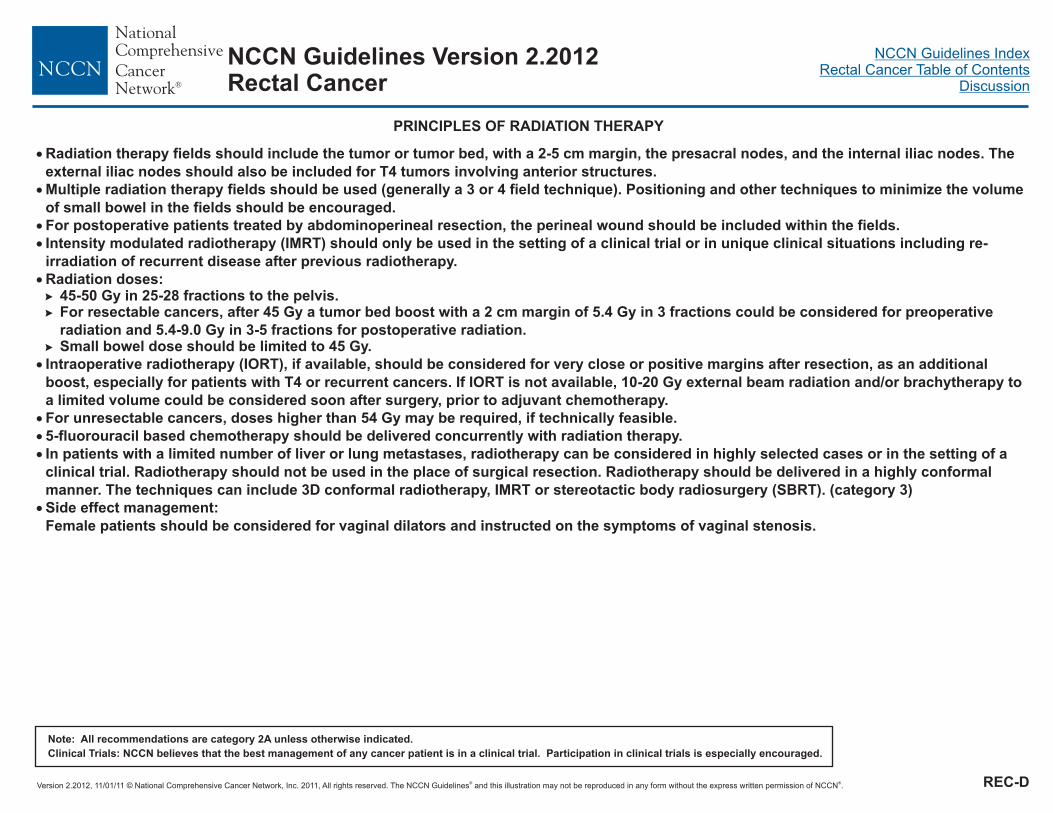
NCCN®
Version 2.2012, 11/01/11 © National Comprehensive Cancer Network, Inc. 2011, All rights reserved. The NCCN Guidelines and this illustration may not be reproduced in any form without the express written permission of NCCN .® ®
NCCN Guidelines IndexRectal Cancer Table of Contents
Discussion
Note: All recommendations are category 2A unless otherwise indicated.
Clinical Trials: NCCN believes that the best management of any cancer patient is in a clinical trial. Participation in clinical trials is especially encouraged.
PRINCIPLES OF RADIATION THERAPY
�
�
�
�
�
�
�
�
Radiation therapy fields should include the tumor or tumor bed, with a 2-5 cm margin, the presacral nodes, and the internal iliac nodes. The
external iliac nodes should also be included for T4 tumors involving anterior structures.
Multiple radiation therapy fields should be used (generally a 3 or 4 field technique). Positioning and other techniques to minimize the volume
of small bowel in the fields should be encouraged.
For postoperative patients treated by abdominoperineal resection, the perineal wound should be included within the fields.
Radiation doses:45-50 Gy in 25-28 fractions to the pelvis.For resectable cancers, after 45 Gy a tumor bed boost with a 2 cm margin of 5.4 Gy in 3 fractions could be considered for preoperative
radiation and 5.4-9.0 Gy in 3-5 fractions for postoperative radiation.Small bowel dose should be limited to 45 Gy.
Intraoperative radiotherapy (IORT), if available, should be considered for very close or positive margins after resection, as an additional
boost, especially for patients with T4 or recurrent cancers. If IORT is not available, 10-20 Gy external beam radiation and/or brachytherapy to
a limited volume could be considered soon after surgery, prior to adjuvant chemotherapy.
For unresectable cancers, doses higher than 54 Gy may be required, if technically feasible.
�
�
Intensity modulated radiotherapy (IMRT) should only be used in the setting of a clinical trial or in unique clinical situations including re-
irradiation of recurrent disease after previous radiotherapy.
5-fluorouracil based chemotherapy should be delivered concurrently with radiation therapy.
In patients with a limited number of liver or lung metastases, radiotherapy can be considered in highly selected cases or in the setting of a
clinical trial. Radiotherapy should not be used in the place of surgical resection. Radiotherapy should be delivered in a highly conformal
manner. The techniques can include 3D conformal radiotherapy, IMRT or stereotactic body radiosurgery (SBRT). (category 3)
Side effect management:
Female patients should be considered for vaginal dilators and instructed on the symptoms of vaginal stenosis.
�
�
�
REC-D
NCCN Guidelines Version 2.2012Rectal Cancer

NCCN®
Version 2.2012, 11/01/11 © National Comprehensive Cancer Network, Inc. 2011, All rights reserved. The NCCN Guidelines and this illustration may not be reproduced in any form without the express written permission of NCCN .® ®
NCCN Guidelines IndexRectal Cancer Table of Contents
Discussion
Note: All recommendations are category 2A unless otherwise indicated.
Clinical Trials: NCCN believes that the best management of any cancer patient is in a clinical trial. Participation in clinical trials is especially encouraged.
REC-E1 of 7
CONTINUUM OF CARE - CHEMOTHERAPY FOR ADVANCED OR METASTATIC DISEASE: (PAGE 1 of 7)1
See footnotes on page REC-E 3 of 7
Additional Options on REC-E 2 of 7
NCCN Guidelines Version 2.2012Rectal Cancer
Patient
appropriate
for
intensive
therapy2
FOLFOX ±
bevacizumab or
CapeOX ±
bevacizumab
3
4
5,6
FOLFOX
± panitumumab
(KRAS wild-type
[WT] gene only)
3
6,7
8,9
or
FOLFIRI
or
5,10
orIrinotecan
FOLFIRI + (cetuximab or
panitumumab)
(KRAS WT gene only)or
(Cetuximab or panitumumab)
(KRAS WT gene only) + irinotecan
10
15-18
15-18
10
6,
8
6,
8
Clinical trial or best supportive care19
(Cetuximab or panitumumab) (KRAS WT gene
only) + irinotecan,
patients not able to tolerate combination,
consider single agent (cetuximab or
panitumumab) (KRAS WT gene only)
6,
8
6,15-18 8
15-18
10
FOLFIRI +
bevacizumab
10
5,6
FOLFOXor
3,5 4,5
15-18
10
or CapeOX
(Cetuximab or panitumumab)
(KRAS WT gene only) +
irinotecan, patients not able to
tolerate combination,
consider single agent (cetuximab or
panitumumab)
(KRAS WT gene only)
6,
8
6,15-18
8
FOLFOX3 4or CapeOX
FOLFIRI ±
cetuximab or
panitumumab
(KRAS WT gene
only)
10
6,7
8,9
or
or
Irinotecan10
10
± oxaliplatin
orFOLFIRI
5-FU/leucovorin or
Capecitabine± bevacizumab
11
5,6,13
12
FOLFOXIRI
(category 2B)
14
Irinotecan10
or(Cetuximab or panitumumab) (KRAS WT gene
only) + irinotecan,patients not able to tolerate combination,
consider single agent (cetuximab or
panitumumab) (KRAS WT gene only)
6,15
6,15-18 8
-18
10
FOLFOX or CapeOX3,5 4,5
or
or
(Cetuximab or panitumumab) (KRAS WT gene
only) + irinotecan,
patients not able to tolerate combination,
consider single agent (cetuximab or
panitumumab) (KRAS WT gene only)
6,
8
6,15-18 8
15-18
10
(Cetuximab or panitumumab)
(KRAS WT gene only) + irinotecan,
patients not able to tolerate
combination, consider single agent
(cetuximab or panitumumab)
(KRAS WT gene only)
6,
8
6,15-18
8
15-18
10
Initial therapy Therapy after First Progression Therapy after Second Progression

NCCN®
Version 2.2012, 11/01/11 © National Comprehensive Cancer Network, Inc. 2011, All rights reserved. The NCCN Guidelines and this illustration may not be reproduced in any form without the express written permission of NCCN .® ®
NCCN Guidelines IndexRectal Cancer Table of Contents
Discussion
Note: All recommendations are category 2A unless otherwise indicated.
Clinical Trials: NCCN believes that the best management of any cancer patient is in a clinical trial. Participation in clinical trials is especially encouraged.
REC-E2 of 7
CONTINUUM OF CARE - CHEMOTHERAPY FOR ADVANCED OR METASTATIC DISEASE: (PAGE 2 of 7)1
Initial therapy Therapy after First Progression
See footnotes on page REC-E 3 of 7
Patient not
appropriate
for
intensive
therapy2
Infusional 5-FU + leucovorin or
Capecitabine ± bevacizumab Improvement in
functional status
No improvement in
functional status
Consider Initial Therapy
as 20REC-E 1 of 7
Best supportive careSee NCCN Palliative Care Guidelines
Cetuximab (KRAS wild-type
gene only) (category 2B)8,9
or
Panitumumab (KRAS wild-type
gene only) (category 2B)8,9
or
NCCN Guidelines Version 2.2012Rectal Cancer

NCCN®
Version 2.2012, 11/01/11 © National Comprehensive Cancer Network, Inc. 2011, All rights reserved. The NCCN Guidelines and this illustration may not be reproduced in any form without the express written permission of NCCN .® ®
NCCN Guidelines IndexRectal Cancer Table of Contents
Discussion
Note: All recommendations are category 2A unless otherwise indicated.
Clinical Trials: NCCN believes that the best management of any cancer patient is in a clinical trial. Participation in clinical trials is especially encouraged.
REC-E3 of 7
CHEMOTHERAPY FOR ADVANCED OR METASTATIC DISEASE (PAGE 3 of 7)
1
3
4
2
5
10
11
12
13
15
16
17
19
20
For chemotherapy references,
PET-CT should not be used to monitor progress of therapy. CT with contrast orMRI is recommended.
Discontinuation of oxaliplatin should be strongly considered from FOLFOX orCapeOX after 3-4 months of therapy (or sooner if significant neurotoxicity
develops grade 2) with other drugs maintained (fluoropyrimidine +bevacizumab) until time of tumor progression. Oxaliplatin may be reintroduced if itwas discontinued previously for neurotoxicity rather than disease progression.Tournigand C, Cervantes A, Figer A, et al. OPTIMOX1: A randomized study ofFOLFOX4 or FOLFOX7 with oxaliplatin in a stop-and-go fashion in advancedcolorectal cancer - A GERCOR Study. J Clin Oncol 2006;24:394-400. There areinsufficient data to support the routine use of Ca/Mg infusion to preventoxaliplatin-related neurotoxicity.
The majority of safety and efficacy data for this regimen have been developed inEurope, where a capecitabine starting dose of 1250 mg/m twice daily for 14days, repeated every 21 days, is standard. Some data suggest that NorthAmerican patients may experience greater toxicity with capecitabine (as well aswith other fluoropyrimidines) than European patients, and may require a lowerdose of capecitabine. The relative efficacy of CapeOx with lower starting doses ofcapecitabine has not been addressed in large scale randomized trials. For goodperformance status patients, the 1000 mg/m twice daily dose is therecommended starting dose, with close monitoring in the first cycle for toxicity,and dose adjustments as indicated.
There are insufficient data to support continuation of bevacizumab with a second-line regimen after progression on a bevacizumab-containing first line regimen,and such continuation of bevacizumab beyond progression is not recommended.If bevacizumab is not used in initial therapy, it may be appropriate to consider, ifthere is no contraindication to therapy. There is an increased risk of stroke and
other arterial events especially in age 65. The use of bevacizumab may interferewith wound healing.
Combination therapy involving cytotoxics, anti-EGFRs and anti-VEGFs is notrecommended. Hecht JR, Mitchell E, Chidiac T, et al. A randomized phase IIIB trialof chemotherapy, bevacizumab, and panitumumab compared with chemotherapyand bevacizumab alone for metastatic colorectal cancer. J Clin Oncol2009;27:672-80. Tol J, Koopman M, Cats A, et al. Chemotherapy, bevacizumab,and cetuximab in metastatic colorectal cancer. N Engl J Med 2009;360(6):563-572.
If cetuximab or panitumumab are used as initial therapy, then neither cetuximabnor panitumumab should be used in second or subsequent lines of therapy.
- KRAS and BRAF MutationTesting.
Patients with a V600E BRAF mutation appear to have a poorer prognosis.Retrospective subset analyses suggest potential benefit from anti-EGFRmonoclonal antibodies in the first-line setting with active chemotherapyregardless of V600E mutation status.
Irinotecan should be used with caution and with decreased doses in patientswith Gilbert's disease or elevated serum bilirubin. There is a commerciallyavailable test for UGT1A1. Guidelines for use in clinical practice have not beenestablished.
Infusional 5-FU is preferred.
Patients with diminished creatinine clearance may require dose modification ofcapecitabine.
A treatment option for patients not able to tolerate oxaliplatin or irinotecan.
Data are not mature for the addition of biologic agents to FOLFOXIRI.
Cetuximab is indicated in combination with irinotecan-based therapy or assingle agent therapy for patients who cannot tolerate irinotecan.
EGFR testing has no demonstrated predictive value, and therefore routineEGFR testing is not recommended. No patient should be included or excludedfrom cetuximab or panitumumab therapy on the basis of EGFR test results.
There are no data, nor is there a compelling rationale, to support the use ofpanitumumab after clinical failure on cetuximab, or the use of cetuximab afterclinical failure on panitumumab. As such, the use of one of these agents aftertherapeutic failure on the other is not recommended.
Patients with a V600E BRAF mutation appear to have a poorer prognosis.Limited available data suggest lack of antitumor activity from anti-EGFRmonoclonal antibodies in the presence of a V600E mutation when used afterpatient has progressed on first-line therapy.
Single agent or combination therapy with capecitabine, mitomycin, orgemcitabine has not been shown to be effective in this setting.
The use of single agent capecitabine as a salvage therapy after failure on afluoropyrimidine-containing regimen has been shown to be ineffective, and this istherefore not recommended.
2
2
6
7
8
9
14
18
�
�
see Chemotherapy Regimens and References(REC-E pages 4 - 7).
See Principles of Pathologic Review (REC-A 5 of 6)
NCCN Guidelines Version 2.2012Rectal Cancer

NCCN®
Version 2.2012, 11/01/11 © National Comprehensive Cancer Network, Inc. 2011, All rights reserved. The NCCN Guidelines and this illustration may not be reproduced in any form without the express written permission of NCCN .® ®
NCCN Guidelines IndexRectal Cancer Table of Contents
Discussion
Note: All recommendations are category 2A unless otherwise indicated.
Clinical Trials: NCCN believes that the best management of any cancer patient is in a clinical trial. Participation in clinical trials is especially encouraged.
REC-E4 of 7
CHEMOTHERAPY FOR ADVANCED OR METASTATIC DISEASE - CHEMOTHERAPY REGIMENS (PAGE 4 of 7)
FOLFOXmFOLFOX 6Oxaliplatin 85 mg/m IV over 2 hours, day 1Leucovorin* 400 mg/m IV over 2 hours, day 15-FU 400 mg/m IV bolus on day 1, then 1200 mg/m /day x 2 days(total 2400 mg/m over 46-48 hours) IV continuous infusionRepeat every 2 weeks
CapeOXOxaliplatin 130 mg/m IV over 2 hours, day 1Capecitabine 850-1000 mg/m twice daily PO for 14 daysRepeat every 3 weeks
CapeOX + BevacizumabOxaliplatin 130 mg/m IV over 2 hours, day 1Capecitabine 850-1000‡ mg/m PO twice daily for 14 daysBevacizumab 7.5 mg/kg IV, day 1Repeat every 3 weeks
2
2
2 2
2
2
2
2
2
†
†
†
1,5
‡
1,2
1,5,¶
mFOLFOX6 + BevacizumabOxaliplatin 85 mg/m IV over 2 hours, day 1Leucovorin* 400 mg/m IV over 2 hours, day 15-FU 400 mg/m IV bolus on day 1, then 1200 mg/m /day x 2 days(total 2400 mg/m2 over 46-48 hours) IV continuous infusionBevacizumab 5 mg/kg IV, day 1Repeat every 2 weeks
mFOLFOX6 + PanitumumabOxaliplatin 85 mg/m IV over 2 hours, day 1Leucovorin* 400 mg/m IV over 2 hours, day 15-FU 400 mg/m IV bolus on day 1, then 1200 mg/m /day x 2 days(total 2400 mg/m2 over 46-48 hours) IV continuous infusionPanitumumab 6 mg/kg IV over 60 minutes, day 1Repeat every 2 weeks
2,3,¶
2,4
2
2
2 2
2
2
2 2
* is the equivalent of levoleucovorin 200 mg/mLeucovorin 400 mg/m2 .2
2 2
2
†NCCN recommends limiting chemotherapy orders to 24 h units (ie, 1200 mg/m /day NOT 2400 mg/m over 48 hours) to minimize medication errors.‡The majority of safety and efficacy data for this regimen have been developed in Europe, where a capecitabine starting dose of 1000 mg/m twice daily for 14 days,repeated every 21 days, is standard. Evidence suggests that North American patients may experience greater toxicity with capecitabine (as well as with otherfluoropyrimidines) than European patients, and may require a lower dose of capecitabine. The relative efficacy of CapeOx with lower starting doses of capecitabine hasnot been addressed in large scale randomized trials.
Bevacizumab may be safely given at a rate of 0.5 mg/kg/minute (5 mg/kg over 10 minutes and 7.5 mg/kg over 15 minutes).¶
See References on page REC-E 7 of 7
IMPORTANT NOTE REGARDING LEUCOVORIN
SHORTAGE, PLEASE SEE MS-14
NCCN Guidelines Version 2.2012Rectal Cancer
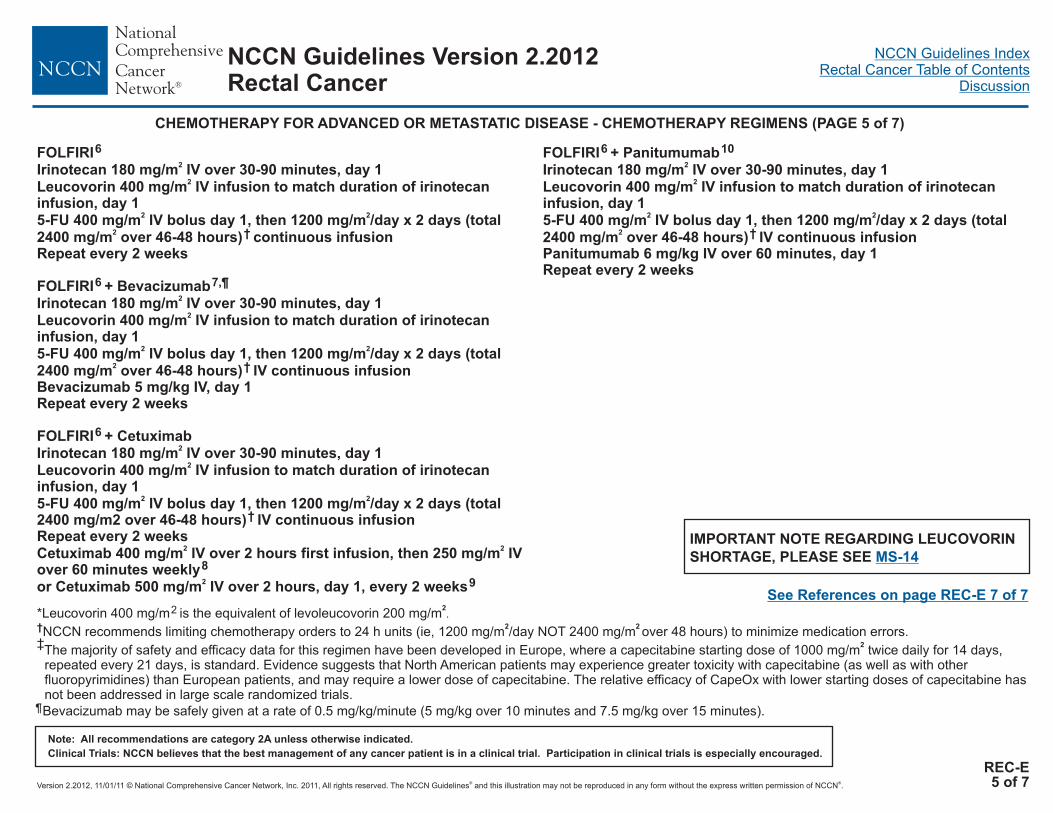
NCCN®
Version 2.2012, 11/01/11 © National Comprehensive Cancer Network, Inc. 2011, All rights reserved. The NCCN Guidelines and this illustration may not be reproduced in any form without the express written permission of NCCN .® ®
NCCN Guidelines IndexRectal Cancer Table of Contents
Discussion
Note: All recommendations are category 2A unless otherwise indicated.
Clinical Trials: NCCN believes that the best management of any cancer patient is in a clinical trial. Participation in clinical trials is especially encouraged.
REC-E5 of 7
CHEMOTHERAPY FOR ADVANCED OR METASTATIC DISEASE - CHEMOTHERAPY REGIMENS (PAGE 5 of 7)
FOLFIRIIrinotecan 180 mg/m IV over 30-90 minutes, day 1Leucovorin 400 mg/m IV infusion to match duration of irinotecaninfusion, day 15-FU 400 mg/m IV bolus day 1, then 1200 mg/m /day x 2 days (total2400 mg/m over 46-48 hours) continuous infusionRepeat every 2 weeks
FOLFIRI + BevacizumabIrinotecan 180 mg/m IV over 30-90 minutes, day 1Leucovorin 400 mg/m IV infusion to match duration of irinotecaninfusion, day 15-FU 400 mg/m IV bolus day 1, then 1200 mg/m /day x 2 days (total2400 mg/m over 46-48 hours) IV continuous infusionBevacizumab 5 mg/kg IV, day 1Repeat every 2 weeks
FOLFIRI + CetuximabIrinotecan 180 mg/m IV over 30-90 minutes, day 1Leucovorin 400 mg/m IV infusion to match duration of irinotecaninfusion, day 15-FU 400 mg/m IV bolus day 1, then 1200 mg/m /day x 2 days (total2400 mg/m2 over 46-48 hours) IV continuous infusionRepeat every 2 weeksCetuximab 400 mg/m IV over 2 hours first infusion, then 250 mg/m IVover 60 minutes weeklyor Cetuximab 500 mg/m IV over 2 hours, day 1, every 2 weeks
FOLFIRI + PanitumumabIrinotecan 180 mg/m IV over 30-90 minutes, day 1Leucovorin 400 mg/m IV infusion to match duration of irinotecaninfusion, day 15-FU 400 mg/m IV bolus day 1, then 1200 mg/m /day x 2 days (total2400 mg/m over 46-48 hours) IV continuous infusionPanitumumab 6 mg/kg IV over 60 minutes, day 1Repeat every 2 weeks
6
2
2
2 2
2
2
2
2 2
2
2
2
2 2
2 2
2
2
2
2 2
2†
6 7,¶
†
6
†
8
9
6 10
†
* is the equivalent of levoleucovorin 200 mg/mLeucovorin 400 mg/m2 .2
2 2
2
†NCCN recommends limiting chemotherapy orders to 24 h units (ie, 1200 mg/m /day NOT 2400 mg/m over 48 hours) to minimize medication errors.‡The majority of safety and efficacy data for this regimen have been developed in Europe, where a capecitabine starting dose of 1000 mg/m twice daily for 14 days,repeated every 21 days, is standard. Evidence suggests that North American patients may experience greater toxicity with capecitabine (as well as with otherfluoropyrimidines) than European patients, and may require a lower dose of capecitabine. The relative efficacy of CapeOx with lower starting doses of capecitabine hasnot been addressed in large scale randomized trials.
Bevacizumab may be safely given at a rate of 0.5 mg/kg/minute (5 mg/kg over 10 minutes and 7.5 mg/kg over 15 minutes).¶
See References on page REC-E 7 of 7
IMPORTANT NOTE REGARDING LEUCOVORIN
SHORTAGE, PLEASE SEE MS-14
NCCN Guidelines Version 2.2012Rectal Cancer

NCCN®
Version 2.2012, 11/01/11 © National Comprehensive Cancer Network, Inc. 2011, All rights reserved. The NCCN Guidelines and this illustration may not be reproduced in any form without the express written permission of NCCN .® ®
NCCN Guidelines IndexRectal Cancer Table of Contents
Discussion
Note: All recommendations are category 2A unless otherwise indicated.
Clinical Trials: NCCN believes that the best management of any cancer patient is in a clinical trial. Participation in clinical trials is especially encouraged.
REC-E6 of 7
CHEMOTHERAPY FOR ADVANCED OR METASTATIC DISEASE - CHEMOTHERAPY REGIMENS (PAGE 6 of 7)
Capecitabine11
850-1250 mg/m PO twice daily, days 1-14Repeat every 3 weeks
Capecitabine + Bevacizumab850-1250 mg/m PO twice daily, days 1-14Repeat every 3 weeksBevacizumab 5 mg/kg IV, day 1 weekly
Bolus or infusional 5-FU/leucovorin
then 1200 mg/m /day x 2days (total 2400 mg/m over 46-48 hours) continuous infusion
Weekly
5-FU 2600 mg/m by 24 h infusion plus leucovorin 500 mg/mRepeat every week
IROXOxaliplatin 85 mg/m IV over 2 hours, followed by irinotecan 200mg/m over 30 or 90 minutes every 3 weeks
FOLFOXIRIIrinotecan 165 mg/m IV day 1, oxaliplatin 85 mg/m day 1, leucovorin
mg/m day 1, fluorouracil 3,200 mg/m over 48 h continuousinfusion starting on day 1Repeat every 2 weeks
IrinotecanIrinotecan 125 mg/m IV over 30-90 minutes, days 1, 8Repeat every 3 weeksIrinotecan 300-350 mg/m IV over 30-90 minutes, day 1Repeat every 3 weeks
Cetuximab (KRAS wild-type gene only) ± irinotecanCetuximab 400 mg/m 1st infusion, then 250 mg/m IV weeklyor Cetuximab 500 mg/m IV every 2 weeks±Irinotecan 300-350 mg/m IV every 3 weeksor Irinotecan 180 mg/m IV every 2 weeksor Irinotecan 125 mg/m on days 1, 8 and repeat every 3 weeks
Cetuximab (KRAS wild-type gene only)Cetuximab 400 mg/m 1st infusion, then 250 mg/m IV weeklyor Cetuximab 500 mg/m2 IV over 2 hours, day 1, every 2 weeks
Panitumumab (KRAS wild-type gene only)Panitumumab 6 mg/kg IV over 60 minutes every 2 weeks
2
2
2
2
2
2 2
2
2 2
2 2
2
2
2 2
2 2
2
2
2 2
2
2
2
2
2 2
11 ¶
15
16
17,18
9,19
19
9
Roswell-Park regimenLeucovorin 500 mg/m IV over 2 hours, days 1, 8, 15, 22, 29, and 365-FU 500 mg/m IV bolus 1 hour after start of leucovorin,days 1, 8, 15, 22, 29, 36Repeat every 8 weeks
Simplified biweekly infusional 5-FU/LV (sLV5FU2)Leucovorin 400 mg/m IV over 2 hours on day 1,followed by 5-FU bolus 400 mg/m and
Repeat every 2 weeks
Leucovorin 20 mg/m IV over 2 hours on day 1, 5-FU 500 mg/m IVbolus injection 1h after the start of leucovorin. Repeat weekly.
400*
12
6
†
13
14
9
20
* is the equivalent of levoleucovorin 200 mg/mLeucovorin 400 mg/m2 .2
2 2†NCCN recommends limiting chemotherapy orders to 24 h units (ie, 1200 mg/m /day NOT 2400 mg/m over 48 hours) to minimize medication errors.
Bevacizumab may be safely given at a rate of 0.5 mg/kg/minute (5 mg/kg over 10 minutes and 7.5 mg/kg over 15 minutes).¶
See References on page REC-E 7 of 7
IMPORTANT NOTE REGARDING LEUCOVORIN
SHORTAGE, PLEASE SEE MS-14
NCCN Guidelines Version 2.2012Rectal Cancer

NCCN®
Version 2.2012, 11/01/11 © National Comprehensive Cancer Network, Inc. 2011, All rights reserved. The NCCN Guidelines and this illustration may not be reproduced in any form without the express written permission of NCCN .® ®
NCCN Guidelines IndexRectal Cancer Table of Contents
Discussion
Note: All recommendations are category 2A unless otherwise indicated.
Clinical Trials: NCCN believes that the best management of any cancer patient is in a clinical trial. Participation in clinical trials is especially encouraged.
REC-E7 of 7
CHEMOTHERAPY FOR ADVANCED OR METASTATIC DISEASE - REFERENCES (PAGE 7 of 7)
NCCN Guidelines Version 2.2012Rectal Cancer
1
5
6
7
8
12
13
17
18
19
20
Cassidy J, Clarke S, Diaz Rubio E, et al. Randomized phase III study ofcapecitabine plus oxaliplatin compared with fluorouracil/folinic acid plus oxaliplatinas first-line therapy for metastatic colorectal cancer. J Clin Oncol 2008;26:2006-12.Cheeseman SL, Joel SP, Chester JD, et al. A 'modified de Gramont' regimen offluorouracil, alone and with oxaliplatin, for advanced colorectal cancer. Br J Cancer2002;87:393-399. Available at: .Emmanouilides C, Sfakiotaki G, Androulakis N, et al. Front-line bevacizumab incombination with oxaliplatin, leucovorin and 5-fluorouracil (FOLFOX) in patientswith metastatic colorectal cancer: a multicenter phase II study. BMC Cancer2007;7:91.Douillard JY, Siena S, Cassidy J, et al. Randomized, phase III trial of panitumumabwith infusional fluorouracil, leucovorin, and oxaliplatin (FOLFOX4) versusFOLFOX4 alone as first-line treatment in patients with previously untreatedmetastatic colorectal cancer: the PRIME study. J Clin Oncol 2010;28:4697-4705.European studies showing equivalent efficacy for CapeOX used at a higher dose;however, European patients consistently tolerate capecitabine with less toxicitythan American patients.
Cunningham D, Humblet Y, Siena S, et al. Cetuximab monotherapy and cetuximabplus irinotecan in irinotecan-refractory metastatic colorectal cancer. N Engl J Med2004;351:337-345.Martín-Martorell P, Roselló S, Rodríguez-Braun E, et al. Biweekly cetuximab andirinotecan in advanced colorectal cancer patients progressing after at least oneprevious line of chemotherapy: results of a phase II single institution trial. Br JCancer 2008;99:455-458.Peeters M, Price TJ, Cervantes A, et al. Randomized phase III study of
panitumumab with fluorouracil, leucovorin, and irinotecan (FOLFIRI) compared withFOLFIRI alone as second-line treatment in patients with metastatic colorectalcancer. J Clin Oncol 2010;28:4706-4713. Available at:
.
Wolmark N, Rockette H, Fisher B, et al. The benefit of leucovorin-modulatedfluorouracil as postoperative adjuvant therapy for primary colon cancer: resultsfrom National Surgical Adjuvant Breast and Bowel Protocol C-03. J Clin Oncol1993;11:1879-1887.Jäger E, Heike M, Bernhard H, et al. Weekly high-dose leucovorin versus low-
dose leucovorin combined with fluorouracil in advanced colorectal cancer: resultsof a randomized multicenter trial. J Clin Oncol 1996;14:2274-2279.Douillard JY, Cunningham D, Roth AD, et al. Irinotecan combined with
fluorouracil compared with fluorouracil alone as first-line treatment for metastaticcolorectal cancer:a multicentre randomised trial. The Lancet 2000;355:1041-47.Haller DG, Rothenberg ML, Wong AO, et al. Oxaliplatin plus irinotecan
compared with irinotecan alone as second-line treatment after single agentfluoropyrimidine therapy for metastatic colorectal carcinoma. J Clin Oncol2008;26:4544-4550.Falcone A, Ricci S, Brunetti I, et al. Phase III trial of infusional fluorouracil,
leucovorin, oxaliplatin, and irinotecan (FOLFOXIRI) compared with infusionalfluorouracil, leucovorin, and irinotecan (FOLFIRI) as first-line treatment formetastatic colorectal cancer: The Gruppo Oncologico Nord Ovest. J Clin Oncol2007;25(13):1670-1676.Cunningham D, Pyrhonen S, James R, et al. Randomised trial of irinotecan plus
supportive care versus supportive care alone after fluorouracil failure for patientswith metastatic colorectal cancer. The Lancet 1998;352:1413-1418.Fuchs CS, Moore MR, Harker G, et al. Phase III comparison of two irinotecan
dosing regimens in second-line therapy of metastatic colorectal cancer. J ClinOncol 2003;21:807-814.Van Custem E, Humblet H, Gelderblom J, et al. Cetuximab dose-escalation in
patients with metastatic colorectal cancer with no or slight skin reactions oncetuximab standard dose treatment (EVEREST): Pharmacokinetic and efficacydata of a randomized study. 2007 Gastrointestinal Cancers Symposium. Abstract237.Van Custem E, Peeters M, Siena S, et al. Open-label phase III trial of
panitumumab plus best supportive care compared with best supportive carealone in patients with chemotherapy-refractory metastatic colorectal cancer. JClin Oncol 2007;25:1658-1664.
2
3
4
9
10
11
14
15
16Andre T, Louvet C, Maindrault-Goebel F, et al. CPT-11 (irinotecan) addition tobimonthly, high-dose leucovorin and bolus and continous-infusion 5-fluorouracil(FOLFIRI) for pretreated metastatic colorectal cancer. Eur J Cancer1999;35(9):1343-7.Fuchs CS, Marshall J, Mitchell E, et al. Randomized, controlled trial of irinotecanplus infusional, bolus, or oral fluoropyrimidines in first-line treatment of metastaticcolorectal cancer: results from the BICC-C Study. J Clin Oncol 2007;25:4779-4786.Available at:
VanCutsem E, Twelves C, Cassidy J, et al. Oral capecitabine compared withintravenous fluorouracil plus leucovorin in patients with metastatic colorectalcancer: results of a large phase III study. J Clin Oncol 2001;19:4097-4106.
http://www.ncbi.nlm.nih.gov/pubmed/12177775
http://www.ncbi.nlm.nih.gov/pubmed/20921462
http://www.ncbi.nlm.nih.gov/pubmed/17947725

NCCN®
Version 2.2012, 11/01/11 © National Comprehensive Cancer Network, Inc. 2011, All rights reserved. The NCCN Guidelines and this illustration may not be reproduced in any form without the express written permission of NCCN .® ®
NCCN Guidelines IndexRectal Cancer Table of Contents
Discussion
Note: All recommendations are category 2A unless otherwise indicated.
Clinical Trials: NCCN believes that the best management of any cancer patient is in a clinical trial. Participation in clinical trials is especially encouraged.
REC-F
PRINCIPLES OF SURVIVORSHIP - Colorectal Long-term Follow-up Care
Colorectal Cancer Surveillance:SeeLong term surveillance should be carefully managed with routine goodmedical care and monitoring, including cancer screening, routinehealth care, and preventive care.Routine CEA monitoring and routine CT scanning are notrecommended beyond 5 years.
Management of Late Sequelae of Disease or Treatment:Chronic Diarrhea or Incontinence
Consider anti-diarrheal agents, bulk-forming agents, dietmanipulation, and protective undergarments.
Urogenital Dysfunction after Resection and/or Pelvic Radiation
Screen for urinary incontinence, frequency, and urgencyConsider referral to urologist or gynecologist for persistent
symptoms.Prescription for Survivorship and Transfer of Care to Primary CarePhysician: (If primary physician will be assuming cancer surveillanceresponsibilities)
Include overall summary of treatment, including all surgeries, radiationtreatments, and chemotherapy receivedDescribe possible clinical course, including expected time to resolutionof acute toxicities, long-term effects of treatment, and possible latesequelae of treatmentInclude surveillance recommendationsDelineate appropriate timing of transfer of care with specificresponsibilities identified for PCP and oncologist.
Cancer Screening Recommendations:These recommendations are for average risk patients.Recommendations for high risk individuals should be made on anindividual basis.
Breast Cancer: See theCervical Cancer: See the
Prostate Cancer: See theCounseling Regarding Healthy Lifestyle and Wellness:
Maintain a healthy body weight throughout life.Adopt a physically active lifestyle (At least 30 minutes of moderateintensity activity on most days of the week). Activityrecommendations may require modification based on treatmentsequelae (i.e. ostomy, neuropathy).Consume a healthy diet with emphasis on plant sources.Limit alcohol consumption.Smoking cessation counseling as appropriate.
Additional health monitoring and immunizations should beperformed as indicated under the care of a primary care physician.Survivors are encouraged to maintain a therapeutic relationshipwith a primary care physician throughout their lifetime.
�
�
�
�
�
�
�
�
�
�
�
�
�
�
�
�
�
1-5
6,7
8
9
�
�
�
�
Screen for sexual dysfunction, erectile dysfunction, dyspareunia,
and vaginal dryness
REC-7
NCCN Breast Cancer Screening GuidelinesNCCN Cervical Cancer Screening
GuidelinesNCCN Prostate Early Detection Guidelines
1
2
3
4
5
6
7
Schneider EC, Malin JL, Kahn KL, et al. Surviving colorectal cancer. Cancer 2007;110: 2075-2082.Sprangers MAG, Taal BG, Aaronson NK, et al. Quality of life in colorectal cancer: stoma vs.nonstoma patients. Dis Colon Rectum 1995;38:361-369.Gami B, Harrington K, Blake P, et al. How patients manage gastrointestinal symptoms afterpelvic radiotherapy. Alimentary Pharmacology and Therapeutics 2003;18:987-994.DeSnoo L, Faithfull S. A qualitative study of anterior resection syndrome: the experiences ofcancer survivors who have undergone resection surgery. European Journal of Cancer Care2006;15:244-251.McGough C, Baldwin C, Frost C, Andreyev HJN. Role of nutritional intervention in patientstreated with radiotherapy for pelvic malignancy. British Journal of Cancer 2004;90:2278-2287.
Lange MM, Mass CP, Marijnen CAM, et al. Risk factors for sexual dysfunction after rectalcancer treatment. Eur J Cancer 2009;45:1578-88.Lange MM, Mass CP, Marijnen CAM, et al. Urinary dysfunction after rectal cancertreatment is mainly caused by surgery. Brit J Cancer 2008;95:1020-28.
8
9
Hewitt M, Greenfield S, Stovall E. From Cancer Patient to Cancer Survivor: Lost inTransition. Washington, D.C.:The National Academies Press;2006.Kushi LH, Byers T, Doyle C, et al and The American Cancer Society 2006 Nutrition andPhysical Activity Guidelines Advisory Committee. American Cancer Society Guidelineson Nutrition and Physical Activity for Cancer Prevention: Reducing the Risk of CancerWith Healthy Food Choices and Physical Activity CA Cancer J Clin 2006;56:254-281.
NCCN Guidelines Version 2.2012Rectal Cancer

NCCN®
Version 2.2012, 11/01/11 © National Comprehensive Cancer Network, Inc. 2011, All rights reserved. The NCCN Guidelines and this illustration may not be reproduced in any form without the express written permission of NCCN .® ®
NCCN Guidelines IndexRectal Cancer Table of Contents
Discussion
Table 1. Definitions for T, N, MPrimary Tumor (T)
Regional Lymph Nodes (N)
Distant Metastasis (M)
TX Primary tumor cannot be assessedT0 No evidence of primary tumorTis Carcinoma in situ: intraepithelial or invasion of lamina propriaT1 Tumor invades submucosaT2 Tumor invades muscularis propriaT3 Tumor invades through the muscularis propria into the pericolorectal tissuesT4a Tumor penetrates to the surface of the visceral peritoneumT4b Tumor directly invades or is adherent to other organs or structures
NX Regional lymph nodes cannot be assessedN0 No regional lymph node metastasisN1 Metastasis in 1-3 regional lymph nodesN1a Metastasis in one regional lymph nodeN1b Metastasis in 2-3 regional lymph nodesN1c Tumor deposit(s) in the subserosa, mesentery, or nonperitonealized
pericolic or perirectal tissues without regional nodal metastasisN2 Metastasis in four or more regional lymph nodesN2a Metastasis in 4-6 regional lymph nodesN2b Metastasis in seven or more regional lymph nodes
M0 No distant metastasisM1 Distant metastasisM1a Metastasis confined to one organ or site
(eg, liver, lung, ovary, nonregional node)M1b Metastases in more than one organ/site or the peritoneum
a
b
b,c
ST-1
Used with the permission of the American Joint Committee on Cancer (AJCC), Chicago, Illinois. The original and primary source for this information is the AJCCCancer Staging Manual, Seventh Edition (2010) published by Springer Science and Business Media LLC (SBM). (For complete information and data supportingthe staging tables, visit .) Any citation or quotation of this material must be credited to the AJCC as its primary source. The inclusion of thisinformation herein does not authorize any reuse or further distribution without the expressed, written permission of Springer SBM, on behalf of the AJCC.
www.springer.com
Note : cTNM is the clinical classification, pTNM is the pathologicclassification. The y prefix is used for those cancers that are classifiedafter neoadjuvant pretreatment (e.g., ypTNM). Patients who have acomplete pathologic response are ypT0N0cM0 that may be similar toStage Group 0 or I. The r prefix is to be used for those cancers thathave recurred after a disease-free interval (rTNM).
*Dukes B is a composite of better (T3 N0 M0) and worse (T4 N0 M0)prognostic groups, as is Dukes C (Any TN1 M0 and Any T N2 M0). MACis the modified Astler-Coller classification.
Table 2. Anatomic Stage/Prognostic GroupsStage T N M Dukes MAC* *
0 Tis N0 M0 - -I T1 N0 M0 A A
T2 N0 M0 A B1IIA T3 N0 M0 B B2IIB T4a N0 M0 B B2IIC T4b N0 M0 B B3IIIA T1-T2 N1/N1c M0 C C1
T1 N2a M0 C C1IIIB T3-T4a N1/N1c M0 C C2
T2-T3 N2a M0 C C1/C2T1-T2 N2b M0 C C1
IIIC T4a N2a M0 C C2T3-T4a N2b M0 C C2T4b N1-N2 M0 C C3
IVA Any T Any N M1a - -IVB Any T Any N M1b - -
NCCN Guidelines Version 2.2012 StagingRectal Cancer
a
b
c
Tis includes cancer cells confined within the glandular basement membrane (intraepithelial) or mucosal lamina propria (intramucosal) with no extension through themuscularis mucosae into the submucosa.
Direct invasion in T4 includes invasion of other organs or other segments of the colorectum as a result of direct extension thourhg the serosa, as confirmed onmicroscopic examination (for example, invasion of the sigmoid colon by a carcinoma of the cecum) or, for cancers in a retroperitoneal or subperitoneal location, directinvasion of other organs or structures by virtue of extension beyond the muscularis propria (i.e., respectively, a tumor on the posterior wall of the descending coloninvading the left kidney or lateral abdominal wall; or a mid or distal rectal cancer with invasion of prostate, seminal vesicles, cervix, or vagina).
Tumor that is adherent to other organs or structures, grossly, is classified cT4b. However, if no tumor is present in the adhesion, microscopically, the classificationshould be pT1-4a depending on the anatomical depth of wall invasion. The V and L classifications should be used to identify the presence or absence of vascular orlymphatic invasion whereas the PN site-specific factor should be used for perineural invasion.

Version 2.2012, 11/01/11 © National Comprehensive Cancer Network, Inc. 2011, All rights reserved. The NCCN Guidelines® and this illustration may not be reproduced in any form without the express written permission of NCCN®. MS-1
NCCN Guidelines IndexRectal Cancer Table of Contents
Discussion
NCCN Guidelines Version 2.2012 Rectal Cancer
Discussion
NCCN Categories of Evidence and Consensus
Category 1: Based upon high-level evidence, there is uniform NCCN consensus that the intervention is appropriate.
Category 2A: Based upon lower-level evidence, there is uniform NCCN consensus that the intervention is appropriate.
Category 2B: Based upon lower-level evidence, there is NCCN consensus that the intervention is appropriate.
Category 3: Based upon any level of evidence, there is major NCCN disagreement that the intervention is appropriate.
All recommendations are category 2A unless otherwise noted.
Overview In 2011, an estimated 39,870 new cases of rectal cancer will occur in the United States (22,910 cases in men; 16,960 cases in women). During the same year, it is estimated that 49,380 people will die from rectal and colon cancer combined.1 Although colorectal cancer is ranked as the fourth most frequently diagnosed cancer and the second leading cause of cancer death in the U.S., the incidence per 100,000 population of colon and rectal cancers has decreased from 60.5 in 1976 to 46.4 in 2005.2 In addition, mortality from colorectal cancer has decreased by more almost 35% from 1990 to 2007.1 This decrease may be due to both earlier diagnoses through screening and better treatment modalities.
The recommendations in these clinical practice guidelines are classified as category 2A except where noted, meaning that there is uniform
NCCN consensus, based on lower-level evidence (including clinical experience), that the recommendation is appropriate. The panel unanimously endorses patient participation in a clinical trial over standard or accepted therapy. This is especially true for cases of advanced disease and for patients with locally aggressive colorectal cancer who are receiving combined modality treatment. The clinical practice guidelines for managing rectal cancer overlap considerably with the NCCN Colon Cancer Guidelines. First-degree relatives of patients with newly diagnosed adenomas or invasive carcinoma are at increased risk for colorectal cancer.3, 4 Therefore, all rectal cancer patients should be counseled regarding their family history as outlined in the NCCN Colorectal Cancer Screening Guidelines.
TNM Staging The NCCN Rectal Cancer Guidelines adhere to the current TNM staging system of the 7th edition of the American Joint Committee on Cancer’s (AJCC) Cancer Staging Manual (Table 1 of the guidelines).5 Several changes to the staging of colorectal cancer were made in the 7th edition.6 For instance, based on new data showing differential prognosis,7 T4 lesions have now been subdivided into T4a (tumor penetrates to the surface of the visceral peritoneum) and T4b (tumor directly invades or is adherent to other organs or structures). Another change of note is the subdivision of N1 into N1a (metastasis in 1 node), N1b (metastasis in 2-3 nodes), and N1c (without regional nodal metastases, but with tumor deposits in the subserosa, mesentery, or non-peritonealized pericolic or perirectal tissues); and of N2 into N2a (metastasis in 4-6 nodes) and N2b (metastasis in 7 or more nodes). These subsets reflect new data showing that the number of involved nodes influences prognosis8 and new data on the prognostic value of tumor deposits within the lymph drainage area of the primary tumor.9-11 Stage I rectal cancer is defined as T1-T2, N0, M0. Stage II disease is

Version 2.2012, 11/01/11 © National Comprehensive Cancer Network, Inc. 2011, All rights reserved. The NCCN Guidelines® and this illustration may not be reproduced in any form without the express written permission of NCCN®. MS-2
NCCN Guidelines IndexRectal Cancer Table of Contents
Discussion
NCCN Guidelines Version 2.2012 Rectal Cancer
subdivided into IIA (if the primary tumor is T3, N0, M0), IIB (for T4a, N0, M0 lesions), and IIC (for T4b, N0, M0). Stage III disease is subdivided into IIIA (T1-2, N1/N1c, M0 or T1, N2a, M0), IIIB (T3-4a, N1/N1c, M0 or T2-T3, N2a, M0 or T1-T2, N2b, M0), and IIIC (T4a, N2a, M0 or T3-4a, N2b, M0 or T4b, N1-2, M0). Stage IVA disease is defined as any T, any N, and the presence of distant metastasis confined to 1 organ or site (M1a). Stage IVB disease is defined as any T, any N, with metastases in more than 1 organ or site or in the peritoneum (M1b).5 The prefixes “p” and “yp” used in TNM staging denote pathologic staging and pathologic staging following neoadjuvant therapy, respectively.5
Pathology Pathologic staging information is provided by examination of the surgical specimen. Some of the information that should be detailed in the report of the pathologic evaluation of rectal cancer12 includes: (1) gross description of the tumor and specimen; (2) grade of the cancer; (3) depth of penetration and extension to adjacent structures (T); (4) number of regional lymph nodes evaluated; (5) number of positive regional lymph nodes (N); (6) the presence of distant metastases to other organs or sites including non-regional lymph nodes (M); (7) the status of proximal, distal, and circumferential (radial) margins5, 12-17; (8) neoadjuvant treatment effect5, 12, 18, 19; (9) lymphovascular invasion5, 12,
20; (10) perineural invasion21-23; and (11) the number of tumor deposits.11, 24
The 7th edition of the AJCC staging manual includes the suggestion that the surgeon mark the area of the specimen with the deepest tumor penetration so that the pathologist can directly evaluate the status of the resection margins.5 The completeness of the resection is scored as R0 for complete tumor resection with all margins negative; R1 for incomplete tumor resection with microscopic involvement of a margin;
and R2 for incomplete tumor resection with gross residual tumor that was not resected.5
The circumferential margin or circumferential resection margin (CRM) is an important pathologic staging parameter in rectal cancer.25 Whereas the radial margin for resected segments of the colon that are completely encased by a peritonealized (serosal) surface is also referred to as the peritoneal margin, the CRM is very important in segments of the colon or rectum that are either not encased or only partially encased in peritoneum.25 The CRM is the closest radial margin between the deepest penetration of the tumor and the edge of resected soft tissue around the rectum (ie, the retroperitoneal or subperitoneal aspect of the tumor) or from the edge of a lymph node and should be measured in millimeters. Identification of the CRM is determined through evaluation of the outer circumference of the rectal and mesorectal specimen that often requires inking of the outer surfaces and “bread-loaf” slicing of the specimen.26 The panel defines a positive CRM as tumor within 1 mm from the transected margin.15, 17, 27
Accurate pathologic assessment of the CRM of resected rectal tumor specimens is very important since the CRM has been shown to be a strong predictor of both local recurrence and overall survival,25, 27-29 including in patients undergoing neoadjuvant therapy,16 and is an important consideration when post-operative treatment decisions are made. Furthermore, in a retrospective study of over 17,000 patients with rectal cancer, CRM was found to be a better predictor of local recurrence for patients undergoing surgery as initial therapy than for those who had received preoperative therapy.16 CRM positivity based solely on intranodal tumor should be noted as such; some studies have shown that positive intranodal CRM is associated with lower recurrence rates than a positive CRM by direct tumor extension. Additional components of the pathological evaluation of the surgical specimen

Version 2.2012, 11/01/11 © National Comprehensive Cancer Network, Inc. 2011, All rights reserved. The NCCN Guidelines® and this illustration may not be reproduced in any form without the express written permission of NCCN®. MS-3
NCCN Guidelines IndexRectal Cancer Table of Contents
Discussion
NCCN Guidelines Version 2.2012 Rectal Cancer
following a total mesorectal excision (TME) are described under ‘Surgical Approaches,’ below.
The AJCC and the College of American Pathologists (CAP) recommend evaluation of 10-14 and 12-18 lymph nodes to accurately identify early stage colorectal cancers, respectively.5, 12, 25 The number of lymph nodes that can be retrieved varies with age and gender of the patient and on tumor grade or site.30 The literature lacks consensus regarding the minimal number of lymph nodes needed to accurately identify early stage rectal cancer.31 Most of these studies have combined rectal and colon cancers and reflect those cases with surgery as the initial treatment. Two studies confined only to rectal cancer have reported 14 and >10 lymph nodes as the minimal number to accurately identify stage II rectal cancer.32, 33 Furthermore, the mean number of lymph nodes retrieved from rectal cancers treated with neoadjuvant therapy is significantly less than those treated by surgery alone (13 vs. 19, P<0.05; 7 vs. 10, P≤0.0001).34, 35 The panel recommends a minimum of 12 lymph nodes be examined.
Results of studies evaluating the sentinel node for micrometastatic disease through use of hematoxylin and eosin (H&E) staining to identify small foci of tumor cells and the identification of particular tumor antigens through immunohistochemical (IHC) analysis have been reported.36, 37 Although results of some of these studies seem promising, there is no uniformity in the definition of “true” clinically relevant metastatic carcinoma. Some studies have considered detection of single cells by IHC or by H&E, so-called isolated tumor cells (ITC), to be micrometastasis.37, 38 In addition, results of one study demonstrated that, following neoadjuvant radiotherapy for rectal cancer, the sensitivity for the sentinel node procedure was only 40%.39 Furthermore, in a recent study involving 156 patients with colon cancer and 44 patients with rectal cancer, this “ultra-staging” of lymph nodes
only changed the staging for 1% of patients.40 Others have noted that micrometastasis found in node-negative patients did not predict outcome.41 Presently, the use of sentinel lymph nodes and detection of cancer cells by IHC should be considered investigational, and the results should be used with caution in clinical management decisions.
The 7th edition of the AJCC Staging Manual and the most recent College of American Pathologists Guidelines require that the pathology report comment on treatment effects of neoadjuvant therapy.5, 12 The minimum requirement is a yes/no whether a definitive treatment effect is identified. However, it is the opinion of the panel, as well as of the College of American Pathologists, that the tumor response should be graded on a scale of 0 (complete response – no viable cancer cells observed) to 3 (poor response – minimal or no tumor kill; extensive residual cancer).5, 12, 18, 19
Several studies have demonstrated that the presence of perineural invasion (PNI) is associated with a significantly worse prognosis.21-23 For example, one retrospective analysis of 269 consecutive patients who had colorectal tumors resected at 1 institution found a 4-fold greater 5-year survival in patients without perineural invasion versus patient whose tumors invaded nearby neural structures.22 Multivariate analysis of patients with stage II rectal cancer showed that patients with PNI have a significantly worse 5-year disease-free survival compared to those without PNI (29% vs. 82%; P=0.0005).23 Similar results were seen for patients with stage III disease.21
Extra-nodal tumor deposits, or satellite nodules, are irregular discrete tumor deposits in the pericolic or perirectal fat that show no evidence of residual lymph node tissue, but that are within the lymphatic drainage of the primary tumor. They are not counted as lymph nodes replaced by tumor. Most of these tumor deposits are thought to be due to

Version 2.2012, 11/01/11 © National Comprehensive Cancer Network, Inc. 2011, All rights reserved. The NCCN Guidelines® and this illustration may not be reproduced in any form without the express written permission of NCCN®. MS-4
NCCN Guidelines IndexRectal Cancer Table of Contents
Discussion
NCCN Guidelines Version 2.2012 Rectal Cancer
lymphovascular invasion or occasionally perineural invasion. The number of extra-nodal tumor deposits should be recorded in the pathology report, since they have been shown to be associated with reductions in disease-free and overall survival.11, 24, 42 Multivariate survival analysis in one study showed that patients with pN0 tumors without satellite nodules had a 91.5% 5-year survival rate compared to 37.0% for patients with pN0 tumors and the presence of satellite nodules (P<0.0001)11 Extra-nodal tumor deposits are classified as pN1c.5
The Role of Vitamin D in Colorectal Cancer Prospective studies have suggested that vitamin D deficiency may contribute to colorectal cancer incidence and that vitamin D supplementation may decrease colorectal cancer risk.43, 44 Furthermore, 2 prospective studies showed that low vitamin D levels were associated with increased mortality of patients with colorectal cancer, especially in stage III and IV disease.45 Moreover, in a study of 515 patients with stage IV colorectal cancer, 82% of patients were found to be vitamin D insufficient (levels <30 ng/mL) and 50% found to be vitamin D deficient (<20 ng/mL).46 Nonetheless, no study has yet examined whether vitamin D supplementation improves patient outcomes. In a recent report, the Institute of Medicine concluded that data supporting a role for vitamin D was only conclusive in bone health, not in cancer and other diseases.47 Citing this report and the lack of level 1 evidence, the panel does not recommend routine screening for vitamin D deficiency or supplementation of vitamin D in patients with colorectal cancer at this time.
Clinical Presentation and Treatment of Nonmetastatic Disease Management of Polypoid Cancer Before making a decision about surgical resection for an endoscopically resected adenomatous polyp or villous adenoma, physicians should review pathology48 and consult with the patient. A malignant rectal polyp is defined as one with cancer invading through the muscularis mucosae and into the submucosa (pT1).49 Conversely, polyps classified as carcinoma in situ (pTis) have not penetrated into the submucosa and are therefore incapable of regional nodal metastasis.25 The panel recommends marking the cancerous polyp site at the time of colonoscopy or within 2 weeks.
In patients with pedunculated polyps with invasive cancer (tubular, tubulovillous, or villous adenoma), no additional surgery is required if the polyp has been completely resected with favorable histological features.48 Favorable histological features include lesions of grade 1 or 2 without angiolymphatic invasion and with a negative resection margin.48 For patients with a completely removed, single-specimen, sessile polyp (pT1) with favorable histological features and clear margins, observation may be considered, with the understanding that there is significantly greater incidence of adverse outcomes (residual disease, recurrent disease, mortality, and hematogenous metastasis, but not lymph node metastasis) than polyploid malignant polyps. Also see the section on ‘Endoscopically removed malignant polyp’ in ‘Principles of Pathologic Review’ (REC-A) in the guidelines. Rectal surgery is also an option for these patients.
Rectal surgery is also recommended for patients with polyps with unfavorable histological features or when the specimen is fragmented or margins cannot be assessed. Unfavorable histological features for

Version 2.2012, 11/01/11 © National Comprehensive Cancer Network, Inc. 2011, All rights reserved. The NCCN Guidelines® and this illustration may not be reproduced in any form without the express written permission of NCCN®. MS-5
NCCN Guidelines IndexRectal Cancer Table of Contents
Discussion
NCCN Guidelines Version 2.2012 Rectal Cancer
adenomas are grade 3 or 4, angiolymphatic invasion, or a positive margin of resection. In such cases, risk of nodal involvement is higher. It should be noted that there is currently no consensus as to the definition of what constitutes a positive margin of resection. A positive margin for an endoscopically removed polyp has been defined as the presence of tumor within 1-2 mm from the transected margin or by the presence of tumor cells within the diathermy of the transected margin.48,
50-52
For a polyp with fragmented specimen or margins that cannot be assessed, either a transanal excision or a transabdominal resection is recommended. In patients with unfavorable pathologic features, transabdominal resection should be considered in order to include lymphadenectomy. Results from a preoperative endoscopic ultrasound evaluation may provide additional information to guide choice of surgical approach, although the accuracy of this method to detect residual cancer is limited (see section on ‘Clinical Evaluation/Staging,’ below).53 All patients who have resected polyps should undergo surveillance as described in the guidelines.54
Management of Rectal Cancer Rectal cancer has been defined as a cancerous lesion located within 12 cm of the anal verge by rigid proctoscopy.55 Some support for this definition comes from the study of Kapiteijn et al,56 which included a subgroup analysis of the risk of recurrence of rectal cancer based on tumor location. Univariate analyses indicated that local recurrence rates were low for patients who had tumors with an inferior margin of 10.1 cm or more from the anal verge, and that no significant differences between patients in this group receiving radiotherapy and surgery were observed when they were compared to those undergoing surgery alone.56 A recent retrospective review of patients with rectal or
rectosigmoid cancer demonstrated that treatment options were impacted by whether the location of the rectal lesion was characterized by rigid proctoscopy or colonoscopy.57
The determination of an optimal treatment plan for an individual patient with rectal cancer is a complex process. In addition to decisions relating to the intent of rectal cancer surgery (ie, curative or palliative), consideration must also be given to the likely functional results of treatment, including the probability of maintaining or restoring normal bowel function/anal continence and preserving genitourinary functions. For patients with distal rectal cancer, in particular, the simultaneous achievement of the goals of cure and of minimal impact on quality of life can be challenging.58 Furthermore, the risk of pelvic recurrence is higher in patients with rectal cancer compared to those with colon cancer, and locally recurrent rectal cancer has frequently been associated with a poor prognosis.59-61 Careful patient selection with respect to particular treatment options and the use of sequenced multimodality therapy that combines chemoradiation (chemoRT) with operative treatment for selected patients are recommended.
Clinical Evaluation/Staging The initial clinical workup of patients with rectal cancer provides important preoperative information on the clinical stage of disease. Since the clinical stage of the disease is used to direct decisions regarding choice of primary treatment, including surgical intent (eg, curative or palliative) and approaches, and whether to recommend preoperative chemoRT, the implications of either clinically under-staging or over-staging rectal cancer can be substantial.
Patients who present with rectal cancer appropriate for resection require a complete staging evaluation, including total colonoscopy to evaluate for synchronous lesions or other pathologic conditions of the

Version 2.2012, 11/01/11 © National Comprehensive Cancer Network, Inc. 2011, All rights reserved. The NCCN Guidelines® and this illustration may not be reproduced in any form without the express written permission of NCCN®. MS-6
NCCN Guidelines IndexRectal Cancer Table of Contents
Discussion
NCCN Guidelines Version 2.2012 Rectal Cancer
colon and rectum; and rigid proctoscopy to provide a determination of the location of the cancer (ie, measurement of the distance of the tumor from the anal verge should be performed by the responsible surgeon using rigid proctoscopy). They also require a complete physical examination, including carcinoembryonic antigen (CEA) determination and assessment of performance status to determine operative risk. In addition, the accessibility of rectal cancer to evaluation by certain imaging modalities, such as endorectal ultrasound and magnetic resonance imaging (MRI) makes possible preoperative assessments of depth of tumor penetration and the presence of local lymph nodal metastases.62 The consensus of the panel is that a positron emission tomography (PET) scan is not routinely indicated at baseline in the absence of evidence of synchronous metastatic disease, but the panel recognizes new data that suggest that PET-CT may be helpful in some scenarios.63 Additional information regarding the extent of disease and the occurrence of distant metastases can be determined preoperatively through CT scans. Thus, endorectal ultrasound or pelvic MRI, and CT scans of the chest, abdomen, and pelvis are recommended for the preoperative staging of rectal cancer.
Results from a meta-analysis of 90 studies involving the accuracy of endoscopic ultrasound, MRI, and CT in preoperatively staging rectal cancer demonstrated that endoscopic ultrasound and MRI have similarly high sensitivities for evaluating the depth of tumor penetration into the muscularis propria (94%), although endoscopic ultrasound was found to be more specific than MRI in the evaluation of local tumor invasion (86% vs. 69%).64 Only a very limited number of studies using CT for the purpose of T-staging have been performed, and it is not currently considered to be an optimal method for staging the extent of tumor penetration.64, 65 Accurate assessment of nodal status is one of the greatest challenges in the preoperative staging of rectal cancer. In
the meta-analysis of Bipat et al,64 the sensitivities and specificities of the 3 imaging modalities for accurately evaluating lymph node involvement were comparable: CT (55% and 74%); endoscopic ultrasound (67% and 78%); and MRI (66% and 76%). However, only CT and MRI can evaluate iliac and mesenteric or retroperitoneal nodes.64 Results from another recent meta-analysis of 84 articles, indicated that none of the 3 imaging modalities were significantly superior to another method with respect to an accurate determination of tumor N-stage.66 A disadvantage of endoscopic ultrasound is a high degree of operator dependence.64 An advantage of MRI is its ability to provide accurate images of soft tissue structures in the mesorectum, including the mesorectal fascia.64 Hence, MRI evaluation of patients with more advanced rectal cancer has the potential to provide information useful in the prediction of the CRM prior to radical surgery.65-67
Clinical staging is also based on histopathologic examination of the specimen obtained via biopsy or local excision (eg, excised polyps). Endoscopic biopsy specimens of the lesion should undergo careful pathology review for evidence of invasion into the muscularis mucosa. If removal of the rectum is contemplated, early consultation with an enterostomal therapist is recommended for preoperative marking of the site and patient teaching purposes.
Surgical Approaches A variety of surgical approaches, depending on the location and extent of disease, are used to treat primary rectal cancer lesions.68, 69 These methods include local procedures, such as polypectomy, transanal excision, and transanal endoscopic microsurgery (TEM), and more invasive procedures involving a transabdominal resection (eg, low anterior resection [LAR], proctectomy with total mesorectal excision
Version 2.2012, 11/01/11 © National Comprehensive Cancer Network, Inc. 2011, All rights reserved. The NCCN Guidelines® and this illustration may not be reproduced in any form without the express written permission of NCCN®. MS-7
NCCN Guidelines IndexRectal Cancer Table of Contents
Discussion
NCCN Guidelines Version 2.2012 Rectal Cancer
[TME] and coloanal anastomosis, or abdominoperineal resection [APR]).68, 69
Transanal excision may be appropriate for selected T1, N0 early-stage cancers. Small (<3 cm), well to moderately differentiated tumors that are within 8 cm of the anal verge and limited to less than 30% of the rectal circumference and for which there is no evidence of nodal involvement can be approached with transanal excision with negative margins. Transanal endoscopic microsurgery (TEM) can facilitate excision of small tumors through the anus when lesions can be adequately identified in the rectum. TEM may be technically feasible for more proximal lesions. Both transanal excision and TEM involve a full thickness excision performed perpendicularly through the bowel wall into the perirectal fat. Negative (>3 mm) deep and mucosal margins are required, and tumor fragmentation should be avoided. The excised specimen should be oriented and pinned before fixation and brought to the pathologist by the surgeon to facilitate an oriented histopathologic evaluation of the specimen. Advantages of a local procedure include minimal morbidity (eg, a sphincter-sparing procedure) and mortality and rapid postoperative recovery.58, 70 If pathologic examination reveals adverse features such as high grade, positive margins, lymphovascular invasion (LVI) or perineural invasion, a more radical resection is recommended. Data are limited on long-term patient outcomes, including risk of local recurrence, for patients undergoing local excision for T2 tumors.70
Limitations of a transanal excision include the absence of pathologic staging of nodal involvement. Further, there is evidence to indicate that lymph node micrometastases are both common in early rectal lesions and unlikely to be identified by endorectal ultrasound.71 These observations may underlie the findings that patients undergoing local excision have a higher local recurrence rate than those undergoing
radical resection.70, 72 A recent retrospective study of 282 patients undergoing either transanal excision or radical resection for T1 rectal cancer from 1985 to 2004 showed respective local recurrence rates of 13.2% and 2.7% for these 2 groups (P=0.001).72 A similar retrospective study of 2,124 patients showed local recurrence rates of 12.5% and 6.9% for patients undergoing local excision versus standard resection, respectively (P=0.003).70
Patients with rectal cancer who do not meet requirements for local surgery should be treated with a transabdominal resection. Organ-preserving procedures that maintain sphincter function are preferable, but not possible in all cases. Preoperative chemoRT may result in tumor downsizing and a decrease in tumor bulk (see section on ‘Neoadjuvant/Adjuvant Therapy,’ below); sphincter preservation may become possible in cases where initial tumor bulk prevented consideration of such surgery and exposure to the tumor is improved by chemoRT.
In transabdominal resections, total mesorectal excision (TME) is recommended. A TME involves an en bloc removal of the mesorectum, including associated vascular and lymphatic structures, fatty tissue, and mesorectal fascia as a “tumor package” through sharp dissection and is designed to spare the autonomic nerves.58, 69, 73 The lymphatic drainage regions of rectal tumors are influenced by their position in the rectum. More distal tumors are more likely to be characterized by both upward and lateral lymphatic drainage whereas the likelihood of only upward mesorectal drainage is much higher for more proximal tumors.74 The TME approach is designed to radically remove lymphatic drainage regions of tumors located above the level of the levator muscles.75 The panel does not recommend extension of nodal dissection beyond the field of resection (eg, into the distribution of iliac lymph nodes) unless these nodes are clinically suspicious. In cases where anal function is

Version 2.2012, 11/01/11 © National Comprehensive Cancer Network, Inc. 2011, All rights reserved. The NCCN Guidelines® and this illustration may not be reproduced in any form without the express written permission of NCCN®. MS-8
NCCN Guidelines IndexRectal Cancer Table of Contents
Discussion
NCCN Guidelines Version 2.2012 Rectal Cancer
intact and distal clearance is adequate, the TME may be followed by creation of a coloanal anastomosis.
For lesions in the mid to upper rectum, a low anterior resection (LAR) extended 4-5 cm below the distal edge of the tumor, followed by creation of a colorectal anastomosis, is the treatment of choice. Where creation of an anastomosis is not possible, colostomy is required. Wide TME is recommended in order to facilitate adequate lymphadenectomy and improve the probability of achieving negative circumferential margins.
An abdominoperineal resection (APR) should be performed when the tumor directly involves the anal sphincter or the levator muscles. An APR is also necessary in cases where a margin-negative resection of the tumor would result in loss of anal sphincter function and incontinence. An APR involves en bloc resection of the rectosigmoid, the rectum, and the anus, as well as the surrounding mesentery, mesorectum (TME), and perianal soft tissue, and it necessitates creation of a colostomy.76
Pathologists play a key role in evaluating the surgical specimen following TME which includes a macroscopic assessment of both its external appearance/completeness and the CRM.77, 78 Detailed descriptions of how the quality of the mesorectal specimens should be scored were provided in the Dutch Rectal Cancer Trial, and these guidelines are endorsed by the NCCN panel.15
Recent retrospective comparisons of the outcomes of patients undergoing an APR versus a LAR in the treatment of rectal cancer have shown those treated with an APR to have worse local control and overall survival.79, 80 Whether these differences can be attributed to the surgical procedure alone, to patient- and tumor-related characteristics,
or some combination of these factors is presently unclear. However, results from a recent retrospective study of 3,633 patients with T3-4 rectal cancer tumors included in 5 large European trials suggest that there is an association between the APR procedure itself and the increased risks of recurrence and death.79
Data from randomized studies evaluating use of laparoscopic surgery in the treatment of patients with rectal cancer are limited.81, 82 In the CLASICC trial comparing laparoscopically-assisted resection to open resection, nearly half of the 794 patients were diagnosed with rectal cancer.81 No significant differences in local recurrence, DFS, or overall survival were observed between the 2 groups of patients with colon or rectal cancer based on surgical approach. A 5-year follow-up of the CLASICC trial showed that this lack of difference in local recurrence, DFS, or overall survival was maintained for patients with rectal cancer, despite a trend towards better 5-year overall survival after open surgery (52.9% and 62.8% for open and laparoscopic surgery, respectively; P= 0.132).83 Factors that may confound conclusions drawn from randomized studies comparing open surgery to laparoscopically-assisted surgery for colorectal cancer have been described,84 and longer-term outcomes from laparoscopic rectal surgery have not been reported. Additional clinical trials exploring open versus laparoscopic surgery for rectal cancer are ongoing (including clinicaltrials.gov NCT00297791 and NCT00470951). At this time, laparoscopic surgery for rectal cancer is preferred in the setting of a clinical trial.
Neoadjuvant and Adjuvant Therapy for Resectable Nonmetastatic Disease Neoadjuvant/adjuvant therapy of stage II (T3-4, node-negative disease with tumor penetration through the muscle wall) or stage III (node-positive disease without distant metastasis) rectal cancer often includes locoregional treatment due to the relatively high risk of locoregional

Version 2.2012, 11/01/11 © National Comprehensive Cancer Network, Inc. 2011, All rights reserved. The NCCN Guidelines® and this illustration may not be reproduced in any form without the express written permission of NCCN®. MS-9
NCCN Guidelines IndexRectal Cancer Table of Contents
Discussion
NCCN Guidelines Version 2.2012 Rectal Cancer
recurrence. This risk is associated with the close proximity of the rectum to pelvic structures and organs, the absence of a serosa surrounding the rectum, and technical difficulties associated with obtaining wide surgical margins at resection. In contrast, adjuvant treatment of colon cancer is more focused on preventing distant metastases since this disease is characterized by lower rates of local recurrence.
Although radiation therapy has been associated with decreased rates of local recurrence of rectal cancer, it is also associated with increased toxicity (eg, radiation-induced injury, hematologic toxicities, etc.) relative to surgery alone.26, 85 It has been suggested that some patients with disease at lower risk of local recurrence (eg, proximal rectal cancer staged as T3, N0, M0, characterized by clear margins and favorable prognostic features) may be adequately treated with surgery and adjuvant chemotherapy.26, 86, 87 However, 22% of 188 patients clinically staged with T3, N0 rectal cancer by either EUS or MRI who subsequently received preoperative chemoRT had positive lymph nodes following pathologic review of the surgical specimens according to results of a recent retrospective multicenter study,88 suggesting that many patients are under-staged and would benefit from chemoRT. Therefore, the guidelines recommend preoperative chemoRT for patients with T3, N0 disease.
Combined-modality therapy consisting of surgery, radiation therapy (RT), and chemotherapy is recommended for the majority of patients with stage II or stage III rectal cancer. Use of perioperative pelvic RT in the treatment of patients with stage II/III rectal cancer continues to evolve. In these patients, the current guidelines recommend concurrent fluoropyrimidine-based chemotherapy with ionizing radiation to the pelvis preoperatively and chemotherapy postoperatively. A total of 6 months perioperative chemotherapy with or without RT is preferred.
Preoperative versus postoperative radiation Several studies have compared the administration of radiation preoperatively versus postoperatively.89, 90 A large prospective, randomized trial from the German Rectal Cancer Study Group compared preoperative versus postoperative chemoRT in the treatment of clinical stage II/III rectal cancer.89 Results of this study indicated that preoperative therapy was associated with a significant reduction in local recurrence (6% vs 13%; P=0.006) and treatment-associated toxicity (27% vs 40%; P=0.001), although overall survival was similar in the 2 groups.
Putative advantages to preoperative radiation, as opposed to radiation given postoperatively, are related to both tumor response and preservation of normal tissue.89-91 First of all, reducing tumor volume may facilitate resection and increase the likelihood of a sphincter-sparing procedure. Although some studies have indicated that preoperative radiation or chemoRT is associated with increased rates of sphincter preservation in rectal cancer patients,89, 90 this conclusion has not been supported by 2 recent meta-analyses of randomized trials involving preoperative chemoRT in the treatment of rectal cancer.92, 93 Second, irradiating tissue that is surgery-naïve and thus better oxygenated may result in increased sensitivity to RT. Third, preoperative radiation can avoid the occurrence of radiation-induced injury to small bowel trapped in the pelvis by post-surgical adhesions. Finally, preoperative radiation that includes structures that will be resected increases the likelihood that an anastomosis with healthy colon can be performed (ie, the anastomosis remains unaffected by the effects of RT because irradiated tissue is resected). One disadvantage of using preoperative RT is the possibility of over-treating early-stage tumors that do not require adjuvant radiation.89, 94 Improvements in preoperative staging techniques, such as MRI or CT scans, have

Version 2.2012, 11/01/11 © National Comprehensive Cancer Network, Inc. 2011, All rights reserved. The NCCN Guidelines® and this illustration may not be reproduced in any form without the express written permission of NCCN®. MS-10
NCCN Guidelines IndexRectal Cancer Table of Contents
Discussion
NCCN Guidelines Version 2.2012 Rectal Cancer
allowed for more accurate staging, but the risk of over-staging disease has not been eliminated.88 Weighing these advantages and disadvantages, the panel recommends preoperative chemoRT for patients with stage II/III rectal cancer.
Postoperative chemoRT is recommended when stage I rectal cancer is upstaged to stage II or III after pathologic review of the surgical specimen. Postoperative chemoRT regimens commonly employ a “sandwich” approach – whereby chemotherapy (typically 5-FU based) is administered before and after the chemoRT regimen.87, 95, 96 The use of FOLFOX or capecitabine chemotherapy before and after postoperative chemoRT is an extrapolation of the available data in colon cancer.97, 98
Concurrent chemotherapy with radiation A number of randomized trials have evaluated the effectiveness of the addition of chemotherapy to radiation administered either preoperatively following clinical evaluation/staging (eg, T3-4 by endoscopic ultrasound) or postoperatively following pathologic staging of rectal cancer as pT3 and/or N1-2.99, 100 Putative benefits of the addition of chemotherapy concurrent with either pre- or postoperative RT include local RT sensitization and systemic control of disease (ie, eradication of micrometastases). Preoperative chemoRT also has the potential to increase rates of pathologic complete response and sphincter preservation.
In a study of patients with T3-4 rectal cancer without evidence of distant metastases who were randomly assigned to receive either preoperative RT alone or preoperative concurrent chemoRT with 5-FU/LV, no difference in overall survival or sphincter preservation was observed in the 2 groups, although patients receiving chemoRT were significantly more likely to exhibit a pathologic complete response (11.4% vs 3.6%;
P<0.05) and grade 3/4 toxicity (14.6% vs 2.7%; P<0.05) and less likely to exhibit local recurrence of disease (8.1% vs 16.5%; P<0.05).100 These conclusions have been supported in a recent systematic review that included 4 randomized controlled trials.99
Preliminary results of a phase III trial that included an evaluation of the addition of chemotherapy to preoperative RT in patients with T3-4 resectable rectal cancer demonstrated that use of 5-FU/LV chemotherapy enhanced the tumoricidal effect of RT when the 2 approaches were used concurrently.101 Significant reductions in tumor size, pTN stage, and lymphatic, vascular, and perineural invasion rates were observed with use of combined-modality therapy compared with use of RT and surgery without chemotherapy.101 More mature results from this trial, however, which included 4 treatment groups (preoperative RT; preoperative chemoRT; preoperative RT plus postoperative chemotherapy; and preoperative chemoRT plus postoperative chemotherapy), indicated that no significant differences in overall survival were associated with adding 5-FU-based chemotherapy preoperatively or postoperatively.102 Although local recurrence rates were significantly higher in the group receiving preoperative RT alone, the addition of chemotherapy after concurrent chemoRT did not significantly impact local recurrence rates. In subsequent exploratory analyses of data from the group of patients in this trial who underwent complete tumor resection without evidence of distant disease before or at surgery, those patients with disease characterized as ypT0-2 showed significant benefit from adjuvant chemotherapy with respect to DFS and overall survival.103 These findings may indicate that patients are more likely to benefit from adjuvant therapy if their disease can be down-staged by chemoRT. Of note, patients with stage II/III rectal cancer enrolled in this trial were

Version 2.2012, 11/01/11 © National Comprehensive Cancer Network, Inc. 2011, All rights reserved. The NCCN Guidelines® and this illustration may not be reproduced in any form without the express written permission of NCCN®. MS-11
NCCN Guidelines IndexRectal Cancer Table of Contents
Discussion
NCCN Guidelines Version 2.2012 Rectal Cancer
found to be 2.6-times more likely to develop distant metastases than local recurrence of disease after a median follow-up of over 5 years.102
With respect to the type of chemotherapy administered concurrently with RT, results from the Intergroup 0114 trial, showed bolus 5-FU as part of adjuvant therapy for rectal cancer to be non-inferior to bolus 5-FU plus LV.87 After a median follow-up of 4 years, neither the rate of local control nor survival differed among 3 different combinations of modulated 5-FU chemotherapy. Furthermore, the equivalence of bolus 5-FU/LV and infusional 5-FU in concurrent chemoRT for rectal cancer is supported by the results of a phase III trial (median follow-up of 5.7 years) in which similar outcomes with respect to overall survival and relapse-free survival were observed when an infusion of 5-FU or bolus 5-FU plus LV was administered concurrently with postoperative RT, although hematologic toxicity was greater in the group of patients receiving bolus 5-FU.96 On the other hand, results from an earlier trial from the North Central Cancer Treatment Group (NCCTG) showed that postoperative administration of infusional 5-FU during pelvic irradiation was associated with longer overall survival when compared to bolus 5-FU.95 Most of the patients in this study had node-positive disease.
Recent studies have shown that capecitabine is equivalent to 5-FU in perioperative chemoradiation therapy.104, 105 The randomized NSABP R-04 trial compared the preoperative use of infusional 5-FU with or without oxaliplatin to capecitabine with or without oxaliplatin in 1,608 patients with stage II or III rectal cancer.104 No differences in complete pathologic response, sphincter-saving surgery, or surgical downstaging were seen between the regimens, while toxicity was increased with the inclusion of oxaliplatin. Similarly, a phase III randomized trial in which 401 patients with stage II or III rectal cancer received capecitabine- or 5-FU-based chemoradiation either pre- or postoperatively showed that capecitabine was non-inferior to 5-FU with regards to 5-year OS
(capecitabine 75.7% vs. 5-FU 66.6%; P=0.0004), with capecitabine showing showed borderline significance for superiority (P=0.053).105 Furthermore, in this trial capecitabine demonstrated a significant improvement in 3-year disease-free survival (75.2% vs 66.6%; P=0.034).105 Because of these studies, capecitabine given concurrently with radiation therapy is now listed in the guidelines as a category 2A recommendation. The panel feels that capecitabine in an acceptable alternative to infusional 5-FU in those patients who are able to manage the responsibilities inherent in self-administered, oral chemotherapy.
In attempts to improve on the outcomes achieved with neoadjuvant 5-FU/RT or capecitabine/RT, several large randomized phase III trials addressed the addition of oxaliplatin to the regimens. In a planned interim report of primary tumor response in the STAR-01 trial, grade 3 and 4 adverse events occurred more frequently in patients receiving infusional 5-FU/oxaliplatin/RT than in those receiving infusional 5-FU/RT (24% vs. 8%, P<0.001), while there was no difference in pathologic response between the 2 arms of the study (16% pathologic complete response in both arms).106 Similar results were seen in the ACCORD 12/0405-Prodige 2 trial, in which capecitabine/RT was compared to CapeOx/RT and the primary endpoint was pathologic complete response (ypCR).107 Here, the grade 3 and 4 toxicity rates were 25% and 11% (P<0.001), and the ypCR rates were 19.2% and 13.9% (P=0.09) for the oxaliplatin-containing arm and the control arm, respectively. Finally, recently reported results of the NSABP R-04 trial also showed that the addition of oxaliplatin did not improve clinical outcomes including the endpoints of ypCR, sphincter-saving surgery, and surgical downstaging, while it increased toxicity.104 The addition of oxaliplatin to neoadjuvant chemoradiation for non-metastatic disease is thus not recommended at this time. However, it is interesting to note that the patients treated with oxaliplatin in the ACCORD 12 trial had an

Version 2.2012, 11/01/11 © National Comprehensive Cancer Network, Inc. 2011, All rights reserved. The NCCN Guidelines® and this illustration may not be reproduced in any form without the express written permission of NCCN®. MS-12
NCCN Guidelines IndexRectal Cancer Table of Contents
Discussion
NCCN Guidelines Version 2.2012 Rectal Cancer
increased rate of minimal residual disease at the time of surgery (39.4% vs. 28.9%, P=0.008), which has the potential to translate over time to improved local recurrence rates.107 Therefore, further follow-up of these trials is necessary to see if there is a difference in local recurrence rates and PFS over time. The primary endpoints of overall survival for the STAR-01 trial and local tumor control for the R-04 trial as well as rates of local recurrence, survival, and late toxicity for the ACCORD 12 trial will be reported in the future.
Technical aspects of radiation therapy With respect to administration of RT, multiple RT fields should include the tumor or tumor bed with a 2-5 cm margin, presacral nodes, and the internal iliac nodes. The external iliac nodes should also be included for T4 tumors involving anterior structures; inclusion of the inguinal nodes for tumors invading into the distal anal canal can also be considered. Recommended doses of radiation are typically 45-50 Gy in 25-28 fractions to the pelvis using 3 or 4 fields. Positioning and other techniques to minimize radiation to the small bowel are encouraged. Intensity modulated radiotherapy (IMRT) should only be used in the setting of a clinical trial or in unique clinical situations including re-irradiation of recurrent disease.
Coordination of preoperative therapy, surgery, and adjuvant chemotherapy is important. For patients treated with preoperative chemoRT, the panel recommends an interval of 5 to 10 weeks following completion of full-dose 5-½ week chemoRT prior to surgical resection in order to allow patient recuperation from chemoRT-associated toxicities. Although longer intervals from completion of chemoRT to surgery have been shown to be associated with an increase in pathologic complete response rates,108-110 it is unclear whether such longer intervals are associated with clinical benefit. Nevertheless, when longer intervals are
clinically necessary, they do not appear to increase the blood loss, time associated with surgery, or positive margin rate.111
Short-course radiation Several European studies have looked at the efficacy of a shorter course of preoperative radiation (25 Gy over 5 days), not combined with chemotherapy, for the treatment of rectal cancer. The results of the Swedish Rectal Cancer Trial evaluating the use of short-course RT administered preoperatively for resectable rectal cancer showed a survival advantage and a decreased rate of local recurrence with this approach compared with surgery alone.112 However, a follow-up study published in 2005 showed that the patients with short-course preoperative RT had increased relative risk for postoperative hospitalization due to bowel obstructions and other gastrointestinal complications.113 A number of other studies also investigating the effectiveness of preoperative short-course RT in patients with rectal cancer staged as T1-3 have demonstrated that overall survival was not significantly affected despite improvements in local control of disease.56,
114, 115 A recent multicenter, randomized study of 1,350 patients with rectal cancer compared (a) short-course preoperative RT and no postoperative treatment with (b) no preoperative RT and a postoperative approach that included chemoRT in selected patients (ie, those with a positive CRM following resection) and no RT in patients without evidence of residual disease following surgery.116 Results indicated that patients in the preoperative RT arm (a) had significantly lower local recurrence rates and a 6% absolute improvement in 3-year DFS (P=0.03), although no difference in overall survival was observed between the 2 arms of the study.116, 117
Long-term (12-year) follow-up of one of the short-course radiation trials (the Dutch TME trial114) was recently reported.118 The analysis showed that 10-year survival was significantly improved in stage III patients with

Version 2.2012, 11/01/11 © National Comprehensive Cancer Network, Inc. 2011, All rights reserved. The NCCN Guidelines® and this illustration may not be reproduced in any form without the express written permission of NCCN®. MS-13
NCCN Guidelines IndexRectal Cancer Table of Contents
Discussion
NCCN Guidelines Version 2.2012 Rectal Cancer
a negative circumferential margin in the radiotherapy plus surgery group compared to the group that received surgery alone (50% vs. 40%; P=0.032).118 However, this long follow-up showed that secondary malignancies and other non-rectal cancer causes of death were more frequent in the radiotherapy group than in the control group (14% vs 9% for secondary malignancies), negating any survival advantage in the node-negative subpopulation.
Few studies have directly compared preoperative short-course radiation and more conventional preoperative long-course chemoradiation. One randomized study of 312 patients in Poland showed no differences in local recurrence or survival.119 Similarly, a Australian/New Zealand trial that randomized 326 patients to short-course radiation or long-course chemoradiation found no differences in local recurrence and overall survival rates.120
Overall, it appears that short-course RT gives effective local control and the same overall survival as more conventional RT schedules, and therefore may be an appropriate choice in some situations.
Adjuvant chemotherapy Adjuvant chemotherapy is recommended for all patients with stage II/III rectal cancer following neoadjuvant chemoRT/surgery regardless of the surgical pathology results, although few studies have evaluated the effect of adjuvant chemotherapy in patients with rectal cancer, and its role is not well defined.121 The addition of 5-FU-based adjuvant chemotherapy to preoperative chemoRT provided no benefit to the rate of local recurrence in the European Organization for Research and Treatment of Cancer (EORTC) Radiotherapy Group Trial 22921.102 However, this study did show an improvement in disease-free survival (hazard ratio=0.87; 95% CI, 0.72-1.04; P=0.13) of patients receiving
adjuvant chemotherapy (+/- RT) following preoperative RT (+/- 5-FU-based chemotherapy).102
Most of the support for use of FOLFOX or capecitabine as adjuvant chemotherapy in rectal cancer is an extrapolation from the data available for colon cancer.97, 98 The phase III ECOG E3201 trial was designed to investigate the effect of adding either oxaliplatin (FOLFOX) or irinotecan (FOLFIRI) to 5-FU/LV-based adjuvant chemotherapy administered to stage II/III rectal cancer patients following either preoperative or postoperative chemoRT. This study was replaced with an alternative trial with bevacizumab, but results from an initial 165 patients indicate that adjuvant FOLFOX can be safely used in this patient population.122 Nevertheless, the optimal duration of treatment with adjuvant FOLFOX in rectal cancer is still unclear.123, 124 In the MOSAIC trial, patients with stage II/III colon cancer were treated with 6 months of adjuvant FOLFOX.125 The use of a shorter course of adjuvant FOLFOX in rectal cancer (ie, 4 months) is justified when preoperative chemoRT is administered. In addition, the NSABP-07 trial demonstrated similar disease-free survival benefits to those reported in the MOSAIC trial with the FLOX regimen (bolus 5-FU/LV with oxaliplatin).126
A recent systematic review and meta-analysis of 10 studies involving more than 15,000 patients with colorectal cancer looked at the effect of timing of adjuvant therapy following resection.127 Results of this analysis showed that each 4-week delay in chemotherapy results in a 14% decrease in overall survival, indicating that adjuvant therapy should be administered as soon as the patient is medically able. These results are consistent with other similar analyses.128

Version 2.2012, 11/01/11 © National Comprehensive Cancer Network, Inc. 2011, All rights reserved. The NCCN Guidelines® and this illustration may not be reproduced in any form without the express written permission of NCCN®. MS-14
NCCN Guidelines IndexRectal Cancer Table of Contents
Discussion
NCCN Guidelines Version 2.2012 Rectal Cancer
Leucovorin shortage There is currently a shortage of leucovorin in the United States. There are no specific data to guide management under these circumstances, and all proposed strategies are empiric. The panel recommends several possible options to help alleviate the problems associated with this shortage. One is the use of levo-leucovorin, which is commonly used in Europe. A dose of 200 mg/m2 of levo-leucovorin is equivalent to 400 mg/m2 of standard leucovorin. Another option is for practices or institutions to use lower doses of leucovorin for all doses in all patients, since the panel feels that lower doses are likely to be as efficacious as higher doses, based on several studies. The QUASAR study found that 175 mg leucovorin gave similar survival and 3-year recurrence rates as 25 mg leucovorin when given with bolus 5-FU to patients as adjuvant therapy following R0 resections for colorectal cancer.129 Another study showed no difference in response rate or survival in patients with metastatic colorectal cancer receiving bolus 5-FU with either high dose (500 mg/m2) or low dose (20 mg/m2) leucovorin.130 Also, the Mayo Clinic and North Central Cancer Treatment (NCTTG) group determined that there was no therapeutic difference between the use of high (200 mg/m2) or low (20 mg/m2) dose leucovorin with bolus 5-FU in the treatment of advanced colorectal cancer, although 5-FU doses were different in the 2 arms.131 Finally, if none of the above options are available, treatment without leucovorin would be reasonable. For patients who tolerate this without grade II or higher toxicity, a modest increase in 5-FU dose (in the range of 10%) may be considered.
Recommendations for patients with T1 and T2 lesions Node-negative T1 lesions are treated with transabdominal resection or transanal excision, as appropriate (see section on ‘Surgical Approaches,’ above). If pathology review after local excision reveals a poorly differentiated histology, positive margins, or LVI or if the tumor is
restaged to T2, then a transabdominal re-resection should be performed. For such high-risk patients who cannot undergo additional surgery, systemic chemotherapy with chemoradiation (a “sandwich regimen” as described below) should be considered as an adjuvant treatment in order to avoid the risk of undertreatment, being that the lymph node status is unknown.
Node-negative T2 lesions are treated with transabdominal resection, since local recurrence rates of 11% to 45% have been observed for T2 lesions following local excision alone.58, 132, 133 In selected lesions that are staged by endoscopic ultrasound or MRI as T1-2, N0 and without adverse pathologic features (eg, no lymphovascular invasion [LVI] or perineural invasion; size less than 3 cm; well to moderately differentiated), local excision with negative margins may give results comparable to transabdominal resection.134
Following transabdominal resection, patients with tumors staged as pT1-2, N0, M0 require no further treatment. If pathology review reveals pT3, N0, M0 or node-positive disease, a “sandwich regimen,” consisting of (1) an optional first round of adjuvant chemotherapy with 5-FU with or without LV or FOLFOX or capecitabine with or without oxaliplatin,135 followed by (2) concurrent 5-FU/RT (infusional (preferred) or bolus infusion along with LV) or capecitabine/RT (preferred), followed by (3) 5-FU with or without LV or FOLFOX or capecitabine with or without oxaliplatin, is recommended.
The panel recommends perioperative therapy for a total duration of approximately 6 months. For patients with pathologic evidence of proximal T3, N0, M0 disease with clear margins and favorable prognostic features following an upfront resection, the incremental benefit of RT is likely to be small and chemotherapy alone can be

Version 2.2012, 11/01/11 © National Comprehensive Cancer Network, Inc. 2011, All rights reserved. The NCCN Guidelines® and this illustration may not be reproduced in any form without the express written permission of NCCN®. MS-15
NCCN Guidelines IndexRectal Cancer Table of Contents
Discussion
NCCN Guidelines Version 2.2012 Rectal Cancer
considered, although most patients are not likely to be part of this subset.
Recommendations for patients with T3 lesions and lesions with nodal involvement Patients clinically staged as having resectable T3, N0 or T any, N1-2 lesions should initially be treated with preoperative combined-modality therapy unless medically contraindicated. Preoperative infusional 5-FU/RT or capecitabine/RT are the preferred treatment options (category 1 for both). An alternative regimen is bolus 5-FU/LV/RT. Patients who receive preoperative radiotherapy should undergo transabdominal resection 5 to 10 weeks following completion of neoadjuvant therapy. The panel recommends postoperative adjuvant therapy for a duration giving approximately 6 months total of pre- and postoperative chemotherapy (regardless of surgical pathology results) with 5-FU with or without LV; or FOLFOX; or capecitabine with or without oxaliplatin.
Upfront surgery for patients with disease characterized as T3, N0 or T any, N1-2 should be reserved for those patients with medical contraindications to chemoRT. Following initial transabdominal resection, patients with subsequent pathologic staging of disease as pT1-2, N0, M0 can be followed with observation only. For patients with disease pathologically staged as pT3, N0, M0 or pT1-3, N1-2, M0, approximately 6 months of postoperative chemotherapy “sandwich regimen” (see ‘Recommendations for patients with T1 and T2 lesions,’ above) should be reconsidered. For some patients with pathologic evidence of proximal T3, N0, M0 disease with clear margins and favorable prognostic features following transabdominal resection, the incremental benefit of RT is likely to be small and chemotherapy alone can be considered, although this subset of patients is small.
Recommendations for patients with T4 lesions and/or locally unresectable disease
Patients with T4 and/or locally unresectable disease are treated with preoperative infusional 5-FU/RT or bolus 5-FU with LV/RT or capecitabine/RT. If possible, resection should be considered following preoperative chemoRT. For patients with T4 tumors or recurrent cancers or if margins are very close or positive, intraoperative radiotherapy (IORT),136-138 which involves direct exposure of tumors to RT during surgery while removing normal structures from the field of treatment, should be considered as an additional boost to facilitate resection. If IORT is not available, 10-20 Gy and/or brachytherapy to a limited volume can be considered soon after surgery, prior to adjuvant chemotherapy. Adjuvant therapy to complete 6 months with either 5-FU with or without LV; or FOLFOX; or capecitabine with or without oxaliplatin is recommended regardless of the surgical pathology results.
For unresectable cancers, doses higher than 54 Gy may be required, and the dose of radiation to the small bowel should be limited to 45 Gy.
Principles of the Management of Metastatic Disease Approximately 50%-60% of patients diagnosed with colorectal cancer will develop colorectal metastases,139-141 and 80-90% of these patients are initially evaluated to have unresectable metastatic disease.142-144 Approximately 20%-34% of patients with colorectal cancer present with synchronous liver metastases.145, 146 Metastatic disease more frequently develops metachronously following treatment for colorectal cancer, with the liver as a common site of involvement.147 There is some evidence to indicate that synchronous metastatic colorectal liver disease is associated with a more disseminated disease state and a worse prognosis than metastatic colorectal disease that develops metachronously. In a retrospective study of 155 patients who underwent hepatic resection for colorectal liver metastases, patients

Version 2.2012, 11/01/11 © National Comprehensive Cancer Network, Inc. 2011, All rights reserved. The NCCN Guidelines® and this illustration may not be reproduced in any form without the express written permission of NCCN®. MS-16
NCCN Guidelines IndexRectal Cancer Table of Contents
Discussion
NCCN Guidelines Version 2.2012 Rectal Cancer
with synchronous liver metastases had more sites of liver involvement (P=0.008) and more bilobar metastases (P=0.016) when compared with patients diagnosed with metachronous liver metastases.148 For patients presenting with synchronous, unresectable metastases and an intact primary that is not acutely obstructed, palliative resection of the primary is rarely indicated, and systemic chemotherapy is the preferred initial maneuver.149
It has been estimated that over one-half of patients who die of colorectal cancer have liver metastases at autopsy, and that metastatic liver disease is the cause of death in the majority of these patients.150 Results from reviews of autopsy reports of patients dying from colorectal cancer showed that the liver was the only site of metastatic disease in one-third of patients.144 Furthermore, rates of 5-year survival for patients with metastatic liver disease not undergoing surgery have been shown to be quite low in a number of studies.140, 151 However, studies of selected patients undergoing surgery to remove colorectal liver metastases have demonstrated that cure is possible in this population and should be the goal for many patients with colorectal metastatic liver disease.140, 152 Certain clinicopathologic factors, such as the presence of extrahepatic metastases and a disease-free interval of <12 months, have been associated with a poor prognosis in patients with colorectal cancer metastatic to the liver,145, 153 although the ability of these factors to predict outcome following resection may be limited.154-157 Recent reports have shown 5-year survival rates following resection of hepatic colorectal metastases exceeding 50%.154, 157 Therefore, decisions relating to patient suitability, or potential suitability, and subsequent selection for metastatic colorectal surgery are critical junctures in the management of metastatic colorectal liver disease.158
Most of the treatment recommendations discussed for metastatic colorectal liver disease also apply to the treatment of colorectal
pulmonary metastases. Combined pulmonary and hepatic resections of resectable metastatic disease have been performed in selected cases.159, 160
Resection is the standard of care for the local treatment of metastatic disease that is initially resectable and for patients with significant response to preoperative chemotherapy who are converted from unresectable to resectable status.161 Other patients who cannot undergo resection due to comorbidity, location of the metastatic lesion(s), or an estimate of inadequate liver volume following resection may be candidates for tumor ablation therapy.162 A number of retrospective studies have compared radiofrequency ablation (RFA) and liver resection in the treatment of liver metastases.163-166 Most of these studies have shown RFA to be inferior to resection with respect to rates of local recurrence and 5-year overall survival.161 It is presently unclear whether the differences in outcome observed for patients with liver metastases treated with RFA versus resection alone are due to patient selection bias, technological limitations of RFA or a combination of these 2 factors.163 A recent American Society of Clinical Oncology (ASCO) clinical evidence review determined that RFA has not been well studied in the setting of colorectal cancer liver metastases, with no randomized controlled trials having been reported.166 The ASCO panel concluded that there is a compelling need for more research in this area.
The NCCN panel does not consider ablation to be a substitute for resection in patients with completely resectable disease. In addition, resection or ablation (either alone or in combination with resection) should be reserved for patients with disease that is completely amenable to local therapy. Use of surgery, ablation, or the combination of the two with the goal of less than complete resection/ablation of all known sites of disease is not recommended.
Version 2.2012, 11/01/11 © National Comprehensive Cancer Network, Inc. 2011, All rights reserved. The NCCN Guidelines® and this illustration may not be reproduced in any form without the express written permission of NCCN®. MS-17
NCCN Guidelines IndexRectal Cancer Table of Contents
Discussion
NCCN Guidelines Version 2.2012 Rectal Cancer
Placement of a hepatic arterial port or implantable pump during surgical intervention for liver resection with subsequent administration of chemotherapy directed to the liver metastases through the hepatic artery (ie, HAI) is listed in the guidelines as an option (category 2B). In a randomized study of patients who had undergone hepatic resection, administration of floxuridine with dexamethasone by HAI and intravenous 5-FU with or without LV was shown to be superior to a similar systemic chemotherapy regimen alone with respect to 2-year survival free of hepatic disease.144, 167 The study was not powered for long-term survival, but there was a trend (not significant) towards better long term outcome in the group receiving HAI at later follow-up periods.144, 168 A number of other clinical trials have shown significant improvement in response or time to hepatic disease progression when HAI therapy was compared with systemic chemotherapy, although most have not shown a survival benefit of HAI therapy.144 Some of the uncertainties regarding patient selection for preoperative chemotherapy are also relevant to the application of HAI.152 However, limitations on the use of HAI therapy include the potential for biliary toxicity144 and the requirement for specific technical expertise. The consensus of the panel is that HAI therapy should be considered selectively and only at institutions with extensive experience in both the surgical and medical oncologic aspects of the procedure.
A number of non-extirpative liver-directed therapies are available for the treatment of unresectable metastatic disease in highly selected patients, although the role of such therapies of colorectal metastases is controversial. These therapies include arterial radioembolization with yttrium-90 microspheres,169-171 arterial chemoembolization,170 and conformal (stereotactic) radiation therapy.172 Use of arterial-directed embolic therapy is a category 3 recommendation for select patients with predominant hepatic metastases, and conformal external beam
radiation therapy is not recommended for patients with liver or lung metastases unless the patient is symptomatic or it is used in the setting of a clinical trial, and it should not be used indiscriminately in patients who are potentially resectable. See sections on ‘Recommendations for the Treatment of Synchronous Metastases/Unresectable Disease’ and ‘Recommendations for the Treatment of Metachronous Metastases.’
Radiotherapy to the metastatic site can be considered in highly selected cases in which the patient has a limited number of liver or lung metastases (category 3 recommendation) or in the setting of a clinical trial. It should be delivered in a highly conformal manner and should not be used in place of surgical resection. The possible techniques include 3D conformal radiotherapy, stereotactic body radiosurgery,143 and intensity modulated radiotherapy (IMRT), which uses computer-imaging to focus RT to the tumor site and potentially decrease toxicity to normal tissue.173-176
Determining Resectability The consensus of the panel is that patients diagnosed with potentially resectable metastatic colorectal cancer should undergo an upfront evaluation by a multidisciplinary team, including surgical consultation (ie, with an experienced hepatic surgeon in cases involving liver metastases) to assess resectability status. The criteria for determining patient suitability for resection of metastatic disease are the likelihood of achieving complete resection of all evident disease with negative surgical margins, while maintaining adequate liver reserve.177-180 It should be noted that size alone is rarely a contraindication to resection of a tumor. Resectability differs fundamentally from endpoints that focus more on palliative measures. Instead, the resectability endpoint is focused on the potential of surgery to cure the disease.181 Resection should not be undertaken unless complete removal of all known tumor

Version 2.2012, 11/01/11 © National Comprehensive Cancer Network, Inc. 2011, All rights reserved. The NCCN Guidelines® and this illustration may not be reproduced in any form without the express written permission of NCCN®. MS-18
NCCN Guidelines IndexRectal Cancer Table of Contents
Discussion
NCCN Guidelines Version 2.2012 Rectal Cancer
is realistically possible (R0 resection), since incomplete resection or debulking has not been shown to be beneficial.141, 177
Conversion to Resectability The majority of patients diagnosed with metastatic colorectal disease are initially classified as having unresectable disease. However, for those with liver-limited unresectable disease that cannot be resected unless regression is accomplished, preoperative chemotherapy is being increasingly employed to downsize colorectal metastases in highly selected cases in order to convert these lesions to a resectable status (ie, conversion chemotherapy). Patients presenting with large numbers of metastatic sites within the liver or lung are unlikely to achieve an R0 resection simply on the basis of a favorable response to chemotherapy, as the probability of complete eradication of a metastatic deposit by chemotherapy alone is low, and should be regarded as having unresectable disease not amenable to conversion therapy.
In the study of Pozzo et al, it was reported that chemotherapy with irinotecan combined with 5-FU/LV enabled a significant portion (32.5%) of the patients with initially unresectable liver metastases to undergo liver resection.179 Median time to progression was 14.3 months with all of these patients alive at a median follow-up of 19 months. In a phase II study conducted by the North Central Cancer Treatment Group (NCCTG),142 42 patients with unresectable liver metastases were treated with FOLFOX. Twenty-five patients (60%) had tumor reduction and 17 patients (40%; 68% of the responders) were able to undergo resection after a median period of 6 months of chemotherapy. In another study, 1,104 initially unresectable patients with colorectal liver disease were treated with chemotherapy that included oxaliplatin in the majority of cases. Of these patients, 138 (12.5%) classified as “good responders” underwent secondary hepatic resection following
preoperative chemotherapy.153 The 5-year survival rate for these 138 patients overall was 33%. In addition, results from a retrospective analysis of 795 previously untreated patients with metastatic colorectal cancer enrolled in the Intergroup N9741 randomized phase III trial evaluating the efficacy of mostly oxaliplatin-containing chemotherapy regimens indicated that 24 patients (3.3%) were able to undergo curative resection (16 were hepatic) following treatment.182 The median overall survival time in this group was 42.4 months.
There have been recent favorable reports of randomized clinical trials evaluating FOLFIRI or FOLFOX as conversion therapies in combination with anti-EGFR inhibitors.183, 184 For instance, in the CELIM phase II trial, patients were randomized to receive cetuximab with either FOLFOX6 or FOLFIRI.183 Retrospective analysis showed that, in both treatment arms combined, resectability increased from 32% to 60% following chemotherapy in patients with wild-type KRAS (P< 0.0001).
In addition, first-line FOLFOXIRI (infusional 5-FU, LV, oxaliplatin, irinotecan) has been compared with FOLFIRI in 2 randomized clinical trials of patients with unresectable colorectal liver metastases.185, 186 In both studies, FOLFOXIRI led to an increase in R0 secondary resection rates: 6% versus 15%, P = 0.033 in the Gruppo Oncologico Nord Ovest (GONO) trial185; and 4% versus 10%, P = 0.08 in the Gastrointestinal Committee of the Hellenic Oncology Research Group (HORG) trial.186 In a follow-up study of the GONO trial, the 5-year survival rate was higher in the group receiving FOLFOXIRI (15% vs. 8%), with a median overall survival of 23.4 vs 16.7 months (P = 0.026).187
When chemotherapy is planned for patients with initially unresectable disease, the panel recommends that a surgical re-evaluation should be planned 2 months after initiation of chemotherapy, and that those patients who continue to receive chemotherapy undergo surgical re-

Version 2.2012, 11/01/11 © National Comprehensive Cancer Network, Inc. 2011, All rights reserved. The NCCN Guidelines® and this illustration may not be reproduced in any form without the express written permission of NCCN®. MS-19
NCCN Guidelines IndexRectal Cancer Table of Contents
Discussion
NCCN Guidelines Version 2.2012 Rectal Cancer
evaluation every 2 months thereafter.188-191 Other reported risks associated with chemotherapy include the potential for development of liver steatosis or steatohepatitis when oxaliplatin or irinotecan-containing chemotherapeutic regimens are administered.192 To limit the development of hepatotoxicity, it is therefore recommended that surgery should be performed as soon as possible after the patient becomes resectable.
Neoadjuvant and Adjuvant Therapy for Resectable Metastatic Disease Chemotherapy is recommended in conjunction with liver resection, particularly in those patients who are chemotherapy naïve. However, the optimal sequencing of chemotherapy remains unclear. Patients with initially resectable disease may undergo liver resection first, followed by postoperative adjuvant chemotherapy. Alternatively, perioperative (neoadjuvant plus postoperative) chemotherapy can be used. This question is the subject of an ongoing NCI-sponsored cooperative trial (NSABP C-11; clinicaltrials.gov NCT01189227).
Potential advantages of the preoperative chemotherapy approach include: earlier treatment of micrometastatic disease; determination of responsiveness to chemotherapy, which can be prognostic and help plan postoperative therapy; and avoidance of local therapy in those who progress early. Potential disadvantages include: chemotherapy-induced liver injury; missing the “window of opportunity” for resection through the possibility of disease progression; and achievement of a complete response, thereby making it difficult to identify areas for resection.144, 193, 194 Furthermore, results from a study of colorectal cancer patients receiving preoperative chemotherapy indicated that viable cancer was still present in most of the original sites of metastases when these sites were examined pathologically despite achievement of a complete response as evaluated on CT scan.194, 195 It
is therefore essential that during treatment with preoperative chemotherapy, frequent evaluations are undertaken and close communication is maintained between medical oncologists, radiologists, surgeons, and patients so that a treatment strategy can be developed which optimizes exposure to the preoperative regimen and facilitates an appropriately-timed surgical intervention.192
It is important to note that some of the treatment approaches for patients diagnosed with rectal cancer and resectable synchronous lung or liver metastases differ relative to those for patients diagnosed with similarly staged colon cancer. In particular, initial treatment options for synchronous resectable rectal cancer include: preoperative chemoRT directed toward treatment of the primary cancer; preoperative combination chemotherapy regimen plus a biologic agent to target metastatic disease; and a surgical approach (ie, staged or synchronous resection of metastases and rectal lesion). Advantages of an initial chemoRT approach include a possible decreased risk of pelvic failure following surgery, while a disadvantage is that preoperative pelvic RT may decrease tolerance to systemic bevacizumab-containing adjuvant regimens, thereby limiting subsequent treatment of systemic disease. Data to guide decisions regarding optimal treatment approaches in this population of patients are very limited.
Based largely on extrapolation from stage III disease and limited randomized data for stage IV disease, the panel recommends the use of postoperative adjuvant chemotherapy in patients who have undergone liver or lung resection and who have received preoperative chemoRT. The total chemotherapy duration should be approximately 6 months. Postoperative chemoRT is recommended for patients with synchronous metastases who have not received prior chemoRT and who are at higher risk for pelvic recurrence following staged or

Version 2.2012, 11/01/11 © National Comprehensive Cancer Network, Inc. 2011, All rights reserved. The NCCN Guidelines® and this illustration may not be reproduced in any form without the express written permission of NCCN®. MS-20
NCCN Guidelines IndexRectal Cancer Table of Contents
Discussion
NCCN Guidelines Version 2.2012 Rectal Cancer
synchronous resection of metastases and rectal lesion (ie, patients with disease staged as pT3-4, Any N, M1 or Any T, N1-2, M1). The panel recommends consideration of administration of a course of an active systemic chemotherapy regimen for metastatic disease, for a total perioperative treatment time of approximately 6 months, for most patients undergoing liver or lung resection, to increase the likelihood that residual microscopic disease will be eradicated. The choice of chemotherapy regimen in the preoperative setting is dependent on a number of factors including the response rates and safety/toxicity issues associated with the regimens. Regimens recommended for adjuvant therapy and neoadjuvant therapy are the same.
Although the benefits of preoperative or postoperative chemotherapy for patients with liver metastases have not yet been fully validated in randomized clinical trials, a recent European Organization for Research and Treatment of Cancer (EORTC) phase III study evaluating use of perioperative FOLFOX (6 cycles before and 6 cycles after surgery) for patients with initially resectable liver metastases demonstrated absolute improvements in 3-year progression-free survival of 8.1% (P=0.041) and 9.2% (P=0.025) for all eligible patients and all resected patients, respectively, when chemotherapy in conjunction with surgery was compared with surgery alone.196 The partial response rate after preoperative FOLFOX was 40%, and operative mortality was <1% in both treatment groups.
The efficacy of bevacizumab in combination with FOLFOX and FOLFIRI (infusional 5-FU, LV, irinotecan) in the treatment of unresectable metastatic disease (see section on ‘Chemotherapy for Advanced or Metastatic Disease’ in NCCN Colon Cancer Guidelines) has led to its use in combination with these regimens in the preoperative setting, although the safety of administering bevacizumab
pre- or postoperatively, in combination with 5-FU-based regimens has not been adequately evaluated. A retrospective evaluation of data from 2 randomized trials of 1,132 patients receiving chemotherapy with or without bevacizumab as initial therapy for metastatic colorectal cancer indicated that the incidence of wound healing complications was increased for the group of patients undergoing a major surgical procedure while receiving a bevacizumab-containing regimen when this population was compared to the group receiving chemotherapy alone while undergoing major surgery (13% vs 3.4%, respectively; P=0.28).197 However, when chemotherapy plus bevacizumab or chemotherapy alone was administered prior to surgery, the incidence of wound healing complications in either group of patients was low (1.3% vs 0.5%; P=0.63). The panel recommends at least a 6-week interval (which corresponds to 2 half-lives of the drug198) between the last dose of bevacizumab and elective surgery. Further support for this recommendation comes from results of a single center, nonrandomized phase II trial of patients with potentially resectable liver metastases.199 This study showed no increase in bleeding or wound complications when the bevacizumab component of CapeOX plus bevacizumab therapy was stopped 5 weeks prior to surgery (ie, bevacizumab excluded from the sixth cycle of therapy). In addition, no significant differences in bleeding, wound, or hepatic complications were observed in a retrospective trial evaluating effects of preoperative bevacizumab stopped ≤8 weeks vs. >8 weeks prior to resection of liver colorectal metastases for patients receiving oxaliplatin- or irinotecan-containing regimens.200
A recent meta-analysis of randomized controlled trials demonstrated that the addition of bevacizumab to chemotherapy is associated with a higher incidence of treatment-related mortality than with chemotherapy alone (RR = 1.33, 95% CI, 1.02-1.73; P = 0.04); hemorrhage (23.5%),

Version 2.2012, 11/01/11 © National Comprehensive Cancer Network, Inc. 2011, All rights reserved. The NCCN Guidelines® and this illustration may not be reproduced in any form without the express written permission of NCCN®. MS-21
NCCN Guidelines IndexRectal Cancer Table of Contents
Discussion
NCCN Guidelines Version 2.2012 Rectal Cancer
neutropenia (12.2%), and gastrointestinal perforation (7.1%) were the most common causes of fatality.201 Venous thromboembolisms, on the other hand, were not increased in patients receiving bevacizumab with chemotherapy versus those receiving chemotherapy alone.202
The Role of KRAS and BRAF Status The receptor for epidermal growth factor (EGFR) has been shown to be overexpressed in 19% of colorectal tumors.203 Cetuximab and panitumumab are monoclonal antibodies directed against EGFR that inhibit its downstream signaling pathways, but EGFR status is not predictive of treatment efficacy.204, 205 Furthermore, cetuximab and panitumumab are only effective in approximately 10-20% of patients with colorectal cancer.204-206 The RAS/RAF/MAPK pathway is downstream of EGFR; mutations in components of this pathway have been studied in hopes to find predictive markers for response to these therapies.
A sizable body of literature has demonstrated that mutations in codons 12 and 13 in exon 2 of the coding region of the KRAS gene predict lack of response to cetuximab or panitumumab therapy.207-216 Therefore, the panel strongly recommends genotyping of tumor tissue (either primary tumor or metastasis) in all patients with metastatic colorectal cancer at the time of diagnosis of stage IV disease. The recommendation for KRAS testing at this point is not meant to indicate a preference regarding regimen selection in the first-line setting, but rather, this early establishment of KRAS status is appropriate in order to plan for the treatment continuum, so that the information may be obtained in a non-time-sensitive manner, and the patient and provider can discuss the implications of a KRAS mutation, if present, while other treatment options still exist. KRAS mutations are early events in colorectal cancer formation, and there is a tight correlation between mutation status in the primary tumor and in the metastases.217, 218 For this reason, KRAS
genotyping can be done on archived specimens of either the primary tumor or a metastasis. Fresh biopsies should not be obtained solely for the purpose of KRAS genotyping if an archived specimen from either the primary tumor or a metastasis is available. The panel recommends that KRAS gene testing be performed only in laboratories that are certified under the clinical laboratory improvement amendments of 1988 (CLIA-88) as qualified to perform highly complex molecular pathology testing.
Patients with a V600E BRAF mutation appear to have a poorer prognosis.219, 220 In one study, patients with BRAF V600E mutations in primary colorectal tumors showed significantly decreased progression-free survival (2.7 vs 9.8 months; P<0.001) and median overall survival (14 vs 30 months; P<0.001) than patients whose tumors contained wild-type BRAF.219 Retrospective subset analyses suggest potential benefit from anti-EGFR monoclonal antibodies in the first-line setting with active chemotherapy regardless of V600E mutation status.221 Limited available data suggest a lack of antitumor activity from anti-EGFR monoclonal antibodies in the presence of a V600E mutation when used after a patient has progressed on first-line therapy.222 Testing for the BRAF V600E mutation can be performed on formalin-fixed paraffin-embedded tissues and is usually performed by PCR amplification and direct DNA sequence analysis. Allele-specific PCR is another acceptable method for detecting this mutation. As with KRAS testing, BRAF testing should be performed only in CLIA-88 molecular pathology laboratories.
For a more detailed discussion of the role of KRAS and BRAF, please see the NCCN Colon Cancer Guidelines.

Version 2.2012, 11/01/11 © National Comprehensive Cancer Network, Inc. 2011, All rights reserved. The NCCN Guidelines® and this illustration may not be reproduced in any form without the express written permission of NCCN®. MS-22
NCCN Guidelines IndexRectal Cancer Table of Contents
Discussion
NCCN Guidelines Version 2.2012 Rectal Cancer
Chemotherapy for Advanced or Metastatic Disease The continuum of care approach to the management of patients with metastatic rectal cancer is the same as described for patients with metastatic colon cancer. Please refer to the corresponding section in the Colon Cancer Guidelines – ‘Chemotherapy for Advanced or Metastatic Disease.’
Recommendations for Treatment of Resectable Synchronous Metastases As part of the pre-treatment work-up, the panel recommends tumor KRAS gene status testing for all patients with metastatic colorectal cancer at the time of diagnosis of metastatic disease. If KRAS is found to be wild-type, BRAF testing can be considered (see ‘The Role of KRAS and BRAF Status,’ above).
When patients present with colorectal cancer and synchronous liver metastases, resection of the primary tumor and liver can be done in a simultaneous or staged approach.223-226 When the remnant liver is insufficient in size based on cross-sectional imaging volumetrics, preoperative portal vein embolization of the involved liver can be done to expand the future liver remnant.227 In other cases, complete resection can be safely achieved using 2-stage liver resection.223
Surgery can be the initial treatment, or it can be preceded by combination chemotherapy for 2 to 3 months (FOLFOX, CapeOX, or FOLFIRI regimens with or without bevacizumab; or FOLFOX or FOLFIRI with panitumumab (for KRAS wild-type tumors only); or FOLFIRI with cetuximab (for KRAS wild-type tumors only)) with or without subsequent chemoRT (infusional 5-FU/pelvic RT (preferred) or bolus 5-FU with LV/pelvic RT or capecitabine/RT (preferred)). Alternatively, surgery can be preceded by the same chemoRT options without combination therapy. For patients receiving neoadjuvant
therapy, surgery should be performed 5 to 10 weeks following completion of such treatment. Upfront systemic treatment has the goal of early eradication of micrometastases, while the goal of consolidating chemoRT is local control of disease prior to surgery. ChemoRT (same options) can be considered postoperatively for patients who did not receive it before resection.
Adjuvant therapy for patients undergoing initial surgery for synchronous metastatic disease without neoadjuvant treatment is dependent on pathologic staging of disease. The panel recommends that those at higher risk for pelvic failure relative to systemic disease (ie, disease pathologically staged as pT3-4, Any N or Any T, N1-2) undergo postoperative chemoRT using the “sandwich” approach (ie, chemotherapy followed by concurrent chemoRT followed by chemotherapy for 6 months total duration).95, 96 The sandwich approach recommended is (1) 5-FU with or without LV or FOLFOX or capecitabine with or without oxaliplatin, followed by (2) infusional 5-FU/RT (preferred) or bolus 5-FU with LV/RT or capecitabine/RT (preferred), followed by (3) 5-FU with or without LV or FOLFOX or capecitabine with or without oxaliplatin. The panel acknowledges that not all patients with rectal cancer and resectable liver or lung metastases need to be treated with chemoRT. For example, in the population of patients with pT1-2, N0 disease, the competing risk of distant metastases is considered to be higher than that of locoregional recurrence. Therefore, the panel recommends that these patients receive 6 months of adjuvant therapy as for stage III disease. The adjuvant therapy recommendation for patients who have received neoadjuvant chemoRT without combination therapy is an active chemotherapy regimen for advanced disease (see below; total duration of pre- plus postoperative chemotherapy should be 6 months). Those

Version 2.2012, 11/01/11 © National Comprehensive Cancer Network, Inc. 2011, All rights reserved. The NCCN Guidelines® and this illustration may not be reproduced in any form without the express written permission of NCCN®. MS-23
NCCN Guidelines IndexRectal Cancer Table of Contents
Discussion
NCCN Guidelines Version 2.2012 Rectal Cancer
patients undergoing preoperative combination therapy followed by preoperative chemoRT should not receive postoperative chemotherapy.
Recommendations for Treatment of Unresectable Synchronous Metastases Patients with any unresectable or medically inoperable metastases are treated according to whether they are symptomatic or asymptomatic. Symptomatic patients are treated with chemotherapy alone, combined modality therapy with 5-FU/RT or capecitabine/RT (category 2B), resection of the involved rectal segment, laser canalization, diverting colostomy, or stenting. Primary treatment should be followed by an active chemotherapy regimen for advanced or metastatic disease.
For patients with asymptomatic liver or lung disease that is deemed to be unresectable, the panel recommends chemotherapy for advanced or metastatic disease to attempt to render these patients candidates for resection (see sections on ‘Determining Resectability’ and ‘Conversion to Resectability,’ above). Chemotherapy regimens with high response rates should be considered for patients with potentially convertible disease,228 and these patients should be re-evaluated for resection after 2 months of chemotherapy and every 2 months thereafter while undergoing such therapy.
Primary treatment of unresectable synchronous liver or lung metastases by palliative surgery to remove the primary tumor should be considered only if the patient has an unequivocal imminent risk of obstruction or acute significant bleeding. It should be noted that symptomatic improvement in the primary is often seen with first-line systemic chemotherapy, even within the first 1 to 2 weeks, and routine palliative resection of a synchronous primary lesion should not be done in the absence of overt, serious symptoms.149 Complications from the
primary lesion are uncommon in these circumstances, and its removal delays initiation of systemic chemotherapy.
An intact primary tumor is not a contraindication to bevacizumab use. The risk of gastrointestinal perforation in the setting of bevacizumab is not decreased by removal of the primary tumor, as large bowel perforations, in general, and perforation of the primary lesion, in particular, are rare (see section on ‘Chemotherapy for Advanced or Metastatic Disease’ in the discussion section of the NCCN Colon Cancer Guidelines).
Ablative therapy of metastatic disease, either alone or in combination with resection, can also be considered when all measurable metastatic disease can be treated (see ‘Principles of the Management of Metastatic Disease,’ above).162
There was no consensus of the panel regarding the use of arterially-directed embolic therapy. For select patients with chemotherapy resistant/refractory disease characterized by predominant liver metastases and no obvious systemic disease, use of these interventions was supported by some panel members but not others (category 3). The consensus of the panel is that conformal external radiation therapy should not be used for patients with liver or lung metastases unless the patient is symptomatic or it is administered in the context of a clinical trial (category 3).
Recommendations for Treatment of Metachronous Metastases Upon documentation of metachronous potentially resectable metastatic disease by dedicated contrast-enhanced CT or MRI, characterization of the extent of disease by PET scan should be considered. PET is used at this juncture to promptly characterize the extent of metastatic disease, and to identify possible sites of extrahepatic disease which

Version 2.2012, 11/01/11 © National Comprehensive Cancer Network, Inc. 2011, All rights reserved. The NCCN Guidelines® and this illustration may not be reproduced in any form without the express written permission of NCCN®. MS-24
NCCN Guidelines IndexRectal Cancer Table of Contents
Discussion
NCCN Guidelines Version 2.2012 Rectal Cancer
could preclude surgery.229, 230 Specifically, Joyce et al reported that preoperative PET changed or precluded curative-intent liver resection in 25% of patients.229 Routine use of PET to monitor for disease recurrence is not recommended. It should be noted that the CT that accompanies a “PET/CT” is usually a non-contrast CT, and thus not of ideal quality for routine surveillance.
As with other conditions in which stage IV disease is diagnosed, KRAS genotyping should be performed (on metastases or original primary) in order to define whether anti-EGFR agents can be considered in the list of potential options for this patient (see ‘The Role of KRAS and BRAF Status,’ above). Close communication between members of the multidisciplinary treatment team is recommended, including upfront evaluation by a surgeon experienced in the resection of hepatobiliary and lung metastases.
The management of metachronous metastatic disease is distinguished from that of synchronous disease by also including an evaluation of the chemotherapy history of the patient, and by the absence of transabdominal resection as a treatment option. Patients with resectable disease are classified according to whether or not they have received chemotherapy within the previous 12 months. For patients who have not received prior chemotherapy and who have resectable metastatic disease, primary treatment options include initial resection followed by 6 months of chemotherapy as for stage III disease or neoadjuvant chemotherapy for 2 to 3 months followed by resection. These latter patients are given FOLFOX, or they repeat their neoadjuvant therapy if no progression is observed; observation or an active chemotherapy regimen is recommended for patients without progression following neoadjuvant therapy and resection. For patients with a history of previous chemotherapy, the treatment options are the
same; with the exception that initial resection can be followed by either an active chemotherapy regimen or observation.
Patients determined by cross-sectional imaging or PET scan to have unresectable (including those considered potentially convertible or unconvertible) disease should receive an active chemotherapy regimen based on prior chemotherapy history. Specifically, patients exhibiting disease progression on FOLFOX administered within the previous 12 months should be switched to a FOLFIRI regimen with the option of inclusion of bevacizumab or cetuximab/panitumumab (KRAS wild type only). Patients progressing on FOLFOX administered more than 12 months previously or on 5-FU/LV or capecitabine or with no previous chemotherapy should undergo an active chemotherapy regimen. There are insufficient data to support continuation of bevacizumab with a second-line regimen after progression on a bevacizumab-containing first-line regimen, and such continuation beyond progression is not recommended. Patients potentially convertible to resectability should be re-evaluated for disease conversion to a resectable status every 2 months; those with chemotherapy-responsive disease who are converted to a resectable state should undergo resection followed by observation or postoperative therapy for a total of 6 months of perioperative therapy. In the case of liver metastases only, HAI therapy with or without systemic 5-FU/LV (category 2B) remains at option at centers with experience in the surgical and medical oncologic aspects of this procedure.
Patients with unresectable metastatic disease not responding to therapy should receive chemotherapy for advanced or metastatic disease (see the Colon Cancer Guidelines discussion section, ‘Chemotherapy for Advanced or Metastatic Disease’), with treatment selection based, in part, on whether the patient is or is not an appropriate candidate for intensive therapy. Patients receiving palliative

Version 2.2012, 11/01/11 © National Comprehensive Cancer Network, Inc. 2011, All rights reserved. The NCCN Guidelines® and this illustration may not be reproduced in any form without the express written permission of NCCN®. MS-25
NCCN Guidelines IndexRectal Cancer Table of Contents
Discussion
NCCN Guidelines Version 2.2012 Rectal Cancer
chemotherapy should be monitored with CT or MRI scans approximately every 2 to 3 months. PET scans are not recommended for routine monitoring of the progression of metastatic disease.
The goal of treatment for most abdominal/peritoneal metastases is palliative, rather than curative. The panel currently considers the treatment of disseminated carcinomatosis with cytoreductive surgery (ie, peritoneal stripping surgery) and perioperative hyperthermic intraperitoneal chemotherapy231, 232 to be investigational and does not endorse such therapy outside of a clinical trial. However, the panel recognizes the need for randomized clinical trials that will address the risks and benefits associated with each of these modalities.
Treatment of Locally Recurrent Disease Locally recurrent rectal cancer is characterized by isolated pelvic/anastomotic recurrence of disease. In a single-center study, Yu et al reported low rates of 5-year local recurrence (ie, 5-year locoregional control rate of 91%) for patients with rectal cancer treated with surgery and either RT or chemoRT, and 49% of recurrences occurred in the low pelvic and presacral regions with an additional 14% occurring in the mid and high pelvis.233 Patients with disease recurrence at the anastomotic site are more likely than those with an isolated pelvic recurrence to be cured following re-resection.234, 235
Potentially resectable isolated pelvic/anastomotic recurrence is optimally managed with resection followed by adjuvant chemoRT or with preoperative RT and concurrent infusional 5-FU. Intraoperative radiotherapy (IORT) or brachytherapy should be considered with resection if it can be safely delivered.236-238 In a study of 43 consecutive patients with advanced pelvic recurrence of colorectal cancer who had not undergone prior RT, treatment with 5 weeks of 5-FU by infusion concurrent with RT enabled the majority of patients (77%) to undergo
re-resection with curative intent.235 Studies of patients who previously received pelvic radiotherapy show that re-irradiation can be effective, with acceptable rates of toxicity.239, 240 In one such study of 48 patients with recurrent rectal cancer and a history of pelvic radiation, the 3-year rate of grade 3 to 4 late toxicity was 35%, and 36% of treated patients were able to undergo surgery following radiation.239 IMRT can be used in this setting of re-irradiation.
Patients with unresectable lesions are treated with chemotherapy with or without radiation according to their ability to tolerate therapy. Debulking that results in gross residual cancer is not recommended.
Post-Treatment Surveillance Following curative-intent surgery, post-treatment surveillance of patients with colorectal cancer is performed to evaluate for possible therapeutic complications, discover a recurrence that is potentially resectable for cure, and to identify new metachronous neoplasms at a preinvasive stage. The approach to monitoring and surveillance of patients with rectal cancer is similar to that described for colon cancer with the addition of proctoscopy to evaluate the rectal anastomosis for local recurrence for patients who have undergone an LAR. The optimal timing for surveillance of the rectal anastomosis is not known. Furthermore, no specific data clearly support the use of rigid versus flexible proctoscopy, and the utility of endoscopic ultrasound for early surveillance is not defined.
Advantages of more intensive follow-up of stage II and/or stage III patients have been demonstrated prospectively in several studies241-243 and in 3 meta-analyses of randomized controlled trials designed to compare low-intensity and high-intensity programs of surveillance.244-246 Other studies impacting on the issue of post-treatment surveillance of colorectal cancer include results from an analysis of data from 20,898

Version 2.2012, 11/01/11 © National Comprehensive Cancer Network, Inc. 2011, All rights reserved. The NCCN Guidelines® and this illustration may not be reproduced in any form without the express written permission of NCCN®. MS-26
NCCN Guidelines IndexRectal Cancer Table of Contents
Discussion
NCCN Guidelines Version 2.2012 Rectal Cancer
patients enrolled in 18 large adjuvant colon cancer randomized trials.247 The meta-analysis demonstrated that 80% of recurrences were in the first 3 years after surgical resection of the primary tumor. However, in the final analysis of Intergroup trial 0114 comparing bolus 5-FU to bolus 5-FU/LV in patients with surgically resectable rectal cancer, local recurrence rates continued to rise after 5 years.87 Further, a population-based report indicated that long-term survival is possible in patients treated for local recurrence of rectal cancer (overall 5-year relative survival rate of 15.6%), thereby providing support for more intensive post-treatment follow-up in these patients.248 Nevertheless, controversies remain regarding selection of optimal strategies for following up patients after potentially curative colorectal cancer surgery.249, 250
The following panel recommendations for post-treatment surveillance pertain to patients with stage I-stage III disease who have undergone successful treatment (ie, no known residual disease): history and physical examination every 3 to 6 months for 2 years, and then every 6 months for a total of 5 years; and a CEA test at baseline and every 3 to 6 months for 2 years, then every 6 months for the next 5 years if the patient is a potential candidate for resection of isolated metastases.244,
251, 252 Colonoscopy is recommended at approximately 1 year following resection (or at approximately 3 to 6 months post-resection if not performed preoperatively due to an obstructing lesion). Repeat colonoscopy is typically recommended at 3 years, and then every 5 years thereafter, unless follow-up colonoscopy indicates advanced adenoma (villous polyp, polyp >1 cm, or high-grade dysplasia), in which case colonoscopy should be repeated in 1 year.253 More frequent colonoscopies may be indicated in patients who present with colorectal cancer before age 50.253 Proctoscopy should be considered every 6 months for 5 years to evaluate for local recurrence at the rectal
anastomosis for patients who have undergone an LAR, as discussed above. Chest, abdominal, and pelvic CT scans are recommended annually for the first 3-5 years in stage II and III patients (ie, patients considered at high risk of recurrence, for example those with lymphatic or venous invasion by the tumor or with poorly differentiated tumors).244,
250 Routine CEA monitoring and CT scanning are not recommended beyond 5 years. PET/CT scanning is not routinely recommended and should generally not be obtained either as a routine pre-operative baseline study or for routine surveillance.
Initial follow-up office visits at 3 months intervals for history and physical examination may be more useful for patients diagnosed with stage III disease, whereas patients with a diagnosis of stage I disease may not need to be seen as frequently (ie, can be seen once every 6 months). This principle also applies to CEA testing,254 which is used primarily to monitor for recurrence of the original disease (see section on ‘Managing an Increasing CEA Level,’ below), although post-treatment CEA testing is recommended only if the patient is a potential candidate for further intervention.251 Surveillance colonoscopies are primarily aimed at identifying and removing metachronous polyps since data show that patients with a history of colorectal cancer have an increased risk of developing second cancers,255 particularly in the first 2 years following resection; the use of post-treatment surveillance colonoscopy has not been shown to improve survival through the early detection of recurrence of the original colorectal cancer.253 CT scan is recommended to monitor for the presence of potentially resectable metastatic lesions, primarily in the lung and the liver. Hence, CT scan is not routinely recommended in patients who are not candidates for potentially curative resection of liver or lung metastases.

Version 2.2012, 11/01/11 © National Comprehensive Cancer Network, Inc. 2011, All rights reserved. The NCCN Guidelines® and this illustration may not be reproduced in any form without the express written permission of NCCN®. MS-27
NCCN Guidelines IndexRectal Cancer Table of Contents
Discussion
NCCN Guidelines Version 2.2012 Rectal Cancer
Managing an Increasing Carcinoembryonic Antigen Level Management of patients with an elevated CEA level after resection should include colonoscopy; chest, abdominal, and pelvic CT scans; and consideration of a PET scan. If imaging study results are normal in the face of a rising CEA, a PET scan should be performed, with repeat CT scans recommended every 3 months or until either disease is identified or CEA stabilizes or declines. The opinion of the panel on the usefulness of PET scan in the scenario of an elevated CEA with negative, good-quality CT scans was divided (ie, some panel members favored use of PET in this scenario while others noted that the likelihood of PET identifying surgically curable disease in the setting of negative good-quality CT scans is vanishingly small). Use of PET scans in this scenario is permissible within these guidelines. The panel does not recommend a so-called "blind” or “CEA-directed” laparotomy or laparoscopy for patients whose workup for an increased CEA level is negative,256 nor do they recommend the use of anti-CEA-radiolabeled scintigraphy. Patients with positive findings should be treated as described above for pelvic/anastomotic recurrence or metachronous metastases as appropriate.
Survivorship Post-treatment surveillance also includes a survivorship care plan involving disease preventive measures such as early disease detection through periodic screening for second primary cancers (eg, breast, cervical, or prostrate cancers) and routine good medical care and monitoring. Additional health monitoring and immunizations should be performed as indicated under the care of a primary care physician. Survivors are encouraged to maintain a therapeutic relationship with a primary care physician throughout their lifetime.
Other recommendations include monitoring for late sequelae of rectal cancer or of the treatment of rectal cancer,257 such as chronic diarrhea or incontinence (eg, patients with stoma).258-261 Urogenital dysfunction following resection and/or pelvic irradiation is common.258, 262-264 Patients should be screened for sexual dysfunction, erectile dysfunction, dyspareunia, and vaginal dryness, as well urinary incontinence, frequency, and urgency. Referral to a gynecologist or urologist can be considered for persistent symptoms. Specific management interventions to address side effects of colorectal cancer are described in a recent review,265 and a survivorship care plan for patients with colorectal cancer has recently been published.266
There is also evidence to indicate that certain lifestyle characteristics, such as smoking cessation, maintaining a healthy body mass index (BMI), engaging in regular exercise, and making certain dietary choices are associated with improved outcomes following treatment for colon cancer. For example, a retrospective study of patients with stage II and III colon cancer enrolled in NSABP trials from 1989 to 1994 showed that patients with a BMI ≥35 kg/m2 had an increased risk of disease recurrence and death.267 In a prospective observational study of patients with stage III colon cancer enrolled in the CALGB 89803 adjuvant chemotherapy trial, disease-free survival was found to be directly dependent on how much exercise these patients received.268 Furthermore, a diet consisting of more fruits, vegetables, poultry, and fish, and less red meat, as well as diets higher in whole grains and lower in refined grains and concentrated sweets was found to be associated with an improved outcome in terms of cancer recurrence and death.269 A discussion of lifestyle characteristics that may be associated with a decreased risk of colorectal cancer recurrence, such as those recommended by the American Cancer Society,270 also provides “a teachable moment” for the promotion of overall health, and

Version 2.2012, 11/01/11 © National Comprehensive Cancer Network, Inc. 2011, All rights reserved. The NCCN Guidelines® and this illustration may not be reproduced in any form without the express written permission of NCCN®. MS-28
NCCN Guidelines IndexRectal Cancer Table of Contents
Discussion
NCCN Guidelines Version 2.2012 Rectal Cancer
an opportunity to encourage patients to make choices and changes compatible with a healthy lifestyle.
The panel recommends that a prescription for survivorship and transfer of care to the primary care physician be written if the primary physician will be assuming cancer surveillance responsibilities.271 The prescription should include an overall summary of treatments received, including surgeries, radiation treatments, and chemotherapy. The possible clinical course should be described, including the expected time to resolution of acute toxicities, long-term effects of treatment, and possible late sequelae of treatment. Surveillance recommendations should be included, as should a delineation of the appropriate timing of transfer of care with specific responsibilities identified for the primary care physician and the oncologist.
Summary
The NCCN Rectal Cancer panel believes that a multidisciplinary approach, including representation from gastroenterology, medical oncology, surgical oncology, radiation oncology, and radiology is necessary for treating patients with rectal cancer. Adequate pathologic assessment of the resected lymph nodes is important with a goal of evaluating at least 12 nodes when possible. Patients with very early stage tumors that are node-negative by endorectal ultrasound or endorectal or pelvic MRI and who meet carefully defined criteria can be managed with a transanal excision. A transabdominal resection is appropriate for all other rectal lesions. Preoperative chemoRT is preferred for the majority of patients with suspected or proven T3-4 disease and/or regional node involvement, and adjuvant chemotherapy is recommended. Patients with recurrent localized disease should be considered for resection with or without radiotherapy.
A patient with metastatic disease in the liver or lung should be considered for surgical resection if he or she is a candidate for surgery and if complete resection (R0) can be achieved. Preoperative chemotherapy can be considered as initial therapy in patients with synchronous or metachronous resectable metastatic disease (ie, neoadjuvant therapy) or when a response to chemotherapy may convert a patient from an unresectable to a resectable state (ie, conversion therapy). Other options for patients with resectable synchronous metastases are initial treatment with chemoRT or chemotherapy with or without bevacizumab or cetuximab/panitumumab (KRAS wild type tumor only) followed by consolidating chemoRT. Resection should be followed by adjuvant therapy based on prior therapy received.
The recommended post-treatment surveillance program for rectal cancer patients includes serial CEA determinations, as well as periodic chest, abdominal, and pelvic CT scans, and periodic evaluations by colonoscopy and proctoscopy. Recommendations for patients with disseminated metastatic disease represent a continuum of care in which lines of treatment are blurred rather than discrete. Principles to consider at the start of therapy include pre-planned strategies for altering therapy for patients in both the presence and absence of disease progression and plans for adjusting therapy for patients who experience certain toxicities. Recommended initial therapy options for advanced or metastatic disease depend on whether or not the patient is appropriate for intensive therapy. The more intensive initial therapy options include FOLFOX, FOLFIRI, CapeOX, and FOLFOXIRI (category 2B). Addition of a biologic agent (ie, bevacizumab, cetuximab, or panitumumab) is either recommended or listed as an option in combination with some of these regimens, depending on available data. Chemotherapy options for patients with progressive

Version 2.2012, 11/01/11 © National Comprehensive Cancer Network, Inc. 2011, All rights reserved. The NCCN Guidelines® and this illustration may not be reproduced in any form without the express written permission of NCCN®. MS-29
NCCN Guidelines IndexRectal Cancer Table of Contents
Discussion
NCCN Guidelines Version 2.2012 Rectal Cancer
disease are dependent on the choice of initial therapy. The panel endorses the concept that treating patients in a clinical trial has priority over standard treatment regimens.

Version 2.2012, 11/01/11 © National Comprehensive Cancer Network, Inc. 2011, All rights reserved. The NCCN Guidelines™ and this illustration may not be reproduced in any form without the express written permission of NCCN®. REF-1
NCCN Guidelines IndexRectal Cancer Table of Contents
Discussion
NCCN Guidelines Version 2.2012 Rectal Cancer
References 1. Siegel R, Ward E, Brawley O, Jemal A. Cancer statistics, 2011: The impact of eliminating socioeconomic and racial disparities on premature cancer deaths. CA Cancer J Clin 2011. Available at: http://www.ncbi.nlm.nih.gov/pubmed/21685461.
2. Cheng L, Eng C, Nieman LZ, et al. Trends in Colorectal Cancer Incidence by Anatomic Site and Disease Stage in the United States From 1976 to 2005. Am J Clin Oncol 2011. Available at: http://www.ncbi.nlm.nih.gov/pubmed/21217399.
3. Ahsan H, Neugut AI, Garbowski GC, et al. Family history of colorectal adenomatous polyps and increased risk for colorectal cancer. Ann Intern Med 1998;128:900-905. Available at: http://www.ncbi.nlm.nih.gov/pubmed/9634428.
4. Bonelli L, Martines H, Conio M, et al. Family history of colorectal cancer as a risk factor for benign and malignant tumours of the large bowel. A case-control study. Int J Cancer 1988;41:513-517. Available at: http://www.ncbi.nlm.nih.gov/pubmed/3356486.
5. Edge SBB, D.R.; Compton, C.C.; Fritz, A.G.; Greene, F.L.; Trotti, A. , ed AJCC Cancer Staging Manual (ed 7th Edition). New York: Springer; 2010.
6. Jessup JM, Gunderson LL, Greene FL, et al. 2010 staging system for colon and rectal carcinoma. Ann Surg Oncol 2011;18:1513-1517. Available at: http://www.ncbi.nlm.nih.gov/pubmed/21445673.
7. Altekruse SF, Kosary CL, Krapcho M, et al. SEER Cancer Statistics Review, 1975-2007. National Cancer Institute, Bethesda, MD 2010. Available at: http://seer.cancer.gov/csr/1975_2007/.
8. Gunderson LL, Jessup JM, Sargent DJ, et al. Revised tumor and node categorization for rectal cancer based on surveillance, epidemiology, and end results and rectal pooled analysis outcomes. J Clin Oncol 2010;28:256-263. Available at: http://www.ncbi.nlm.nih.gov/pubmed/19949015.
9. Nagtegaal ID, Quirke P. Colorectal tumour deposits in the mesorectum and pericolon; a critical review. Histopathology 2007;51:141-149. Available at: http://www.ncbi.nlm.nih.gov/pubmed/17532768.
10. Puppa G, Maisonneuve P, Sonzogni A, et al. Pathological assessment of pericolonic tumor deposits in advanced colonic carcinoma: relevance to prognosis and tumor staging. Mod Pathol 2007;20:843-855. Available at: http://www.ncbi.nlm.nih.gov/pubmed/17491597.
11. Ueno H, Mochizuki H, Hashiguchi Y, et al. Extramural cancer deposits without nodal structure in colorectal cancer: optimal categorization for prognostic staging. Am J Clin Pathol 2007;127:287-294. Available at: http://www.ncbi.nlm.nih.gov/pubmed/17210518.
12. Washington MK, Berlin J, Branton P, et al. Protocol for the examination of specimens from patients with primary carcinoma of the colon and rectum. Arch Pathol Lab Med 2009;133:1539-1551. Available at: http://www.ncbi.nlm.nih.gov/pubmed/19792043.
13. Compton CC, Fielding LP, Burgart LJ, et al. Prognostic factors in colorectal cancer. College of American Pathologists Consensus Statement 1999. Arch Pathol Lab Med 2000;124:979-994. Available at: http://www.ncbi.nlm.nih.gov/pubmed/10888773.
14. Compton CC, Greene FL. The staging of colorectal cancer: 2004 and beyond. CA Cancer J Clin 2004;54:295-308. Available at: http://www.ncbi.nlm.nih.gov/pubmed/15537574.
15. Nagtegaal ID, Marijnen CA, Kranenbarg EK, et al. Circumferential margin involvement is still an important predictor of local recurrence in rectal carcinoma: not one millimeter but two millimeters is the limit. Am J Surg Pathol 2002;26:350-357. Available at: http://www.ncbi.nlm.nih.gov/pubmed/11859207.
16. Nagtegaal ID, Quirke P. What is the role for the circumferential margin in the modern treatment of rectal cancer? J Clin Oncol

Version 2.2012, 11/01/11 © National Comprehensive Cancer Network, Inc. 2011, All rights reserved. The NCCN Guidelines™ and this illustration may not be reproduced in any form without the express written permission of NCCN®. REF-2
NCCN Guidelines IndexRectal Cancer Table of Contents
Discussion
NCCN Guidelines Version 2.2012 Rectal Cancer
2008;26:303-312. Available at: http://www.ncbi.nlm.nih.gov/pubmed/18182672.
17. Wibe A, Rendedal PR, Svensson E, et al. Prognostic significance of the circumferential resection margin following total mesorectal excision for rectal cancer. Br J Surg 2002;89:327-334. Available at: http://www.ncbi.nlm.nih.gov/pubmed/11872058.
18. Gavioli M, Luppi G, Losi L, et al. Incidence and clinical impact of sterilized disease and minimal residual disease after preoperative radiochemotherapy for rectal cancer. Dis Colon Rectum 2005;48:1851-1857. Available at: http://www.ncbi.nlm.nih.gov/pubmed/16132481.
19. Rodel C, Martus P, Papadoupolos T, et al. Prognostic significance of tumor regression after preoperative chemoradiotherapy for rectal cancer. J Clin Oncol 2005;23:8688-8696. Available at: http://www.ncbi.nlm.nih.gov/pubmed/16246976.
20. Nissan A, Stojadinovic A, Shia J, et al. Predictors of recurrence in patients with T2 and early T3, N0 adenocarcinoma of the rectum treated by surgery alone. J Clin Oncol 2006;24:4078-4084. Available at: http://www.ncbi.nlm.nih.gov/pubmed/16943525.
21. Fujita S, Shimoda T, Yoshimura K, et al. Prospective evaluation of prognostic factors in patients with colorectal cancer undergoing curative resection. J Surg Oncol 2003;84:127-131. Available at: http://www.ncbi.nlm.nih.gov/pubmed/14598355.
22. Liebig C, Ayala G, Wilks J, et al. Perineural invasion is an independent predictor of outcome in colorectal cancer. J Clin Oncol 2009;27:5131-5137. Available at: http://www.ncbi.nlm.nih.gov/pubmed/19738119.
23. Quah HM, Chou JF, Gonen M, et al. Identification of patients with high-risk stage II colon cancer for adjuvant therapy. Dis Colon Rectum 2008;51:503-507. Available at: http://www.ncbi.nlm.nih.gov/pubmed/18322753.
24. Lo DS, Pollett A, Siu LL, et al. Prognostic significance of mesenteric tumor nodules in patients with stage III colorectal cancer. Cancer 2008;112:50-54. Available at: http://www.ncbi.nlm.nih.gov/pubmed/18008365.
25. Compton CC. Key issues in reporting common cancer specimens: problems in pathologic staging of colon cancer. Arch Pathol Lab Med 2006;130:318-324. Available at: http://www.ncbi.nlm.nih.gov/pubmed/16519558.
26. Lai LL, Fuller CD, Kachnic LA, Thomas CR, Jr. Can pelvic radiotherapy be omitted in select patients with rectal cancer? Semin Oncol 2006;33:S70-74. Available at: http://www.ncbi.nlm.nih.gov/pubmed/17178292.
27. Glynne-Jones R, Mawdsley S, Novell JR. The clinical significance of the circumferential resection margin following preoperative pelvic chemo-radiotherapy in rectal cancer: why we need a common language. Colorectal Dis 2006;8:800-807. Available at: http://www.ncbi.nlm.nih.gov/pubmed/17032329.
28. Adam IJ, Mohamdee MO, Martin IG, et al. Role of circumferential margin involvement in the local recurrence of rectal cancer. Lancet 1994;344:707-711. Available at: http://www.ncbi.nlm.nih.gov/pubmed/7915774.
29. Mawdsley S, Glynne-Jones R, Grainger J, et al. Can histopathologic assessment of circumferential margin after preoperative pelvic chemoradiotherapy for T3-T4 rectal cancer predict for 3-year disease-free survival? Int J Radiat Oncol Biol Phys 2005;63:745-752. Available at: http://www.ncbi.nlm.nih.gov/pubmed/16199310.
30. Sarli L, Bader G, Iusco D, et al. Number of lymph nodes examined and prognosis of TNM stage II colorectal cancer. Eur J Cancer 2005;41:272-279. Available at: http://www.ncbi.nlm.nih.gov/pubmed/15661553.

Version 2.2012, 11/01/11 © National Comprehensive Cancer Network, Inc. 2011, All rights reserved. The NCCN Guidelines™ and this illustration may not be reproduced in any form without the express written permission of NCCN®. REF-3
NCCN Guidelines IndexRectal Cancer Table of Contents
Discussion
NCCN Guidelines Version 2.2012 Rectal Cancer
31. Wong SL, Ji H, Hollenbeck BK, et al. Hospital lymph node examination rates and survival after resection for colon cancer. JAMA 2007;298:2149-2154. Available at: http://www.ncbi.nlm.nih.gov/pubmed/18000198.
32. Pocard M, Panis Y, Malassagne B, et al. Assessing the effectiveness of mesorectal excision in rectal cancer: prognostic value of the number of lymph nodes found in resected specimens. Dis Colon Rectum 1998;41:839-845. Available at: http://www.ncbi.nlm.nih.gov/pubmed/9678368.
33. Tepper JE, O'Connell MJ, Niedzwiecki D, et al. Impact of number of nodes retrieved on outcome in patients with rectal cancer. J Clin Oncol 2001;19:157-163. Available at: http://www.ncbi.nlm.nih.gov/pubmed/11134208.
34. Baxter NN, Morris AM, Rothenberger DA, Tepper JE. Impact of preoperative radiation for rectal cancer on subsequent lymph node evaluation: a population-based analysis. Int J Radiat Oncol Biol Phys 2005;61:426-431. Available at: http://www.ncbi.nlm.nih.gov/pubmed/15667963.
35. Wichmann MW, Muller C, Meyer G, et al. Effect of preoperative radiochemotherapy on lymph node retrieval after resection of rectal cancer. Arch Surg 2002;137:206-210. Available at: http://www.ncbi.nlm.nih.gov/pubmed/11822961.
36. Turner RR, Nora DT, Trocha SD, Bilchik AJ. Colorectal carcinoma nodal staging. Frequency and nature of cytokeratin-positive cells in sentinel and nonsentinel lymph nodes. Arch Pathol Lab Med 2003;127:673-679. Available at: http://www.ncbi.nlm.nih.gov/pubmed/12741889.
37. Wood TF, Nora DT, Morton DL, et al. One hundred consecutive cases of sentinel lymph node mapping in early colorectal carcinoma: detection of missed micrometastases. J Gastrointest Surg 2002;6:322-329; discussion 229-330. Available at: http://www.ncbi.nlm.nih.gov/pubmed/12022982.
38. Yasuda K, Adachi Y, Shiraishi N, et al. Pattern of lymph node micrometastasis and prognosis of patients with colorectal cancer. Ann Surg Oncol 2001;8:300-304. Available at: http://www.ncbi.nlm.nih.gov/pubmed/11352302.
39. Braat AE, Oosterhuis JW, Moll FC, et al. Sentinel node detection after preoperative short-course radiotherapy in rectal carcinoma is not reliable. Br J Surg 2005;92:1533-1538. Available at: http://www.ncbi.nlm.nih.gov/pubmed/16231281.
40. Wiese D, Sirop S, Yestrepsky B, et al. Ultrastaging of sentinel lymph nodes (SLNs) vs. non-SLNs in colorectal cancer--do we need both? Am J Surg 2010;199:354-358; discussion 358. Available at: http://www.ncbi.nlm.nih.gov/pubmed/20226909.
41. Noura S, Yamamoto H, Miyake Y, et al. Immunohistochemical assessment of localization and frequency of micrometastases in lymph nodes of colorectal cancer. Clin Cancer Res 2002;8:759-767. Available at: http://www.ncbi.nlm.nih.gov/pubmed/11895906.
42. Ueno H, Mochizuki H. Clinical significance of extrabowel skipped cancer infiltration in rectal cancer. Surg Today 1997;27:617-622. Available at: http://www.ncbi.nlm.nih.gov/pubmed/9306563.
43. Gorham ED, Garland CF, Garland FC, et al. Optimal vitamin D status for colorectal cancer prevention: a quantitative meta analysis. Am J Prev Med 2007;32:210-216. Available at: http://www.ncbi.nlm.nih.gov/pubmed/17296473.
44. Lappe JM, Travers-Gustafson D, Davies KM, et al. Vitamin D and calcium supplementation reduces cancer risk: results of a randomized trial. Am J Clin Nutr 2007;85:1586-1591. Available at: http://www.ncbi.nlm.nih.gov/pubmed/17556697.
45. Ng K, Meyerhardt JA, Wu K, et al. Circulating 25-hydroxyvitamin d levels and survival in patients with colorectal cancer. J Clin Oncol 2008;26:2984-2991. Available at: http://www.ncbi.nlm.nih.gov/pubmed/18565885.

Version 2.2012, 11/01/11 © National Comprehensive Cancer Network, Inc. 2011, All rights reserved. The NCCN Guidelines™ and this illustration may not be reproduced in any form without the express written permission of NCCN®. REF-4
NCCN Guidelines IndexRectal Cancer Table of Contents
Discussion
NCCN Guidelines Version 2.2012 Rectal Cancer
46. Ng K, Sargent DJ, Goldberg RM, et al. Vitamin D status in patients with stage IV colorectal cancer: findings from Intergroup trial N9741. J Clin Oncol 2011;29:1599-1606. Available at: http://www.ncbi.nlm.nih.gov/pubmed/21422438.
47. Dietary Reference Intakes for Calcium and Vitamin D. Institute of Medicine of the National Academies; 2010. Available at: http://www.iom.edu/Reports/2010/Dietary-Reference-Intakes-for-Calcium-and-Vitamin-D.aspx. Accessed August 24, 2011.
48. Cooper HS, Deppisch LM, Gourley WK, et al. Endoscopically removed malignant colorectal polyps: clinicopathologic correlations. Gastroenterology 1995;108:1657-1665. Available at: http://www.ncbi.nlm.nih.gov/pubmed/7768369.
49. Hamilton SR, Bosman FT, Boffetta P, et al. Carcinoma of the colon and rectum. In: Bosman FT, Carneiro F, Hruban RH, Theise ND, eds. WHO Classification of Tumours of the Digestive System. Lyon: IARC; 2010.
50. Seitz U, Bohnacker S, Seewald S, et al. Is endoscopic polypectomy an adequate therapy for malignant colorectal adenomas? Presentation of 114 patients and review of the literature. Dis Colon Rectum 2004;47:1789-1796. Available at: http://www.ncbi.nlm.nih.gov/pubmed/15622570.
51. Ueno H, Mochizuki H, Hashiguchi Y, et al. Risk factors for an adverse outcome in early invasive colorectal carcinoma. Gastroenterology 2004;127:385-394. Available at: http://www.ncbi.nlm.nih.gov/pubmed/15300569.
52. Volk EE, Goldblum JR, Petras RE, et al. Management and outcome of patients with invasive carcinoma arising in colorectal polyps. Gastroenterology 1995;109:1801-1807. Available at: http://www.ncbi.nlm.nih.gov/pubmed/7498644.
53. Garcia-Aguilar J, Hernandez de Anda E, Rothenberger DA, et al. Endorectal ultrasound in the management of patients with malignant
rectal polyps. Dis Colon Rectum 2005;48:910-916; discussion 916-917. Available at: http://www.ncbi.nlm.nih.gov/pubmed/15868240.
54. Winawer SJ, Zauber AG, Fletcher RH, et al. Guidelines for colonoscopy surveillance after polypectomy: a consensus update by the US Multi-Society Task Force on Colorectal Cancer and the American Cancer Society. CA Cancer J Clin 2006;56:143-159; quiz 184-145. Available at: http://www.ncbi.nlm.nih.gov/pubmed/16737947.
55. Nelson H, Petrelli N, Carlin A, et al. Guidelines 2000 for colon and rectal cancer surgery. J Natl Cancer Inst 2001;93:583-596. Available at: http://www.ncbi.nlm.nih.gov/pubmed/11309435.
56. Kapiteijn E, Marijnen CA, Nagtegaal ID, et al. Preoperative radiotherapy combined with total mesorectal excision for resectable rectal cancer. N Engl J Med 2001;345:638-646. Available at: http://www.ncbi.nlm.nih.gov/pubmed/11547717.
57. Schoellhammer HF, Gregorian AC, Sarkisyan GG, Petrie BA. How important is rigid proctosigmoidoscopy in localizing rectal cancer? Am J Surg 2008;196:904-908; discussion 908. Available at: http://www.ncbi.nlm.nih.gov/pubmed/19095107.
58. Baxter NN, Garcia-Aguilar J. Organ preservation for rectal cancer. J Clin Oncol 2007;25:1014-1020. Available at: http://www.ncbi.nlm.nih.gov/pubmed/17350952.
59. Rajput A, Bullard Dunn K. Surgical management of rectal cancer. Semin Oncol 2007;34:241-249. Available at: http://www.ncbi.nlm.nih.gov/pubmed/17560986.
60. Weiser MR, Landmann RG, Wong WD, et al. Surgical salvage of recurrent rectal cancer after transanal excision. Dis Colon Rectum 2005;48:1169-1175. Available at: http://www.ncbi.nlm.nih.gov/pubmed/15793645.

Version 2.2012, 11/01/11 © National Comprehensive Cancer Network, Inc. 2011, All rights reserved. The NCCN Guidelines™ and this illustration may not be reproduced in any form without the express written permission of NCCN®. REF-5
NCCN Guidelines IndexRectal Cancer Table of Contents
Discussion
NCCN Guidelines Version 2.2012 Rectal Cancer
61. Wiig JN, Larsen SG, Giercksky KE. Operative treatment of locally recurrent rectal cancer. Recent Results Cancer Res 2005;165:136-147. Available at: http://www.ncbi.nlm.nih.gov/pubmed/15865028.
62. Bartram C, Brown G. Endorectal ultrasound and magnetic resonance imaging in rectal cancer staging. Gastroenterol Clin North Am 2002;31:827-839. Available at: http://www.ncbi.nlm.nih.gov/pubmed/12481733.
63. Buijsen J, van den Bogaard J, Janssen MH, et al. FDG-PET provides the best correlation with the tumor specimen compared to MRI and CT in rectal cancer. Radiother Oncol 2011;98:270-276. Available at: http://www.ncbi.nlm.nih.gov/pubmed/21295874.
64. Bipat S, Glas AS, Slors FJM, et al. Rectal cancer: local staging and assessment of lymph node involvement with endoluminal US, CT, and MR imaging--a meta-analysis. Radiology 2004;232:773-783. Available at: http://www.ncbi.nlm.nih.gov/pubmed/15273331.
65. Klessen C, Rogalla P, Taupitz M. Local staging of rectal cancer: the current role of MRI. Eur Radiol 2007;17:379-389. Available at: http://www.ncbi.nlm.nih.gov/pubmed/17008990.
66. Lahaye MJ, Engelen SM, Nelemans PJ, et al. Imaging for predicting the risk factors--the circumferential resection margin and nodal disease--of local recurrence in rectal cancer: a meta-analysis. Semin Ultrasound CT MR 2005;26:259-268. Available at: http://www.ncbi.nlm.nih.gov/pubmed/16152740.
67. Beets-Tan RG, Beets GL. Rectal cancer: review with emphasis on MR imaging. Radiology 2004;232:335-346. Available at: http://www.ncbi.nlm.nih.gov/pubmed/15286305.
68. Guillem JG, Cohen AM. Current issues in colorectal cancer surgery. Semin Oncol 1999;26:505-513. Available at: http://www.ncbi.nlm.nih.gov/pubmed/10528898.
69. Lindsetmo RO, Joh YG, Delaney CP. Surgical treatment for rectal cancer: an international perspective on what the medical gastroenterologist needs to know. World J Gastroenterol 2008;14:3281-3289. Available at: http://www.ncbi.nlm.nih.gov/pubmed/18528924.
70. You YN, Baxter NN, Stewart A, Nelson H. Is the increasing rate of local excision for stage I rectal cancer in the United States justified?: a nationwide cohort study from the National Cancer Database. Ann Surg 2007;245:726-733. Available at: http://www.ncbi.nlm.nih.gov/pubmed/17457165.
71. Landmann RG, Wong WD, Hoepfl J, et al. Limitations of early rectal cancer nodal staging may explain failure after local excision. Dis Colon Rectum 2007;50:1520-1525. Available at: http://www.ncbi.nlm.nih.gov/pubmed/17674104.
72. Nash GM, Weiser MR, Guillem JG, et al. Long-term survival after transanal excision of T1 rectal cancer. Dis Colon Rectum 2009;52:577-582. Available at: http://www.ncbi.nlm.nih.gov/pubmed/19404055.
73. Heald RJ, Husband EM, Ryall RD. The mesorectum in rectal cancer surgery--the clue to pelvic recurrence? Br J Surg 1982;69:613-616. Available at: http://www.ncbi.nlm.nih.gov/pubmed/6751457.
74. Steup WH, Moriya Y, van de Velde CJH. Patterns of lymphatic spread in rectal cancer. A topographical analysis on lymph node metastases. Eur J Cancer 2002;38:911-918. Available at: http://www.ncbi.nlm.nih.gov/pubmed/11978516.
75. Schlag PM. Surgical Sphincter Preservation in Rectal Cancer. Oncologist 1996;1:288-292. Available at: http://www.ncbi.nlm.nih.gov/pubmed/10388006.
76. Marr R, Birbeck K, Garvican J, et al. The modern abdominoperineal excision: the next challenge after total mesorectal excision. Ann Surg 2005;242:74-82. Available at: http://www.ncbi.nlm.nih.gov/pubmed/15973104.

Version 2.2012, 11/01/11 © National Comprehensive Cancer Network, Inc. 2011, All rights reserved. The NCCN Guidelines™ and this illustration may not be reproduced in any form without the express written permission of NCCN®. REF-6
NCCN Guidelines IndexRectal Cancer Table of Contents
Discussion
NCCN Guidelines Version 2.2012 Rectal Cancer
77. Nagtegaal ID, van de Velde CJ, van der Worp E, et al. Macroscopic evaluation of rectal cancer resection specimen: clinical significance of the pathologist in quality control. J Clin Oncol 2002;20:1729-1734. Available at: http://www.ncbi.nlm.nih.gov/pubmed/11919228.
78. Parfitt JR, Driman DK. The total mesorectal excision specimen for rectal cancer: a review of its pathological assessment. J Clin Pathol 2007;60:849-855. Available at: http://www.ncbi.nlm.nih.gov/pubmed/17046842.
79. den Dulk M, Putter H, Collette L, et al. The abdominoperineal resection itself is associated with an adverse outcome: the European experience based on a pooled analysis of five European randomised clinical trials on rectal cancer. Eur J Cancer 2009;45:1175-1183. Available at: http://www.ncbi.nlm.nih.gov/pubmed/19128956.
80. Pahlman L, Bohe M, Cedermark B, et al. The Swedish rectal cancer registry. Br J Surg 2007;94:1285-1292. Available at: http://www.ncbi.nlm.nih.gov/pubmed/17661309.
81. Jayne DG, Guillou PJ, Thorpe H, et al. Randomized trial of laparoscopic-assisted resection of colorectal carcinoma: 3-year results of the UK MRC CLASICC Trial Group. J Clin Oncol 2007;25:3061-3068. Available at: http://www.ncbi.nlm.nih.gov/pubmed/17634484.
82. Ng SSM, Leung KL, Lee JFY, et al. Laparoscopic-assisted versus open abdominoperineal resection for low rectal cancer: a prospective randomized trial. Ann Surg Oncol 2008;15:2418-2425. Available at: http://www.ncbi.nlm.nih.gov/pubmed/18392659.
83. Jayne DG, Thorpe HC, Copeland J, et al. Five-year follow-up of the Medical Research Council CLASICC trial of laparoscopically assisted versus open surgery for colorectal cancer. Br J Surg 2010;97:1638-1645. Available at: http://www.ncbi.nlm.nih.gov/pubmed/20629110.
84. Wagman LD. Laparoscopic and open surgery for colorectal cancer: reaching equipoise? J Clin Oncol 2007;25:2996-2998. Available at: http://www.ncbi.nlm.nih.gov/pubmed/17634477.
85. Peeters KC, van de Velde CJ, Leer JW, et al. Late side effects of short-course preoperative radiotherapy combined with total mesorectal excision for rectal cancer: increased bowel dysfunction in irradiated patients--a Dutch colorectal cancer group study. J Clin Oncol 2005;23:6199-6206. Available at: http://www.ncbi.nlm.nih.gov/pubmed/16135487.
86. Gunderson LL, Sargent DJ, Tepper JE, et al. Impact of T and N stage and treatment on survival and relapse in adjuvant rectal cancer: a pooled analysis. J Clin Oncol 2004;22:1785-1796. Available at: http://www.ncbi.nlm.nih.gov/pubmed/15067027.
87. Tepper JE, O'Connell M, Niedzwiecki D, et al. Adjuvant therapy in rectal cancer: analysis of stage, sex, and local control--final report of intergroup 0114. J Clin Oncol 2002;20:1744-1750. Available at: http://www.ncbi.nlm.nih.gov/pubmed/11919230.
88. Guillem JG, Diaz-Gonzalez JA, Minsky BD, et al. cT3N0 rectal cancer: potential overtreatment with preoperative chemoradiotherapy is warranted. J Clin Oncol 2008;26:368-373. Available at: http://www.ncbi.nlm.nih.gov/pubmed/18202411.
89. Sauer R, Becker H, Hohenberger W, et al. Preoperative versus postoperative chemoradiotherapy for rectal cancer. N Engl J Med 2004;351:1731-1740. Available at: http://www.ncbi.nlm.nih.gov/pubmed/15496622.
90. Wagman R, Minsky BD, Cohen AM, et al. Sphincter preservation in rectal cancer with preoperative radiation therapy and coloanal anastomosis: long term follow-up. Int J Radiat Oncol Biol Phys 1998;42:51-57. Available at: http://www.ncbi.nlm.nih.gov/pubmed/9747819.
91. Kachnic LA. Should preoperative or postoperative therapy be administered in the management of rectal cancer? Semin Oncol 2006;33:S64-69. Available at: http://www.ncbi.nlm.nih.gov/pubmed/17178291.

Version 2.2012, 11/01/11 © National Comprehensive Cancer Network, Inc. 2011, All rights reserved. The NCCN Guidelines™ and this illustration may not be reproduced in any form without the express written permission of NCCN®. REF-7
NCCN Guidelines IndexRectal Cancer Table of Contents
Discussion
NCCN Guidelines Version 2.2012 Rectal Cancer
92. Bujko K, Kepka L, Michalski W, Nowacki MP. Does rectal cancer shrinkage induced by preoperative radio(chemo)therapy increase the likelihood of anterior resection? A systematic review of randomised trials. Radiother Oncol 2006;80:4-12. Available at: http://www.ncbi.nlm.nih.gov/pubmed/16730086.
93. Wong RK, Tandan V, De Silva S, Figueredo A. Pre-operative radiotherapy and curative surgery for the management of localized rectal carcinoma. Cochrane Database Syst Rev 2007:CD002102. Available at: http://www.ncbi.nlm.nih.gov/pubmed/17443515.
94. Madoff RD. Chemoradiotherapy for rectal cancer--when, why, and how? N Engl J Med 2004;351:1790-1792. Available at: http://www.ncbi.nlm.nih.gov/pubmed/15496630.
95. O'Connell MJ, Martenson JA, Wieand HS, et al. Improving adjuvant therapy for rectal cancer by combining protracted-infusion fluorouracil with radiation therapy after curative surgery. N Engl J Med 1994;331:502-507. Available at: http://www.ncbi.nlm.nih.gov/pubmed/8041415.
96. Smalley SR, Benedetti JK, Williamson SK, et al. Phase III trial of fluorouracil-based chemotherapy regimens plus radiotherapy in postoperative adjuvant rectal cancer: GI INT 0144. J Clin Oncol 2006;24:3542-3547. Available at: http://www.ncbi.nlm.nih.gov/pubmed/16877719.
97. Andre T, Boni C, Navarro M, et al. Improved overall survival with oxaliplatin, fluorouracil, and leucovorin as adjuvant treatment in stage II or III colon cancer in the MOSAIC trial. J Clin Oncol 2009;27:3109-3116. Available at: http://www.ncbi.nlm.nih.gov/pubmed/19451431.
98. Twelves C, Wong A, Nowacki MP, et al. Capecitabine as adjuvant treatment for stage III colon cancer. N Engl J Med 2005;352:2696-2704. Available at: http://www.ncbi.nlm.nih.gov/pubmed/15987918.
99. Ceelen WP, Van Nieuwenhove Y, Fierens K. Preoperative chemoradiation versus radiation alone for stage II and III resectable
rectal cancer. Cochrane Database Syst Rev 2009:CD006041. Available at: http://www.ncbi.nlm.nih.gov/pubmed/19160264.
100. Gerard JP, Conroy T, Bonnetain F, et al. Preoperative radiotherapy with or without concurrent fluorouracil and leucovorin in T3-4 rectal cancers: results of FFCD 9203. J Clin Oncol 2006;24:4620-4625. Available at: http://www.ncbi.nlm.nih.gov/pubmed/17008704.
101. Bosset JF, Calais G, Mineur L, et al. Enhanced tumorocidal effect of chemotherapy with preoperative radiotherapy for rectal cancer: preliminary results--EORTC 22921. J Clin Oncol 2005;23:5620-5627. Available at: http://www.ncbi.nlm.nih.gov/pubmed/16009958.
102. Bosset JF, Collette L, Calais G, et al. Chemotherapy with preoperative radiotherapy in rectal cancer. N Engl J Med 2006;355:1114-1123. Available at: http://www.ncbi.nlm.nih.gov/pubmed/16971718.
103. Collette L, Bosset J-F, den Dulk M, et al. Patients with curative resection of cT3-4 rectal cancer after preoperative radiotherapy or radiochemotherapy: does anybody benefit from adjuvant fluorouracil-based chemotherapy? A trial of the European Organisation for Research and Treatment of Cancer Radiation Oncology Group. J Clin Oncol 2007;25:4379-4386. Available at: http://www.ncbi.nlm.nih.gov/pubmed/17906203.
104. Roh MS, Yothers GA, O'Connell MJ, et al. The impact of capecitabine and oxaliplatin in the preoperative multimodality treatment in patients with carcinoma of the rectum: NSABP R-04 [abstract]. J Clin Oncol 2011;29 (suppl):3503. Available at: http://abstract.asco.org/AbstView_102_76910.html.
105. Hofheinz R, Wenz FK, Post S, et al. Capecitabine (Cape) versus 5-fluorouracil (5-FU)–based (neo)adjuvant chemoradiotherapy (CRT) for locally advanced rectal cancer (LARC): Long-term results of a randomized, phase III trial [abstract]. J Clin Oncol 2011;29 (suppl):3504. Available at: http://abstract.asco.org/AbstView_102_77485.html

Version 2.2012, 11/01/11 © National Comprehensive Cancer Network, Inc. 2011, All rights reserved. The NCCN Guidelines™ and this illustration may not be reproduced in any form without the express written permission of NCCN®. REF-8
NCCN Guidelines IndexRectal Cancer Table of Contents
Discussion
NCCN Guidelines Version 2.2012 Rectal Cancer
106. Aschele C, Cionini L, Lonardi S, et al. Primary tumor response to preoperative chemoradiation with or without oxaliplatin in locally advanced rectal cancer: pathologic results of the STAR-01 randomized phase III trial. J Clin Oncol 2011;29:2773-2780. Available at: http://www.ncbi.nlm.nih.gov/pubmed/21606427.
107. Gerard JP, Azria D, Gourgou-Bourgade S, et al. Comparison of two neoadjuvant chemoradiotherapy regimens for locally advanced rectal cancer: results of the phase III trial ACCORD 12/0405-Prodige 2. J Clin Oncol 2010;28:1638-1644. Available at: http://www.ncbi.nlm.nih.gov/pubmed/20194850.
108. Habr-Gama A, Perez RO, Proscurshim I, et al. Interval between surgery and neoadjuvant chemoradiation therapy for distal rectal cancer: does delayed surgery have an impact on outcome? Int J Radiat Oncol Biol Phys 2008;71:1181-1188. Available at: http://www.ncbi.nlm.nih.gov/pubmed/18234443.
109. Moore HG, Gittleman AE, Minsky BD, et al. Rate of pathologic complete response with increased interval between preoperative combined modality therapy and rectal cancer resection. Dis Colon Rectum 2004;47:279-286. Available at: http://www.ncbi.nlm.nih.gov/pubmed/14991488.
110. Tulchinsky H, Shmueli E, Figer A, et al. An interval >7 weeks between neoadjuvant therapy and surgery improves pathologic complete response and disease-free survival in patients with locally advanced rectal cancer. Ann Surg Oncol 2008;15:2661-2667. Available at: http://www.ncbi.nlm.nih.gov/pubmed/18389322.
111. Tran C-L, Udani S, Holt A, et al. Evaluation of safety of increased time interval between chemoradiation and resection for rectal cancer. Am J Surg 2006;192:873-877. Available at: http://www.ncbi.nlm.nih.gov/pubmed/17161111.
112. Improved survival with preoperative radiotherapy in resectable rectal cancer. Swedish Rectal Cancer Trial. N Engl J Med
1997;336:980-987. Available at: http://www.ncbi.nlm.nih.gov/pubmed/9091798.
113. Birgisson H, Pahlman L, Gunnarsson U, Glimelius B. Adverse effects of preoperative radiation therapy for rectal cancer: long-term follow-up of the Swedish Rectal Cancer Trial. J Clin Oncol 2005;23:8697-8705. Available at: http://www.ncbi.nlm.nih.gov/pubmed/16314629.
114. Peeters KCMJ, Marijnen CAM, Nagtegaal ID, et al. The TME trial after a median follow-up of 6 years: increased local control but no survival benefit in irradiated patients with resectable rectal carcinoma. Ann Surg 2007;246:693-701. Available at: http://www.ncbi.nlm.nih.gov/pubmed/17968156.
115. Siegel R, Burock S, Wernecke KD, et al. Preoperative short-course radiotherapy versus combined radiochemotherapy in locally advanced rectal cancer: a multi-centre prospectively randomised study of the Berlin Cancer Society. BMC Cancer 2009;9:50. Available at: http://www.ncbi.nlm.nih.gov/pubmed/19200365.
116. Sebag-Montefiore D, Stephens RJ, Steele R, et al. Preoperative radiotherapy versus selective postoperative chemoradiotherapy in patients with rectal cancer (MRC CR07 and NCIC-CTG C016): a multicentre, randomised trial. Lancet 2009;373:811-820. Available at: http://www.ncbi.nlm.nih.gov/pubmed/19269519.
117. Stephens RJ, Thompson LC, Quirke P, et al. Impact of short-course preoperative radiotherapy for rectal cancer on patients' quality of life: data from the Medical Research Council CR07/National Cancer Institute of Canada Clinical Trials Group C016 randomized clinical trial. J Clin Oncol 2010;28:4233-4239. Available at: http://www.ncbi.nlm.nih.gov/pubmed/20585099.
118. van Gijn W, Marijnen CA, Nagtegaal ID, et al. Preoperative radiotherapy combined with total mesorectal excision for resectable rectal cancer: 12-year follow-up of the multicentre, randomised

Version 2.2012, 11/01/11 © National Comprehensive Cancer Network, Inc. 2011, All rights reserved. The NCCN Guidelines™ and this illustration may not be reproduced in any form without the express written permission of NCCN®. REF-9
NCCN Guidelines IndexRectal Cancer Table of Contents
Discussion
NCCN Guidelines Version 2.2012 Rectal Cancer
controlled TME trial. Lancet Oncol 2011;12:575-582. Available at: http://www.ncbi.nlm.nih.gov/pubmed/21596621.
119. Bujko K, Nowacki MP, Nasierowska-Guttmejer A, et al. Long-term results of a randomized trial comparing preoperative short-course radiotherapy with preoperative conventionally fractionated chemoradiation for rectal cancer. Br J Surg 2006;93:1215-1223. Available at: http://www.ncbi.nlm.nih.gov/pubmed/16983741.
120. Ngan S, Fisher R, Goldstein D, et al. A randomized trial comparing local recurrence (LR) rates between short-course (SC) and long-course (LC) preoperative radiotherapy (RT) for clinical T3 rectal cancer: An intergroup trial (TROG, AGITG, CSSANZ, RACS) [abstract]. J Clin Oncol 2010;28; 15s (suppl):3509. Available at: http://www.asco.org/ascov2/Meetings/Abstracts?&vmview=abst_detail_view&confID=74&abstractID=42290.
121. Wolmark N, Wieand HS, Hyams DM, et al. Randomized trial of postoperative adjuvant chemotherapy with or without radiotherapy for carcinoma of the rectum: National Surgical Adjuvant Breast and Bowel Project Protocol R-02. J Natl Cancer Inst 2000;92:388-396. Available at: http://www.ncbi.nlm.nih.gov/pubmed/10699069.
122. Benson AB, Catalan P, Meropol NJ, et al. ECOG E3201: Intergroup randomized phase III study of postoperative irinotecan, 5- fluorouracil (FU), leucovorin (LV) (FOLFIRI) vs oxaliplatin, FU/LV (FOLFOX) vs FU/LV for patients (pts) with stage II/ III rectal cancer receiving either pre or postoperative radiation (RT)/ FU [abstract]. J Clin Oncol 2006;24 (June 20 suppl):3526. Available at: http://meeting.ascopubs.org/cgi/content/abstract/24/18_suppl/3526.
123. Fakih M. Treating rectal cancer: key issues reconsidered. Oncology (Williston Park) 2008;22:1444-1446. Available at: http://www.ncbi.nlm.nih.gov/pubmed/19322952.
124. Minsky BD, Guillem JG. Multidisciplinary management of resectable rectal cancer. New developments and controversies.
Oncology (Williston Park) 2008;22:1430-1437. Available at: http://www.ncbi.nlm.nih.gov/pubmed/19086601.
125. Andre T, Boni C, Mounedji-Boudiaf L, et al. Oxaliplatin, fluorouracil, and leucovorin as adjuvant treatment for colon cancer. N Engl J Med 2004;350:2343-2351. Available at: http://www.ncbi.nlm.nih.gov/pubmed/15175436.
126. Kuebler JP, Wieand HS, O'Connell MJ, et al. Oxaliplatin combined with weekly bolus fluorouracil and leucovorin as surgical adjuvant chemotherapy for stage II and III colon cancer: results from NSABP C-07. J Clin Oncol 2007;25:2198-2204. Available at: http://www.ncbi.nlm.nih.gov/pubmed/17470851.
127. Biagi JJ, Raphael MJ, Mackillop WJ, et al. Association between time to initiation of adjuvant chemotherapy and survival in colorectal cancer: a systematic review and meta-analysis. JAMA 2011;305:2335-2342. Available at: http://www.ncbi.nlm.nih.gov/pubmed/21642686.
128. Des Guetz G, Nicolas P, Perret GY, et al. Does delaying adjuvant chemotherapy after curative surgery for colorectal cancer impair survival? A meta-analysis. Eur J Cancer 2010;46:1049-1055. Available at: http://www.ncbi.nlm.nih.gov/pubmed/20138505.
129. Comparison of flourouracil with additional levamisole, higher-dose folinic acid, or both, as adjuvant chemotherapy for colorectal cancer: a randomised trial. QUASAR Collaborative Group. Lancet 2000;355:1588-1596. Available at: http://www.ncbi.nlm.nih.gov/pubmed/10821362.
130. Jager E, Heike M, Bernhard H, et al. Weekly high-dose leucovorin versus low-dose leucovorin combined with fluorouracil in advanced colorectal cancer: results of a randomized multicenter trial. Study Group for Palliative Treatment of Metastatic Colorectal Cancer Study Protocol 1. J Clin Oncol 1996;14:2274-2279. Available at: http://www.ncbi.nlm.nih.gov/pubmed/8708717.

Version 2.2012, 11/01/11 © National Comprehensive Cancer Network, Inc. 2011, All rights reserved. The NCCN Guidelines™ and this illustration may not be reproduced in any form without the express written permission of NCCN®.REF-10
NCCN Guidelines IndexRectal Cancer Table of Contents
Discussion
NCCN Guidelines Version 2.2012 Rectal Cancer
131. O'Connell MJ. A phase III trial of 5-fluorouracil and leucovorin in the treatment of advanced colorectal cancer. A Mayo Clinic/North Central Cancer Treatment Group study. Cancer 1989;63:1026-1030. Available at: http://www.ncbi.nlm.nih.gov/pubmed/2465076.
132. Garcia-Aguilar J, Mellgren A, Sirivongs P, et al. Local excision of rectal cancer without adjuvant therapy: a word of caution. Ann Surg 2000;231:345-351. Available at: http://www.ncbi.nlm.nih.gov/pubmed/10714627.
133. Sengupta S, Tjandra JJ. Local excision of rectal cancer: what is the evidence? Dis Colon Rectum 2001;44:1345-1361. Available at: http://www.ncbi.nlm.nih.gov/pubmed/11584215.
134. Willett CG, Compton CC, Shellito PC, Efird JT. Selection factors for local excision or abdominoperineal resection of early stage rectal cancer. Cancer 1994;73:2716-2720. Available at: http://www.ncbi.nlm.nih.gov/pubmed/8194011.
135. Schmoll HJ, Cartwright T, Tabernero J, et al. Phase III trial of capecitabine plus oxaliplatin as adjuvant therapy for stage III colon cancer: a planned safety analysis in 1,864 patients. J Clin Oncol 2007;25:102-109. Available at: http://www.ncbi.nlm.nih.gov/pubmed/17194911.
136. Hahnloser D, Haddock MG, Nelson H. Intraoperative radiotherapy in the multimodality approach to colorectal cancer. Surg Oncol Clin N Am 2003;12:993-1013, ix. Available at: http://www.ncbi.nlm.nih.gov/pubmed/14989129.
137. Valentini V, Balducci M, Tortoreto F, et al. Intraoperative radiotherapy: current thinking. Eur J Surg Oncol 2002;28:180-185. Available at: http://www.ncbi.nlm.nih.gov/pubmed/11884054.
138. Willett CG, Czito BG, Tyler DS. Intraoperative radiation therapy. J Clin Oncol 2007;25:971-977. Available at: http://www.ncbi.nlm.nih.gov/pubmed/17350946.
139. Lee WS, Yun SH, Chun HK, et al. Pulmonary resection for metastases from colorectal cancer: prognostic factors and survival. Int J Colorectal Dis 2007;22:699-704. Available at: http://www.ncbi.nlm.nih.gov/pubmed/17109105.
140. Van Cutsem E, Nordlinger B, Adam R, et al. Towards a pan-European consensus on the treatment of patients with colorectal liver metastases. Eur J Cancer 2006;42:2212-2221. Available at: http://www.ncbi.nlm.nih.gov/pubmed/16904315.
141. Yoo PS, Lopez-Soler RI, Longo WE, Cha CH. Liver resection for metastatic colorectal cancer in the age of neoadjuvant chemotherapy and bevacizumab. Clin Colorectal Cancer 2006;6:202-207. Available at: http://www.ncbi.nlm.nih.gov/pubmed/17026789.
142. Alberts SR, Horvath WL, Sternfeld WC, et al. Oxaliplatin, fluorouracil, and leucovorin for patients with unresectable liver-only metastases from colorectal cancer: a North Central Cancer Treatment Group phase II study. J Clin Oncol 2005;23:9243-9249. Available at: http://www.ncbi.nlm.nih.gov/pubmed/16230673.
143. Dawood O, Mahadevan A, Goodman KA. Stereotactic body radiation therapy for liver metastases. Eur J Cancer 2009;45:2947-2959. Available at: http://www.ncbi.nlm.nih.gov/pubmed/19773153.
144. Kemeny N. Management of liver metastases from colorectal cancer. Oncology (Williston Park) 2006;20:1161-1176, 1179; discussion 1179-1180, 1185-1166. Available at: http://www.ncbi.nlm.nih.gov/pubmed/17024869.
145. Hayashi M, Inoue Y, Komeda K, et al. Clinicopathological analysis of recurrence patterns and prognostic factors for survival after hepatectomy for colorectal liver metastasis. BMC Surg 2010;10:27. Available at: http://www.ncbi.nlm.nih.gov/pubmed/20875094.
146. Muratore A, Zorzi D, Bouzari H, et al. Asymptomatic colorectal cancer with un-resectable liver metastases: immediate colorectal resection or up-front systemic chemotherapy? Ann Surg Oncol

Version 2.2012, 11/01/11 © National Comprehensive Cancer Network, Inc. 2011, All rights reserved. The NCCN Guidelines™ and this illustration may not be reproduced in any form without the express written permission of NCCN®.REF-11
NCCN Guidelines IndexRectal Cancer Table of Contents
Discussion
NCCN Guidelines Version 2.2012 Rectal Cancer
2007;14:766-770. Available at: http://www.ncbi.nlm.nih.gov/pubmed/17103261.
147. Fong Y, Cohen AM, Fortner JG, et al. Liver resection for colorectal metastases. J Clin Oncol 1997;15:938-946. Available at: http://www.ncbi.nlm.nih.gov/pubmed/9060531.
148. Tsai M-S, Su Y-H, Ho M-C, et al. Clinicopathological features and prognosis in resectable synchronous and metachronous colorectal liver metastasis. Ann Surg Oncol 2007;14:786-794. Available at: http://www.ncbi.nlm.nih.gov/pubmed/17103254.
149. Poultsides GA, Servais EL, Saltz LB, et al. Outcome of primary tumor in patients with synchronous stage IV colorectal cancer receiving combination chemotherapy without surgery as initial treatment. J Clin Oncol 2009;27:3379-3384. Available at: http://www.ncbi.nlm.nih.gov/pubmed/19487380.
150. Foster JH. Treatment of metastatic disease of the liver: a skeptic's view. Semin Liver Dis 1984;4:170-179. Available at: http://www.ncbi.nlm.nih.gov/pubmed/6205450.
151. Stangl R, Altendorf-Hofmann A, Charnley RM, Scheele J. Factors influencing the natural history of colorectal liver metastases. Lancet 1994;343:1405-1410. Available at: http://www.ncbi.nlm.nih.gov/pubmed/7515134.
152. Venook AP. The Kemeny Article Reviewed Management of Liver Metastases From Colorectal Cancer: Review 2. Oncology 2006;20. Available at: http://www.cancernetwork.com/display/article/10165/108033.
153. Adam R, Delvart V, Pascal G, et al. Rescue surgery for unresectable colorectal liver metastases downstaged by chemotherapy: a model to predict long-term survival. Ann Surg 2004;240:644-657; discussion 657-648. Available at: http://www.ncbi.nlm.nih.gov/pubmed/15383792.
154. Choti MA, Sitzmann JV, Tiburi MF, et al. Trends in long-term survival following liver resection for hepatic colorectal metastases. Ann Surg 2002;235:759-766. Available at: http://www.ncbi.nlm.nih.gov/pubmed/12035031.
155. Elias D, Liberale G, Vernerey D, et al. Hepatic and extrahepatic colorectal metastases: when resectable, their localization does not matter, but their total number has a prognostic effect. Ann Surg Oncol 2005;12:900-909. Available at: http://www.ncbi.nlm.nih.gov/pubmed/16184442.
156. Fong Y, Salo J. Surgical therapy of hepatic colorectal metastasis. Semin Oncol 1999;26:514-523. Available at: http://www.ncbi.nlm.nih.gov/pubmed/10528899.
157. Pawlik TM, Scoggins CR, Zorzi D, et al. Effect of surgical margin status on survival and site of recurrence after hepatic resection for colorectal metastases. Ann Surg 2005;241:715-722, discussion 722-714. Available at: http://www.ncbi.nlm.nih.gov/pubmed/15849507.
158. Charnsangavej C, Clary B, Fong Y, et al. Selection of patients for resection of hepatic colorectal metastases: expert consensus statement. Ann Surg Oncol 2006;13:1261-1268. Available at: http://www.ncbi.nlm.nih.gov/pubmed/16947009.
159. Headrick JR, Miller DL, Nagorney DM, et al. Surgical treatment of hepatic and pulmonary metastases from colon cancer. Ann Thorac Surg 2001;71:975-979; discussion 979-980. Available at: http://www.ncbi.nlm.nih.gov/pubmed/11269484.
160. Pulitano C, Bodingbauer M, Aldrighetti L, et al. Liver Resection for Colorectal Metastases in Presence of Extrahepatic Disease: Results from an International Multi-institutional Analysis. Ann Surg Oncol 2011;18:1380-1388. Available at: http://www.ncbi.nlm.nih.gov/pubmed/21136180.
161. Abdalla EK. Commentary: Radiofrequency ablation for colorectal liver metastases: do not blame the biology when it is the technology.

Version 2.2012, 11/01/11 © National Comprehensive Cancer Network, Inc. 2011, All rights reserved. The NCCN Guidelines™ and this illustration may not be reproduced in any form without the express written permission of NCCN®.REF-12
NCCN Guidelines IndexRectal Cancer Table of Contents
Discussion
NCCN Guidelines Version 2.2012 Rectal Cancer
Am J Surg 2009;197:737-739. Available at: http://www.ncbi.nlm.nih.gov/pubmed/18789420.
162. Abdalla EK, Vauthey J-N, Ellis LM, et al. Recurrence and outcomes following hepatic resection, radiofrequency ablation, and combined resection/ablation for colorectal liver metastases. Ann Surg 2004;239:818-825. Available at: http://www.ncbi.nlm.nih.gov/pubmed/15166961.
163. Gleisner AL, Choti MA, Assumpcao L, et al. Colorectal liver metastases: recurrence and survival following hepatic resection, radiofrequency ablation, and combined resection-radiofrequency ablation. Arch Surg 2008;143:1204-1212. Available at: http://www.ncbi.nlm.nih.gov/pubmed/19075173.
164. Hur H, Ko YT, Min BS, et al. Comparative study of resection and radiofrequency ablation in the treatment of solitary colorectal liver metastases. Am J Surg 2009;197:728-736. Available at: http://www.ncbi.nlm.nih.gov/pubmed/18789428.
165. Reuter NP, Woodall CE, Scoggins CR, et al. Radiofrequency ablation vs. resection for hepatic colorectal metastasis: therapeutically equivalent? J Gastrointest Surg 2009;13:486-491. Available at: http://www.ncbi.nlm.nih.gov/pubmed/18972167.
166. Wong SL, Mangu PB, Choti MA, et al. American Society of Clinical Oncology 2009 clinical evidence review on radiofrequency ablation of hepatic metastases from colorectal cancer. J Clin Oncol 2010;28:493-508. Available at: http://www.ncbi.nlm.nih.gov/pubmed/19841322.
167. Kemeny N, Huang Y, Cohen AM, et al. Hepatic arterial infusion of chemotherapy after resection of hepatic metastases from colorectal cancer. N Engl J Med 1999;341:2039-2048. Available at: http://www.ncbi.nlm.nih.gov/pubmed/10615075.
168. Kemeny NE, Gonen M. Hepatic arterial infusion after liver resection. N Engl J Med 2005;352:734-735. Available at: http://www.ncbi.nlm.nih.gov/pubmed/15716576.
169. Benson A, Mulcahy MF, Siskin G, et al. Safety, response and survival outcomes of Y90 radioembolization for liver metastases: Results from a 151 patient investigational device exemption multi-institutional study [abstract]. Journal of Vascular and Interventional Radiology 2011;22 (suppl):S3. Available at: http://www.jvir.org/article/S1051-0443(11)00003-0/fulltext.
170. Hong K, McBride JD, Georgiades CS, et al. Salvage therapy for liver-dominant colorectal metastatic adenocarcinoma: comparison between transcatheter arterial chemoembolization versus yttrium-90 radioembolization. J Vasc Interv Radiol 2009;20:360-367. Available at: http://www.ncbi.nlm.nih.gov/pubmed/19167245.
171. Mulcahy MF, Lewandowski RJ, Ibrahim SM, et al. Radioembolization of colorectal hepatic metastases using yttrium-90 microspheres. Cancer 2009;115:1849-1858. Available at: http://www.ncbi.nlm.nih.gov/pubmed/19267416.
172. Katz AW, Carey-Sampson M, Muhs AG, et al. Hypofractionated stereotactic body radiation therapy (SBRT) for limited hepatic metastases. Int J Radiat Oncol Biol Phys 2007;67:793-798. Available at: http://www.ncbi.nlm.nih.gov/pubmed/17197128.
173. ACR–ASTRO Practice Guideline for Intensity-Modulated Radiation Therapy (IMRT). The American College of Radiology; 2007. Available at: http://www.acr.org/SecondaryMainMenuCategories/quality_safety/guidelines/ro/imrt.aspx. Accessed 2010.
174. Chang DT, Swaminath A, Kozak M, et al. Stereotactic body radiotherapy for colorectal liver metastases: A pooled analysis. Cancer 2011. Available at: http://www.ncbi.nlm.nih.gov/pubmed/21432842.
175. Hong TS, Ritter MA, Tome WA, Harari PM. Intensity-modulated radiation therapy: emerging cancer treatment technology. Br J Cancer 2005;92:1819-1824. Available at: http://www.ncbi.nlm.nih.gov/pubmed/15856036.
Version 2.2012, 11/01/11 © National Comprehensive Cancer Network, Inc. 2011, All rights reserved. The NCCN Guidelines™ and this illustration may not be reproduced in any form without the express written permission of NCCN®.REF-13
NCCN Guidelines IndexRectal Cancer Table of Contents
Discussion
NCCN Guidelines Version 2.2012 Rectal Cancer
176. Meyer J, Czito B, Yin F-F, Willett C. Advanced radiation therapy technologies in the treatment of rectal and anal cancer: intensity-modulated photon therapy and proton therapy. Clin Colorectal Cancer 2007;6:348-356. Available at: http://www.ncbi.nlm.nih.gov/pubmed/17311699.
177. Altendorf-Hofmann A, Scheele J. A critical review of the major indicators of prognosis after resection of hepatic metastases from colorectal carcinoma. Surg Oncol Clin N Am 2003;12:165-192. Available at: http://www.ncbi.nlm.nih.gov/pubmed/12735137.
178. Pawlik TM, Schulick RD, Choti MA. Expanding criteria for resectability of colorectal liver metastases. Oncologist 2008;13:51-64. Available at: http://www.ncbi.nlm.nih.gov/pubmed/18245012.
179. Pozzo C, Basso M, Cassano A, et al. Neoadjuvant treatment of unresectable liver disease with irinotecan and 5-fluorouracil plus folinic acid in colorectal cancer patients. Ann Oncol 2004;15:933-939. Available at: http://www.ncbi.nlm.nih.gov/pubmed/15151951.
180. Vauthey J-N, Zorzi D, Pawlik TM. Making unresectable hepatic colorectal metastases resectable--does it work? Semin Oncol 2005;32:118-122. Available at: http://www.ncbi.nlm.nih.gov/pubmed/16399448.
181. Folprecht G, Grothey A, Alberts S, et al. Neoadjuvant treatment of unresectable colorectal liver metastases: correlation between tumour response and resection rates. Ann Oncol 2005;16:1311-1319. Available at: http://www.ncbi.nlm.nih.gov/pubmed/15870084.
182. Delaunoit T, Alberts SR, Sargent DJ, et al. Chemotherapy permits resection of metastatic colorectal cancer: experience from Intergroup N9741. Ann Oncol 2005;16:425-429. Available at: http://www.ncbi.nlm.nih.gov/pubmed/15677624.
183. Folprecht G, Gruenberger T, Bechstein WO, et al. Tumour response and secondary resectability of colorectal liver metastases following neoadjuvant chemotherapy with cetuximab: the CELIM
randomised phase 2 trial. Lancet Oncol 2010;11:38-47. Available at: http://www.ncbi.nlm.nih.gov/pubmed/19942479.
184. Tan BR, Zubal B, Hawkins W, et al. Preoperative FOLFOX plus cetuximab or panitumumab therapy for patients with potentially resectable hepatic colorectal metastases. 2009 Gastrointestinal Cancers Symposium 497. Available at: http://www.asco.org/ASCOv2/Meetings/Abstracts?&vmview=abst_detail_view&confID=63&abstractID=10593.
185. Falcone A, Ricci S, Brunetti I, et al. Phase III trial of infusional fluorouracil, leucovorin, oxaliplatin, and irinotecan (FOLFOXIRI) compared with infusional fluorouracil, leucovorin, and irinotecan (FOLFIRI) as first-line treatment for metastatic colorectal cancer: the Gruppo Oncologico Nord Ovest. J Clin Oncol 2007;25:1670-1676. Available at: http://www.ncbi.nlm.nih.gov/pubmed/17470860.
186. Souglakos J, Androulakis N, Syrigos K, et al. FOLFOXIRI (folinic acid, 5-fluorouracil, oxaliplatin and irinotecan) vs FOLFIRI (folinic acid, 5-fluorouracil and irinotecan) as first-line treatment in metastatic colorectal cancer (MCC): a multicentre randomised phase III trial from the Hellenic Oncology Research Group (HORG). Br J Cancer 2006;94:798-805. Available at: http://www.ncbi.nlm.nih.gov/pubmed/16508637.
187. Masi G, Vasile E, Loupakis F, et al. Randomized trial of two induction chemotherapy regimens in metastatic colorectal cancer: an updated analysis. J Natl Cancer Inst 2011;103:21-30. Available at: http://www.ncbi.nlm.nih.gov/pubmed/21123833.
188. Adam R, Avisar E, Ariche A, et al. Five-year survival following hepatic resection after neoadjuvant therapy for nonresectable colorectal. Ann Surg Oncol 2001;8:347-353. Available at: http://www.ncbi.nlm.nih.gov/pubmed/11352309.
189. Pawlik TM, Olino K, Gleisner AL, et al. Preoperative chemotherapy for colorectal liver metastases: impact on hepatic histology and

Version 2.2012, 11/01/11 © National Comprehensive Cancer Network, Inc. 2011, All rights reserved. The NCCN Guidelines™ and this illustration may not be reproduced in any form without the express written permission of NCCN®.REF-14
NCCN Guidelines IndexRectal Cancer Table of Contents
Discussion
NCCN Guidelines Version 2.2012 Rectal Cancer
postoperative outcome. J Gastrointest Surg 2007;11:860-868. Available at: http://www.ncbi.nlm.nih.gov/pubmed/17492335.
190. Rivoire M, De Cian F, Meeus P, et al. Combination of neoadjuvant chemotherapy with cryotherapy and surgical resection for the treatment of unresectable liver metastases from colorectal carcinoma. Cancer 2002;95:2283-2292. Available at: http://www.ncbi.nlm.nih.gov/pubmed/12436433.
191. Vauthey J-N, Pawlik TM, Ribero D, et al. Chemotherapy regimen predicts steatohepatitis and an increase in 90-day mortality after surgery for hepatic colorectal metastases. J Clin Oncol 2006;24:2065-2072. Available at: http://www.ncbi.nlm.nih.gov/pubmed/16648507.
192. Bilchik AJ, Poston G, Curley SA, et al. Neoadjuvant chemotherapy for metastatic colon cancer: a cautionary note. J Clin Oncol 2005;23:9073-9078. Available at: http://www.ncbi.nlm.nih.gov/pubmed/16361615.
193. Leonard GD, Brenner B, Kemeny NE. Neoadjuvant chemotherapy before liver resection for patients with unresectable liver metastases from colorectal carcinoma. J Clin Oncol 2005;23:2038-2048. Available at: http://www.ncbi.nlm.nih.gov/pubmed/15774795.
194. van Vledder MG, de Jong MC, Pawlik TM, et al. Disappearing colorectal liver metastases after chemotherapy: should we be concerned? J Gastrointest Surg 2010;14:1691-1700. Available at: http://www.ncbi.nlm.nih.gov/pubmed/20839072.
195. Benoist S, Brouquet A, Penna C, et al. Complete response of colorectal liver metastases after chemotherapy: does it mean cure? J Clin Oncol 2006;24:3939-3945. Available at: http://www.ncbi.nlm.nih.gov/pubmed/16921046.
196. Nordlinger B, Sorbye H, Glimelius B, et al. Perioperative chemotherapy with FOLFOX4 and surgery versus surgery alone for resectable liver metastases from colorectal cancer (EORTC Intergroup
trial 40983): a randomised controlled trial. Lancet 2008;371:1007-1016. Available at: http://www.ncbi.nlm.nih.gov/pubmed/18358928.
197. Scappaticci FA, Fehrenbacher L, Cartwright T, et al. Surgical wound healing complications in metastatic colorectal cancer patients treated with bevacizumab. J Surg Oncol 2005;91:173-180. Available at: http://www.ncbi.nlm.nih.gov/pubmed/16118771.
198. Package Insert. AVASTIN® (bevacizumab). South San Francisco, C: Genentech, Inc.; 2009. Available at: http://dailymed.nlm.nih.gov/dailymed/drugInfo.cfm?id=14571&CFID=54483017&CFTOKEN=2b48b8f5210987e0-3B42D442-0B95-3FCF-32E159177FB92553&jsessionid=ca30e3863753695d5d38. Accessed 2010.
199. Gruenberger B, Tamandl D, Schueller J, et al. Bevacizumab, capecitabine, and oxaliplatin as neoadjuvant therapy for patients with potentially curable metastatic colorectal cancer. J Clin Oncol 2008;26:1830-1835. Available at: http://www.ncbi.nlm.nih.gov/pubmed/18398148.
200. Reddy SK, Morse MA, Hurwitz HI, et al. Addition of bevacizumab to irinotecan- and oxaliplatin-based preoperative chemotherapy regimens does not increase morbidity after resection of colorectal liver metastases. J Am Coll Surg 2008;206:96-9106. Available at: http://www.ncbi.nlm.nih.gov/pubmed/18155574.
201. Ranpura V, Hapani S, Wu S. Treatment-related mortality with bevacizumab in cancer patients: a meta-analysis. JAMA 2011;305:487-494. Available at: http://www.ncbi.nlm.nih.gov/pubmed/21285426.
202. Hurwitz HI, Saltz LB, Van Cutsem E, et al. Venous Thromboembolic Events With Chemotherapy Plus Bevacizumab: A Pooled Analysis of Patients in Randomized Phase II and III Studies. J Clin Oncol 2011;29:1757-1764. Available at: http://www.ncbi.nlm.nih.gov/pubmed/21422411.

Version 2.2012, 11/01/11 © National Comprehensive Cancer Network, Inc. 2011, All rights reserved. The NCCN Guidelines™ and this illustration may not be reproduced in any form without the express written permission of NCCN®.REF-15
NCCN Guidelines IndexRectal Cancer Table of Contents
Discussion
NCCN Guidelines Version 2.2012 Rectal Cancer
203. Ooi A, Takehana T, Li X, et al. Protein overexpression and gene amplification of HER-2 and EGFR in colorectal cancers: an immunohistochemical and fluorescent in situ hybridization study. Mod Pathol 2004;17:895-904. Available at: http://www.ncbi.nlm.nih.gov/pubmed/15143334.
204. Cunningham D, Humblet Y, Siena S, et al. Cetuximab monotherapy and cetuximab plus irinotecan in irinotecan-refractory metastatic colorectal cancer. N Engl J Med 2004;351:337-345. Available at: http://www.ncbi.nlm.nih.gov/pubmed/15269313.
205. Saltz LB, Meropol NJ, Loehrer PJ, et al. Phase II trial of cetuximab in patients with refractory colorectal cancer that expresses the epidermal growth factor receptor. J Clin Oncol 2004;22:1201-1208. Available at: http://www.ncbi.nlm.nih.gov/pubmed/14993230.
206. Van Cutsem E, Peeters M, Siena S, et al. Open-label phase III trial of panitumumab plus best supportive care compared with best supportive care alone in patients with chemotherapy-refractory metastatic colorectal cancer. J Clin Oncol 2007;25:1658-1664. Available at: http://www.ncbi.nlm.nih.gov/pubmed/17470858.
207. Amado RG, Wolf M, Peeters M, et al. Wild-type KRAS is required for panitumumab efficacy in patients with metastatic colorectal cancer. J Clin Oncol 2008;26:1626-1634. Available at: http://www.ncbi.nlm.nih.gov/pubmed/18316791.
208. Baselga J, Rosen N. Determinants of RASistance to anti-epidermal growth factor receptor agents. J Clin Oncol 2008;26:1582-1584. Available at: http://www.ncbi.nlm.nih.gov/pubmed/18316790.
209. Bokemeyer C, Bondarenko I, Makhson A, et al. Fluorouracil, leucovorin, and oxaliplatin with and without cetuximab in the first-line treatment of metastatic colorectal cancer. J Clin Oncol 2009;27:663-671. Available at: http://www.ncbi.nlm.nih.gov/pubmed/19114683.
210. Dahabreh IJ, Terasawa T, Castaldi PJ, Trikalinos TA. Systematic review: Anti-epidermal growth factor receptor treatment effect
modification by KRAS mutations in advanced colorectal cancer. Ann Intern Med 2011;154:37-49. Available at: http://www.ncbi.nlm.nih.gov/pubmed/21200037.
211. De Roock W, Piessevaux H, De Schutter J, et al. KRAS wild-type state predicts survival and is associated to early radiological response in metastatic colorectal cancer treated with cetuximab. Ann Oncol 2008;19:508-515. Available at: http://www.ncbi.nlm.nih.gov/pubmed/17998284.
212. Karapetis CS, Khambata-Ford S, Jonker DJ, et al. K-ras mutations and benefit from cetuximab in advanced colorectal cancer. N Engl J Med 2008;359:1757-1765. Available at: http://www.ncbi.nlm.nih.gov/pubmed/18946061.
213. Khambata-Ford S, Garrett CR, Meropol NJ, et al. Expression of epiregulin and amphiregulin and K-ras mutation status predict disease control in metastatic colorectal cancer patients treated with cetuximab. J Clin Oncol 2007;25:3230-3237. Available at: http://www.ncbi.nlm.nih.gov/pubmed/17664471.
214. Lievre A, Bachet J-B, Boige V, et al. KRAS mutations as an independent prognostic factor in patients with advanced colorectal cancer treated with cetuximab. J Clin Oncol 2008;26:374-379. Available at: http://www.ncbi.nlm.nih.gov/pubmed/18202412.
215. Tejpar S, Peeters M, Humblet Y, et al. Relationship of efficacy with KRAS status (wild type versus mutant) in patients with irinotecan-refractory metastatic colorectal cancer (mCRC), treated with irinotecan (q2w) and escalating doses of cetuximab (q1w): The EVEREST experience (preliminary data) [abstract]. J Clin Oncol 2008;26 (May 20 suppl):4001. Available at: http://meeting.ascopubs.org/cgi/content/abstract/26/15_suppl/4001.
216. Van Cutsem E, Kohne CH, Hitre E, et al. Cetuximab and chemotherapy as initial treatment for metastatic colorectal cancer. N Engl J Med 2009;360:1408-1417. Available at: http://www.ncbi.nlm.nih.gov/pubmed/19339720.

Version 2.2012, 11/01/11 © National Comprehensive Cancer Network, Inc. 2011, All rights reserved. The NCCN Guidelines™ and this illustration may not be reproduced in any form without the express written permission of NCCN®.REF-16
NCCN Guidelines IndexRectal Cancer Table of Contents
Discussion
NCCN Guidelines Version 2.2012 Rectal Cancer
217. Artale S, Sartore-Bianchi A, Veronese SM, et al. Mutations of KRAS and BRAF in primary and matched metastatic sites of colorectal cancer. J Clin Oncol 2008;26:4217-4219. Available at: http://www.ncbi.nlm.nih.gov/pubmed/18757341.
218. Etienne-Grimaldi M-C, Formento J-L, Francoual M, et al. K-Ras mutations and treatment outcome in colorectal cancer patients receiving exclusive fluoropyrimidine therapy. Clin Cancer Res 2008;14:4830-4835. Available at: http://www.ncbi.nlm.nih.gov/pubmed/18676755.
219. Saridaki Z, Papadatos-Pastos D, Tzardi M, et al. BRAF mutations, microsatellite instability status and cyclin D1 expression predict metastatic colorectal patients' outcome. Br J Cancer 2010;102:1762-1768. Available at: http://www.ncbi.nlm.nih.gov/pubmed/20485284.
220. Samowitz WS, Sweeney C, Herrick J, et al. Poor survival associated with the BRAF V600E mutation in microsatellite-stable colon cancers. Cancer Res 2005;65:6063-6069. Available at: http://www.ncbi.nlm.nih.gov/pubmed/16024606.
221. Bokemeyer C, Kohne C, Rougier P, et al. Cetuximab with chemotherapy (CT) as first-line treatment for metastatic colorectal cancer (mCRC): Analysis of the CRYSTAL and OPUS studies according to KRAS and BRAF mutation status [abstract]. J Clin Oncol 2010;28 (May 20 suppl):3506. Available at: http://meeting.ascopubs.org/cgi/content/abstract/28/15_suppl/3506.
222. Di Nicolantonio F, Martini M, Molinari F, et al. Wild-type BRAF is required for response to panitumumab or cetuximab in metastatic colorectal cancer. J Clin Oncol 2008;26:5705-5712. Available at: http://www.ncbi.nlm.nih.gov/pubmed/19001320.
223. Adam R, Miller R, Pitombo M, et al. Two-stage hepatectomy approach for initially unresectable colorectal hepatic metastases. Surg Oncol Clin N Am 2007;16:525-536. Available at: http://www.ncbi.nlm.nih.gov/pubmed/17606192.
224. Boostrom SY, Vassiliki LT, Nagorney DM, et al. Synchronous rectal and hepatic resection of rectal metastatic disease. J Gastrointest Surg 2011;15:1583-1588. Available at: http://www.ncbi.nlm.nih.gov/pubmed/21748454.
225. Chen J, Li Q, Wang C, et al. Simultaneous vs. staged resection for synchronous colorectal liver metastases: a metaanalysis. Int J Colorectal Dis 2011;26:191-199. Available at: http://www.ncbi.nlm.nih.gov/pubmed/20669024.
226. Reddy SK, Pawlik TM, Zorzi D, et al. Simultaneous resections of colorectal cancer and synchronous liver metastases: a multi-institutional analysis. Ann Surg Oncol 2007;14:3481-3491. Available at: http://www.ncbi.nlm.nih.gov/pubmed/17805933.
227. Covey AM, Brown KT, Jarnagin WR, et al. Combined portal vein embolization and neoadjuvant chemotherapy as a treatment strategy for resectable hepatic colorectal metastases. Ann Surg 2008;247:451-455. Available at: http://www.ncbi.nlm.nih.gov/pubmed/18376189.
228. Bartlett DL, Berlin J, Lauwers GY, et al. Chemotherapy and regional therapy of hepatic colorectal metastases: expert consensus statement. Ann Surg Oncol 2006;13:1284-1292. Available at: http://www.ncbi.nlm.nih.gov/pubmed/16955384.
229. Joyce DL, Wahl RL, Patel PV, et al. Preoperative positron emission tomography to evaluate potentially resectable hepatic colorectal metastases. Arch Surg 2006;141:1220-1226; discussion 1227. Available at: http://www.ncbi.nlm.nih.gov/pubmed/17178965.
230. Pelosi E, Deandreis D. The role of 18F-fluoro-deoxy-glucose positron emission tomography (FDG-PET) in the management of patients with colorectal cancer. Eur J Surg Oncol 2007;33:1-6. Available at: http://www.ncbi.nlm.nih.gov/pubmed/17126522.
231. Esquivel J, Sticca R, Sugarbaker P, et al. Cytoreductive surgery and hyperthermic intraperitoneal chemotherapy in the management of peritoneal surface malignancies of colonic origin: a consensus

Version 2.2012, 11/01/11 © National Comprehensive Cancer Network, Inc. 2011, All rights reserved. The NCCN Guidelines™ and this illustration may not be reproduced in any form without the express written permission of NCCN®.REF-17
NCCN Guidelines IndexRectal Cancer Table of Contents
Discussion
NCCN Guidelines Version 2.2012 Rectal Cancer
statement. Society of Surgical Oncology. Ann Surg Oncol 2007;14:128-133. Available at: http://www.ncbi.nlm.nih.gov/pubmed/17072675.
232. Yan TD, Black D, Savady R, Sugarbaker PH. Systematic review on the efficacy of cytoreductive surgery combined with perioperative intraperitoneal chemotherapy for peritoneal carcinomatosis from colorectal carcinoma. J Clin Oncol 2006;24:4011-4019. Available at: http://www.ncbi.nlm.nih.gov/pubmed/16921055.
233. Yu TK, Bhosale PR, Crane CH, et al. Patterns of locoregional recurrence after surgery and radiotherapy or chemoradiation for rectal cancer. Int J Radiat Oncol Biol Phys 2008;71:1175-1180. Available at: http://www.ncbi.nlm.nih.gov/pubmed/18207667.
234. Hoffman JP, Riley L, Carp NZ, Litwin S. Isolated locally recurrent rectal cancer: a review of incidence, presentation, and management. Semin Oncol 1993;20:506-519. Available at: http://www.ncbi.nlm.nih.gov/pubmed/8211198.
235. Lowy AM, Rich TA, Skibber JM, et al. Preoperative infusional chemoradiation, selective intraoperative radiation, and resection for locally advanced pelvic recurrence of colorectal adenocarcinoma. Ann Surg 1996;223:177-185. Available at: http://www.ncbi.nlm.nih.gov/pubmed/8597512.
236. Dresen RC, Gosens MJ, Martijn H, et al. Radical resection after IORT-containing multimodality treatment is the most important determinant for outcome in patients treated for locally recurrent rectal cancer. Ann Surg Oncol 2008;15:1937-1947. Available at: http://www.ncbi.nlm.nih.gov/pubmed/18389321.
237. Kuehne J, Kleisli T, Biernacki P, et al. Use of high-dose-rate brachytherapy in the management of locally recurrent rectal cancer. Dis Colon Rectum 2003;46:895-899. Available at: http://www.ncbi.nlm.nih.gov/pubmed/12847362.
238. Wang JJ, Yuan HS, Li JN, et al. CT-guided radioactive seed implantation for recurrent rectal carcinoma after multiple therapy. Med Oncol 2010;27:421-429. Available at:
239. Das P, Delclos ME, Skibber JM, et al. Hyperfractionated accelerated radiotherapy for rectal cancer in patients with prior pelvic irradiation. Int J Radiat Oncol Biol Phys 2010;77:60-65. Available at: http://www.ncbi.nlm.nih.gov/pubmed/19695792.
240. Valentini V, Morganti AG, Gambacorta MA, et al. Preoperative hyperfractionated chemoradiation for locally recurrent rectal cancer in patients previously irradiated to the pelvis: A multicentric phase II study. Int J Radiat Oncol Biol Phys 2006;64:1129-1139. Available at: http://www.ncbi.nlm.nih.gov/pubmed/16414206.
241. Pietra N, Sarli L, Costi R, et al. Role of follow-up in management of local recurrences of colorectal cancer: a prospective, randomized study. Dis Colon Rectum 1998;41:1127-1133. Available at: http://www.ncbi.nlm.nih.gov/pubmed/9749496.
242. Rodriguez-Moranta F, Salo J, Arcusa A, et al. Postoperative surveillance in patients with colorectal cancer who have undergone curative resection: a prospective, multicenter, randomized, controlled trial. J Clin Oncol 2006;24:386-393. Available at: http://www.ncbi.nlm.nih.gov/pubmed/16365182.
243. Secco GB, Fardelli R, Gianquinto D, et al. Efficacy and cost of risk-adapted follow-up in patients after colorectal cancer surgery: a prospective, randomized and controlled trial. Eur J Surg Oncol 2002;28:418-423. Available at: http://www.ncbi.nlm.nih.gov/pubmed/12099653.
244. Desch CE, Benson AB, Somerfield MR, et al. Colorectal cancer surveillance: 2005 update of an American Society of Clinical Oncology practice guideline. J Clin Oncol 2005;23:8512-8519. Available at: http://www.ncbi.nlm.nih.gov/pubmed/16260687.

Version 2.2012, 11/01/11 © National Comprehensive Cancer Network, Inc. 2011, All rights reserved. The NCCN Guidelines™ and this illustration may not be reproduced in any form without the express written permission of NCCN®.REF-18
NCCN Guidelines IndexRectal Cancer Table of Contents
Discussion
NCCN Guidelines Version 2.2012 Rectal Cancer
245. Jeffery M, Hickey BE, Hider PN. Follow-up strategies for patients treated for non-metastatic colorectal cancer. Cochrane Database Syst Rev 2007. Available at: http://www.ncbi.nlm.nih.gov/pubmed/17253476.
246. Renehan AG, Egger M, Saunders MP, O'Dwyer ST. Impact on survival of intensive follow up after curative resection for colorectal cancer: systematic review and meta-analysis of randomised trials. BMJ 2002;324:813-813. Available at: http://www.ncbi.nlm.nih.gov/pubmed/11934773.
247. Sargent DJ, Wieand HS, Haller DG, et al. Disease-free survival versus overall survival as a primary end point for adjuvant colon cancer studies: individual patient data from 20,898 patients on 18 randomized trials. J Clin Oncol 2005;23:8664-8670. Available at: http://www.ncbi.nlm.nih.gov/pubmed/16260700.
248. Guyot F, Faivre J, Manfredi S, et al. Time trends in the treatment and survival of recurrences from colorectal cancer. Ann Oncol 2005;16:756-761. Available at: http://www.ncbi.nlm.nih.gov/pubmed/15790673.
249. Li Destri G, Di Cataldo A, Puleo S. Colorectal cancer follow-up: useful or useless? Surg Oncol 2006;15:1-12. Available at: http://www.ncbi.nlm.nih.gov/pubmed/16891116.
250. Pfister DG, Benson AB, 3rd, Somerfield MR. Clinical practice. Surveillance strategies after curative treatment of colorectal cancer. N Engl J Med 2004;350:2375-2382. Available at: http://www.ncbi.nlm.nih.gov/pubmed/15175439.
251. Locker GY, Hamilton S, Harris J, et al. ASCO 2006 update of recommendations for the use of tumor markers in gastrointestinal cancer. J Clin Oncol 2006;24:5313-5327. Available at: http://www.ncbi.nlm.nih.gov/pubmed/17060676.
252. Macdonald JS. Carcinoembryonic antigen screening: pros and cons. Semin Oncol 1999;26:556-560. Available at: http://www.ncbi.nlm.nih.gov/pubmed/10528904.
253. Rex DK, Kahi CJ, Levin B, et al. Guidelines for colonoscopy surveillance after cancer resection: a consensus update by the American Cancer Society and US Multi-Society Task Force on Colorectal Cancer. CA Cancer J Clin 2006;56:160-167. Available at: http://www.ncbi.nlm.nih.gov/pubmed/16737948.
254. Grossmann I, de Bock GH, Meershoek-Klein Kranenbarg WM, et al. Carcinoembryonic antigen (CEA) measurement during follow-up for rectal carcinoma is useful even if normal levels exist before surgery. A retrospective study of CEA values in the TME trial. Eur J Surg Oncol 2007;33:183-187. Available at: http://www.ncbi.nlm.nih.gov/pubmed/17174516.
255. Green RJ, Metlay JP, Propert K, et al. Surveillance for second primary colorectal cancer after adjuvant chemotherapy: an analysis of Intergroup 0089. Ann Intern Med 2002;136:261-269. Available at: http://www.ncbi.nlm.nih.gov/pubmed/11848723.
256. Martin EW, Minton JP, Carey LC. CEA-directed second-look surgery in the asymptomatic patient after primary resection of colorectal carcinoma. Ann Surg 1985;202:310-317. Available at: http://www.ncbi.nlm.nih.gov/pubmed/4037904.
257. Schneider EC, Malin JL, Kahn KL, et al. Surviving colorectal cancer : patient-reported symptoms 4 years after diagnosis. Cancer 2007;110:2075-2082. Available at: http://www.ncbi.nlm.nih.gov/pubmed/17849466.
258. Desnoo L, Faithfull S. A qualitative study of anterior resection syndrome: the experiences of cancer survivors who have undergone resection surgery. Eur J Cancer Care (Engl) 2006;15:244-251. Available at: http://www.ncbi.nlm.nih.gov/pubmed/16882120.
259. Gami B, Harrington K, Blake P, et al. How patients manage gastrointestinal symptoms after pelvic radiotherapy. Aliment Pharmacol Ther 2003;18:987-994. Available at: http://www.ncbi.nlm.nih.gov/pubmed/14616164.

Version 2.2012, 11/01/11 © National Comprehensive Cancer Network, Inc. 2011, All rights reserved. The NCCN Guidelines™ and this illustration may not be reproduced in any form without the express written permission of NCCN®.REF-19
NCCN Guidelines IndexRectal Cancer Table of Contents
Discussion
NCCN Guidelines Version 2.2012 Rectal Cancer
260. McGough C, Baldwin C, Frost G, Andreyev HJ. Role of nutritional intervention in patients treated with radiotherapy for pelvic malignancy. Br J Cancer 2004;90:2278-2287. Available at: http://www.ncbi.nlm.nih.gov/pubmed/15162154.
261. Sprangers MA, Taal BG, Aaronson NK, te Velde A. Quality of life in colorectal cancer. Stoma vs. nonstoma patients. Dis Colon Rectum 1995;38:361-369. Available at: http://www.ncbi.nlm.nih.gov/pubmed/7720441.
262. Baxter NN, Habermann EB, Tepper JE, et al. Risk of pelvic fractures in older women following pelvic irradiation. JAMA 2005;294:2587-2593. Available at: http://www.ncbi.nlm.nih.gov/pubmed/16304072.
263. Lange MM, Maas CP, Marijnen CA, et al. Urinary dysfunction after rectal cancer treatment is mainly caused by surgery. Br J Surg 2008;95:1020-1028. Available at: http://www.ncbi.nlm.nih.gov/pubmed/18563786.
264. Lange MM, Marijnen CA, Maas CP, et al. Risk factors for sexual dysfunction after rectal cancer treatment. Eur J Cancer 2009;45:1578-1588. Available at: http://www.ncbi.nlm.nih.gov/pubmed/19147343.
265. Denlinger CS, Barsevick AM. The challenges of colorectal cancer survivorship. J Natl Compr Canc Netw 2009;7:883-893. Available at: http://www.ncbi.nlm.nih.gov/pubmed/19755048.
266. Faul LA, Shibata D, Townsend I, Jacobsen PB. Improving survivorship care for patients with colorectal cancer. Cancer Control 2010;17:35-43. Available at: http://www.ncbi.nlm.nih.gov/pubmed/20010517.
267. Dignam JJ, Polite BN, Yothers G, et al. Body mass index and outcomes in patients who receive adjuvant chemotherapy for colon cancer. J Natl Cancer Inst 2006;98:1647-1654. Available at: http://www.ncbi.nlm.nih.gov/pubmed/17105987.
268. Meyerhardt JA, Heseltine D, Niedzwiecki D, et al. Impact of physical activity on cancer recurrence and survival in patients with stage III colon cancer: findings from CALGB 89803. J Clin Oncol 2006;24:3535-3541. Available at: http://www.ncbi.nlm.nih.gov/pubmed/16822843.
269. Meyerhardt JA, Niedzwiecki D, Hollis D, et al. Association of dietary patterns with cancer recurrence and survival in patients with stage III colon cancer. JAMA 2007;298:754-764. Available at: http://www.ncbi.nlm.nih.gov/pubmed/17699009.
270. Kushi LH, Byers T, Doyle C, et al. American Cancer Society Guidelines on Nutrition and Physical Activity for cancer prevention: reducing the risk of cancer with healthy food choices and physical activity. CA Cancer J Clin 2006;56:254-281; quiz 313-254. Available at: http://www.ncbi.nlm.nih.gov/pubmed/17005596.
271. Hewitt M, Greenfield S, Stovall E. From Cancer Patient to Cancer Survivor: Lost in Transition. 2006. Available at: http://www.nap.edu/openbook.php?isbn=0309095956.