Embed Size (px)
Citation preview
Case capsule
ZeeshanSurgery-III
History• 54 M• No comorbidities• Difficulty in swallowing solids for 5 months No dysphagia to liquids• Progressively worsening
• ?Relevant history

Differentials
Neurologic
- CVA- Parkinson’s disease- Brainstem tumors- Degenerative diseases (ALS/MS)- Poliomyelitis- Syphilis- Myasthenia gravis
Non-neurologic
• Obstructive lesions- Tumors- Zenker’s diverticulum- Esophageal webs- Anterior mediastinal masses- Strictures- Schatzki’s ring
• Spastic motor disorders:- Diffuse esophageal spasm- Hypertensive LES- Nutcracker esophagus

Investigations
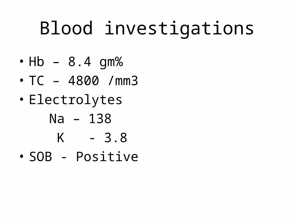
Blood investigations
• Hb – 8.4 gm%• TC – 4800 /mm3• Electrolytes Na – 138 K - 3.8• SOB - Positive

What imaging?
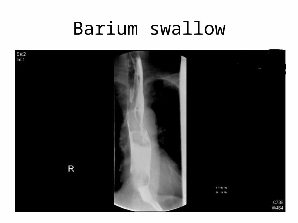
Barium swallow

How would you like to proceed?

Diagnosis
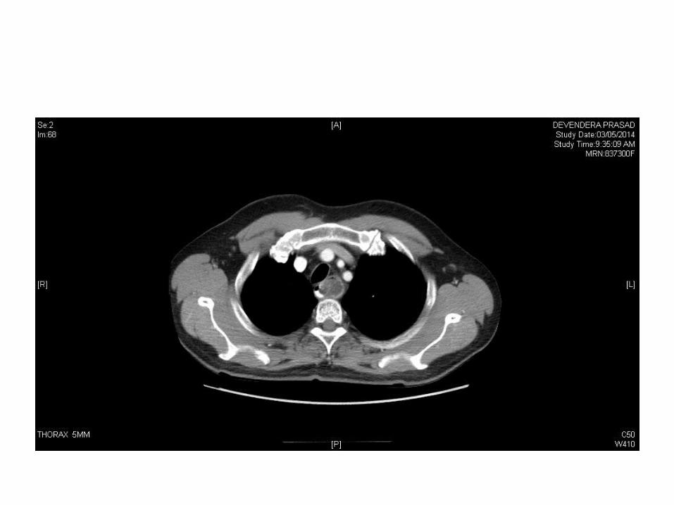
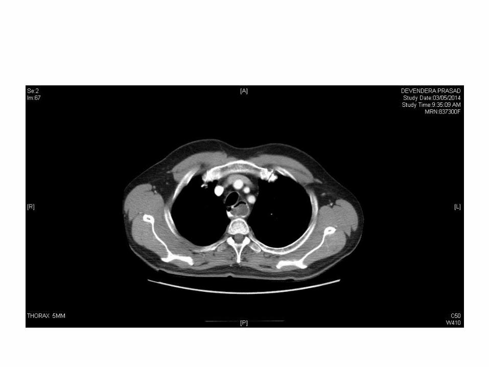
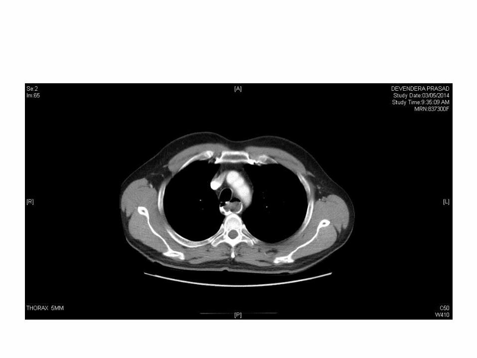
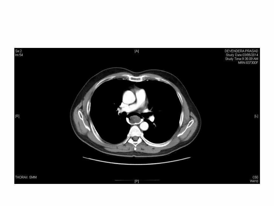
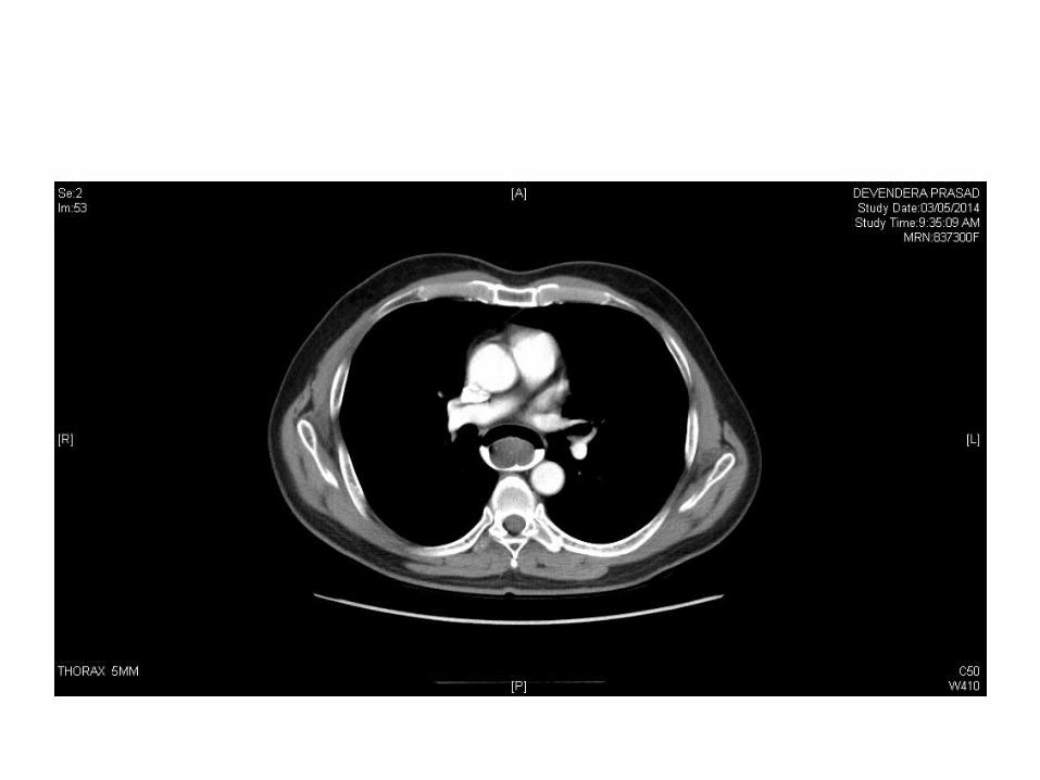
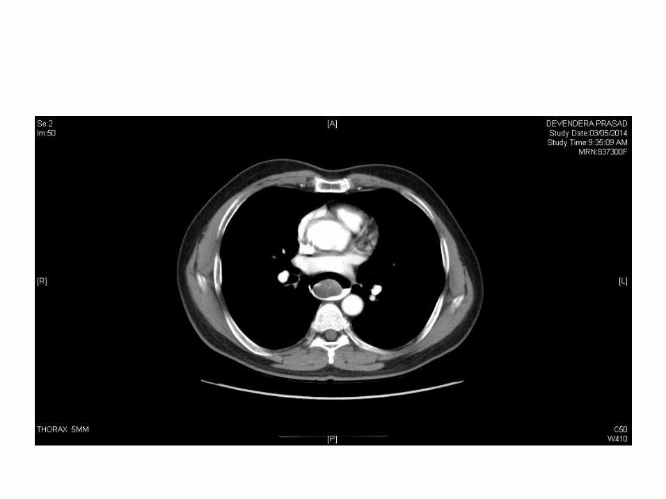
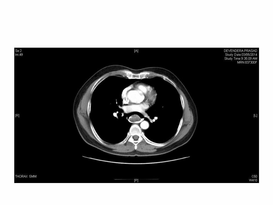
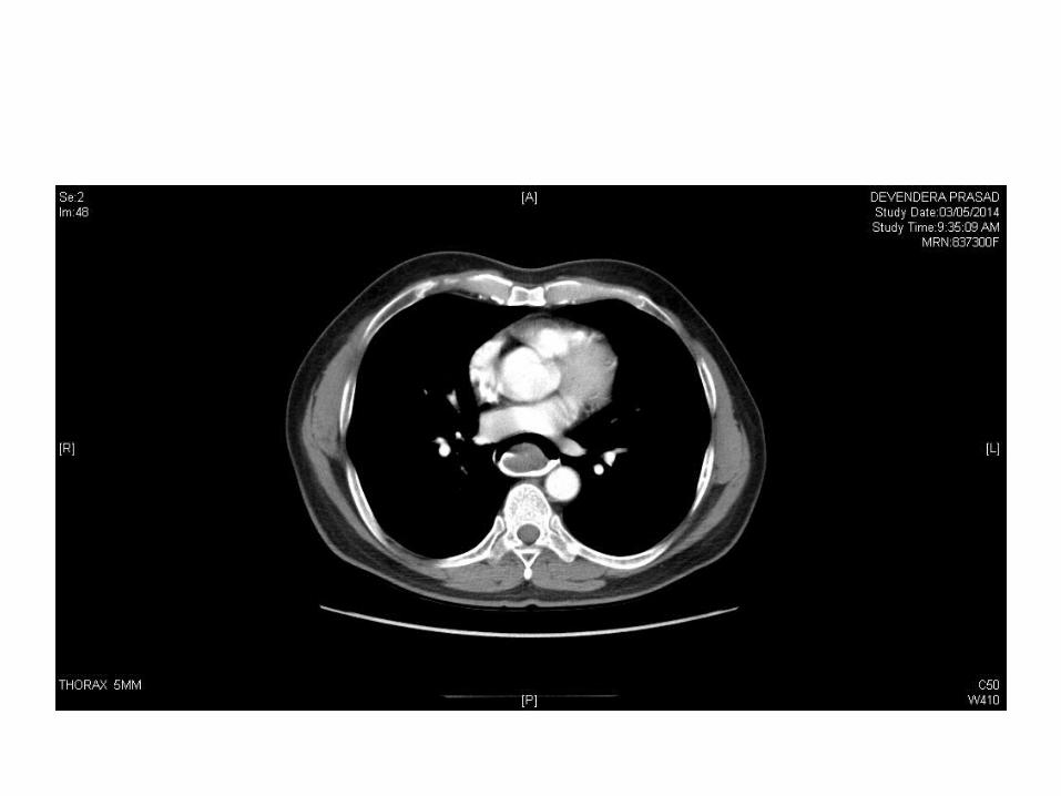
CT neck and thorax
• A large elongated polypoid soft tissue density lesion is seen extending inferiorly from the level of cervical esophagus upto GE junction, the lesion is filling and distending the lumen of esophagus causing luminal narrowing
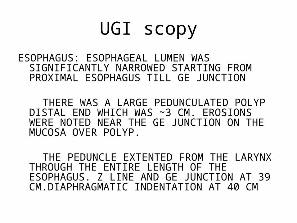
UGI scopyESOPHAGUS: ESOPHAGEAL LUMEN WAS SIGNIFICANTLY
NARROWED STARTING FROM PROXIMAL ESOPHAGUS TILL GE JUNCTION
THERE WAS A LARGE PEDUNCULATED POLYP DISTAL END WHICH WAS ~3 CM. EROSIONS WERE NOTED NEAR THE GE JUNCTION ON THE MUCOSA OVER POLYP.
THE PEDUNCLE EXTENTED FROM THE LARYNX THROUGH THE ENTIRE LENGTH OF THE ESOPHAGUS. Z LINE AND GE JUNCTION AT 39 CM.DIAPHRAGMATIC INDENTATION AT 40 CM
EUS• ESOPHAGUS: A LONG PEDUNCULATED POLYP WITH A
THICK STALK OF ~ 2.0 CMS AND MEASURING ~ 22 CMS IN LENGTH STARTING FROM 18 CMS AND EXTENDING TO THE GE JUNCTION SEEN.
• THE TIP OF THE POLYP AT THE LEVEL OF GE JUNCTION WAS ULCERATED.
• EUS: SHOWED A POLYP ARISING FROM SUBMUCOSA. THERE WAS NO MAJOR VESSELS RUNNING WITHIN THE POLYP
FINAL DIAGNOSIS
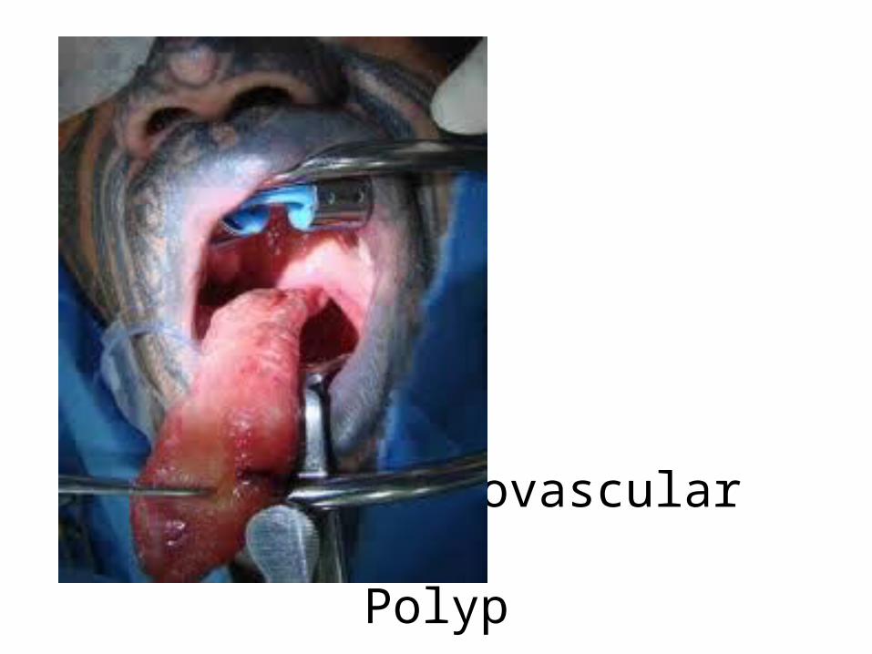
Fi Fibrovascular Polyp

How will you proceed?
Fibrovascular polyp
Zeeshan
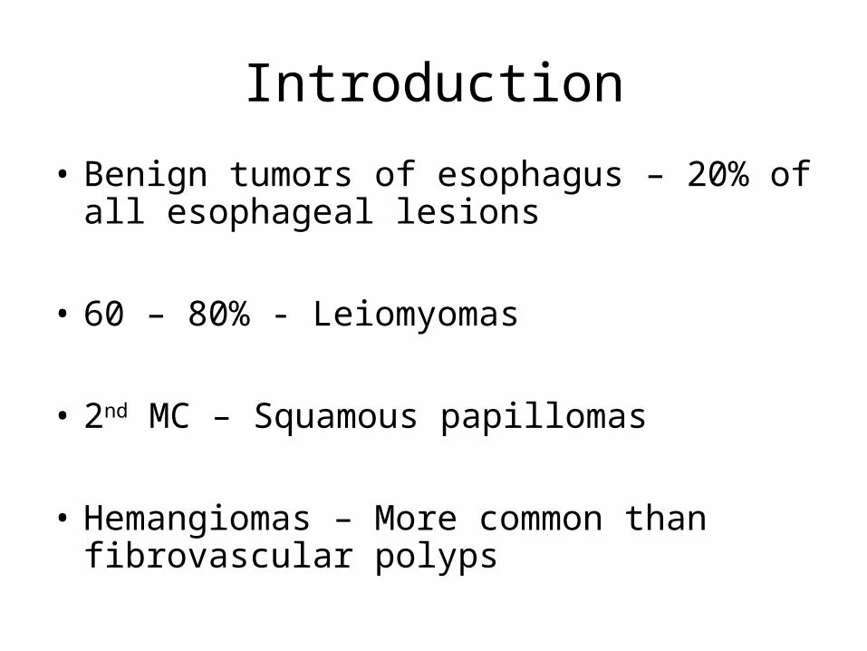
Introduction
• Benign tumors of esophagus – 20% of all esophageal lesions
• 60 – 80% - Leiomyomas
• 2nd MC – Squamous papillomas
• Hemangiomas – More common than fibrovascular polyps
Incidence
• 1-2% of esophageal tumors
• MC in males
• Age group – 50 years
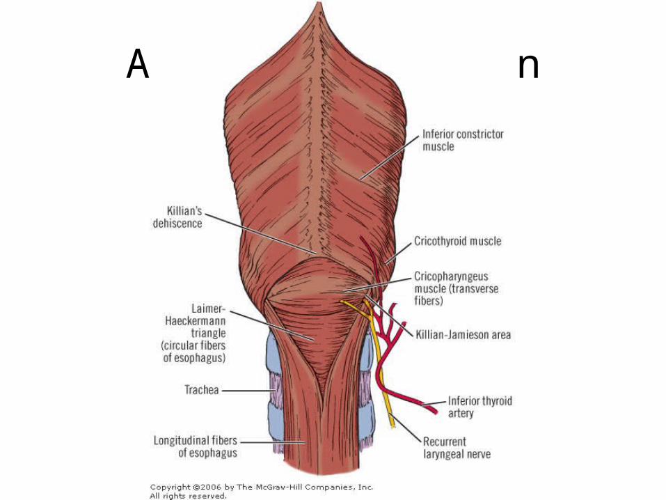
Anatomy of origin
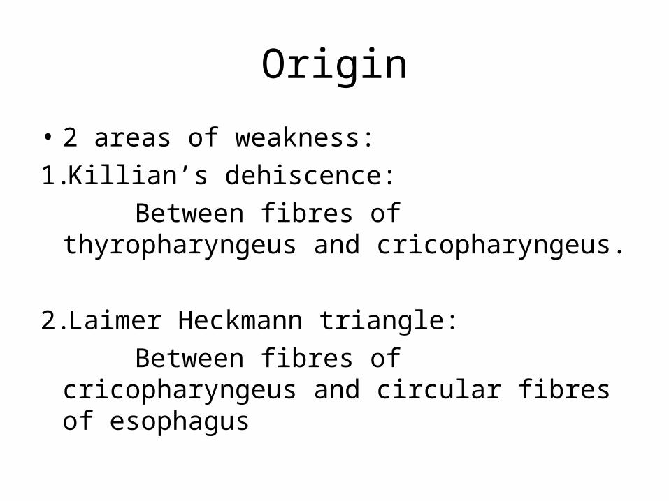
Origin
• 2 areas of weakness:1.Killian’s dehiscence: Between fibres of thyropharyngeus and
cricopharyngeus.
2.Laimer Heckmann triangle: Between fibres of cricopharyngeus and
circular fibres of esophagus
Factors
• Anatomical weakness
• Redundant submucosal folds
• Pronounced peristaltic activity
Presentation
• Dysphagia – MC symptom
• Other complaints:- Retrosternal/ epigastric discomfort- Odynophagia- Vomiting- Weight loss- Respiratory symptoms : Shortness of breath;
persistent cough
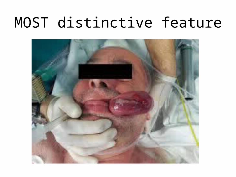
MOST distinctive feature
Complications• Asphyxia
• Laryngeal obstruction
• Aspiration pneumonia
• Hemorrhage – Secondary to twisting
• Occult GI bleed leading to anemia – Ulceration of tip
• Malignancy – Very rare
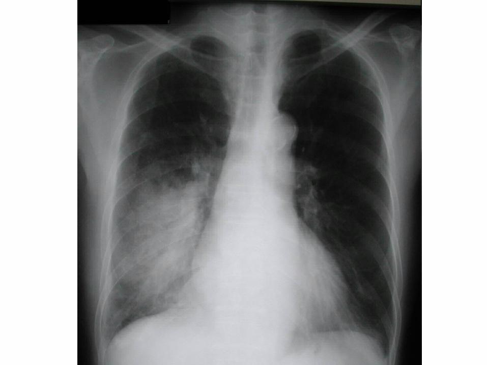
Investigations• CXR – Posterior mediastinal mass
• Barium contrast studies
• Endoscopy
• CT Neck and thorax
• EUS
Management
• Endoscopic resection of polyp - Predominantly fat - Less vascularity
• Surgical excision – 1st line of therapy
PRIOR to surgery
• Assess fitness of patient
• NPL scopy – Assess vocal cords
• Involve Plastic Sx – For local flap cover
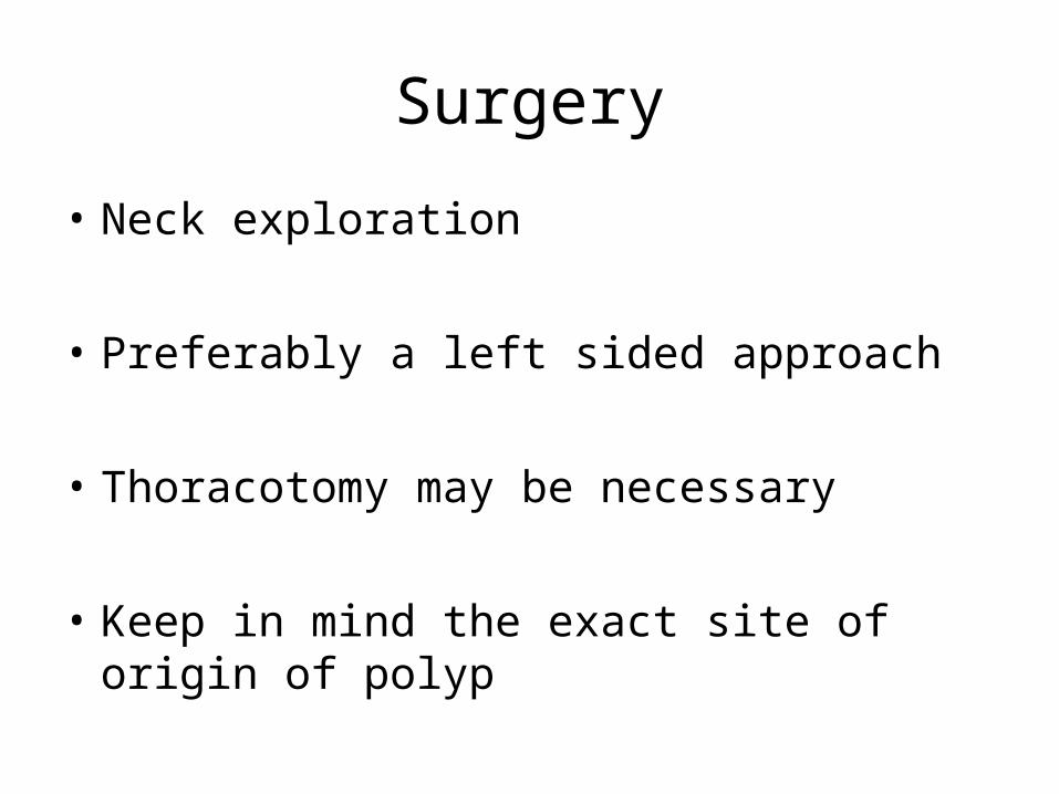
Surgery
• Neck exploration
• Preferably a left sided approach
• Thoracotomy may be necessary
• Keep in mind the exact site of origin of polyp
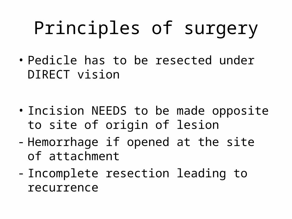
Principles of surgery
• Pedicle has to be resected under DIRECT vision
• Incision NEEDS to be made opposite to site of origin of lesion
- Hemorrhage if opened at the site of attachment
- Incomplete resection leading to recurrence
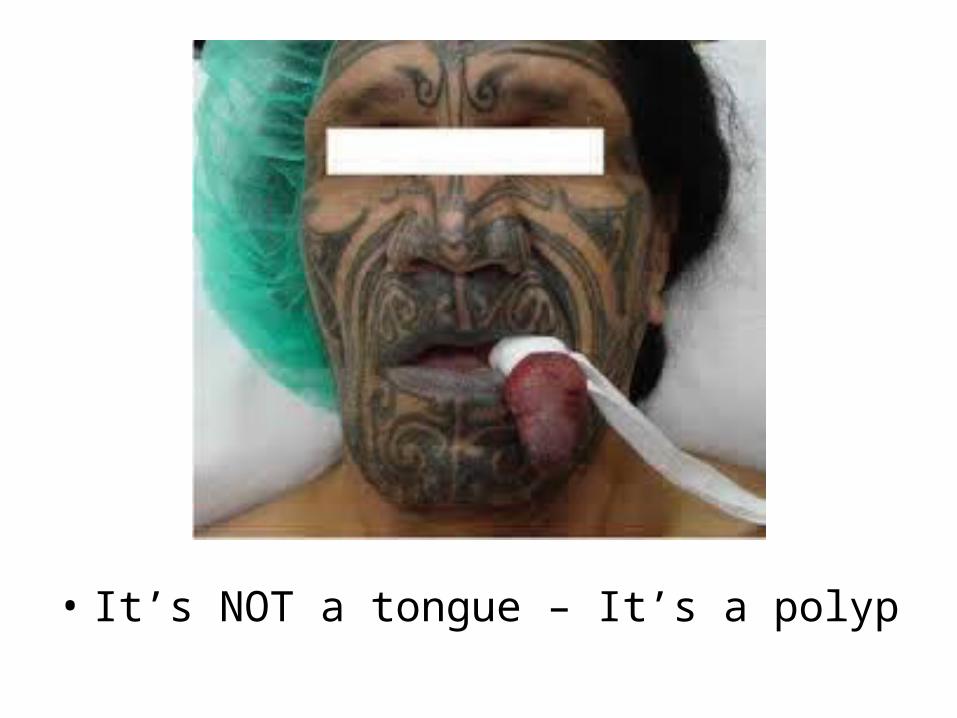
• It’s NOT a tongue – It’s a polyp
Reference • Timmons B, Sedwitz JL, Oiler DW: Benign fibrovascular polyp of the
esophagus. South Med J 1991, 84:1370-1372
• Drenth J, Wobbes T, Bonenkamp JJ, Nagengast FM: Recurrent esophageal fibrovascular polyps: case history and review of the literature.
Dig Dis Sci 2002, 47:2598-2604
• Levine MS, Buck JL, Pantongrag-Brown L, Buetow PC, Hallman JR, Sobin LH:Fibrovascular polyps of the esophagus: clinical, radiographic, and pathologic findings in 16 patients.
• AJR 1996, 166:781-787.