Embed Size (px)
Citation preview

345
RECOMMENDATIONS
StandardsThe data generated from studies that only include participants with burn injuries are inconclusive and methodologically limited. Thus, the following guidelines have been based on literature generated
from participants with scars originating from any dermal injury or disease. It is recommended that further randomized controlled trials (RCTs) that are adequately powered, use objective instrumentation to evaluate scars, and are methodologically rigorous be conducted with burn survivors.
Practice guidelines
• Gels or gel sheets should be applied to burn scars that have a high probability of forming hypertrophic scars (HTS) (ie, wounds that require ≥21 days to heal, personal factors, etc.) as soon as the wound has re-epithelialized.
• Only immature scars should be treated with gels or gel sheets, as mature burn scars have not been shown to respond.
• There are no clear benefits to using gels versus gel sheets or nonsilicone versus silicone prod-ucts with respect to the treatment effect, but there appear to be fewer adverse reactions when using silicone gels compared to gel sheets.
Options
• Keloids may benefit from treatment with gels or gel sheets.
Copyright © 2014 by the American Burn Association 1559-047X/2014
DOI: 10.1097/BCR.0000000000000124
J Burn Care Res
The objective of this review was to systematically evaluate available clinical evidence for the application of nonsilicone or silicone gels and gel sheets on hypertrophic scars and keloids after a burn injury so that practice guidelines could be proposed. This review provides evidence based recommendations, specifically for the rehabilitation interventions required for the treatment of aberrant wound healing after burn injury with gels or gel sheets. These guidelines are designed to assist all healthcare providers who are responsible for initiating and supporting scar management interventions prescribed for burn survivors. Summary recommendations were made after the literature, retrieved by systematic review, was critically appraised and the level of evidence determined according to Oxford Centre for Evidence-based Medicine criteria.1 (J Burn Care Res 2015;36:345–374)
From the *School of Physical and Occupational Therapy, McGill University, Montreal, Quebec, Canada; †Centre de recherche, Centre hospitalier de l’Université de Montréal (CRCHUM), Canada; ‡Hôpital de réadaptation Villa Medica, Montreal, Quebec, Canada; §Health Sciences Centre, Winnipeg, Mani-toba, Canada; ‖University of California Irvine Medical Center, Orange; ¶Our Lady’s Hospice and Care Services, Dublin, Ireland; #Shriners Hospitals for Children, Northern Califor-nia, Sacramento; **Surgical Services Massachusetts General Hospital, Shriners Hospitals for Children, Spaulding Rehabilita-tion Hospital, Harvard Medical School, Boston, Massachusetts; ††Shriners Hospitals for Children, Galveston, Texas; ‡‡Mas-sachusetts General Hospital, Shriners Hospitals for Children, Spaulding Rehabilitation Hospital, Department of Physical Medicine and Rehabilitation, Harvard Medical School, Boston; §§Cincinnati Children’s Medical Center, Ohio; and ‖‖McGill University, Montreal, Quebec, Canada.
Address correspondence to Bernadette Nedelec, PhD, McGill University, Faculty of Medicine, 3654 Promenade Sir William Osler, Montreal, Quebec, Canada H3G 1Y5.
Practice Guidelines for the Application of Nonsilicone or Silicone Gels and Gel Sheets After Burn Injury
Bernadette Nedelec, BSc OT(c), PhD,*†‡ Alissa Carter, BSc OT,‡ Lisa Forbes, BMR(OT), MSc,§ Shu-Chuan Chen Hsu, MA, OTR/L, CHT,‖ Margaret McMahon, MAppSc(Physio),¶ Ingrid Parry, MS, PT,# Colleen M. Ryan, MD,** Michael A. Serghiou, OTR, MBA,†† Jeffrey C. Schneider, MD,**‡‡ Patricia A. Sharp, OTD, MS, OTR/L,§§ Ana de Oliveira, BSc,† and Jill Boruff, MLIS‖‖
PRACTICE GUIDELINES

Journal of Burn Care & Research346 Nedelec et al May/June 2015
OVERVIEW
PurposeThe purpose of these guidelines was to review evi-dence evaluating the application of nonsilicone or silicone gels and gel sheets after a burn injury. Since literature specific to burn survivors was lim-ited, the search was expanded to include participants who developed scars as a result of other injuries and disorders. Rehabilitation specific information was extracted from the literature to provide further guid-ance for clinicians.
UsersThese guidelines are designed to aid burn care team members (nurses, occupational therapists, physical therapists, physicians, etc.) who are responsible for initiating and supporting the treatment of HTS or keloids after a burn injury. Additionally, the recom-mended guidelines can be implemented by health-care professionals who do not routinely treat burn patients at their facilities, such as ambulatory care centers, outpatient clinics, etc.
Clinical ProblemDeep burn wounds frequently heal with the forma-tion of HTS or keloids,2,3 which may profoundly affect a patient’s functional and psychosocial recov-ery and quality of life.4–6 Although a precise defi-nition and method for differentiating HTS from keloids has not been agreed upon in the literature, keloids proliferate or originate beyond the confines of the original injury and are recognized as having important pathological differences.7 International clinical recommendations published in 20028 sup-ported the use of silicone gel sheets for the treat-ment of scar after a burn injury, but limited burn injury specific literature and literature addressing the use of nonsilicone gel sheets and gels available at that time. The objective of this review was to systemati-cally evaluate the available evidence for the applica-tion of nonsilicone or silicone gels and gel sheets on HTS and keloids after burn injury so that practice guidelines could be proposed that specifically outline required rehabilitation interventions.
PROCESS
The steps taken to develop the practice guidelines reported here are those outlined by Bowker and col-leagues.9 These steps included setting up a guideline development group, forging links with stakeholder
groups, agreeing on the scope of the guidelines, for-mulating clinically relevant PICO (population, inter-vention, condition, outcome) questions, searching the literature for evidence, systematically appraising the evidence found, and making recommendations. The guideline development group consisted of an international assembly of occupational therapists, physicians and physiotherapists who were members of the American Burn Association Rehabilitation Committee, clinicians recruited from the American Burn Association, and clinicians who had previous experience with practice guideline development.10 The scope of the guidelines is limited to the PICO question: “Do gels or gel sheets reduce the thickness and vascularity or increase the pliability of postburn hypertrophic scar or keloids?”
Search StrategyA broad computerized search was conducted in the following databases: Ovid MEDLINE, the Cumu-lative Index of Nursing & Allied Health Literature (CINAHL), Ovid EMBASE and the Cochrane Library, from the earliest available date until Decem-ber 2, 2011. The search strategy was developed for MEDLINE by a medical librarian (J.B.) as described in Appendix 1, then adapted for the other databases. A total of 697 results were retrieved from all sources; 254 duplicates were removed, yielding 443 records for eligibility screening. Additional publications were retrieved by scanning reference lists in the articles reviewed.
Selection for InclusionSince studies focusing on this clinical question were expected to be sparse, all study designs that provided original data on patients were selected. Examples of the cause of wounds included trauma, mammoplasty, surgery (ie. cancer excision), burn injuries, immuni-zation sites, and acne. The title and abstract of each article were assessed. Only full length articles, in Eng-lish or French, were selected for review, with review articles being excluded to allow the critical appraisal of original publications. Ultimately, 55 articles were deemed appropriate for the full review process. An additional six articles were added after scanning the reference lists of these articles (Figure 1).
Data Extraction and AnalysisAll studies were systematically critiqued and scored by at least two independent reviewers, drawing on the critical appraisal form designed by Law and col-leagues.11 Fourteen items comprised in the scoring of this form relate to study purpose, literature review,

Journal of Burn Care & Research Volume 36, Number 3 Nedelec et al 347
study sample, outcomes, interventions, results, con-clusions, and clinical implications. The two reviewers independently extracted details required to com-plete the critical appraisal form. Each item was rated numerically as (1) for Yes and (0) for No or Not applicable. A total score was then calculated and compared to the second reviewer’s results. If there were minor differences (±2 points), the discrepan-cies were discussed by both reviewers until a consen-sus was reached. When larger differences occurred, a third reviewer was called upon to critique the article and consensus was achieved among all three review-ers. After this process, one article was removed12 because the data were also included in a subsequent article,13 one because there were no data provided14 and one because the authors’ clinical question was not addressed.15
RESULTS
Study CharacteristicsTable 1 summarizes the critique results for the 58 citations retained. Citations are presented in three categories, based on the population of patients included: 1) Burn scars only (denoted with double asterisk); 2) Combined burn and nonburn scar eti-ology (denoted by a single asterisk); and 3) non-burn or unspecified scars. As can be seen in the final column of this table, 16 of the 58 citations (28%) received a score of <5 out of a possible total score of
14, but were maintained for the sake of complete-ness. Of the remaining citations, 28 (48%) received a score of 5 to 9 and 14 (24%) received a score ≥10. The 11 studies that included burn scars only were more likely to score <5 compared to the total group, where six (55%) received a score of <5, two (18%) received a score of 5 to 9 and three (27%) received a score of ≥10 out of a possible total score of 14.
Table 2 presents the study characteristics of the 58 citations in detail. Twenty- seven were RCTs,16–42 six were cohort studies,13,43–47 24 case series,48–71 and one an expert opinion.72 The sample size of all the studies ranged from 5 to 276 and those that included burn scars only ranged from 30 to 94 participants. Sample size of the RCTs ranged from 11 to 155 and for burn scar only studies, from 32 to 104 subjects. The level of evidence was assigned according to the updated Oxford Centre for Evidence-based Medi-cine Levels of Evidence.1
Eleven of the citations study samples contained combined burn and nonburn scar etiology: four RCTs,17,25,26,30 two cohort studies,13,43 and five case series.52,57,59,60,63 Eleven studies included burn scar subjects only: five were RCTs,22,23,28,38,39 five were case series,48,58,66,68,69 and one was an expert opinion.72
Scientific FoundationThe initial description of the use of silicone gel sheets (SGSs) was published by Perkins and colleagues,72 who treated 42 patients with scars resulting from burn injuries. All of the patients were children, and approximately half were also wearing pressure gar-ments. Their scar age ranged from newly closed wounds to 12 years post injury. No scar outcome data were provided, but the authors concluded that all of the patients showed “significant improve-ment.” This early report was followed by a number of reports by Quinn and colleagues,68,69 who reported on a series of patients and also explored some plausi-ble mechanisms of action by which SGS were having an effect. The first report included 40 patients and the second report 125. It appears that all patients included in these two reports were burn survivors and completely different populations, but this was never clearly articulated. In both the reports, out-come measures included color and elevation, as well as “texture”, which were quantified with an exten-someter in the second report. In the first report, all patients improved within the first 2 months with respect to at least one of the skin characteristics that was evaluated, and in the second report 75 out of 125 improved. In the second report, 37 were lost
Databases searched: CINAHL, Cochrane library, EMBASE, Medline
Citations identified = 697Duplicates removed = 254
Citation titles and abstracts screened = 433
Articles included for detailed evaluation = 55
[Primary search]
Articles identified by manual search from
references = 6[Secondary search]
Total articles included in full review process = 61
Articles retained after full review process = 58
Excluded during review process = 3
Reasons:Data included in subsequent article = 1
No data provided = 1Did not address PICO question = 1
Figure 1. Flow chart for search strategy.

Journal of Burn Care & Research348 Nedelec et al May/June 2015
Table 1. Evidentiary table: evaluation of the quality of intervention studies
CitationStudy
PurposeLiterature
Review Design
Sample Outcomes Intervention Results
Size Details Justified Reliable ValidDetailed
Description Contamination CointerventionStatistical
Signif icanceAnalysis
AppropriateClinical
ImportanceDrop Outs Reported
Conclusions Appropriate
Total Score
Ahn et al 1991 1 1 CO 48† 1 0 1 1 1 0 0 1 1 1 1 1 11Argirova et al 2006 0 0 CS 276* 0 0 0 0 0 0 0 0 0 1 0 0 1Baum and Busuito 1998 0 1 CS 34 1 0 0 0 0 0 0 0 0 0 1 0 3Berman and Flores 1999 0 1 RCT 22 1 0 0 0 1 0 0 0 0 1 1 1 6Boutli-Kasapidou et al 2005 1 0 CO 30† 1 0 0 0 0 0 0 1 1 1 1 1 7Carney et al 1994 1 1 RCT 42† 1 0 0 0 0 1 1 1 0 1 0 1 8Cassuto et al 2010 0 1 CS 37 1 0 0 0 1 0 0 0 0 0 0 0 3Chan et al 2005 1 1 RCT 100 1 1 0 0 0 0 0 1 1 1 1 1 9Chernoff et al 2007 1 1 CO 30 1 0 0 0 1 0 0 0 0 1 0 0 5Chuangsuw anich et al 2000 1 0 CS 18 0 0 0 0 0 0 0 1 0 0 1 1 4Cruz-Korchin 1996 1 1 CO 20 1 0 0 0 1 1 0 1 1 1 1 1 10de Giorgi et al 2009 1 1 RCT 110 1 0 0 0 1 0 1 1 1 1 1 1 10de Oliveira et al 2001 1 1 RCT 26 1 0 0 0 1 1 1 1 1 1 0 1 10Dockery and Nilson 1994 1 1 CS 94† 1 0 0 0 1 0 0 0 0 1 1 1 7Eishi et al 2003 1 1 CS 6 1 0 0 0 1 0 1 1 1 1 0 1 9Fulton 1995 1 0 CS 20 1 0 0 0 1 0 0 0 0 1 0 0 4Gibbons et al 1994 0 1 CS 5 0 0 0 0 0 0 0 0 0 1 1 0 3Gold 1993 0 1 CS 10 0 0 0 0 0 0 0 0 1 1 1 1 5Gold 1994 0 1 CS 34† 0 0 0 0 0 0 0 0 0 1 0 1 3Gold et al 2001 1 1 RCT 96 1 0 0 0 0 0 0 0 0 1 1 1 6Hamanova and Broz 2002 1 0 CS 60* 0 0 0 0 0 0 0 0 0 1 0 1 3Harte et al 2009 1 1 RCT 22* 1 1 1 0 1 0 0 1 1 1 1 1 11Hirshowitz et al 1993 1 1 CS 32† 0 0 0 0 1 0 0 0 1 1 1 1 7Hirshowitz et al 1998 1 1 CS 30† 0 0 0 0 1 0 0 0 0 1 1 1 6Hosnuter et al 2007 1 1 CO 60 1 0 0 0 1 0 0 1 1 1 0 1 8Karagoz et al 2009 1 1 RCT 32* 1 0 0 0 1 0 0 1 1 1 1 1 9Katz 1995 1 1 CS 48 0 0 0 0 0 0 0 0 0 1 0 1 4Klopp et al 2000 1 1 RCT 12 0 0 0 0 0 0 1 1 1 1 0 1 7Lacarrubba et al 2008 1 1 CS 8 1 1 0 0 0 0 0 0 0 1 1 0 6Lee et al 1996 1 1 CS 26† 1 0 0 0 1 0 0 0 0 0 1 0 5Li-Tsang et al 2006 0 1 RCT 45† 1 0 1 1 0 0 0 1 1 1 1 0 8Li-Tsang et al 2010 1 1 RCT 104† 1 0 1 1 1 0 1 1 1 1 1 1 12Majan 2006 1 1 RCT 11 1 0 0 0 1 0 0 0 1 0 1 1 7Mercer 1989 0 0 CS 18 0 0 0 0 0 0 0 0 0 1 1 1 2Momeni et al 2009 1 1 RCT 38* 1 0 0 0 1 0 0 1 1 1 1 1 9Murison and James 2005 1 0 CS 6 1 0 0 0 1 0 0 0 0 1 1 0 5Niessen et al 1998 1 1 RCT 155 1 0 1 1 1 1 0 1 1 1 1 1 12Ohmori 1988 0 0 CS 46* 0 0 0 0 0 0 0 0 0 1 1 0 2Palmieri et al 1995 1 1 RCT 80† 0 0 0 0 0 0 1 1 0 0 1 0 5Perez et al 2010 1 0 RCT 30 1 0 0 0 1 0 1 1 1 1 1 1 9Perkins et al 1983 1 0 EO 42* 1 0 0 0 0 0 0 0 0 1 0 0 3Phillips et al 1996 1 1 RCT 20 1 0 1 1 1 0 0 1 1 1 0 0 9Puri and Talwar 2009 0 1 CS 30 0 0 0 0 0 0 0 0 0 1 0 0 2Quinn et al 1985 0 0 CS 40* 1 0 0 0 0 0 0 0 0 0 0 1 2Quinn 1987 1 0 CS 125* 0 0 0 0 0 0 0 0 0 1 1 1 4Rhee et al 2010 1 1 RCT 40 1 0 0 0 1 0 1 1 1 1 1 1 10Sakuraba et al 2011 1 0 CS 9 0 0 0 0 1 0 0 1 0 0 0 0 3Scuderi et al 2010 1 1 RCT 150 1 0 0 0 1 0 0 1 1 1 1 1 9Scuderi et al 2011 1 1 RCT 85 1 0 0 0 1 0 0 1 0 1 1 1 8Signori and Clementonit 2007 1 1 RCT 148 1 0 0 0 1 0 1 1 0 1 1 0 8Spencer 2010 1 0 CS 7 0 0 0 1 1 0 0 0 0 1 1 1 6Sprout et al 1992 1 1 RCT 14 0 1 1 1 1 0 1 1 1 1 0 1 11Steinstraesser et al 2011 1 1 RCT 38* 1 0 1 1 0 0 0 1 1 1 1 1 10Tan et al 1999 1 1 CO 17 1 0 0 0 1 0 1 1 1 1 1 1 10van der Wal et al 2010 1 1 RCT 23* 1 0 1 1 1 1 0 1 1 1 1 1 12Widgerow et al 2008 0 1 RCT 120 0 0 1 1 0 0 0 1 1 1 0 0 6Wigger-Alberti et al 2009 1 1 RCT 60 1 1 1 1 1 1 0 1 1 1 1 1 13Wittenberg et al 1999 1 1 RCT 19 1 1 0 0 1 0 0 1 1 1 1 1 10CS, case series; CO, cohort; EO, expert opinion; RCT, randomized controlled trial.*Only burn survivors.†Burn survivors included; YES = 1; NO or N/A = 0.

Journal of Burn Care & Research Volume 36, Number 3 Nedelec et al 349
Table 1. Evidentiary table: evaluation of the quality of intervention studies
CitationStudy
PurposeLiterature
Review Design
Sample Outcomes Intervention Results
Size Details Justified Reliable ValidDetailed
Description Contamination CointerventionStatistical
Signif icanceAnalysis
AppropriateClinical
ImportanceDrop Outs Reported
Conclusions Appropriate
Total Score
Ahn et al 1991 1 1 CO 48† 1 0 1 1 1 0 0 1 1 1 1 1 11Argirova et al 2006 0 0 CS 276* 0 0 0 0 0 0 0 0 0 1 0 0 1Baum and Busuito 1998 0 1 CS 34 1 0 0 0 0 0 0 0 0 0 1 0 3Berman and Flores 1999 0 1 RCT 22 1 0 0 0 1 0 0 0 0 1 1 1 6Boutli-Kasapidou et al 2005 1 0 CO 30† 1 0 0 0 0 0 0 1 1 1 1 1 7Carney et al 1994 1 1 RCT 42† 1 0 0 0 0 1 1 1 0 1 0 1 8Cassuto et al 2010 0 1 CS 37 1 0 0 0 1 0 0 0 0 0 0 0 3Chan et al 2005 1 1 RCT 100 1 1 0 0 0 0 0 1 1 1 1 1 9Chernoff et al 2007 1 1 CO 30 1 0 0 0 1 0 0 0 0 1 0 0 5Chuangsuw anich et al 2000 1 0 CS 18 0 0 0 0 0 0 0 1 0 0 1 1 4Cruz-Korchin 1996 1 1 CO 20 1 0 0 0 1 1 0 1 1 1 1 1 10de Giorgi et al 2009 1 1 RCT 110 1 0 0 0 1 0 1 1 1 1 1 1 10de Oliveira et al 2001 1 1 RCT 26 1 0 0 0 1 1 1 1 1 1 0 1 10Dockery and Nilson 1994 1 1 CS 94† 1 0 0 0 1 0 0 0 0 1 1 1 7Eishi et al 2003 1 1 CS 6 1 0 0 0 1 0 1 1 1 1 0 1 9Fulton 1995 1 0 CS 20 1 0 0 0 1 0 0 0 0 1 0 0 4Gibbons et al 1994 0 1 CS 5 0 0 0 0 0 0 0 0 0 1 1 0 3Gold 1993 0 1 CS 10 0 0 0 0 0 0 0 0 1 1 1 1 5Gold 1994 0 1 CS 34† 0 0 0 0 0 0 0 0 0 1 0 1 3Gold et al 2001 1 1 RCT 96 1 0 0 0 0 0 0 0 0 1 1 1 6Hamanova and Broz 2002 1 0 CS 60* 0 0 0 0 0 0 0 0 0 1 0 1 3Harte et al 2009 1 1 RCT 22* 1 1 1 0 1 0 0 1 1 1 1 1 11Hirshowitz et al 1993 1 1 CS 32† 0 0 0 0 1 0 0 0 1 1 1 1 7Hirshowitz et al 1998 1 1 CS 30† 0 0 0 0 1 0 0 0 0 1 1 1 6Hosnuter et al 2007 1 1 CO 60 1 0 0 0 1 0 0 1 1 1 0 1 8Karagoz et al 2009 1 1 RCT 32* 1 0 0 0 1 0 0 1 1 1 1 1 9Katz 1995 1 1 CS 48 0 0 0 0 0 0 0 0 0 1 0 1 4Klopp et al 2000 1 1 RCT 12 0 0 0 0 0 0 1 1 1 1 0 1 7Lacarrubba et al 2008 1 1 CS 8 1 1 0 0 0 0 0 0 0 1 1 0 6Lee et al 1996 1 1 CS 26† 1 0 0 0 1 0 0 0 0 0 1 0 5Li-Tsang et al 2006 0 1 RCT 45† 1 0 1 1 0 0 0 1 1 1 1 0 8Li-Tsang et al 2010 1 1 RCT 104† 1 0 1 1 1 0 1 1 1 1 1 1 12Majan 2006 1 1 RCT 11 1 0 0 0 1 0 0 0 1 0 1 1 7Mercer 1989 0 0 CS 18 0 0 0 0 0 0 0 0 0 1 1 1 2Momeni et al 2009 1 1 RCT 38* 1 0 0 0 1 0 0 1 1 1 1 1 9Murison and James 2005 1 0 CS 6 1 0 0 0 1 0 0 0 0 1 1 0 5Niessen et al 1998 1 1 RCT 155 1 0 1 1 1 1 0 1 1 1 1 1 12Ohmori 1988 0 0 CS 46* 0 0 0 0 0 0 0 0 0 1 1 0 2Palmieri et al 1995 1 1 RCT 80† 0 0 0 0 0 0 1 1 0 0 1 0 5Perez et al 2010 1 0 RCT 30 1 0 0 0 1 0 1 1 1 1 1 1 9Perkins et al 1983 1 0 EO 42* 1 0 0 0 0 0 0 0 0 1 0 0 3Phillips et al 1996 1 1 RCT 20 1 0 1 1 1 0 0 1 1 1 0 0 9Puri and Talwar 2009 0 1 CS 30 0 0 0 0 0 0 0 0 0 1 0 0 2Quinn et al 1985 0 0 CS 40* 1 0 0 0 0 0 0 0 0 0 0 1 2Quinn 1987 1 0 CS 125* 0 0 0 0 0 0 0 0 0 1 1 1 4Rhee et al 2010 1 1 RCT 40 1 0 0 0 1 0 1 1 1 1 1 1 10Sakuraba et al 2011 1 0 CS 9 0 0 0 0 1 0 0 1 0 0 0 0 3Scuderi et al 2010 1 1 RCT 150 1 0 0 0 1 0 0 1 1 1 1 1 9Scuderi et al 2011 1 1 RCT 85 1 0 0 0 1 0 0 1 0 1 1 1 8Signori and Clementonit 2007 1 1 RCT 148 1 0 0 0 1 0 1 1 0 1 1 0 8Spencer 2010 1 0 CS 7 0 0 0 1 1 0 0 0 0 1 1 1 6Sprout et al 1992 1 1 RCT 14 0 1 1 1 1 0 1 1 1 1 0 1 11Steinstraesser et al 2011 1 1 RCT 38* 1 0 1 1 0 0 0 1 1 1 1 1 10Tan et al 1999 1 1 CO 17 1 0 0 0 1 0 1 1 1 1 1 1 10van der Wal et al 2010 1 1 RCT 23* 1 0 1 1 1 1 0 1 1 1 1 1 12Widgerow et al 2008 0 1 RCT 120 0 0 1 1 0 0 0 1 1 1 0 0 6Wigger-Alberti et al 2009 1 1 RCT 60 1 1 1 1 1 1 0 1 1 1 1 1 13Wittenberg et al 1999 1 1 RCT 19 1 1 0 0 1 0 0 1 1 1 1 1 10CS, case series; CO, cohort; EO, expert opinion; RCT, randomized controlled trial.*Only burn survivors.†Burn survivors included; YES = 1; NO or N/A = 0.

Journal of Burn Care & Research350 Nedelec et al May/June 2015
Tab
le 2
. Cha
ract
eris
tics
of in
clud
ed s
tudi
es
Aut
hors
Des
ign
Sam
ple
Out
com
e
Mea
sure
sIn
terv
enti
onR
esul
tsL
evel
of
Evi
denc
e
Ahn
et
al
1991
Intr
a indi
vidu
al
coho
rt
•n
= 48
•G
roup
1 =
29
Sx
sca
rs, a
ll bu
t tw
o <4
0 yr
, n
= 10
wer
e lo
st t
o fo
llow
-up
•G
roup
2 =
19
HT
S, a
ge
rang
e 3–
78 y
r, bu
rns,
Sx
sca
rs, s
pide
r bi
te
•B
asel
ine,
1 a
nd
2/12
•Sc
ar v
olum
e•
Phot
ogra
ph•
Ela
stom
eter
•H
omem
ade
glob
al
scar
ass
essm
ent
•T
x =
SGS
secu
red
with
ad
hesi
ve t
ape
and
wor
n >1
2 hr
s/d
•C
ontr
ol =
no
trea
tmen
t•
Gro
up 1
—ha
lf of
the
su
rgic
al e
xcis
ion
assi
gned
to
Tx
or c
ontr
ol•
Gro
up 2
—ha
lf of
the
su
rfac
e ar
ea o
f HT
S as
sign
ed t
o T
x or
con
trol
•In
crea
sed
scar
vol
ume
of su
rgic
al sc
ars a
t co
ntro
l site
s (1/
12 P
= .0
3; 2
/12
P =
.003
)•
No
stat
istic
ally
sig
nific
ant
redu
ctio
n in
sc
ar v
olum
e of
HT
S•
Incr
ease
d sc
ar e
last
icity
in T
x’d
HT
S co
mpa
red
to b
asel
ine
at 1
/12
(P =
.019
) an
d 2/
12 (P
= .0
001)
and
com
pare
d to
the
cont
rol (
1/12
, P =
.005
; 2/
12, P
= .0
001)
•H
TS
elas
ticity
trea
tmen
t eff
ect p
late
aued
at
2/
12
3
Arg
irov
a et
al
2006
Cas
e se
ries
•n
= 27
6 ch
ildre
n•
Bur
ns•
Bas
elin
e, 4
, 8, 1
2,
24/
52•
VSS
•Ph
otog
raph
•G
roup
1—
(n =
232
) −
com
pres
sion
(24
hrs
/d)
+
intr
ales
iona
l TC
A (
10–
40 m
g/m
l) +
SG
S (8
–24
hrs/
d)•
Gro
up 2
— (
n =
44)
− co
mpr
essi
on (
24 h
rs/
d) +
SG
S (8
–24
hrs/
d)
•N
o st
atis
tical
com
pari
sons
pro
vide
d•
Gra
phic
out
com
es p
rese
nted
for
pigm
enta
tion,
hei
ght,
vas
cula
rity
, itc
h,
and
plia
bilit
y•
17 p
atie
nts
deve
lope
d sk
in r
eact
ion
4
Bau
m a
nd
Bus
uito
199
8C
ase
seri
es•
n =
34•
Sx e
xcis
ions
•H
omem
ade
eval
uatio
n (e
ffec
tive
vs n
ot
effe
ctiv
e)
•N
SGS
(>12
hrs
/d
× 8–
12
wks
) ap
plie
d po
stsu
rgic
ally
+
com
pres
sion
(n
= 3)
•N
o st
atis
tical
com
pari
sons
pro
vide
d•
Stat
ed t
hat
31/
34 s
cars
wer
e ef
fect
ivel
y tr
eate
d
4
Ber
man
and
Fl
ores
199
9R
CT
•N
= 2
2 (1
0 H
TS;
22
kelo
ids)
•n
= 9
lost
to
follo
w-u
p•
Age
ran
ge 2
5–70
yrs
•M
ean
7 m
os p
ostin
jury
•E
tiolo
gy n
ot s
peci
fied
•Sc
ar v
olum
e•
Hom
emad
e ev
alua
tion
(col
or,
tend
erne
ss,
itchi
ng, i
ndur
atio
n,
patie
nt s
atis
fact
ion)
•G
roup
1—
silic
one
gel fi
lled
cush
ion
•G
roup
2—
SGS
•G
roup
1 a
nd 2
—>1
0 hr
s/d
•N
o st
atis
tical
ly s
igni
fican
t di
ffer
ence
be
twee
n th
e gr
oups
•61
% o
f the
pat
ient
s re
port
ed a
sat
isfa
ctio
n le
vel o
f >9/
10•
one
patie
nt d
evel
oped
ski
n re
actio
n
2
Bou
tli-K
asap
idou
et
al 2
005
CO
•n
= 30
•G
roup
1 =
11F
, 9M
•
Gro
up 2
= 6
F, 4
M•
Mea
n ag
e 24
yrs
•K
eloi
ds a
nd H
TS
•Sx
, acn
e, im
mun
izat
ion,
bu
rns,
spo
ntan
eous
ke
loid
s
•R
atin
g sc
ale
•Pa
tient
ass
essm
ent:
pr
uritu
s, b
urni
ng,
tens
ion
•D
r as
sess
men
t:
flatt
enin
g,
func
tiona
lity,
so
ftne
ss
•G
roup
1—
poly
ther
apy
(cry
othe
rapy
, int
rale
sion
al
cort
ison
e, S
G [
3x/
d ×
12
mos
])•
Gro
up 2
—SG
S (2
0 hr
s/d
× 12
mos
)
•Pa
tient
sat
isfa
ctio
n: G
roup
1 in
crea
se
6.05
± 0
.66
vs g
roup
2 in
crea
se 1
.8 ±
0.2
5 (P
< .0
001)
•D
r as
sess
men
t: g
roup
1 in
crea
se
6.4
± 0.
06 v
s gr
oup
2 in
crea
se 1
.7 ±
0.2
1 (P
< .0
001)
•Yo
unge
r (2
.9 ±
1.5
1-yr
-old
) ke
loid
s w
ere
mor
e lik
ely
to r
espo
nd t
han
olde
r (5
.36
± 1-
yr-o
ld)
(P <
.001
)
3

Journal of Burn Care & Research Volume 36, Number 3 Nedelec et al 351
Car
ney
et a
l 199
4In
trai
ndiv
idua
l R
CT
•n
= 42
•M
ean
23.2
(2
–60
yrs
)•
92%
bur
ns
•E
xten
som
eter
•H
omem
ade
eval
uatio
n (c
olor
, te
xtur
e, g
ener
al
cond
ition
, pat
ient
s’
opin
ion)
•G
roup
1—
SGS
and
cont
rol
(int
rain
divi
dual
)•
Gro
up 2
—C
ica
care
and
co
ntro
l (in
trai
ndiv
idua
l)•
Gro
up 1
and
2—
wor
n as
m
uch
as p
ossi
ble
•G
roup
1—
impr
ovem
ent
was
sig
nific
antly
gr
eate
r in
Tx’
d vs
con
trol
at
2/12
(e
xten
sibi
lity
P <
.000
1; c
olor
P =
0.0
05;
text
ure
P .0
01)
and
6/12
(ex
tens
ibili
ty
P <
.03;
tex
ture
P =
.012
)•
Gro
up 2
—im
prov
emen
t was
sig
nific
antly
gr
eate
r in
Tx’
d vs
con
trol
at
1/12
(e
xten
sibi
lity
P <
.03)
, 2/
12
(ext
ensi
bilit
y P
< .0
01; c
olor
P =
.008
; te
xtur
e P
.001
) an
d 6/
12 (
exte
nsib
ility
P
< .0
4; c
olor
P =
.007
; tex
ture
P =
.002
)•
Adv
erse
rea
ctio
ns: p
ruri
tus,
ski
n ir
rita
tion
•N
o st
atis
tical
diff
eren
ce b
etw
een
the
grou
ps
2
Cas
suto
et
al 2
010
Cas
e se
ries
•n
= 37
(31
F, 6
M)
(n
= 4
8 sc
ars;
34
HT
S +
14 k
eloi
ds)
•M
ean
34 (
8–67
yrs
)•
Mea
n sc
ar a
ge 9
(3–
35
mos
)•
Etio
logy
not
spe
cifie
d
•B
asel
ine
and
4 w
ks a
fter
eve
ry T
x (m
axim
um T
x 21
)•
VSS
•Ph
otog
raph
•L
aser
(53
2 nm
mill
isec
ond)
+
SGS
(16–
24 h
rs/
d)•
No
stat
istic
al c
ompa
riso
ns p
rovi
ded
•V
SS im
prov
ed fr
om 1
2.6
(bas
elin
e) t
o 3.
3 (fi
nal)
4
Cha
n et
al 2
005
Intr
aind
ivid
ual
RC
T•
n =
50•
Med
ian
age
61
(26–
77 y
rs)
•St
erna
l sca
r po
stca
rdia
c su
rger
y
•2/
52, 6
/52
, 3/
12
post
oper
ativ
e•
VSS
•Pa
in (
3 po
int
scal
e)•
Itch
(3
poin
t sc
ale)
•D
igita
l im
age
(rat
er b
linde
d to
gr
oup
allo
catio
n)•
com
plia
nce
•U
pper
and
low
er p
ortio
n of
th
e sc
ar r
ando
mly
ass
igne
d to
Tx
and
the
cont
rol g
roup
•T
x gr
oup—
SG (
sem
i-liq
uid
stic
k)•
Con
trol
—N
SGS
•A
pplie
d 2x
/d
•Su
bjec
ts b
linde
d to
gro
up
allo
catio
n•
3/12
Tx
•T
x gr
oup
sign
ifica
ntly
bet
ter
on a
ll ou
tcom
e m
easu
res
(pig
men
tatio
n
P =
.02;
vas
cula
rity
P =
.001
; plia
bilit
y
P =
.001
; hei
ght
P =
.001
; pai
n P
= .0
01;
itch
P =
0.02
)•
Tx
grou
p gr
adua
lly im
prov
ed b
etw
een
6/52
and
3/
12•
Con
trol
gro
up g
radu
ally
wor
sene
d ov
er
time
•74
% p
erfe
ct c
ompl
ianc
e, 2
4% fo
rgot
so
met
imes
, 2%
forg
ot m
ost
of t
he t
ime
2
Che
rnof
f et
al 2
007
Intr
aind
ivid
ual
coho
rt•
n =
30 b
ilate
ral a
ctiv
e sc
ars
•H
TS,
kel
oids
or
post
lase
r ex
folia
tion
eryt
hem
a•
Etio
logy
of H
TS
and
kelo
ids
not
spec
ified
•O
ptic
al p
rofil
om-
etry
(su
rfac
e to
polo
gy, e
leva
tion)
•H
omem
ade
eval
u-at
ion
(ery
them
a,
scar
sym
ptom
s [i
rrita
tion,
ski
n m
acer
atio
n] a
nd
diffi
culty
usi
ng)
•G
roup
1 (
n =
10)—
SG
(2x/
d)•
Gro
up 2
(n
= 10
)—SG
S (m
orni
ng a
nd n
ight
)•
Gro
up 3
(n
= 10
)—SG
(m
orni
ng)
and
SGS
(nig
ht)
•B
ilate
ral s
car—
no t
reat
men
t co
ntro
l•
Tx
time
for
all g
roup
s =
90 d
•G
roup
1, 2
, and
3 w
ere
sign
ifica
ntly
less
el
evat
ed (
P <
.001
) an
d gr
oups
1 a
nd 2
w
ere
sign
ifica
ntly
less
red
(P
< .0
01),
less
sy
mpt
omat
ic (
P <
.001
) co
mpa
red
to
untr
eate
d co
ntro
l sca
r•
SG w
as e
asie
r to
use
tha
t SG
S (P
< .0
01)
•G
roup
3 p
artic
ipan
ts r
ated
impr
ovem
ent
mos
t fa
vora
bly
3
(Con
tinu
ed)

Journal of Burn Care & Research352 Nedelec et al May/June 2015
Chu
angs
uwan
ich
et
al 2
000
Cas
e se
ries
•n
= 18
•M
ean
age
21 (
6–33
yr)
•M
ean
scar
age
5.7
yrs
(3
mos
–20
yrs)
•H
TS
and
kelo
ids
•E
tiolo
gy n
ot s
peci
fied
•B
asel
ine,
1, 2
, 4,
6 m
os•
Hei
ght,
wei
ght,
an
d co
lor
•Ph
otog
raph
s•
Scar
vol
ume
•Pa
tient
rat
ing
(poo
r, fa
ir,
good
)
•T
x gr
oup:
SG
S,
12 h
rs/
d, 8
/52
Tx
•H
eigh
t of
sca
r af
ter
Tx
66.6
7% b
ette
r
(P =
.058
)•
Wei
ght
of t
he s
car
afte
r T
x 55
.55%
im
prov
ed (
P =
.09)
•C
olor
of t
he s
car
afte
r T
x 36
.84%
im
prov
ed•
Patie
nt r
atin
g: 6
6.67
% g
ood
4
Cru
z-K
orch
in
1996
Intr
aind
ivid
ual
coho
rt•
n =
20•
Bila
tera
l mam
mop
last
y 14
d p
osto
pera
tivel
y
•B
asel
ine,
1, 2
, and
6
mos
•A
ppea
ranc
e (r
aise
d vs
flat
)•
Phot
ogra
phy
•O
bser
vatio
n
•B
ilate
ral b
reas
ts: o
ne
cont
rol,
one
Tx
•SG
S po
stop
erat
ive
day
14,
12 h
rs/
d ×
2/12
•A
t 2/
12—
25%
of t
reat
ed b
reas
ts w
ere
hype
rtro
phic
, 60%
of n
ontr
eate
d w
ere
hype
rtro
phic
(P
< .0
5)•
At
6/12
—25
% o
f tre
ated
bre
asts
wer
e hy
pert
roph
ic, 5
5% o
f non
trea
ted
wer
e hy
pert
roph
ic (
P <
.05)
•Sk
in ir
rita
tion
in t
wo
patie
nts
3
De
Geo
rgi e
t al
20
09R
CT
•n
= 11
0 (5
5F, 5
5M)
•G
roup
A =
65
(32F
, 33
M)
(mea
n ag
e 52
yrs
ra
nge
26–8
1)•
Gro
up B
= 4
5 (2
3F,
22M
) (m
ean
age
48 y
rs.
rang
e 23
–76)
•Fr
esh
Sx s
cars
•1,
2, 3
, 4, 6
, 8 m
os
post
Sx
•Sc
ar a
ltera
tions
(k
eloi
ds, H
TS,
di
asta
sic
scar
, st
roph
ic s
car)
•Pa
in a
nd it
ch V
AS
•G
roup
A =
SG
+ z
inc
oxid
e 2x
/d
× 60
d•
Gro
up B
= z
inc
oxid
e cr
eam
•A
bnor
mal
sca
r fo
rmat
ion:
gro
up A
27%
vs
gro
up B
55%
(P
= .0
38)
−Kel
oids
: gro
up A
0%
vs
grou
p B
11%
−HT
S: g
roup
A 9
% v
s gr
oup
B 2
2%−P
ain:
gro
up A
20%
vs
grou
p B
47%
−Itc
h: g
roup
A 9
% v
s gr
oup
B 3
5%
2
De
Oliv
eira
et
al 2
001
Intr
aind
ivid
ual
RC
T•
n =
26 (
41 H
TS
and
kelo
ids)
•15
–53
yrs
•Sc
ars
>3 m
os o
ld•
Acn
e, S
x, s
pont
aneo
us,
trau
ma,
ear
rin
g,
infe
cted
wou
nd, h
erpe
s
•B
asel
ine,
30,
60,
90
, 120
, 135
d•
Pain
, itc
h (r
elie
f vs
no r
elie
f)•
Indu
ratio
n•
Lin
ear
mea
sure
•C
olor
•In
trac
icat
ricia
l pr
essu
re (
only
m
easu
red
at 1
35 d
)
•2
scar
s T
x’d
sam
e pa
tient
: 1
SGS,
1 N
SGS
•U
ntre
ated
con
trol
sca
r if
mor
e th
an t
wo
scar
s•
Wea
r 24
hrs
/d
× 4.
5 m
os
•N
o di
ffer
ence
in li
near
mea
sure
s SG
an
d no
n-SG
(le
ngth
P =
.524
7; w
idth
P
= .3
354)
•N
o di
ffer
ence
SG
vs
NSG
for
colo
r, itc
h, p
ain,
indu
ratio
n, a
nd in
trac
icat
rici
al
pres
sure
•Si
gnifi
cant
whe
n co
mpa
red
to c
ontr
ol
(len
gth
P =
.013
9; w
idth
P =
.001
1;
colo
r P
< .0
01; i
ndur
atio
n P
< .0
001;
in
trac
icat
rici
al p
ress
ure
= .0
152)
2
Tab
le 2
. (C
onti
nued
)
Aut
hors
Des
ign
Sam
ple
Out
com
e
Mea
sure
sIn
terv
enti
onR
esul
tsL
evel
of
Evi
denc
e
Journal of Burn Care & Research Volume 36, Number 3 Nedelec et al 353
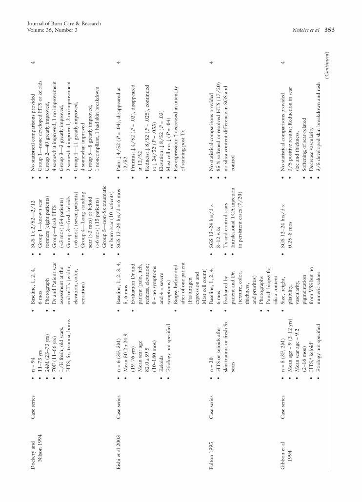
Doc
kery
and
N
ilson
199
4C
ase
seri
es•
n =
94•
11–7
3 yr
s•
24M
(23
–73
yrs)
•70
F (1
1–66
yrs
)•
L/
E fr
esh,
old
sca
rs,
HT
S, S
x, t
raum
a, b
urns
•B
asel
ine,
1, 2
, 4,
6 m
os•
Phot
ogra
ph•
Dr
and
Patie
nt s
car
asse
ssm
ent
at t
he
end
of T
x (w
idth
, el
evat
ion,
col
or,
sens
atio
n)
•SG
S T
x 2/
52—
2/12
•G
roup
1—
know
n sc
ar
form
ers
(eig
ht p
atie
nts)
•G
roup
—fr
esh
HT
S
(<3
mos
) (5
4 pa
tient
s)•
Gro
up 3
—fr
esh
kelo
ids
(<
6 m
os)
(sev
en p
atie
nts)
•G
roup
4—
Lon
g st
andi
ng
scar
(>3
mos
) or
kel
oid
(>
6 m
os)
(15
patie
nts)
•G
roup
5—
non-
Sx t
raum
atic
or
bur
n sc
ar (
10 p
atie
nts)
•N
o st
atist
ical
com
paris
ons
prov
ided
•G
roup
1—
none
dev
elop
ed H
TS
or k
eloi
ds•
Gro
up 2
—49
gre
atly
impr
oved
, 4
som
ewha
t im
prov
ed, 1
no
impr
ovem
ent
•G
roup
3—
3 gr
eatly
impr
oved
, 2
som
ewha
t im
prov
ed, 2
no
impr
ovem
ent
•G
roup
4—
11 g
reat
ly im
prov
ed,
4 so
mew
hat i
mpr
oved
•G
roup
5—
8 gr
eatly
impr
oved
, 1
nonc
ompl
iant
, 1 h
ad s
kin
brea
kdow
n
4
Eis
hi e
t al
200
3C
ase
seri
es•
n =
6 (3
F, 3
M)
•M
ean
50.2
± 2
4.9
(19–
76 y
rs)
•M
ean
scar
age
82
.0 ±
59.
5
(10–
180
mos
)•
Kel
oids
•E
tiolo
gy n
ot s
peci
fied
•B
asel
ine,
1, 2
, 3, 4
, 5,
6 m
os•
Eva
luat
ion
Dr
and
patie
nt (
pain
, itc
h,
redn
ess,
ele
vatio
n;
0 =
no s
ympt
oms
an
d 4
= se
vere
sy
mpt
oms)
•B
iops
y be
fore
and
af
ter
of o
ne p
atie
nt
(Fas
ant
igen
ex
pres
sion
and
M
ast
cell
coun
t)
•SG
S 12
–24
hrs/
d ×
6 m
os•
Pain
: ↓ 4
/52
(P
= .0
4), d
isap
pear
ed a
t 12
/52
•Pr
uritu
s: ↓
4/
52 (
P =
.02)
, dis
appe
ared
at
12/
52•
Red
ness
: ↓ 8
/52
(P
= .0
25),
con
tinue
d to
↓ 2
4/52
(P
= .0
33)
•E
leva
tion:
↓ 8
/52
(P
= .0
3)•
Mas
t ce
ll no
: ↓ (
P =
.04)
•Fa
s ex
pres
sion
: ↑ d
ecre
ased
in in
tens
ity
of s
tain
ing
post
Tx
4
Fulto
n 19
95C
ase
seri
es•
n =
20•
HT
S or
kel
oids
aft
er
skin
tra
uma
or fr
esh
Sx
scar
s
•B
asel
ine,
1, 2
, 4,
6 m
os•
Eva
luat
ed b
y pa
tient
and
Dr.
(tex
ture
, col
or,
thic
knes
s,
and
prur
itus)
•Ph
otog
raph
s•
Punc
h bi
opsy
for
silic
a co
nten
t
•SG
S 12
–24
hrs/
d ×
8–
12 w
ks•
Tx
and
cont
rol s
cars
•In
tral
esio
nal T
CA
inje
ctio
n in
per
sist
ent
case
s (7
/20
)
•N
o st
atis
tical
com
pari
sons
pro
vide
d•
85 %
sof
tene
d or
res
olve
d H
TS
(17/
20)
•no
sili
ca c
onte
nt d
iffer
ence
in S
GS
and
cont
rol
4
Gib
bon
et a
l 19
94C
ase
seri
es•
n =
5 (3
F, 2
M)
•M
ean
age
= 9
(2–1
2 yr
s)•
Mea
n sc
ar a
ge =
9.2
(2
–16
mos
)•
HT
S,4
kelo
id1
•E
tiolo
gy n
ot s
peci
fied
•Si
ze, h
eigh
t,
plia
bilit
y,
vasc
ular
ity,
pigm
enta
tion
fr
om V
SS b
ut n
o nu
mer
ic v
alue
s
•SG
S 12
–24
hrs/
d ×
0.
25–8
mos
•N
o st
atis
tical
com
pari
sons
pro
vide
d•
3/5
posi
tive
resu
lts: R
educ
tion
in s
car
size
and
thi
ckne
ss.
•So
ften
ing
of s
car-
rela
ted
•D
ecre
ase
vasc
ular
ity•
3/5
deve
lope
d sk
in b
reak
dow
n an
d ra
sh
4
(Con
tinu
ed)

Journal of Burn Care & Research354 Nedelec et al May/June 2015
Gol
d 19
93C
ase
seri
es•
n =
10 (
16 s
cars
)•
Sx a
nd t
raum
atic
•B
asel
ine,
1, 2
, 3
mos
•E
valu
ated
by
D
r. an
d pa
tient
(t
hick
ness
, co
lor,
over
all
effe
ctiv
enes
s;
0 no
cha
nge
and
3
com
plet
e re
solu
tion)
•SG
S >1
2 hr
s/d
×
12 w
ks•
No
stat
istic
al c
ompa
riso
ns p
rovi
ded
•Pa
tient
s re
port
ed m
oder
ate
impr
ovem
ent
in t
hick
ness
, col
or, o
vera
ll ef
fect
iven
ess
(81.
25%
, 75%
, 100
%)
•D
rs r
epor
ted
com
plet
e re
solu
tion
(6
.25%
, 6.2
5%, 6
.25%
) or
mod
erat
e ch
ange
(50
%, 6
8.75
%, 9
3.75
%)
in
thic
knes
s, c
olor
, ove
rall
effe
ctiv
enes
s
4
Gol
d 19
94C
ase
seri
es•
n =
34 (
n =
21 g
roup
1;
n =
8 gr
oup
2;
n =
5 gr
oup
3)•
Sx a
nd b
urn
•B
asel
ine,
1, 2
3
mos
•E
valu
ated
by
Dr.
and
patie
nt
(thi
ckne
ss,
colo
r, ov
eral
l ef
fect
iven
ess;
0
no c
hang
e an
d 3
com
plet
e re
solu
tion)
•SG
S >1
2 hr
s/d
× 12
wks
•G
roup
1—
17 H
TS,
4
kelo
ids
whe
re h
alf o
f the
sc
ar w
as T
x’d,
hal
f of t
he
scar
was
con
trol
•G
roup
2—
Sx s
ite w
as
trea
ted
afte
r re
mov
al o
f ke
loid
•G
roup
3—
HT
S fr
om b
urn
•N
o st
atis
tical
com
pari
sons
pro
vide
d•
Gro
up 1
—sc
ar t
hick
ness
mod
erat
e re
duct
ion:
Pat
ient
= 3
3.3%
, phy
sici
an =
47
.6%
; Col
or c
hang
e m
oder
ate:
Pa
tient
= 1
9.1%
, Dr
= 42
.9%
; Ove
rall
mod
erat
e ef
fect
iven
ess:
Pat
ient
= 2
3.8%
, D
r =
52.3
%•
Gro
up 2
—1
trea
ted
site
res
ulte
d in
re
curr
ent
kelo
id•
Gro
up 3
—4
of 5
min
imal
cha
nge;
on
e m
oder
ate
4
Gol
d et
al
2001
RC
T•
N =
96
•St
ratifi
ed in
to lo
w r
isk
(n =
50)
and
hig
h ri
sk
(n =
46)
•M
ean
38.5
yrs
(lo
w
risk
); 3
4.8
yrs
(hig
h ri
sk)
•R
emov
al o
f ski
n le
sion
s (b
enig
n an
d m
alig
nant
tu
mor
s, s
ebac
eous
cys
ts,
HT
S, k
eloi
ds)
•2,
4, 8
, 12,
16,
20,
24
/52
pos
t-Sx
•Ph
ysic
ian
obse
rvat
ion
•Pa
tient
’s o
pini
on
(dis
com
fort
, em
barr
assm
ent,
co
lor
heig
ht,
text
ure,
func
tion)
•Ph
otog
raph
ic
asse
ssm
ent
•N
o de
tails
pro
vide
d ab
out a
ny o
f the
sc
ales
•T
x gr
oup—
SGS
(12–
24
hrs/
d ap
plie
d 48
hrs
/po
st
surg
ery,
sut
ures
/st
aple
s re
mov
ed 7
–14
d)•
Con
trol
gro
up—
rout
ine
post
oper
ativ
e ca
re•
6/12
Tx
•L
ow r
isk
grou
p—no
diff
eren
ce b
etw
een
the
trea
tmen
t an
d co
ntro
l•
Hig
h ri
sk g
roup
—re
duct
ion
in
perc
enta
ge w
ho d
evel
oped
HT
S or
kel
oid
(P =
.072
) bu
t no
t st
atis
tical
ly s
igni
fican
t•
Indi
vidu
als
unde
rgoi
ng s
car
revi
sion
su
rger
y—In
crea
sed
rate
of p
reve
ntio
n
(P =
.035
) (n
= 4
Tx
grou
p vs
n =
10
co
ntro
l)
2
Tab
le 2
. (C
onti
nued
)
Aut
hors
Des
ign
Sam
ple
Out
com
e
Mea
sure
sIn
terv
enti
onR
esul
tsL
evel
of
Evi
denc
e

Journal of Burn Care & Research Volume 36, Number 3 Nedelec et al 355
Ham
anov
a
and
Bro
z
2002
Cas
e se
ries
•n
= 60
(80
site
s) (
24F,
36
M)
•18
Adu
lts, 4
2 ch
ildre
n•
Mea
n ag
e 28
.5 (5
–55
yrs)
•10
Pat
ient
s with
old
, m
atur
e H
TS,
50
patie
nts
with
imm
atur
e sc
ar•
Bur
ns
•B
asel
ine
1, 2
, 4,
6 m
os•
Hei
ght,
wei
ght,
co
lor
of s
car
•Pa
tient
sub
ject
ive
repo
rt
•SG
S T
x up
to
2 yr
s•
>20
hrs/
d w
inte
r,
>12
hrs/
d su
mm
er•
Min
imum
siz
e of
sc
ar: 1
0 ×
40 m
m,
3 m
m h
eigh
t
•N
o st
atis
tical
com
pari
sons
pro
vide
d•
Posi
tive
effe
ct in
red
uctio
n im
mat
ure
HT
S bu
t no
t in
mat
ure
scar
(10
pat
ient
s)•
40%
of t
he p
atie
nts
repo
rted
impr
ovem
ent
in a
ll th
e te
sted
cha
ract
erist
ics
•10
% o
f the
sub
ject
s de
velo
ped
a sk
in
reac
tion
4
Har
te e
t al
20
09R
CT
•n
= 22
(9F
, 13M
)•
Mea
n ag
e =
36.8
(1
6–64
yrs
)•
Mea
n T
BSA
15
(1–5
5%)
•14
U/
E, 8
L/
E•
<6 m
os p
ostin
jury
•H
TS
post
burn
inju
ry
•B
asel
ine,
12
and
24
wks
•V
SS•
Patie
nt d
iary
•G
roup
A—
pres
sure
the
rapy
•G
roup
B—
pres
sure
the
rapy
+
SGS
•23
hrs
/d,
rep
lace
eve
ry 7
d
•O
vera
ll re
duct
ion
of V
SS s
core
s fo
r bo
th
grou
ps a
t 24
wks
•N
o di
ffer
ence
bet
wee
n gr
oup
A a
nd B
at
12
or 2
4 w
ks•
Pow
er a
naly
sis
dete
rmin
ed 1
92
part
icip
ants
per
gro
up w
ould
be
requ
ired
, the
refo
re r
esul
ts a
re u
nder
po
wer
ed
2
Hir
show
itz
et a
l 199
3C
ase
seri
es•
N =
32
•M
ean
age
= 27
.3
(5–5
1 yr
s)•
Mea
n sc
ar a
ge 2
3 (1
0–60
mos
)•
Post
-Sx,
21 p
ostb
urn,
13
post
tra
uma6
•1
m, e
very
2–3
m
os fo
r 12
mos
.•
Scar
app
eara
nce:
ha
rdne
ss, e
leva
tion,
co
lor
•Pa
in, i
tch,
di
scom
fort
su
bjec
tive
•D
igita
l pho
togr
aphs
•SG
S•
No
stat
istic
al c
ompa
riso
ns p
rovi
ded
•12
.5%
No
impr
ovem
ent
•15
.6 %
Slig
ht im
prov
emen
t•
37.5
% M
oder
ate
impr
ovem
ent
•34
.4%
Exc
elle
nt im
prov
emen
t
4
Hir
show
itz
et a
l 199
8C
ase
seri
es•
n =
30•
Scar
age
> 4
mos
•Sx
sca
rs a
nd b
urn
•H
ardn
ess,
el
evat
ion,
col
or,
pain
, itc
h,
disc
omfo
rt
•0.
75 m
m th
ick
silic
one
cush
ion
fille
d w
ith si
licon
e oi
l•
12 m
os T
x•
33.3
% h
ad in
tral
esio
nal
cort
icos
tero
id in
ject
ions
•N
o st
atis
tical
com
pari
sons
pro
vide
d•
63.3
% A
llevi
atio
n of
sym
ptom
s at
6 m
os•
33.3
% h
ad r
ecal
citr
ant
scar
s
4
(Con
tinu
ed)

Journal of Burn Care & Research356 Nedelec et al May/June 2015
Hos
nute
r
et a
l 200
7N
on ra
ndom
i zed
co
ntro
lled
coho
rt
•n
= 60
(36
F, 2
4M)
•H
TS39
and
kel
oids
21
diff
eren
t•
Etio
logy
not
spe
cifie
d•
Mea
n ag
e 40
.3 ±
9.6
(2
2–61
yrs
)•
Scar
age
ran
ge 1
–6 m
os
•M
onth
ly fo
r 6
mos
•C
olor
, hei
ght,
itc
h, p
ain
(0
= n
one,
1 =
mild
, 2
= pr
onou
nced
)•
Har
dnes
s
(0 =
nor
mal
, 1
= m
ild,
2 =
mod
erat
e,
3 =
pron
ounc
ed)
•T
hera
peut
ic in
dex
(com
plet
e he
alin
g,
clea
r im
prov
emen
t,
mod
erat
e, p
oor)
•G
roup
1—
topi
cal o
nion
ex
trac
t (n
= 2
1), 4
x/d
•G
roup
2—
SGS
(n =
19)
, 24
hr/
d•
Gro
up 3
—SG
S (2
4 hr
s/d)
+
onio
n ex
trac
t (2
x/d)
(n
= 2
0)•
6 m
os T
x
•Im
prov
emen
t w
ith t
ime:
Gro
up
1—co
lor,
hard
ness
P <
.001
, pai
n
P <
.05;
Gro
up 2
—co
lor,
hard
ness
, itc
h
P <
.01,
hei
ght
P <
.001
; Gro
up
3—co
lor,
heig
ht, h
ardn
ess
P <
.001
, itc
h P
< .0
1•
Mor
e co
lor
redu
ctio
n in
gro
up 1
vs
grou
p 2
(P <
.01)
•M
ore
heig
ht r
educ
tion
in g
roup
3 v
s gr
oup
1 (P
< .0
5)•
No
sign
ifica
nt d
iffer
ence
in
impr
ovem
ents
in h
ardn
ess,
itch
, pai
n be
twee
n th
e gr
oups
•Fi
ve w
ithdr
awal
s du
e to
itch
ing
3
Kar
agoz
et
al 2
009
RC
T•
n =
32 (
45 s
cars
)
(20F
, 12M
)•
Mea
n ag
e 24
(3–
55 y
rs)
•Sc
ar a
ge <
6 m
os•
HT
S po
stbu
rn in
jury
•B
asel
ine
and
6
mos
•V
SS•
Phot
ogra
ph
•G
roup
1—
silic
one
gel,
2x/
d•
Gro
up 2
—SG
S 24
hrs
/d
•G
roup
3—
onio
n ex
trac
t,
2x/
d•
6 m
os T
x
•B
efor
e an
d af
ter
Tx
diff
eren
ces
for
each
gr
oup
(P <
.05)
•Si
gnifi
cant
ly b
ette
r im
prov
emen
t in
gr
oup
1 vs
3 a
nd 2
vs
3 (P
< .0
5)•
No
diff
eren
ce b
etw
een
grou
p 1
and
2•
Skin
mac
erat
ion:
tw
o pa
tient
s in
gro
up 2
2
Kat
z 19
95C
ase
seri
es•
n =
34 (
36 c
hron
ic
scar
s >3
mos
)•
n =
14 (
14 fr
esh
scar
s <3
mos
)
•B
asel
ine,
2, 6
mos
•D
r an
d pa
tient
as
sess
ed r
edne
ss,
elev
atio
n ba
sed
on
phot
ogra
phs
•SG
S >1
2 hr
s/d
× 2
mos
•N
o st
atis
tical
com
pari
sons
pro
vide
d•
20/
36 C
hron
ic s
cars
impr
oved
aft
er T
x•
11/
14 F
resh
sca
r—no
HT
S de
velo
ped
afte
r 6
mos
•D
erm
atiti
s oc
curr
ed w
ith t
hree
pat
ient
s
4
Klo
pp e
t al
20
00In
trai
ndiv
idua
lR
CT
•n
= 12
•L
/E
sca
rs a
fter
rem
oval
of
vei
ns fo
r ca
rdia
c su
rger
y•
Scar
s 2.
5–4
yrs
•Si
tes
rand
omly
ass
igne
d
•V
enul
ar fl
ow r
ate
•M
icro
vess
el le
ngth
•N
o. o
f blo
od
cell-
perf
used
nod
al
poin
ts in
a d
efine
d tis
sue
volu
me
•Sk
in t
empe
ratu
re•
Surf
ace
qual
ity
•G
roup
1—
NSG
S (p
olyu
reth
ane)
alo
ne•
Gro
up 2
—N
SGS
(pol
yure
than
e) +
pre
ssur
e•
Gro
up 3
—pr
essu
re a
lone
•G
roup
4—
SGS
+ pr
essu
re•
Pres
sure
adm
inis
tere
d us
ing
stre
tch
band
ages
•15
mm
bet
wee
n T
x si
tes
•8
wks
Tx
•R
epor
ted
sign
ifica
nt c
hang
es fo
r al
l m
easu
rem
ent
liste
d fo
r al
l gro
ups
but
no P
val
ues
wer
e pr
ovid
ed (
dec
reas
e in
ves
sel l
engt
h an
d ve
nula
r flo
w r
ate;
de
crea
se in
the
no.
of a
ggre
gate
d er
ythr
ocyt
es; i
ncre
ase
in t
he s
kin
tem
pera
ture
; dec
reas
e in
sur
face
ro
ughn
ess)
2
Tab
le 2
. (C
onti
nued
)
Aut
hors
Des
ign
Sam
ple
Out
com
e
Mea
sure
sIn
terv
enti
onR
esul
tsL
evel
of
Evi
denc
e

Journal of Burn Care & Research Volume 36, Number 3 Nedelec et al 357
Lac
arru
bba
20
08C
ase
seri
es•
n =
8 (6
F, 2
M)
•M
ean
age
29
(12–
49 y
rs)
•<9
0 d
post
trau
ma
or
surg
ery
•B
asel
ine,
1, 2
, 3
mos
•Sc
ar t
hick
ness
by
ultr
asou
nd•
Clin
ical
eva
luat
ion
of s
ize,
red
ness
and
te
xtur
e
•Si
licon
e ge
l app
lied
2x/
d•
6 m
os T
x•
Thr
ee p
atie
nts
with
con
trol
sc
ars
•N
o st
atis
tical
com
pari
sons
pro
vide
d•
Mea
n sc
ar t
hick
ness
red
uced
by
37%
(4
mm
bef
ore
Tx
[3.4
–6.1
]; 2
.5 m
m a
fter
[2
.1–3
.2])
•T
wo
patie
nts
c/o
itch
4
Lee
et
al
1996
Cas
e se
ries
•n
= 26
par
ticip
ants
(1
5F, 1
1M)
with
45
scar
s•
Mea
n ag
e 26
(3–
52 y
rs)
•Sc
ars
<6/
12 o
ld•
Bur
ns, o
pera
tive
scar
s,
tatt
oo s
cars
, kel
oids
•B
asel
ine,
mon
thly
fo
r 6
mos
•3
poin
t sc
ale
(0 =
wor
se, 1
=
rem
aine
d th
e sa
me,
2
= im
prov
ed)
for
colo
r, te
xtur
e,
thic
knes
s, a
nd
regu
lari
ty
•G
roup
1—
SGS
(Sil-
K)
•G
roup
2—
SGS
epid
erm
•H
eld
in p
lace
with
adh
esiv
e st
rip,
tub
igri
p or
ban
dage
•6/
12 T
x
•N
o st
atis
tical
com
pari
sons
pro
vide
d•
Impr
ovem
ent
repo
rted
in b
oth
the
grou
ps (
90%
impr
ovem
ent
in c
olor
and
te
xtur
e, 8
0% im
prov
emen
t in
reg
ular
ity,
50%
impr
ovem
ent
in t
hick
ness
)•
Som
e pa
tient
s co
mpl
aine
d of
itch
and
m
acer
atio
n
4
Li-
Tsa
ng e
t al
200
6R
CT
•n
= 45
•M
ean
29.6
5 ±
17.6
yrs
•C
hine
se s
ubje
cts
with
sc
ar >
3 m
m t
hick
•B
urns
, tra
uma
•B
asel
ine,
1, 2
, 4,
6 m
os•
Spec
troc
olor
imet
er
(col
or)
•T
issu
e ul
tras
ound
pa
lpat
ion
syst
em
(thi
ckne
ss)
•V
SS (
plia
bilit
y)•
Pain
(V
AS)
•It
ch (
VA
S)
•T
x gr
oup—
SGS
(24
hrs/
d) +
15
min
mas
sage
2x
/d
with
lano
linC
ontr
ol
grou
p—15
min
mas
sage
2x
/d
with
lano
lin6
mos
Tx
• D
ecre
ased
thi
ckne
ss o
ver
6/12
in T
x gr
oup
rela
tive
to c
ontr
ol (
P <
0.01
)•
Inc
reas
ed p
liabi
lity
over
6/
12 in
Tx
grou
p re
lativ
e to
con
trol
(P
< 0.
01)
•N
o st
atis
tical
ly s
igni
fican
t di
ffer
ence
in
itch,
pai
n, o
r co
lor
2
Li-
Tsa
ng e
t al
201
0R
CT
•n
= 10
4 (8
4 co
mpl
eted
al
l ass
essm
ent)
•M
ean
age
21.8
± 1
8.7
•M
ean
scar
age
14
.9 ±
30.
8 m
os•
Bur
ns, t
raum
a
•B
asel
ine
2, 4
, 6
mos
•Sp
ectr
ocol
orim
eter
(c
olor
)•
Tis
sue
ultr
asou
nd
palp
atio
n sy
stem
(t
hick
ness
)•
VSS
(pl
iabi
lity)
•Pa
in (
VA
S)•
Itch
(V
AS)
•Pa
tient
sat
isfa
ctio
n (i
nter
view
)
•G
roup
1—
pres
sure
ga
rmen
ts (
24 h
r/d
exce
pt
hygi
ene)
•G
roup
2—
SGS
•G
roup
3—
pres
sure
ga
rmen
ts +
SG
S•
Gro
up 4
—co
ntro
l•
6 m
os T
x•
Pres
sure
gar
men
ts a
nd S
GS
wor
n 24
hrs
/d
exce
pt fo
r hy
gien
e
•T
hick
ness
: gro
up 3
impr
oved
at
2/12
, 4/
12, a
nd 6
/12
and
gro
up 1
at
6/12
(P
< .0
01)
rela
tive
to c
ontr
ol b
ut n
o di
ffer
ence
bet
wee
n T
x gr
oups
(P
= .0
66)
•Pl
iabi
lity:
at
6/12
all
grou
ps im
prov
ed
(P <
.001
); g
roup
3 w
as s
igni
fican
tly
mor
e pl
iabl
e th
an c
ontr
ol a
t 2/
12
(P =
.002
) an
d 4/
12 (
P <
.000
1)•
Pigm
enta
tion:
at
6/12
all
grou
ps w
ere
light
er a
nd m
ore
yello
w (
P <
.001
)•
Pain
: gro
up 2
(P
= .0
01)
and
grou
p 3
(P
= .0
04)
redu
ced
rela
tive
to c
ontr
ol•
Itch
: aut
hors
rep
ort
redu
ced
itch
for
all
Tx
grou
ps b
ut d
ata
indi
cate
s an
incr
ease
fo
r gr
oup
1 fr
om b
asel
ine
to 6
/12
2
(Con
tinu
ed)

Journal of Burn Care & Research358 Nedelec et al May/June 2015
Maj
an 2
006
RC
T•
n =
11 (
11F)
•C
ontr
ol g
roup
mea
n ag
e 28
.8 (
20––
43 y
rs)
•T
x gr
oup
mea
n ag
e 29
.4 (
22–4
0 yr
s)•
Post
-Sx
scar
s
•B
asel
ine,
mon
thly
un
til 6
mos
and
12
mos
•V
SS•
Dr
and
patie
nt
over
all a
sses
smen
t at
base
line
and
12 m
os
•T
x gr
oup—
SGS
•C
ontr
ol—
no t
reat
men
t•
Wea
r up
to
23 h
rs/
d•
Tx
initi
ated
2/
52—
2/12
po
st-S
x an
d co
ntin
ued
for
12 m
os
•N
o st
atis
tical
com
pari
sons
pro
vide
d•
All
scar
s im
prov
ed (
VSS
)•
Ove
rall
asse
ssm
ent:
Dr
rate
d 5/
5 in
Tx
grou
p as
ver
y go
od o
r go
od, p
atie
nts
rate
d 4/
5 ve
ry g
ood
or g
ood
•O
ne p
atie
nt e
xper
ienc
ed s
kin
reac
tion
2
Mer
cer
1989
Cas
e se
ries
•n
= 18
(7F
, 11M
)
(22
kelo
ids)
•M
ean
age
19 (
3–64
yrs
)•
Mea
n sc
ar a
ge 3
5 (9
–120
mos
)
•B
asel
ine,
1, 2
–3,
6 m
os•
Hei
ght,
col
or,
text
ure
•Pr
ogre
ssiv
ely
incr
ease
d w
ear
time
of S
GS
to 2
4 hr
s/d
•N
o st
atis
tical
com
pari
sons
pro
vide
d•
86%
Sca
rs p
ositi
vely
res
pond
ed t
o SG
S (t
extu
re 8
6%, c
olor
84%
, hei
ght
68%
)•
3 Sc
ars
(14%
) sh
owed
no
resp
onse
to
TX
•T
hree
Pat
ient
s ex
peri
ence
d sk
in r
eact
ion
4
Mom
eni e
t al
20
09In
trai
ndiv
idua
l R
CT
•n
= 38
(18
F, 1
6M)
•M
ean
age
22
(1.5
–60
yrs)
•H
TS
post
burn
inju
ry
•V
SS•
Bas
elin
e, 1
, 4 m
os•
Pigm
enta
tion,
va
scul
arity
, pl
iabi
lity,
pai
n, it
ch
•H
TS
divi
ded
into
2
side
s—si
de 1
= S
GS;
sid
e 2
= N
SGS
(pro
pyle
ne g
lyco
l an
d hy
drox
yeth
yl c
ellu
lose
sh
eetin
g)(r
efer
red
to a
s pl
aceb
o co
ntro
l)•
Tx
initi
ated
2–4
mo
post
inju
ry•
Wor
n 24
hrs
/d
•Si
de 1
—at
4/
12 a
ll sc
ores
exc
ept
pain
w
ere
sign
ifica
ntly
low
er in
the
SG
S gr
oup
than
the
NSG
S (P
< .0
5)•
Side
2—
No
sign
ifica
nt c
hang
e at
1/
12
or 4
/12
2
Mur
ison
and
Ja
mes
200
6C
ase
seri
es•
n =
6•
Age
ran
ge 3
0–58
yrs
•Sc
ar a
ge 3
–6 m
os t
o 4–
5 yr
s•
Etio
logy
not
spe
cifie
d
•D
igita
l pho
togr
aphs
•M
odifi
ed V
SS:
redn
ess,
ele
vatio
n,
hard
ness
, itc
h,
tend
erne
ss, p
ain
•Sp
ectr
opho
tom
etric
in
trac
utan
eous
sc
ope
(SIS
)
•SG
2x/
d x
8/12
•N
o st
atis
tical
com
pari
sons
pro
vide
d•
Mod
ified
VSS
Im
prov
ed fo
r al
l sub
ject
s•
SIS—
7.2%
dec
reas
e in
col
lage
n, 3
%
incr
ease
in b
lood
flow
4
Tab
le 2
. (C
onti
nued
)
Aut
hors
Des
ign
Sam
ple
Out
com
e
Mea
sure
sIn
terv
enti
onR
esul
tsL
evel
of
Evi
denc
e

Journal of Burn Care & Research Volume 36, Number 3 Nedelec et al 359
Nie
ssen
et
al
1998
RC
T•
n =
155
(all
F w
omen
)•
Mea
n ag
e 31
(1
4–69
yrs
)•
Bre
ast
redu
ctio
n Sx
sc
ars
•B
asel
ine,
1, 2
, 4,
6 m
os•
Thi
ckne
ss
(ultr
asou
nd)
•B
lood
flow
(la
ser
dopp
ler)
•C
olor
(c
hrom
amet
er)
•Pa
in (
VA
S)•
Itch
(V
AS)
•Pu
nch
biop
sy, r
uler
•G
roup
1—
SGS
(Sil-
K)
(n
= 8
0)•
Gro
up 2
—SG
S (e
pide
rm)
(n =
75)
•G
roup
1 a
nd 2
wer
e ra
ndom
ly a
ssig
ned
to le
ft
late
ral a
nd ri
ght m
edia
l or
left
med
ial a
nd ri
ght l
ater
al
with
the
rem
aind
er a
s con
trol
•T
x in
itiat
ed 3
d
post
oper
ativ
ely,
24
hrs/
d ×
3 m
os•
SGS
appl
ied
with
str
etch
te
nsio
n an
d fix
ed w
ith
mic
ropo
re•
Con
trol
—m
icro
pore
onl
y
•11
9 pa
rtic
ipan
ts c
ompl
eted
the
stu
dy•
Dev
elop
ed H
TS:
64.
3% a
t 3
mos
, 56.
6%
at 6
mos
, 35.
3% a
t 12
mos
•O
ccur
renc
e of
HT
S in
crea
ses
with
eas
e of
ta
nnin
g (P
< .0
2)•
No
diff
eren
ce b
etw
een
2 si
licon
e T
x’d
site
s•
incr
ease
d H
TS
at T
x si
tes
rela
tive
to
cont
rol:
at 6
mos
(P
= .0
06);
at
12 m
os
(P =
.02)
•Se
vera
l pat
ient
s de
velo
ped
skin
irri
tatio
n
2
Ohm
ori 1
988
Cas
e se
ries
•n
= 46
(48
sca
rs)
•K
eloi
ds•
Bur
ns, a
cne,
tra
uma
•Su
bjec
tive
scal
e•
Red
ness
, ele
vatio
n,
subj
ectiv
e co
mpl
aint
s (it
ch, p
ain)
•1
mm
SG
S•
8–12
hrs
/d
•N
o st
atis
tical
com
pari
sons
pro
vide
d•
Exc
elle
nt =
6; G
ood
= 24
; Fai
r =
12;
Poor
= 6
4
Palm
ieri
et
al
1995
RC
T•
n =
80•
18–6
3 yr
s•
Scar
age
: 3 m
os–2
yrs
po
stin
jury
•H
TS
or k
eloi
ds
post
trau
ma70
or
burn
s10
•E
valu
atio
n at
4
and
8 w
ks•
Phot
ogra
phs f
or
colo
r, siz
e an
d co
smet
ic a
ppea
ranc
e•
Itch
, pai
n
•G
roup
A—
SGS
+ vi
tam
in E
•G
roup
B—
SGS
•W
ore
betw
een
10 p
m &
8
am
•A
t 4/
12 g
roup
A 8
5% im
prov
ed b
y m
ore
than
50%
and
gro
up B
55%
(P
< .0
1)•
At 8
/12
gro
up A
95%
impr
ovem
ent b
y m
ore
than
50%
and
Gro
up B
75%
(P
< .0
5)
2
(Con
tinu
ed)

Journal of Burn Care & Research360 Nedelec et al May/June 2015
Pere
z et
al
2010
RC
T•
n =
30•
HT
S or
kel
oids
•E
tiolo
gy n
ot s
peci
fied
•B
asel
ine,
4, 8
, 12
, 16
wks
sca
r vo
lum
e•
Inve
stig
ator
s ev
alua
tion
of
indu
ratio
n,
eryt
hem
a,
pigm
enta
tion,
pa
in, i
tch,
cos
met
ic
asse
ssm
ent
•Pa
tient
sat
isfa
ctio
n•
Spec
trom
etri
c in
trac
utan
eous
sc
ope
•G
roup
1—
TC
A +
SG
S +
vita
min
E (
2x/
d)•
Gro
up 2
—on
ion
extr
act
gel
(3–4
x/d)
•G
roup
3—
plac
ebo
cont
rol
(2x/
d)•
Tx
peri
od 1
6/52
•n
= 15
pat
ient
s co
mpl
eted
stu
dy•
At
16/
52 r
elat
ive
to b
asel
ine:
gro
up 1
im
prov
ed o
n vo
lum
e (P
= .0
1), l
engt
h
(P =
.02)
, ind
urat
ion
(P <
.01)
, ery
them
a (<
.01)
, pig
men
tatio
n, (
P <
.01)
, in
vest
igat
or e
valu
atio
n (P
< .0
1), p
atie
nt
satis
fact
ion
(P =
.04)
; gro
up 2
vol
ume
(P
= .0
1), l
engt
h (P
= .0
2), w
idth
(P =
.02)
, in
dura
tion
(P =
.03)
; gro
up 3
vol
ume
(P
= .0
2), p
atie
nt sa
tisfa
ctio
n (P
= .0
1)•
Sign
ifica
nt im
prov
emen
t: gr
oup
1 vs
3—
in
vest
igat
or c
osm
etic
eva
luat
ion
(P <
.01)
, in
dura
tion
(P <
.001
), p
igm
enta
tion
(P <
.0
01),
ery
them
a (P
= .0
1); g
roup
2 v
s 3:
in
vest
igat
or c
osm
etic
eva
luat
ion
(P <
.01)
, in
dura
tion
(P <
.001
), p
igm
enta
tion
(P
< .0
01),
tend
erne
ss (
P <
.05)
•O
vera
ll sa
tisfa
ctio
n: g
roup
1 a
nd 2
>7
6/10
0
2
Perk
ins
et a
l 19
83C
ase
seri
es•
n =
42•
Age
ran
ge 4
mos
–16
yrs
•N
ewly
hea
led
to m
atur
e (1
2 yr
s) b
urn
scar
s
•Sc
ar im
prov
emen
t,
pain
•SG
S (s
penc
o)•
20/
42 p
atie
nts
with
pr
essu
re t
hera
py
•N
o ob
ject
ive
resu
lts o
r st
atis
tical
co
mpa
riso
ns p
rovi
ded
•Su
bjec
tive
clai
ms
of b
enefi
ts
5
Phill
ips
et a
l 19
96R
CT
•n
= 20
•H
TS
and
kelo
ids
•M
ean
age
= 33
.3 y
rs•
Mea
n sc
ar a
ge 8
.5–9
yrs
•E
tiolo
gy n
ot s
peci
fied
•B
asel
ine,
2,
4, 8
wks
, and
1
mo
post
Tx
term
inat
ion
•V
SS•
Scar
vol
ume
•Ph
otog
raph
s•
Tra
nscu
tane
ous
oxyg
en m
easu
res
•G
roup
1—
NSG
S (h
ydro
collo
id)
for
7 d,
re
plac
ed if
it b
ecam
e di
slod
ged
•G
roup
2—
cont
rol
moi
stur
izer
1x/
d•
8 m
os T
x
•N
o di
ffer
ence
bet
wee
n T
x gr
oups
•Si
gnifi
cant
red
uctio
n in
itch
(P
< .0
3) in
bo
th t
he g
roup
s w
ith t
ime
•A
dver
se s
kin
reac
tions
in 4
pat
ient
s
2
Puri
and
T
alw
ar 2
009
Cas
e se
ries
•n
= 30
(2:
1 m
ale:
fem
ale
ratio
)•
Age
ran
ge 5
–60
yrs
•H
TS
and
kelo
id•
Scar
age
ran
ge:
<1/
12–>
6/12
•E
tiolo
gy n
ot s
peci
fied
•E
valu
atio
n 1x
/12
× 6
mos
•G
rade
d: I
=
norm
al, I
I =
mild
ly
hype
rtro
phic
, II
I =
hype
rtro
phic
, IV
= k
eloi
d
•SG
2x/
d x
0.25
–8 m
os•
No
stat
istic
al c
ompa
riso
ns p
rovi
ded
•Im
prov
emen
t af
ter
Tx:
No.
of c
ases
in
crea
sed
from
0–6
0% (
grad
e I)
and
de
crea
sed
26.6
–20%
(gr
ade
II),
50–
10
(gra
de I
II),
23.
3–10
% (
grad
e IV
)
4
Tab
le 2
. (C
onti
nued
)
Aut
hors
Des
ign
Sam
ple
Out
com
e
Mea
sure
sIn
terv
enti
onR
esul
tsL
evel
of
Evi
denc
e

Journal of Burn Care & Research Volume 36, Number 3 Nedelec et al 361
Qui
nn e
t al
19
85C
ase
seri
es•
n =
40•
1–67
yrs
•Sc
ar a
ge:1
–144
mos
•H
TS
and
kelo
ids
•E
tiolo
gy n
ot s
peci
fied
but
pres
umed
bur
n in
jury
•E
valu
ated
sca
r qu
aliti
es t
extu
re,
colo
r, el
evat
ion
•SG
S w
ith b
anda
ge, a
dhes
ive
tape
, sili
cone
bas
e ad
hesi
ve
+ pr
essu
re g
arm
ent
•N
o st
atis
tical
com
pari
sons
pro
vide
d•
7/40
Im
prov
ed o
n 3
scar
qua
litie
s, 1
8/40
on
2 a
nd 1
5/40
on
1 af
ter
2 m
os T
x•
Mod
e of
act
ion
of S
GS
does
not
invo
lve
pres
sure
, tem
pera
ture
, oxy
gen
tens
ion
or
occl
usio
n
4
Qui
nn 1
987
Cas
e se
ries
•n
= 12
5 (1
29 s
cars
)•
Etio
logy
not
spe
cifie
d bu
t pr
esum
ed b
urn
inju
ry
•E
valu
ated
sca
r qu
aliti
es t
extu
re
(ext
enso
met
er),
co
lor,
elev
atio
n
•SG
S he
ld in
pla
ce b
y di
ffer
ent
met
hods
•12
–24
hrs/
d x
2 m
os
•10
/12
9 Im
prov
ed o
n 3
scar
qua
litie
s,
28/
129
on 2
and
37/
129
on 1
aft
er 2
m
os T
x•
14 D
evel
oped
a r
ash,
3 r
esul
ted
in t
issu
e br
eakd
own,
37
wer
e lo
st t
o fo
llow
up
•SG
S ha
s w
ater
vap
or t
rans
mis
sion
rat
e lo
wer
tha
n no
rmal
ski
n
4
Rhe
e et
al
2010
RC
T•
n =
40•
Mea
n ag
e =
31.7
(1
6–51
yrs
)•
Asi
an p
artic
ipan
ts a
fter
sc
ar r
evis
ion
or m
ass
exci
sion
•E
valu
atio
n at
2/
52, 1
/12
, and
3/
12•
Pigm
enta
tion,
va
scul
arity
, hei
ght
usin
g a
phot
ogra
ph
•G
roup
1—
SGS
x 12
hrs
/d
x 3
mos
•G
roup
2—
cont
rol
•N
o di
ffer
ence
s at
2/
52•
1/12
SG
S sh
owed
dec
reas
e in
hei
ght
(P
< .0
024)
•3/
12, S
GS
show
ed d
ecre
ase
in
pigm
enta
tion
(P <
.000
2), v
ascu
lari
ty
(P <
.000
2) a
nd h
eigh
t (P
< .0
0001
)•
2 pa
tient
s de
velo
ped
skin
ras
h
2
Saku
raba
et
al
2011
Cas
e se
ries
•n
= 9
(5F,
4M
)•
Mea
n ag
e =
41.1
(1
8–67
yrs
)•
Etio
logy
not
spe
cifie
d
•Pr
otub
eran
ce,
redn
ess,
rub
bing
, cr
ampi
ng, a
nd
itchi
ng r
ated
on
a sc
ale
of
0 =
negl
igib
le–4
=
seve
re
•SG
S•
24 h
rs/
d, 2
wks
pos
t-op
for
24 w
ks
•R
ubbi
ng (
P =
.035
), c
ram
ping
(P
= .0
02)
and
itchi
ng (
P =
.021
) im
prov
ed4
Scud
eri e
t al
20
10R
CT
•n
= 15
0 (8
9F, 6
1M)
•M
ean
age
= 32
(18
–52
yrs)
•H
TS
age
= 6–
24 m
os•
Sx a
nd t
raum
atic
sca
r
•B
asel
ine,
1, 3
, 6,
12 m
os•
Glo
bal s
car
asse
ssm
ent
•C
olor
(pho
togr
aph)
, ov
eral
l sof
teni
ng,
wid
th (
rule
r),
leng
th (
calip
er),
el
evat
ion
(opt
ical
pr
ofilo
met
ry)
•V
AS—
itch,
col
or,
plia
bilit
y, t
hick
ness
, re
lief
•G
roup
A—
(n
= 10
7)
(asy
mm
etri
cal)
: NSG
(c
yano
acry
late
s)•
Gro
up B
— (
n =
40)
(lin
ear)
: hal
f SG
, 2x/
d; h
alf
NSG
(cy
anoa
cryl
ates
) (3
la
yers
eve
ry 3
–5 d
)•
Tx
3 m
os
•G
roup
A—
89.7
% im
prov
ed o
n gl
obal
sc
ar a
sses
smen
t•
Gro
up B
—Sc
ar e
leva
tion
redu
ced
in
both
the
gro
ups
at 1
/12
(SG
P <
.000
1)
(NSG
P <
.000
1) a
nd 1
yr
(SG
P <
.0
001)
(N
SG P
< .0
001)
•O
ne p
atie
nt d
evel
oped
ski
n re
actio
n
2
(Con
tinu
ed)

Journal of Burn Care & Research362 Nedelec et al May/June 2015
Scud
eri e
t al
201
1In
tra in
divi
dual
R
CT
•n
= 85
•M
ean
age
32
(18–
52 y
rs)
•B
ilate
ral m
amm
opla
sty
•B
asel
ine,
1, 3
, 6,
12 m
os•
Scar
wid
th (
rule
r),
leng
th (
calip
er),
el
evat
ion
(opt
ical
pr
ofilo
met
ry),
te
xtur
e•
VA
S: it
ch, c
olor
, pl
iabi
lity,
thi
ckne
ss,
relie
f
•O
ne b
reas
t T
x’d
with
NSG
(c
yano
acry
late
) ev
ery
3–5
d fo
r 3m
os•
One
bre
ast
Tx’
d w
ith S
G,
2x/
d x
3/12
•E
leva
tion:
incr
ease
d be
twee
n ba
selin
e an
d 1/
12 (
NSG
and
SG
P <
.000
1);
decr
ease
d be
twee
n ba
selin
e an
d 1
yr
(NSG
P =
.018
2; S
G P
= .0
174)
•Sc
ar w
idth
: inc
reas
ed b
etw
een
base
line
and
1 yr
(N
SG a
nd S
G P
< .0
001)
; in
crea
sed
in w
idth
sig
nific
antly
less
with
N
SG T
x (P
< .0
001)
•3
patie
nts
expe
rien
ced
skin
rea
ctio
n to
N
SG
2
Sign
orin
i and
C
lem
ento
nit
2007
RC
T•
n =
148
•M
ean
age
= 53
.5
(5–8
2 yr
s)•
Scar
age
: 10
d–3
wks
•R
ecen
t Sx
sca
r
•B
asel
ine,
1, 2
, 3, 4
, an
d 6
mos
•O
vera
ll ev
alua
tion
(Gra
de 1
=
norm
al, 2
= m
ildly
hy
pert
roph
ic,
3 =
hype
rtro
phic
, 4
= ke
loid
)
•T
x gr
oup:
SG
(2x
/d
x 4
mos
)•
Con
trol
gro
up
•G
rade
of T
x vs
con
trol
sca
rs a
t 6
mos
: 1
= 67
vs
28%
; 2 =
26
vs 4
6%; 3
= 6
vs
0%; 4
= 1
vs
26%
whi
ch w
as s
igni
fican
tly
diff
eren
t (P
< .0
01)
2
Spen
cer
2010
Cas
e se
ries
•n
= 7
•Sx
exc
isio
n—sk
in c
ance
r•
Bas
elin
e, 1
, 3 m
os•
VSS
and
Dr’
s ev
alua
tions
•M
exam
eter
for
eryt
hem
a an
d pi
gmen
tatio
n
•L
inea
r sc
ar d
ivid
ed in
to
two:
hal
f with
SG
, hal
f co
ntro
l•
SG 2
x/d
for
3 m
os
•N
o st
atis
tical
com
pari
sons
pro
vide
d•
No
diff
eren
ce in
ery
them
a an
d pi
gmen
tatio
n be
twee
n th
e gr
oups
•5/
7 SG
gro
up w
ere
bett
er t
han
cont
rol
as a
sses
sed
by p
hysi
cian
•4/
7 SG
gro
up w
ere
bett
er t
han
cont
rol
on V
SS
4
Spro
at e
t al
199
2In
tra in
divi
dual
R
CT
•n
= 14
•M
ean
age
= 41
–81
yrs
•Sc
ar a
ge: 4
mos
–3 y
rs•
Post
ste
rnot
omy
scar
s
•Ph
otog
raph
s,
leng
th, w
idth
, he
ight
, blin
ded
asse
ssm
ent
•V
AS
for
pain
in
ject
ion
•Sc
ar d
ivid
ed in
to t
wo:
hal
f T
x w
ith S
GS
12 h
rs/
d x
12/
52, o
ther
hal
f Tx
with
T
CA
inje
ctio
n 40
mg/
ml
•11
/14
pat
ient
s fa
vore
d SG
S (P
< .0
5)•
82%
of t
he b
linde
d ob
serv
ers
pref
erre
d SG
S re
sults
•Pa
in w
ith in
ject
ion
= 6.
7 ±
3.0
VA
S•
Sym
ptom
atic
com
plai
nts
impr
oved
with
SG
S 2.
9 ±
1.44
d so
oner
than
TC
A (P
< .0
5)•
Scar
hei
ght
decr
ease
s w
ith b
oth
Tx
•Si
de e
ffec
ts: 6
4% w
ith T
CA
, 1%
with
SG
S
2
Tab
le 2
. (C
onti
nued
)
Aut
hors
Des
ign
Sam
ple
Out
com
e
Mea
sure
sIn
terv
enti
onR
esul
tsL
evel
of
Evi
denc
e
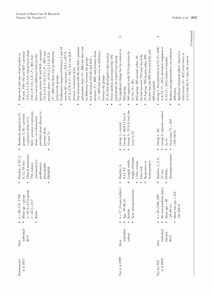
Journal of Burn Care & Research Volume 36, Number 3 Nedelec et al 363
Stei
nstr
aess
er
et a
l 201
1In
tra in
divi
dual
R
CT
•n
= 38
(11
F, 1
7M)
•M
ean
age
= gr
oup
1—42
.3 ±
13.
3; g
roup
2—
43.1
± 1
3.7
•B
urns
•B
asel
ine,
2/
52, 2
, 6,
12,
18
mos
•C
linic
al a
sses
smen
t, V
SS, r
edne
ss
(chr
onom
eter
),
profi
lom
etry
, ph
otog
raph
s•
PRIM
OS
•R
ando
mly
ass
igne
d to
Tx
grou
ps: 1
) SG
+ p
ress
ure
vs p
ress
ure
alon
e or
2)
SGS
+ pr
essu
re v
s pr
essu
re
alon
e—no
info
rmat
ion
prov
ided
abo
ut a
lloca
te t
o pr
essu
re a
lone
•12
mos
Tx
•Im
prov
ed w
ith t
ime
from
bas
elin
e to
18
mos
: VSS
—SG
and
SG
S +
pres
sure
(5
.9 ±
1.5
–1.9
± 1
.6)
and
pres
sure
alo
ne
(5.8
± 1
.9–1
.8 ±
1.3
) (P
< .0
01)
but
ther
e w
as n
o di
ffer
ence
bet
wee
n th
e gr
oups
; red
ness
SG
and
SG
S +
pres
sure
(1
5.4
± 2.
2–11
.0 ±
2.1
) (P
< .0
01)
and
pres
sure
alo
ne (
15.9
± 2
.2–1
1.3
± 2.
3)
(P =
.003
) bu
t th
ere
was
no
diff
eren
ce
betw
een
the
grou
ps•
Profi
lom
etry
impr
oved
bet
wee
n 2
and
18
mos
for
SG +
pre
ssur
e (3
23.1
± 1
97.5
–25
7.6
± 17
8.3;
P =
.047
) bu
t no
t SG
S +
pres
sure
or
pres
sure
alo
ne•
Pain
dec
reas
ed w
ith S
G a
nd S
GS
+ pr
essu
re
and
pres
sure
alo
ne (
P =
.005
) bu
t the
re w
as
no d
iffer
ence
bet
wee
n th
e gr
oups
•It
ch d
ecre
ased
with
SG
and
SG
S +
pres
sure
(P
= .0
05)
and
pres
sure
alo
ne
(P =
.003
) bu
t th
ere
was
no
diff
eren
ce
betw
een
the
grou
ps•
31.5
% S
GS
deve
lope
d a
skin
rea
ctio
n•
Pow
er a
naly
sis
dete
rmin
ed t
hat
192
part
icip
ants
are
req
uire
d pe
r gr
oup
Tan
et
al 1
999
Intr
a indi
vidu
al
coho
rt
•n
= 17
(3
scar
s/su
bjec
t)•
Age
: 19–
40 y
rs•
Kel
oids
•A
cne
and
spon
tane
ous
•B
asel
ine,
4,
8,12
/52
•L
engt
h, w
idth
, he
ight
of k
eloi
ds•
Col
or, t
extu
re•
Pain
/itc
h•
Res
pons
ive
or
nonr
espo
nsiv
e
•G
roup
1—
cont
rol
•G
roup
2—
SGS
12 h
rs/
d•
Gro
up 3
—T
CA
0.1
ml/
site
ev
ery
4/52
•N
o si
gnifi
cant
cha
nge
for
the
cont
rol o
r SG
S gr
oup
•94
% im
prov
ed w
ith T
CA
inje
ctio
ns b
y 12
/52
(P
< .0
5)•
SGS
grou
p: 1
8% b
ecam
e so
fter
, 6%
be
cam
e le
ss r
ed, 7
5% w
ere
less
itch
y•
TC
A g
roup
: 70%
bec
ame
softe
r, 59
%
beca
me
less
red
, 50%
wer
e le
ss it
chy,
and
60
% le
ss p
ainf
ul
3
Van
der
Wal
et
al 2
010
Intr
a indi
vidu
al
blin
ded
RC
T
•n
= 23
(13
M, 1
0F)
•46
sca
rs p
ostb
urn
inju
ry•
Mea
n ag
e =
38
(18–
69 y
rs)
•M
ean
scar
age
= 1
23
(36–
338
d)
•B
asel
ine,
1, 3
, 6,
12 m
os•
POSA
S,
derm
aspe
ctro
met
er
•G
roup
1–
SG•
Gro
up 2
—pl
aceb
o co
ntro
l•
2x/
d•
mea
n tim
e T
x =
263
(195
–347
d)
•G
roup
1: d
ecre
ased
itch
at 3
/12
(P
= .0
18)
at 6
/12
(P
= .0
13);
dec
reas
ed ro
ughn
ess a
t 3/
12 (
P =
.014
) re
lativ
e to
bas
elin
e•
Red
ness
and
pig
men
tatio
n, n
o st
atis
tical
di
ffer
ence
•Si
gnifi
cant
tre
atm
ent
effe
ct: o
bser
ver’
s re
lief s
core
s (P
= .0
12)
wer
e lo
wer
at
3/12
with
SG
Tx
than
the
con
trol
2
(Con
tinu
ed)

Journal of Burn Care & Research364 Nedelec et al May/June 2015
Wid
gero
w e
t al
20
09R
CT
•n
= 12
0 (1
70 s
cars
)•
18––
82 y
rs•
Gro
up 1
(n
= 60
)—Sx
ex
cisi
on a
ssig
ned
to T
x (n
= 3
0) o
r co
ntro
l (n
= 3
0)•
Gro
up 2
(n
= 20
)—2
Sx
exci
sion
site
s/pa
tient
s as
sign
ed t
o T
x or
co
ntro
l•
Gro
up 3
(n
= 10
)—
bila
tera
l bre
ast
Sx R
&
L a
ssig
ned
to T
x or
co
ntro
l•
Gro
up 4
(n
= 30
) co
smet
ic p
roce
dure
s co
mpa
red
to h
isto
rica
l co
ntro
ls
•PO
SAS
•H
omem
ade
4 po
int
scar
cl
assi
ficat
ion
1)
norm
al, 2
) m
ildly
hy
pert
roph
ic, 3
) hy
pert
roph
ic, 4
) ke
loid
•T
x gr
oup—
Mic
ropo
rous
ta
pe +
NSG
S•
Con
trol
gro
up—
Mic
ropo
rous
tap
e (a
pplie
d po
st s
urgi
cally
)
•Sc
ar C
lass
ifica
tion
•G
roup
1—
sign
ifica
ntly
mor
e T
x’d
scar
s w
ere
mild
sca
rs a
t 2/
12 a
nd 6
/12
(P
< .0
001)
•G
roup
2—
sign
ifica
ntly
mor
e T
x’d
scar
s w
ere
mild
sca
rs a
t 6/
12 (
P =
.006
)•
Gro
up 3
—si
gnifi
cant
ly m
ore
Tx’
d w
ith
mild
sca
rs a
t 2/
12 (
P =
.032
)•
Gro
up 4
—si
gnifi
cant
red
uctio
n in
sca
r se
veri
ty fr
om 1
/12
–6/
12•
POSA
•G
roup
1—
stiff
ness
(P =
.000
3) a
nd th
ickn
ess
(P <
.000
1) w
ere
supe
rior i
n T
x pa
tient
s•
Gro
up 2
—st
iffne
ss (
P =
.06)
and
thic
knes
s (P
= .0
022)
wer
e su
perio
r in
Tx
patie
nts
•G
roup
3—
Tx’
d sc
ars
show
ed s
igni
fican
t im
prov
emen
t in
itch
ines
s•
OSA
S•
Gro
up 1
—al
l par
amet
ers w
ere
signi
fican
tly
bett
er in
Tx
grou
p (P
< .0
05)
•G
roup
2—
all p
aram
eter
s ex
cept
vas
cula
rity
signi
fican
tly im
prov
ed in
Tx
grou
p
2
Wig
ger-
Alb
ert
et a
l 20
09In
tra in
divi
dual
R
CT
n =
60 (
44F,
16M
)M
ean
age
= 38
.2
(16–
61 y
rs)
Scar
age
= 4
9.6
(3
–355
mos
)E
tiolo
gy n
ot s
peci
fied
Bas
elin
e, 4
, 8, a
nd
12 w
ksPr
imar
y ou
tcom
e:
Patie
nt’s
qu
estio
nnai
re
over
all S
IR
edne
ss (c
hrom
amet
er)
Clin
ical
sca
r as
sess
men
t m
odifi
ed
Scar
div
ided
into
tw
o: h
alf T
x w
ith S
GS
and
half
Tx
NSG
S (p
olyu
reth
ane)
12 w
ks T
xR
emov
e SG
S an
d N
SGS
up t
o 1
hr/
d
SI: s
igni
fican
tly im
prov
ed a
t 4/
52
(P <
0.0
001)
and
8/
52 (
P =
.012
);
impr
ovem
ent
was
sig
nific
antly
mor
e pr
onou
nced
for
poly
uret
hane
at
4/12
(P
< 0
.000
1) a
nd 8
/12
(P
= .0
12)
Red
ness
: low
er fo
r po
lyur
etha
ne a
t 8/
52
(P =
.001
6)
2
Tab
le 2
. (C
onti
nued
)
Aut
hors
Des
ign
Sam
ple
Out
com
e
Mea
sure
sIn
terv
enti
onR
esul
tsL
evel
of
Evi
denc
e

Journal of Burn Care & Research Volume 36, Number 3 Nedelec et al 365
to followup, 14 developed a rash and three experi-enced skin breakdown. These articles received a cri-tique rating of <5 (Table 1), but have been described due to their historic importance. The remainder of the detailed review will be restricted to reports that obtained a rating ≥5, as the validity of the conclu-sions of poorly rated reports is questionable.
Burn Scar–Only LiteratureThere were no case series in the burn scar–only lit-erature reviewed that received a rating ≥5. How-ever, since 2009 there have been five RCTs that recruited exclusively burn survivors for their stud-ies. Harte and colleagues22 examined 22 adult burn survivors who were treated with either pressure or pressure and SGS. Using the Vancouver Scar Scale (VSS) to evaluate scar outcome, there was no dif-ference between treatment groups at 12 or 24 weeks. A power analysis of their data revealed that 384 participants would be required before any con-clusions could be drawn. Karagoz and colleagues23 examined 32 burn survivors aged three to 44 years old. Participants were allocated to silicone gel (SG), SGS or onion extract. The VSS was used to evalu-ate outcome, and all groups showed significant improvements after treatment. The improvements were significantly better for the SG vs onion extract group and for the SGS vs onion extract group, but there was no difference between the SG and SGS groups. Momeni and colleagues28 examined 38 burn survivors between the ages of 1.5 and 60 years. Participants had a HTS that was divided into two sides, one treated with SGS, the other with nonsili-cone gel sheet (NSGS) (self adhesive propylene gly-col and hydroxyethyl cellulose sheeting, which the authors refer to as a placebo control). The VSS was used to compare outcomes between the SGS and NSGS in terms of scar itch, pigmentation, pliability and vascularity. The VSS scores of the SGS-treated scars were significantly lower at 4 months compared to those of the NSGS-treated scars. Steinstraesser and colleagues38 examined 38 burn survivors. Par-ticipants were randomly assigned to SG and pres-sure or SGS and pressure. Half of their scars also received only pressure therapy. Using the VSS as an outcome measure, the SG and pressure as well as SGS and pressure, and pressure alone improved from baseline to 18 months, but there was no dif-ference between the groups. Using the chromameter to measure redness, an improvement was reported in SG and SGS and pressure, and pressure alone groups at 18 months, but again no difference was demon-strated between groups. Profilometry (scar surface W
itten
berg
et
al
1999
Intr
a indi
vidu
al
RC
T
n =
20 (
19 s
cars
)
(15F
, 5M
)M
ean
age
= 49
yrs
.M
ean
scar
age
= 3
2 m
osSx
sca
rs
Bas
elin
e, 8
, 16,
24,
40
wks
Blo
od fl
ow/
volu
me
(las
er d
oppl
er),
el
astic
ity, p
unch
bi
opsy
Self
asse
ssm
ent
of
pain
, itc
h, a
nd
burn
ing
Scar
vol
ume
Sam
e sc
ar in
3 g
roup
s: S
GS,
la
ser,
and
cont
rol
SGS
>12
hrs/
d x
24 w
ksL
aser
4 T
x at
8 w
k in
terv
als
No
diff
eren
ce b
etw
een
Tx
and
cont
rol
grou
psIt
ch (
P =
.005
), b
urni
ng (
P =
.01)
, and
blo
od
flow
(P
= .0
01)
decr
ease
d w
ith ti
me
Scar
vol
ume:
ove
rall
time
effe
ct (
P =
.02)
, onl
y co
ntro
l sho
wed
a si
gnifi
cant
red
uctio
n fr
om b
asel
ine
to 4
0 w
ks (
P =
.007
)
2
c/o,
com
plai
ned
of;
CO
, co
hort
; D
r, d
octo
r; F
, fe
mal
e; H
TS,
hyp
ertr
ophi
c sc
ars;
M,
mal
e; L
, le
ft;
L/E
, lo
wer
ext
rem
ity;
n,
sam
ple
size
num
ber;
NSG
S, n
onsi
licon
e ge
l she
ets;
PO
SAS,
Pat
ient
and
Obs
erve
r Sc
ar A
sses
smen
t Sc
ales
; R, r
ight
; RC
T, r
ando
miz
ed c
ontr
olle
d tr
ials
; SG
, sili
cone
gel
; SG
S, s
ilico
ne g
el s
heet
s; S
I, s
car
inde
x; S
x, s
urge
ry; T
CA
, tri
amci
nolo
ne a
ceto
nide
; Tx,
tre
atm
ent;
U/E
, upp
er e
xtre
mit
y;
VAS,
vis
ual a
nalo
gue
scal
e; V
SS, V
anco
uver
sca
r sc
ale;
x/5
2, n
umbe
r of
wee
ks; x
/12,
num
ber
of m
onth
s.

Journal of Burn Care & Research366 Nedelec et al May/June 2015
microtopography) was measured using the PRIMOS optical three dimensional measuring system (GFM, Berlin, Germany). Profilometry improved between two and 18 months for SG and pressure, but not for SGS and pressure or pressure alone. Pain and itch were also reduced in all groups, but there was no difference between the groups. A power analysis of their data revealed that 192 participants per group would be required before any conclusions could be drawn. Van der Wal and colleagues39 examined 23 adult burn survivors using the Patient and Observer Scar Assessment Scale (POSAS) and a dermaspec-trometer. The participants’ scars were divided in half and treated with SG or a placebo control. There was a reduction in itch and roughness with time, but no change in redness and pigmentation. The observer’s relief score of the POSAS did show a significantly lower score with SG treatment at 3 months, but the difference was no longer significant at 6 months.
All of the burn scar–only studies include differ-ent treatments and comparison groups; therefore, taken together it is difficult to develop confident conclusions. All of these studies used either the VSS or POSAS as their outcome measure but two also included profilometry and a colorimeter (chro-mameter or dermaspectrometer). Using the VSS as an outcome measure, two of the studies23,28 reported better results with SG and SGS versus onion extract or SGS versus NSGS, but two reported no difference between the groups22,38 when comparing pressure and pressure plus SGS or SG plus pressure and SGS plus pressure compared to pressure alone. Using profilometry, the later study did find a significant improvement with SG and pressure treatment com-pared to SGS and pressure or pressure alone. The final study39 used the POSAS and dermaspectrom-eter and only reported significant differences on the observer’s relief score at 3 months but not 6 months. Thus, the findings of these burn scar–only studies evaluating the use of gels and gel sheets are inconsis-tent and inconclusive.
Several issues should be taken into consideration, including the small number of participants, with 38 participants allocated to each treatment by Momeni and colleagues28 being by far the largest number in any study (range 11–38). In fact, two of the studies performed a post hoc power analysis that determined that they were under-powered,22,38 confirming that their results must be interpreted with caution. In addition, the VSS has been shown to be far less reli-able than objective instrumentation that measures the same skin characteristics73,74; thus, the outcome measures used may not have been sensitive enough to detect change. These two methodological limitations
both potentially contribute to the possibility of type 1 errors; therefore, it cannot be concluded that gels and gel sheets have no effect on scars. Another meth-odological issue that must be considered is that none of these studies had a “no-treatment control group” limiting comparison to the alternative treatment that was being investigated. Two of the studies stated that they had a placebo control,28,39 but did not report on the occlusive properties of the products being used. Since one of the currently proposed mechanisms of action of gels and gel sheets is occlusion,8,75 it cannot be concluded that these were true placebo controls unless the occlusive properties of these products has been objectively documented and investigated to determine whether they have a therapeutic benefit.
Combined Burn and Nonburn Scar Etiology LiteratureFor the studies that included burn scars in addition to participants with scars resulting from other etiolo-gies, there were five case series, all of which noted an improvement across time, and four out of the five received a rating of ≥5. Dockery and Nilson52 examined 94 participants treated with SGS that were divided into five different groups (known scar form-ers, fresh HTS, fresh keloids, long standing scars and traumatic or burn scars). The participants were mon-itored for 6 months, based upon photographs and physician and participant scar assessments. Although no statistical comparison was provided, the author reported either a lack of development of scar when placed on newly healed wounds or improvement in most participants after treatment. In 1993, Hir-showitz and colleagues59 examined a series of 32 participants treated with SGS who developed scars after surgery, trauma or burns. The authors reported that 71.9% of the participants showed moderate or excellent improvement over a 12-month period. Then in 1998, Hirshowitz and colleagues60 exam-ined 30 participants treated with thick silicone cush-ions filled with silicone oil who developed scars after surgery or burns. Alleviation of symptoms was noted after 6 months in 63.3% of the participants, with the remainder being reported as having recalcitrant scars. Lee and colleagues63 reported on 26 partici-pants who were treated with two different types of SGS, with improvements being reported in both groups for color and texture (90%), regularity (80%), and thickness (50%).
An intra individual cohort study reported by Ahn and colleagues in 1991 included participants who developed scars after burns, surgery and spi-der bites. There were two groups of participants

Journal of Burn Care & Research Volume 36, Number 3 Nedelec et al 367
who had either a recent surgical excision or existing HTS. The sites were divided in half, with half being treated with SGS and half receiving no treatment. For the recent surgical excision group, the no treat-ment control site significantly increased in volume at months 1 and 2, but the treatment site did not. The HTS sites did not reduce in volume but elastic-ity did improve, both with time and compared to the no treatment control, as measured by the elastom-eter. Boutli-Kasapidou and colleagues44 examined 30 participants in a cohort study that had either HTS or keloids, but the authors never indicated the pro-portion of each and throughout the article referred to all as keloids, who received either polytherapy (cryotherapy, intralesional cortisone, SG) or SGS. The results of both the participants’ level of satisfac-tion and the physician’s assessment saw significantly greater improvement in the polytherapy group. The authors also reported significantly better improve-ment in less mature keloids.
Of the four RCTs that combined burn and non-burn scar etiology, one was an intra individual study17 and the others randomly assigned participants to different groups.25,26,30 The intra individual study by Carney and colleagues included two groups that were treated with two different SGS and a randomly assigned control site that did not receive any treat-ment.17 Using an extensometer and subjective evalua-tion, improvements in extensibility, color and texture were significantly greater in both groups compared to their no treatment control site, but there was no significant difference between the two different SGS. Li-Tsang and colleagues25 conducted an RCT exam-ining the effect of SGS plus massage compared to only massage in 45 participants who developed scars after traumatic or burn injuries. The SGS plus mas-sage treatment group showed significantly greater reductions in thickness and increases in pliability after 6 months compared to the massage only group when assessed with the tissue ultrasound palpation system and VSS respectively. There was no significant dif-ference in itch, pain, or color. In 2010, Li-Tsang and colleagues randomly assigned 104 participants to four different groups (pressure garments, SGS, pressure garments and SGS, and no treatment con-trol). The evaluation results at 6 months included 84 participants and demonstrated a significant improve-ment in thickness with pressure garments alone or pressure garments and SGS, compared to no treat-ment control. The pressure garment and SGS group was significantly more pliable than the no-treatment control at 2 and 4 months, but by 6 months pliabil-ity had improved for all groups compared to base-line and there was no significant difference between
the groups. Pain significantly improved in the SGS and pressure garments and SGS group compared to the no treatment control group. Pigmentation sig-nificantly improved with time for all the groups, but the improvement did not differ between the groups. Palmieri and colleagues30 randomly assigned 80 par-ticipants to treatment with SGS plus vitamin E or SGS alone. Participants were evaluated at 4 and 8 weeks using photographs to assess color, size and cos-metic appearance that revealed a significantly greater percentage of participants rated as improved when treated with SGS plus vitamin E.
As a group, the RCTs that included combined burn and nonburn scar etiology were more likely to find a positive effect with SGS than the compari-son treatment. Three of the four studies had a no treatment control group, with the fourth compar-ing SGS to SGS plus vitamin E, where the latter had significantly better results. Three of these studies used objective instrumentation (extensometer, tis-sue ultrasound palpation system and spectrometer) in addition to subjective outcome measures.
Nonburn or Unspecified Scar LiteratureOf the case series that did not include burn scars or did not specify the etiology of the condition that preceded the scar formation, five received a rating of ≥5.53,56,62,65,71 Eishi and colleagues53 treated six participants’ scars with SGS for 6 months. Pain and itch were significantly reduced at 4 weeks and had disappeared by 12 weeks. Redness and elevation were significantly reduced by 8 weeks, with a further reduction in redness at 6 months. Gold56 reported moderate improvement in thickness (81.25%), color (75%), and overall effectiveness (100%) when he evaluated 10 participants who were treated with SGS over a 3-month period. Physician’s evalua-tion of outcome reported complete resolution in a small percentage (6.25%) or moderate change in thickness (50%), color (68.75%), and overall effec-tiveness (93.75%). Lacarrubba and colleagues62 examined eight participants treated with SG and reported a 37% reduction in the mean thickness of scars, using high frequency ultrasound as an evalua-tion tool. Murison and James65 evaluated six partici-pants treated with SG, which they evaluated using a modified VSS and spectrophotometric intracutane-ous scope (SIS). All participants improved on the VSS and showed a 7.2% reduction in collagen and 3% increase in blood flow with the SIS. Spencer71 reported on seven participants who had SG applied to half of their post surgical excision while the other half received no treatment. After 3 months of treat-ment, the SG treated half was rated as better by the

Journal of Burn Care & Research368 Nedelec et al May/June 2015
physician in five of the seven excision scars and using the VSS, four out of seven were rated better.
There were four cohort studies, three of which were intra individual. Chernoff and colleagues44 assigned participants with bilateral scars to one of the three treatment groups; 1) SG 2X/day; 2) SGS day and night; or 3) SG (day) and SGS (night) with their bilateral scar serving as an untreated control scar. After 90 treatment days, all the three treat-ment groups were significantly less elevated, red, itchy, and irritated in the SG and SGS group than the untreated control. Participants rated SG as easier to apply and combined SG and SGS as producing the most favorable outcome. Cruz-Korchin45 examined 20 participants after bilateral mammoplasty where one side was treated with SGS and the other was an untreated control. Six months post surgery, HTS were found in 25% of the treated breasts and 55% of the untreated breasts. Hosnuter and colleagues46 assigned 60 participants to three treatment groups: topical onion extract, SG, or SGS plus topical onion extract. Improvement was reported over time for all the groups, with significantly more color reduction reported with topical onion extract compared to SG and significantly greater height reduction with SGS plus topical onion extract compared to topical onion extract alone. Tan and colleagues47 reported the outcome of 17 participants with three keloids each who were assigned to SGS treatment, triamcinolone acetonide (TCA) injections, or no treatment. Statis-tically significant improvements were seen after 12 weeks of TCA injections, but not in the SGS-treated keloids or the untreated keloids.
There were 18 RCTs reviewed that included nonburn and unspecified scar etiology, all of which received a rating of ≥5. Berman and Flores17 reported on 22 participants treated with either a silicone gel-filled cushion or SGS. There was no significant dif-ference between the groups. Chan and colleagues18 reported an intra individual trial where the upper and lower portions of the scar of 50 participants were randomly allocated to SG or NSG. The SG group was significantly better for all subscales of the VSS after 3 months of treatment. De Giorgi and colleagues19 reported on 110 participants who had recently undergone surgery and were assigned to apply either SG with zinc oxide or zinc oxide cream alone to their excision site after staple removal. There were significantly fewer participants who developed abnormal scar in the SG group (27% vs 55%) dur-ing the 8-month followup period. De Oliveira and colleagues20 reported on 26 participants with 41 scars. One scar was treated with SGS and a second with NSGS. If the participant had more than two
scars, then the third served as an untreated control scar. There was no significant difference between the SGS and NSGS treated scars with respect to length, width, color, itch, pain, induration, and intracicatri-cial pressure, but when compared to the untreated control there were significant differences for length, width, color, induration, and pressure. Gold and colleagues21 reported on 96 participants who were stratified into low risk (n = 50) and high risk (n = 46) participants who were treated with SGS or routine postoperative care. There was no statistically signifi-cant difference for either low or high risk groups, although participants who were undergoing scar revision surgery had an increased rate of scar pre-vention. Klopp and colleagues24 reported on an intra individual RCT where 12 participants’ scars were treated with NSGS, NSGS plus pressure, pressure, and SGS plus pressure. Vessel length, venular flow rate, erythrocyte, and aggregate number were all evaluated using vital microscopic methods in addi-tion to the skin temperature and surface roughness. The authors reported that all the measured char-acteristics significantly improved in all groups at P < .05. The improvement seen with SGS and NSGS plus pressure was substantially greater than that for NSGS or pressure alone. Majan27 reported on 12 participants whose post surgical scars were ran-domly assigned to SGS or no treatment. Although the authors presented positive VSS results and physi-cians’ overall impressions, no statistical comparisons were included. Niessen and colleagues29 randomly assigned 155 women who had undergone breast reductions to two different types of SGS. Each group had one side treated with the SGS and the other side an untreated control. The authors reported the per-centage of participants who developed HTS, but it was unclear whether this was on the treatment or control side, or both. There was no difference in the outcome between the two different SGS treated sites, but there were significantly more HTS that formed on the SGS treated side than on the untreated con-trol side. Perez and colleagues31 randomly assigned 30 participants to one of three treatments (TCA, SGS and vitamin E, onion extract gel, or placebo control). Fifteen of the participants completed the 16-week treatment period, five in each group. All the three groups significantly improved with time, as described in Table 2. When the treatment groups were compared to the placebo control, the investi-gator’s cosmetic evaluation, induration and pigmen-tation showed significantly more improvement for both the groups, as well as erythema for group 1 and tenderness for group 2. Phillips and colleagues32 ran-domly assigned 20 participants to NSGS treatment

Journal of Burn Care & Research Volume 36, Number 3 Nedelec et al 369
or control moisturizer for 8 weeks. There was no significant difference between treatment groups, but both the groups reported a significant reduction with itching over time. Rhee and colleagues33 randomly assigned 40 participants to SGS or untreated control groups after scar revision and mass excision surgeries. At 1 month, the SGS treated scars were significantly decreased in height compared to the untreated con-trol scars. Pigmentation and vascularity were also reduced in the SGS treated participants at 3 months. Scuderi and colleagues34 had two groups of partici-pants. The first group received treatment with NSG, with improvement reported for 89.7% of the partici-pants based on a global scar assessment. The second group had a linear scar, with half being treated with a SG and half treated with a NSG. Both treatments resulted in reduced scar elevation at 1 month and 1 year. Scuderi and colleagues35 recruited 85 par-ticipants who underwent bilateral mammoplasties and randomly assigned each breast to NSG or SG. In both the groups, scar elevation, as measured by optical profilometry, increased from baseline to 1 month but reduced from 1 month to 1 year. The scar width increased across time for both the groups, but was significantly less in the NSG-treated group. Signorini and Chementontit36 randomly assigned 148 participants with recent surgical scars to either SG treatment or to an untreated control group. At 6 months, post treatment scars were classified as nor-mal, mildly hypertrophic, hypertrophic, or keloids. Significantly more incisions were classified normal when treated with SG than the untreated control. Sproat and colleagues37 performed an intra individ-ual RCT with 14 participants who had sternal scars post sternotomy. Half of the scar was treated with SGS and half with TCA. Significantly more partici-pants favored the SGS treated half and 82% of the blinded evaluators preferred the SGS treated portion based upon photographic assessments. Widgerow and colleagues40 evaluated 120 participants who were divided into four groups: 1) n = 60 participants post surgical incisions, with half of their surgical scar assigned to treatment with NSGS plus microporous tape and half assigned to the control group which was treated with microporous tape only; 2) n = 20 participants with two surgical sites, one assigned to the treatment group and the other to the control group; 3) n = 10 participants post bilateral breast surgery, each assigned to treatment or control; and 4) n = 30 participants who received treatment and compared to historic controls. Using the POSAS, scars were graded as normal, mildly hypertrophic, hypertrophic, or keloids. At 2 and 6 months, the treated scars in group 1 were significantly more likely
to be graded as mildly hypertrophic. They were also less thick and stiff. At 6 months, the treated incision scar in group 2 was more likely to be graded as mildly hypertrophic and was less stiff and thick. At 2 months, the breasts in group 3 that received treatment were more likely to be graded as mildly hypertrophic and less itchy. In group 4, there was a significant reduc-tion in scar severity compared to historic controls. Wigger-Albert and colleagues41 reported on 60 par-ticipants who received SGS treatment to half of their scar and NSGS to the other half for 12 weeks. Using a homemade patient scar index questionnaire, both scars significantly improved at 4 and 8 weeks, but to a greater extent for the NSGS-treated scars. Redness, as assessed by the chromameter, was also significantly less at 8 weeks in the NSGS treated group. Witten-berg and colleagues42 reported on 20 participants whose scars were divided into three sections that were treated with SGS, laser, and untreated controls. There was no difference between the treatment and control scars except for scar volume, which showed a significant reduction with time in the control group after 40 weeks. Itching, burning sensation, and blood flow significantly reduced in all groups over time.
Of the RCTs that included nonburn or unspeci-fied scars, the majority (14 out of 18) reported a positive treatment effect, although the compari-son groups were variable including NSG, NSGS, zinc oxide, TCA, laser, or untreated controls. None of the studies reported whether they performed a power analysis, including those studies that had a negative effect17,29,32,42; therefore, it is not possible to determine whether their sample size was too small to detect an effect, potentially resulting in a type 1 error. Seven of the RCTs applied gels or gel sheets to newly healed incisions and all but one29 reported less scar formation with the application of SG, SGS, or NSGS18,19,21,33,36,40 compared to controls, but no dif-ference when NSG was compared to SG.35 Ten of the RCTs applied gels or gel sheets to established scars.17,
18,20,24,31,32,34,37,41,42 Three of these studies did not find an effect, but Berman and Flores17 compared SGS to silicone cushions and Phillips and colleagues32 and Wittenberg and colleagues42 only included 20 and 19 scars respectively, in their analysis. Of the remain-ing seven studies, all found a positive treatment effect using SG,18,20,34 NSG,20,34 SGS,24,37 NSGS24,41 or SGS in combination with Vitamin E and TCA.31
Rehabilitation-Specific Treatment InformationThe rehabilitation-specific information within cita-tions was usually limited, but those that did provide

Journal of Burn Care & Research370 Nedelec et al May/June 2015
information made the following recommendations. The gel sheet or gel should extend approximately 5 mm beyond the scar margins, should be applied for several hours the first day and then increased by two-hour increments every other day until the patient is able to wear it for up to 23 hours/day. It should be taken off daily and gently washed in clear water with a hypoallergenic soap and air dried or patted dry with a lint free cloth. The description of how the gel sheets were held in place was very variable, including skin tape (ie, HypafixTM/Fixumull®), Tubigrip®, custom fabricated pressure garments, self adherent properties of gel sheet, etc., with no apparent con-sensus or formal evaluation of which method was most advantageous.
Not all publications reported on whether any adverse events occurred, but those reported included skin reactions, dermatitis, itch, or skin breakdown. These issues occurred with both SGS and NSGS.16,17,23,27,29,32–35,37,38,45,46,48,52,55,58,61–64,69 Gels or gel sheets are contraindicated if a patient has a known allergy, but since different products con-tain different ingredients, individual products may be well tolerated by some individuals and not oth-ers. Interestingly, a number of studies also reported a reduction in itching and/or pain,18,19,26,31,32,37–
40,44,46–48,53,60,70 suggesting that these symptoms may actually improve with gel or gel sheet treatment. If an adverse skin reaction does occur, the gel sheet should be discontinued until complete recovery has occurred. When reinitiated, this process should progress more slowly and particular attention should be paid to skin hygiene. If the skin reaction reoc-curs, the gel sheet should again be discontinued until complete recovery, but when it is reinitiated the maximum wearing time should be limited to 12 hours/day (Figure 2). The gel sheet should be replaced when it begins to fray or becomes difficult to handle. It should be noted that gels and gel sheets may constitute a choking hazard for small children; therefore they should only be applied if the child is unable to gain access to them.
The study that was removed after full review due to the fact that it did not address the authors’ PICO question did examine the effect of patient education on compliance/adherence.15 The authors reported that participants who viewed a 26-minute instruc-tional video tape, in addition to standard verbal and written instructions (a one-page handout), wore their SGS for more than twice as long as the par-ticipants who only received standard verbal and writ-ten instructions (21.8 ± 3.0 hours/day vs 10.1 ± 7.5 hours/day). They also reported that participants who had enhanced patient education had improved
outcomes on the VSS and subjective participant eval-uations, suggesting that thorough education should be included as part of the rehabilitation intervention plan.
The majority of studies that applied SG had the participants apply the products 2x/day18,19,23,30,34,
35,39,44,62,65,67,71; however, one study44 had the par-ticipants apply it 3x/day. One study examined the application of SG during the day and SGS dur-ing the night44; thus the participants were asked to apply it only once per day. The one group that reported on NSG had the participants apply three layers every three to five days. No adverse reactions were reported for the SG, but they were reported for the NSG.34,35 The authors reported that incorrect application occurred when excessive quantities were applied and that reinstruction on correct application of the product resulted in improved participant com-fort in applying the product.35
It must be emphasized that gel or gel sheet prod-uct prescription requires proper clinician followup and good clinical judgment when evaluating the wearing schedule and management of any adverse events.
DISCUSSION
The objective of the review was to systematically evaluate the available evidence for the use of gels and gel sheets for the treatment of hypertrophic scars and keloids that occur after burn injury. Unfortunately, the number of studies and quality of evidence specifi-cally focusing on burn survivors is lacking; thus, the decision was made to include all patient populations to strengthen the conclusions and recommendations. Confident conclusions cannot be drawn from the lit-erature that restricted recruitment to burn survivors, since level one data does not exist and findings from the RCTs were inconsistent. The inconsistency of the findings may be a result of methodological limi-tations of the studies and/or the fact that they were underpowered.
In total, there were 23 case series reviewed, with the vast majority reporting a positive effect. It is dif-ficult, however, to conclude that this constitutes a real treatment effect, since HTS is well known to spontaneously improve with time. It has been recom-mended by a panel of experts that in order to provide proof of efficacy of therapies intended to prevent or reduce scar formation, intra individual or self con-trolled RCT designs should be used where the treat-ment and control sites are anatomically matched.76 Of the 27 RCTs reviewed, the vast majority reported positive results, but comparison between studies is

Journal of Burn Care & Research Volume 36, Number 3 Nedelec et al 371
difficult since many studies were making compari-sons to other treatments rather than to a no treat-ment control. Therefore, if their data confirmed that the gels or gel sheets were as effective as the compar-ison treatment and resulted in a significant change over time that does not rule out the possibility that the improvement could be due to spontaneous reso-lution. If only those RCTs that recruited exclusively burn survivors are summarized, no conclusions about treatment benefit can be drawn, but the methodolog-ical limitations of these studies, as described above, must be considered. There does seem to be some evidence that pathological scar formation can be pre-vented in fresh surgical wounds, but this finding has not been investigated in burn survivors. A number of studies have reported including participants with
keloids,16,20,21,30–32,43,44,46,50,51,54,55,63,67,68 but did not report diagnostic details for their classification or whether these participants responded in a similar manner to the other participants. Three case series included only participants with keloids,53,64,66 all of which reported a positive treatment effect, but two received a rating of <5 on the critical appraisal.64,66 One study reported itch reduction with SGS,47 but described superior outcomes after TCA injections. Only one study58 reported treating mature scars and showed no treatment benefit.
Many of the reports, both in the burn scar–only literature and other populations, continue to use subjective or home made scar evaluations to deter-mine if there is a treatment effect. There has been extensive discussion in the literature recently about
Wound Re-epithelization
Decision to Tx with NSG or SGDecision to Tx with NSGS or SGS
Apply 2-3x/dayApply 2 hrs/day the 1st day & ↑ by 2 hrs every other day up to 23 hrs/day*
No skin rash Skin rash
Continue to Tx until scar has matured
Continue to Tx until scar has matured
Discontinue gel sheet until healed
Re-initiate gel slowly with ↑ ’d attention to hygiene care
No skin rash Skin rash
Continue to Tx until scar has matured
Re-initiate gel slowly up to 12 hrs/day with ’d attention
to hygiene care
Continue to Tx until scar has matured
*Can be held in place with pressure garments, microtape, tensor bandage, etc.
Discontinue gel sheet until healed
Provided thorough education Provided thorough education
↑
Figure 2. Expert opinion and evidence based rehabilitation specific algorithm for postburn scar treatment with nonsilicone or silicone gels and gel sheets. NSGS, nonsilicone gel sheet; SGS, silicone gel sheet; Tx, treat; ↑, increase.
Journal of Burn Care & Research372 Nedelec et al May/June 2015
the clinimetric properties of scar assessment tools.76–80 It is imperative that any subjective scar scales used for scar evaluation be published tools that have well established clinimetric properties, so that study find-ings can be repeated by other investigators and com-parisons can be made between the studies. Objective measurement tools have been recognized as the accepted standard for the assessment of skin char-acteristics,73,74,80 while recognizing that the patient’s opinion of their scar should also be evaluated.77–79
There is a need for methodologically rigorous studies that only include scars that form after a burn injury, so confident conclusions can be drawn about whether burn survivors’ scars benefit from the appli-cation of gels or gel sheets. Future studies would allow for stronger conclusions if they addressed the limitations in the current literature. An appropriate a priori power analysis should be performed to deter-mine the sample size that would be required to avoid a type 1 error. Post hoc analyses performed in two separate investigations22,38 concluded that 192 par-ticipants per group would be required when using the VSS as an outcome measure, but this analysis would need to be repeated if a different outcome measure was used. Objective instrumentation should be used to evaluate scar characteristics such as thick-ness, pliability, vascularity, and profile or contour, in addition to patient opinions about the benefits of treatment. Intra individual and no treatment con-trols should be included to limit the intra individual variations associated with scar formation and treat-ment response, as well as spontaneous resolution over time. The question of whether scarring after a burn injury can be prevented has yet to be addressed. This would require that contralateral or anatomically adjacent wounds with the same injury and recov-ery profile be compared across time, which would be difficult due to the traumatic and unpredictable nature of burn injury; however, this would make an important contribution to the literature. Laser dop-pler imaging to determine the depth of the burn may help to select comparable wounds and has been cor-related with longterm scar outcomes.81 In order to recruit sufficient participant numbers and increase the generalizability of the results, multicentre trials using clinimetrically sound subjective and objective scar assessment tools are strongly recommended.
ACKNOWLEDGMENTSThis work was supported by the Fondation des pompiers du Québec pour les Grands Brûlés. The editorial assistance of Kathe Lieber, Research Support Office, Research Centre, CHUM, is gratefully acknowledged.
REFERENCES
1. Howick J, Chalmers I, Glasziou P, et al. Oxford Centre for Evidence-Based Medicine Working Group. The Oxford 2011 Levels of Evidence 2011; http://www.cebm.net/index.aspx?o=5653
2. Gangemi EN, Gregori D, Berchialla P, et al. Epidemiology and risk factors for pathologic scarring after burn wounds. Arch Facial Plast Surg 2008;10:93–102.
3. Deitch EA, Wheelahan TM, Rose MP, Clothier J, Cotter J. Hypertrophic burn scars: analysis of variables. J Trauma 1983; 23:895–8.
4. Wiechman SA. Psychosocial recovery, pain, and itch after burn injuries. Phys Med Rehabil Clin N Am 2011;22:327–45.
5. Esselman PC, Thombs BD, Magyar-Russell G, Fauerbach JA. Burn rehabilitation: state of the science. Am J Phys Med Rehabil 2006;85:383–413.
6. Leblebici B, Adam M, Bağiş S, et al. Quality of life after burn injury: the impact of joint contracture. J Burn Care Res 2006;27:864–8.
7. Atiyeh BS, Costagliola M, Hayek SN. Keloid or hypertro-phic scar: the controversy: review of the literature. Ann Plast Surg 2005;54:676–80.
8. Mustoe TA. Evolution of silicone therapy and mecha-nism of action in scar management. Aesthetic Plast Surg 2008;32:82–92.
9. Bowker R, Lakhanpaul M, Atkinson M, Armon K, MacFaul R, Stephenson T. How to write a guideline from start to finish: a handbook for healthcare professionals. Toronto, Elsevier; 2008.
10. Nedelec B, Serghiou MA, Niszczak J, McMahon M, Healey T. Practice guidelines for early ambulation of burn survivors af-ter lower extremity grafts. J Burn Care Res 2012;33:319–29.
11. Law M, Stewart D, Pollock N, Letts L, Bosch J, Westmorland M. McMaster University 1998; http://www.s r s -mcmas te r. c a/Resea r chResour ce snbsp/ResearchGroups/CentreforEvidenceBasedRehabilitation/EvidenceBasedPracticeResearchGroup/tabid/630/Default.asp
12. Ahn ST, Monafo WW, Mustoe TA. Topical silicone gel: a new treatment for hypertrophic scars. Surgery 1989;106:781–6; discussion 786–7.
13. Ahn ST, Monafo WW, Mustoe TA. Topical silicone gel for the prevention and treatment of hypertrophic scar. Arch Surg 1991;126:499–504.
14. Padmanabhan TV, Mohmad K, Gupta RK. Prosthodontist contribution in treating post-burn hypertrophic facial scars. Indian J Dent Res 2010;21:299–301.
15. So K, Umraw N, Scott J, Campbell K, Musgrave M, Cartotto R. Effects of enhanced patient education on compliance with silicone gel sheeting and burn scar outcome: a randomized prospective study. J Burn Care Rehabil 2003;24:411–7; discussion 410.
16. Berman B, Flores F. Comparison of a silicone gel-filled cush-ion and silicon gel sheeting for the treatment of hypertro-phic or keloid scars. Dermatol Surg 1999;25:484–6.
17. Carney SA, Cason CG, Gowar JP, et al. Cica-Care gel sheet-ing in the management of hypertrophic scarring. Burns 1994;20(2):163–7.
18. Chan KY, Lau CL, Adeeb SM, Somasundaram S, Nasir-Zahari M. A randomized, placebo-controlled, double-blind, prospective clinical trial of silicone gel in prevention of hyper-trophic scar development in median sternotomy wound. Plast Reconstr Surg 2005; 116: 1013–20; discussion 1021–2.
19. de Giorgi V, Sestini S, Mannone F, et al. The use of silicone gel in the treatment of fresh surgical scars: a randomized study. Clin Exp Dermatol 2009;34:688–93.
20. de Oliveira GV, Nunes TA, Magna LA, et al. Silicone versus nonsilicone gel dressings: a controlled trial. Dermatol Surg 2001;27:721–6.
21. Gold MH, Foster TD, Adair MA, Burlison K, Lewis T. Prevention of hypertrophic scars and keloids by the

Journal of Burn Care & Research Volume 36, Number 3 Nedelec et al 373
prophylactic use of topical silicone gel sheets following a surgical procedure in an office setting. Dermatol Surg 2001;27:641–4.
22. Harte D, Gordon J, Shaw M, Stinson M, Porter-Armstrong A. The use of pressure and silicone in hypertrophic scar man-agement in burns patients: a pilot randomized controlled trial. J Burn Care Res 2009;30:632–42.
23. Karagoz HF, Yuksel F, Ulkur E, et al. Comparison of ef-ficacy of silicone gel, silicone gel sheeting, and topical onion extract including heparin and allantoin for the treatment of postburn hypertrophic scars. Burns 2009;35(8):1097–103.
24. Klopp R, Niemer W, Fraenkel M, von der Weth A. Effect of four treatment variants on the functional and cosmetic state of mature scars. J Wound Care 2000;9:319–24.
25. Li-Tsang CW, Lau JC, Choi J, Chan CC, Jianan L. A prospective randomized clinical trial to investigate the ef-fect of silicone gel sheeting (Cica-Care) on post-traumatic hypertrophic scar among the Chinese population. Burns 2006;32:678–83.
26. Li-Tsang CWP, Zheng YP, Lau JCM. A randomized clinical trial to study the effect of silicone gel dressing and pressure therapy on posttraumatic hypertrophic scars. J Burn Care Res 2010;31(3):448–57.
27. Maján JIC. Evaluation of a self-adherent soft silicone dress-ing for the treatment of hypertrophic postoperative scars. J Wound Care 2006;15(5):193–6.
28. Momeni M, Hafezi F, Rahbar H, Karimi H. Effects of sili-cone gel on burn scars. Burns 2009;35:70–4.
29. Niessen FB, Spauwen PH, Robinson PH, Fidler V, Kon M. The use of silicone occlusive sheeting (Sil-K) and silicone oc-clusive gel (Epiderm) in the prevention of hypertrophic scar formation. Plast Reconstr Surg 1998;102: 1962–72.
30. Palmieri B, Gozzi G, Palmieri G. Vitamin E added silicone gel sheets for treatment of hypertrophic scars and keloids. Int J Dermatol 1995;34:506–9.
31. Perez OA, Viera MH, Patel JK, et al. A comparative study evaluating the tolerability and efficacy of two topical thera-pies for the treatment of keloids and hypertrophic scars. J Drugs Dermatol 2010;9:514–8.
32. Phillips TJ, Gerstein AD, Lordan V. A randomized con-trolled trial of hydrocolloid dressing in the treatment of hy-pertrophic scars and keloids. Dermatol Surg 1996;22:775–8.
33. Rhee SH, Koh SH, Lee DW, Park BY, Kim YO. Aesthetic effect of silicone gel on surgical scars in Asians. J Craniofac Surg 2010;21:706–10.
34. Scuderi N, Dessy LA, Mazzocchi M, Chiummariello S, Onesti MG. Efficacy of topical cyanoacrylates compared to topical silicone gel in the treatment of hypertrophic scars. In Vivo 2010;24:591–7.
35. Scuderi N, Dessy LA, Bucheri EM, et al. Phase 2 cross-over multicenter trial on the efficacy and safety of topical cyanoc-rylates compared with topical silicone gel in the prevention of pathologic scars. Aesth Plast Surgery 2011;35:373–81.
36. Signorini M, Clementoni MT. Clinical evaluation of a new self-drying silicone gel in the treatment of scars: a prelimi-nary report. Aesthetic Plast Surg 2007;31(2):183–7.
37. Sproat JE, Dalcin A, Weitauer N, Roberts RS. Hypertrophic sternal scars: silicone gel sheet versus Kenalog injection treatment. Plast Reconstr Surg 1992;90:988–92.
38. Steinstraesser L, Flak E, Witte B, et al. Pressure garment therapy alone and in combination with silicone for the pre-vention of hypertrophic scarring: randomized controlled trial with intraindividual comparison. Plast Reconstr Surg 2011;128:306e–13e.
39. van der Wal MBA, van Zuijlen PP, van de Ven P, Middelkoop E. Topical silicone gel versus placebo in promoting the mat-uration of burn scars: a randomized controlled trial. Plast Reconstr Surg 2010;126:524–31.
40. Widgerow AD, Chait LA, Stals PJ, Stals R, Candy G. Multimodality scar management program. Aesthic Plast Surg 2009;33:533–43.
41. Wigger-Albert W, Kuhlmann M, Wilhelm D, et al. Efficacy of a polyurethane dressing versus a soft silicone sheet on hy-pertrophic scars. J Wound Care 2009;18(5):208–14.
42. Wittenberg GP, Fabian BG, Bogomilsky JL, et al. Prospective, single-blind, randomized, controlled study to assess the efficacy of the 585-nm flashlamp-pumped pulsed-dye laser and silicone gel sheeting in hypertrophic scar treat-ment. Arch Dermatol 1999;135:1049–55.
43. Boutli-Kasapidou F, Tsakiri A, Anagnoustou E, Mourellou O. Hypertrophic and keloidal scars: an approach to poly-therapy. Int J Dermatol 2005;44:324–7.
44. Chernoff WG, Cramer H, Su-Huang S. The efficacy of topi-cal silicone gel elastomers in the treatment of hypertrophic scars, keloid scars, and post-laser exfoliation erythema. Aesth Plast Surg 2007;31(5):495–500.
45. Cruz-Korchin NI. Effectiveness of silicone sheets in the prevention of hypertrophic breast scars. Ann Plast Surg 1996;37:345–8.
46. Hosnuter M, Payasli C, Isikdemir A, Tekerekoglu B. The effects of onion extract on hypertrophic and keloid scars. J Wound Care 2007;16:251–4.
47. Tan E, Chua SH, Lim JTE. Topical silicone gel sheet ver-sus intralesional injection of triamcinolone acetonide in the treatment of keloids- a patient controlled comparative clini-cal trial”. J Dermatol Treatment 1999;10:251–4.
48. Argirova M, Hadjiski O, Victorova A. Non-operative treat-ment of hypertrophic scars and keloids after burns in chil-dren. Ann Burns Fire Disasters 2006;19:80–7.
49. Baum TM, Busuito MJ. Use of a glycerin-based gel sheeting in scar management. Adv Wound Care 1998;11:40–3.
50. Cassuto DA, Scrimali L, Sirago P. Treatment of hypertrophic scars and keloids with an LBO laser (532 nm) and silicone gel sheeting. J Cosmet Laser Ther 2010;12(1):32–7.
51. Chuangsuwanich A, Osathalert V, Muangssombut S. Self-adhesive silicone gel sheet: a treatment for hypertrophic scars and keloids. J Med Assoc Thai 2000;83:439–44.
52. Dockery GL, Nilson RZ. Treatment of hypertrophic and ke-loid scars with SILASTIC Gel Sheeting. J Foot Ankle Surg 1994;33:110–9.
53. Eishi K, Bae SJ, Ogawa F, Hamasaki Y, Shimizu K, Katayama I. Silicone gel sheets relieve pain and pruritus with clini-cal improvement of keloid: possible target of mast cells. J Dermatolog Treat 2003;14:248–52.
54. Fulton JE Jr. Silicone gel sheeting for the prevention and management of evolving hypertrophic and keloid scars. Dermatol Surg 1995;21:947–51.
55. Gibbons M, Zuker R, Brown M, Candlish S, Snider L, Zimmer P. Experience with silastic gel sheeting in pediatric scarring. J Burn Care Rehabil 1994;15:69–73.
56. Gold MH. Topical silicone gel sheeting in the treatment of hypertrophic scars and keloids. A dermatologic experience. J Dermatol Surg Oncol 1993;19:912–6.
57. Gold MH. A controlled clinical trial of topical silicone gel sheeting in the treatment of hypertrophic scars and keloids. J Am Acad Dermatol 1994;30:506–7.
58. Hamanová H, Broz L. Topigel in the treatment of hypertro-phic scars after burn injuries. Acta Chir Plast 2002;44:18–22.
59. Hirshowitz B, Lindenbaum E, Har-Shai Y, Feitelberg L, Tendler M, Katz D. Static-electric field induction by a sili-cone cushion for the treatment of hypertrophic and keloid scars. Plast Reconstr Surg 1998;101:1173–83.
60. Hirshowitz B, Ullmann Y, Har-Shai Y, et al. Silicone oc-clusive sheeting (SOS) in the management of hypertrophic and keloid scarring, including the possible mode of action of silicone, by static electricity. Euro J Plast Surg 1993;16 (1):5–9.
61. Katz BE. Silicone gel sheeting in scar therapy. Cutis 1995; 56(1):65–7.
62. Lacarrubba F, Patania L, Perrotta R, Stracuzzi G, Nasca MR, Micali G. An open-label pilot study to evaluate the efficacy and tolerability of a silicone gel in the treatment of

Journal of Burn Care & Research374 Nedelec et al May/June 2015
Appendix 1. Search strategy. 1. Silicone gels/ 2. Silicone elastomers/ 3. Silicon*.tw. 4. (gels or gel).tw. 5. Cica care.tw. 6. Or/1–5 7. Cicatrix, hypertrophic/ 8. (hypertrophic adj3 scar*).tw.
9. Hypertrophic cicatrices.tw.10. Hypertrophic cicatrix.tw.11. Or/7–1012. 6 and 1113. Cicatrix, hypertrophic/th 14. Limit 13 to “review” 15. 12 or 14
hypertrophic scars using clinical and ultrasound assessments. J Dermatol Treat 2008;19:50–3.
63. Lee SM, Ngim CK, Chan YY, Ho MJ. A comparison of Sil-K and Epiderm in scar management. Burns 1996;22: 483–7.
64. Mercer NSG. Silicone gel in the treatment of keloid scars. Br J Plast Surg 1989;42:83–7.
65. Murison M, James W. Preliminary evaluation of the efficacy of dermatix silicone gel in the reduction of scar elevation and pigmentation. J Plast Reconstr Aesthet Surg 2006;59: 437–9.
66. Ohmori S. Effectiveness of silastic sheet coverage in the treatment of scar keloid (hypertrophic scar). Aesthetic Plast Surg 1988;12:95–9.
67. Puri N, Talwar A. The efficacy of silicone gel for the treat-ment of hypertrophic scars and keloids. J Cutan Aesthet Surg 2009;2(2):104–6.
68. Quinn KJ, Evans JH, Courtney JM, Gaylor JD, Reid WH. Non-pressure treatment of hypertrophic scars. Burns Incl Therm Inj 1985;12:102–8.
69. Quinn KJ. Silicone gel in scar treatment. Burns Incl Therm Inj 1987;13 Suppl:S33–S40.
70. Sakuraba M, Takahashi N, Akahoshi T, Miyasaka Y, Suzuki K. Use of silicone gel sheets for prevention of keloid scars after median sternotomy. Surg Today 2011;41:496–9.
71. Spencer JM. Case series: evaluation of a liquid silicone gel on scar appearance following excisional surgery–a pilot study. J Drugs Dermatol 2010;9:856–8.
72. Perkins K, Davey RB, Wallis KA. Silicone gel: a new treat-ment for burn scars and contractures. Burns Incl Therm Inj 1983;9:201–4.
73. Nedelec B, Correa JA, Rachelska G, Armour A, LaSalle L. Quantitative measurement of hypertrophic scar: intrarater reliability, sensitivity, and specificity. J Burn Care Res 2008;29: 489–500.
74. Nedelec B, Correa JA, Rachelska G, Armour A, LaSalle L. Quantitative measurement of hypertrophic scar: inter-rater reliability and concurrent validity. J Burn Care Res 2008;29:501–11.
75. Gallant-Behm CL, Mustoe TA. Occlusion regulates epider-mal cytokine production and inhibits scar formation. Wound Rep Reg 2010;18:235–44.
76. Bush JA, McGrouther DA, Young VL, et al. Recommendations on clinical proof of efficacy for potential scar prevention and reduction therapies. Wound Rep Reg 2011;19 Suppl 1:s32–7.
77. Durani P, McGrouther DA, Ferguson MWJ. Current scales for assessing human scarring: a review. J Plast Reconstr Aesthet Surg 2009;62:713–20.
78. Brusselaers M, Pirayesh A, Hoeksema H, Verbelen J, Blot S, Monstrey S. Burn scar assessment: a systematic review of dif-ferent scar scales. J Surg Res 2010;164:e115–23.
79. Tyack Z, Simons M, Spinks A, Wasiak J. A systematic review of the quality of burn scar rating scales for clinical and re-search use. Burns 2012;38:6–18.
80. Verhaegan PD, van der Wal MB, Middelkoop E, van Zuijlen PP. Objective scar assessment tools: a clinimetric appraisal. Plast Reconstr Surg 2011;127:1561–70.
81. Wang XQ, Mill J, Kravchuk O, et al. Ultrasound assessed thickness of burn scars in association with laser Doppler im-aging determined depth of burns in paediatric patients. Burns 2010;36:1254–62.