Embed Size (px)
Citation preview
DOI 10.1212/WNL.0b013e3181b783f7 2009;73;887Neurology
Stephen Ashwal, David Michelson, Lauren Plawner, et al.Practice Committee of the Child Neurology Society
Subcommittee of the American Academy of Neurology and theevidence-based review) : Report of the Quality Standards
Practice Parameter: Evaluation of the child with microcephaly (an
April 24, 2013This information is current as of
http://www.neurology.org/content/73/11/887.full.html
located on the World Wide Web at: The online version of this article, along with updated information and services, is
Neurology. All rights reserved. Print ISSN: 0028-3878. Online ISSN: 1526-632X.since 1951, it is now a weekly with 48 issues per year. Copyright © 2009 American Academy of
® is the official journal of the American Academy of Neurology. Published continuouslyNeurology
Practice Parameter: Evaluation of the child withmicrocephaly (an evidence-based review)Report of the Quality Standards Subcommittee of the American Academy ofNeurology and the Practice Committee of the Child Neurology Society
Stephen Ashwal, MDDavid Michelson, MDLauren Plawner, MDWilliam B. Dobyns, MD
ABSTRACT
Objective: To make evidence-based recommendations concerning the evaluation of the child withmicrocephaly.
Methods: Relevant literature was reviewed, abstracted, and classified. Recommendations werebased on a 4-tiered scheme of evidence classification.
Results: Microcephaly is an important neurologic sign but there is nonuniformity in its definitionand evaluation. Microcephaly may result from any insult that disturbs early brain growth and canbe seen in association with hundreds of genetic syndromes. Annually, approximately 25,000infants in the United States will be diagnosed with microcephaly (head circumference ��2 SD).Few data are available to inform evidence-based recommendations regarding diagnostic testing.The yield of neuroimaging ranges from 43% to 80%. Genetic etiologies have been reported in15.5% to 53.3%. The prevalence of metabolic disorders is unknown but is estimated to be 1%.Children with severe microcephaly (head circumference ��3 SD) are more likely (�80%) to haveimaging abnormalities and more severe developmental impairments than those with milder micro-cephaly (�2 to �3 SD; �40%). Coexistent conditions include epilepsy (�40%), cerebral palsy(�20%), mental retardation (�50%), and ophthalmologic disorders (�20% to �50%).
Recommendations: Neuroimaging may be considered useful in identifying structural causes in theevaluation of the child with microcephaly (Level C). Targeted and specific genetic testing may beconsidered in the evaluation of the child with microcephaly who has clinical or imaging abnormali-ties that suggest a specific diagnosis or who shows no evidence of an acquired or environmentaletiology (Level C). Screening for coexistent conditions such as cerebral palsy, epilepsy, and sen-sory deficits may also be considered (Level C). Further study is needed regarding the yield ofdiagnostic testing in children with microcephaly. Neurology® 2009;73:887–897
GLOSSARYCP � cerebral palsy; GDD � global developmental delay; HC � head circumference; MRE � medically refractory epilepsy;OMIM � Online Mendelian Inheritance in Man.
Microcephaly is an important neurologic sign butthere is nonuniformity in the definition of micro-cephaly and inconsistency in the evaluation of af-fected children.1,2 Microcephaly is usually defined asa head circumference (HC) more than 2 SDs belowthe mean for age and gender.2,3 Some academics haveadvocated for defining severe microcephaly as an HCmore than 3 SDs below the mean.4-7 Other thanwhere specified, this parameter uses the usual defini-
tion of microcephaly. Recommended methods forHC measurement are described in appendix 2.
If HC is normally distributed, 2.3% of childrenshould by definition be microcephalic. However,published estimates for HC ��2 SD at birth are farlower, at 0.56%8 and 0.54%.9 The difference may beaccounted for by a non-normal distribution, postna-tal development of microcephaly, or incomplete as-certainment. Severe microcephaly would be expected
Supplemental data atwww.neurology.org
From the Division of Child Neurology (S.A., D.M.), Department of Pediatrics, Loma Linda University School of Medicine, CA; Division of PediatricNeurology (L.P.), Children’s Hospital Regional Medical Center, Seattle, WA; and The University of Chicago (W.D.), Department of HumanGenetics, IL.
Appendices e-1 through e-6 and references e1–e12 are available on the Neurology� Web site at www.neurology.org.
Approved by the Quality Standards Subcommittee on November 5, 2008; by the Child Neurology Society (CNS) Practice Committee on August 2,2009; by the AAN Practice Committee on November 20, 2008; and by the AAN Board of Directors on July 7, 2009.
Disclosure: Author disclosures are provided at the end of the article.
Address correspondence andreprint requests to the AmericanAcademy of Neurology, 1080Montreal Avenue, St. Paul, [email protected]
SPECIAL ARTICLE
Copyright © 2009 by AAN Enterprises, Inc. 887
in 0.1% of children if normal distribution is as-sumed, which agrees with the published estimate of0.14% of neonates.9
Microcephaly may be described as syndromic oras pure, primary, or true (microcephalia vera), de-pending on the presence or absence of extracranialmalformations or dysmorphic facial features. Theseterms do not imply a distinct etiology and can beseen with either genetic or environmental causes ofneurodevelopmental impairment. Some of the morecommon causes are outlined in table 1.
A comprehensive history, growth records for thechild and close relatives, and a detailed physical ex-amination will often suggest a diagnosis or directionfor further testing. Advances in neuroimaging andgenetics have improved understanding of the causesof microcephaly, suggesting new approaches to classi-fication and testing. In developing diagnostic algo-rithms for microcephaly defined as congenital or ofpostnatal onset, we also examined whether the diagnos-tic yield depended on the severity of microcephaly.
DESCRIPTION OF THE ANALYTIC PROCESSLiterature examined for this parameter (1966 –2007) included 4,500 titles and abstracts, of which150 articles were selected for review. See appendicese-1A–e-1C on the Neurology® Web site at www.neurology.org for information on databases, searchterms, and article classification.
ANALYSIS OF EVIDENCE What is the role of diag-nostic testing of children with microcephaly? Neuroimaging.
CT data are available from 2 Class III studies involv-ing 143 children with microcephaly (table e-1).10,11
In one study, 61% of 85 children with microcephaly(��2 SD) had abnormal CT findings.10 Patientswith a known history of perinatal or postnatal braininjury (n � 22) had the highest percentage of imag-ing abnormalities (91%). Patients with one or moreextracranial congenital anomalies (n � 30) had anintermediate yield (67%). The lowest yield (36%)was in patients (n � 33) with no evidence by historyor examination of a brain injury, although 4 patientshad major CNS malformations that were not sus-pected clinically. Imaging findings were classifiedinto 4 groups: normal (39%), mild atrophy/ventricu-lar dilatation (31%), moderate to severe atrophy/ventricular dilatation (28%), and isolated parenchymalabnormalities (2%). The degree of microcephaly corre-lated with the severity of cerebral atrophy or ventriculardilatation. Five cases (6%) had findings that led to amore specific diagnosis (e.g., schizencephaly, holo-prosencephaly). In a second study of 58 children withmicrocephaly (HC ��2 SD), head size did not corre-late with CT findings, but there were correlations be-tween CT findings and mental retardation, motor
disturbance, and epilepsy.11 CT was felt to be useful fordetermining prognosis.
Data from 2 Class III MRI studies of 88 childrenwith microcephaly found abnormalities in 67% and80% (table e-1).12,13 In one study, abnormalities weredetected in 68% of children in whom a genetic disor-der was suspected or diagnosed, with the most fre-quent findings being neuronal migrational disordersor callosal malformations.13 In the children withpostnatal onset microcephaly, 100% showed abnor-malities, with hydranencephaly and infarction beingmost common. The second study classified MRI abnor-malities into 4 groups: congenital cytomegalovirus(CMV) infection (n � 6), cerebral malformations/myelination disorders (n � 16), unclassifiable patho-logic findings (n � 8), and normal (n � 3).12 The highprevalence of CMV infection was due to case selectionbias. In this small study, the authors did not find a cor-relation between the severity of the cerebral malforma-tion and neurodevelopmental disturbances.
Two Class III studies examined the diagnostic yieldof either CT or MRI and the severity of microcephaly(table e-1).14,15 In one study, children with mild micro-cephaly (��2 SD) had a yield of 68.8% whereas thosewith severe microcephaly (��3 SD) had a yield of75%. A second study also found that children with se-vere microcephaly were more likely to have imaging ab-normalities (80%) than those with mild (�2 to �3SD) microcephaly (43%).15 There was correlation be-tween imaging findings and neurodevelopmental out-comes as measured by the Bayley Scales of InfantDevelopment or the McCarthy Scales of Children’sAbilities, depending on the patient’s age.15 Develop-mental quotients in the normal imaging group were 70or greater, whereas quotients in the abnormal imaginggroup were 52 or less.
Conclusions. Data from 6 Class III studies (2 CT, 2MRI, 2 CT/MRI) of 292 children with microceph-aly found diagnostic yields ranging from 43% to80%. In 2 studies, children with severe microcephaly(��3 SD) were more likely (i.e., 75%, 80%) to havean abnormal MRI than those with milder micro-cephaly. MRI detected brain abnormalities typicallybeyond the sensitivity of CT.
Recommendation. Neuroimaging may be considereduseful in identifying structural causes in the evaluationof the child with microcephaly (Level C).
Clinical context. MRI often reveals findings thatare more difficult to visualize on CT, such as migra-tional disorders, callosal malformations, structuralabnormalities in the posterior fossa, and disorders ofmyelination, and is considered the superior diagnos-tic test.16 An MRI-based classification scheme of mi-crocephaly is outlined in appendix 3.17 While basedon retrospective review, its usefulness is apparent as
888 Neurology 73 September 15, 2009
certain malformations (e.g., lissencephaly, schizen-cephaly) are well known to be associated with severeneurologic impairment and specific gene abnormali-ties have been found in several of these disorders.Thus, MRI is often helpful for definitive diagnosis,prognosis, and genetic counseling.
Genetic testing. There are very few data as to theprevalence and specific type of genetic abnormali-ties in children with microcephaly. In one Class IIstudy of 58 children referred for evaluation of mi-crocephaly, 9 (15.5%) were found to have a ge-netic etiology.18 One patient had Angelmansyndrome, 1 had tuberous sclerosis, 2 had multiplecongenital anomalies, and 5 had a family historyof microcephaly. A Class III study of 30 infants inwhom prenatal microcephaly was diagnosed by ul-trasound found associations with a chromosomedisorder in 23.3%, multiple congenital anomaliesin 23.3%, and specific genetic syndromes in20%.19 In this cohort, an additional 16.7% hadholoprosencephaly, a malformation often associ-ated with genetic abnormalities.19
Conclusions. Genetic etiologies may be found in15.5% (Class II, n � 58) to 53.3% (Class III, n �
30) of children with microcephaly. MRI studies maydetect specific malformations associated with well-described genetic conditions.
Recommendation. Specific targeted genetic testingmay be considered in the evaluation of the child withmicrocephaly in order to determine a specific etiol-ogy (Level C).
Clinical context. Microcephaly has been associatedwith numerous genetic etiologies (appendix 4), in-cluding syndromes whose causes are as yet unidenti-fied but which may be elucidated by furtherresearch.20 Because the genetics of microcephaly is arapidly evolving field, currently available data likelyunderestimate the importance and relevance of ge-netic testing as part of the diagnostic evaluation ofchildren with microcephaly.20 Many of the micro-cephaly genes identified to date have been associatedwith specific phenotypes, allowing more targetedclinical testing. Available screening tests for chromo-somal deletions and duplications include karyotyp-ing, subtelomeric fluorescent in situ hybridization, andbacterial artificial chromosome or oligo-based compara-tive genomic hybridization.2,20,21 As the diagnosticyields of these tests in children with microcephaly iscurrently unknown, specific recommendations regard-ing their use cannot be made at this time.
Metabolic testing. Metabolic disorders rarely presentwith nonsyndromic congenital microcephaly, with 3notable exceptions: maternal phenylketonuria, in whichthe fetal brain is exposed to toxic levels of phenylala-nine; phosphoglycerate dehydrogenase deficiency, a dis-
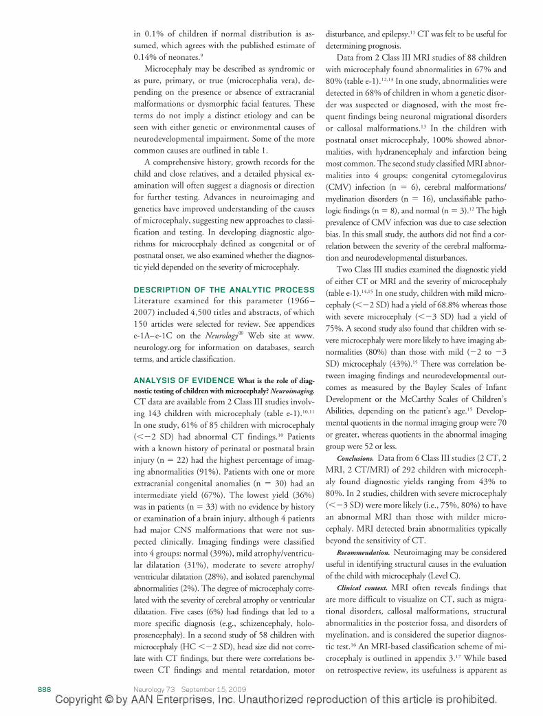
Table 1 Etiologies of congenital and postnatal onset microcephaly
Congenital Postnatal onset
Genetic Genetic
Isolated Inborn errors of metabolism
Autosomal recessive microcephaly Congenital disorders of glycosylation
Autosomal dominant microcephaly Mitochondrial disorders
X-linked microcephaly Peroxisomal disorders
Chromosomal (rare: “apparently”balanced rearrangements and ringchromosomes)
Menkes disease
Amino acidopathies and organic acidurias
Glucose transporter defect
Syndromic Syndromic
Chromosomal
Trisomy 21, 13, 18
Unbalanced rearrangements
Contiguous gene deletion Contiguous gene deletion
4p deletion (Wolf-Hirschhorn syndrome) 17p13.3 deletion (Miller-Dieker syndrome)
5p deletion (cri-du-chat syndrome)
7q11.23 deletion (Williams syndrome)
22q11 deletion (velocardiofacialsyndrome)
Single gene defects Single gene defects
Cornelia de Lange syndrome Rett syndrome
Holoprosencephaly (isolated orsyndromic)
Nijmegen breakage syndrome
Smith-Lemli-Opitz syndrome Ataxia-telangiectasia
Seckel syndrome Cockayne syndrome
Aicardi-Goutieres syndrome
XLAG syndrome
Cohen syndrome
Acquired Acquired
Disruptive injuries Disruptive injuries
Death of a monozygous twin Traumatic brain injury
Ischemic stroke Hypoxic-ischemic encephalopathy
Hemorrhagic stroke Hemorrhagic and ischemic stroke
Infections Infections
TORCHES (toxoplasmosis, rubella,cytomegalovirus, herpes simplex,syphilis) and HIV
Meningitis and encephalitis
Congenital HIV encephalopathy
Teratogens Toxins
Alcohol, hydantoin, radiation Lead poisoning
Maternal phenylketonuria Chronic renal failure
Poorly controlled maternal diabetes
Deprivation Deprivation
Maternal hypothyroidism Hypothyroidism
Maternal folate deficiency Anemia
Maternal malnutrition Malnutrition
Placental insufficiency Congenital heart disease
Reprinted with permission from Elsevier from: Abuelo D. Microcephaly syndromes. SeminPediatr Neurol 2007;14:118 –127. Note that there are approximately 500 listings for mi-crocephaly in OMIM (http://www.ncbi.nlm.nih.gov/omim).
Neurology 73 September 15, 2009 889
order of L-serine biosynthesis; and Amish lethalmicrocephaly, which is associated with 2-ketoglutaricaciduria.22 Metabolic disorders associated with syn-dromic congenital microcephaly are listed in appendixe-2. Metabolic disorders are more likely to cause postna-tal onset microcephaly and are typically associated withglobal developmental delay (GDD). As was publishedin a practice parameter on the topic, the diagnostic yieldof routine screening for inborn errors of metabolism inchildren with GDD is about 1% and the yield mayincrease to 5% in specific situations, such as when mi-crocephaly is present.23
Conclusions. The prevalence of metabolic disordersamong children with microcephaly is unknown.Based on prior analysis of studies of children withGDD, it is likely 1% to 5%.
Recommendation. There is insufficient evidence tosupport or refute obtaining metabolic testing on aroutine basis for the evaluation of the newborn orinfant with microcephaly (Level U).
Clinical context. Microcephaly is common in GDDand the yield of metabolic testing may be higher whenthe following are present: a parental history of consan-guinity, a family history of similar symptoms in rela-tives, episodic symptoms (seizures, ataxia, vomiting,
encephalopathy), developmental regression, extracra-nial organ failure, or specific findings on neuroimag-ing.23 Metabolic testing may also have a higher yield inchildren whose microcephaly remains unexplained afterother evaluations have been done. There are insufficientdata to recommend when and how metabolic testingshould be done, although it is reasonable to test infantswith severe primary congenital microcephaly for the el-evated urine alpha-ketoglutaric acid found in Amish le-thal microcephaly.23
What neurologic disorders are associated with micro-cephaly? Epilepsy. Data from one Class III study involv-ing 66 children with microcephaly (��2 SD) foundan overall prevalence of epilepsy of 40.9%.24 Two ClassIII studies suggest that epilepsy is more common inpostnatal onset than in congenital microcephaly. In onestudy, epilepsy occurred in 50% of children with post-natal onset microcephaly compared to only 35.7% ofthose with congenital microcephaly.24 The second studyfound that epilepsy was 4 times more common in post-natal onset microcephaly.25
Microcephaly is a significant risk factor for medi-cally refractory epilepsy (MRE).26-28 In a Class IIIstudy of 30 children, microcephaly was found in58% of those with MRE compared to 2% in whomseizures were controlled (odds ratio 67.67; p �0.001).27
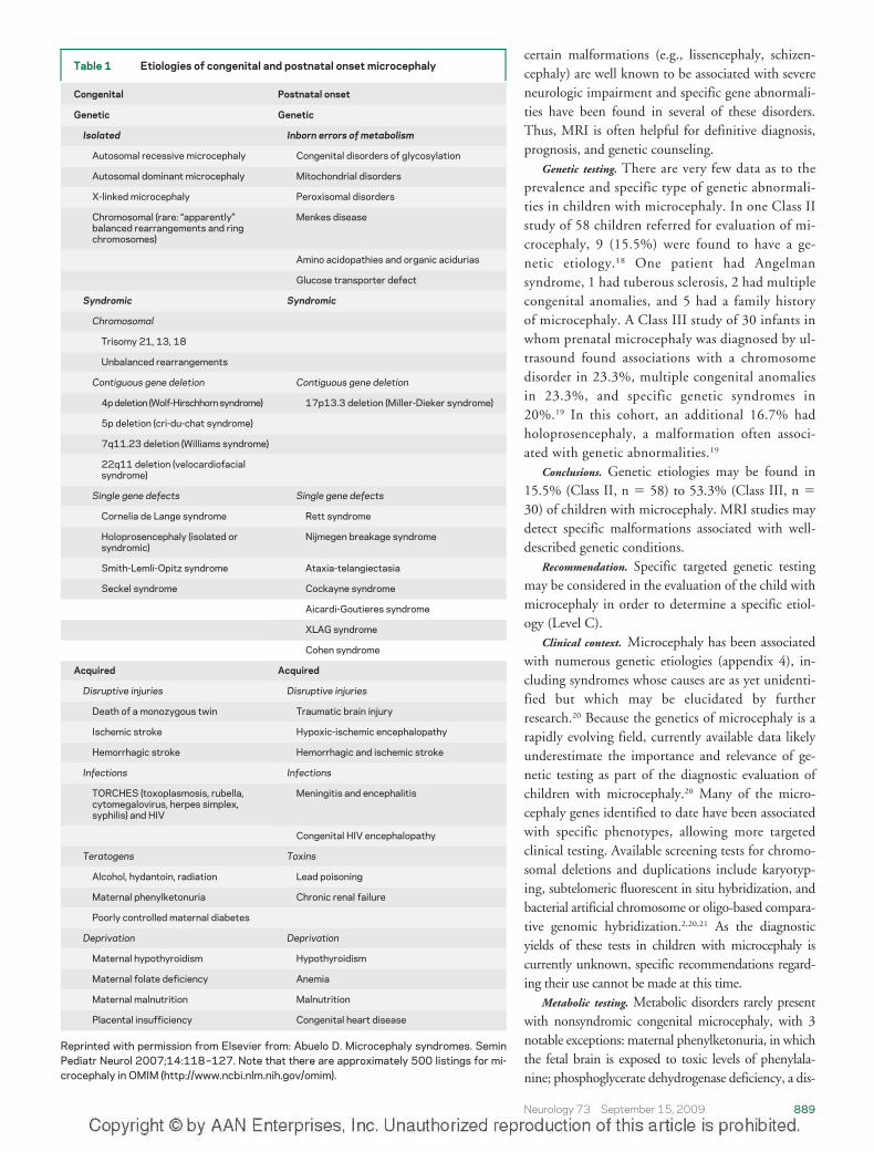
Epilepsy is a prominent feature of some types ofsyndromic microcephaly, which are summarizedin table 2. Studies have not examined the role ofobtaining a routine EEG in children with micro-cephaly. In one Class III study of children withmicrocephaly, EEG abnormalities were found in51% of 39 children who either had no seizures orhad occasional febrile seizures.24 EpileptiformEEG abnormalities were present in 78% of 18children with MRE.
Conclusions. Children with microcephaly are morelikely to have epilepsy, particularly epilepsy that is diffi-cult to treat. Certain microcephaly syndromes are asso-ciated with a much higher prevalence of epilepsy. Thereare no systematic studies regarding EEG testing of chil-dren with microcephaly with and without epilepsy.
Recommendations.
1. Because children with microcephaly are at risk forepilepsy, physicians may consider educating caregiv-ers of children with microcephaly on how to recog-nize clinical seizures (Level C).
2. There are insufficient data to support or refuteobtaining a routine EEG in a child with micro-cephaly (Level U).
Cerebral palsy. Data from a Class II study of chil-dren with developmental disabilities found cerebralpalsy (CP) in 21.4% of the 216 children with micro-
Table 2 Severe epilepsy and microcephaly associated genetic syndromes
Disorder Gene(s)
Structural malformations
Classic lissencephaly (isolated LIS sequence) Lis1, DCX, TUBA1A
Lissencephaly: X-linked with abnormalgenitalia
ARX
Lissencephaly: autosomal recessive withcerebellar hypoplasia
RELN
Bilateral frontoparietal polymicrogyria (COB) GPR56
Periventricular heterotopia with microcephaly ARFGEF2
Schizencephaly EMX2 (rare)
Holoprosencephaly HPE1 21q22.3 HPE 6 2q37.1
HPE2 2p21 HPE7 9q22.3
HPE3 7q36 HPE 8 14q13
HPE4 18p11.3 HPE9 2q14
HPE5 13q32
Syndromes
Wolf-Hirschhorn syndrome 4p�
Angelman syndrome UBE3A,15q11-q13
Rett syndrome Xp22, Xq28
MEHMO (mental retardation, epilepsy,hypogonadism, microcephaly, obesity)
Xp22.13-p21.1
Mowat-Wilson syndrome (microcephaly,mental retardation, distinct facial featureswith/without Hirschsprung disease)
ZFHX1B, 2q22
Data extracted from OMIM (http://www.ncbi.nlm.nih.gov/omim) and the reader is referred tothat source for updated information as new entries are added and data are revised. The readercan also go directly to GeneTests (http://www.genetests.org), to which OMIM links, for updatedinformation regarding the availability of genetic testing on a clinical or research basis.
890 Neurology 73 September 15, 2009
cephaly compared to 8.8% of the 1,159 normoce-phalic children (p � 0.001).29
Two Class I (n � 2,445) studies and one ClassIII (n � 540) study of children with CP found anaverage incidence of congenital microcephaly of1.8%.30-32 In 3 Class III (n � 338) studies, thecombined prevalence of congenital and postnatalonset microcephaly ranged from 32.5% to 81%and averaged 47.9%.33-35 In one of these studies(n � 96), 68% were diagnosed with postnatal on-set microcephaly and 13% had congenital micro-cephaly.33 Others have shown that the yield ofdetermining the etiology of CP is higher when mi-crocephaly is present.36
Conclusions. CP is a common disability in childrenwith microcephaly. Microcephaly, particularly ofpostnatal onset and identifiable etiology, is morecommon in children with CP.
Recommendations.
1. Because children with microcephaly are at risk forCP, physicians and other care providers may con-sider monitoring them for early signs so that sup-portive treatments can be initiated (Level C).
2. Because children with CP are at risk for develop-ing acquired microcephaly, serial HC measure-ments should be followed (Level A).
Mental retardation. What is the prevalence of microcephaly
in different populations? Prevalence estimates of micro-cephaly in Class III surveys of institutionalized pa-tients vary widely from 6.5%35 to 53%.37 Forchildren seen in neurodevelopmental clinics, 3 ClassIII studies (n � 933) found an average prevalence ofmicrocephaly (��2 SD) of 24.7% (range 6% to40.4%).38-40 Similarly, a high rate of severe (��3SD) microcephaly (20%) was found in a Class IIIstudy of 836 children undergoing evaluation formental retardation.e1
A number of studies have looked at the prevalenceand significance of microcephaly in children with ap-parently normal intelligence. One Class II study of1,006 students in mainstream classrooms found that1.9% had mild (�2 to �3 SD) and none hadsevere (��3 SD) microcephaly.e2 The studentswith microcephaly had a similar mean IQ to thenormocephalic group (99.5 vs 105) but had lowermean academic achievement scores (49 vs 70). AClass III study looking at the records of 1,775 nor-mally intelligent adolescents followed by pediatri-cians found 11 (0.6%) with severe microcephaly(��3 SD).e3 Among a separate sample of 106 ad-olescents with mental retardation, the prevalenceof severe microcephaly was 11%.
What is the prevalence of developmental disability in individu-
als with microcephaly? Three Class I studies based on theNational Institute of Neurological Disorders and
Stroke Collaborative Perinatal Project examined dataon microcephaly. In an early report (n � 9,379), halfof the children with microcephaly (males ��2.3SD, females ��2.4 SD) at 1 year of age were foundto have an IQ �80 at 4 years of age.e4 A subsequentstudy (n � 35,704) found congenital microcephaly(��2 SD) in 1.3% and in certain populations thisconferred a twofold risk of mental retardation at 7years of age (15.3% vs 7%).e5 The third study (n �
28,820) found that of normocephalic children, 2.6%were mentally retarded (IQ �70) and 7.4% had bor-derline IQ scores (71–80). Of the 114 (0.4%) chil-dren with mild microcephaly (�2 to �3 SD),10.5% were mentally retarded and 28% had border-line IQ scores.9 Severe microcephaly (��3 SD) wasfound in 41 (0.14%) children, of whom 51.2% werementally retarded and 17% had borderline IQ scores.These findings have been supported by several ClassIII studies.29,e6
A Class II retrospective study of 212 childrenwith microcephaly found a significant correlation be-tween the degree of microcephaly and the presence ofmental retardation. Among the 113 subjects withmild microcephaly (�2 to �3 SD), mental retarda-tion was found in 11%. Mental retardation was diag-nosed in 50% of the 99 subjects with severemicrocephaly (��3 SD) and in all of those with anHC less than �7 SD.e7
A number of Class III studies of children withmicrocephaly have examined other clinical factors.There are conflicting data as to whether proportion-ate microcephaly (i.e., similar weight, height, andhead size percentiles) is predictive of developmentaland learning disabilities.9,e2 Other Class III studieshave shown that early medical illness or brain injuryare associated with microcephaly and mental retarda-tion.e8 The pattern of head growth can thus be apredictor of outcome: infants with normal birthHCs who acquire microcephaly by 1 year of ageare likely to be severely delayed. On the otherhand, studies of children from countries withemerging economies have shown that when micro-cephaly and developmental delay are acquired as aconsequence of malnutrition, poverty, and lack ofstimulation, there is significant potential for reha-bilitation.e9 The findings from these and other se-lected studies are summarized in appendix e-3.
Conclusions. Microcephaly is commonly found indevelopmentally and cognitively impaired children.Children with microcephaly are at a higher risk formental retardation and there is a correlation betweenthe degree of microcephaly and the severity of cogni-tive impairment.
Recommendation. Because children with microceph-aly are at risk for developmental disability, physicians
Neurology 73 September 15, 2009 891
should periodically assess development and academicachievement to determine whether further testing andrehabilitative efforts are warranted (Level A).
Ophthalmologic and audiologic disorders. One Class Istudy of 360 children with severe microcephaly (��3SD) found eye abnormalities in 6.4%, but in only 0.2%of 3,600 age-matched normocephalic controls.e10 A re-lated study found 145 cases of congenital eye malforma-tions in 212,479 consecutive births, but prevalences forindividual malformations could not be ascertained.e11
Microcephaly was among the associated malformationsin 56% of these children.
Appendix e-4 lists microcephaly syndromes inwhich prominent ophthalmologic involvement hasbeen reported. A Boolean search of the Online Men-delian Inheritance in Man (OMIM) database of the499 genetic syndromes associated with microcephalyfound that 241 (48%) of the entries mentioned vari-ous ophthalmologic abnormalities. This searchmethod provides an upper estimate of the frequencywith which ophthalmologic abnormalities might befound in patients with syndromic microcephaly.
A study of 100 children with complex ear anomaliesreported that 85 had neurologic involvement and 13had microcephaly.e12 There are no published studiesregarding the frequency of audiologic disorders in chil-dren with microcephaly. Appendix e-5 lists OMIM mi-crocephaly syndromes in which prominent audiologicinvolvement has been reported. A Boolean search ofOMIM listings of genetic syndromes associated with
microcephaly found 113 (23%) in which hearing losshad been described.
Conclusions. Ophthalmologic disorders are morecommon in children with microcephaly but the fre-quency, nature, and severity of this involvement has notbeen studied. Data on the prevalence of audiologicdisorders in children with microcephaly have notbeen reported.
Recommendation. Screening for ophthalmologic ab-normalities in children with microcephaly may beconsidered (Level C).
Clinical context. Certain microcephaly syndromesare classically characterized by sensory impairments,as listed in appendices e-4 and e-5. Early identifica-tion of visual and hearing deficits may help with boththe identification of a syndromic diagnosis and thesupportive care of the child.
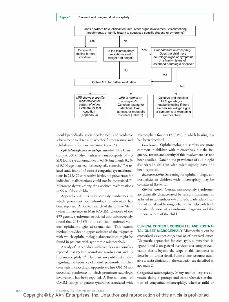
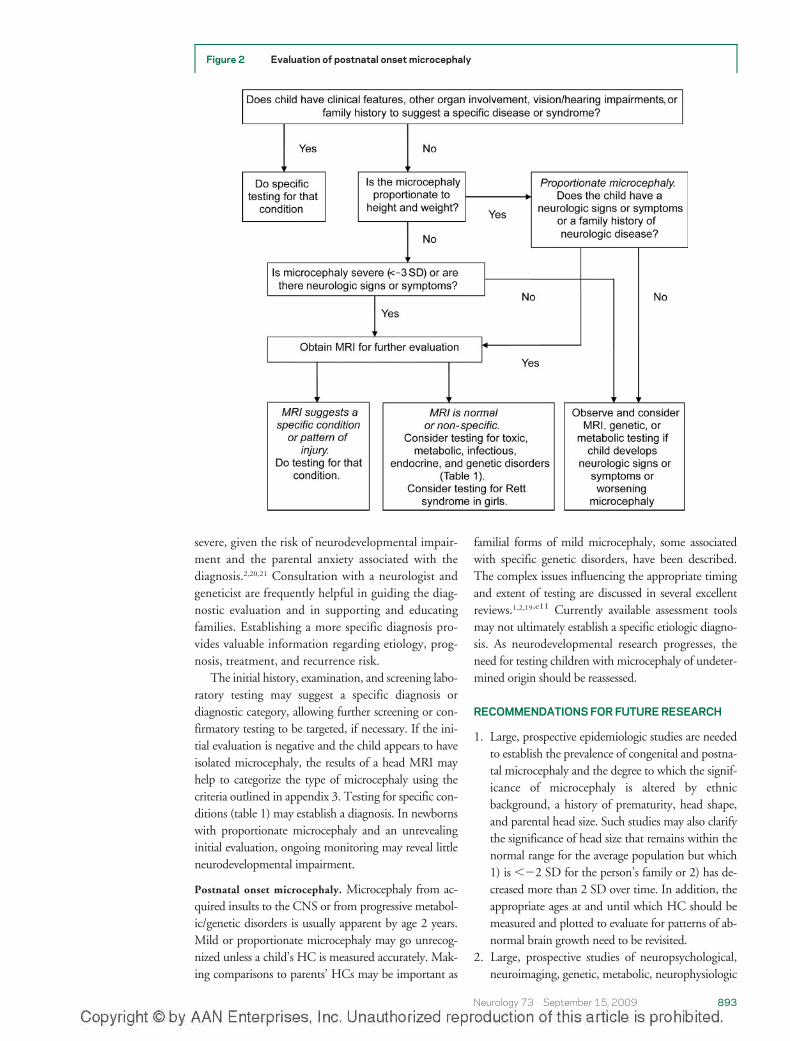
CLINICAL CONTEXT: CONGENITAL AND POSTNA-TAL ONSET MICROCEPHALY Microcephaly can becategorized as either congenital or of postnatal onset.Diagnostic approaches for each type, summarized infigures 1 and 2, are general overviews of a complex eval-uation that is beyond the scope of this parameter todescribe in further detail. Some online resources avail-able to assist clinicians in the evaluation are described inappendix 2.
Congenital microcephaly. Many medical experts ad-vocate doing a prompt and comprehensive evalua-tion of congenital microcephaly, whether mild or
Figure 1 Evaluation of congenital microcephaly
892 Neurology 73 September 15, 2009
severe, given the risk of neurodevelopmental impair-ment and the parental anxiety associated with thediagnosis.2,20,21 Consultation with a neurologist andgeneticist are frequently helpful in guiding the diag-nostic evaluation and in supporting and educatingfamilies. Establishing a more specific diagnosis pro-vides valuable information regarding etiology, prog-nosis, treatment, and recurrence risk.
The initial history, examination, and screening labo-ratory testing may suggest a specific diagnosis ordiagnostic category, allowing further screening or con-firmatory testing to be targeted, if necessary. If the ini-tial evaluation is negative and the child appears to haveisolated microcephaly, the results of a head MRI mayhelp to categorize the type of microcephaly using thecriteria outlined in appendix 3. Testing for specific con-ditions (table 1) may establish a diagnosis. In newbornswith proportionate microcephaly and an unrevealinginitial evaluation, ongoing monitoring may reveal littleneurodevelopmental impairment.
Postnatal onset microcephaly. Microcephaly from ac-quired insults to the CNS or from progressive metabol-ic/genetic disorders is usually apparent by age 2 years.Mild or proportionate microcephaly may go unrecog-nized unless a child’s HC is measured accurately. Mak-ing comparisons to parents’ HCs may be important as
familial forms of mild microcephaly, some associatedwith specific genetic disorders, have been described.The complex issues influencing the appropriate timingand extent of testing are discussed in several excellentreviews.1,2,19,e11 Currently available assessment toolsmay not ultimately establish a specific etiologic diagno-sis. As neurodevelopmental research progresses, theneed for testing children with microcephaly of undeter-mined origin should be reassessed.
RECOMMENDATIONS FOR FUTURE RESEARCH
1. Large, prospective epidemiologic studies are neededto establish the prevalence of congenital and postna-tal microcephaly and the degree to which the signif-icance of microcephaly is altered by ethnicbackground, a history of prematurity, head shape,and parental head size. Such studies may also clarifythe significance of head size that remains within thenormal range for the average population but which1) is ��2 SD for the person’s family or 2) has de-creased more than 2 SD over time. In addition, theappropriate ages at and until which HC should bemeasured and plotted to evaluate for patterns of ab-normal brain growth need to be revisited.
2. Large, prospective studies of neuropsychological,neuroimaging, genetic, metabolic, neurophysiologic
Figure 2 Evaluation of postnatal onset microcephaly
Neurology 73 September 15, 2009 893
(i.e., EEG), and ancillary (vision and hearing) testsshould be undertaken in children with microcephalyto establish the diagnostic yields of these tests andinform the development of an evidence-based algo-rithmic approach to evaluation.
3. The burden of neurodevelopmental disability andcomorbid medical illness in children with micro-cephaly should be more thoroughly studied toguide the provision of preventative and rehabilita-tive services that might improve outcomes.
DISCLOSUREDr. Ashwal serves on the scientific advisory board of the Tuberous Sclero-
sis Association and the International Pediatric Stroke Society; serves as an
editor of Pediatric Neurology; and receives research support from the NIH
[1 R01 NS059770-01A2 (PI), 1 R01 NS054001-01A1 (PI), and R01
CA107164-03 (PI)]. Dr. Michelson reports no disclosures. Dr. Plawner
receives royalties from publishing PEMSoft: The Pediatric Emergency Med-
icine Software (2007 and 2008); receives research support from the NIH
[NO1-HD-3-3351 (Co-investigator); and has served as an expert consul-
tant in a legal proceeding. Dr. Dobyns serves on the editorial advisory
boards of the American Journal of Medical Genetics and Clinical Dysmor-
phology and receives research support from the NIH [1R01-NS050375
(PI) and 1R01-NS058721 (PI)].
DISCLAIMER
This statement is provided as an educational service of the American
Academy of Neurology and the Child Neurology Society. It is based on an
assessment of current scientific and clinical information. It is not intended
to include all possible proper methods of care for a particular neurologic
problem or all legitimate criteria for choosing to use a specific procedure.
Neither is it intended to exclude any reasonable alternative methodolo-
gies. The AAN and the Child Neurology Society recognize that specific
patient care decisions are the prerogative of the patient and the physician
caring for the patient, based on all of the circumstances involved. The
clinical context section is made available in order to place the evidence-
based guideline(s) into perspective with current practice habits and chal-
lenges. No formal practice recommendations should be inferred.
CONFLICT OF INTEREST STATEMENTThe American Academy of Neurology is committed to producing inde-
pendent, critical, and truthful clinical practice guidelines (CPGs). Signifi-
cant efforts are made to minimize the potential for conflicts of interests to
influence the recommendation of this CPG. To the extent possible, the
AAN keeps separate those who have a financial stake in the success or
failure of the products appraised in the CPGs and the developers of the
guidelines. Conflict of interest forms were obtained from all authors and
reviewed by an oversight committee prior to project initiation. AAN lim-
its the participation of authors with substantial conflicts of interest. The
AAN forbids commercial participation in, or funding of, guideline
projects. Drafts of the guidelines have been reviewed by at least three AAN
committees, a network of neurologists, Neurology® peer reviewers, and
representatives from related fields. The AAN Guideline Author Conflict
of Interest Policy can be viewed at http://www.aan.com.
APPENDIX 1AQuality Standards Subcommittee Members 2007–2009: Jacqueline French,
MD, FAAN (Chair); Charles E. Argoff, MD; Eric Ashman, MD; Stephen
Ashwal, MD, FAAN (Ex-Officio); Christopher Bever, Jr., MD, MBA,
FAAN; John D. England, MD, FAAN; Gary M. Franklin, MD, MPH,
FAAN (Ex-Officio); Deborah Hirtz, MD, FAAN (Ex-Officio); Robert G.
Holloway, MD, MPH, FAAN; Donald J. Iverson, MD, FAAN; Steven R.
Messe, MD; Leslie A. Morrison, MD; Pushpa Narayanaswami, MD,
MBBS; James C. Stevens, MD, FAAN (Ex-Officio); David J. Thurman,
MD, MPH (Ex-Officio); Dean M. Wingerchuk, MD, MSc, FRCP(C);
Theresa A. Zesiewicz, MD, FAAN.
APPENDIX 1BChild Neurology Society Practice Committee Members: Bruce Cohen, MD
(Chair); Diane Donley, MD; Bhuwan Garg, MD; Michael Goldstein
(Emeritus); Brian Grabert, MD; David Griesemer, MD; Edward Kovnar,
MD; Agustin Legido, MD; Leslie Morrison, MD; Ben Renfroe, MD;
Shlomo Shinnar, MD; Russell Snyder, MD; Carmela Tardo, MD; Greg
Yim, MD.
APPENDIX 2Resources for evaluating children with microcephaly
1. Accurate head circumference (HC) measurement is obtained with a
flexible non-stretchable measuring tape pulled tightly across the most
prominent part on the back (occiput) and front (supraorbital ridges) of
the head.
Standardized growth charts in percentiles for boys and girls from birth
to age 36 months are available online from the Web site of the National
Center for Health Statistics.
Growth charts for HC for boys and girls from birth to age 5 years and
plotted as standard deviations from the mean are available through the
World Health Organization Web site. These charts, updated in 2006,
are based on data from 8,500 well-nourished children from Brazil,
Ghana, India, Norway, Oman, and the United States.
Measurements from patients older than 36 months can be evaluated
using charts derived in 1968 from pooled data from a few countries
(Nellhaus G. Head circumference from birth to eighteen years: com-
posite international and interracial graphs. Pediatrics 1968;41:106–
110), made available online through the Web site of the Department of
Neurology at Emory University.
Growth charts for premature infants and for children born in countries
other than the United States, including China, India, Korea, and Viet-
nam, can be found online at Web sites specializing in information for
prospective adoptive parents, such as that of the Center for Adoption
Medicine. There is evidence that extremely premature infants (�1 kg)
who survive never catch up to infants with birth weights over 1 kg. In
these cases, if one uses standard HC graphs, many normal children will
appear microcephalic and might be subjected to unnecessary evalua-
tions. (Sheth RD, Mullett MD, Bodensteiner JB, Hobbs GR. Longitu-
dinal head growth in developmentally normal preterm infants. Arch
Pediatr Adolesc Med 1995;149:1358–1361.)
Web sites:
http://www.cdc.gov/growthcharts
http://www.who.int/childgrowth/standards/hc_for_age/en/index.html
http://www.pediatrics.emory.edu/divisions/neurology/hc.pdf
http://www.adoptmed.org/topics/growth-charts.html
2. The freely searchable Online Mendelian Inheritance in Man (OMIM)
database contains close to 500 entries for genetic disorders associated
with microcephaly.
Web site: http://www.ncbi.nlm.nih.gov/omim
3. GeneTests is a publicly funded medical genetics information resource
that provides reviews of genetic disorders causing microcephaly and a
directory of laboratories that perform confirmatory genetic and enzy-
matic testing.
Web site: http://www.genetests.org
4. Pictures of Standard Syndromes and Undiagnosed Malformations
(POSSUM) is a computer-based system that can be purchased for
approximately $1,000. It contains information on more than 3,000
syndromes, including chromosomal and metabolic disorders associated
with multiple malformations and skeletal dysplasias.
Web site: http://www.possum.net.au
5. The London Dysmorphology Database, London Neurogenetics Data-
base, and Dysmorphology Photo Library on CD-ROM (2001, 3rd
edition) combine 2 comprehensive databases and an extensive photo
library onto a single CD-ROM that can be purchased for $2,495.
894 Neurology 73 September 15, 2009
There are more than 3,400 nonchromosomal syndromes in the dys-
morphology database and nearly 3,300 neurologic disorders in the
neurogenetics database, with online updates made available to regis-
tered users.
Web site: http://www.lmdatabases.com
APPENDIX 3MRI-based classification of microcephaly*
1. Microcephaly with normal to thin cortex
a. Autosomal recessive microcephaly
i. Autosomal recessive microcephaly with normal or slightly
short stature and high function
(a) MCPH1 mutations
(b) ASPM mutations
(c) CDK5RAP2 mutations
(d) CENPJ mutations
ii. Autosomal recessive microcephaly with normal or minor short
stature and very poor function
(a) Profound microcephaly—Amish-type lethal microcephaly
(SLC25A19 mutations)
(b) Less severe microcephaly with periventricular nodular het-
erotopia (ARFGEF2 mutations)
(c) Less severe microcephaly with abnormal frontal cortex
and thin corpus callosum—Warburg micro syndrome
(RAB3GAP mutations)
b. Extreme microcephaly with simplified gyral pattern and normal
stature
i. Extreme microcephaly with jejunal atresia
ii. Microcephaly with pontocerebellar hypoplasia
c. Primary microcephaly, not otherwise classified
2. Microlissencephaly (extreme microcephaly with thick cortex)
a. MLIS with thick cortex (Norman-Roberts syndrome)
b. MLIS with thick cortex, severe brainstem and cerebellar hypopla-
sia (Barth MLIS syndrome)
c. MLIS with severe, proportional short stature—Seckel syndrome
(ATR mutation)
d. MLIS with mildly to moderately thick (6-mm to 8-mm) cortex,
callosal agenesis
3. Microcephaly with polymicrogyria or other cortical dysplasias
a. Extreme microcephaly with diffuse or asymmetric polymicrogyria
b. Extreme microcephaly with ACC and cortical dysplasia
*Adapted from Barkovich AJ, Kuzniecky RI, Jackson GD, Guerrini R,
Dobyns WB. A developmental and genetic classification for malforma-
tions of cortical development. Neurology 2005;65:1873–1887.
The reader can also go directly to GeneTests (http://www.genetests.
org), to which OMIM links, for updated information regarding the availabil-
ity of genetic testing on a clinical or research basis.
APPENDIX 4Syndromic classification of primary microcephaly andassociated genes*
Autosomal recessive microcephaly (OMIM 251200)
MCPH1 (Microcephalin; 8p22-pter)
MCPH2 (19q13.1-13.2)
MCPH3 (CDK5RAP2; 9q34)
MCPH4 (15q15-q21)
MCPH5 (ASPM; 1q31 )
MCPH6 (CENPJ; 13q12.2 )
Microcephaly with severe IUGR
ATR Seckel syndrome
PCNT2 microcephalic osteodysplastic primordial dwarfism, type
2; Seckel syndrome
Microcephaly with a simplified gyral pattern (OMIM 603802)
Autosomal dominant microcephaly (OMIM 156580)
Amish lethal microcephaly (OMIM 607196)
Other genes
AKT3 severe postnatal microcephaly
SLC25A19 Amish lethal microcephaly
LIS1 lissencephaly
DCX lissencephaly (X-linked)
SHH holoprosencephaly
ZIC2 holoprosencephaly
TGIF holoprosencephaly
SIX3 holoprosencephaly
DHCR7 Smith-Lemli-Opitz syndrome
CREBBP Rubinstein-Taybi syndrome
PAK3 X-linked mental retardation
NBS1 Nijmegen breakage syndrome
MECP2 Rett syndrome (X-linked)
*Inheritance is autosomal recessive excepted where noted. Data collated
from Mochida and Walsh20; Alderton GK, Galbiati L, Griffith E, et al.
Regulation of mitotic entry by microcephalin and its overlap with ATR
signaling. Nat Cell Biol 2006;8:725–733; Bond J, Woods CG. Cytoskel-
etal genes regulating brain size. Curr Opin Cell Biol 2006;18:95–101;
Woods CG, Bond J, Enard W. Autosomal recessive primary microcephaly
(MCPH): a review of clinical, molecular, and evolutionary findings. Am J
Hum Genet 2005;76:717–728.
The reader can also go directly to GeneTests (http://www.genetests.
org), to which OMIM links, for updated information regarding the avail-
ability of genetic testing on a clinical or research basis.
Received December 18, 2008. Accepted in final form July 7, 2009.
REFERENCES1. Leviton A, Holmes LB, Allred EN, Vargas J. Method-
ologic issues in epidemiologic studies of congenital micro-cephaly. Early Hum Dev 2002;69:91–105.
2. Opitz JM, Holt MC. Microcephaly: general considerationsand aids to nosology. J Craniofac Genet Dev Biol 1990;10:75–204.
3. Roche AF, Mukherjee D, Guo SM, Moore WM. Headcircumference reference data: birth to 18 years. Pediatrics1987;79:706–712.
4. Barkovich AJ, Ferriero DM, Barr RM, et al. Microlissen-cephaly: a heterogeneous malformation of cortical devel-opment. Neuropediatrics 1999;29:113–119.
5. Dobyns WB, Andermann E, Andermann F, et al. X-linkedmalformations of neuronal migration. Neurology 1996;47:331–339.
6. Woods CG. Human microcephaly. Curr Opin Neurobiol2004;14:112–117.
7. Jackson AP, Eastwood H, Bell SM, et al. Identification ofmicrocephalin, a protein implicated in determining the size ofthe human brain. Am J Hum Genet 2002;71:136–142.
8. Vargas JE, Allred EN, Leviton A, Holmes LB. Congenitalmicrocephaly: phenotypic features in a consecutive sampleof newborn infants. J Pediatr 2001;139:210–214.
9. Dolk H. The predictive value of microcephaly during thefirst year of life for mental retardation at seven years. DevMed Child Neurol 1991;33:974–983.
10. Jaworski M, Hersh JH, Donat J, Shearer LT, Weisskopf B.Computed tomography of the head in the evaluation ofmicrocephaly. Pediatrics 1986;78:1064–1069.
11. Ito M, Okuno T, Mikawa H. Computed tomographicstudy of children with microcephaly. No To Hattatsu1989;21:440–444.
12. Steinlin M, Zurrer M, Martin E, Boesch CH, Largo RH,Bottshauser E. Contribution of MRI in the evaluation ofmicrocephaly. Neuropediatrics 1991;22:184–189.
13. Sugimoto T, Yasuhara A, Nishida N, Murakami K, WooM, Kobayashi Y. MRI of the head in the evaluation ofmicrocephaly. Neuropediatrics 1993;24:4–7.
Neurology 73 September 15, 2009 895
14. Pandey A, Phadke SR, Gupta N, Phadke RV. Neuroimagingin mental retardation. Indian J Pediatr 2004;71:203–209.
15. Custer DA, Vezina LG, Vaught DR, et al. Neurodevelop-mental and neuroimaging correlates in nonsyndromal mi-crocephalic children. J Dev Behav Pediatr 2000;21:12–18.
16. Wycliffe ND, Thompson JR, Holshouser BA, Ashwal S. Pe-diatric neuroimaging. In: Swaiman KF, Ashwal S, FerrieroDM. Pediatric Neurology Principles & Practice, Fourth edi-tion. Philadelphia: Mosby Elsevier; 2006:167–202.
17. Barkovich AJ, Kuzniecky RI, Jackson GD, Guerrini R,Dobyns WB. A developmental and genetic classificationfor malformations of cortical development. Neurology2005;65:1873–1887.
18. Lalaguna-Mallada P, Alonso-del Val B, Abio-Albero S,Pena-Segura JL, Rebage V, Lopez-Pison J. Microcephalusas the reason for visiting a regional referral neuropaediatricservice. Rev Neurol 2004;38:106–110.
19. den Hollander NS, Wessels MW, Los FJ, Ursem NT,Niermeijer MF, Wladimiroff JW. Congenital micro-cephaly detected by prenatal ultrasound: genetic aspectsand clinical significance. Ultrasound Obstet Gynecol2000;15:282–287.
20. Mochida GH, Walsh CA. Molecular genetics of humanmicrocephaly. Curr Opin Neurol 2001;14:151–156.
21. Abuelo D. Microcephaly syndromes. Semin Pediatr Neu-rol 2007;14:118–127.
22. Kelley RI, Robinson D, Puffenberger EG, Strauss KA, Mor-ton DH. Amish lethal microcephaly: a new metabolic disor-der with severe congenital microcephaly and 2-ketoglutaricaciduria. Am J Med Genet 2002;112:318–326.
23. Shevell M, Ashwal S, Donley D, et al. Practice parameter:evaluation of the child with global developmental delay:report of the Quality Standards Subcommittee of theAmerican Academy of Neurology and The Practice Com-mittee of the Child Neurology Society. Neurology 2003;60:367–380.
24. Abdel-Salam GM, Halasz AA, Czeizel AE. Association ofepilepsy with different groups of microcephaly. Dev MedChild Neurol 2000;42:760–767.
25. Qazi QH, Reed TE. A problem in diagnosis of primaryversus secondary microcephaly. Clin Genet 1973;4:46–52.
26. Berg AT, Levy SR, Novotny EJ, Shinnar S. Predictors ofintractable epilepsy in childhood: a case-control study.Epilepsia 1996;37:24–30.
27. Chawla S, Aneja S, Kashyap R, Mallika V. Etiology andclinical predictors of intractable epilepsy. Pediatr Neurol2002;27:186–191.
28. Aneja S, Ahuja B, Taluja V, Bhatia VK. Epilepsy in childrenwith cerebral palsy. Indian J Pediatr 2001;68:111–115.
29. Watemberg N, Silver S, Harel S, Lerman-Sagie T. Signifi-cance of microcephaly among children with developmentaldisabilities. J Child Neurol 2002;17:117–122.
30. Croen LA, Grether JK, Curry CJ, Nelson KB. Congenitalabnormalities among children with cerebral palsy: moreevidence for prenatal antecedents. J Pediatr 2001;138:804–810.
31. Pharoah PO. Prevalence and pathogenesis of congenitalanomalies in cerebral palsy. Arch Dis Child Fetal NeonatalEd 2007;92:F489–443.
32. Laisram N, Srivastava VK, Srivastava RK. Cerebral palsy:an etiological study. Indian J Pediatr 1992;59:723–728.
33. Edebol-Tysk K. Epidemiology of spastic tetraplegic cere-bral palsy in Sweden: I: impairments and disabilities. Neu-ropediatrics 1989;20:41–45.
34. Lubis MU, Tjipta GD, Marbun MD, Saing B. Cerebralpalsy. Paediatr Indones 1990;30:65–70.
35. Suzuki J, Ito M, Tomiwa K, Okuno T. A clinical study ofcerebral palsy in Shiga; 1977–1986: II: severity of the dis-ability and complications in various types of cerebral palsy.No To Hattatsu 1999;31:336–342.
36. Shevell MI, Majnemer A, Morin I. Etiologic yield of cere-bral palsy: a contemporary case series. Pediatr Neurol2003;28:352–359.
37. Roboz P. Microcephaly. Aust J Ment Retard 1973;2:173–179.
38. Smith RD. Abnormal head circumference in learning-disabled children. Dev Med Child Neurol 1981;23:626–632.
39. Martin HP. Microcephaly and mental retardation. Am JDis Child 1970;119:128–131.
40. Desch LW, Anderson SK, Snow JH. Relationship of headcircumference to measures of school performance. Clin Pe-diatr 1990;29:389–392.
896 Neurology 73 September 15, 2009
Editor’s Note to Authors and Readers: Levels of Evidence in Neurology®
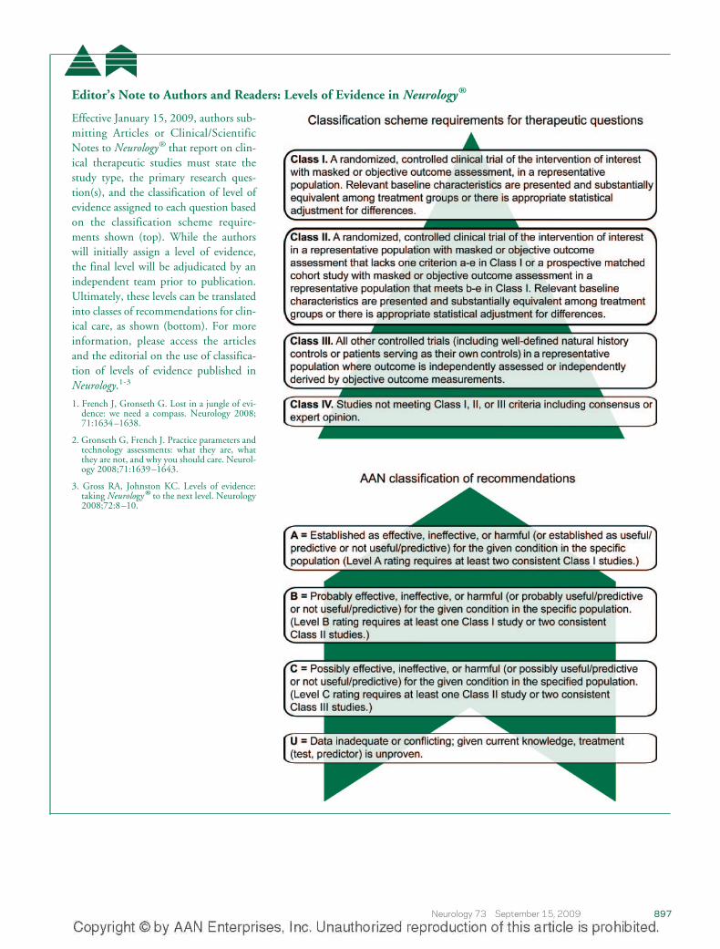
Effective January 15, 2009, authors sub-mitting Articles or Clinical/ScientificNotes to Neurology® that report on clin-ical therapeutic studies must state thestudy type, the primary research ques-tion(s), and the classification of level ofevidence assigned to each question basedon the classification scheme require-ments shown (top). While the authorswill initially assign a level of evidence,the final level will be adjudicated by anindependent team prior to publication.Ultimately, these levels can be translatedinto classes of recommendations for clin-ical care, as shown (bottom). For moreinformation, please access the articlesand the editorial on the use of classifica-tion of levels of evidence published inNeurology.1-3
1. French J, Gronseth G. Lost in a jungle of evi-dence: we need a compass. Neurology 2008;71:1634–1638.
2. Gronseth G, French J. Practice parameters andtechnology assessments: what they are, whatthey are not, and why you should care. Neurol-ogy 2008;71:1639–1643.
3. Gross RA, Johnston KC. Levels of evidence:taking Neurology® to the next level. Neurology2008;72:8–10.
Neurology 73 September 15, 2009 897
DOI 10.1212/WNL.0b013e3181b783f7 2009;73;887Neurology
Stephen Ashwal, David Michelson, Lauren Plawner, et al.Academy of Neurology and the Practice Committee of the Child Neurology Society
review) : Report of the Quality Standards Subcommittee of the American Practice Parameter: Evaluation of the child with microcephaly (an evidence-based
April 24, 2013This information is current as of
ServicesUpdated Information &
http://www.neurology.org/content/73/11/887.full.htmlincluding high resolution figures, can be found at:
Supplementary Material
DC2.htmlhttp://www.neurology.org/content/suppl/2009/09/13/73.11.887.
DC1.htmlhttp://www.neurology.org/content/suppl/2009/09/13/73.11.887.
DC3.htmlhttp://www.neurology.org/content/suppl/2010/09/12/73.11.887.Supplementary material can be found at:
Referenceshttp://www.neurology.org/content/73/11/887.full.html#ref-list-1This article cites 39 articles, 8 of which can be accessed free at:
Citations
rlshttp://www.neurology.org/content/73/11/887.full.html#related-uThis article has been cited by 7 HighWire-hosted articles:
Subspecialty Collections
http://www.neurology.org/cgi/collection/mental_retardationMental retardation
rshttp://www.neurology.org/cgi/collection/developmental_disordeDevelopmental disorders
http://www.neurology.org/cgi/collection/all_geneticsAll Geneticsfollowing collection(s):This article, along with others on similar topics, appears in the
Permissions & Licensing
http://www.neurology.org/misc/about.xhtml#permissionstables) or in its entirety can be found online at: Information about reproducing this article in parts (figures,
Reprints http://www.neurology.org/misc/addir.xhtml#reprintsus
Information about ordering reprints can be found online: