Embed Size (px)
DESCRIPTION
Frank A. Voelker, DVM, DACVP Pathology Experts LLC. Practical Image Analysis from a Pathologist’s Perspective. Topics……. Introduction General Concepts and Approaches Guidelines and Pitfalls Analytical Strategies Applications and using Genie™ Summary. General Analytical Approaches……. - PowerPoint PPT Presentation
Citation preview
Frank A. Voelker, DVM, DACVPPathology Experts LLC
Practical Image Analysis from a Pathologist’s Perspective
2/4/2008Image Analysis in Pathology 2
Topics…….
Introduction
General Concepts and Approaches
Guidelines and Pitfalls
Analytical Strategies
Applications and using Genie™
Summary
2/4/2008Image Analysis in Pathology 3
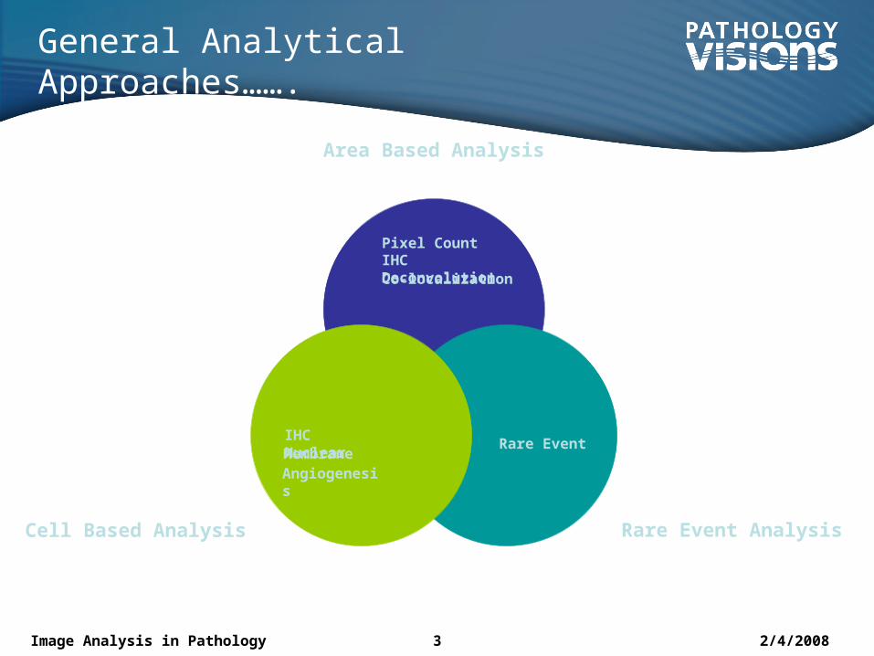
General Analytical Approaches…….
Area Based Analysis
Rare Event AnalysisCell Based Analysis
Pixel CountIHC DeconvolutionCo-localization
Rare EventIHC NuclearMembraneAngiogenesis
2/4/2008Image Analysis in Pathology 4
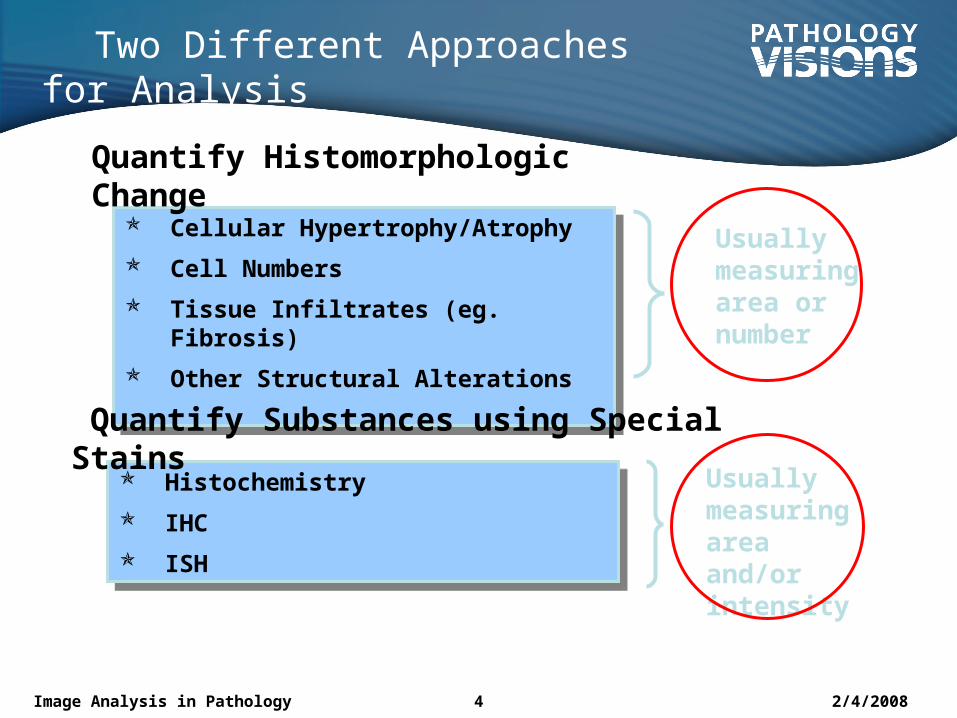
Two Different Approaches for Analysis
Cellular Hypertrophy/Atrophy
Cell Numbers
Tissue Infiltrates (eg. Fibrosis)
Other Structural Alterations
Cellular Hypertrophy/Atrophy
Cell Numbers
Tissue Infiltrates (eg. Fibrosis)
Other Structural Alterations
Histochemistry
IHC
ISH
Histochemistry
IHC
ISH
Quantify Substances using Special Stains
Usually measuring area or number
Usually measuring area and/or intensity
Quantify Histomorphologic Change
2/4/2008Image Analysis in Pathology 5
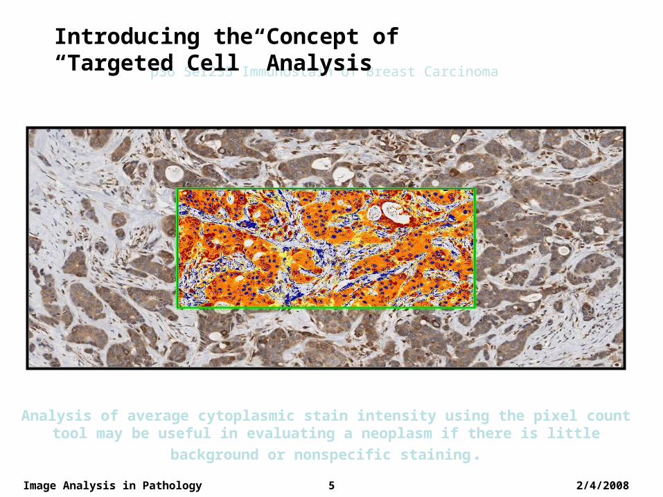
pS6 Ser235 Immunostain of Breast Carcinoma
Analysis of average cytoplasmic stain intensity using the pixel count tool may be useful in evaluating a neoplasm if there is little background or
nonspecific staining.
Introducing the Concept of “Targeted Cell” Analysis
2/4/2008Image Analysis in Pathology 6
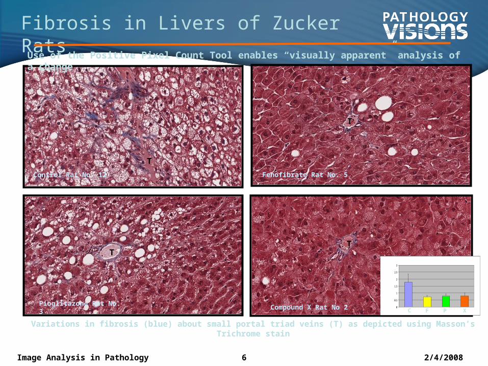
Fibrosis in Livers of Zucker Rats
Control Rat No. 12 Fenofibrate Rat No. 5
Pioglitazone Rat No. 3
Variations in fibrosis (blue) about small portal triad veins (T) as depicted using Masson’s Trichrome stain
C
Compound X Rat No 2
T
T
T
T
0
0.5
1
1.5
2
2.5
3
C F P X
Use of the Positive Pixel Count Tool enables “visually apparent” analysis of a change
2/4/2008Image Analysis in Pathology 7
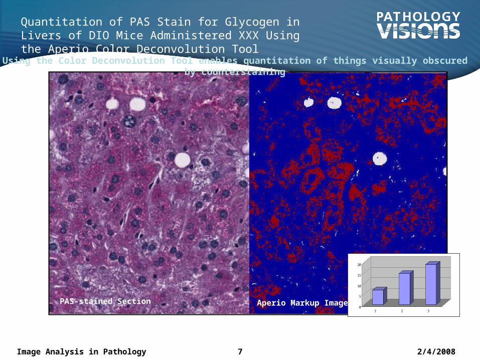
Quantitation of PAS Stain for Glycogen in Livers of DIO Mice Administered XXX Using the Aperio Color Deconvolution Tool
PAS-stained Section Aperio Markup Image
0
5
10
15
20
1 2 3
Using the Color Deconvolution Tool enables quantitation of things visually obscured by counterstaining
8 Image Analysis in DD / Voelker / 09/12/06
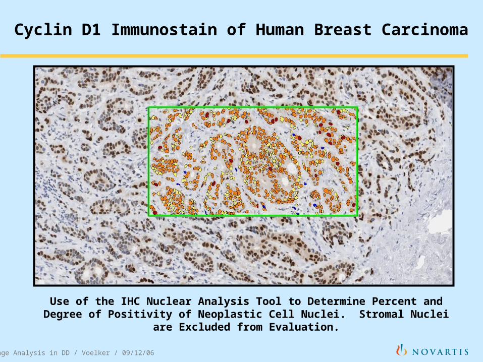
Cyclin D1 Immunostain of Human Breast Carcinoma
Use of the IHC Nuclear Analysis Tool to Determine Percent and Degree of Positivity of Neoplastic Cell Nuclei. Stromal Nuclei are Excluded
from Evaluation.
2/4/2008Image Analysis in Pathology 9
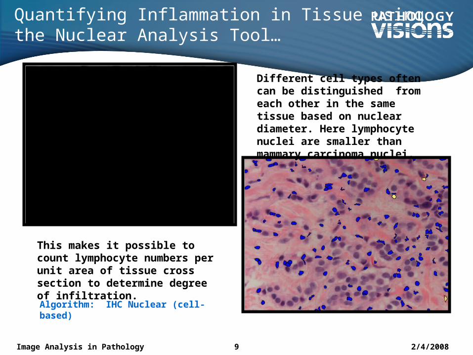
Quantifying Inflammation in Tissue using the Nuclear Analysis Tool…
Different cell types often can be distinguished from each other in the same tissue based on nuclear diameter. Here lymphocyte nuclei are smaller than mammary carcinoma nuclei.
This makes it possible to count lymphocyte numbers per unit area of tissue cross section to determine degree of infiltration.Algorithm: IHC Nuclear (cell-based)
2/4/2008Image Analysis in Pathology 10
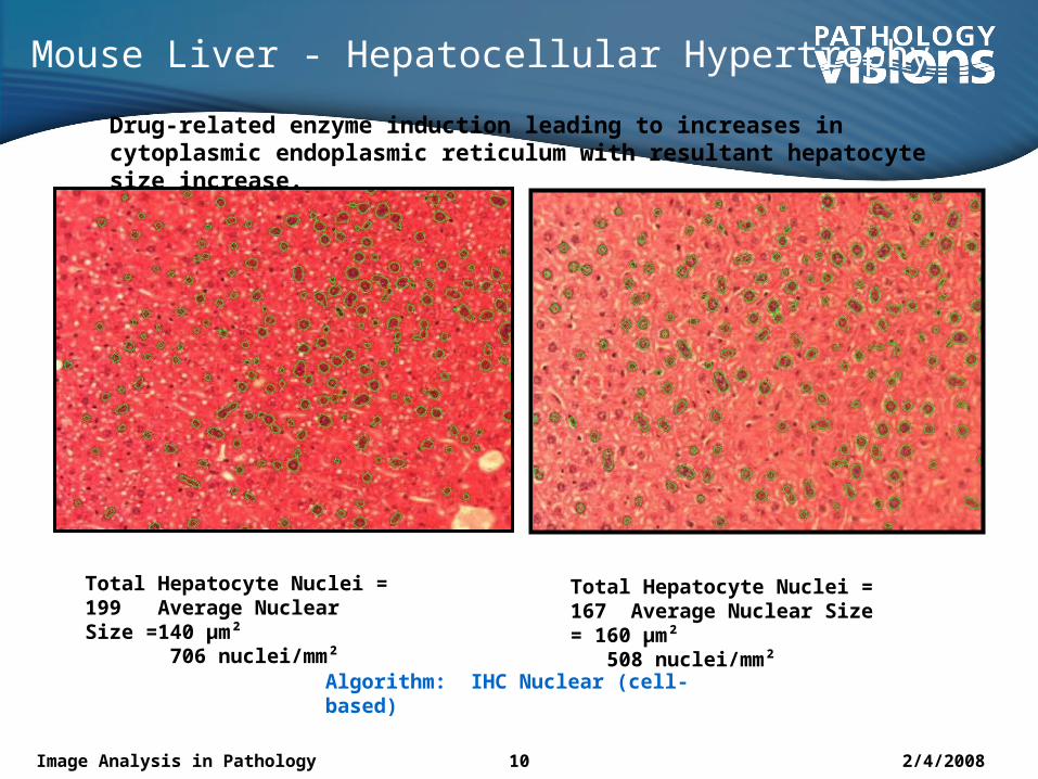
Mouse Liver - Hepatocellular Hypertrophy
Total Hepatocyte Nuclei = 167 Average Nuclear Size = 160 µm² 508 nuclei/mm²
Total Hepatocyte Nuclei = 199 Average Nuclear Size =140 µm² 706 nuclei/mm²
Algorithm: IHC Nuclear (cell-based)
Drug-related enzyme induction leading to increases in cytoplasmic endoplasmic reticulum with resultant hepatocyte size increase.
2/4/2008Image Analysis in Pathology 11
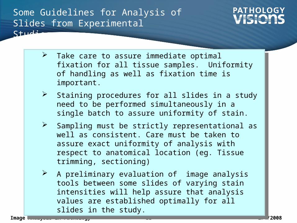
Some Guidelines for Analysis of Slides from Experimental Studies
Take care to assure immediate optimal fixation for all tissue samples. Uniformity of handling as well as fixation time is important.
Staining procedures for all slides in a study need to be performed simultaneously in a single batch to assure uniformity of stain.
Sampling must be strictly representational as well as consistent. Care must be taken to assure exact uniformity of analysis with respect to anatomical location (eg. Tissue trimming, sectioning)
A preliminary evaluation of image analysis tools between some slides of varying stain intensities will help assure that analysis values are established optimally for all slides in the study.
2/4/2008Image Analysis in Pathology 12
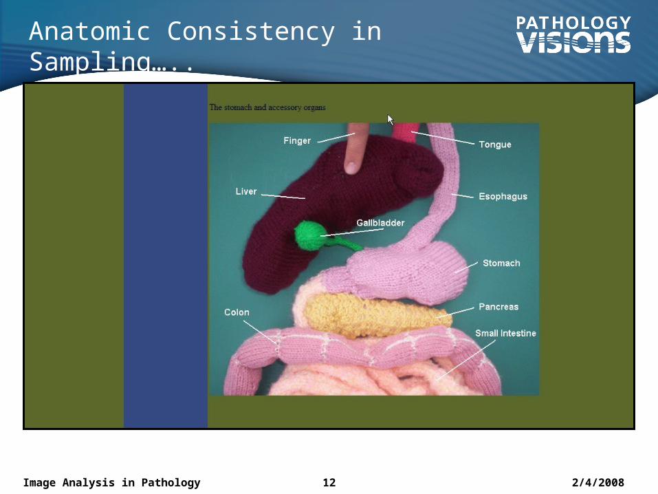
Anatomic Consistency in Sampling…..
2/4/2008Image Analysis in Pathology 13
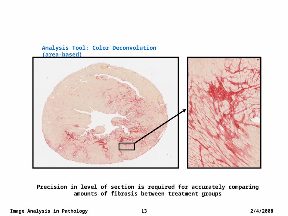
Sirius Red Stain Depicting Myocardial Fibrosis in a Mouse
Precision in level of section is required for accurately comparing amounts of fibrosis between treatment groups
Analysis Tool: Color Deconvolution (area-based)
2/4/2008Image Analysis in Pathology 14
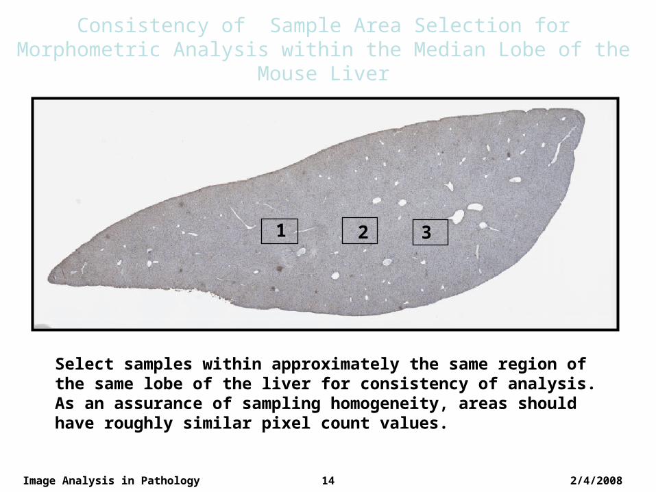
Consistency of Sample Area Selection for Morphometric Analysis within the Median Lobe of the Mouse Liver
1 2 3
Select samples within approximately the same region of the same lobe of the liver for consistency of analysis. As an assurance of sampling homogeneity, areas should have roughly similar pixel count values.
2/4/2008Image Analysis in Pathology 15
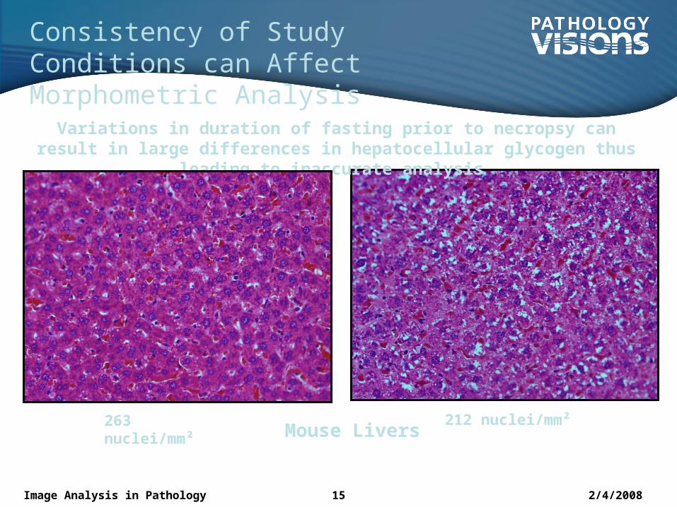
Consistency of Study Conditions can Affect Morphometric Analysis Variations in duration of fasting prior to necropsy can result in
large differences in hepatocellular glycogen thus leading to inaccurate analysis
Mouse Livers
263 nuclei/mm²
212 nuclei/mm²
2/4/2008Image Analysis in Pathology 16
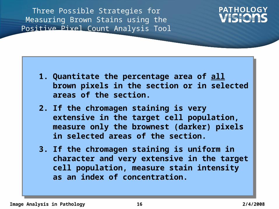
Three Possible Strategies for Measuring Brown Stains using the Positive Pixel Count Analysis Tool
1. Quantitate the percentage area of all brown pixels in the section or in selected areas of the section.
2. If the chromagen staining is very extensive in the target cell population, measure only the brownest (darker) pixels in selected areas of the section.
3. If the chromagen staining is uniform in character and very extensive in the target cell population, measure stain intensity as an index of concentration.
2/4/2008Image Analysis in Pathology 17
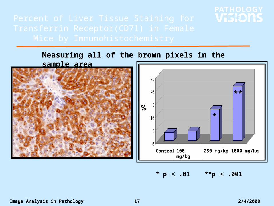
Percent of Liver Tissue Staining for Transferrin Receptor(CD71) in Female Mice by
Immunohistochemistry
* p .01 **p .001
0
5
10
15
20
25
1 2 3 4
*
**
Control 100 mg/kg
250 mg/kg1000 mg/kg
%
Measuring all of the brown pixels in the sample area
18 Aperio in TBD / Voelker / 08/24/06
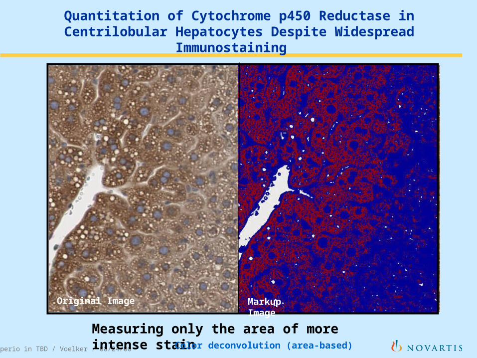
Quantitation of Cytochrome p450 Reductase in Centrilobular Hepatocytes Despite Widespread Immunostaining
Original Image Markup Image
Measuring only the area of more intense stainColor deconvolution (area-based)
2/4/2008Image Analysis in Pathology 19
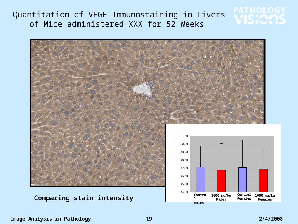
Quantitation of VEGF Immunostaining in Livers of Mice administered XXX for 52 Weeks
44.00
45.00
46.00
47.00
48.00
49.00
50.00
51.00
Control Males
Control Females
1000 mg/kg Males
1000 mg/kg FemalesComparing stain intensity
2/4/2008Image Analysis in Pathology 20
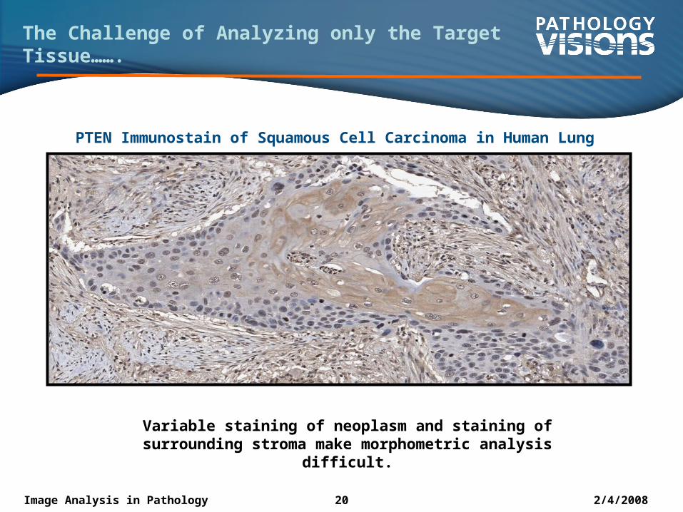
Variable staining of neoplasm and staining of surrounding stroma make morphometric analysis difficult.
PTEN Immunostain of Squamous Cell Carcinoma in Human Lung
The Challenge of Analyzing only the Target Tissue…….
2/4/2008Image Analysis in Pathology 21
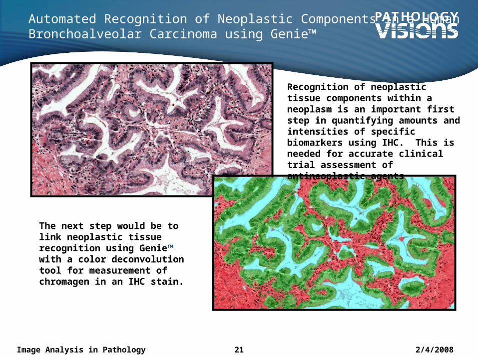
Automated Recognition of Neoplastic Components in a Human Bronchoalveolar Carcinoma using Genie™
Recognition of neoplastic tissue components within a neoplasm is an important first step in quantifying amounts and intensities of specific biomarkers using IHC. This is needed for accurate clinical trial assessment of antineoplastic agents
The next step would be to link neoplastic tissue recognition using Genie™ with a color deconvolution tool for measurement of chromagen in an IHC stain.
2/4/2008Image Analysis in Pathology 22
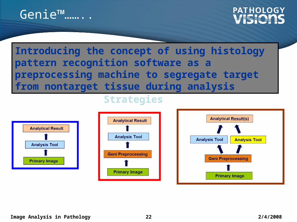
Genie™……..
Introducing the concept of using histology pattern recognition software as a preprocessing machine to segregate target from nontarget tissue during analysis
Strategies
2/4/2008Image Analysis in Pathology 23
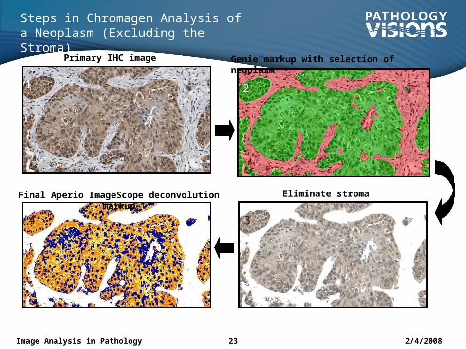
Steps in Chromagen Analysis of a Neoplasm (Excluding the Stroma)
Primary IHC image Genie™markup with selection of neoplasm
Eliminate stroma Final Aperio ImageScope deconvolution markup
1 2
34
2/4/2008Image Analysis in Pathology 24
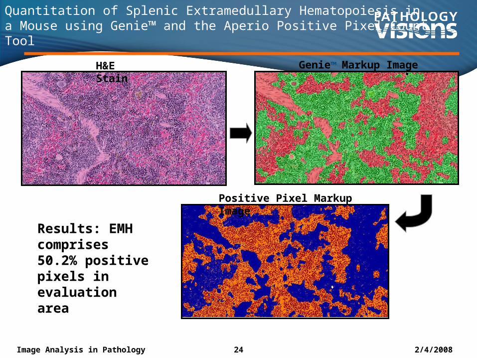
Quantitation of Splenic Extramedullary Hematopoiesis in a Mouse using Genie™ and the Aperio Positive Pixel Count Tool
H&E Stain
Genie™ Markup Image
Positive Pixel Markup Image
Results: EMH comprises 50.2% positive pixels in evaluation area
2/4/2008Image Analysis in Pathology 25
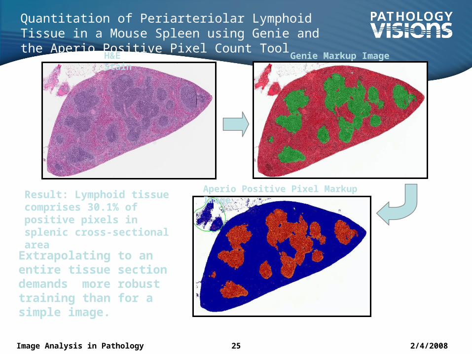
Quantitation of Periarteriolar Lymphoid Tissue in a Mouse Spleen using Genie and the Aperio Positive Pixel Count Tool
Aperio Positive Pixel Markup Image
H&E Stain
Genie Markup Image
Result: Lymphoid tissue comprises 30.1% of positive pixels in splenic cross-sectional area
Extrapolating to an entire tissue section demands more robust training than for a simple image.

2/4/2008Image Analysis in Pathology 26
Analysis of Study Sample Groups by Genie™
Targeted Tissue Selection and Isolation by Genie™
Subsequent Uniform Analysis of Isolated Target Tissue for area/intensity
Morphologically Variable Samples Trained Individually for Genie Target Tissue Selection
Separate target tissue training of each sample does not adversely affect final analysis.
2/4/2008Image Analysis in Pathology 27
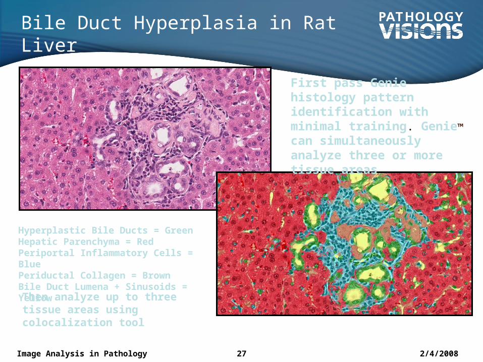
Bile Duct Hyperplasia in Rat Liver
Hyperplastic Bile Ducts = GreenHepatic Parenchyma = RedPeriportal Inflammatory Cells = BluePeriductal Collagen = BrownBile Duct Lumena + Sinusoids = Yellow
First pass Genie histology pattern identification with minimal training. Genie™ can simultaneously analyze three or more tissue areas
Then analyze up to three tissue areas using colocalization tool
2/4/2008Image Analysis in Pathology 28
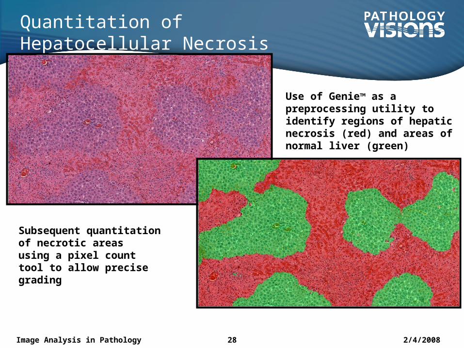
Quantitation of Hepatocellular Necrosis
Use of Genie™ as a preprocessing utility to identify regions of hepatic necrosis (red) and areas of normal liver (green)
Subsequent quantitation of necrotic areas using a pixel count tool to allow precise grading
2/4/2008Image Analysis in Pathology 29
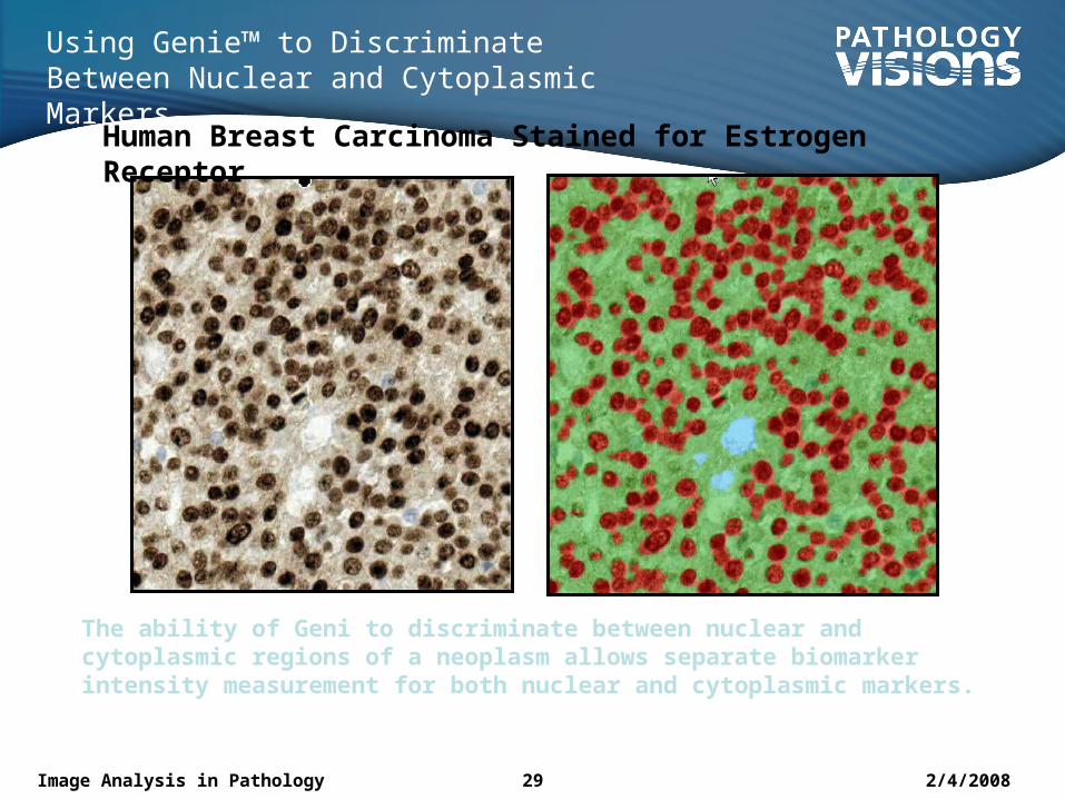
Using Genie™ to Discriminate Between Nuclear and Cytoplasmic Markers
Human Breast Carcinoma Stained for Estrogen Receptor
The ability of Geni to discriminate between nuclear and cytoplasmic regions of a neoplasm allows separate biomarker intensity measurement for both nuclear and cytoplasmic markers.
2/4/2008Image Analysis in Pathology 30
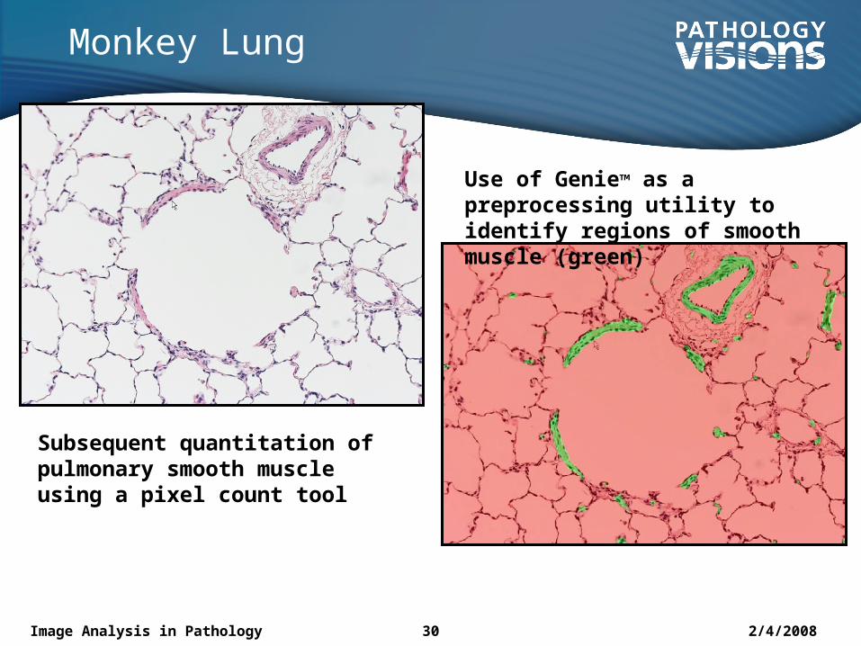
Monkey Lung
Use of Genie™ as a preprocessing utility to identify regions of smooth muscle (green)
Subsequent quantitation of pulmonary smooth muscle using a pixel count tool
2/4/2008Image Analysis in Pathology 31
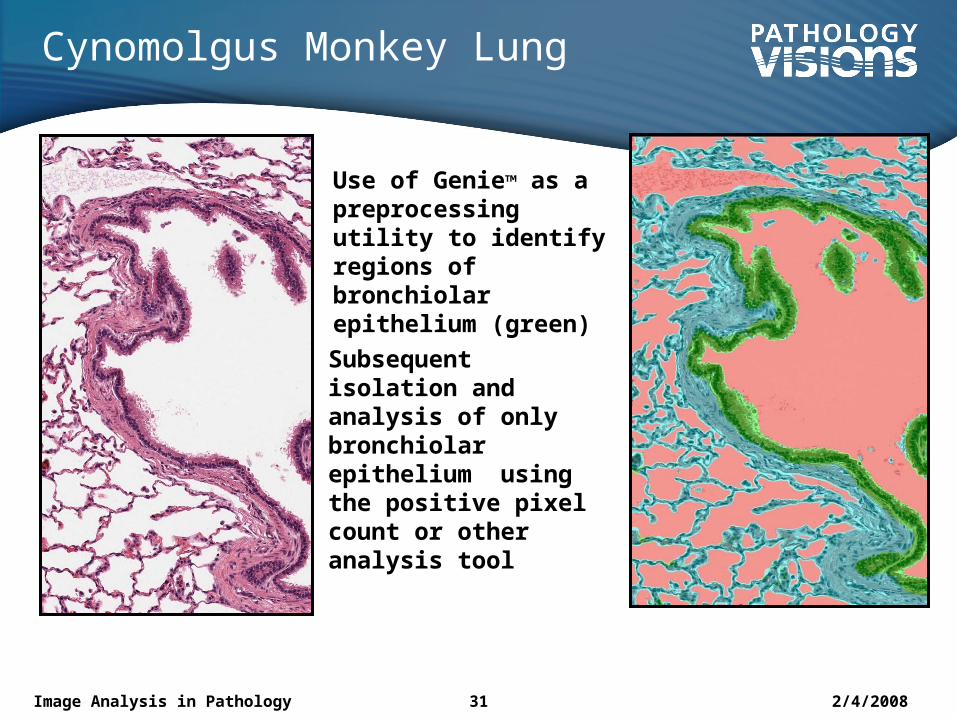
Cynomolgus Monkey Lung
Use of Genie™ as a preprocessing utility to identify regions of bronchiolar epithelium (green)
Subsequent isolation and analysis of only bronchiolar epithelium using the positive pixel count or other analysis tool
2/4/2008Image Analysis in Pathology 32
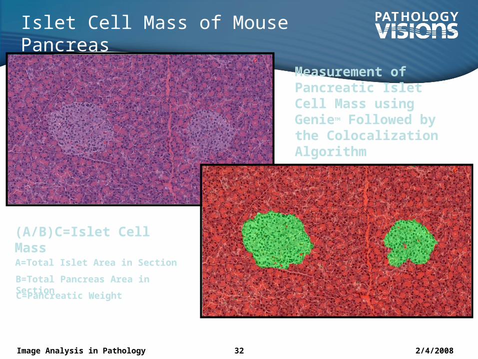
Islet Cell Mass of Mouse Pancreas
Measurement of Pancreatic Islet Cell Mass using Genie™ Followed by the Colocalization Algorithm
(A/B)C=Islet Cell MassA=Total Islet Area in Section
B=Total Pancreas Area in SectionC=Pancreatic Weight
2/4/2008Image Analysis in Pathology 33
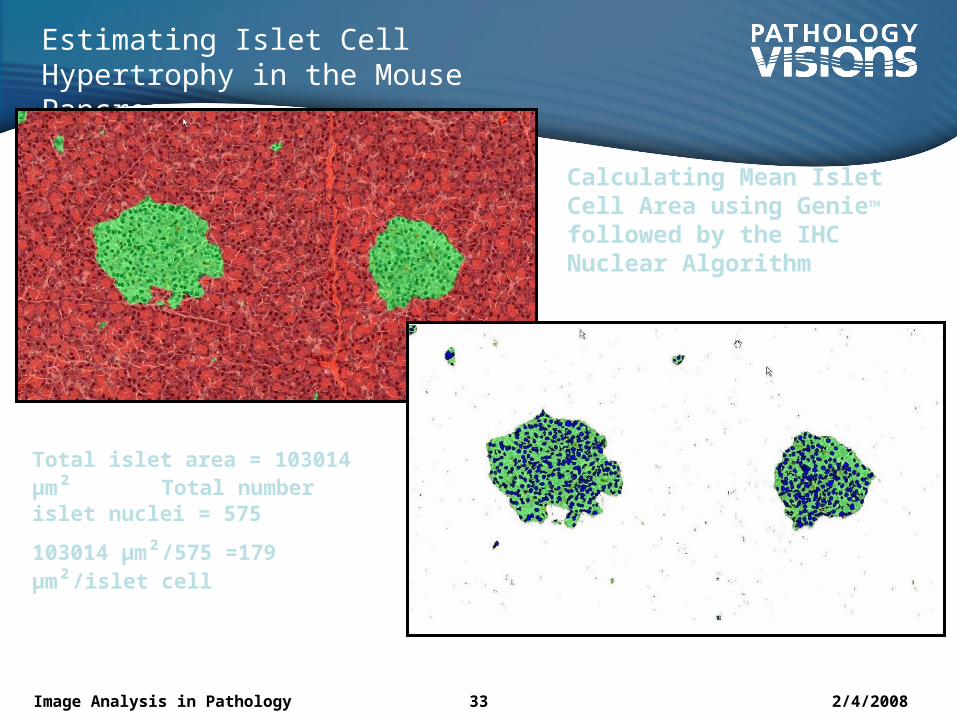
Estimating Islet Cell Hypertrophy in the Mouse Pancreas
Calculating Mean Islet Cell Area using Genie™ followed by the IHC Nuclear Algorithm
Total islet area = 103014 µm² Total number islet nuclei = 575
103014 µm²/575 =179 µm²/islet cell
2/4/2008Image Analysis in Pathology 34
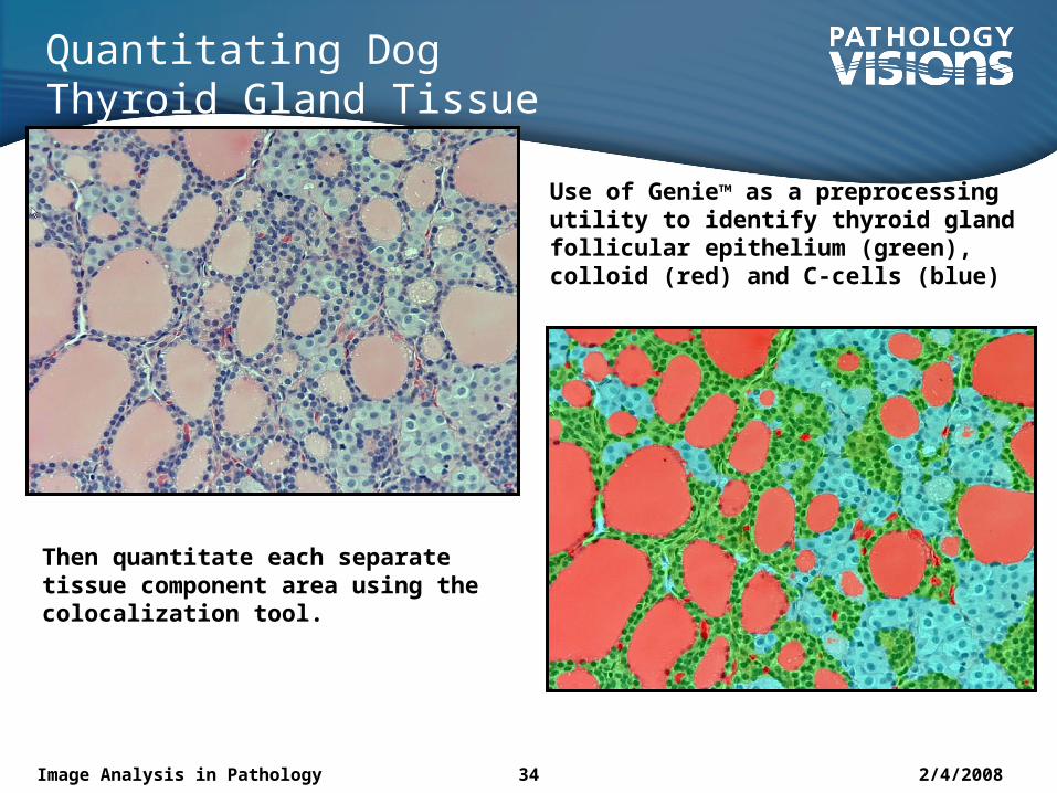
Quantitating Dog Thyroid Gland Tissue Components
Use of Genie™ as a preprocessing utility to identify thyroid gland follicular epithelium (green), colloid (red) and C-cells (blue)
Then quantitate each separate tissue component area using the colocalization tool.
2/4/2008Image Analysis in Pathology 35
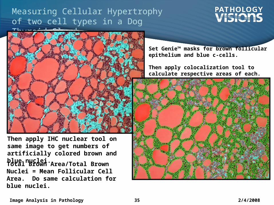
Measuring Cellular Hypertrophy of two cell types in a Dog Thyroid Gland
Then apply IHC nuclear tool on same image to get numbers of artificially colored brown and blue nuclei.
Set Genie™ masks for brown follicular epithelium and blue c-cells.
Then apply colocalization tool to calculate respective areas of each.
Total Brown Area/Total Brown Nuclei = Mean Follicular Cell Area. Do same calculation for blue nuclei.
2/4/2008Image Analysis in Pathology 36
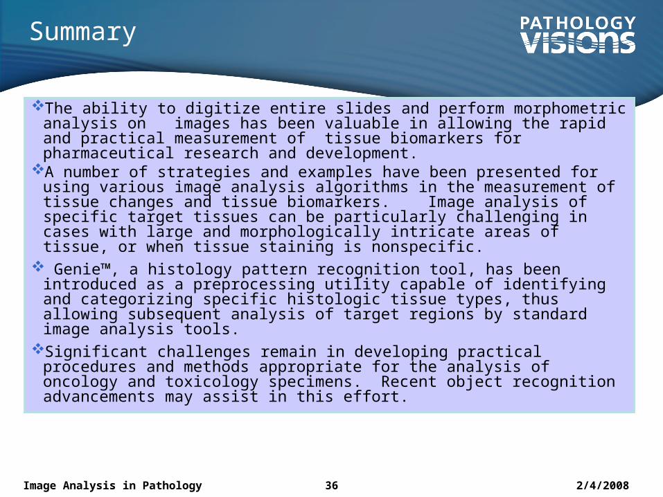
Summary
The ability to digitize entire slides and perform morphometric analysis on images has been valuable in allowing the rapid and practical measurement of tissue biomarkers for pharmaceutical research and development.
A number of strategies and examples have been presented for using various image analysis algorithms in the measurement of tissue changes and tissue biomarkers. Image analysis of specific target tissues can be particularly challenging in cases with large and morphologically intricate areas of tissue, or when tissue staining is nonspecific.
Genie™, a histology pattern recognition tool, has been introduced as a preprocessing utility capable of identifying and categorizing specific histologic tissue types, thus allowing subsequent analysis of target regions by standard image analysis tools.
Significant challenges remain in developing practical procedures and methods appropriate for the analysis of oncology and toxicology specimens. Recent object recognition advancements may assist in this effort.
2/4/2008Image Analysis in Pathology 37
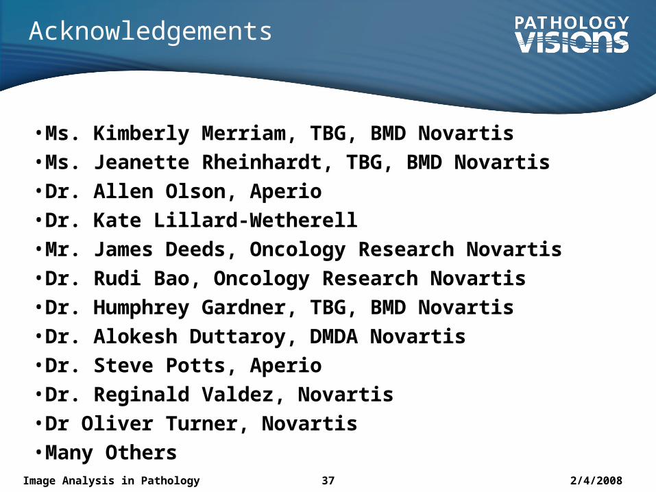
Acknowledgements
• Ms. Kimberly Merriam, TBG, BMD Novartis
• Ms. Jeanette Rheinhardt, TBG, BMD Novartis
• Dr. Allen Olson, Aperio
• Dr. Kate Lillard-Wetherell
• Mr. James Deeds, Oncology Research Novartis
• Dr. Rudi Bao, Oncology Research Novartis
• Dr. Humphrey Gardner, TBG, BMD Novartis
• Dr. Alokesh Duttaroy, DMDA Novartis
• Dr. Steve Potts, Aperio
• Dr. Reginald Valdez, Novartis
• Dr Oliver Turner, Novartis
• Many Others
2/4/2008Image Analysis in Pathology 38
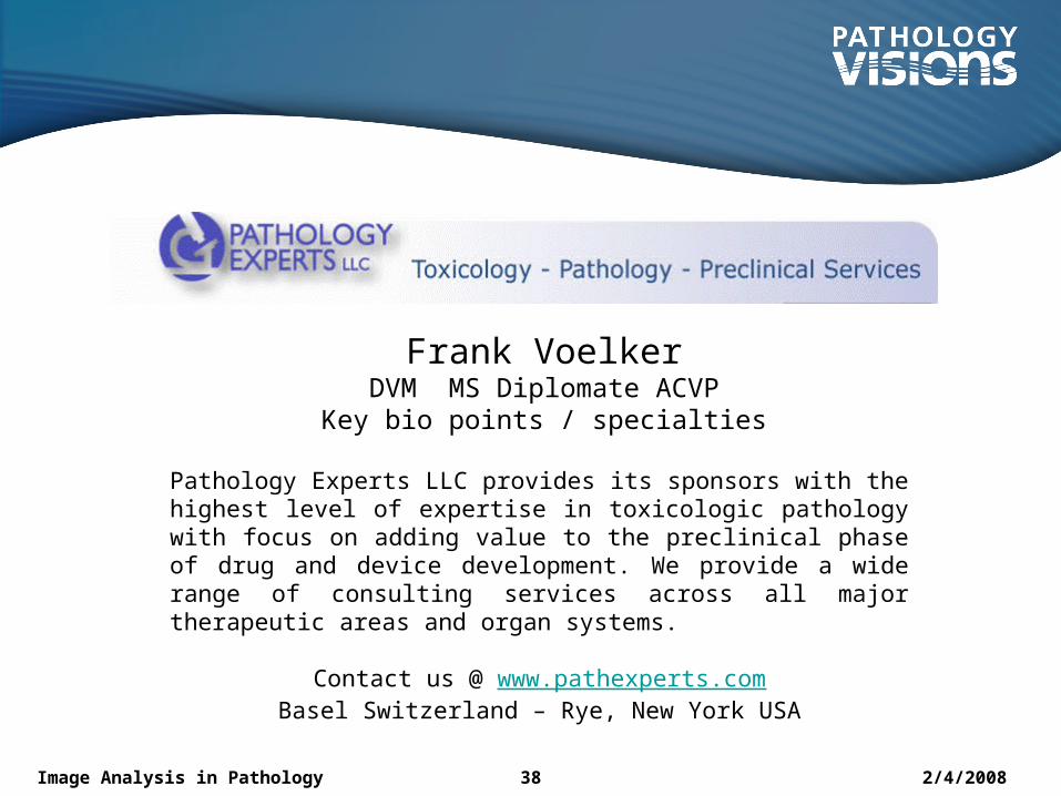
Frank VoelkerDVM MS Diplomate ACVPKey bio points / specialties
Pathology Experts LLC provides its sponsors with the highest level of expertise in toxicologic pathology with focus on adding value to the preclinical phase of drug and device development. We provide a wide range of consulting services across all major therapeutic areas and organ systems.
Contact us @ www.pathexperts.comBasel Switzerland – Rye, New York USA


![a new perspective on Equality, a practical handbook [preview]](https://img.pdfslide.us/doc/110x75/568c37791a28ab02359bb7e8/a-new-perspective-on-equality-a-practical-handbook-preview.jpg)