Embed Size (px)
Citation preview

Implant restoration in the aesthetic zone using guided surgery and immediate functional loadingDigital Workflow: clinical patient information digitally obtained and shared
Prachatipat Hospital Prathumtani ProvinceDr. Nawakamon Suriyan
Imag
e fr
om
ww
w.b
angk
okb
on
ehar
vest
.co
m/o
ur-d
oct
ors
/dr-
naw
akam
on
-sur
iyan
Case informationMale patient, 72 years old, with a 3 months prior extract from failed root canal treatment on supported bridge 13–22. Patient would like a fix prosthodontics for missing teeth. Patient is a medical doctor and more concerned with restorative rather than aesthetic success.
The patient’s general periodontal condition is fair with medical history of cardiovascular disease and on Warfarin at 5 mg per day. Clinical and radiographic evaluation performed on initial visit.

Implant restoration in the aesthetic zone using guided surgery and immediate functional loading 2
Fig.1 Pre-operation – shows available bone at the implant site
Treatment Options
The patient was presented with the following treatment options for the replacement of the anterior maxilla. The first option was a 3-unit fixed prosthodontic using teeth #14, #22 and #23 as abutments. Second, an implant restoration for the replacement of the edentulous space at teeth #13, #11, #21 and #22 was determined to be the more conservative option. Option number 2, replacement of the edentulous space at teeth #13, #11, #21 or #22 was accepted.
Fig.2 Pre-operation, Cross-sectional shows bone available at the implant site
Fig.3 The Orthopanoramic scan shows bone in the edentulous area and no bone pathology
Treatment description
An inclusive extraoral examination, followed by intraoral examination was performed (Fig 1, 2). The occlusion on the right and left side showed an unclassified molar relationship due to the missing tooth (Fig 3, 4). The SAC classification for implant dentistry was used.
An implant site-specific evaluation was then carried out, including an evaluation of the inner occlusal space, in addition to both a hard- and a soft-tissue assessment, showing good ridge dimensions and keratinized tissue volume.
Keratinized tissue width was 8 mm. The CT scan revealed the buccolingual cortical width was 7 mm, mesio-distal width 32 mm. The CT scan revealed the buccolingual cortical width at 7 mm. From the Orthopantomograph, the crest to floor of nose was 16mm (Fig 3). An intraoral periapical radiograph was also taken (Fig 4-6).
Fig.4 The other teeth show normal periodontal with no pathology in the apical area

Implant restoration in the aesthetic zone using guided surgery and immediate functional loading 3
Fig.5 Bone at the edentulous area
Fig.6 Neighbouring teeth show no bone pathology
Fig.7 Virtual crowns (3Shape, Implant Studio) used for prosthetic driven implant planning and surgical guide design
The patient was scanned using an intraoral scanner (TRIOS, 3Shape). CBCT scans (GiANO, Newtom, Italy) and digital impressions were merged using 3Shape Implant Studio® software.
Implant placements (CAMLOG, Germany) were planned using software (Implant Studio) to identify direction and positioning and to create the CAD/CAM guide surgical template.
A CBCT was taken before and after implant placement. During surgery, the drilled template was used as a drill guide. The implant-supported bridge for #13 to #11 and #21 designed using 3Shape software (Dental System™).
After implant placement, postoperative CBCTs were recorded and compared with the preoperative using the McNemar test.
One hour before surgery the patient was given 2g of Amoxillin (400 mg) or Ibuprofen (1 g) or paracetamol then the mucoperiosteal flap from tooth 14 to 22 started.
Preparation of the implant bed started with a bone drill sequence. It was planned to place the definitive prefabricated abutment and provisional restoration immediately after surgery.

Implant restoration in the aesthetic zone using guided surgery and immediate functional loading 4
Fig.9b Abutment designed and manufactured including the provisional bridge prior to surgery (#13, #11, #21) (3Shape, Dental System)
Fig.8 Cone beam (CBCT) and intraoral (3Shape, TRIOS) 3D digital impression files merged in software to create and approve implant positions
Fig.9a
Summary
During the restorative phase, an impression was taken at implant level according to protocol. This scan set assists in determining the dimensions for assuming the axial alignment and gingival height. An appropriate space was obtained and a good emergence profile of the cement-retained restoration was selected for the case. All information gathered before the actual surgery, together with the guided surgical template ensured an accurate, functional and aesthetic final restoration.

Implant restoration in the aesthetic zone using guided surgery and immediate functional loading 5
Fig.10 Tooth-supported surgical guide (SurgiGuided, 3Shape, Implant Studio) designed using the stereolithographic scan. (STL scan)
Fig.11 A guided template placed before the start of the operation
Fig.12 Implant placement
Digital Workflow Benefits
A comprehensive pre-operative workup is essential for planning the number and type of implants, as well as in identifying the optimum location for implant placement.
Prosthesis design and manufacture based on three-dimen- sional imaging data facilitates the preparation of a guide for the surgical intervention.
Using a prosthetic-driven workflow (3Shape Implant Studio) by first placing virtual crowns, helped to ensure proper depths and angulations. Implants were then positioned to accommodate the maximum amount of bone, while establishing optimal support to achieve an aesthetic outcome for the proposed restoration.
The result was less postoperative morbidity, due to the surgery being minimally invasive. Guided surgery technique and immediate functional loading helped to create a more predictable procedure.
The digital workflow increases patient satisfaction because the procedure provides an immediate aesthetic and functional restoration.

Implant restoration in the aesthetic zone using guided surgery and immediate functional loading 6
Fig.13 Final abutments designed in 3Shape Dental System
Fig.14 Fix titanium base with the CAD /CAM abutment
Fig.15 The emergence profile of the zirconia abutment on Ti-base was sandblast and fixed using resin cement (Multilink Automix, Ivoclar Vivadent)
Advantages of the digital workflow:
• More efficient communications between the dentist, dental technician and patient
• Diagnostic and restorative software tools allow for the precise, predictable placement of dental implants
• Detailed visualization of the prosthetic outcome prior to the actual treatment due to intraoral scanning, digital treatment planning, guided surgery, and CAD/CAM software
• Guided surgery technique and immediate functional loading create a predictable procedure
• Patient satisfaction increased due to immediate provisionalization and an aesthetic and functional restoration

Implant restoration in the aesthetic zone using guided surgery and immediate functional loading 7
Fig.18 Zirconia abutment position and preparation of teeth before crown insertion
Fig.16 Final monolithic zirconia crowns fabricated
Fig.17 Adjustments reglazed and polished
Discussion/challenges
Patients must meet the right criteria for being treated with a digital workflow. The restorative team needs a thorough understanding of the techniques and limitations in the digital workflow to ensure a high success rate in the aesthetic zone.
Implant restoration in the aesthetic zone using guided surgery and immediate functional loading 8
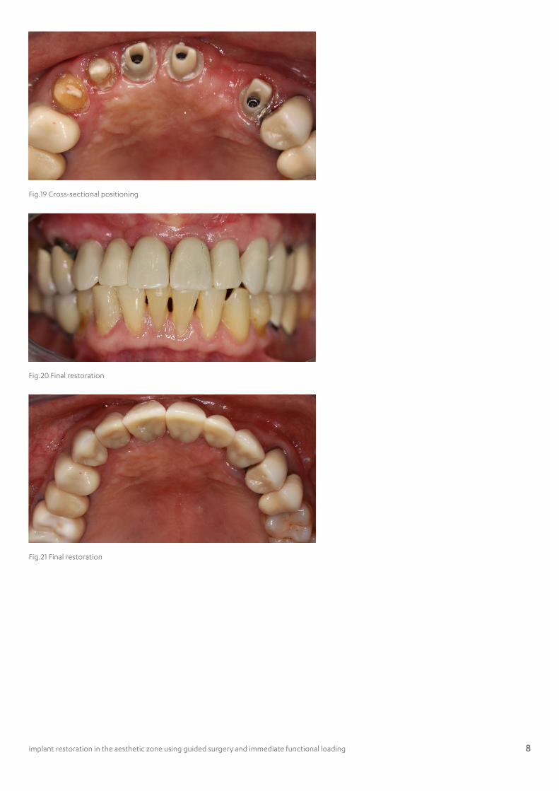
Fig.19 Cross-sectional positioning
Fig.20 Final restoration
Fig.21 Final restoration
Implant restoration in the aesthetic zone using guided surgery and immediate functional loading 9
About Dr. Nawakamon SuriyanPrachatipat Hospital Prathumtani Province
EducationDoctor of dental surgery Chulalongkorn University 1997.Postgraduate oral and maxillofacial surgery Chiangmai University 2001.International oral and maxillofacial surgery Prince of Songkha University 2004.Master public health Sukhothaithammatirat University 2008.Fellowship international congress of oral implantologist 2009.Doctor of philosophy Chulalongkorn University 2011.Msc. Implant dentistry Mahidol University 2015.IMC Master Implant dentistry Program at Muenster University 2015.
Professional Career 2006 – Present : Dentist in Prachatipat Hospital Prathumtani Province2015 – Present : Lecturer in Msc. Implant Dentistry at Thammasat University2015 – Present : Research Consultant at Dental Institute
AcknowledgementMaster of Science Program in Implant Dentistry (International Program), Rodolphe Saidnattar (Dental technician/Digital advancement technology, Accord Corporation Limited)
About 3Shape
3Shape is changing dentistry together with dental professionals across the world by developing innovations that provide superior dental care for patients. Our portfolio of 3D scanners and CAD/CAM software solutions for the dental industry includes the multiple award-winning 3Shape TRIOS intraoral scanner, the upcoming 3Shape X1 CBCT scanner, and market leading scanning and design software solutions for dental labs.
Two graduate students founded 3Shape in Denmark’s capital in the year 2000. Today, 3Shape has over 1,000 employees servicing customers in over 100 countries from an ever-growing number of 3Shape offices around the world.
3Shape’s products and innovations con- tinue to challenge traditional methods, enabling dental professionals to treat more patients more effectively.
Let’s change dentistry together


![4,400 117,000 130M · needs and/or with the ideal emergence proile of the missing tooth [21]. 2. Classiication of implant abutments Varying types of implant abutments have been reported](https://img.pdfslide.us/doc/110x75/5f01f8077e708231d401ed17/4400-117000-130m-needs-andor-with-the-ideal-emergence-proile-of-the-missing-tooth.jpg)