Embed Size (px)
Citation preview
7/10/2015
1
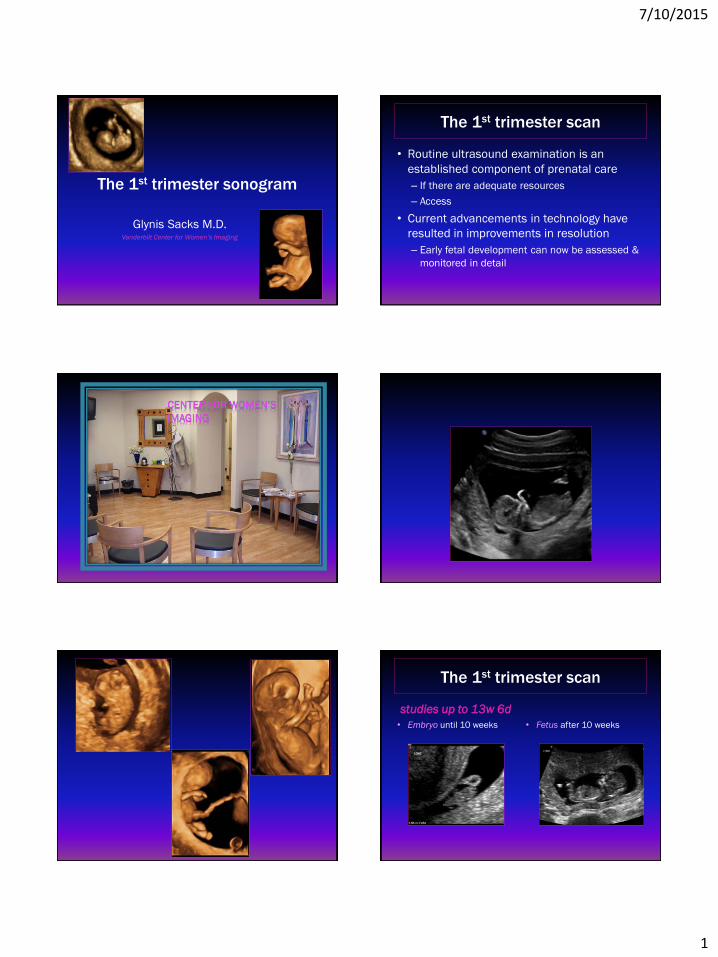
The 1st trimester sonogram
Glynis Sacks M.D. Vanderbilt Center for Women’s Imaging
The 1st trimester scan
• Routine ultrasound examination is an
established component of prenatal care
– If there are adequate resources
– Access
• Current advancements in technology have
resulted in improvements in resolution
– Early fetal development can now be assessed &
monitored in detail
The 1st trimester scan
studies up to 13w 6d
• Embryo until 10 weeks • Fetus after 10 weeks
7/10/2015
2
The 1st trimester scan
• Provide information which will optimize
prenatal care and lead to the best possible
outcomes for both mother and fetus.
The 1st trimester scan
• Confirm viability
• Establish gestational age
• Evaluate for possible ectopic gestation
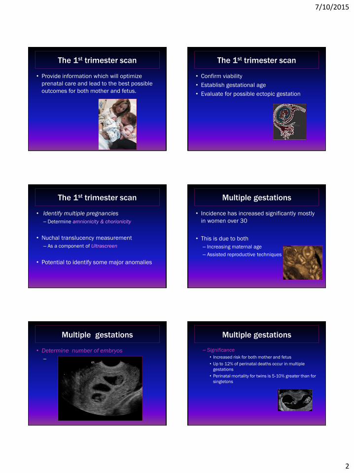
The 1st trimester scan
• Identify multiple pregnancies
– Determine amnionicity & chorionicity
• Nuchal translucency measurement
– As a component of Ultrascreen
• Potential to identify some major anomalies
Multiple gestations
• Incidence has increased significantly mostly
in women over 30
• This is due to both
– Increasing maternal age
– Assisted reproductive techniques
Multiple gestations
• Determine number of embryos
–
Multiple gestations
– Significance
• Increased risk for both mother and fetus
• Up to 12% of perinatal deaths occur in multiple
gestations
• Perinatal mortality for twins is 5-10% greater than for
singletons
7/10/2015
3
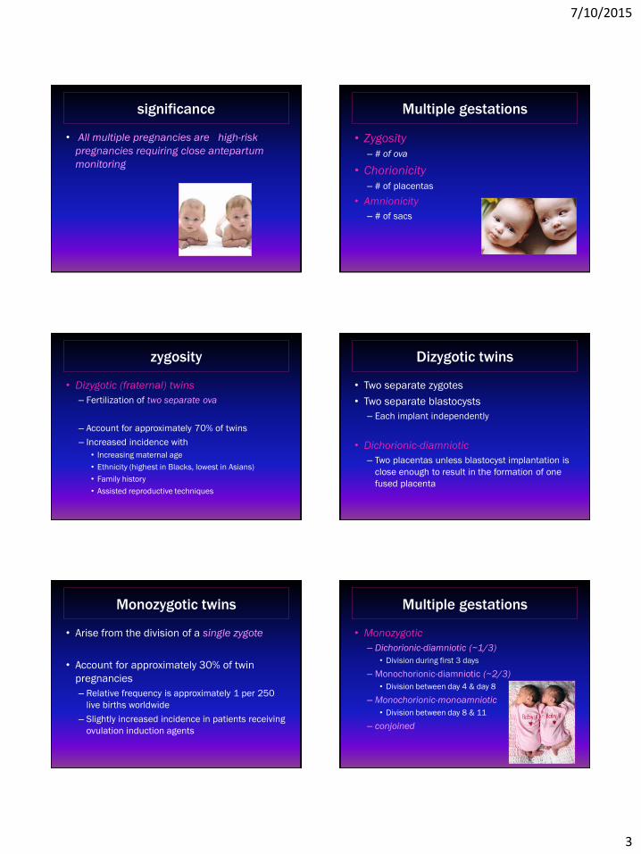
significance
• All multiple pregnancies are high-risk
pregnancies requiring close antepartum
monitoring
Multiple gestations
• Zygosity
– # of ova
• Chorionicity
– # of placentas
• Amnionicity
– # of sacs
zygosity
• Dizygotic (fraternal) twins
– Fertilization of two separate ova
– Account for approximately 70% of twins
– Increased incidence with
• Increasing maternal age
• Ethnicity (highest in Blacks, lowest in Asians)
• Family history
• Assisted reproductive techniques
Dizygotic twins
• Two separate zygotes
• Two separate blastocysts
– Each implant independently
• Dichorionic-diamniotic
– Two placentas unless blastocyst implantation is
close enough to result in the formation of one
fused placenta
Monozygotic twins
• Arise from the division of a single zygote
• Account for approximately 30% of twin
pregnancies
– Relative frequency is approximately 1 per 250
live births worldwide
– Slightly increased incidence in patients receiving
ovulation induction agents
Multiple gestations
• Monozygotic
– Dichorionic-diamniotic (~1/3)
• Division during first 3 days
– Monochorionic-diamniotic (~2/3)
• Division between day 4 & day 8
– Monochorionic-monoamniotic
• Division between day 8 & 11
– conjoined
7/10/2015
4
Multiple gestations
• dichorionic-diamniotic twins
– Perinatal mortality ~ 10%
• Preterm birth
• IUGR
• Slightly increased risk of anomalies
Multiple gestations
• Monochorionic-diamniotic
– Perinatal morbidity & mortality 3-5 times > dichorionic twins
• Preterm delivery
• IUGR
• Anomalies
– Placental
• Twin-twin transfusion
• TRAP
• Twin embolization syndrome
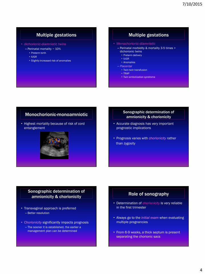
Monochorionic-monoamniotic
• Highest mortality because of risk of cord
entanglement
Sonographic determination of
amnionicity & chorionicity
• Accurate diagnosis has very important
prognostic implications
• Prognosis varies with chorionicity rather
than zygosity
Sonographic determination of
amnionicity & chorionicity
• Transvaginal approach is preferred
– Better resolution
• Chorionicity significantly impacts prognosis
– The sooner it is established, the earlier a
management plan can be determined
Role of sonography
• Determination of chorionicity is very reliable
in the first trimester
• Always go to the initial exam when evaluating
multiple pregnancies
• From 6-9 weeks, a thick septum is present
separating the chorionic sacs
7/10/2015
5
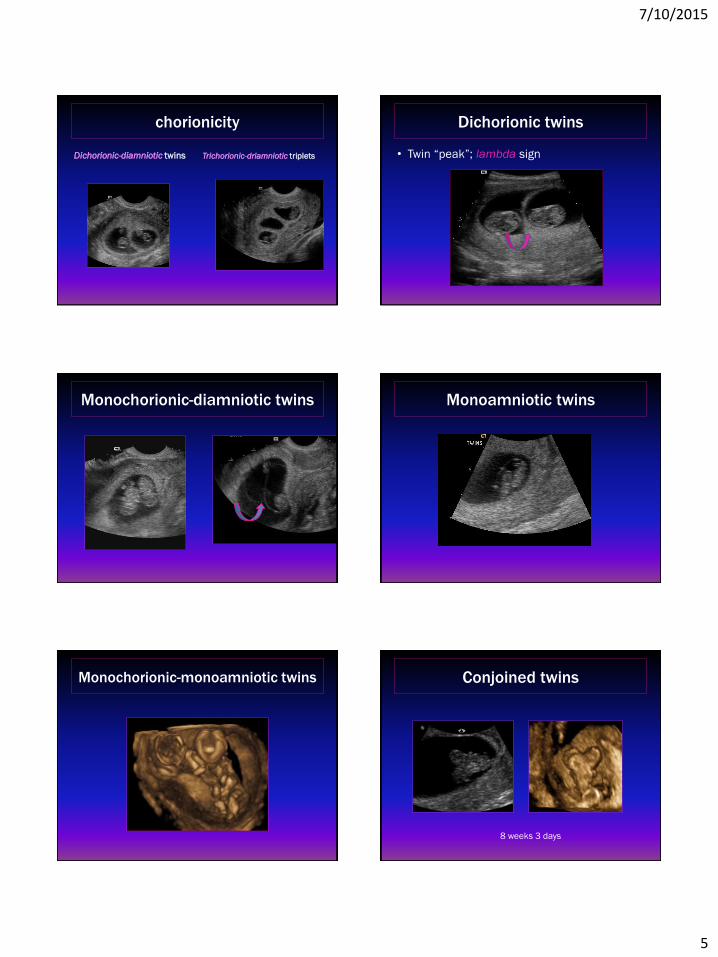
chorionicity
Dichorionic-diamniotic twins Trichorionic-driamniotic triplets
Dichorionic twins
• Twin “peak”; lambda sign
Monochorionic-diamniotic twins
Monoamniotic twins
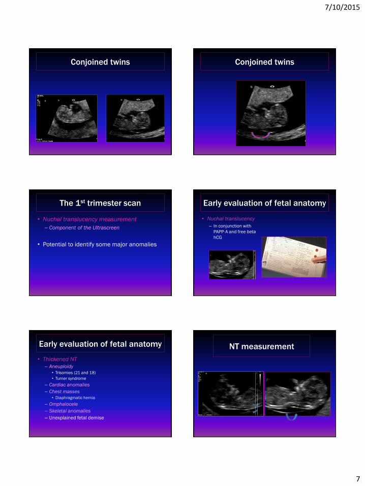
Monochorionic-monoamniotic twins Conjoined twins
8 weeks 3 days
7/10/2015
6
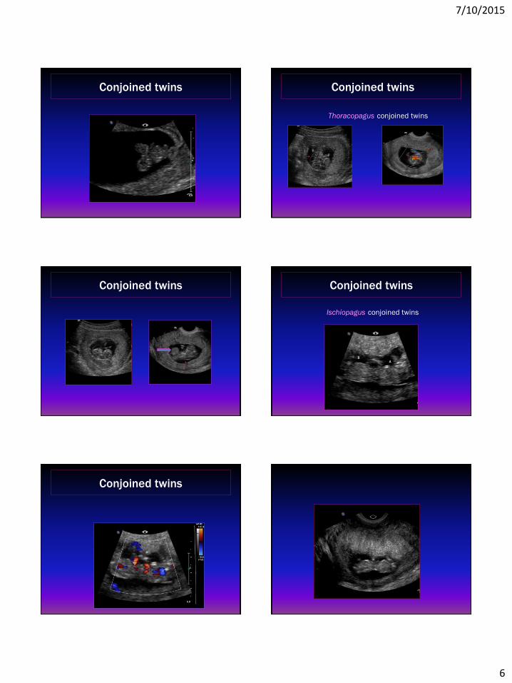
Conjoined twins Conjoined twins
Thoracopagus conjoined twins
Conjoined twins Conjoined twins
Ischiopagus conjoined twins
Conjoined twins
7/10/2015
7
Conjoined twins Conjoined twins
The 1st trimester scan
• Nuchal translucency measurement
– Component of the Ultrascreen
• Potential to identify some major anomalies
Early evaluation of fetal anatomy
• Nuchal translucency
– In conjunction with
PAPP-A and free beta
hCG
Early evaluation of fetal anatomy
• Thickened NT
– Aneuploidy
• Trisomies (21 and 18)
• Turner syndrome
– Cardiac anomalies
– Chest masses
• Diaphragmatic hernia
– Omphalocele
– Skeletal anomalies
– Unexplained fetal demise
NT measurement
7/10/2015
8
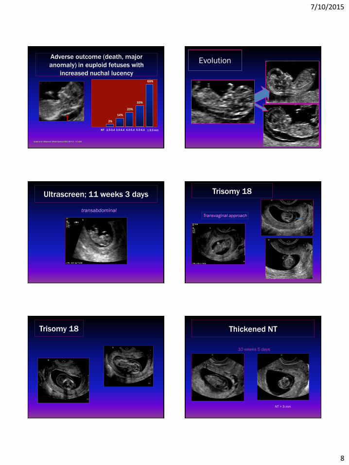
Adverse outcome (death, major
anomaly) in euploid fetuses with
increased nuchal lucency
Souka et al. Ultrasound Obstet Gynecol 2001;18:9-17; n=1,320
69%
> 6.5 mm
14%
3.5-4.4 2.5-3.4 NT
3%
5.5-6.4
33%
4.5-5.4
23%
Evolution
Ultrascreen; 11 weeks 3 days
transabdominal
Trisomy 18
Transvaginal approach
Trisomy 18 Thickened NT
10 weeks 5 days
NT > 3 mm
7/10/2015
9
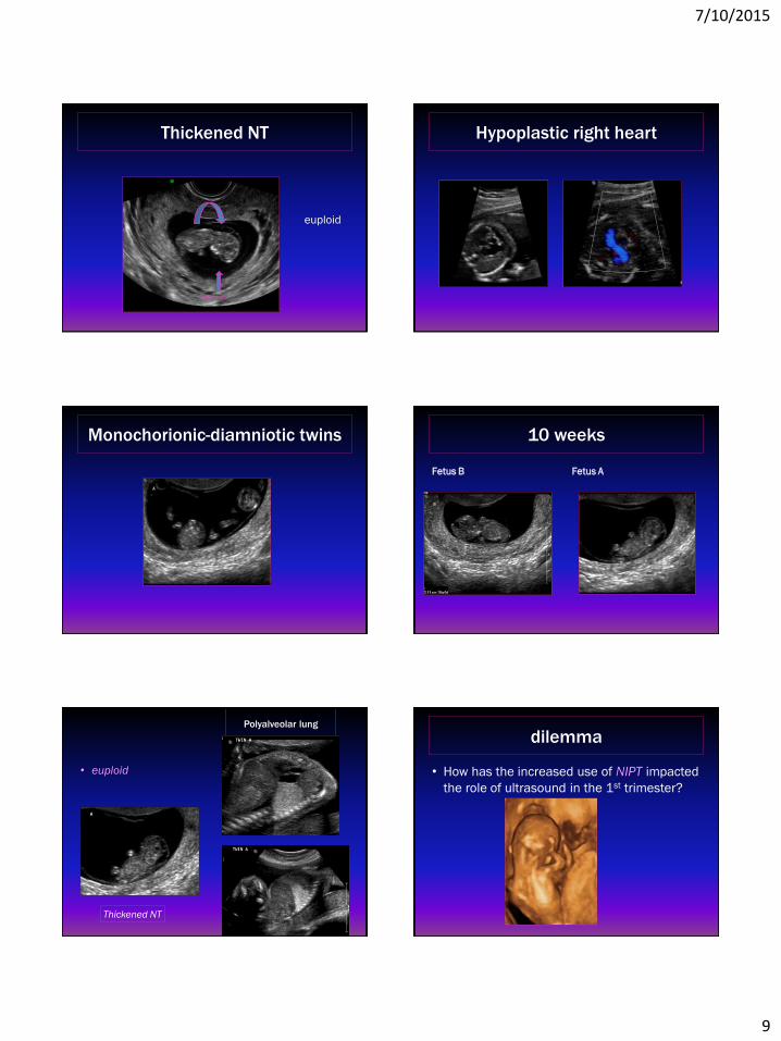
Thickened NT
amnion
euploid
Hypoplastic right heart
Monochorionic-diamniotic twins 10 weeks
Fetus B Fetus A
Polyalveolar lung
• euploid
Thickened NT
dilemma
• How has the increased use of NIPT impacted
the role of ultrasound in the 1st trimester?
7/10/2015
10
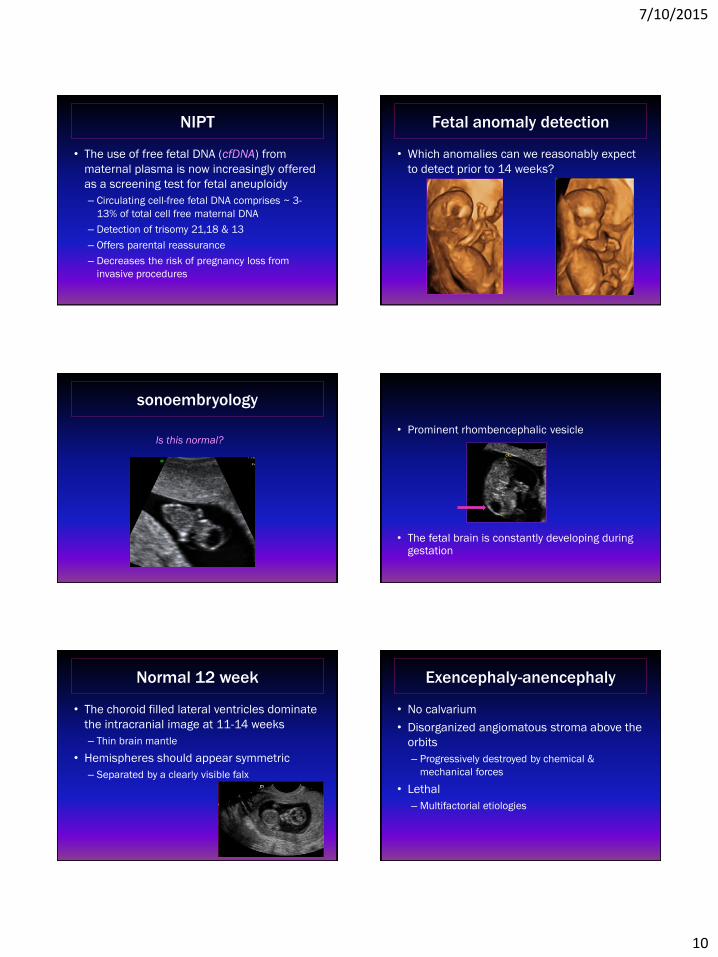
NIPT
• The use of free fetal DNA (cfDNA) from
maternal plasma is now increasingly offered
as a screening test for fetal aneuploidy
– Circulating cell-free fetal DNA comprises ~ 3-
13% of total cell free maternal DNA
– Detection of trisomy 21,18 & 13
– Offers parental reassurance
– Decreases the risk of pregnancy loss from
invasive procedures
Fetal anomaly detection
• Which anomalies can we reasonably expect
to detect prior to 14 weeks?
sonoembryology
Is this normal? • Prominent rhombencephalic vesicle
• The fetal brain is constantly developing during gestation
Normal 12 week
• The choroid filled lateral ventricles dominate
the intracranial image at 11-14 weeks
– Thin brain mantle
• Hemispheres should appear symmetric
– Separated by a clearly visible falx
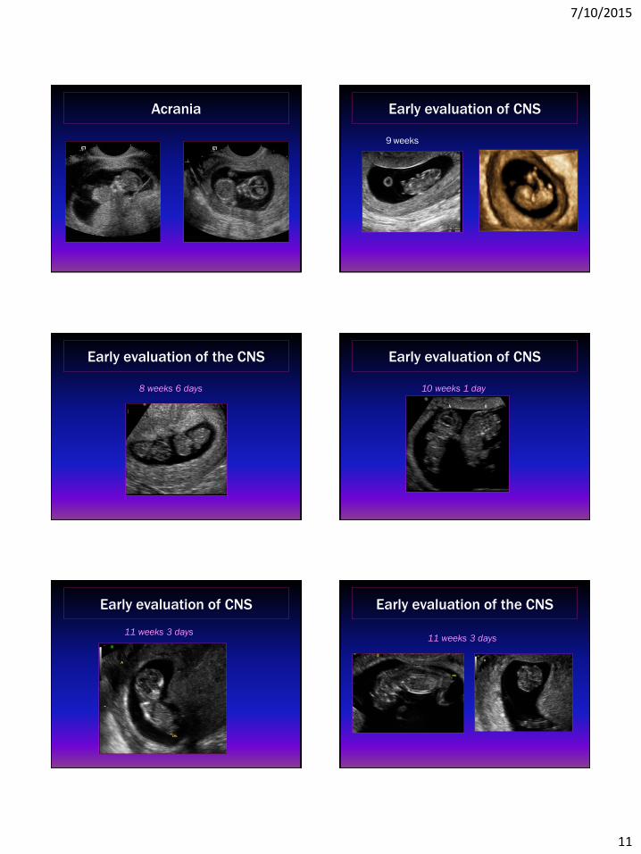
Exencephaly-anencephaly
• No calvarium
• Disorganized angiomatous stroma above the
orbits
– Progressively destroyed by chemical &
mechanical forces
• Lethal
– Multifactorial etiologies
7/10/2015
11
Acrania
Early evaluation of CNS
9 weeks
Early evaluation of the CNS
8 weeks 6 days
Early evaluation of CNS
10 weeks 1 day
Early evaluation of CNS
11 weeks 3 days
Early evaluation of the CNS
11 weeks 3 days
7/10/2015
12
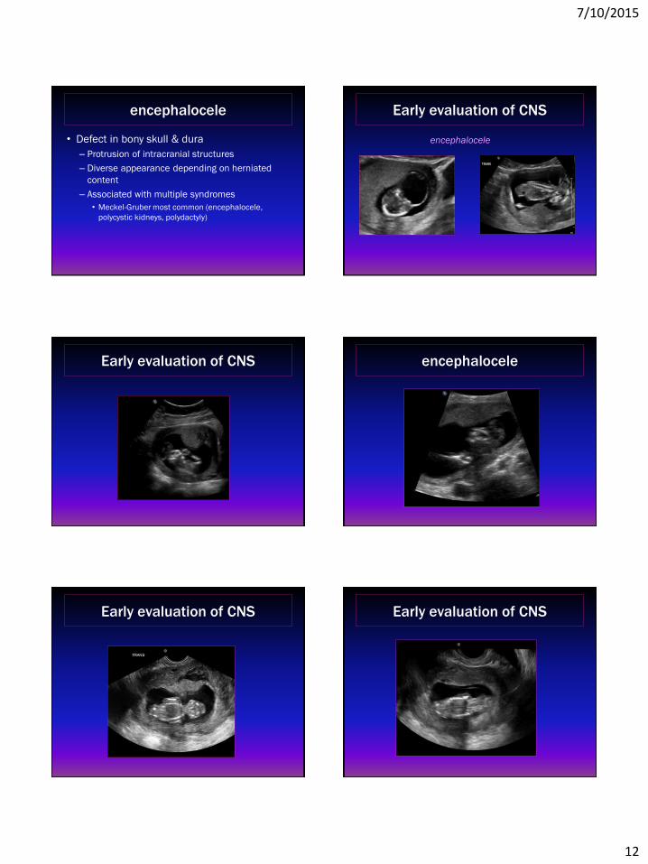
encephalocele
• Defect in bony skull & dura
– Protrusion of intracranial structures
– Diverse appearance depending on herniated
content
– Associated with multiple syndromes
• Meckel-Gruber most common (encephalocele,
polycystic kidneys, polydactyly)
Early evaluation of CNS
encephalocele
Early evaluation of CNS encephalocele
Early evaluation of CNS Early evaluation of CNS
7/10/2015
13
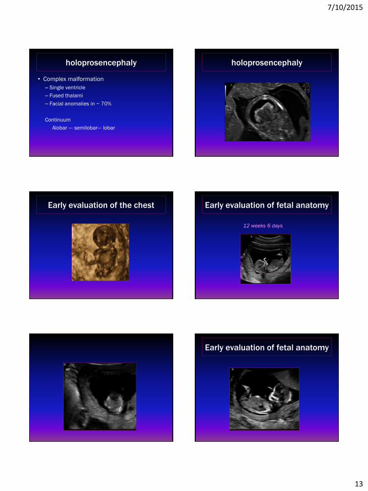
holoprosencephaly
• Complex malformation
– Single ventricle
– Fused thalami
– Facial anomalies in ~ 70%
Continuum
Alobar --- semilobar--- lobar
holoprosencephaly
Early evaluation of the chest Early evaluation of fetal anatomy
12 weeks 6 days
Early evaluation of fetal anatomy
7/10/2015
14
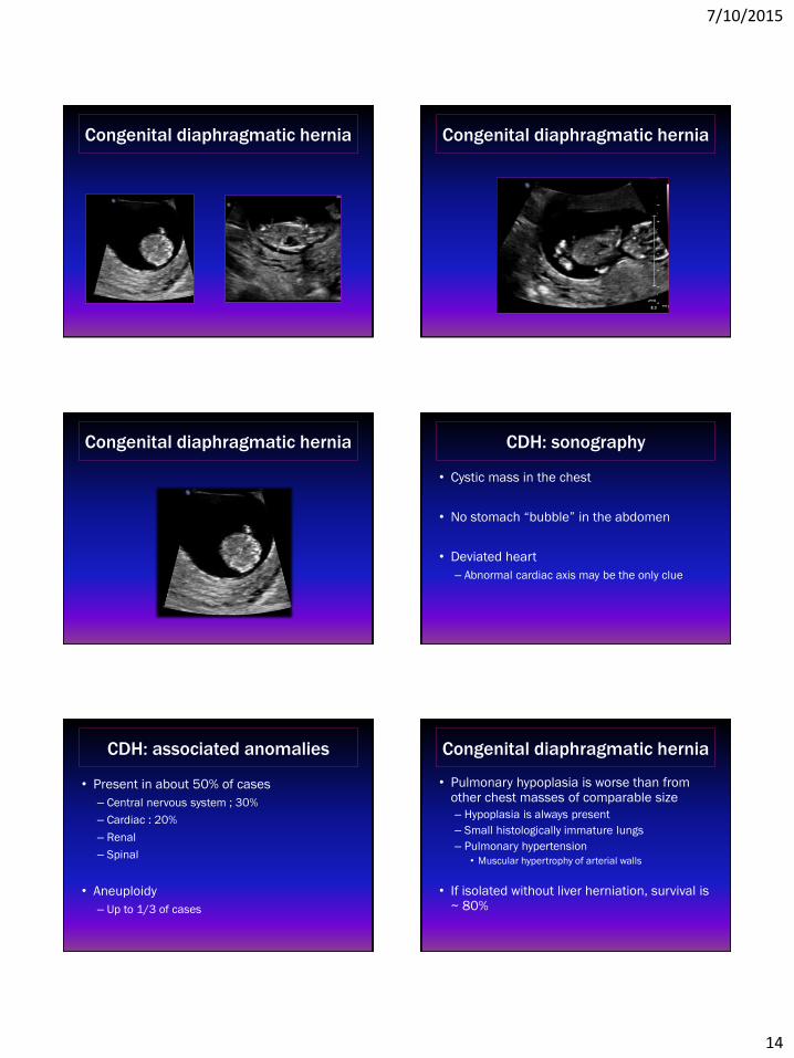
Congenital diaphragmatic hernia Congenital diaphragmatic hernia
Congenital diaphragmatic hernia CDH: sonography
• Cystic mass in the chest
• No stomach “bubble” in the abdomen
• Deviated heart
– Abnormal cardiac axis may be the only clue
CDH: associated anomalies
• Present in about 50% of cases
– Central nervous system ; 30%
– Cardiac : 20%
– Renal
– Spinal
• Aneuploidy
– Up to 1/3 of cases
Congenital diaphragmatic hernia
• Pulmonary hypoplasia is worse than from other chest masses of comparable size
– Hypoplasia is always present
– Small histologically immature lungs
– Pulmonary hypertension
• Muscular hypertrophy of arterial walls
• If isolated without liver herniation, survival is ~ 80%
7/10/2015
15
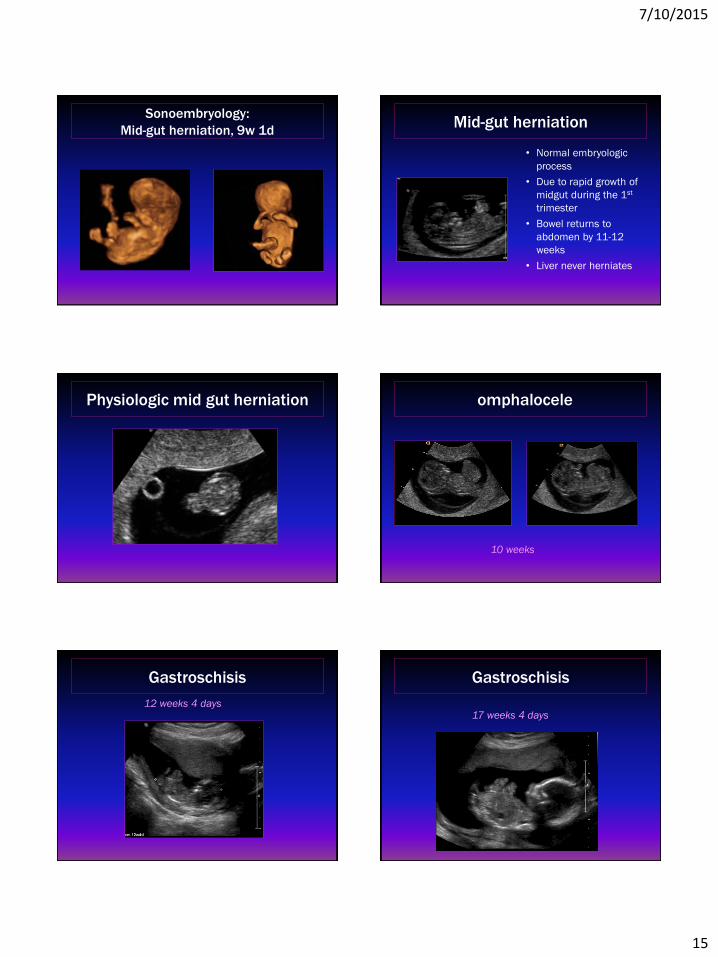
Sonoembryology:
Mid-gut herniation, 9w 1d Mid-gut herniation
• Normal embryologic
process
• Due to rapid growth of
midgut during the 1st
trimester
• Bowel returns to
abdomen by 11-12
weeks
• Liver never herniates
Physiologic mid gut herniation omphalocele
10 weeks
Gastroschisis
12 weeks 4 days
Gastroschisis
17 weeks 4 days
7/10/2015
16
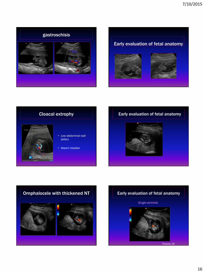
gastroschisis
Early evaluation of fetal anatomy
Cloacal extrophy
• Low abdominal wall
defect
• Absent bladder
Early evaluation of fetal anatomy
Omphalocele with thickened NT Early evaluation of fetal anatomy
Single ventricle
Trisomy 18
7/10/2015
17
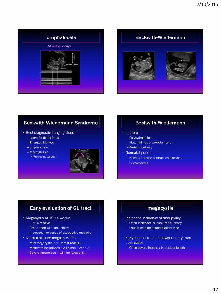
omphalocele
14 weeks 2 days
Beckwith-Wiedemann
Beckwith-Wiedemann Syndrome
• Best diagnostic imaging clues
– Large for dates fetus
– Enlarged kidneys
– omphalocele
– Macroglossia
• Protruding tongue
Beckwith-Wiedemann
• In utero
– Polyhydramnios
– Maternal risk of preeclampsia
– Preterm delivery
• Neonatal period
– Neonatal airway obstruction if severe
– hypoglycemia
Early evaluation of GU tract
• Megacystis at 10-14 weeks
– ~ 50% resolve
– Association with aneuploidy
– Increased incidence of obstructive uropathy
• Normal bladder length < 6 mm
– Mild megacystis 7-11 mm (Grade 1)
– Moderate megacystis 12-15 mm (Grade 2)
– Severe megacystis > 15 mm (Grade 3)
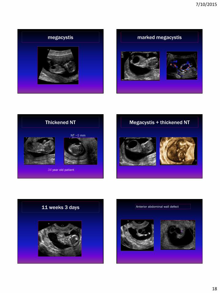
megacystis
• Increased incidence of aneuploidy
– Often increased Nuchal Translucency
– Usually mild-moderate bladder size
• Early manifestation of lower urinary tract
obstruction
– Often severe increase in bladder length
7/10/2015
18
megacystis marked megacystis
Thickened NT
NT ~5 mm
34 year old patient
Megacystis + thickened NT
11 weeks 3 days Anterior abdominal wall defect
7/10/2015
19
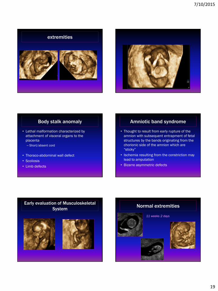
extremities
Body stalk anomaly
• Lethal malformation characterized by
attachment of visceral organs to the
placenta
– Short/absent cord
• Thoraco-abdominal wall defect
• Scoliosis
• Limb defects
Amniotic band syndrome
• Thought to result from early rupture of the
amnion with subsequent entrapment of fetal
structures by the bands originating from the
chorionic side of the amnion which are
“sticky”
• Ischemia resulting from the constriction may
lead to amputation
• Bizarre asymmetric defects
Early evaluation of Musculoskeletal
System Normal extremities
11 weeks 2 days
7/10/2015
20
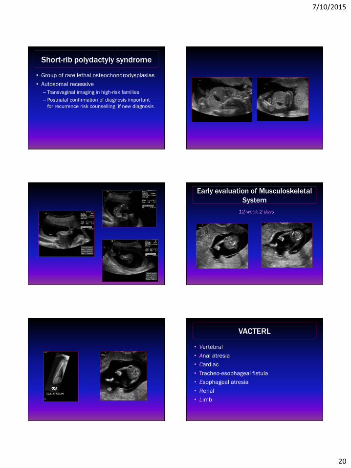
Short-rib polydactyly syndrome
• Group of rare lethal osteochondrodysplasias
• Autosomal recessive
– Transvaginal imaging in high-risk families
– Postnatal confirmation of diagnosis important
for recurrence risk counselling if new diagnosis
Early evaluation of Musculoskeletal
System
12 week 2 days
VACTERL
• Vertebral
• Anal atresia
• Cardiac
• Tracheo-esophageal fistula
• Esophageal atresia
• Renal
• Limb
7/10/2015
21
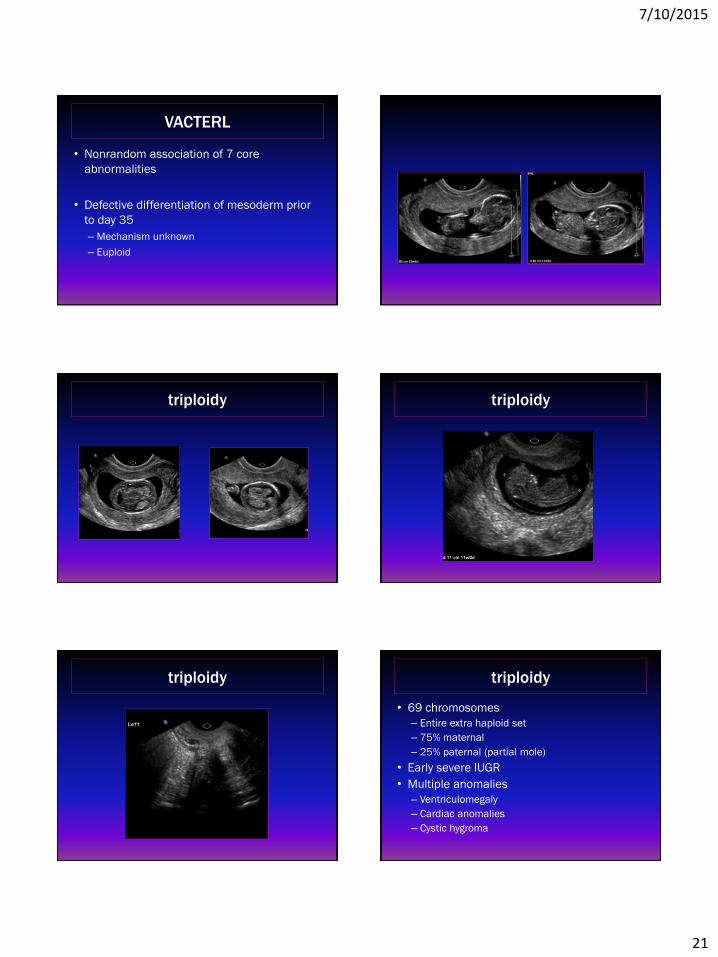
VACTERL
• Nonrandom association of 7 core
abnormalities
• Defective differentiation of mesoderm prior
to day 35
– Mechanism unknown
– Euploid
triploidy triploidy
triploidy triploidy
• 69 chromosomes
– Entire extra haploid set
– 75% maternal
– 25% paternal (partial mole)
• Early severe IUGR
• Multiple anomalies
– Ventriculomegaly
– Cardiac anomalies
– Cystic hygroma
7/10/2015
22
conclusions
• A comprehensive sonogram in the first
trimester provides a evaluation of both the
pregnancy and the maternal pelvis
– Consequent improvement in obstetric care
• An awareness of normal anatomy in the 1st
trimester is necessary
– Even if the study is performed for other reasons
conclusions
• Anomalies evident in the 1st trimester are
often
– More complex
– Higher association with aneuploidy
– Increased likelihood of hydrops
– May be transient
conclusions
• In the uncomplicated pregnancy without
clinical concerns, the 1st trimester scan
should be scheduled at ~ 11-14 weeks
– This provides the opportunity to evaluate fetal
anatomy as well as confirming gestational age,
viability and determining fetal number
conclusions
• Earlier diagnosis offers many advantages
– Patient learns of the fetal problem with sufficient time to consider management options
– Terminations earlier in pregnancy are safer & cheaper
– Less traumatic for the mother both psychologically & physically with a greater likelihood of preserving patient privacy
– Availability of termination in the latter half of the 2nd trimester is soon to be less available
conclusions
• Cannot overemphasize the value of parental
reassurance in patients with a previous
history of a fetus with a major anomaly or
genetic abnormality
Thank you!
• Meredith Bennett
• Sandra Crabtree
• Nancy Cross
• Jessica Delaney
• Casey Duke
• Susan Garrett
• Jan Herndon
• Kristin Kuhn
• Julie Malone
• Vickie Matthews
• Alana Northcott
• Stephanie Perry
• Nicole Redmon
• Jen Rohrs
• Stephanie Smith
• Mitzi Sonafelt
• Karen Tisdale

7/10/2015
23