Embed Size (px)
Citation preview

2 swedish dental journal vol. 25 issue 1 2001 swedish dental journal vol. 25 issue 1 2001 3
Swedish Dental JournalSwedish Dental Journal is the scientific journal of
The Swedish Dental Association and of The Swedish Dental Society
Pos t t idn ing B
contentsThe fit of crowns produced using digi-tal impression systemsVennerström, Fakhary, Vult von Steyern 101
Implementation of laser technology and treatment at county level in the Swedish Public Dental ServiceBergholm, Östberg, Gabre 111
Orthodontic treatment by general practitioners in consultation with orthodontists – a survey of appliances recommended by Swedish orthodont-ists Petrén, Bjerklin, Ecorcheville, Hedrén 121
Outcome of orthodontic care and residual treatment need in Swedish 19-year-oldsGöranson, Lundström, Bågesund 133
Anamnestic findings from patients with recurrent aphthous stomatitisBratel, Hakeberg 143
Adverse events in Public Dental Service in a Swedish county – a survey of reported cases over two years Jonsson, Gabre 151
Swedish Dental JournalScientific Journal of The Swedish Dental Association
No. 3/14Vol.38 Pages 101–160
The fit of crowns produced using digital impression systems page 101
Omslag nr 3 2014.indd 2-3 2014-10-07 09:49

Instructions to authors
Swedish Dental Journal Scientific journalof the Swedish Dental Associationand the Swedish Dental Societyissn: 0347-9994
Editor-in-chiefProfessor Göran Koch, Jönköping
Associate EditorsProfessor Gunnar Dahlén, GöteborgProfessor Björn Klinge, MalmöProfessor Ulf Lerner, UmeåProfessor Lars Matsson, Malmö
Advisory Editorial BoardAssoc. prof. Michael Ahlqvist, StockholmProfessor Krister Bjerklin, JönköpingAssoc. prof. Annika Björkner, GöteborgProfessor Dan Ericson, MalmöProfessor Malin Ernberg, StockholmProfessor Anders Gustafsson, StockholmProfessor Anders Hugoson, JönköpingProfessor Ingegerd Johansson, UmeåProfessor Åke Larsson, MalmöProfessor Tomas Magnusson, JönköpingProfessor Margareta Molin Thorén, UmeåAssoc. prof. Peter Nilsson, JönköpingProfessor Arne Petersson, MalmöOdont. dr. Karin Sjögren, GöteborgProfessor Svante Twetman, KöpenhamnProfessor Jan van Dijken, UmeåProfessor Ulf Örtengren, Tromsø/Göteborg
ProductionLasse Mellquist, Tfn +46 (0)8 666 15 [email protected]
Editorial addressSwedish Dental JournalOdontologiska InstitutionenBox 1030, SE-551 11 Jönköping, SwedenTfn: +46 (0)36 32 46 04Fax: +46 (0)36 71 22 35
Subscription/business addressSwedish Dental JournalBox 1217, SE-111 82 Stockholm, SwedenTfn: +46 (0)8 666 15 00Fax: +46 (0)8 662 58 42e-mail: [email protected]: Skandinaviska Enskilda BankenBankgiro: 404-4699 Postgiro: 45 86 34-3
SubscriptionsSweden: SEK 950 Others: SEK 1 260(Supplements are not included.)For subscriptions delivered to adresses within the European Union. Please notice: If you have a VAT registration number you must provide this. Otherwise, please add your local VAT to the above price in SEK.
Printing officeLjungbergs Tryckeri AB 264 22 Klippan
IntroductionSwedish Dental Journal, the scientific journal of The Swedish Dental Associa-tion and the Swedish Dental Society, is pu-blished 4 times a year to promote practice, education and research within odonto-logy. Manuscripts containing original research are accepted for considerationif neither the article nor any part of its essential substance has been or will be published elsewhere. Reviews (after con-sultations with the editors), Case Reports and Short Communications will also be considered for publication. All manuscript will be exposed to a referee process.
The ManuscriptThree complete copies of the manuscript should be sent to the Editor-in-chief Professor Göran Koch at the Editorial address (see beside). The paper should be in English using English spelling,be typed double-spaced with one-inch margins. The format of the manuscript should be arranged as follows:
Title Page, Abstract, Sammanfattning (in Swedish including title), Introduction, Material and Methods, Results, Discussion, Acknowledgements, References, Figures Legends, and Tables.
The letter attached to the manuscript should be signed by all the authors.When the paper has been accepted for publication the author will be asked to supply an updated final manuscript on disk together with two complete manu-scripts.
The Title Page should contain in the following order: A concise and covering title, authors’ full names (without titles), affiliation(s) of the author(s) including city and country, Key-words (according to Index Medicus and not more than 5), Running title and name and contact information of the corresponding author.
The Abstract should be short and con-cise and not exceeding 300 words. The Swedish Sammanfattning can be somewhat more extensive.
ReferencesIn the reference list the references should be arranged in alphabetical order and numbered consecutively by Arabicnumerals. Indicate references in therunning text by using the Arabic numeral within brackets.
Abbreviations should follow ”Listof Journals indexed in Index Medicus”. (http://www.nlm.nih.gov). Examples of references are presented below.
Article:Helm S, Seidler B. Timing of permanent tooth emergence in Danish children.Community Dent Oral Epidemiol 1974; 2:122–9
Book: Andreasen JO, Petersen JK, Laskin DM, eds. Textbook and color atlas of tooth im-pactions. Copenhagen: Munksgaard, 1997
Illustrations should be numbered in sequence with Arabic numerals. Legends to all the illustrations should be on a separate sheet. Author’s name and figure number should be written on the back of each illustration. No extra cost for coulor figures. Each Table should be written on a separate sheet. They should be numbered with Arabic numerals and each should have a heading.
Galley proof will be sent to the author and should be returned to the Editor without delay.
Page charge will be due if the article is longer than 6 printed pages. For excess of pages the charge is 1 000 SEK per page.
Reprints are not generally available. The Swedisch Dental Journal is available in PDF format at www.tandlakarforbundet.se - Log in - In English.
Supplements can be arranged, the full cost beeing paid by the author. Contact the Editor.
supplements to swedish dental journal
The supplements can be ordered from Swedish Dental Journal, Box 1217, SE-111 82 Stockholm, Sweden. Subscription of the supplements can be arranged.
216. Molar Incisor Hypomineralization. Morphological and chemical aspects, onset and possible etiological factors Tobias Fagrell (2011) 400 SEK217. Bonding of porcelain to titanium- clinical and technical possibilities Per Haag (2011) 400 SEK218. Factors Shaping Demand for Prosthetic Dentistry Treatment with Special Focus on Implant Dentistry Birger Narby (2011) 400 SEK219. Cone Beam Computed Tomography in Evaluations of Some Side Effects of Orthodontic Treatment Henrik Lund (2011) 400 SEK220. Chronic intraoral pain — assessment of diagnostic methods and prognosis Maria Pigg (2011) 400 SEK221. The functional importance of estrogen in the periodontium Daniel Nebel (2012) 400 SEK222. Enamel of primary teeth Morphological and chemical aspects Nina Sabel (2012) 400 SEK223. Oral health and oral treatment need of adults in Sweden - as perceived by patients and dentists Nina Lundegren (2012) 400 SEK224. Preterm infants - Odontological aspects Marianne Rythén (2012) 400 SEK225. Dentofacial morphology in Turner syndrome karyotypes Sara Rizell (2012) 400 SEK226. On the repair of the dentine barrier Helena Fransson (2012) 400 SEK227. On temporomandibular disorders - time trends, associated factors, treatment need and treatment outcome Alkisti Anastassaki Köhler (2012) 400 SEK228. Experimental Tooth Clenching - A Model for Studying Mechanisms of Muscle Pain Andreas Dawson (2013) 400 SEK229. Evaluation of surgically assisted rapid maxillary expansion and orthodontic treatment Anders Magnusson (2013) 400 SEK230. On implementation of an endodontic program Margaretha Koch (2013) 400 SEK231. Masticatory function and temporomandibular disorders in patients with dentofacial deformities Cecilia Abrahamsson (2013) 400 SEK232. On dental caries and dental erosion in Swedish young adults. Prevalences and related factors Helén Isaksson (2013) 400 SEK
Omslag nr 3 2014.indd 4-5 2014-10-07 09:49

swedish dental journal vol. 38 issue 3 2014 101
swed dent j 2014; 38: 101–110 vennerström, fakhary, vult von steyern
The fit of crowns produced using digital impression systemsMicael Vennerström1, Mobin Fakhary1, Per Vult von Steyern2
Abstract Compare the marginal and internal fit of crowns manufactured using four different digital impression systems with crowns manufactured using conventional impression technique, that served as a control group.
Fifty all-ceramic crowns were fabricated using 50 standardized dies divided into five groups, each group representing one impression system. Each crown was cemented onto its respective model and sectioned into four segments. The marginal and internal fit were measured at 8 predefined points. A total of 1567 measurements were made, statis-tically analyzed and compared with crowns fabricated using the five systems.
The following was found: (1) No significant difference was found with regard to mar-ginal gap when comparing the control group to any of the digital systems. (2) Lava™ had smaller marginal gaps than CEREC® and iTero®, (3) CEREC and Lava had smaller gaps in the chamfer compared to iTero and the control, (4) E4D® showed smaller gaps than CE-REC at measuring points 4-8 and CEREC a smaller gap at point 2, (5) Lava showed smaller gaps than CEREC at measuring points 1, 3 and 5-8. (6) Lava had smaller gaps than iTero at measuring points 1-4, 7 and 8. All differences presented were significant.
In conclusions, crowns manufactured using digital impressions present a marginal and internal fit equal to, or better than, crowns made using a conventional impression method. The marginal and internal fit of reconstructions made using digital impression techniques could improve with a lower initial setting of the spacer.
Key wordsInternal fit, marginal fit, CAD/CAM, intraoral scanning
1 Faculty of Odontology, Malmö University, Malmö, Sweden2 Department of Materials Science and Technology, Faculty of Odontology, Malmö University, Malmö, Sweden
1038.indd 101 2014-10-07 09:57

102 swedish dental journal vol. 38 issue 3 2014
swed dent j 2014; 38: 101–110 vennerström, fakhary, vult von steyern
Kronors passform framställda genom digitala avtryckstekniker
Micael Vennerström, Mobin Fakhary, Per Vult von Steyern
Sammanfattning Syfte: Att jämföra marginal och intern passform på kronor framställda genom fyra olika digitala avtryckssystem med kronor framställda genom konventionell avtryckstek-nik, som kontrollgrupp.Metod: Femtio helkeramiska kronor framställdes individuellt på 50 standardiserade stansar, som delades in i fem grupper, där varje grupp representerade ett avtryckssystem. Varje krona cementerades på sin respektive stans och sektionerades till fyra segment. Den marginala och interna passformen mättes i 8 fördefinierade punkter. Totalt 1567 mätningar utfördes, analyserades och jämfördes.
Resultat: (1) Avseende marginal spalt jämfört med inställt värde kunde inga signifi-kanta skillnader hittas då kontrollgruppen jämfördes med vart och ett av de digitala systemen. (2) Lava™ hade mindre avvikelser i marginala spalter än CEREC® och iTero®, (3) CEREC och Lava hade mindre avvikelser spalter i chamfern jämfört med iTero och kon-troll, (4) E4D® visade mindre avvikelser än CEREC i mätpunkter 4-8 och CEREC visade en mindre avvikelse i mätpunkt 2, (5) Lava visade mindre avvikelser än CEREC i mätpunkter 1, 3 och 5-8. (6) Lava visade även mindre avvikelser än iTero i mätpunkter 1-4, 7 och 8. Alla ovan presenterade differenser var signifikanta.
Slutsatser: Kronor framställda genom digital avtrycksteknik kan uppvisa marginal och intern passform som är likvärdig eller bättre än kronor framställda genom konventionell avtrycksteknik. Marginal och intern passform på rekonstruktioner framställda genom digitala avtryckssystem kan eventuellt vinna på att sänka den förinställda spaltdimensio-nen.
1038.indd 102 2014-10-07 09:57

swedish dental journal vol. 38 issue 3 2014 103
internal fit, marginal fit, cad/cam, intraoral scanning
IntroductionDental reconstructions are traditionally made from a detailed replica of the teeth and the tissues in the oral cavity, which is made by pouring an impres-sion with die stone plaster (7,30). The quality and accuracy of this reproduction is highly dependent on the impression technique employed, the impres-sion material used and the accuracy of the gypsum reproduction (4,9).
Even though modern impression materials have the ability to reproduce a high level of detail, many of the impressions sent to dental laboratories con-tain defects (7). It has been shown that as many as 89 % of conventional impressions contain at least one error (27) and that common errors are artifacts in the marginal area which may directly affect the fit of the reconstruction to be manufactured (4,7,9,27). If an impression fails to reproduce the preparation ac-curately, the result may be an ill-fitting crown which might lead in turn to greater marginal gaps that could be directly related to gingival inflammation if placed subgingivally or caries if placed supragingi-vally (10,33).
The fit of a crown has been defined as its axial and occlusal fit (internal fit) in combination with its marginal adaptation to the preparation (marginal fit) (25). Although the marginal and internal fit are both key factors, regardless of which cement is used, the general consensus is that the marginal fit is the most critical point, clinically, which therefore makes it a crucial factor in deciding whether or not to continue and cement the crown permanently dur-ing try-in in the mouth (25,26). Furthermore, it was concluded in a review that defective margins were responsible for 10% of failed reconstructions (16).
Comparably few studies have been made to an-alyze the effect of internal fit on the durability of crowns. However, Tuntiprawon & Wilson showed a decrease in fracture strength of porcelain crowns with a cement thickness exceeding 70 µm compared to crowns with accurate internal fit (34).
Patients often associate impression making with discomfort and generally describe the process as “greasy and unpleasant” (5,6,12). Other disadvan-tages are that impression materials are highly tech-nique sensitive and susceptible to deformation and artifacts, making impression-making a difficult task. However the technique is well-known and relatively inexpensive (6).
CAD/CAM technology has been used in dental technology for decades and although its introduc-tion was cautious initially, the last ten years have
brought about a complete change in the workflow of the modern dental laboratory. Currently CAD/CAM technology accounts for the majority of all production at the expense of traditional manufac-turing techniques. But, despite this development, the patient data forming the basis for production is still acquired by scanning a master cast poured from an impression.
Recently intraoral scanning has been offered as an alternative to traditional impression techniques to create an impression of the dental arch and the prep-arations. Today there are several different intraoral scanners available on the market, all using different technology to acquire the necessary data. (5,6).
With these new technologies, it is possible to eliminate multiple steps in the process and thus re-duce the source of errors compared to traditional impressions. Many patients have also found it to be more comfortable than the conventional techniques (6,12). Henkel showed in a blind study that crowns created by means of a digital impression using an intraoral scanner (iTero®, Cadent, Carlstadt, USA) were preferred over crowns created using conven-tional impression methods in nearly 70% of the cases, regarding time of adjustment, marginal fit, contacts and occlusion (14). Syrek et al. found in a clinical study comparing marginal gap size between all-ceramic crowns created using digital impressions (Lava™ C.O.S., 3M™ ESPE™, St. Paul, USA) and us-ing the conventional 2-step impression technique, that the median marginal gap size of crowns created using digital impressions was lower than that of the crowns created using conventional methods, 49 µm and 71 µm respectively (32).
The question remains, however, as to whether the accuracy of detail reproduction between the tech-niques is identical or whether the conventional tech-niques still have an advantage, and if so, how large are these differences?
AimThe aim of the present study was to investigate and compare the marginal and internal fit of crowns manufactured using four different digital impres-sion systems under the null hypothesis that the re-sult would equal that of crowns made using conven-tional impression technique.
1038.indd 103 2014-10-07 09:57

104 swedish dental journal vol. 38 issue 3 2014
Material and methods Fifty all-ceramic crowns, divided into five groups of 10, were made using 50 standardized dies that were scanned with an intraoral scanner (n=40) or repro-duced via a-silicone impressions (n=10). After fabri-cation, each crown was cemented onto its respective die, embedded in acrylic resin and sectioned into four segments. Subsequently, the marginal and in-ternal fit were measured at 8 predefined points on each segment using a light microscope. A total of 1567 measurements were made, statistically analyzed and compared for crowns fabricated using the five different systems. The different steps are described in detail below:
Master model and reproduction of the diesAn acrylic molar tooth was prepared according to general principles for all-ceramic crowns in pros-thetic rehabilitation (deep cervical chamfer and al-lowing for a material thickness of 1.5 mm in gen-eral and 2 mm occlusally). The prepared tooth was placed in an arch and scanned with a lab-scanner.1 Subsequently, 50 standardized models (prepara-tions) were milled in a polymer material2 by means of computer-aided manufacturing (CAM)3 based on a master preparation and a master jig in the form of a jaw section as seen in Figure 1. The models were
thereafter divided into five randomized groups, each group representing one of the intraoral scanners or the control group. All 50 models were manufactured to fit on the master jig which was designed in such a way as to prevent the models from rotating during impression making and to ensure that the prerequi-sites for each impression were the same.
Production of crownsThe crowns of the control group were produced us-ing a conventional two-step impression technique with addition silicone4 in rigid (perforated) metal stock trays according to the manufacturer’s instruc-tions. Master casts were poured and cast by a dental technician within 48 h, using type IV dental stone5
according to the manufacturer’s recommendations. The dies were sent to a dental laboratory where they were scanned6 and the crowns7 were manufactured by one dental technician.
The preparations of the remaining groups were scanned with the respective system8,9,10,11 and subse-quently sent to a chair-side milling unit or to a dental laboratory for manufacture of the crownsVII12. The scanning procedures were carried out by the same operator according to the manufacturers’ instruc-tions and under the supervision of a representative from the manufacturer of each system. The recom-mended cement spacer settings for each system were used. Table 1 shows the specifics of, and settings for, each of the systems used.
Cementation and sectioning of the crowns The crowns produced were cemented to their respec-tive dies with Variolink® II resin cement13 according to the manufacturer’s instructions by one operator. To facilitate the measurement of the cement gap in the light microscope, the cement was mixed with a red dye color14 which resulted in a higher contrast between the materials. The crowns were fitted using firm finger pressure followed by a standardized ver-tical pressure of 15N in a table-mounted fixture until light curing was accomplished. A small amount of putty was placed on top of each crown before pres-sure was applied, to ensure that it would be equally distributed over the crowns.
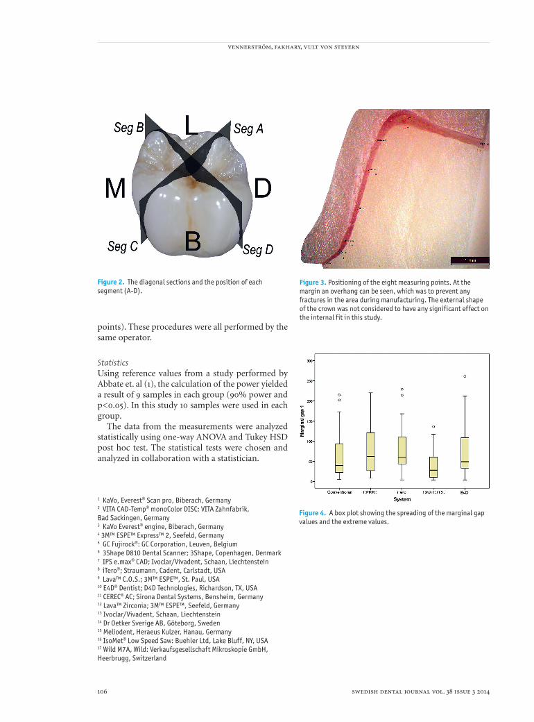
The preparation with the cemented crown was embedded in an acrylic resin block15 and sectioned diagonally into four segments (Figure 2) using a low speed saw with a 0.3 mm thick diamond blade16. Eight predefined points (Figure 3) on each seg-ment were measured using a light microscope17. To standardize the analysis of the predefined points, a
Figure 1. The 50 manufactured preparations were created to fit on a special jig in the form of a jaw section, which was designed in a manner to prevent any rotation.
vennerström, fakhary, vult von steyern
1038.indd 104 2014-10-07 09:57

swedish dental journal vol. 38 issue 3 2014 105
for the respective segments in all groups. Measure-ments were performed at the marginal gap (accord-ing to Holmes (15)), in the chamfer (2 points), on the axial wall (3 points) and on the occlusal surface (2
Table 1. The specifics of each system and milling settings.
Control group CEREC® AC (Sirona) iTero® (Cadent)Lava™ C.O.S. (3M™ ESPE™)
E4D® Dentist (D4D Technologies)
Technique
A-Silicone (3M™ ESPE™ Express™ 2)3Shape D810 Dental Scanner
Active triangulation and confocal micro-scopy (Blue LED)
Parallel confocal imaging (laser)
Active wave-front sampling (blue light)
Optical coherence tomography and confocal microscopy (laser)
Spacer 60 µm 100 µm 60 µm30 µm (0.8-1.5 mm) and 80 µm (from 1.5 mm)
100 µm
Spacer starting point from preparation margin
0.8 mm 0.8 mm 0.8 mm 0.8 mm 0.8 mm
Treatment of preparation before scanning
-
Titanium Dioxide powder. CEREC® Optispray (Sirona)(Grain size – NA. Optimal thickness of the coat – 40-60 µm) (8)
-
Titanium Dioxide powder (3M™ ESPE™ Lava™ Powder for C.O.S) (Grain size – 20 µm)
-***
Manufacturing
Kavo Everest® 467 at dental lab (Dental Syd Malmö)
CEREC® MC XL (chair- side milling unit)
Kavo Everest® 467 at dental lab (Dental Syd Malmö)
CNC 240 (3M™ Danmark AS, Glostrup)
E4D Mill (chair- side milling unit)
Material used for milling of crowns
IPS e.max® CAD (Ivoclar Vivadent)
IPS e.max® CAD (Ivoclar Vivadent)
IPS e.max® CAD (Ivoclar Vivadent)
Lava™ Zirconia (3M™ ESPE™)
IPS e.max® CAD (Ivoclar Vivadent)
After treatmentCrystallization* and etching**
Crystallization* and etching**
Crystallization* and etching**
SinteringCrystallization* and etching**
* The crystallization processes were performed according to the manufacturer’s instructions in an Ivoclar Vivadent Programat P500 crystallization furnace. ** The crowns were etched using IPS Ceramic Etching Gel <5% HF according to the manufacturer’s instructions. *** In areas with high translucency silver nitrate is needed, however this was not the case in this study.
translucent grid was used with predefined reference points specially created from a sectioned master crown (segment A-D). The four grids were individu-ally designed for segments A-D and thereafter used
internal fit, marginal fit, cad/cam, intraoral scanning
1038.indd 105 2014-10-07 09:57
106 swedish dental journal vol. 38 issue 3 2014
points). These procedures were all performed by the same operator.
Statistics Using reference values from a study performed by Abbate et. al (1), the calculation of the power yielded a result of 9 samples in each group (90% power and p<0.05). In this study 10 samples were used in each group.
The data from the measurements were analyzed statistically using one-way ANOVA and Tukey HSD post hoc test. The statistical tests were chosen and analyzed in collaboration with a statistician.
Figure 2. The diagonal sections and the position of each segment (A-D).
Figure 4. A box plot showing the spreading of the marginal gap values and the extreme values.
1 KaVo, Everest® Scan pro, Biberach, Germany2 VITA CAD-Temp® monoColor DISC: VITA Zahnfabrik, Bad Sackingen, Germany3 KaVo Everest® engine, Biberach, Germany4 3M™ ESPE™ Express™ 2, Seefeld, Germany5 GC Fujirock®: GC Corporation, Leuven, Belgium6 3Shape D810 Dental Scanner; 3Shape, Copenhagen, Denmark 7 IPS e.max® CAD; Ivoclar/Vivadent, Schaan, Liechtenstein8 iTero®; Straumann, Cadent, Carlstadt, USA 9 Lava™ C.O.S.; 3M™ ESPE™, St. Paul, USA10 E4D® Dentist; D4D Technologies, Richardson, TX, USA11 CEREC® AC; Sirona Dental Systems, Bensheim, Germany12 Lava™ Zirconia; 3M™ ESPE™, Seefeld, Germany13 Ivoclar/Vivadent, Schaan, Liechtenstein14 Dr Oetker Sverige AB, Göteborg, Sweden15 Meliodent, Heraeus Kulzer, Hanau, Germany16 IsoMet® Low Speed Saw: Buehler Ltd, Lake Bluff, NY, USA17 Wild M7A, Wild: Verkaufsgesellschaft Mikroskopie GmbH, Heerbrugg, Switzerland
Figure 3. Positioning of the eight measuring points. At the margin an overhang can be seen, which was to prevent any fractures in the area during manufacturing. The external shape of the crown was not considered to have any significant effect on the internal fit in this study.
vennerström, fakhary, vult von steyern
1038.indd 106 2014-10-07 09:57

swedish dental journal vol. 38 issue 3 2014 107
ResultsForty-nine crowns were cemented and analyzed. One crown was excluded (E4D) due to problems during the cementation process, which caused an obvious dislocation that could not be associated with the fit of the crown. One measuring point (point 8, iTero) was excluded because of an obvious defect in the crown itself which gave a large non-representable value.
Table 2 shows the mean values for each point measured on the manufactured crowns of each group, together with the standard deviation as well as the deviation from the desired values at each measuring point.
Figure 4 shows a box plot illustrating the data scatter for the marginal gaps.
The statistical analysis showed no significant dif-ference with regard to marginal gap when compar-ing the control group to any of the digital systems. However, significant difference was found between the digital systems, where Lava (Lava C.O.S) had sig-nificantly smaller marginal gaps than CEREC and iTero (p<0.05). The numerical result showed differ-ences between Lava and the E4D system but the dif-ference was not significant (p=0.052).
The gaps of the crowns from the Lava group were smaller than the control group at all measuring points. With exception of the marginal gap, the re-sults were significantly different (p<0.001 for points 2-4 and 7, p<0.05 for points 5, 6 and 8). Regarding the chamfer, measuring points 2 and 3, the crowns from the CEREC and Lava groups had gaps that were significantly smaller than crowns in both the iTero and the control group (p<0.001).
Significant difference was also found between E4D and CEREC. The crowns in the E4D group had smaller gaps than CEREC at measuring points 4-8 (p<0.05 for point 4 and 5, p<0.001 for points 6-8). At measuring point 2, the crowns in the CEREC group had smaller gaps than the crowns in the E4D group (p<0.05).
The crowns in the Lava group showed significant-ly smaller gaps than the crowns in the CEREC group at measuring points 1, 3 and 5-8 (p<0.05 for points 1 and 5, p<0.001 for points 3, 6, 7 and 8). Significant difference was also found between Lava and iTero, where the crowns in the Lava group showed smaller gaps at measuring points 1-4, 7 and 8 (p<0.05 for points 1 and 4, p<0.001 for points 2, 3, 7 and 8).
Table 2. Mean values for each measuring point in µm. Standard deviations are shown in the parentheses. The values in the first column represent the measured gap size and the values in the second column represent their deviations from the set spacer settings. The negative values indicate a mean value smaller than the set spacer gap.
Control CEREC iTero Lava E4D
Measuring Points Mean (SD) Diff Mean (SD) Diff Mean (SD) Diff Mean (SD) Diff Mean (SD) Diff
1: Marginal 66 (61) 66 79 (58) 79 80 (54) 80 39 (34) 39 73 (63) 732: Chamfer 1 146 (62) 146 85 (55) 85 145 (44) 145 61 (33) 61 127 (46) 1273: Chamfer 2 189 (66) 189 113 (64) 113 176 (57) 176 64 (28) 64 125 (46) 1254: Axial 1 150 (85) 90 117 (62) 17 131 (71) 71 85 (34) 5 76 (50) (-) 245: Axial 2 148 (103) 88 140 (71) 40 120 (101) 60 91 (42) 11 80 (53) (-) 206: Axial 3 157 (107) 97 194 (103) 94 145 (110) 85 93 (43) 13 82 (54) (-) 187: Occlusal 1 268 (97)* 208 316 (84)* 216 357 (100)* 297 140 (41) 60 148 (61)* 488: Occlusal 2 218 (106) 158 327 (89) 227 332 (95) 272 167 (42) 87 204 (54) 104
*Measuring point which represents the area where milling compensation could be observed
internal fit, marginal fit, cad/cam, intraoral scanning
1038.indd 107 2014-10-07 09:57

108 swedish dental journal vol. 38 issue 3 2014
DiscussionIn terms of clinically acceptable marginal gap size, there is no real consensus. In an in vitro study ex-amining the solubility rate of type I zinc phosphate cement Jacobs & Windeler found a significant but slight increase in dissolution at 150 µm gaps com-pared to 75 µm gaps (17). McLean has set the clini-cal acceptable marginal gap size to be up to 120 µm (21,22). Abbate et al. obtained results for marginal gaps ranging from 56 to 81 µm for different crown systems - all-ceramic and metal-ceramic crowns, while Fransson et al. reported gaps of more than 50 µm, with 30-50% being 150 µm or above and having a mean thickness over 100 µm (1,11).
In some of the specimens in this study a large scatter could be seen between the minimum and maximum values. The cause of these varying results is difficult to identify as the process consists of nu-merous steps, many of which are beyond the opera-tor’s control.
The scanner highlights areas with insufficient data so that the operator can re-scan the surface in question. Therefore the scanning could be con-sidered a process which is more dependent on the scanner and the interpreting software than on the operator. However, as some systems require a con-trast medium, this could be considered as an op-erator dependent step. Even though the effect of it was not investigated in this study, it is interesting to discuss how it affects the scanning; negatively by increasing the gap through an uneven application, or positively by reducing the risk of receiving inac-curate data because of transparency and reflective surfaces. Insufficient seating of a crown will create a large internal gap occlusally. (24,31) By comparing the gaps at the marginal and occlusal points, one can see if a seating failure has occurred (31). In the pre-sent study this relationship indicates that the crowns were seated adequately. Each segment was measured at 8 points. The points were chosen on the basis of clinical importance as well as in positions which pre-vious studies have shown to be difficult areas to re-produce during milling of crowns. Larger gaps have been observed in areas around the cusps and in the chamfer (19,24). Several studies have shown that the marginal gap is the most important point regarding crown survival. (16,17,21,22,25,26)
The analysis of the specimens showed that larger cement gaps were generally observed over the cusp areas. This is probably the result of an excess of mill-ing around the cusps by the milling unit to avoid interferences in these areas. Over-milling (or drill
compensation) in certain areas, to compensate for size of the milling tool, has been discussed in stud-ies and should be taken into consideration when analyzing the results (24). The clinical significance of the drill compensation, however, is unknown but since this excess of milling disrupts the uniformity of the future cement film, it might also reduce its strength and affect the seating of the crown (19,29).
Drill compensation was observed on crowns in all groups, with the exception of the Lava group. This might be explained by the fact that they were milled in pre-sintered zirconia, which means that the milled crowns are proportionally larger before sintering. This, however, introduces another source of error: shrinkage during sintering. IPS e.max CAD was chosen as it was available for all of the systems except Lava, is an often-selected material in general dentistry, and because it is milled in scale one to one. It could, however, be argued whether the choice of material had any effect on the fit of the crown, since the material used was different in one group. The decision was made to keep Lava in the study but mill the crowns in zirconia instead. Studies on respective materials have shown similar values regarding mar-ginal fit (13,38). Therefore it is considered that the use of the two different materials would not influ-ence the results sufficiently regarding precision.
A large value was measured on one of the speci-mens from the iTero group. The internal shape in this specimen (specimen 8A) differed greatly from the other specimen in this specific region (measur-ing point 8). Therefore it was regarded to be a defect of unknown cause and was excluded, as it was not considered to be a representable value.
No statistical significance was found when com-paring all the digital systems with the control group. Therefore, with regard to marginal gap, the null hypothesis could not be rejected. The mean value of marginal gap measured on the crowns manu-factured in this study using the Lava system was 39 µm, which is comparable to previous clinical studies, where a mean marginal gap of 49 µm was measured on crowns manufactured using the same system (28,32). Akbar et al. used the CEREC scanner to manufacture crowns in vitro, on which the mean marginal gap measured was 66 µm, which is some-what smaller than the mean marginal gap of the CEREC crowns in this study of 79 µm. Nakamura et al. showed in his study a marginal gap between 53 µm and 108 µm, depending on the software setting and convergence of the preparation. However, the measuring methods used were different (2,23).
vennerström, fakhary, vult von steyern
1038.indd 108 2014-10-07 09:57

swedish dental journal vol. 38 issue 3 2014 109
The statistical analysis yielded many significant values in relation to the other measuring points. It could be questioned as to how representative the statistical analysis is, since there were differences between the spacer values. It is therefore of greater interest to compare the mean deviation from the aimed spacer value instead of analyzing the mean gap size itself, as this better represents the accuracy of the systems. However, from a clinical point of view, a smaller gap is more desirable.
Table 2 illustrates the mean deviations of the ce-ment gap from the set cement spacer for the systems in this study. Due to the possible drill compensations or vertical misfit the occlusal points are regarded as less representable. Consideration should also be given to the values at the marginal gap, as well as on the values at the chamfer as these are, in theory, sup-posed to be 0 µm. However, this is not a practically-achievable value due to the fact that the cement will create a gap per se. The axial points are considered to be less affected by a vertical misfit, and because a horizontal misfit is compensated in the sense that a gap smaller than the spacer on one side is followed by a gap larger than the spacer on the other side. This could be observed in some specimens. However the results may still be considered representative as the mean value would equalize the lateral movement.
With the exception of E4D, the manufactured crowns showed axial gaps, more or less, larger than the set spacer value (see Table 2). The system with the smallest deviations was Lava, followed by E4D, CEREC, iTero and the control group.
The point on a crown where the tensile stress is the greatest is located directly below the point of applied occlusal load (3). Theoretically, cusps are areas that are exposed to high force of load during function, which might be areas located above where excess milling has occurred. Significant variations in thickness of ceramic crowns might lead to stress concentrations during load (3,20), which theo-retically means that the mechanical properties of crowns might be reduced when variations in thick-ness exist (3,20,35).
Previous studies have shown that resin cements often give a greater film thickness compared to other cements (36). This is reflected in the current ISO-standard (ISO 4049:2009), which suggests that the viscosity of resin cements should be so low that their thickness does not exceed 50 µm during work-ing time. However, more recent studies demonstrate that the use of several different resin cements leads to gaps less than 25 µm (18,37). This suggests that it is
not necessary for clinicians to provide extra cement space to compensate for the high viscosity of resin cements (18).
Finally, it must be considered that the crowns were manufactured using different milling units with different spacer values although the manufac-turers’ recommendations were followed. Since this study evaluates the systems per se, and not the scan-ning devices only, the milling must be considered to be a part of the system, as recommended values were used. This is, however, feasible as the measurements where done with respect to the settings made for each group studied.
ConclusionsWithin the limitations of this in vitro study the fol-lowing conclusions were drawn.
Crowns made with the aid of digital impressions show marginal and internal fit that is equal to or bet-ter than crowns made using a conventional impres-sion method.
The marginal and internal fit of reconstructions made using digital impression techniques could possibly improve with lower initial CAD setting of the cement gap/spacer, provided that the prepara-tions to be scanned meet standard prerequisites, e.g. not having undercuts.
AcknowledgementsThe authors thank the representative from Ivoclar Vivadent (AB, Solna), Straumann AB (Göteborg, Sweden), 3M™ Svenska AB (Sollentuna, Sweden), Dental Syd Sweden AB (Malmö, Sweden), Plandent Forsbergs Dental AB (Stockholm, Sweden), DAB Dental AB (Upplands Väsby, Sweden) and Dent-house AB (Sundbyberg, Sweden) for support and supplies.
References 1. Abbate MF, Tjan AH, Fox WM. Comparison of the
marginal fit of various ceramic crown systems. J Prosthet Dent 1989 May;61(5):527-31.
2. Akbar JH, Petrie CS, Walker MP, Williams K, Eick JD. Marginal adaptation of Cerec 3 CAD/CAM composite crowns using two different finish line preparation designs. J Prosthodont 2006 May-Jun;15(3):155-63.
3. Anusavice KJ. Dental Ceramics. In: Anusavice KJ, editor. Phillips’ Science of Dental Materials. 11th ed. St. Louis: W.B. Saunders; 2003. p. 655-719.
4. Birnbaum NS, Aaronson HB. Dental impressions using 3D digital scanners: virtual becomes reality. Compend Contin Educ Dent 2008 Oct;29(8):494, 496, 498-505.
internal fit, marginal fit, cad/cam, intraoral scanning
1038.indd 109 2014-10-07 09:57

110 swedish dental journal vol. 38 issue 3 2014
5. Christensen GJ. Impressions are changing: deciding on conventional, digital or digital plus in-office milling. J Am Dent Assoc 2009 Oct;140(10):1301-4.
6. Christensen GJ. The challenge to conventional impressions. J Am Dent Assoc 2008 Mar;139(3):347-9.
7. Donovan TE, Chee WW. A review of contemporary impression materials and techniques. Dent Clin North Am 2004 Apr;48(2):vi-vii, 445-70.
8. Ender A, Mehl A. CEREC Basic Information 3.8 A Clinical Guide. Germany: Sirona Dental Systems GmbH; 2011.
9. Faria AC, Rodrigues RC, Macedo AP, Mattos Mda G, Ribeiro RF. Accuracy of stone casts obtained by different impression materials. Braz Oral Res 2008 Oct-Dec;22(4):293-8.
10. Felton DA, Kanoy BE, Bayne SC, Wirthman GP. Effect of in vivo crown margin discrepancies on periodontal health. J Prosthet Dent 1991 Mar;65(3):357-64.
11. Fransson B, Oilo G, Gjeitanger R. The fit of metal-ceramic crowns, a clinical study. Dent Mater 1985 Oct;1(5):197-9.
12. Garg AK. Cadent iTero’s digital system for dental impressions: the end of trays and putty? Dent Implantol Update 2008 Jan;19(1):1-4.
13. Gonzalo E, Suarez MJ, Serrano B, Lozano JF. A comparison of the marginal vertical discrepancies of zirconium and metal ceramic posterior fixed dental prostheses before and after cementation. J Prosthet Dent 2009 Dec;102(6):378-84.
14. Henkel GL. A comparison of fixed prostheses generated from conventional vs digitally scanned dental impressions. Compend Contin Educ Dent 2007 Aug;28(8):422-4, 426-8, 430-1.
15. Holmes JR, Bayne SC, Holland GA, Sulik WD. Considerations in measurement of marginal fit. J Prosthet Dent 1989 Oct;62(4):405-8.
16. Hunter AJ, Hunter AR. Gingival margins for crowns: a review and discussion. Part II: Discrepancies and configurations. J Prosthet Dent 1990 Dec;64(6):636-42.
17. Jacobs MS, Windeler AS. An investigation of dental luting cement solubility as a function of the marginal gap. J Prosthet Dent 1991 Mar;65(3):436-42.
18. Kious AR, Roberts HW, Brackett WW. Film thicknesses of recently introduced luting cements. J Prosthet Dent 2009 Mar;101(3):189-92.
19. Kokubo Y, Ohkubo C, Tsumita M, Miyashita A, Vult von Steyern P, Fukushima S. Clinical marginal and internal gaps of Procera AllCeram crowns. J Oral Rehabil 2005 Jul;32(7):526-30.
20. Larsson C, Madhoun SE, Wennerberg A, Vult von Steyern P. Fracture strength of yttria-stabilized tetragonal zirconia polycrystals crowns with different design: an in vitro study. Clin Oral Implants Res 2011 Jun 2.
21. McLean JW. Polycarboxylate cements. Five years’ experience in general practice. Br Dent J 1972 Jan 4;132(1):9-15.
22. McLean JW, von Fraunhofer JA. The estimation of cement film thickness by an in vivo technique. Br Dent J 1971 Aug 3;131(3):107-11.
23. Nakamura T, Dei N, Kojima T, Wakabayashi K. Marginal and internal fit of Cerec 3 CAD/CAM all-ceramic crowns. Int J Prosthodont 2003 May-Jun;16(3):244-8.
24. Ortorp A, Jonsson D, Mouhsen A, Vult von Steyern P. The fit of cobalt-chromium three-unit fixed dental prostheses fabricated with four different techniques: a comparative in vitro study. Dent Mater 2011 Apr;27(4):356-63.
25. Rahme HY, Adib SM, Zebouni EA, Bechara BB, Rifai KT. Comparison of the fit of Procera crowns made from stone with those made from polyurethane resin. Gen Dent 2009 Mar-Apr;57(2):171-9.
26. Reich S, Uhlen S, Gozdowski S, Lohbauer U. Measurement of cement thickness under lithium disilicate crowns using an impression material technique. Clin Oral Investig 2010 May 12.
27. Samet N, Shohat M, Livny A, Weiss EI. A clinical evaluation of fixed partial denture impressions. J Prosthet Dent 2005 Aug;94(2):112-7.
28. Scotti R, Cardelli P, Baldissara P, Monaco C. Clinical fitting of CAD/CAM zirconia single crowns generated from digital intraoral impressions based on active wavefront sampling. J Dent 2011 Oct 17.
29. Shen C. Dental Cements. In: Anusavice KJ, editor. Phillips’ Science of Dental Materials. 11th ed. St. Louis: W.B. Saunders; 2003. p. 443-94.
30. Shen C. Impression Materials. In: Anusavice KJ, editor. Phillips’ Science of Dental Materials. 11th ed. St. Louis: W.B. Saunders; 2003. p. 205-54.
31. Shillingburg HT, Hobo S, Whitsett LD, Jacobi R, Brackett SE. Fundamentals of fixed prosthodontics. 3rd ed. Chicago: Quintessence Publishing Co, Inc; 1997.
32. Syrek A, Reich G, Ranftl D, Klein C, Cerny B, Brodesser J. Clinical evaluation of all-ceramic crowns fabricated from intraoral digital impressions based on the principle of active wavefront sampling. J Dent 2010 Jul;38(7):553-9.
33. Tan K, Pjetursson BE, Lang NP, Chan ES. A systematic review of the survival and complication rates of fixed partial dentures (FPDs) after an observation period of at least 5 years. Clin Oral Implants Res 2004 Dec;15(6):654-66.
34. Tuntiprawon M, Wilson PR. The effect of cement thickness on the fracture strength of all-ceramic crowns. Aust Dent J 1995 Feb;40(1):17-21.
35. Vult von Steyern P, Ebbesson S, Holmgren J, Haag P, Nilner K. Fracture strength of two oxide ceramic crown systems after cyclic pre-loading and thermocycling. J Oral Rehabil 2006 Sep;33(9):682-89.
36. White SN, Yu Z. Film thickness of new adhesive luting agents. J Prosthet Dent 1992 Jun;67(6):782-5.
37. Wu JC, Wilson PR. Optimal cement space for resin luting cements. Int J Prosthodont 1994 May-Jun;7(3):209-15.
38. Zhang Y, Li J, Xue XQ, Chen ZY, Li XJ. A comparison of three-dimensional marginal adaptation among three all-ceramic crown systems. Shanghai Kou Qiang Yi Xue 2011 Oct;20(5):494-9.
Corresponding author:Dr Micael Vennerström Department of Materials Science and Technology Faculty of Odontology Malmö University SE-205 06 Malmö, Sweden E-mail: [email protected]
vennerström, fakhary, vult von steyern
1038.indd 110 2014-10-07 09:57

swedish dental journal vol. 38 issue 3 2014 111
Implementation of laser technology and treatment at county level in the Swedish Public Dental ServiceÅsa Bergholm1, Anna-lena Östberg2,3, Pia Gabre1,3
Abstract The aim of this study was to obtain an understanding of the factors that affected the way new technology and methods were used in dentistry after a training program.
A qualitative research method was used to collect data. Nine dentists working in the Public Dental Service (PDS) in Uppsala County in Sweden agreed to be interviewed in the study. They worked in five different clinics, all with laser equipment, and had received training in the use of lasers. The interviews were tape recorded and transcribed, and were analysed using manifest and latent qualitative content analysis.
The categories in this study were identified as “Prerequisites and obstacles to imple-mentation”, “Attitudes to laser technology and treatments” and “Laser technology in the future”. The dentists described working with lasers as complicated and problematic. They had concerns about the method relating to the working environment, evidence of efficacy of treatment, costs, and benefits for patients and dentists. The main finding was that the decision to adopt the technology seemed to be based on individual perceptions of the value of lasers compared to other ways of achieving the same goal. They provided uniform proposals regarding how an organization should implement new methods, including an emphasis on the importance of preparation and having opportunities to be able to test and evaluate the technology. Another important factor was support from surrounding staff, colleagues and management. Despite all the barriers, the respondents were positive about working with lasers in the future, mainly due to their belief that patients would demand laser treatment.
In conclusion both individual and organizational factors affected the extent to which the respondents used the laser. The main finding was the individual perception of the value of lasers compared to other methods which could achieve the same goal.
Key words Dentists, implementation, laser technology
1 Public Dental Service, Uppsala County Council, Uppsala, Sweden, 2 Public Dental Service, Region Västra Götaland, Sweden, 3 Institute of Odontology, the Sahlgrenska Academy, University of Gothenburg, Gothenburg, Sweden
swed dent j 2014; 38: 111-120 bergholm, östberg,gabre
1031.indd 111 2014-10-07 09:58

112 swedish dental journal vol. 38 issue 3 2014
Implementation av laserteknologi inom tandvården
Åsa Bergholm, Anna-lena Östberg, Pia Gabre
Sammanfattning
Syftet med studien var att skapa förståelse för vilka faktorer som avgjorde hur tandlä-kare använde laser efter utbildningsinsats.
En kvalitativ metod användes för att samla in och analysera data. Nio tandläkare som arbetade i Folktandvården, Uppsala län, med olika bakgrund, ålder och erfarenhet inter-vjuades. En semistrukturerad intervjuguide valdes med fokus på tandläkarnas upplevel-ser av att börja använda laser som behandlingsmetod. De utskrivna intervjuerna analy-serades genom kvalitativ innehållsanalys.
Kategorierna som framkom i resultatet var ”Förutsättningar och hinder för implemen-tering”, ”Attityder till laserteknik och laserbehandlingar” samt ”Lasertekniken i framti-den”. Subkategorier identifierades inom samtliga kategorier.
Deltagarna i studien beskrev det som omständligt och komplicerat att arbeta med laser. Tandläkarna hade trott att lasertekniken skulle underlätta för både patient och behandlare men beskrev det som en hinderbana att börja arbeta med laser. Hinder som nämndes var organisatoriska faktorer, att behandlingarna tog lång tid samt att man kände sig osäker på om lasermetoden var evidensbaserad och effektiv i relation till kost-naderna. Huvudresultat var att beslutet att använda laser eller inte, berodde på bedöm-ningen av värdet av lasern i jämförelse med andra sätt att uppnå samma mål, och om det fanns en vinst för patienten.
Respondenterna hade enhetliga förslag till hur en organisation bör gå tillväga när man inför en ny behandlingsmetod och ny teknik. Man poängterade vikten av förberedelse, kunskap och möjlighet att pröva och utvärdera en metod. Även stöd från tandsköterskan, kolleger och ledning nämndes som betydelsefullt. Trots de egna erfarenheterna av att det är komplicerat att använda laser, ansåg de intervjuade tandläkarna att metoden kommit för att stanna, framförallt eftersom patienterna efterfrågar behandlingen.
Slutsatsen var att såväl individuella som organisatoriska faktorer påverkade hur man använde lasertekniken. Det viktigaste var om man upplevde att laserbehandlingen inne-bar en vinst för patienten.
swed dent j 2014; 38: 111-120 bergholm, östberg, gabre
1031.indd 112 2014-10-07 09:58
swedish dental journal vol. 38 issue 3 2014 113
implementation of laser technology
IntroductionIn healthcare systems, development and improve-ment are continuous processes. Important factors for improving clinical practice are studied, as well as factors that highlight the implementation of new treatment methods, policies and technologies (9). Studies in medicine show that there are no “magic bullets” for improving the quality of healthcare, but there are a wide range of interventions available that, if used appropriately, can lead to important im-provements in professional practice and patient out-come (19). Scientific research into implementation is underway in several areas including healthcare, psy-chology, sociology and economics. However, barri-ers to change can occur at multiple levels - in indi-viduals, groups or organizations, and it is important to be able to use the right strategy to overcome them. As Grol &Grimshaw write: “Plans for change should be based on characteristics of the evidence or guide-line itself and barriers and facilitators to change. In general, evidence shows that none of the approaches for transferring evidence to practice is superior to all changes in all situations” (11).
There is a complex interplay between the distri-bution and application of new knowledge which is influenced by factors including leadership, the working environment, available resources and indi-vidual circumstances (9). People’s abilities to adopt new knowledge and techniques can be described as a normal distribution curve, where those with the greatest openness to change will be found at the edge of the curve, i.e. “early adopters” (21). Dentists who are early adopters in one area have been shown to be likely to be in the forefront in other areas too (5).
In dental practice, knowledge of professional bar-riers to embracing new guidelines can actually fa-cilitate the change process (23). Dentists alter their clinical therapies based on research, colleagues’ opinions, training and “ad hoc” evaluation of their own work (18, 13).
A Swedish study demonstrates how well-struc-tured training, which is followed up over time, can achieve good results in getting dentists to use new treatment technologies in endodontics (15). Change in clinical endodontic treatment was achieved by us-ing a combination of strategies including lectures, briefings to all staff, clinical training and reminders. Determining how successful the introduction of new technologies is may be related to how the user per-ceives the “usefulness” and the “ease of use” of these technologies. The concept “usefulness” describes the degree of benefit the new technology brings to the
user, and “ease of use” refers to how easy the tech-nology is to use. The considered benefits of new technology appears to be more important in how frequently the new method is used, rather than how easy it is to use (3).
In the Public Dental Service (PDS) in the county of Uppsala, Sweden, laser technology was intro-duced in 2008. Laser technology in caries treatment has been evaluated in a systematic review. The re-sult showed that laser seemed to work as well as the conventional drill when removing caries-damaged tissue. Patients find laser treatment less uncomfort-able than drilling, but treatment with laser is more time-consuming. It was not possible to draw any conclusions regarding biological or technical com-plications and the method can not be considered cost-effective (14). Another disadvantage is that la-sers cannot be used to remove metal fillings and do not perform well when being used to remove plastic fillings. However, laser technique may be an option for soft-tissue surgery in children, and when treating short maxillary or lingual frenula (16).
Despite the studies described above, knowledge of dentists’ experiences of learning new, advanced methods of treatment is limited. This study intends to examine the introduction of laser treatment in PDS dental care in a Swedish county. The aim of the study is to create an understanding of the factors that affect how new methods and technology are used in dentistry after a training program.
Material and methodsThe study was approved by the Regional Ethical Review Board at the University of Uppsala (No. 2011/158). Informed consent was obtained from all subjects prior to the start of the study. The aim of the study was to gain a deeper understanding of dentists’ strategies in adopting new technology. A qualitative research method was used and data was collected in individual interviews.
SubjectsNine dentists, seven women and two men, from five different clinics with laser equipment in the PDS in Uppsala County were interviewed. To obtain a relevant strategic selection, dentists of different ages, genders, professional experience and differ-ent practice orientations, i.e. general practitioners and specialized dentists, were selected. Two dentists from the same clinic chose not to participate, and therefore one clinic with laser equipment was not represented in the study. The age range among the
1031.indd 113 2014-10-07 09:58

114 swedish dental journal vol. 38 issue 3 2014
participants was between 29 and 65 years (median 45 years), and they had worked between four and 40 years in the profession (median 20 years). The interviewees received their laser education between 2007 and 2010, four having undergone professional training in Germany and five in Sweden.
InterviewsThe interviews were performed by a dentist and clin-ic manager with experience in interviewing (ÅB). The interviewer was known by the participants, but had no working relationship with any of them. The interviews took place at the participants’ workplaces or other PDS premises and lasted between 45 and 60 minutes. A semi-structured interview guide was used. Interview focus was on the dentists’ own expe-riences of using laser technology and also contained the following perspectives: the participants’ profes-sional backgrounds and careers, how they used la-ser in patient treatment, and how an organization should act when introducing new technology and new treatments. The interview guide was adapted to new perspectives expressed by the participants in subsequent interviews. All the interviews were tape recorded and transcribed verbatim by a transcrip-tion office. The interviews were performed and tran-scribed in Swedish and the analysis was conducted with the Swedish text as a basis. A professional trans-lator translated the quotations used in this paper from Swedish into English.
AnalysisThe transcribed text was studied using content anal-ysis; both manifest and latent analyses were carried out. Both types of analysis involve interpretation, although they differ in depth and level of abstrac-tion (8). The unit of analysis was nine interviews. The analysis was made by two of the authors (ÅB and PG) who independently read all the inter-views several times to gain an overall understand-ing. Units of meaning, combinations of words that relate to the same meaning, were identified in the text. The analysis continued by condensing the units of meaning into codes, and in this process an ab-straction took place involving analyzing on a higher logical level. The codes were then clustered into cat-egories and sorted into subcategories. Descriptions of statements were sorted into manifest categories, while latent categories included interpretations of the underlying meanings of statements, at varying levels of abstraction. The authors suggested tentative categories and subcategories, evaluated the group-
ings, revised the material and finally reached con-sensus. Quotations from most of the interviews are presented in all the categories to help the reader to assess the trustworthiness of the results. The orga-nization of codes, subcategories and categories are shown in Figure 1.
ResultsThe respondents described the use and applications of the laser technique in different ways. Some re-spondents used the laser as soon as the education was completed, but then stopped using this form of treatment. Others used laser about once a month or even less, while others used it every week. The appli-cations were caries treatment, primarily in patients with dental anxiety, soft tissue surgery, endodontics and treatment of sensitive root surfaces. A few re-spondents conducted research on laser as a method for caries treatment and endodontics. The categories in this study were identified as “Prerequisites and obstacles for implementation”, “Attitudes to laser technology and treatments” and “Laser technology in the future”.
Prerequisites and obstacles to implementationThe category “Prerequisites and obstacles to im-plementation” was composed of the subcategories Education and training, Preparation and practice, Obstacles and Motivation for using laser (Fig. 1).
Education and trainingSome participants described how the introduction of laser technology was a decision taken by the lo-cal PDS while others became interested at dental congresses. Regardless of who took the initiative, respondents stated that they had made their own decisions to participate. It was pointed out by re-spondents that it is crucial that individuals have a personal interest in learning a method and take their own initiatives.
”If you’ve asked for it yourself ….you do it …you want it and you persevere ….”
The participants’ knowledge of laser prior to training was described as varied. Some had heard that patients who received laser treatment were sat-isfied, and others had read in the newspapers about laser use in dentistry and wanted to know more. Once they had decided to attend the program, some participants obtained information of their own about lasers and had contact with companies selling laser equipment.
The theoretical training was described as diffi-
bergholm, östberg, gabre
1031.indd 114 2014-10-07 09:58

swedish dental journal vol. 38 issue 3 2014 115
cult, but extensive and thorough. The dentists’ opin-ions of the quality of the training differed - some found it very good while others were less satisfied. Those who had received their education in Sweden seemed to be more satisfied with the training pro-gram than those trained in Germany. The main rea-son mentioned was the educational content.
“I think it was good….there was a lot… of phys-ics in it and I think that was great and important to have in and I think …. it’s good that everybody who is going to use lasers has had training so you don’t miss anything.”
Preparation and practiceThe importance of preparation was stated, for in-stance visiting others working in the same field in order to learn from their experiences. Participants
also stressed the importance of opportunities for people to find out themselves how it was to work with the technology, as well as the importance of testing and evaluation in practice.
Some dentists considered that the practical train-ing prepared them adequately to be able to work with laser equipment in the clinics, while others stat-ed that they would have appreciated more “hands-on” training and more opportunity to practice. They were of the opinion that when starting to work at their own clinics, there was no opportunity for tu-toring. There they had to try out techniques on their own, using trial and error:
“No I didn’t really feel ready; instead it was a bit ….. try and see for yourself. Which settings the equipment should have, which was best and all that…”
- initiative- knowledge- theoretical training
- the importance of preparation- trial and error- support
- complicated and problematic- assistant personnel not trained- time consuming – the importance of time
- benefits for patient- benefits for therapist- anticipations for utilization
-easier for patient- benefits for therapist- demand for laser treatment- unfulfilled expections created disappointment
- scientific evidence and credibility-worries about safety- high costs in relation to benefits
- benefits for patients a must- to be in the frontline
- other applications for laser- improved laser devices- contribution to research- overcome resistance
Education and training
Preparation and practice
Obstacles
Motivation for using laser
Expectations of laser
Insecurity associated with laser treatment
Find new motivation for using laser
Ongoing work and further progress
Prerequisites and obstacles of implentation
Attitudes to laser technology and treatments
Laser technology in the future
Figure 1. Codes, subcategories and categories in the analyses.
implementation of laser technology
Codes Subcategories Categories
1031.indd 115 2014-10-07 09:58

116 swedish dental journal vol. 38 issue 3 2014
The dentists expressed different needs for support while they gained experience in methods of treat-ment. Since there were just a few dentists working with laser technology at their clinics, participants lacked input from colleagues. Respondents also felt that it would have been an advantage to have had dentists from other clinics to collaborate with, i.e. a professional network. The general view was that it was advantageous when the clinic manager had an understanding of safety issues and the need for training before performing laser treatments.
ObstaclesParticipants had many comments on how it was to start using the laser machine: operating it was com-plicated and consideration had to be paid to space, availability, ergonomics and time. Working with la-ser was described as a complicated and problematic process, and was compared to an obstacle course.
“It’s like a little obstacle course, you’ve got to sort of jump over a lot of obstacles … You have to man-age it and on your own too, so it gets quite hard.”
The laser was not available in the treatment room and a lot of preparation, involving safety regulations and the settings on the equipment, was necessary be-fore performing the actual treatment. They felt that the machine was clumsy and that it took up a lot of space in the room. Respondents described the work environment as unpleasant; the device made a lot of noise, it smelled bad, and the seating position was uncomfortable. On the other hand, some made the point that it was not so difficult to use the device and that a “Quick Reference guide” was available.
Respondents stated that one difficulty was that as-sistant personnel were not trained in the technique and had to be trained during treatment sessions. Participants described time as a very important fac-tor when using laser technique. Treatment time was perceived as longer than when using a conventional drill. The dentists had a clear awareness of the im-portance of the financial outcome in relation to the time spent.
“I was going to say that time is money. Put it like this, time is really everything to us.”
Another time-related aspect was that the dentists felt they had a stressful work situation since there were lots of patients waiting for treatment. It took time to inform colleagues and patients about the option of laser treatment. It was felt that the den-tist should be able to set aside time both to practice laser treatment and to inform and educate people about it.
Motivation for using laserAn important factor that was mentioned as a mo-tivation to use laser technology was to discover whether or not there were any benefits for the pa-tient and the therapist.
“I have to feel that it’s good for me, and first and foremost for the patient”
Some respondents said that they were convinced that this method had advantages for the patient, es-pecially for those with dental anxiety, and that pa-tients would choose laser treatment if they had the opportunity. Others did not believe that there were any reasons to use the laser, or thought any advan-tages were insignificant.
Participants said that they felt the management anticipated that they would use modern technol-ogy and would utilize the investment that had been made. Some participants stated that they felt com-pelled to work with this method since they had re-ceived extensive and expensive training. Others said they experienced a feeling of guilt when the equip-ment was not used.
Attitudes to laser technology and treatmentsIn this category, subcategories Expectations of laser treatment and Insecurity associated with laser treat-ment were identified.
Expectations of laser treatmentParticipants said that their expectations of what could be achieved with laser treatment were high before they started. Above all, the main aim was to make it easier for patients, particularly for those with negative experiences of the drill. It was expect-ed that it would be painless for the patient and that a local anesthetic would not be necessary.
“I had child patients and very many were scared of the injection, there was a lot of fear of needles and of pain …..so I thought it would be a great opportunity to work with lasers….”
The respondents voiced an expectation that la-ser treatment would be faster and easier to use than conventional drilling, which would also be an ad-vantage for the therapist.
The dentists reported that the image of laser treat-ment was that it would produce good results, that it could be used for most things in dentistry includ-ing caries treatment and surgery, and that it would be very good for root canal treatments and patients with disabilities.The respondents considered that positive expectations were created both by those who marketed laser appliances and also by those
bergholm, östberg, gabre
1031.indd 116 2014-10-07 09:58

swedish dental journal vol. 38 issue 3 2014 117
who gave the training. However, more skeptical at-titudes were also expressed. Some participants had seen the laser used in the past and did not think it seemed to work as well as described. Respondents expected that a large number of patients would de-mand laser treatment in the future.
Since the dentists´ expectations were not fulfilled they reported a feeling of disappointment. The general opinion was that treatment took a long time, the machine malfunctioned, the technology was too complicated and patients did not ask for laser treat-ment in the first place.
Insecurity associated with laser treatmentThe scientific evidence and credibility of the laser method was stated as important for dentists in the study.
It was considered that there was not enough evi-dence from research in the field. Gradually the re-spondents began to doubt if what they had learned during training was credible and they expressed their disappointment with the low quality of com-pleted studies:
“If we use lasers without evidence that’s quackery ….. We need evidence and science behind us.”
Participants declared a strong sense of uncertain-ty and insecurity when it came to using laser tech-nology.They expressed worries regarding safety, the machine was described as intimidating, and the costs associated with its use were too high. Uncer-tainty concerning caries treatment and the sterility of laser appliances were also mentioned. In addition, uncertainty was voiced about the working environ-ment.
“We know enough to be scared of the laser, but not enough to be certain about how to use it.”
Laser technology in the futureWithin this category, two subcategories were identi-fied; Finding new motivation for using lasers and On-going work and further progress.
Finding new motivation for using laserThe respondents often repeated that the most cru-cial aspect of laser treatment was benefits for the pa-tients. Many expressed the belief that patients would choose laser treatments if they had the option.
Several respondents expressed the opinion that laser technology was modern, which was an im-portant reason for wanting to learn to use it. They believed that their organization wanted to be in the front line of technological advances.
“I think it’s great that people are investing in new technology …that we’re at the cutting edge rather than falling behind, I think it’s fun.”
It was also mentioned that laser treatment could attract younger patients, who may consider new technology to be exciting. However, opinions on the laser were ambivalent. On one hand it was new and modern, on the other hand it might not meet the requirements of evidence-based dental treatment.
Ongoing work and further progressParticipants considered that it was possible to find other applications for laser treatment, including soft tissue surgery, where it would facilitate difficult treatments and bring quicker healing, and scurrying root surfaces. In these areas the laser was mentioned as an equipment and a good complement to conven-tional treatment. The view was expressed that the la-ser has come to stay in dental treatment, since laser devices are improving all the time.
Some interviewees expressed the opinion that they could not rely on existing research, instead it was stated that users themselves had a responsibility to increase their knowledge in the field of laser use and to contribute by carrying out scientific work of their own.
“Just because there aren’t any scientific studies doesn’t mean that it’s not good.”
Some declared that they believed it was possible to make a new start. Despite the resistance that existed and the doubts dentists described, there was also a determination to continue working with laser tech-nology. Some saw it as stimulating to overcome the obstacles they had experienced.
“It’s sort of the future….of course you have to keep on trying.”
DiscussionThis study showed that dentists found it complicat-ed and time-consuming to work with lasers. They compared laser use to an obstacle course and found that the high expectations they had in the beginning were not fulfilled. The interpretation of the results was that respondents felt uncertain regarding the working environment, scientific evidence, costs, and benefits for patients and dentists.The main find-ing was that the decision to adopt new technology seemed to be based on individual perceptions of the value of lasers compared to other ways of achieving the same goal and the meaning, here interpreted as if there was a benefit to the patient (4).
A qualitative research method was chosen in or-
implementation of laser technology
1031.indd 117 2014-10-07 09:58

118 swedish dental journal vol. 38 issue 3 2014
der to obtain useful information about the aspects being studied. The content analysis method makes it possible to draw replicable and valid inferences from data, which in turn can give knowledge and new insights into the issues (16). Criticism of the method includes the opinion that it is not suffi-ciently qualitative in nature, and that its descrip-tions of data are simplistic. Content analysis initially involved a quantitative description of the context, but the method has developed and now includes in-terpretations of latent content (8). Trustworthiness in a qualitative study depends on two aspects. The first is credibility, associated with confidence in how well data and the analysis processes address the in-tended focus, here obtained by the selection of par-ticipants with various backgrounds and the use of open questions adapted to cope with new informa-tion and opinions. The second is dependability, as-sociated with the degree to which data changes over time and alterations made by the researcher during the analysis process (8). A third factor of importance for trustworthiness is transferability, referring to the extent to which results can be transferred to other groups. Carefully describing factors of importance for credibility and dependability in the study enables the reader to assess the level of transferability of the results.
Statements in this study showed that the den-tists used laser technology to different degrees. The results indicated that both organizational and in-dividual factors were of importance for how laser technology was used. This is consistent with another study that recognized that implementation of clini-cal guidelines requires both organizational and indi-vidual changes (10).When it came to working with laser devices, respondents’ descriptions and sugges-tions of what to do when implementing a new treat-ment method, were consistent in this study. Several of the concerns mentioned by the respondents, like the laser being complicated to use, treatment taking more time, the value of practicing with laser equip-ment before attending a training course and obvious benefits of new methods, have previously been de-scribed in a review concerning diffusions of innova-tion in service organizations (9). Descriptions of the obstacles the dentists faced and the frustration they felt about working with laser equipment occupied a large part of the interviews. A consequence of this is that the category “Prerequisites and obstacles to implementation” dominates the results and analysis.
The respondents seemed to have good levels of confidence in their own abilities, a concept known
as “self-efficacy” (1). In the concept described by Schunk & Pajares, people with high self-efficacy got involved in their work faster, made greater efforts, showed greater endurance and achieved better re-sults (22). Although the respondents in our study expressed confidence in their own abilities, several failed to adopt laser technology. When expectations regarding the use of laser were not met, some partic-ipants drew the conclusion that they had not done a good job. Similarities and differences in participants’ attitudes to individual adoption of new technology were found. It was possible to describe them all as “early adopters,” meaning they said that they were stimulated by challenge and were eager to develop their professional skills. But the adoption of an in-novation is more of a process than an event and the grouping of people into “early adopters” and “lag-gards” (20) was not sufficient to explain why some individuals decided to use the new technology and some did not. As described by Greenhalgh et al, peo-ple test and evaluate new treatment methods and guidelines and often find, or fail to find, a meaning in them in dialogue with others (9).
Some of the respondents performed very few treatments, for which they gave different reasons. These included no or few indications, uncertainty concerning results, concerns about sterility and lack of scientific evidence. Lack of time for practice and the practical problems of fetching the machine were also mentioned. A possible conclusion is that the in-novation did not meet identified needs of the den-tists. According to the Concerned Based Adoption Model (12), several of the factors mentioned above are important for the successful adoption of an in-novation. This model points out important prereq-uisites for adoption, such as sufficient information about the innovation, sufficient training and sup-port, feed-back, opportunity to adapt, refine and improve. Sociological research describes how key attributes of the innovation itself are also of impor-tance when it comes to its adoption. Innovations that have a clear advantage in either effectiveness or cost-effectiveness are more easily adopted and implemented (20).
The dentists were also doubtful about the scien-tific evidence relating to laser technology. Accord-ing to Hopper et al., dentists were able to see the importance of evidence, but also considered per-sonal experience important when adopting clinical guidelines (13). The evidence base for technologies and methods was often ambiguous and contested, and therefore needed to be continually interpreted
bergholm, östberg, gabre
1031.indd 118 2014-10-07 09:58
swedish dental journal vol. 38 issue 3 2014 119
and reframed in a local context (7). Laser technology seemed to be hard to introduce even among those describing themselves as interested in new technolo-gy and who, for example, used a microscope in end-odontics. These results may be in line with Davies´ theory that the perceived usefulness of a technology was of greater importance than its ease of use (3). Particular factors, such as involving PDS managers, holding seminars for assisting personnel and giving hands-on practical training with supervision and follow-up, resulted in the successful implementa-tion of new technology in endodontics (15). Similar initiatives might also have facilitated the implemen-tation of laser treatment.
A recurrent point made by participants was the importance of the benefits of laser treatment to the patient and the dentist. The dentists that used the laser expressed a belief in its benefits for the patients, and pointed out that patients´ demands for laser treatment were an important factor. These dentists also mentioned that they had a sense of responsi-bility and a commitment to use the laser appliances since they had received extensive training. They felt that management expected them to use it. According to Eveland, if the conditions mentioned above were fulfilled, the innovation was more likely to be suc-cessful (6). Those dentists who had ceased to work with lasers thought that it was possible to get equally good results with conventional methods and that the laser method was not effective. They saw no ad-vantage in using it, which is important according to Rogers (20). These dentists made the decision not to use the laser because they saw no benefits for the pa-tient. This could be described as a decision-making model called, “the physician as agent,” in which the decision regarding treatment is taken by the dentist, assuming that he or she knows the patient’s prefer-ence (2).
Despite all the obstacles, the respondents were positive about the future of lasers, and they found it possible to continue using lasers in treatment. The meaning attached to an innovation is generally not fixed but can be negotiated and reframed within an organization (7).
ConclusionBoth individual and organizational factors influ-enced the use of laser technology after a training program. The participants in the study described similar experiences of working with laser technol-ogy but they drew different conclusions from their experiences. For some of them laser technology be-
came a part of the daily treatment arsenal, for others it was used occasionally and some had ceased using it. The main finding was that the decision to adopt the technology seemed to be based on individual perceptions of the value of lasers compared to other ways of achieving the same goal. The respondents provided uniform proposals on how organizational factors affect implementation, including an empha-sis on the importance of preparation and evalua-tion. Other important factors were support from as-sistant personnel, colleagues and management. The respondents were positive about working with laser in the future, mainly due to their belief that patients would continue to demand laser treatment and that laser use is a modern technique that will improve.
AcknowledgementWe thank Gwyneth Olofsson for revising the English text. We would also like to thank the Public Dental Service in Uppsala County for financial support.
implementation of laser technology
1031.indd 119 2014-10-07 09:58

120 swedish dental journal vol. 38 issue 3 2014
References1. Bandura A. Self Efficacy. The Exercise of Control;
Basingstoke: WH Freeman 1997 pages 37-41.2. Charles C, Gafni A, Whelan T. Decision-making in the
medical encounter: what does it mean? (or it takes at least two to tango). Soc Sci Med. 1997;44:681-92.
3. Davis FD. Perceived Usefulness, Perceived Ease of Use, and User Acceptance of Information Technology. MIS Quarterly 1989;13:319-40.
4. Dearing JW, And O. Portraying the New: Communication between University Innovators and Potential Users. Science Communication 1994;16:11–42.
5. Dorsey M, Overman P, Hayden WJ, Mayberry W, Requa-Clark B, Krust K. Relationships among and demographic predictors of dentists’ self-reported adherence to national guidelines. Soc Sci Med. 1991;32:1263-8.
6. Eveland JD. Diffusion, Technology Transfer and Implementation. Knowledge: Creation, Diffusion, Utilisation. 1986;8:303–22.
7. Ferlie E, Gabbay J, Fitzgerald L, Locock L, Dopson S. Evidence-Based Medicine and Organisational Change: An Overview of Some Recent Qualitative Research. In Organisational Behaviour and Organisational Studies in Health Care: Reflections on the Future, edited by L. Ashburner. Basingstoke: Palgrave, 2001.
8. Graneheim UH, Lundman B. Qualitative content analysis in nursing research:concepts, procedures and measures to achieve trustworthiness. Nurse Education Today 2004;24:105-12.
9. Greenlagh T, Robert G, Macfarlane F, Bate P, Kyriakidou O. Diffusion of innovation in service organizations: systematic review and recommendations. Milbank Q 2004;82:581-629.
10. Grimshaw JM, Thomas RE, MacLennan G, Fraser C. Ramsay CR, Vale L, Whitty P, Eccles MP, Matowe L, Shirra L, Wensing M, Dikstra R, Donaldson C, Hutchinson A. Effectiveness and Efficiency of Guideline Dissemination and Implementation Strategies. Health Technol Assess 2004;8:1–72.
11. Grol R, Grimshaw J. From best evidence to best practice: effective implementation of change in patient´s care. Lancet 2003;362:1225-30.
12. Hall GE, Hord SM. Change in Schools. Albany: State University of New York Press, 1987.
13. Hopper L, Morris L, Tickle M. How primary care dentists perceive and are influenced by research.Community Dent Oral Epidemiol. 2011;39:97-104.
14. Jacobsen T, Norlund A, Englund GS, Tranæus S. Application of laser technology for removal of caries: a systematic review of controlled clinical trials. Acta Odontol Scand. 2011;69:65-74.
15. Koch M, Eriksson HG, Axelsson S, Tegelberg A. Effect of educational intervention on adoption of new endodontic technology by general dental practitioners: questionnaire survey. Int Endod J 2009;42:313-21.
16. Kotlow L. Lasers and soft tissue treatments for the pediatric dental patient. Alpha Omegan 2008;101:140-51.
17. Krippendorff, K. Content Analysis: An Introduction to Its Methodology 2nd ed. Thousand Oaks, CA: Sage 2004.
18. McGlone P, Watt R, Sheiham A. Evidence-based dentistry: an overview of the challenges in changing professional practice. Br Dent J 2001;190:636–9.
bergholm, östberg, gabre
19. Oxman A, Thomson MA, Davis D, Haynes R. No magic bullets: a systematic review of 102 trials of interventions to improve professional practice. CMAJ 1995;153:1423-31.
20. Rogers EM. Diffusion of innovation. New York: Free Press, 1995.
21. Rogers EM. Diffusions of innovations. 5th ed. New York: Free Press, 2003.
22. Schunk DH, Pajares F. The development of Academic Self- Efficacy, Chapter in Wigfield A & Eccles J (Eds). Development of achievement motivation. San Diego: Academic Press, 2001.
23. Spallek H, Song M, Polk DE, Bekhuis T, Frantsve-Hawley J, Aravamudhan KJ. Barriers to implementing evidence- based clinical guidelines:A survey of early adopters. Evid Based Dent Pract. 2010;10:195-206.
Corresponding author:Dr Åsa BergholmPublic Dental ServiceUlleråkersvägen 21750 17 UppsalaSwedenE-mail: [email protected]
1031.indd 120 2014-10-07 09:58

swedish dental journal vol. 38 issue 3 2014 121
swed dent j 2014; 38: 121-132 petrén, bjerlin, ecorcheville, hedrén
Orthodontic treatment by general practitioners in consultation with orthodontists – a survey of appliances recommended by Swedish orthodontists Sofia Petrén, Krister Bjerklin, Agnès Ecorcheville, Pontus Hedrén
Abstract The aim of the present study was to disclose the treatment procedures most fre-quently recommended by Swedish orthodontists for use by general practitioners and to determine whether these recommendations are reflected in the undergraduate dental program in orthodontics at Malmö University. Potential differences between the ortho-dontists’ recommendations were also investigated.
A questionnaire was sent to 169 consulting orthodontists, seeking their recommenda-tions for appliance therapy to be undertaken by general practitioners: 129 (63 males and 66 females) responded.
The Quad Helix was the appliance most commonly recommended for correction of posterior crossbite, a plate with Z-springs for correction of anterior crossbite and the headgear activator for correction of Class II malocclusions. A significant gender diffe-rence was disclosed with respect to orthodontists’ recommendations for treatment of Class II malocclusions by general practitioners, namely that female orthodontists recom-mended the headgear activator more frequently than males. However, this difference is most likely attributable to the gender distribution among orthodontists qualifying as specialists during the last five decades: more recently qualified orthodontists are predo-minantly female.
The choice of appliances corresponded well with undergraduate training in orthodon-tics at the Faculty of Odontology in Malmö.
Key wordsOrthodontics, orthodontic appliances, functional appliances, general practitioners
Department of Orthodontics, Faculty of Odontology, Malmö University, Malmö, Sweden
1037.indd 121 2014-10-07 09:57

122 swedish dental journal vol. 38 issue 3 2014
Ortodontibehandlingar i allmäntandvården rekommenderade av eller i samarbete med ortodontister
Sofia Petrén, Krister Bjerklin, Pontus Hedrén, Agnès Ecorcheville
SammanfattningStudiens syfte var att undersöka vilka ortodontibehandlingar som är vanligast före-kommande hos allmäntandläkare rekommenderade av svenska ortodontister samt att undersöka om de vanligaste behandlingarna är de som ingår i grundutbildningen vid odontologiska fakulteten i Malmö. Eventuella skillnader mellan ortodontisterna under-söktes också.
Ett frågeformulär utformades och skickades till 169 ortodontister med konsultations-verksamhet för att undersöka vilka olika apparaturer som rekommenderas till allmän-tandläkare. Svarsfrekvensen var 76 %, 129 personer varav 66 kvinnliga och 63 manliga ortodontister.
Den mest använda apparaturen för behandling av korsbett var Quad Helix. För att häva frontal invertering var klammerplåt med Z-fjäder vanligast, för att korrigera Klass II malocklussion var det aktivator med inbyggt EOD. En signifikant skillnad mellan ortodon-tisterna hittades så till vida att kvinnliga ortodontister rekommenderade allmäntand-läkare aktivator med extra oralt drag vid behandling av Klass II malocklussion i större utsträckning än manliga ortodontister.
Valet av apparatur överensstämde till största delen väl med det som används i stu-dentundervisningen vid odontologiska fakulteten i Malmö.
swed dent j 2014; 38: 121-132 petrén, bjerlin, ecorcheville, hedrén
1037.indd 122 2014-10-07 09:57

swedish dental journal vol. 38 issue 3 2014 123
orthodontic treatment in general practice
Introduction In Sweden, children and adolescents are offered an annual examination at the dental clinic at which they are registered. They have the choice of attending a public clinic or a private practice. In either case, den-tal care is subsidised by the Swedish National Health Service until the year the patient reaches 20 years of age. In a few counties, this period is prolonged. At the annual visit, the general practitioner under-takes a clinical examination. In the case of clinical findings that may indicate a need for orthodontic treatment, an appointment will be arranged for con-sultation with an orthodontist. In most counties the orthodontic consultation takes place at the patient’s local clinic: appointments are co-ordinated so that several patients are scheduled for consultation at the one session.
At the consultation, the orthodontist decides whether treatment is indicated. If so, depending on the severity of the case, the recommended treatment may be undertaken by the general practitioner, if necessary in consultation with an orthodontist at the local clinic; in more complex cases the patient will be referred to a specialist orthodontic clinic. In certain Swedish County Councils, almost half of all orthodontic treatment is carried out at the general practitioner’s clinic while in other parts of the coun-try almost all treatment is undertaken at a specialist clinic (1).
After consultation or in collaboration with an orthodontist, the general practitioner carries out interceptive treatment, some functional appliance treatment and tooth extractions on orthodontic indications. Other treatments may be grinding of primary teeth and correction of unilateral posterior crossbites (Figure 1) using expansion plates or Quad
Helix appliances (Figure 2a-b). A plate may also be used for correction of anterior crossbite (Figure 3a-d) using springs or screws. General practitioners sometimes treat Class II malocclusions using func-tional appliances such as activators, with or without extraoral traction, Twin Block or extraoral traction appliances.
Thus it is important that the undergraduate den-tal course in orthodontics provides future general practitioners with the skills and competence neces-sary to undertake any orthodontic treatment which may be delegated to them by the consultant special-ists.
The aim of the present study was to investigate which treatments consultant orthodontists most frequently recommend to general practitioners and to analyse potential correlations between choice of recommended treatment, gender of the orthodon-tists, year of graduation as a dentist and year of spe-cialist qualification. A further aim was to investigate whether undergraduate training in orthodontics at the Faculty of Odontology, Malmö University, re-flects the recommendations of Swedish orthodon-tists.
Figure 1 Unilateral posterior crossbite with lateral mandibular shift.
Figure 2 a) Expansion plate. b) Removable Quad Helix.
Figure 3 a) plate with z-spring b) plate with protrusion spring c) plate with protrusion screw for single tooth d) plate with a screw for anterior teeth
1037.indd 123 2014-10-07 09:57

124 swedish dental journal vol. 38 issue 3 2014
Material and MethodsSubjectsThe subjects involved all Swedish orthodontists with available e-mail address, participating in the Swed-ish system with orthodontic consultations to gen-eral practitioners (GPs) and with current experience of these consultations. The subjects were recruited from the membership register of the Swedish Or-thodontic Society with some additions for non-members. The criteria for inclusion in the survey were that participants had been born in 1942 or later and were currently active as specialist consultant or-thodontists in Sweden, thus the questionnaire was sent to 169 subjects. The distribution was performed by the web-based program Survey Monkey®.
MethodsThe questionnaire included items about which ap-pliances the orthodontists recommend for different orthodontic diagnoses. The responses to the survey questions were in multiple-choice form, with op-tions relating to treatment recommendations and choice of appliances. The questionnaire included some core questions about gender, year of gradua-tion as a dentist and year of qualification as a spe-cialist in orthodontics (Appendix). During the first four weeks, 82 orthodontists responded to the sur-vey. Two weeks later, a follow-up e-mail was sent to encourage the non-respondents to participate in the survey, finally resulting in 129 responses.
To investigate whether undergraduate training in orthodontics at the Faculty of Odontology, Malmö University, reflects the recommendations of Swed-ish orthodontists, the education in orthodontics was thoroughly mapped. The education takes place during semester 8-10 of the dental education. The treatment methods carried out are headgear activa-tors, Quad Helix, expansion plates, plates for ante-rior crossbites, cross elastics and in some cases fixed appliances with sectional arches.
Statistical methodsPearson´s Chi-two test was used to disclose any statistical differences between the options with the highest answer rate. Differences with probabilities of less than 5% (P <0.05) were considered statisti-cally significant. The calculations were performed in SPSS version 20.
ResultsThe response rate to the questionnaire was 129 (76 per cent): 66 females and 63 males. More males than females had graduated as dentists during the earlier decades: 71 per cent of the males had graduated be-tween 1961 and 1980, compared with 40 per cent of the females (Table 1).
The male orthodontists had been specialists for a longer time than the females. Forty males (64 per cent) had qualified as specialists between 1981 and 2000, while 53 (80 per cent) of the females had be-come specialists between 1991 and 2011 (Table 1).
The average number of years as a general practi-tioner, including specialist education before qualify-ing as a specialist was 11.6 years for the males and 15.9 years for the females.
Posterior crossbiteFor unilateral posterior crossbites, 75 per cent of re-spondents (80 per cent of the females and 67 per cent of the males) reported that treatment was “always or often” delegated to general practitioners, under the supervision of an orthodontist (Table 2).
The most frequently recommended appliance was the Quad Helix (62 per cent). In 29 per cent expan-sion plates were recommended, with or without oc-clusal coverage (Table 3). Soldered Quad Helix was the most frequently recommended type of Quad Helix appliance: 54 per cent compared with 38 per cent for the removable one.
There were no significant differences between female and male orthodontists with respect to the type of appliance recommended.
When the orthodontists used a Quad Helix them-selves, most preferred a removable appliance: 57 per cent compared with 35 per cent for the soldered Quad Helix. Eight per cent used an expansion plate or a rapid maxillary expansion appliance. At the Fac-ulty of Odontology in Malmö, the undergraduate course in orthodontics includes training in the use of the removable Quad Helix as the preferred appli-ance for correction of posterior crossbite.
Anterior crossbiteFor correction of anterior crossbite in the early mixed dentition, the most frequently recommended appliance (28 per cent) was a plate with occlusal coverage and Z-springs (Figure 3a). Twenty-five per cent of respondents recommended a plate with oc-clusal coverage, and a screw acting on an anterior acrylic segment (Figure 3b) (Table 4). At the Faculty
petrén, bjerlin, ecorcheville, hedrén
1037.indd 124 2014-10-07 09:57
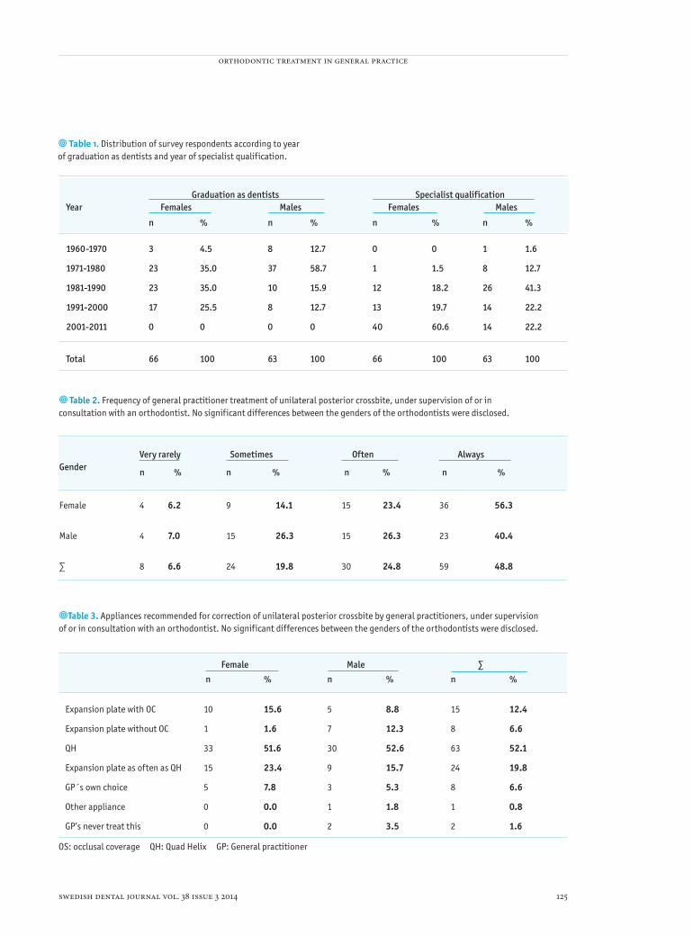
swedish dental journal vol. 38 issue 3 2014 125
Year Graduation as dentists Females Males
Specialist qualification Females Males
n % n % n % n %
1960-1970 3 4.5 8 12.7 0 0 1 1.6
1971-1980 23 35.0 37 58.7 1 1.5 8 12.7
1981-1990 23 35.0 10 15.9 12 18.2 26 41.3
1991-2000 17 25.5 8 12.7 13 19.7 14 22.2
2001-2011 0 0 0 0 40 60.6 14 22.2
Total 66 100 63 100 66 100 63 100
Table 1. Distribution of survey respondents according to year of graduation as dentists and year of specialist qualification.
Table 2. Frequency of general practitioner treatment of unilateral posterior crossbite, under supervision of or in consultation with an orthodontist. No significant differences between the genders of the orthodontists were disclosed.
Gender
Very rarely
n %
Sometimes
n %
Often
n %
Always
n %
Female 4 6.2 9 14.1 15 23.4 36 56.3
Male 4 7.0 15 26.3 15 26.3 23 40.4
∑ 8 6.6 24 19.8 30 24.8 59 48.8
Table 3. Appliances recommended for correction of unilateral posterior crossbite by general practitioners, under supervision of or in consultation with an orthodontist. No significant differences between the genders of the orthodontists were disclosed.
Female Male ∑ n % n % n %
Expansion plate with OC 10 15.6 5 8.8 15 12.4
Expansion plate without OC 1 1.6 7 12.3 8 6.6
QH 33 51.6 30 52.6 63 52.1
Expansion plate as often as QH 15 23.4 9 15.7 24 19.8
GP´s own choice 5 7.8 3 5.3 8 6.6
Other appliance 0 0.0 1 1.8 1 0.8
GP’s never treat this 0 0.0 2 3.5 2 1.6
OS: occlusal coverage QH: Quad Helix GP: General practitioner
orthodontic treatment in general practice
1037.indd 125 2014-10-07 09:57

126 swedish dental journal vol. 38 issue 3 2014
of Odontology in Malmö the appliance of choice for correction of anterior crossbite is a plate with springs.
Anterior crossbite of a single toothWhen correcting anterior crossbite of a single tooth (Figure 3a), 48 per cent of the orthodontists rec-ommended a plate with occlusal coverage and a Z-spring (Table 5).
A plate with occlusal coverage and a screw acting directly on the tooth was recommended by 20 per cent of the respondents.
Ten percent recommended a plate with occlusal coverage, with a protrusion screw to cause anterior movement of the incisors (Figure 3c).
Four orthodontists considered that correction of anterior crossbite of a single tooth required special-ist treatment and did not supervise this treatment by general practitioners. There were no significant differences between female and male orthodontists with respect to recommendation of appliances (Ta-ble 5). A plate with a Z-spring is the appliance of
choice for these cases in the undergraduate program at the Faculty of Odontology in Malmö.
Class II malocclusionSixty-six of the orthodontists (55 per cent) respond-ed that they “often or always” supervised or advised general practitioners in treatment of Class II maloc-clusions: 63 per cent of the female orthodontists and 46 per cent of the males. Eleven per cent of the respondents reported that they very rarely recom-mended or supervised treatment of class II maloc-clusions by general practitioners (Table 6).
The Andresen activator appliance (Figure 4a) was recommended by 28 per cent of the orthodontists but the most frequently recommended appliance for treatment of Class II malocclusions by the general practitioner was an activator with headgear, 63 per cent (Figure 4b). One female and two male ortho-dontists recommended Twin Block (Figure 4c) and two male orthodontists recommended Mono Block appliances.
There was a significant difference between male
Table 4. Distribution of appliances recommended for correction of anterior crossbite in the early mixed dentition by general practitioners, under supervision of or in consultation with an orthodontist. No significant differences between the genders of the orthodontists were disclosed.
Female
n %
Male
n %
∑
n %
Plate with OC and Z-spring 18 29.6
15 26.7
33 28.2
Plate with OC and a screw to an acrylic part
21 34.4 8 14.3 29 24.7
Plate with OC and a screw acting directly on the teeth
8 13.1 9 16.0 17 14.5
Plate without OC and a screw to an acrylic part
5 8.2 8 14.3 3 11.1
Plate with OC and a protrusion spring
3 4.9 3 5.4 6 5.1
None of these treatments are done by general dentist.
3 4.9 9 16.1 12 10.1
GP´s own choice 3 4.9 2 3.6 5 4.3
OC: occlusal coverageGP: General practitioner
petrén, bjerlin, ecorcheville, hedrén
1037.indd 126 2014-10-07 09:57

swedish dental journal vol. 38 issue 3 2014 127
Table 6. Frequency of treatment by general practitioners, under supervision of or recommended by an orthodontist, of Class II malocclusions in the mixed dentition. No significant differences between the genders of the orthodontists were disclosed.
Gender Very rarely
n %
Sometimes
n %
Often
n %
Always
n %
Female 8 12.5 16 25.0 33 51.6 7 10.9
Male 5 8.8 26 45.6 23 40.3 3 5.3
∑ 13 10.7 42 34.7 56 46.3 10 8.3
Table 5. Distribution of appliances recommended for correction of anterior crossbite of a single tooth by general practitioners, under supervision of or in consultation with an orthodontist. No significant differences in relation to orthodontists’ gender were disclosed.
Female
n %
Male
n %
∑
n %
Plate with OC and Z-spring 27 45.0
30 53.5
57 48.3
Plate with OC with a screw acting directly to the tooth 14 23.3 7 12.5 21 19.5
Plate with OC and a screw to an acrylic part
8 13.3 4 7.1 12 10.0
Plate without OC and a screw to an acrylic part 3 5.0 7 12.5 10 8.5
Plate without OC and Z-spring 2 3.3 3 5.4 5 4.2
Plate without OC with a screw acting directly on the tooth 1 1.7 0 0.0 1 0.8
None of these treatments are done by general dentist. 2 3.3 2 3.6 4 3.4
GP´s own choice 3 5.0 3 5.4 6 5.1
OC: occlusal coverageGP: General practitioner
orthodontic treatment in general practice
1037.indd 127 2014-10-07 09:57

128 swedish dental journal vol. 38 issue 3 2014
and female orthodontists with respect to treatment of Class II malocclusions by general practitioners. More male orthodontists recommended the An-dresen appliance, and more female orthodontists recommended activator with headgear.
Regardless of when they qualified as orthodon-tists, more females tended to recommend headgear activator treatment (Table 7). For the male ortho-dontists, almost equal numbers recommended treatment with a headgear activator and an Andres-en appliance, regardless of year of specialist quali-fication. The exception was for those qualifying as orthodontists from 1981 to 1990, who recommended headgear activator treatment (Table 7). At the Facul-ty of Odontology in Malmö, the treatment of choice for Class II malocclusion is the headgear activator.
Sectional arch wireSectional arch wires are very seldom recommended by orthodontists for use by general practitioners, for correction of anterior crossbite or for closing gaps after extraction of premolars. Eighty-six per cent of the female and 82 per cent of the male orthodontists recommend sectional arch wire “very rarely”, 11 per cent of the female and 14 per cent of the male ortho-dontists recommended sectional arch wire “some-times”. Only 4 orthodontists recommended its use “often or always”.
Discussion The aim of the present study was to disclose which appliances orthodontists recommend to general practitioners. The results were used to ascertain
Table 7. Recommended type of activator in relation to year of specialist qualification as an orthodontist.
Females MalesAndresen activator
Headgear Activator
Andresen activator activator
1970-1980 4 2
1981-1990 1 10 7 14
1991-2000 1 9 5 7
2001-2010 8 27 5 6
∑ 10 46 21 29
Figure 4. a) Andresen activator. b) Van Beek appliance. c) Twin block appliance.
Headgear
petrén, bjerlin, ecorcheville, hedrén
1037.indd 128 2014-10-07 09:57

swedish dental journal vol. 38 issue 3 2014 129
whether undergraduate training in orthodontics at the Faculty of Odontology, Malmö University, reflects treatment procedures commonly recom-mended for Swedish general practitioners. This is the first study to investigate the use of different or-thodontic appliances by general dental practitioners in Sweden.
The results disclosed that posterior and anterior crossbites and Class II malocclusions are frequently treated by general practitioners, in consultation or collaboration with an orthodontist. Most of the rec-ommended treatments are consistent with under-graduate education in orthodontics at the Faculty of Odontology in Malmö.
There was a high response rate to the question-naire, 76 per cent: only 40 of the 169 orthodontists failed to respond.
Some respondents questioned whether the or-thodontist should recommend treatment to the general practitioners, or act as a supervisor, treating the patients together with the general practitioner. It is the authors’ opinion that most orthodontists act as supervisors, while the patients remain under the care of the general practitioner, who is also respon-sible for the treatment. The study did not include orthodontists from districts where the role of the or-thodontist is limited to determining which children should be offered the option of specialist treatment free of charge.
Posterior crossbiteThe prevalence of posterior crossbite has been re-ported to be between 7 and 23 per cent (4, 5, 7, 8, 19, 21) and may be of skeletal or dentoalveolar origin and be either bi- or unilateral.
Unilateral posterior crossbites often involve a lat-eral mandibular shift that may cause asymmetrical condylar movement, (6, 12) which in turn might lead to asymmetrical mandibular growth and facial asymmetry (Figure 1) (13, 17, 20).
If left untreated, the malocclusion will persist: spontaneous correction is rare or does not occur (11, 15). Early intervention is recommended for correc-tion of functional posterior crossbites in the mixed dentition (2, 10, 13, 15, 18, 20). The expansion plate and Quad Helix appliances are frequently used for correction of posterior crossbite (Figure 2).
The responses to the survey disclose that most children with posterior crossbite (75 per cent) are treated by general practitioners under supervision of or in consultation with an orthodontist. Of the
soldered and removable Quad Helix appliance, the soldered version is most frequently recommended for use by general practitioners (62 per cent). Sec-ond to this is the expansion plate with or without oc-clusal coverage (29 per cent) (Table 2-3). The expan-sion plate is less effective (15) but in successful cases, it shows long-term stability comparable with Quad Helix. The Quad Helix has the advantage of shorter treatment times and good long-term stability (2, 14). Furthermore, it has been shown that the Quad Helix is more cost-effective (16).
The answers to questions eight and nine showed that almost all the orthodontists are of the opinion that general practitioners should undertake cross-bite correction. However, some orthodontists do not recommend Quad Helix. This may be explained by the fact that the appliance is considered by some to be difficult to manage.
The orthodontists most often recommend sol-dered Quad Helix for use by general practitioners, while the specialists themselves prefer the removable version. One explanation could be that the remov-able Quad Helix is considered to be more difficult to adjust.
Anterior crossbiteIn anterior crossbite, one or more upper incisor teeth are erupting palatally. This occurs in the early mixed dentition and must be differentially diagnosed from hereditary Class III cases. Although anterior cross-bite is quite common and may lead to several com-plications, to date there is no high-quality evidence to indicate which treatment is the most effective (3).
Correction of anterior crossbite in the early mixed dentition is achieved either with a plate with a screw or springs, or with a lingual archwire with protrud-ing springs or sectional arch wire and brackets. The treatment most frequently recommended for use by the general practitioner was a plate with occlusal coverage and Z-springs (28 per cent) (Table 4).
This was also the most frequently recommended treatment for correction of anterior crossbite of a single tooth, followed by a plate with occlusal cov-erage and a screw acting directly on the tooth (20 per cent), more often recommended by female than male orthodontists (Table 5).
Class II malocclusionThe outcome of removable appliance treatment is highly dependent on patient cooperation, timing of the treatment and the dentist’s clinical skills and
orthodontic treatment in general practice
1037.indd 129 2014-10-07 09:57

130 swedish dental journal vol. 38 issue 3 2014
ability to encourage patient compliance. Despite this, only 13 orthodontists reported that
they “very rarely” recommend treatment of Class II malocclusion by general practitioners; 42 responded “sometimes” while more than 50 per cent responded that they “often or always” recommend treatment by general practitioners (Table 6). Thus, general prac-titioners treat the majority of Class II malocclusions in Sweden.
According to the results of this study, the most fre-quently recommended appliance for correction of a Class II malocclusion was a headgear activator (63 per cent) followed by the Andresen activator (28 per cent). This is in accordance with the undergraduate dental program in Malmö.
Only three orthodontists recommended the Twin Block appliance. In this context it is of interest to note that 75 per cent of the orthodontists in the Brit-ish Orthodontic Society preferred the Twin Block as a functional appliance for overjet reduction (9). This may be because the British orthodontists do not share the Scandinavian tradition of using activators.
When recommending treatment of a Class II malocclusion by a general practitioner, the choice of appliance differed in relation to the gender of the orthodontists. This was, however, probably not an effect of the gender itself, but more likely reflected when they qualified as specialists in orthodontics: recently qualified specialists are predominantly fe-male (Table 7).
Before the introduction of the headgear activa-tor system, the Andresen activator was the most frequently used appliance for treatment of Class II malocclusion in Sweden. There was a significant dif-ference in gender ratio when comparing orthodon-tists receiving their specialist qualifications before and after the year 2000. In the year 1990, females comprised only around 27 per cent of orthodontists, whereas in the year 2011, almost 51 per cent of all the orthodontists who responded to the survey were female (Table 1). This may explain why male or-thodontists tended to prefer the Andresen activator more frequently than females and why the headgear activator system was used more frequently by female than male orthodontists .
The results indicate that for correction of Class II malocclusions in general practice, female orthodon-tists recommended the headgear activator system to a greater extent. For all other appliances, there were no differences in relation to the gender of the or-thodontist.
Sectional arch wire In cases of premolar extraction without fixed or-thodontic appliances, there may be residual gaps. In these cases an option could be to reduce or close these gaps with sectional arch wires and elastic chains. However, 112 orthodontists, 84 per cent, re-sponded that they “very rarely” recommend use of sectional arch wires by general practitioners. This procedure is considered difficult to manage and be-cause it is seldom used, brackets and wires are not always readily available in general clinics.
ConclusionsPatients with unilateral posterior crossbite are treat-ed by general practitioners in 75 per cent of cases, after consultation with or under supervision of an orthodontist. Quad Helix was the most frequently recommended appliance.
Correction of Class II malocclusions in Sweden is most frequently undertaken by general practition-ers, after consultation with or under the supervision of an orthodontist.
The only significant difference between female and male orthodontists with respect to recommen-dation of treatment is in correction of Class II maloc-clusions: compared to males, female orthodontists recommend the headgear activator more frequently, while male orthodontists prefer the Andresen appli-ance. However, this difference seems to be related to the fact that more recently qualified orthodontists are predominantly female.
The most frequently recommended treatment modes correspond well with those included in the dental undergraduate program at the Faculty of Od-ontology in Malmö.
petrén, bjerlin, ecorcheville, hedrén
1037.indd 130 2014-10-07 09:57

swedish dental journal vol. 38 issue 3 2014 131
Appendix
1) My gender is: -Male-Female2) I graduated as a dentist in the year: 3) I became a specialist orthodontist in the year: 4) Consultancy: -I work as a consultant with general practitioners -I don’t work as a consultant with general practitioners5) Correction of unilateral posterior crossbite is under-taken by general practitioners in my area:-Very rarely/never-Sometimes (10-20 percent of cases)-Often-Always in cases of forced mandibular shift6) Treatment of children with Angle Class II malocclu-sion in the mixed dentition is undertaken by general practitioners in my area: -Very rarely/never -Sometimes (10-20 per cent of cases)-Often -Always in the absence of clear contraindications such as lack of interest on the part of the patient and/or the parents7) Treatment with sectional arch wires to correct anterior crossbite of a single incisor or to reduce the space in the lateral segments is undertaken by general dentists in my area: -Very rarely/never-Sometimes (10-20 per cent of cases)-Often -Always when treatment is indicated8) When delegating treatment of posterior crossbite to the general practitioner, which appliance do you gener-ally recommend? -Expansion plate with occlusal coverageExpansion plate without occlusal coverage -Quad Helix - Plate or Quad Helix, equally- General practitioners choose appliance themselves -Another appliance -I never recommend this-I almost never treat unilateral crossbites-Unilateral posterior crossbites are corrected without referral to an orthodontist 9) If you recommend Quad Helix to the general practi-tioner, it is mostly:-Quad Helix soldered to the bands-Removable Quad Helix-Soldered about as frequently as removable Quad Helix-Treatment with Quad Helix is undertaken without orthodontic consultation-Treated by orthodontist
10) When correcting a unilateral posterior crossbite you prefer to use:-Quad Helix soldered to the bands -Removable Quad Helix-Expansion plate with occlusal coverage-Expansion plate without occlusal coverage-Fixed appliance for expanding suture/RME11) If you recommend treatment of a single tooth in an anterior crossbite in the early mixed dentition to the general practitioner, what do you suggest they use?-Plate with occlusal coverage and a Z-spring Plate with occlusal coverage and a protrusion spring-Plate without occlusal coverage and a Z-sprin -Plate without occlusal coverage and a protrusion spring-Plate with occlusal coverage and a screw applied directly to the inverted tooth-Plate with occlusal coverage and a screw attached to an acrylic section directly abutting the inverted tooth -Plate without occlusal coverage and a screw attached to an acrylic section directly abutting the inverted tooth-Labial bow with spring against the inverted tooth -General practitioners choose appliance themselves-Treatment is performed without an orthodontist-I do not recommend any of these treatments to the general practitioner 12) What do you recommend for correction of anterior crossbite,? -Plate with occlusal coverage and a Z-spring -Plate without occlusal coverage and a Z-spring -Plate without occlusal coverage and a screw applied directly to the teeth in crossbite -Plate with occlusal coverage and a screw attached to a segment of acrylic-Plate without occlusal coverage and a screw attached to segment of acrylic-Labial bow with spring against the inverted teeth-General practitioners choose appliance themselves -Treatment is performed without an orthodontist-I do not recommend any of these treatments to the general practitioner13) If you are advising a general practitioner about treatment of Angle Class II malocclusion, which appli-ance do you usually recommend?-Activator, Andresen-Headgear-activator combination-Bass appliance -Twin Block-Monoblock-Bionator-Plate to raise the bite -Another appliance-I never recommend this-Treatment is performed without an orthodontist-Personally I do not think any of the above methods should be applied
orthodontic treatment in general practice
1037.indd 131 2014-10-07 09:57

132
References1. Bjerklin K, Lindsten R, Tunge JS, Sjovall C. Orthodontic
treatment need, outcome and residual treatment need in 15- and 20-year-olds. Swed Dent J. 2012; 36: 157-65.
2. Bjerklin K. Follow-up control of patients with unilateral posterior cross-bite treated with expansion plates or the quad-helix appliance. J Orofac Orthop. 2000; 61: 112-24.
3. Borrie F, Bearn D. Early correction of anterior crossbites: a systematic review. J Orthod. 2011; 38: 175-84.
4. da Silva Filho OG, Santamaria M,Jr, Capelozza Filho L. Epidemiology of posterior crossbite in the primary dentition. J Clin Pediatr Dent. 2007; 32: 73-8.
5. Helm S. Malocclusion in Danish children with adolescent dentition: an epidemiologic study. Am J Orthod. 1968; 54: 352-66.
6. Hesse KL, Artun J, Joondeph DR, Kennedy DB. Changes in condylar postition and occlusion associated with maxillary expansion for correction of functional unilateral posterior crossbite. Am J Orthod Dentofacial Orthop. 1997; 111: 410-8.
7. King DL. Functional posterior crossbite in the deciduous and early mixed dentition. Gen Dent. 1978; 26: 36-40.
8. Kutin G, Hawes RR. Posterior cross-bites in the deciduous and mixed dentitions. Am J Orthod. 1969; 59: 491-504.
8. Lee RT, Kyi CS, Mack GJ. A controlled clinical trial of the effects of the Twin Block and Dynamax appliances on the hard and soft tissues. Eur J Orthod. 2007;29:272-82.
10. Lindner A, Henrikson CO, Odenrick L, Modeer T. Maxillary expansion of unilateral cross-bite in preschool children. Scand J Dent Res. 1986; 94: 411-8.
11. Lindner A. Longitudinal study on the effect of early interceptive treatment in 4-year-old children with unilateral cross-bite. Scand J Dent Res. 1989; 97: 432-438.
12. Myers DR, Barenie JT, Bell RA, Williamson EH. Condylar position in children with functional posterior crossbites: before and after crossbite correction. Pediatr Dent. 1980; 2: 190-194.
13. O’Byrn BL, Sadowsky C, Schneider B, BeGole EA. An evaluation of mandibular asymmetry in adults with unilateral posterior crossbite. Am J Orthod Dentofacial Orthop. 1995; 107: 394-400.
14. Petrén S, Bjerklin K, Bondemark L. Stability of unilateral posterior crossbite correction in the mixed dentition: a randomized clinical trial with a 3-year follow-up. Am J Orthod Dentofacial Orthop. 2011; 139: e73-81.
15. Petrén S, Bondemark L. Correction of unilateral posterior crossbite in the mixed dentition: a randomized controlled trial. Am J Orthod Dentofacial Orthop. 2008; 133: 790.e7-790.13.
16. Petrén S, Marké LÅ, Bjerklin K, Bondemark L. Correction of unilateral posterior crossbite in the mixed dentition- a cost-minimization analysis. Eur J Orthod. 2013; 35:14-21.
17. Pirttiniemi P, Kantomaa T, Lahtela P. Relationship between craniofacial and condyle path asymmetry in unilateral cross-bite patients. Eur J Orthod. 1990; 12: 408-13.
18. Schroder U, Schroder I. Early treatment of unilateral posterior crossbite in children with bilaterally contracted maxillae. Eur J Orthod. 1984; 6: 65-9.
19. Tausche E, Luck O, Harzer W. Prevalence of malocclusions in the early mixed dentition and orthodontic treatment need. Eur J Orthod. 2004; 26: 237-44.
20. Thilander B, Bjerklin K. Posterior crossbite and temporomandibular disorders (TMDs): need for orthodontic treatment? Eur J Orthod. 2012;34:667-73.
21. Thilander B, Myhrberg N. The prevalence of malocclusion in Swedish school children. Scand J Dent Res. 1973; 81: 12-20.
Corresponding author:Dr Sofia PetrénDepartment of OrthodonticsFaculty of OdontologyMalmö UniversitySE-205 06 MalmöSwedenE-mail: sofia.petré[email protected]
petrén, bjerlin, ecorcheville, hedrén
1037.indd 132 2014-10-07 09:57

swedish dental journal vol. 38 issue 3 2014 133
swed dent j 2014; 38: 133–142 göranson, lundström, bågesund
Outcome of orthodontic care and residual treatment need in Swedish 19-year-oldsEmma Göranson1, Fredrik Lundström1, mats bågesund2
Abstract The purpose of the study was to assess the outcome of orthodontic care in Linköping, Sweden. The dental records of 207 (107 M, 100 F) 19-year-olds registered at one public dental health clinic were studied. A clinical examination was performed where malocclu-sions were registered, where after residual orthodontic treatment need was measured using the Index of Complexity, Outcome and Need (ICON). The 19-year-olds also filled in a questionnaire regarding residual subjective orthodontic treatment demand. Differences between genders were analysed.
One hundred and ten (47 M, 63 F) individuals (53.1%) had partaken in orthodontic consultations. Orthodontic appliance treatment had been received by 86 (38 M, 48 F) individuals (41.6%).
A residual orthodontic treatment need was registered in 28 (22 M, 6 F) individuals (13.5%).
Residual subjective orthodontic treatment demand was expressed by 9 (3 M, 6 F) indi-viduals (4.3%). Eight (2 M, 6 F) of those had no residual treatment need.
A higher (p=0.006) rate of females (63.0%) than males (44.0%) had participated in ort-hodontic consultations. The proportion of males (35.5%) who had experienced orthodon-tic treatment was not significantly lower (p=0.069) than among the females (48.0%). However, a lower (p=0.009) proportion of treated males (55.3%; n=21 out of 38) than of treated females (81.3%; n=39 out of 48) had received their treatment by orthodontic spe-cialists. At 19 years of age, the proportion of males with residual treatment need (20.6%) was higher (p=0.002) than among the females (6.0%).
Every patient with orthodontic treatment need and -demand at 19 years of age had previously been offered orthodontic treatment. The conclusion was drawn that the orthodontic care scheme had successfully diagnosed and treated orthodontic problems in the population. However, notable differences between genders regarding treatment modalities and the amount of residual treatment need at age 19 were found.
Key wordsOrthodontic treatment, treatment need, treatment demand
1 Centre for Orthodontics and Paediatric Dentistry, County Council of Östergötland, Linköping, Sweden 2 Centre for Orthodontics and Paediatric Dentistry, County Council of Östergötland, Norrköping. Division of Paediatrics, Department of Clinical and Experimental Medicine (IKE), Linköping University, Linköping, Sweden
1040.indd 133 2014-10-07 09:57

134 swedish dental journal vol. 38 issue 3 2014
swed dent j 2014; 38: 133–142 göranson, lundström, bågesund
Utfall av ortodontivården och kvarvarande ortodontiskt behandlingsbehov hos 19-åringar
Emma Göranson, Fredrik Lundström, mats bågesund
Sammanfattning Syftet med studien var att utvärdera utfallet av ortodontivården vid Folktandvården i Östergötland. Tvåhundrasju (107 män, 100 kvinnor) 19-åriga revisionspatienter vid Folk-tandvården Lilla Torget i Linköping undersöktes. Deras journaler granskades avseende ortodontisk behandlingshistorik. Patienterna undersöktes kliniskt beträffande förekomst av bettavvikelser och kvarvarande objektivt ortodontiskt behandlingsbehov bedömdes med hjälp av ICON-indexet. Individerna fick dessutom fylla i ett frågeformulär gällande kvarvarande subjektivt ortodontiskt behandlingsbehov. Skillnader mellan könen analyse-rades.
Bland de undersökta hade 110 (47 män, 63 kvinnor) individer (53.1%) deltagit i en eller flera ortodontikonsultationer. Åttiosex (38 män, 48 kvinnor) personer (41.6%) hade be-handlats med ortodontisk apparatur. Femtiotre (18 män, 35 kvinnor) av de 86 behandlade 19-åringarna (61.6%) hade ortodontisk retention vid 19 års ålder.
Kvarvarande objektivt ortodontiskt behandlingsbehov registrerades hos 28 (22 män, 6 kvinnor) individer (13.5%).
Kvarvarande subjektivt ortodontiskt behandlingsbehov fanns hos nio (3 män, 6 kvin-nor) personer (4.3%). Åtta (2 män, 6 kvinnor) av dessa hade inget kvarvarande objektivt ortodontiskt behandlingsbehov.
En högre (p=0.006) andel kvinnor (63.0%) än män (44.0%) hade deltagit i ortodonti-konsultation. Andelen män (35.5%) som hade behandlats skiljde sig inte signifikant (p=0.069) från andelen kvinnor (48.0%). Däremot var det en lägre (p=0.009) andel av de 38 behandlade männen (55.3%) än av de 48 behandlade kvinnorna (81.3%) som hade fått sin behandling utförd av ortodontist. En annan skillnad mellan könen var att andelen be-handlade män med retention (47.4%) var lägre (p=0.016) än andelen behandlade kvinnor med retention (73.0%). Vid 19 års ålder fanns kvarvarande ortodontiskt behandlingsbehov hos fler (p=0.002) av männen (20.6%) än av kvinnorna (6.0%).
Alla patienter med kvarvarande objektivt och subjektiv ortodontiskt behandlingsbehov vid 19 års ålder hade tidigare erbjudits ortodontisk behandling. Slutsatsen kunde därför dras att ortodontiskt behandlingsbehov framgångsrikt hade upptäckts och behandlats hos den undersökta gruppen av 19-åringar. Däremot upptäcktes anmärkningsvärda skill-nader mellan könen gällande ortodontisk behandlingshistoria och kvarvarande objektivt ortodontiskt behandlingsbehov vid 19 års ålder.
1040.indd 134 2014-10-07 09:57
swedish dental journal vol. 38 issue 3 2014 135
orthodontic outcome in 19-year-olds
Introduction The need for orthodontic treatment has been es-timated to between 30% and 45% in Swedish and Danish populations of children and adolescents (7, 14, 16, 26). Except for a limited number of gross and noticeable occlusal traits, there is little evidence that the presence of a malocclusion has a major impact on oral health (28). It has also been shown that the association between orthodontic treatment need and quality of life is modest (23). Consequently, fac-tors other than the presence of a malocclusion, such as the individuals own opinion concerning their teeth, must be considered when assessing treatment need and -demand.
According to a report from 2005, the average or-thodontic treatment rate in Sweden was 27%, with a range between 21% and 39% in different Swedish Counties (29). Treatment rates on population levels have also been reported from other Nordic Coun-tries. Some of these results are presented in Table 1. Most of these studies report no significant differ-ences between genders.
The outcome of orthodontic care can be assessed by the registration of residual need and demand for treatment when the free of charge dental care ceases (2, 3, 15), which is at the age of 19 years in most Swed-ish Counties. In such an evaluation it is important to include individuals both with and without a history of orthodontic treatment to be able to make valid inferences on the outcome of treatment schemes on a population level (9, 10, 30).
In a recent study, a residual treatment need of 25.6% was found in a population of 20-year olds (7), while in another study, 22% of the examined 19-year-olds leaving the free-of-charge dental care had residual treatment need (22). In these reports residual subjective demand for treatment was found to be 2% and 7%, respectively. These results indi-cate a higher residual objective need than subjective demand for orthodontic treatment in the examined populations. When comparing the genders, some studies indicate a higher residual treatment need in males than in females (15, 19, 31), while others dis-close no differences between genders (5, 10, 22).
The purpose of this study was to assess the out-come of orthodontic care among 19-year-olds in Linköping, Sweden. This was done by examining orthodontic treatment history, frequency of maloc-clusion, and residual need and demand for ortho-dontic treatment in one public dental health clinic. Differences between genders were analysed.
The hypotheses were:
1. The present orthodontic care scheme has been successful in diagnosing and treating orthodon-tic problems.
Every patient with both orthodontic treatment need and -demand at 19 years of age has previ-ously been offered orthodontic treatment. 2. There are no differences between genders at 19
years of age regarding orthodontic treatment history, frequency of malocclusion, residual or-thodontic treatment need or -demand.
Material and MethodsSubjectsThe study population comprised an age cohort of 614 (320 M, 294 F) 19-year-olds registered at the largest public dental clinic in the County Council of Östergötland, Sweden. The clinic is located in the city centre of Linköping. After a random selection of five calendar months of birth (February, June, July, August or October) 281 (143 M, 138 F) individuals born in the year 1993 were chosen for possible par-ticipation in the study.
An exclusion criterion was if dental records were not available from at least the previous five years, why 19 (6 M, 13 F) individuals were excluded. An ad-ditional exclusion criterion was ongoing orthodon-tic treatment, why another 16 (7 M, 9 F) individuals were excluded.
The remaining 246 patients were invited to the dental clinic for a planned routine check-up. When they were called for the check-up, they were in-formed about the study and asked to participate. The 207 (107 M, 100 F) individuals finally included in the study were divided into four subgroups ac-cording to previous orthodontic treatment history:
A. no orthodontic consultation, no orthodontic treatment
B. orthodontic consultation only, no orthodontic treatment
C. orthodontic treatment by GPsD. orthodontic treatment by orthodontists, in-
cluding those treated by both GPs and ortho-dontists
Figure 1 illustrates the distribution of 19-year-olds into subgroups.
Clinical examinationThe clinical examination was carried out by one of the authors (EG). A mouth mirror, a sliding calli-per, and a ruler were used. Malocclusions were di-agnosed, and post-orthodontic retention registered. Further, the occlusion was evaluated applying the
1040.indd 135 2014-10-07 09:57

136 swedish dental journal vol. 38 issue 3 2014
Ta
ble
1. O
rtho
dont
ic tr
eatm
ent r
ates
, res
idua
l ort
hodo
ntic
trea
tmen
t nee
d an
d re
sidu
al o
rtho
dont
ic tr
eatm
ent d
eman
d on
pop
ulat
ion
leve
ls in
dif
fere
nt s
cien
tifi
c pu
blic
atio
ns.
Auth
or(s
)Ye
arCo
untr
ySa
mpl
e si
ze (
n)Ag
e (y
rs)
Trea
tmen
t Rat
eDi
ffer
ence
s be
twee
n ge
nder
s in
trea
tmen
t rat
e
Resi
dual
trea
tmen
t nee
dM
etho
d us
ed fo
r reg
iste
ring
tr
eatm
ent n
eed
Salo
nen
et a
l. (2
7)19
92Sw
eden
669
≥20
0-28
% in
dif
fere
nt
age
grou
ps-
11%
Two
expe
rien
ced
orth
odon
tist
s
Tuom
inen
et a
l. (3
3)19
94Fi
nlan
d22
6M
ean
age
21.5
(s
d=4.
6)38
.5%
n.s.
--
Jaco
bson
et a
l. (1
5)19
96Sw
eden
241
1939
%-
8.3%
mod
erat
e ne
ed, 3
.3%
gr
eat n
eed,
0%
ver
y gr
eat
need
Inde
x of
the
Nati
onal
Boa
rd o
f H
ealt
h
Berg
strö
m e
t al.
(2)
1996
Swed
en94
2M
ean
age
18.8
(s
d=0.
44)
28-4
2% in
the
thre
e di
ffer
ent r
egio
nsn.
s.44
% o
f unt
reat
ed
indi
vidu
als
”Ind
icat
ion
Inde
x”
Birk
elan
d et
al.
(6)
1999
Norw
ay22
415
43.7
%n.
s.-
-Fe
rnan
des
et a
l. (1
0)19
99No
rway
8316
63%
F>M
18%
NOTI
inde
x
Keru
suo
et a
l. (1
9)20
00Fi
nlan
d28
118
-19
46%
F>M
36%
bor
derl
ine
trea
tmen
t ne
ed, 1
5% d
efin
ite
trea
tmen
t nee
d IO
TN in
dex
Lilja
Kar
land
er e
t al.
(22)
2003
Swed
en12
119
36%
of t
he p
opul
atio
n (5
9.5%
of t
he 1
21
exam
ined
indi
vidu
als)
-22
%Si
x de
fine
d m
aloc
clus
ion
trai
ts
Sved
strö
m O
rist
o et
al.
(31)
2009
Finl
and
434
16-2
542
%+
4% s
till
wea
ring
ap
plia
nces
n.s.
15%
bor
derl
ine
trea
tmen
t ne
ed, 2
% d
efin
ite
trea
tmen
t ne
edAC
inde
x
Jose
fsso
n et
al.
(17)
2010
Swed
en31
619
44.4
%n.
s.-
-
Bjer
klin
et a
l. (7
)20
12Sw
eden
96 8215 20
40.6
%46
.3%
- -18
.8%
25.6
%IC
ON in
dex
Pres
ent s
tudy
2014
Swed
en20
719
41.6
%F>
M13
.5%
ICON
inde
x
göranson, lundström, bågesund
1040.indd 136 2014-10-07 09:57

swedish dental journal vol. 38 issue 3 2014 137
tica® software version 10 from StatSoft Inc., Tulsa, Oklahoma, USA. Differences between groups were tested using the chi-square test. Differences between ICON values in men and women were analyzed us-ing the Mann-Whitney U test. Differences between ICON assignment number one and two were cal-culated using the Spearman rank correlation test. P-values less than 0.05 were considered statistically significant.
Ethical considerationsThe Ethics Committee at Linköping University ap-proved the study.
ResultsDrop-out analysis Thirteen (5 M, 8 F) individuals declined participa-tion in the study. Another 26 (17 M, 9 F) individuals failed to turn up at their appointment, despite being given two reminders before falling into the drop-out category. The total drop-out rate was therefore 15.9% (39 out of 246). The 39 drop-outs did not dif-fer significantly from the 207 individuals included in the study regarding gender distribution (p=0.588) or orthodontic treatment history (distribution into group A-D) (p=0.500).
Orthodontic treatment historyAmong the 207 subjects, 110 (47 M, 63 F) individu-als (53.1 %) had partaken in one or more orthodon-tic consultation. Three different orthodontists had been scheduled for orthodontic consultations at the clinic during the specific time period.
Orthodontic appliance treatment had been re-ceived by 86 (38 M, 48 F) individuals (41.6%) (groups C and D). One of these individuals (1 M) had paid for his own orthodontic treatment after being de-nied treatment without cost due to low objective treatment need. Twenty-six (17 M, 9 F) individuals (12.6%) had been treated by GPs (group C). Three of these individuals had received GP treatment with-out prior orthodontic consultation. Fifty-one (16 M, 35 F) individuals had been treated by orthodontists, and nine (5 M, 4 F) by both GPs and orthodontists. In the analyses, the 60 (21 M 39 F), individuals in these two groups constitute group D.
A functional or similar simple appliance such as a quad-helix, transpalatal, or lingual bar was used in 29 (18 M, 11 F) cases (14.0%). The most common treatment was a fixed edgewise appliance, used for 57 (20 M, 37 F) individuals (27.5%). Ten (5 M, 5 F) of the individuals treated with a fixed appliance (17.5%)
Index of Complexity, Outcome and Need (ICON). ICON values of 43 or more indicate objective ortho-dontic treatment need (8).
QuestionnaireThe participants in the study were asked to fill in a questionnaire (Appendix 1). Their answer to the question “Would you like to straighten your teeth with braces? (yes/no/uncertain)” represented sub-jective orthodontic treatment demand.
Clinical examination reliabilityThe subjects were analyzed by a general practitioner (EG), who had calibrated her orthodontic diag-nosing and grading according to the ICON index against two experienced orthodontists before the start of the study. To test for reliability, the ICON index was assessed a second time on 29 of the indi-viduals included in the study who were again clini-cally examined by the same general practitioner 14 days or more after the first assessment.
Statistical methodsStatistical analysis was carried out with the Statis-
Original sample of 19-year-olds
281
Not elegible for study
35 (12.5%)
Elegible for study
246 (87.5%)
Final study group
207 (84.1%)
Drop-outs
39 (15.9%)
Group A.No consultation,
no treatment
94 (4.4%)
Group B.Consultation
only
27 (13.0%)
Group C.Treatment by GP´s (including
3 patients treated by GP´s without prior
cconsultation)
26 (12.6%)
Group D.Treatment by orthodontists
(including 9 individuals treated by both GP´s and
orthodontists)
60 (29.0%)
fig 1 (kopia).jpg.pdf 1 2014-10-02 09:28
Figure 1. Flow chart illustrating 19-year-olds
orthodontic outcome in 19-year-olds
1040.indd 137 2014-10-07 09:57

138 swedish dental journal vol. 38 issue 3 2014
had also undergone earlier treatment with some other sort of appliance. The treatment modalities differed between GPs and orthodontists. Fixed ap-pliances were used in almost all of the orthodontic specialist cases (95.0%; n=57 out of 60) but in none of the GP cases.
Fifty-three (18 M, 35 F) of the treated 19-year-olds (61.6%) had post-orthodontic retention in place at the time of the study. All of these 53 individuals had been treated by orthodontists.
Frequency of malocclusionThe frequencies of malocclusions are presented in Table 2. One or more malocclusions were diagnosed in 75 (42 M, 33 F) of the 207 examined subjects (36.2%). The difference between genders was not significant (p=0.350).
Residual treatment needA residual orthodontic treatment need was reg-istered in 28 (22 M, 6 F) individuals (13.5%). Sub-jects treated by orthodontists less often had residual treatment need than subjects treated by GPs as well as untreated individuals with or without previous orthodontic consultations (all p-values <0.01) (Ta-ble 3).
The mean ICON score in the 207 individuals was 24.8±14.2 (range 7-79). Figure 2 illustrates the ICON score distributions in males and females respectively.
Eight (7 M, 1 F) of the 28 with residual treatment need had previously received orthodontic treatment by general practitioners, but none of them were treated by orthodontists. One (1 M) of them had dis-rupted the GP-treatment. Five males in this group had attained a consultation after the GP treatment but no further treatment was initialised. The reason for this was lack of subjective demand for treatment
Table 2. Residual malocclusions at 19 years of age in the study group (n=207).
Residual malocclusions in the study group No of individuals % of study population
Total with malocclusion(s) 75 (42 M, 33 F) 36.2%
Lower crowding ≥3mm lack of space for alignment of lower 6 anterior incisors
38 (20 M, 18 F) 18.4%
Deep bite without gingival contact5mm or more
21 (12 M, 9 F) 10.1%
Upper crowding ≥3mm lack of space for alignment of upper 6 anterior incisors
16 (10 M, 6 F) 7.7%
Overjet 6mm or more
11 (8 M, 3 F) 5.3%
Lateral open bite if less than 1mm, two teeth or more
9 (2 M, 7 F) 4.3%
Anterior open bite if less than 0mm, two teeth or more
8 (4 M, 4 F) 3.9%
Anterior cross bite 1-4 teeth, incisal edge of maxillary incisors lingual to corresponding mandibular tooth
7 (4 M, 3 F) 3.4%
Anterior spacing if one or more gaps measure 2mm or more
7 (7 M, 0 F) 3.4%
Negative overjet if less than 0mm
2 (1 M, 1 F) 1.0%
Incompetent lip seal when upper incisors are buccal to the lower lip at rest
2 (2 M, 0 F) 1.0%
Lateral cross bite or scissors bitewith forced lateral guidance exceeding 1mm in the intercuspal position
1 (1 M, 0 F) 0.5%
Deep bite with gingival contact 1 (1 M, 0 F) 0.5%
Anterior cross bite with anterior guidance 0 (0 M, 0 F) 0.0%
göranson, lundström, bågesund
1040.indd 138 2014-10-07 09:57

swedish dental journal vol. 38 issue 3 2014 139
Table 3. Frequency of malocclusion and presence of orthodontic treatment need and -demand in 19-year-olds (n=207).
Variables Groups
A. (n=94; 58 M, 36 F) B. (n=27; 11 M, 16 F) C. (n=26; 17 M, 9 F) D. (n=60; 21 M, 39 F) P
n (% of group) n (% of group) n (% of group) n (% of group)
Malocclusion 40 (42.6%) 14 (51.9%) 11 (42.3%) 10 (16.7%) D≠A,B***, D≠C* presentTreatment 14 (14.9%) 6 (22.2%) 8 (30.8%) 0 (0.0%) D≠B,C***, D≠A**needTreatment 7 (7.5%) 0 (0.0%) 2 (7.7%) 0 (0.0%) D≠A,C*demand
A. no orthodontic consultation, no orthodontic treatment; B. orthodontic consultation, no orthodontic treatment; C. orthodontic treatment by general practitioners; D. orthodontic treatment by orthodontists. ≠ difference* P < 0.05** P < 0.01*** P< 0.001
in four of the cases. In the fifth case (1 M) there was no objective need at the time for the consultation, but there were also additional reasons why treatment was not initialised. The last two (1 M, 1 F) individuals in this group had not been offered neither further orthodontic consultations nor treatment after the GP treatment had finished. Two (1 M, 1 F) individu-als had disrupted orthodontic specialist treatment. None of them had any residual orthodontic treat-ment need at the examination at 19 years of age.
Residual subjective treatment demandA residual subjective orthodontic treatment demand could be noted for 9 (3 M, 6 F) individuals (4.3%). Eight (2 M, 6 F) of them had no objective treatment
need. The only (1 M) individual with residual treat-ment need as well as -demand had previously been treated by a GP and been offered further treatment with fixed appliances, but declined the offer. One hundred and fifty-four (82 M, 72 F) of the 207 indi-viduals (74.4%) had no residual treatment demand while 44 (22 M, 22 F) (21.3%) were unsure. The pro-portion of 19-year-olds with residual treatment de-mand (4.3%) did not differ significantly (p=0.493) between the males (2.8%) and the females (6.0%).
Gender differencesA higher (p=0.006) proportion of females (63.0%) than of males (44.0%) had participated in orthodon-tic consultations. The proportion of males (35.5%) who had experienced orthodontic treatment was not significantly lower (p=0.069) than among the females (48.0%). However, a lower (p=0.009) pro-portion of treated males (55.3%; n=21 out of 38) than of treated females (81.3%; n=39 out of 48) had received their treatment by orthodontic special-ists. The proportion of treated males (47.4%) who had post-orthodontic retention in place was lower (p=0.016) than among the treated females (73.0%).
At age 19, a higher (p=0.002) proportion of males (20.6%) than females (6.0%) had residual treatment need. The difference between genders in ICON score distribution was also significant (p=0.016).
Clinical examination reliabilityThe correlation coefficient regarding clinical ex-amination reliability for the ICON index was 0.919, showing high association between the ranks.
Figure 2. Box Plot showing different ICON score distributions between genders (p=0.016).
orthodontic outcome in 19-year-olds
1040.indd 139 2014-10-07 09:57

140 swedish dental journal vol. 38 issue 3 2014
DiscussionResidual treatment needFor the purpose of measuring residual orthodontic treatment need the ICON index (8) was chosen. The use of orthodontic indices has been discussed. Some argue that indices should never serve as a basis for treatment decisions (25). Nevertheless, indices may serve as a guide in clinical decision making and can also be useful for the purpose of clinical studies. Pre-vious studies have validated ICON as an index of or-thodontic treatment need (11). The index has shown a moderate to good reliability for non-calibrated orthodontists and a good reliability for calibrated orthodontists (24).
The prevalence of residual need for orthodontic treatment in a population leaving the free of charge public dental service may be influenced by several aspects; the method used for registering treatment need, the number of patients that have received treatment, and whether the treatment has been suc-cessful or not. A residual unmet treatment need of 13.5% was found in the present investigation. In a recent study by Bjerklin et al. (7), where 46% of the 82 twenty-year-olds had been treated, and the ICON index value 43 had been used as a cut-off point for measuring treatment need, 31.8% of 44 untreated and 18.4% of 38 treated individuals had residual treatment need. The free-of-charge public den-tal health had left 25.6% with a residual treatment need (7). In that study a higher frequency of treat-ments had been performed by GPs as compared to the present study, in which a similar treatment rate (41.6%) but a lower residual treatment need (13.5%) was found. Results from other studies reporting residual treatment need in populations of treated and untreated individuals are presented in Table 1. However these studies use different criteria when defining orthodontic treatment need, and they also report different treatment rates, making the results difficult to compare.
The individuals treated by orthodontic special-ists showed lower residual treatment need than those treated by GPs. This result is in accordance with results from other studies (20, 21). However, it is not certain that these two groups are meaningful to compare. Individuals treated by different means most likely had different varieties and severities of malocclusions to begin with. For example, more complicated cases often need treatment with full fixed appliances, which had not been performed by GPs in this study. It has been shown that treatments with full fixed appliances generally show a more
successful outcome than treatments with removable appliances (4, 5). It is also possible that individuals treated by GPs more often have suffered relapse after treatment, since none of them wore post-orthodon-tic retainers at age 19 and since functional or similar simple appliance treatment by GPs is more often carried out at an earlier age than orthodontic spe-cialist treatment. In addition, functional or similar simple appliances might have been meant to be an introduction to later fixed appliance therapy, but the patient may later on have turned down the suggested fixed appliance.
Residual subjective treatment demandThe present study shows that very few (4.3%) indi-viduals in this region with a treatment rate of 41.6% leave the free dental care with residual demand for orthodontic treatment. This figure is similar to numbers in related studies indicating a residual treatment demand of 2-11.8% (7, 17, 20, 21).
However, comparisons between different studies are complicated since slightly different questions have been used when asking the subjects about sub-jective treatment demand.
Outcome of orthodontic careThe purpose of an orthodontic care scheme must be that individuals with orthodontic treatment need should be given the opportunity to receive ortho-dontic treatment. In a former Swedish study, it was stated that a reasonable quality limit for the dental care was an ICON value <43, along with a require-ment that the patient did not perceive a need for treatment (7). The view was that this quality limit cannot and should not always be fulfilled. While some individuals accept a deviant occlusal status, others have an unrealistic treatment demand (18). In the present study a slightly different definition of a successful orthodontic care scheme is suggested: no individual should have a residual treatment need (ICON value ≥43) along with a subjective treatment demand at the age of 19, without having been of-fered orthodontic treatment. In this study, only one individual had both residual need and demand for treatment. This person had previously been treated by a GP, but declined the offer of further treatment. The conclusion was drawn that the present ortho-dontic care had been successful in diagnosing and treating orthodontic problems, and hypothesis one was accepted.
The clinic where the study was performed is a large inner city clinic in a major Swedish city. It
göranson, lundström, bågesund
1040.indd 140 2014-10-07 09:57

swedish dental journal vol. 38 issue 3 2014 141
needs to be considered that results might have been different if the study had been performed in another clinic in an area with a different socioeconomic sta-tus or in more than one clinic.
Gender differencesDifferences between genders in treatment modali-ties and residual treatment need have been found in this study. Therefore, hypothesis two was rejected. Earlier studies have shown no major differences between genders regarding frequency of malocclu-sion (13, 26, 32). In the present study, males were less often treated by orthodontists than females. Also, males had partaken in orthodontic consultations and wore post-orthodontic retention at the age of 19 less often as compared to females. These facts may explain why residual orthodontic treatment need was found to be higher in males than in females. Our result that the residual subjective treatment de-mand did not differ significantly between genders is in disagreement with many earlier studies report-ing a higher self-perceived treatment need (21) and lower satisfaction with dental appearance (5, 15, 17, 22, 31, 33) in females than in males, while some other studies reveal no differences (10, 18, 19). However, the females in this study had less residual treatment need than the males, which can explain why their residual subjective demand for treatment was not significantly higher than the males´. In a larger ma-terial with more than nine individuals with a sub-jective treatment demand, results might have been different.
The differences between genders may be ex-plained by norms and values in Western society to-day. An attractive physical appearance has in 1990 and 1991 been reported to be more important for girls than for boys (1, 28). Straight teeth has in 1997 also been reported to be more important for females than for males (30). Furthermore, previous studies have found that girls are more willing to accept or-thodontic treatment than boys (12). Consequently, females seem to have a higher demand for ortho-dontic treatment than males.
The reasons for different treatment modalities be-tween genders and a higher residual treatment need in males than in females remains to be explained. Whether the different genders have divergent wish-es, and if parents and responsible dental personnel treat the genders differently because of preconcep-tions, needs to be further analyzed.
Conclusions- The orthodontic care scheme had successfully di-
agnosed and treated orthodontic problems in the population. Every patient with both orthodontic treatment need and -demand at 19 years of age had previously been offered orthodontic treatment.
- The prevalence of residual treatment need was greater than the residual subjective treatment de-mand at age 19.
- The proportion of males with residual treatment need was higher than among the females at 19 years of age.
- Females had more often partaken in orthodontic consultations and were more often treated by or-thodontists as compared to males.
AcknowledgementsThe County Council of Östergötland is acknowl-edged for support of this study.
Our thanks to Anna Carin Dahlgren (DDS, Senior Consultant Specialist in Orthodontics, Head of the Clinic) for initiation of the project and to Margareta Berge Norén (DDS, Senior Consultant Specialist in Orthodontics) for help with the ICON index cali-bration.
Appendix Patient questionnaire.For the purpose of the present study the answer to question 2 was used.
1. Are you satisfied with the appearance of your teeth? Yes ■ □ No ■ □ Don´t know ■□
If no, what are you dissatisfied with (select the best possible answer or answers)?The colour of your teeth? ■□Crowded teeth? ■□Irregular teeth? ■□Protruding teeth? ■□Gaps between teeth? ■□
Other: ___________________________________
2. Would you like to straighten your teeth with braces?Yes ■□No ■□Uncertain ■□
orthodontic outcome in 19-year-olds
1040.indd 141 2014-10-07 09:57

142 swedish dental journal vol. 38 issue 3 2014
References 1. Baldwin DC. Appearance and aesthetics in oral health.
Community Dent Oral Epidemiol 1980; 8:244-56.2. Bergstrom K, Halling A. Orthodontic care provided by
general practitioners and specialists in three Swedish counties with different orthodontic specialist resources. Swed Dent J 1996; 20:35-50.
3. Bergstrom K, Halling A. Orthodontic treatment outcome in three Swedish counties with different orthodontic resources. Swed Dent J 1996; 20:51-60.
4. Bergstrom K, Halling A, Huggare J, Johansson L. Treatment difficulty and treatment outcome in orthodontic care. Eur J Orthod 1998;20:145-57.
5. Birkeland K, Boe OE, Wisth PJ. Relationship between occlusion and satisfaction with dental appearance in orthodontically treated and untreated groups. A longitudinal study. Eur J Orthod 2000;22:509-18.
6. Birkeland K, Katle A, Lovgreen S, Boe OE, Wisth PJ. Factors influencing the decision about orthodontic treatment. A longitudinal study among 11- and 15-year-olds and their parents. J Orofac Orthop/Fortschr Kieferorthop 1999;60:292-307.
7. Bjerklin K, Lindsten R, Tunge JS, Sjovall C. Orthodontic treatment need, outcome and residual treatment need in 15- and 20-year-olds. Swed Dent J 2012;36:157-65.
8. Daniels C, Richmond S. The development of the index of complexity, outcome and need (ICON). J Orthod 2000;27:149-62.
9. Espeland L, Stenvik A. Residual need in orthodontically untreated 16-20-year-olds from areas with different treatment rates. Eur J Orthod 1999;21:523-31.
10. Fernandes LM, Espeland L, Stenvik A. The provision and outcome of orthodontic services in a Norwegian community: A longitudinal cohort study. Community Dent Oral Epidemiol 1999;27:228-34.
11. Firestone AR, Beck FM, Beglin FM, Vig KW. Validity of the index of complexity, outcome, and need (ICON) in determining orthodontic treatment need. Angle Orthod 2002;72:15-20.
12. Gravely JF. A study of need and demand for orthodontic treatment in two contrasting national health service regions. Br J Orthod 1990;17:287-92.
13. Helm S. Malocclusion in Danish children with adolescent dentition: An epidemiologic study. Am J Orthod 1968;54:352-66.
14. Helm S, Kreiborg S, Barlebo J, Caspersen I, Eriksen JH, Hansen W, et al. Estimates of orthodontic treatment need in Danish schoolchildren. Community Dent Oral Epidemiol 1975;3:136-42.
15. Jacobson S, Lennartsson B. Prevalence of malocclusion and awareness of dental appearance in young adults. Swed Dent J 1996;20:113-20.
16. Josefsson E, Bjerklin K, Lindsten R. Malocclusion frequency in Swedish and immigrant adolescents- Influence of origin on orthodontic treatment need. Eur J Orthod 2007;29:79-87.
17. Josefsson E, Bjerklin K, Lindsten R. Self-perceived orthodontic treatment need and prevalence of malocclusion in 18- and 19-year-olds in Sweden with different geographic origin. Swed Dent J 2010;34:95-106.
18. Josefsson E, Lindsten R, Hallberg LR. A qualitative study of the influence of poor dental aesthetics on the lives of
young adults. Acta Odontol Scand 2010;68:19-26.19. Kerosuo H, Kerosuo E, Niemi M, Simola H. The need for
treatment and satisfaction with dental appearance among young Finnish adults with and without a history of orthodontic treatment. J Orofac Orthop/Fortschr Kieferorthop 2000;61:330-40.
20. Lagerstrom L, Stenvik A, Espeland L, Hallgren A. Outcome of a scheme for orthodontic care: A comparison of untreated and treated 19-year-olds. Swed Dent J 2000;24:49-57.
21. Lilja-Karlander E, Kurol J. Outcome of orthodontic care in 19-year-olds attending the public dental service in Sweden: Residual need and demand for treatment. Swed Dent J 2003;27:91-7.
22. Lilja-Karlander E, Kurol J, Josefsson E. Attitudes and satisfaction with dental appearance in young adults with and without malocclusion. Swed Dent J 2003;27:143-50.
23. Liu Z, McGrath C, Hagg U. The impact of malocclusion/orthodontic treatment need on the quality of life. A systematic review. Angle Orthod 2009;79:585-91.
24. Louwerse TJ, Aartman IH, Kramer GJ, Prahl-Andersen B. The reliability and validity of the index of complexity, outcome and need for determining treatment need in Dutch orthodontic practice. Eur J Orthod 2006;28:58-64.
25. Mohlin B, Kurol J. A critical view of treatment priority indices in orthodontics. Swed Dent J 2003;27:11-21.
26. Rolling S. Orthodontic examination of 2,301 Danish children aged 9-10 years in a community dental service. Community Dent Oral Epidemiol 1978;6:146-50.
27. Salonen L, Mohlin B, Gotzlinger B, Hellden L. Need and demand for orthodontic treatment in an adult Swedish population. Eur J Orthod 1992;14:359-68.
28. Shaw WC, O’Brien KD, Richmond S, Brook P. Quality control in orthodontics: Risk/benefit considerations. Br Dent J 1991;170:33-7.
29. Statens beredning för medicinsk utvärdering [SBU]. Bettavvikelser och tandreglering i ett hälsoperspektiv: En systematisk litteraturöversikt. 2005.
30. Stenvik A, Espeland L, Mathisen A. A longitudinal study on subjective and objective orthodontic treatment need. Eur J Orthod 1997;19:85-92.
31. Svedstrom-Oristo AL, Pietila T, Pietila I, Vahlberg T, Alanen P, Varrela J. Acceptability of dental appearance in a group of Finnish 16- to 25-year-olds. Angle Orthod 2009;79:479-83.
32. Thilander B, Myrberg N. The prevalence of malocclusion in Swedish schoolchildren. Scand J Dent Res 1973;81:12-21.
33. Tuominen ML, Tuominen RJ, Nystrom ME. Subjective orthodontic treatment need and perceived dental appearance among young Finnish adults with and without previous orthodontic treatment. Community Dent Health 1994;11:29-33 .
Corresponding author:Dr Emma GöransonCentrum för Ortodonti/PedodontiTorkelbergsgatan 11SE-581 85 LinköpingSwedenE-mail: [email protected]
göranson, lundström, bågesund
1040.indd 142 2014-10-07 09:57

swedish dental journal vol. 38 issue 3 2014 143
swed dent j 2014; 38: 143–149 bratel, hakeberg
Anamnestic findings from patients with recurrent aphthous stomatitisJohn Bratel1, Magnus Hakeberg1,2
Abstract Recurrent aphthous stomatitis (RAS) is a common oral disorder with a prevalence varying between 5% and 66%. RAS appears in three forms; minor, major and herpetiform. The aetiology is unknown. The aim of this study was to evaluate associations between specific anamnestic information and different types of recurrent aphthous stomatitis (RAS). A group of 177 patients (mean age=42.8 years; SD=14.3; range 17-79 years) participa-ted. Data were collected from a structured interview, consisting of 22 questions. Informa-tion about i) health status and medication, ii) predisposing factors, iii) RAS experience, iv) previous treatment methods and v) brand of toothpaste was collected. Sixty-eight per cent of the patients were healthy and 44% of the patients were not taking any medica-tion. Forty-one per cent of the patients did not have any apprehension of the reason for their RAS, while stress (15.8%) was the most common apprehended aetiological fac-tor. Sixty-two per cent had one to three minor ulcers at one time. Forty-eight per cent reported having had a major aphthous ulcer at least once. The most frequent symptom reported was pain (53.7%), followed by a smarting sensation (18.6%) and tenderness (4%). The most common treatment for RAS was Zendium™ toothpaste/mouthrinse (28%), followed by corticosteroids (25%). Fifty-four per cent of the patients experienced no relief from the treatment. When toothpaste habits were investigated, Zendium™ was used by 32% of the patients and toothpaste containing sodium-lauryl-sulfatase was used by 32%. There was no positive correlation between the use of Zendium™ toothpaste and the relief of symptoms or the size, number or frequency of the aphthous ulcers. Sixty-four per cent of the patients had never smoked, while 7% were smokers. No positive correla-tion was found when age, gender, allergy, medication and smoking were correlated to the frequency, number and size of the aphthous ulcers. In conclusion, we found that the aetiology behind RAS is still unclear and probably multifactorial. Standard treatment methods like Zendium™ should perhaps be questioned and this study did not find any support for smoking as a “protective” factor, i.e. having less likelihood of experiencing major problems from RAS.
Key words RAS, symptoms, treatment
1 Special Care Dentistry/Clinic of Oral Medicine odontologen, Gothenburg, Public Dental Service, Västra Götaland Region, Sweden.2 Department of Behavioural and Community Dentistry, Institute of Odontology, the Sahlgrenska Academy, University of Gothenburg, Gothenburg, Sweden
1030.indd 143 2014-10-07 09:58

144 swedish dental journal vol. 38 issue 3 2014
swed dent j 2014; 38: 143–149 bratel, hakeberg
Anamnestiska fynd hos patienter med recidivierande aftös atomatit
John Bratel, Magnus Hakeberg
Sammanfattning Recidivierande aftös stomatit (RAS) är ett vanligt oralt tillstånd med en förekomst som varierar mellan 5 till 66%. RAS förekommer i 3 typer; minor, major och herpetisk form. Orsaken är okänd. Målsättningen med den här studien var att utvärdera sambandet mellan specifik anamnestisk information och olika former av RAS. En grupp bestående av 177 patienter (medelålder 48.2 år; standardavvikelse 14.3; variation 17-79 år) deltog. Data samlades in från ett strukturerat frågeformulär som bestod av 22 frågor. Informa-tion om; i) hälsa och medicinering, ii) predisponerande faktorer, iii) RAS erfarenhet, iv) tidigare behandlingsmetoder och v) tandkrämsmärke samlades in. Sextioåtta procent av patienterna var friska och 44% av patienterna medicinerade inte. Fyrtioen procent av patienterna hade ingen uppfattning om vad som var orsaken till de aftösa besvären, medan stress (15.8%) var den vanligaste uppfattade etiologiska faktorn. Sextioåtta pro-cent hade 1-3 sår av minor typ samtidigt. Fyrtioåtta procent rapporterade att de någon gång hade haft ett afte av major typ. Det vanligaste rapporterade symptomet var smärta (53.7%), följt av sveda (18.6%) och ömhet (4%). Den vanligaste behandlingen mot RAS var Zendium™ tandkräm/munskölj (28%) följt av kortikosteroider (25%). Fyrtiofyra procent av patienterna upplevde ingen förbättring av den insatta behandlingen. När tandkrämsva-norna undersöktes användes Zendium™ av 32 % av patienterna och natrium-lauryl-sulfat innehållande tandkräm användes av 32% av patienterna. Det fanns ingen positiv kor-relation mellan användningen av Zendium™ tandkräm och minskning av besvär, storlek, antalet eller frekvensen av de aftösa såren. Sextifyra procent av patienterna hade aldrig rökt medan 7% var rökare. Ingen positiv korrelation hittades när ålder, kön, allergi, medici-nering och rökning korrelerades till frekvensen, antalet och storleken på de aftösa såren. Vi drog slutsatsen att etiologin bakom RAS fortfarande är oklar och troligtvis multifakto-riell. Standardbehandlingsmetoder såsom Zendium™ kan kanske ifrågasättas.
Denna studie fann inte heller stöd för att rökning är en “skyddande” faktor, som ger mindre sannolikhet att uppleva stora problem med RAS.
1030.indd 144 2014-10-07 09:58
swedish dental journal vol. 38 issue 3 2014 145
anamnestic findings in ras
IntroductionRecurrent aphthous stomatits (RAS) is a common oral disorder with a reported prevalence of about 2% in a Swedish population (3), but, according to others, the prevalence varies between 5% and 66%, depending on the studied population (27). RAS ap-pears in three different clinical forms; minor, major and herpetiform (26-28).
The ulcers are covered by fibrine, surrounded by a red halo indicating inflammation. Most often they are painful and may limit speech, nutrition and so-cial activities and generally adversely affect quality of life (8, 12).
The precise etiopathogenesis of RAS is still un-clear (8), but several immunological mediated mechanisms are suggested to play a role (26). RAS is regarded as multifactorial and several potential trigger factors have therefore been suggested. The most reported are local factors, trauma and lack or reduction of mucosal keratinisation, (27), virus (32), systemic diseases, nutritional deficiencies (27, 33), hypersensitive reactions to foods (27), genetic pre-disposition and immune deficiencies (28).
Independently of the aetiological factors, several treatment strategies have been suggested to improve the resolution of the ulcers; they include chlorhex-idine gluconate (16), chlortetracycline (14), amylo-glucosidase and glucose oxidase (10, 11, 14, 15, 17). Scully and Porter suggested that topical corticoster-oids are a useful drug of choice as they are able to reduce all the symptoms from the ulcers (27). Lon-goVital (a herbal-based tablet which also contains vitamins) has been used in the prevention of RAS and it was shown that the number of ulcers and the days of pain were reduced (6, 16, 21). Recently, some new drugs have been tried to treat RAS, but with varying results; they include colchicine (20), quercetin (13), hyaluronic acid (19), myrthus com-munis (4), polyvylpyrrolidine-sodium hyaluronate (7), diclofenac (25), and HybenX (22). Hence, the management of RAS consists of various agents and strategies but the outcome is limited to symptomatic relief to various degrees (8, 26). Little has been pub-lished on reviewing the patients’ own beliefs about aetiology in relation to RAS.
The overall aim of this study was to evaluate an-amnestic information from structured interview forms in RAS patients in order to find possible as-sociated factors in relation to RAS.
Material and methodsPatientsA group of 177 patients (105 females; 72 males; mean age=42.8 years; SD=14,3 range 17-79 years), all re-ferred to the Clinic of Oral Medicine, Public Dental Service of Gothenburg, Sweden, participated. The patients were included from 3 different clinical tri-als during the years 1996-2009. In order to be in-cluded, the patients had to have had at least 3 three episodes of RAS during a three-months period and be over the age of 18. Patients were excluded if they were pregnant or used corticosteroids or any other agent that was believed to have any effect on RAS. However, the use of Zendium™ was accepted. The RAS diagnosis was based on the following clinical criteria, either by clinical findings or by an anam-nestic description of recurrence and duration. The clinical appearance is characterised by a round to oval shallow ulcer covered by white-yellow pseudo-membrane surrounded by an erythematous halo.
Study designThe data were collected in a structured interview, in which the “Disease history” form consisted of 22 questions. The questions were given and recorded by the examining dentist. The aim of the questionnaire was to obtain information about; i) general health, ii) predisposing factors for RAS, iii) RAS experience, iv) previous treatment modalities and v) brand of toothpaste (Table 1).
The study was approved by the Regional Ethical Review Board, Gothenburg, Sweden.
StatisticsAll the obtained data were computerised and the SPSS program was used for calculation. Cross-tabu-lations were used for correlation analyses and group comparisons of categorical data. A logistic regres-sion analysis was used to evaluate age, gender, al-lergy, medication and smoking in correlation to the frequency, number and size of the aphthous lesions.
Descriptive statistics were shown as the mean, standard deviation (SD). The level of significance was set at p < 0.05.
ResultsAlmost 68% of the patients considered themselves healthy, without any reported diseases. Forty-four per cent of the study population was not taking any medication. Further information about health find-ings is presented in Table 2. Forty-one per cent of the patients did not have any apprehension of the
1030.indd 145 2014-10-07 09:58

146 swedish dental journal vol. 38 issue 3 2014
reason for their aphthous ulcers, while stress (15.8%) was the most common perceived aetiological factor (Table 3). The majority of the patients (62%) had one to three ulcers at a time and nearly 50% had ex-perienced ulcers larger than 1 cm. It is noteworthy that more than 80% of the sample had been affected for more than six years. More data about the num-ber, size, duration and recurrence of RAS are shown in Table 4.
The most frequent symptom reported by the pa-tients was pain (53.7%), followed by a smarting sen-sation (18.6%) and tenderness (4%).
Forty-five per cent of the patients were asked
the question “What kind of handicap does RAS give you?”. The most common handicap was diffi-culty eating (33%), followed by difficulty speaking (18.6%).
However, 43% of the patients did not avoid eat-ing any food of any kind, while 25% avoided fruits and juices, 25% avoided tomatoes and ketchup and 13% avoided spices and spicy food while they had the ulcers.
The most common treatment for RAS in this population was Zendium™ toothpaste/mouthrinse (28%), followed by corticosteroids (25%) (Table 5). Fifty-four percent of the patients experienced no
Table 1. Questionnaire
Which is the most dominant symptom caused by RAS? (open question)What kind of handicap does RAS give you? (open question)Can you give any causes of why you develop RAS? (open question)Do you avoid any kind of food? (open question)How long have you had RAS? (years)How many episodes of RAS have you had during the last month? (number)Does anyone in your family (parents or siblings) have RAS? (yes or no)How many aphthous ulcers do you usually have at one time? (number)Have you had more than 20 aphthous ulcers at one time? (number)Have you had single aphthous ulcers as large as or larger than your small finger nail? (yes or no)What kind of treatment have you received for your aphthous ulcers? (open question)Has any treatment given you relief? (yes or no)Have you had other problems from your oral mucosa? (yes or no)What kind of toothpaste do you use? (open question)Do you smoke or have you previously smoked? (smoker, non-smoker, former smoker)Do you see any connection between your previous smoking and your aphthous ulcers? (yes or no)Do you have any gastro-intestinal problems? (yes or no)Do you have any eye problems? (yes or no)Do you have aching in your joints? (yes or no)Do you take any medication? (yes or no)Do you have any diseases? (yes or no)Do you have any allergies? (yes or no)
Table 2. General anamnestic findings
Anamnstic findings % n
Diseases (allergies excluded)
No diseases 68.4 (121)High blood pressure 3.4 (6)Psoriasis 3.4 (6)Asthma 1.7 (3)Others 27.7 (49)
AllergyNo allergy 70.6 (125)Pollen 14.1 (25)Food (total) 7.9 (14)Others 19.8 (35)
MedicationNo medication 43.5 (77)Allergy medication and 11.3 (20)bronchodilatorOthers* 36.7 (65)
*Other medication such as antibiotics, heart/blood pressure medication and painkillers were only taken to a small extent (< six percent).
Table 3. Self-reported aetiological factors for RAS in the indiduals studied
Aetiological factors % n
No idea about a reason 41.8 (74)Infections (cold included) 9.6 (17)Trauma 10.2 (18) Food 6.8 (12)Stress 15.8 (28)Others 21.5 (38)
bratel, hakeberg
1030.indd 146 2014-10-07 09:58

swedish dental journal vol. 38 issue 3 2014 147
anamnestic findings in ras
relief from the chosen treatment. However, when toothpaste habits were investigated, Zendium™ was used by 32% of the patients and 32% used tooth-paste containing sodium-lauryl-sulfatase. However, there were no positive correlations between the use of Zendium™ toothpaste and the relief of symptoms or the size, number or frequency of the aphthous ul-cers.
Sixty-four per cent of the patients had never smoked, while 7% were smokers and 28% had stopped smoking.
Age, gender, allergy, medication and smoking were tested in a logistic regression analysis in rela-tion to the frequency, number and size of the aph-thous lesions. No positive correlation was found be-tween these factors except for one item; patients on medication had two episodes of ulcers to a greater extent than patients without medication (p<0.05).
DiscussionIn this study, we report the anamnestic findings from a large number of patients. Sixty-eight per cent of these patients were reported as healthy and almost 44% were not taking any medication. These results therefore contradict the hypothesis that sys-temic factors, such as deficiencies of folic acid, vita-min B12 and iron, might play an important role in causing RAS (27, 33).
The majority of the patients in this study had mi-nor RAS, as described by Scully & Porter (1989) (27). However, from our questionnaire, we can conclude that the same patient at different times might have
Table 4. Self-reported distribution and symptoms from RAS in individuals studied
Characteristics % n
Range of aphthous ulcers at one time 1-3 aphthous ulcers 62.1 (110)1-10 aphthous ulcers 33.9 (60)1-20 aphthous ulcers 2.3 (4)
Have had more than 20 aphthous ulcers at one time 6.2 (11)
Have had an aphthous ulcer larger than 1 cm in diameter 48 (85)
Duration of RAS< 1 year 4.5 (8)2-5 yrs 13 (23)6-10 yrs 27.7 (49)> 10 yrs 53.1 (94)
Number of aphthous episodes last month 0 episode 6.2 (11)1 episode 42.4 (75)2 episodes 35.6 (63)3 episodes 12.4 (22)
Symptoms from RASPain 53.7 (95)Smarting sensation 18.6 (33)Tenderness 4.0 (7)Other symptoms 8.5 (15)Missing 15.3 (27)
Limitations caused by RAS1) (n=107)Difficulty eating 55.1 (59)Difficulty speaking 19.6 (21)Other limitations 19.6 (21)No limitation 5.6 (6)
1) This question was added when 70 patients already had answered the questionnaire
Table 5. Self-reported treatment modalities for treatment of RAS in individuals studied
Treatment % n
Zendium™ toothpaste/mouthrinse 28.2 (50) Topical corticosteroids 25.4 (45)Xylocain® or similar 22 (39)LongoVital 16.9 (30)Chlorhexidine 13 (23)Aciclovir/valaciclovir 7.3 (13)Chlortetracycline 6.2 (11)Other 27.7 (49)No treatment 19.2 (34)
Fifty-four percent (95) of patients that received any medication experienced relief from the drugs they used.
1030.indd 147 2014-10-07 09:58

148 swedish dental journal vol. 38 issue 3 2014
had major RAS, as well as the herpetic form (26-28). These results indicate that, even if the different
forms of RAS are defined as different entities, the disease might appear as different forms in the same patient, at different times. These findings lend sup-port to the theory that RAS might be more of a reac-tion pattern than a single disease with one aetiology.
Successful treatment for RAS has been shown to be difficult to find (3, 6), as most of the tested drugs only provide symptomatic relief (3, 4, 11, 12, 18, 19, 25).
In our study, only 56% of the patients experienced relief from the treatment they used. We know that our patients are a selected group of patients and that they have tried different sorts of medication for RAS with limited or no results (6, 9).
Thirty-two percent of the patients in this study used Zendium™ toothpaste/mouthrinse regu-larly and almost 28% of the patients had been us-ing Zendium™ without obtaining full relief from their symptoms. However, in earlier studies in chil-dren and youth, it has been shown that Zendium™ mouthrinse is successful in treating RAS (11). The mechanism is supposed to be antibacterial, by us-ing a combination of the enzymes amyloglucosidase and glucose oxidase. One reason for the lack of ef-ficacy might be the greater severity of RAS in our patients. In this study, the patients were referred and probably had a more severe form of the disease. It can therefore be assumed that Zendium™ is only ef-fective in milder forms of RAS.
Several, but far from all, clinical trials testing dif-ferent relief giving agents define whether the medi-cation is tested on minor or major RAS (6, 21). There has been only a limited discussion about whether the different entities have different aetiological backgrounds. This might be the reason that no suit-able therapeutic drug has been found. This is why it is important to find suitable treatment strategies for treating RAS, which means starting with a less harmful drug and ending up with the most powerful ones, such as steroids or thalidomide (5).
Most of the patients do not have any opinion on why they develop RAS. However, stress, infections and trauma are the factors most frequently associ-ated with RAS. To our knowledge, this is the first time patients’ self-reported view of any association with eliciting factors has been presented. Even if sev-eral patients suspect that infections could be a pos-sible cause, this does not seem likely, as none of the anti-infectious drugs that have been tested appear to be effective enough (1, 12, 14, 32). The difficulty
involved in identifying a single causal factor lend us to strongly support the hypotheses; (i) that RAS has a multifactorial background and (ii) RAS might be more of a reaction pattern triggered by an infection, for example, rather than being caused by the infec-tion.
Trauma is also mentioned as an aetiological factor and, in a study by Wray, patients’ buccal mucosa was injured mechanically and it was found that patients susceptible to RAS developed significantly more ul-cers at the sites of these injuries (34).
It has been suggested that a change or deficiency in keratinisation could play a role in developing RAS (29). Smoking has therefore been mentioned as a protective factor when it comes to developing RAS (2, 23, 30). There is also a report claiming that ceasing smoking increases the frequency of RAS (31). There were only 12 smokers in this study and we found no significant difference between smokers and non-smokers in correlation to RAS frequency, the size of the lesion or the number of aphthous ul-cers. In a later report, smoking was not found to have a “protective” effect on RAS in lower doses, but dose- and time-dependence was found in terms of protec-tion among “heavy” smokers (24). Even though, the number of smokers in this study were few, the role of smoke and nicotine on the oral mucosa and the keratinocytes might be of interest for further inves-tigation.
It appears that our study raises as many questions as it answers. The key question has still not been answered; what triggers the mucosal lining to burst into ulcers in each individual patient?
This study had one major shortcoming; the sam-ple consisted of only referred patients, but on the other hand a large number of patients were included. We can therefore assume representativeness and the possible generalisation of the results for individuals with frequent and severe problems with recurrent aphthous ulcers.
In conclusion, we found that no single aetiologi-cal factor associated with RAS was able to explain factors related to aphthous ulcers. We found that Zendium™ toothpaste/mouthrinse is not an effec-tive treatment in patients with severe RAS. Further-more, smoking as a “protective” factor against RAS should be further investigated.
References
1. Addy M, Carpenter R, Roberts WR. Management of recurrent aphthous ulceration. A trial of chlorhexidine
bratel, hakeberg
1030.indd 148 2014-10-07 09:58

swedish dental journal vol. 38 issue 3 2014 149
anamnestic findings in ras
gluconate gel. Br Dent J 1976; 141: 118-20.2. Axell T, Henricsson V. Association between recurrent
aphthous ulcers and tobacco habits. Scand J Dent Res 1985; 93: 239-42.
3. Axell T, Henricsson V. The occurrence of recurrent aphthous ulcers in an adult Swedish population. Acta Odontol Scand 1985; 43: 121-5.
4. Babaee N, Mansourian A, Momen-Heravi F, Moghadamnia A, Momen-Beitollahi J. The efficacy of a paste containing Myrtus communis (Myrtle) in the management of recurrent aphthous stomatitis: a randomized controlled trial. Clin Oral Investig 2010; 14: 65-70. doi: 10.1007/s00784-009-0267-3. Epub 2009 Mar 21.
5. Barrons RW. Treatment strategies for recurrent oral aphthous ulcers. Am J Health Syst Pharm 2001; 58: 41-50; quiz 51-3.
6. Bratel J, Hakeberg M, Jontell M. The effect of LongoVital on recurrent aphthous stomatitis in a controlled clinical trial. Oral Health Prev Dent 2005; 3: 3-8
7. Buchsel PC. Polyvinylpyrrolidone-sodium hyaluronate gel (Gelclair): a bioadherent oral gel for the treatment of oral mucositis and other painful oral lesions. Expert Opin Drug Metab Toxicol 2008; 4: 1449-54. doi: 10.517/17425255.4.11.1449 .
8. Chavan M, Jain H, Diwan N, Khedkar S, Shete A, Durkar S. Recurrent aphthous stomatitis: a review. J Oral Pathol Med 2012; 41: 577-83. doi: 10.1111/j.600-0714.2012.01134.x. Epub 2012 Mar 13.
9. Coli P, Jontell M, Hakeberg M. The effect of a dentifrice in the prevention of recurrent aphtous stomatitis. Oral Health Prev Dent 2004; 2: 133-41.
10. Donatsky O, Worsaae N, Schiodt M, Johnsen T. Effect of zendium toothpaste on recurrent aphthous stomatitis. Scand J Dent Res 1983; 91: 376-80.
11. Fridh G, Koch G. Effect of a mouth rinse containing amyloglucosidase and glucose oxidase on recurrent aphthous ulcers in children and adolescents. Swed Dent J 1999; 23: 49-57
12. Graykowski EA, Kingman A. Double-blind trial of tetracycline in recurrent aphthous ulceration. J Oral Pathol 1978; 7: 376-82.
13. Hamdy AA, Ibrahem MA. Management of aphthous ulceration with topical quercetin: a randomized clinical trial. J Contemp Dent Pract 2010; 11: E009-16.
14. Henricsson V, Axell T. Treatment of recurrent aphthous ulcers with Aureomycin mouth rinse or Zendium dentifrice. Acta Odontol Scand 1985; 43: 47-52.
15. Hoogendoorn H, Piessens JP. Treatment of aphthous patients by enhancement of the salivary peroxidase system. J Oral Pathol 1987; 16: 425-7.
16. Hunter L, Addy M. Chlorhexidine gluconate mouthwash in the management of minor aphthous ulceration. A double-blind, placebo-controlled cross-over trial. Br Dent J 1987; 162: 106-10.
17. Koch G. [The effect of an enzyme-containing toothpaste on recurrent aphthae]. Tandlakartidningen 1981; 73: 264-72.
18. Merchant HW, Gangarosa LP, Glassman AB, Sobel RE. Betamethasone-17-benzoate in the treatment of recurrent aphthous ulcers. Oral Surg Oral Med Oral Pathol 1978; 45: 870-5.
19. Nolan A, Baillie C, Badminton J, Rudralingham M,
Seymour RA. The efficacy of topical hyaluronic acid in the management of recurrent aphthous ulceration. J Oral Pathol Med 2006; 35: 461-5.
20. Pakfetrat A, Mansourian A, Momen-Heravi F, et al. Comparison of colchicine versus prednisolone in recurrent aphthous stomatitis: A double-blind randomized clinical trial. Clin Invest Med 2010; 33: E189-95.
21. Pedersen A, Hougen HP, Klausen B, Winther K. LongoVital in the prevention of recurrent aphthous ulceration. J Oral Pathol Med 1990; 19: 371-5.
22. Porter SR, Al-Johani K, Fedele S, Moles DR. Randomised controlled trial of the efficacy of HybenX in the symptomatic treatment of recurrent aphthous stomatitis. Oral Dis 2009; 15: 155-61. doi: 10.1111/j.601-0825.2008.01503.x.
23. Rivera-Hidalgo F, Shulman JD, Beach MM. The association of tobacco and other factors with recurrent aphthous stomatitis in an US adult population. Oral Dis 2004; 10: 335-45.
24. Sawair FA. Does smoking really protect from recurrent aphthous stomatitis? Ther Clin Risk Manag 2010; 6:573-7.: 10.2147/TCRM.S15145.
25. Saxen MA, Ambrosius WT, Rehemtula al KF, Russell AL, Eckert GJ. Sustained relief of oral aphthous ulcer pain from topical diclofenac in hyaluronan: a randomized, double-blind clinical trial. Oral Surg Oral Med Oral Pathol Oral Radiol Endod 1997; 84: 356-61.
26. Scully C, Porter S. Oral mucosal disease: recurrent aphthous stomatitis. Br J Oral Maxillofac Surg 2008; 46: 198-206. Epub 2007 Sep 11.
27. Scully C, Porter S. Recurrent aphthous stomatitis: current concepts of etiology, pathogenesis and management. J Oral Pathol Med 1989; 18: 21-7.
28. Slebioda Z, Szponar E, Kowalska A. Etiopathogenesis of Recurrent Aphthous Stomatitis and the Role of Immunologic Aspects: Literature Review. Arch Immunol Ther Exp 2013; 12: 12
29. Subramanyam RV. Occurrence of recurrent aphthous stomatitis only on lining mucosa and its relationship to smoking--a possible hypothesis. Med Hypotheses 2011; 77: 185-7. doi: 10.1016/j.mehy.2011.04.006. Epub 11 May 5.
30. Tuzun B, Wolf R, Tuzun Y, Serdaroglu S. Recurrent aphthous stomatitis and smoking. Int J Dermatol 2000; 39: 358-60.
31. Ussher M, West R, Steptoe A, McEwen A. Increase in common cold symptoms and mouth ulcers following smoking cessation. Tob Control 2003; 12: 86-8.
32. Wormser GP, Mack L, Lenox T, et al. Lack of effect of oral acyclovir on prevention of aphthous stomatitis. Otolaryngol Head Neck Surg 1988; 98: 14-7.
33. Wray D, Ferguson MM, Mason DK, Hutcheon AW, Dagg JH. Recurrent aphthae: treatment with vitamin B12, folic acid, and iron. Br Med J 1975; 2: 490-3.
34. Wray D, Graykowski EA, Notkins AL. Role of mucosal injury in initiating recurrent aphthous stomatitis. Br Med J (Clin Res Ed) 1981; 283: 1569-70.
Corresponding author:Dr John Bratel, Special Care Dentistry/Clinic of Oral Medicine odontology, Public Dental Service, Box 7163, SE-402 33 Gothenburg, Sweden, e-mail: [email protected]
1030.indd 149 2014-10-07 09:58

150 swedish dental journal vol. 38 issue 3 2014
201. On cobalt-chrome frameworks in implant dentistry Lars Hjalmarsson (2009) 400 SEK
202. Dental behaviour management problems among children and adolescents – a matter of understanding? Annika Gustafsson (2010) 400 SEK
203. On oral health in children and adults with myotonic dystrophy Monica Engvall (2010) 400 SEK
204. Methodological studies of orofacial aesthetics, orofacial function and oral health related quality of life Pernilla Larsson (2010) 400 SEK
205. Oligodontia and ectodermal dysplasia Birgitta Bergendal (2010) 400 SEK
206. Resilient appliance therapy of temporomandibular disorders. Subdiagnoses, sense of coherence and treatment outcome Håkan Nilsson (2010) 400 SEK
207. Immigrant background and orthodontic treatment need - quantitative and qualitative studies in Swedish adolescents Eva Josefsson (2010) 400 SEK 208. The preterm child in dentistry. Behavioural aspects and oral health Susanne Brogårdh-Roth (2010) 400 SEK209. Cone Beam computed Tomography - radation dose and image quality assessments Sara Lofthag Hansen (2010) 400 SEK210. Organisation and management of Public Dentistry in Sweden. Past, present and future Sven Ordell (2011) 400 SEK211. Intra-oral soft tissue expansion and volume stability of onlay bone grafts Peter Abrahamsson (2011) 400 SEK212. Correction of posterior crossbite in the mixed dentition - studies of treatment effects, stability and cost-effectiveness. Sofia Petren (2011) 400 SEK213. Zirconium dioxide based dental restorations - studies on clinical survival and fracture behaviour Christel Larsson (2011) 400 SEK214. On tooth movements and associated tissue alterations related to edentulous areas and bone defects Birgitta Lindskog Stokeland (2011) 400 SEK215. On minor salivary gland secretion in children, adolescents and adults Mikael Sonesson(2011) 400 SEK
supplements to swedish dental journal
The supplements can be ordered from Swedish Dental Journal, Box 1217, SE-111 82 Stockholm, Sweden. Subscription of the supplements can be arranged.
1030.indd 150 2014-10-07 09:58

swedish dental journal vol. 38 issue 3 2014 151
swed dent j 2014; 38: 151–160 jonsson, gabre
Adverse events in Public Dental Service in a Swedish county – a survey of reported cases over two years Lena Jonsson1, Pia Gabre1,2
Abstract Adverse events cause suffering and increased costs in health care. The main way of registering adverse event is through dental personnel’s reports, but reports from patients can also contribute to the knowledge of such occurrences.
This study aimed to analyse the adverse events reported by dental personnel and patients in public dental service (PDS) in a Swedish county.
The PDS has an electronic system for reporting and processing adverse events and, in addition, patients can report shortcomings, as regards to reception and treatment, to a patient committee or to an insurance company. The study material consisted of all adver-se events reported in 2010 and 2011, including 273 events reported by dental personnel, 53 events reported by patients to the insurance company and 53 events reported by patients to the patient committee. Data concerning patients´ age and gender, the nature, severity and cause of the event and the dental personnel’s age gender and profession were col-lected and analysed. Furthermore the records describing the dental personnel’s reports from 2011 were studied to investigate if the event had been documented and the patient informed. Age groups 0 to 9 and 20 to 39 years were underrepresented while those between the ages 10 to 19 and 60 to 69 years were overrepresented in dental personnel’s reports.
Among young patients delayed diagnosis and therapy dominated and among patients over 20 years the most frequent reports dealt with inadequate treatments, especially endodontic treatments. In 29% of the events there was no documentation of the adverse event in the records and 49% of cases had no report about patient information. The ma-jority of the reports from dental personnel were made by dentists (69%).
Reporting adverse events can be seen as a reactive way of working with patient safety, but knowledge about frequencies and causes of incidents is the basis of proactive pa-tient safety work.
Key words Adverse events, patient safety, patient and personnel characteristics, record quality
1 Department of Preventive Dentistry, Public Dental Care, Uppsala County Council, Uppsala, Sweden 2 Department of Cariology, Institute of Odontology, The Sahlgrenska Academy, University of Gothenburg, Gothenburg, Sweden
1035.indd 151 2014-10-07 09:58
152 swedish dental journal vol. 38 issue 3 2014
Patientsäkerhetsavvikelser i Folktandvården i Uppsala län – en analys av rapporterade händelser under två år
Lena Jonsson, Pia Gabre
Sammanfattning Vårdskador orsakar lidande och ökande kostnader i vården. Det vanligaste sättet att registrera vårdskador, eller risksituationer för att vårdskador ska uppstå, är behandlar-nas egenrapportering, men även patienters anmälningar kan ge kunskap om oönskade händelser i vården.
Syftet med denna studie var att analysera rapporter från behandlare och patienter om avvikelser som inträffat i en Folktandvårdsorganisation i ett län i Sverige. Organisationen har ett elektroniskt system för att rapportera och hantera avvikelser och dessutom har patienter möjligheten att anmäla händelser till patientnämnden eller till ett försäkrings-bolag.
Studiematerialet bestod av alla händelser som rapporterats under 2010 och 2011, och inkluderade 273 händelser rapporterade av behandlare, 53 rapporterade av patienter till försäkringsbolaget och 53 händelser rapporterade till patientnämnden. Data som beskriver patienternas ålder och kön, karaktären, svårighetsgraden och anledningen till händelsen samt behandlarnas ålder, kön och yrkesgrupp samlades in och analyserades. Dessutom analyserades patientjournalen för de patienter där behandlaren rapporterat avvikelser under 2011 för att se om händelsen fanns beskriven i journalen och om patien-ten var informerad om att något oönskat hade inträffat. Åldersgrupperna 0-9 och 20-39 år var underrapporterade medan grupperna 10-19 och 60-69 år var överpresenterade när behandlarna gjorde anmälningar.
Bland unga patienter dominerade orsakerna försenad diagnos och behandling och bland dem som var äldre än 20 år var brister i behandlingen, och särskilt endodontisk behandling, den vanligaste orsaken. I 29 % fanns ingen dokumentation av händelsen i journalen och i 49 % av fallen noterades inte att patienten var informerad. Majoriteten av händelserna, 69 %, rapporterades av tandläkare.
Att rapportera händelser som inträffat kan ses som ett reaktivt sätt att arbeta med patientsäkerhet, men kunskap om förekomst och anledningar till oönskade händelser är grunden för att proaktivt patientsäkerhetsarbete.
swed dent j 2014; 38: 151–160 jonsson, gabre
1035.indd 152 2014-10-07 09:58

swedish dental journal vol. 38 issue 3 2014 153
adverse events in public dental service in sweden
IntroductionSince approximately 400 BC when the Hippocratic oath was written, the expression ”do not harm” has been recognized as a fundamental principle in medi-cal care (18). Deficiencies in medical care are a pa-tient safety problem, causing human suffering and increased costs. Adverse events have been reported to occur frequently and associated with hospital care errors have been reported to happen in from 4 to 16% of the cases (19). In hospitals, where advanced treatments are performed, the largest challenges to patient safety are found, but at the same time most treatments are performed outside the hospitals, in primary health care. Despite this, the knowledge about adverse events in primary care is limited (7, 23).
Adverse events can also occur in dental care. These coincide to some degree with those in medical care, but there are also events specific to dental care (2, 8-9, 12, 17, 22). Accidental ingestions during dental treat-ments have been reported in several studies, and in-gestion of foreign bodies is more common than for-eign body aspiration (8, 16). In Sweden ingestion/aspiration of objects is one of the most common reasons for reporting adverse events in dentistry (1). A questionnaire showed that almost every second dentist in Sweden had experienced patients having ingested or aspirated objects (3). Complications con-nected with drug use in dentistry have also been re-ported in several studies. When children were sedat-ed during dental treatment the occurrence of adverse events was related to which types of drugs that were used (5). Complications from local anaesthesia, such as the prevalence of paresthesia, have been described and the incidents were found to be caused both by neurotoxicity and needle lesions (6, 9). Other patient safety incidents described in studies are damage to surrounding tissues, extracting the wrong tooth, in-correct drug dosage and the fracture of endodontic files. The last-named injury is often reported as the adverse event that occurs most frequently (4, 8).
There are no available national statistics showing the number of reported adverse events in dentistry in Sweden. In 2010 the National Board of Health and Welfare (21) handled 62 cases reported in accordance with Lex-Maria legislation, which means that the events were assessed by the care providers as being serious. According to the Patient Safety Law in Swe-den, all care organizations should have a system for recording adverse events and the employees have an obligation to report if the need arises (13). The main way of registering an adverse event is through em-
ployees´ reports, but reports from patients can also contribute to the knowledge of such occurrences. Patients in Sweden have the right to complain about their care to a patient committee or call for finan-cial compensation for medical errors from the care organization’s insurance company. However, there is a risk that self-reporting underestimates the real oc-currence of adverse events (7-8, 22, 25) and therefore new methods have been suggested to improve the accuracy of estimates (14). Structured record reviews searching for specific words in the record, trigger tools, for detecting adverse events have shown that it is possible to detect events that were not self-report-ed (10, 15). In a recent dental trial where trigger tools were used to detect adverse events, there have been promising results (11).
Knowledge of the frequency and severity of ad-verse events in dental care is entirely built on self-reporting from dental personnel or patients and experiences from medical care indicate that such events may be underestimated. The aim of this study was to analyse the adverse events reported by dental personnel in the public dental care organization in a Swedish county, and also patients reporting to the patient committee or the patient insurance system.
Material and methodsUppsala County is situated in the middle of Swe-den and has approximately 345,000 inhabitants. The Public Dental Service (PDS) in the county treats about 213,000 of these inhabitants on a regular basis and had in 2010 employed 159 dentists, 88 dental hy-gienists and 242 dental nurses. In terms of full-time positions, the total number of employees was 343. The PDS has an electronic system for reporting and processing adverse events (Medcontrol, Munkeby Systems AB, Malmö, Sweden). In addition, patients can report shortcomings, as regards to reception and treatment, to a patient committee or to an insurance company. Data concerning all adverse events are available for various types of analyses.
The study material consists of all adverse events reported in 2010 and 2011, including:
1. all reports in the electronic system Medcontrol, 2. all reports to the patient committee, and 3. all reports to the insurance company
Reports in the electronic system MedcontrolAll reports were read by one of the authors (PG) several times. Reports were excluded on the follow-ing bases: a) if reports described the same event, one was excluded (duplicates), and b) reports that
1035.indd 153 2014-10-07 09:58

154 swedish dental journal vol. 38 issue 3 2014
clearly had no patient safety perspective, for exam-ple reports describing that the patient had had an incorrect invoice. After exclusion, 123 reports from 2010 and 150 from 2011 were included, making a total of 273 reports.
From the reports data about the patient, the event and the informer (dental personnel) were collected:
1. about the patient: age and gender2.about the events: nature, severity and cause3. about the informer: age, gender and profession
The nature of the event could sometimes be un-clear and more than one description could be ap-plicable. To strengthen the reliability of this variable the following method was used. A consecutive re-view of all reports was made by one of the authors (PG) and a preliminary classification was carried out (21 groups). A new review of all reports was then made, this time group by group. In this process six groups were excluded, one new group included and 50 reports were reclassified. After this a third classi-fication was performed where each group was clas-sified separately. The groups remained unchanged, but three reports were reclassified.
Reports from patient committee and insurance companyAll available reports were included in the study, 53 from patient committee and 53 from the insurance company. From the reports, data about the patient, the event and the informer were collected:
1. about the patient: age and gender2. about the events: nature, outcome and docu-
mentation in the record3. about the clinic: if the event was also reported
in Medcontrol
In most reports from the patient committee it was not possible to link the report to an identified pa-tient since the patients wanted their complaints to be anonymous.
Patient recordsWith the help of the social security numbers in the electronic reports all patients´ records concerning adverse events that occurred in 2011 (N=150) were studied. The record describing the period when the adverse event occurred was read and the following data was collected:
1. Whether the event was described in the record2. Whether there was any record of information
given to the patient about the event.
Ethical considerationsThe compilation of the adverse events and patient complaints are a part of the quality work that the care organization is required to implement in ac-cordance with the law on patients´safety (Ministry of Health and Social Affairs 2010).
Statistical analysisTo test how well the observed distributions of data corresponded to the expected distributions a test of goodness of fit was performed by using Pearson´s chi-square test. The null hypothesis was that adverse events should be reported in the same frequencies in the patients´ different age groups and by personnel of different ages. In addition, the null hypothesis was that adverse events should be reported in the same frequencies by personnel of different categories and gender. Thus, expected distributions would follow the distribution of ages of patients and personnel as well as the distribution of the occupational groups. A p-value <0.05 was considered statistically signifi-cant.
Results There were in total 379 reports of adverse events in-vestigated in this study. These included 273 reports from dental personnel, 53 reports to the insurance company and 53 reports to the patient committee. Data from the analysis of reports to the patient committee was limited since most patients wanted to be anonymous. A description of age and gender is shown in Table 1. Men dominated in age groups up to 19 years (59 %) while they were in the minor-ity in ages over 20 years (45 %). These proportions differed, although not statistically different, to the gender distribution among all patients in the PDS since the proportion of men overall was 52 and 48 % respectively. In Figure 1 the proportion of individu-als in different age groups making reports is related to all PDS patients. Concerning reports from dental personnel, age groups 0 to 9, 20 to 29 and 30 to 39
Table 1. Distribution of age (years) and gender among dental personnel making reports (Medcontrol) and patients making reports to insurance company
Age Gender
Mean (range) Proportion men
Medcontrol (n=273) 38.2 (6-90) 49 %Insurance company (n=53) 44.9 (8-75) 47 %
jonsson, gabre
1035.indd 154 2014-10-07 09:58

swedish dental journal vol. 38 issue 3 2014 155
adverse events in public dental service in sweden
years were underrepresented while those between the ages 10 to 19 and 60 to 69 years were overrepre-sented (p<0.05). When patient reports to the insur-ance company were analysed, children and adoles-cents under 20 years were underrepresented, while those of ages 20 to 29, 50 to 59 and 60 to 69 years were overrepresented (p<0.001).
The nature of adverse events was classified in 13 groups and three subgroups. In Table 2 reports from dental personnel and to insurance company are clas-sified with regard to the nature of the events and the age of the patients. For patients under 20 years of age, two types of reports dominated: delayed diag-nosis and delayed operative therapy. Delayed ortho-dontic diagnosis accounted for 82% of the delays in diagnosis. Among patients over 20 years the most frequent reports from both dental personnel and in-surance company dealt with inadequate treatments. Of these treatments, 56 reports (58%) involved fail-ures connected with endodontic treatments (Table 2). In total, 17% of all adverse advents were associ-ated with endodontic treatments. In six of the 26 reports which concerned ingestion/aspiration a for-eign body had been ingested, but there were no cases
of aspiration. Reports to the patient committee were classified by the staff on the committee and the clas-sification of the 53 reports during 2010-2011 was as follows: financial complaints 51%, poor treatment 30%, poor reception 13% and lack of information 6%.
The decisions from the insurance company had not yet been delivered in six of the cases. Of the remaining 47 reports, 43% had been approved al-though a few cases did not provide any financial compensation since the compensation was under the cut-off point (about 2,100 SEK). All reports concerning loss of sensation after anaesthesia were awarded compensation as were 52% of cases re-lated to endodontic treatments, while only 24% of the cases classified as poor treatment were awarded compensation.
In reports from dental personnel a risk of a seri-ous event made up about 50 % of cases and a se-rious event had occurred in 4% of cases. When se-rious events were identified, delayed orthodontic diagnoses dominated and five cases with irrevers-ible damage to permanent teeth were reported. In a large majority of the cases, 77%, adverse events were
0
5
10
15
20
25
30
35
0-9 19-okt 20-29 30-39 40-49 50-59 60-69 70-79 80-89 90-
Age
Perc
ent i
ndiv
idua
ls
Figure 1. Age distribution of individuals and the reported adverse events related to the total number of patients Distribution of adverse events reported by dental personnel related to age of patients: significant difference, p<0.05 (Pearson´s chi-square test).Distribution of adverse events reported to insurance company related to age of patients: significant difference, p<0.001 (Pearson´s chi-square test).
Reported by dental personnel (n=273) ■ Reported to insurance company (n=53) ■ Total patients in PDS (n=213,906)
1035.indd 155 2014-10-07 09:58

156 swedish dental journal vol. 38 issue 3 2014
Table 2. Distribution of adverse events reported by dental personnel and to insurance company related to nature of events and patients´ age.
Nature of events Age (year) Total
0-19 20-64 65- Unknown Number (%) Number Number Number Number
Delayed examination/diagnosis 35 (11)Dental personnel’s report 26 5 0 4
Delayed operative therapy 25 (8)Dental personnel’s report 15 6 4 0
Referral errors/several caregivers involved 19 (6)Dental personnel’s report 4 3 7 5
Mix up persons, records or radiographs 27 (8)Dental personnel’s report 7 13 4 3
Drug related events 19 (6)Dental personnel’s report 1 9 5 3Report insurance company 0 1 0 0
Mucosal injury 9 (3)Dental personnel’s report 0 8 1 0
Inadequate treatment 96 (29)Dental personnel’s report 3 34 7 4Report insurance company 1 44 5 0
Loss of sensation 9 (3)Dental personnel’s report 1 4 1 0Report insurance company 0 3 0 0
Ingested/aspirated object/risk of 25 (8)Dental personnel’s report 2 9 9 5
Inadequate hygiene routine 10 (3)Dental personnel’s report 1 5 1 3
Radiographic technique errors 24 (7)Dental personnel’s report 7 10 1 6
Inadequate dental technology/ equipment 20 (6)Dental personnel’s report 1 12 1 5Report insurance company 0 1 0 0
Other adverse events 8 (2)Dental personnel’s report 0 2 4 2
jonsson, gabre
1035.indd 156 2014-10-07 09:58

swedish dental journal vol. 38 issue 3 2014 157
adverse events in public dental service in sweden
attributed to operating errors, while about 10% of events were caused by technical and material fail-ures. In only 2% of cases were patients blamed for the adverse event.
The majority of the reports from dental person-nel were made by dentists (69%) followed by dental hygienists (18%) and dental nurses (13%) (p<0.001). The reporting care providers´ ages and professions in relation to all workers in PDS are shown in Ta-ble 3. Most reports were made by dentists younger than 40 years, while most reports from dental nurses came from employees between ages 40 and 59 year of age (p<0.01). The mean age of the reporting per-sonnel was similar in all categories; dentists 44 yrs, dental hygienists 45 yrs and dental nurses 47 yrs. The ages of the reporters were somewhat lower than the mean ages of all personnel in PDS, which were 47, 47 and 52 yrs, respectively. Seventeen percent of the reports were made by men while a total of 12 % of the employees in PDS were men.
The study of the records showed that in 29% of the events there was no documentation of the ad-verse event in the records and 49% of cases had no report that the patient had been informed about the incident (Table 4). Lack of documentation in the records was most commonly found in radiographic technique errors (73%) and delayed operative thera-py (56%). In all cases of loss of sensation, inadequate endodontic treatment, inadequate hygiene routines, mucosal injuries, drug-related events and referral errors, the events were documented in the records. A very high proportion of cases without documented patient information applied to radiography errors (100%).DiscussionThis study shows that inadequate treatments were
the most prevalent adverse events both when dental personnel themselves registered incidents and when patients reported to the insurance company. Self-reported events were not evenly distributed with regard to patients´ ages and the same was true when patients reported undesired events.
Fewer reports from the dental personnel were reg-istered in cases of young children and young adults (between the ages of 20 and 39). The explanation can be that fewer treatments are performed in these ages and complicated treatments in particular are rare. The exception is the category “delayed diagnosis” which is common during adolescence due to the orthodontic needs of this age group. The other age strata with increased registrations is from 60 to 69 years, probably because this is the age when techni-cally advanced care is more frequently performed. At this age many patients also make reports to the insurance company, but the clear peak is patients between the ages of 50 and 59 years, although a cor-responding increase among reports from care pro-viders can not be seen. Also more patients between the ages of 20 and 29 years reported adverse events to the insurance company, a fact hard to explain since people of this age generally have good oral health and receive few treatments. Few adverse events were reported in elderly patients. This may be surprising since several factors make dental treatments more risky in elderly, for example there may be treatment difficulties due to cooperation problems and an im-paired cough reflex (20, 24). In addition, dentists have described that patients with chronic diseases and disabilities were more likely to be involved in incidents (8).
Although there was a tendency for younger dental personnel to make more reports, the average report-
Table 3. Distribution of employees making reports (per cent) related to age and profession and related to total employees in PDS (per cent). Distribution of adverse events reported by dental personnel related to age of personnel: significant difference, p<0.01 (Pearson´s chi-square test).
Profession Age (year)
20-29 30-39 40-49 50-59 60-
Report Total Report Total Report Total Report Total Report Total
Dentists 19 12 27 33 9 16 28 27 17 12Dental 14 19 31 23 24 20 31 29 0 9
hygienistsDental 6 2 6 14 40 33 37 35 11 16
nurses
1035.indd 157 2014-10-07 09:58

158 swedish dental journal vol. 38 issue 3 2014
ing dental personnel was experienced (mean age be-tween 44 and 47 years) and this may partly be sup-ported by the study by Hiivala et al. (8) which found that young dentists reported significantly more inci-dents than older collegues. Dentists reported about two-thirds of the incidents while they represented one-third of the total personnel. Dental nurses, who were 50% of the personnel, reported only 13% of the events, a result contrary to medical care where nurs-es reported more events than doctors (12). In pre-vious studies from dentistry only reports made by dentists have been shown (2-3, 8, 22). In the Swedish county studied, all employees were allowed to report patient safety incidents. In fact, according to the law, they are obliged to do so, which may have increased the number of registrations. However, registrations
were not anonymously which could have led to a re-duced number of reports. There was a tendency for male dental personnel to make more reports, which is not in line with a previous study which found more reports of events among female dentists (8).
As mentioned above, inadequate treatments, and especially failures connected with endodontic treat-ments, were the most common events reported both by dental personnel and patients. Several other studies confirm this result (4, 8). The second most common adverse events was delayed diagnosis, in which the category “delayed orthodontic diagnosis” dominated. However, in the study by Hiivala et al (8) dentists in a questionnaire stated that adverse events relating to orthodontics were unusual. One explanation to the high frequency of reports con-
Table 4. Proportion of events documented in the record and documented information to the patient reported by dental personnel in 2011. N=150
Nature of events Total Record Patient number documented informed percent percent
Delayed examination 10 60 20 Delayed orthodontic diagnosis 25 76 38 Delayed operative therapy 25 44 15 Referral errors 9 100 66 Cooperation problems between care personnel 10 80 80
Mix up of persons or records 27 60 60
Drug-related events 18 100 100 Mucosal injury 9 100 67 Inadequate treatment 15 83 50 Inadequate endodontic treatment 33 100 90
Loss of sensation 6 100 100 Ingested/aspirated object/risk of 25 79 57 Inadequate hygiene routine 10 100 80
Radiographic technique errors 24 27 0 Inadequate dental technology/equipment 19 75 75
jonsson, gabre
1035.indd 158 2014-10-07 09:58
swedish dental journal vol. 38 issue 3 2014 159
adverse events in public dental service in sweden
cerning orthodontic diagnosis could be the fact that Swedish adolescents with orthodontic needs receive treatment free of charge until the age of 19. Thus a delayed diagnosis would have serious financial con-sequences. Ingested or aspirated foreign bodies have received considerable attention in several studies. Berg et al (3) describes how every second dentist has experienced losing an object in a patient´s throat. Ingestion is far more common than aspiration (8, 16, 22) and this is confirmed by this study since none of the 25 incidents with ingestion, or risk of ingestion, resulted in aspiration. Few incidents, 3%, related to inadequate hygiene routines. Deficiencies in such routines risk causing healthcare-associated infec-tions. Infection control is rarely reported in surveys of adverse events (8, 22), but the low frequency may be due to low awareness in dentistry of post-treat-ment infections connected to hygiene routines. In the study by Thusu et al (22) a large proportion of incidents dealt with dental equipment (15%). This did not agree with our study, in which 6 % of the events were caused by equipment, something that was more in agreement with the Finnish study in which 7% of the events were due to equipment (8). Incidents involving mixing up patients, radiographs or teeth, risk causing serious damage and this cate-gory of error was rather common in the study (8%), a similar prevalence as Hiivala et al (8) reported. In the study by Ashkenazi et al. (2) the majority of dentists admitted to such mistakes. The category includes wrong-side treatments and this kind of adverse event in general seems to be more frequent when several care providers are involved in the treat-ment or when a treatment plan is missing (17). Re-ferrals, or other cases when several care providers are involved, constitute an increased risk of adverse events since they place great demands on commu-nication between caregivers and the availability of information (8).
Far from all reported events were considered seri-ous. The estimate of serious incidents was 4% and among serious events irreversible damage to perma-nent teeth due to retained teeth and the ingestion of objects dominated. The prevalence of serious events in this study was lower than the 13% that was consid-ered serious in the study by Hiivala et al. (8). Even if most incidents cause only mild harm they can cause a decrease in patients´ confidence, be stressful for the dental team and costly for PDS and society.
As in medical primary care, the reporting of adverse events is low in dentistry (22). Several in-vestigations indicate that the number of reported
events is underestimated (2, 8, 22). The reasons for not reporting can be various and include not un-derstanding the purpose of reporting, inadequate reporting systems and fear of gaining a bad reputa-tion among patients and the media (3, 7-8). Since life-threatening errors seldom occur in dentistry an important reason for not registering incidents was a lack of knowledge of which incidents should be reported. Structured record reviews have become a tool making it possible to survey the prevalence of adverse events in medical care by identifying words or circumstances, i.e. trigger tools, documented in the electronic records. The triggers can be used for retrospective studies of the records. In a Swedish study a record review using Global Trigger Tool de-tected 41 adverse events from 128 records, although only three of these events had been registered in na-tional and local reporting systems (15). Recently, an adverse event trigger tool has been used in dentistry for some selected treatment patterns, with prom-ising results (11). However, trigger tools need valid record documentation (15). Since this study showed that 29% of the events were not documented in the records, retrospective surveys can be jeopardized. Approximately half of the adverse events had no re-cord that the patient had been informed about the incident. However, the possibility that the patient was informed without this being documented can not be excluded.
Reporting adverse events can be seen as a reac-tive way of working with patient safety, instead of being proactive and creating safe working methods. Proactive work focuses less on individual mistakes and more on making changes to organization struc-tures to prevent adverse events occurring. However, knowledge of the frequency and seriousness of ad-verse events is the first step to creating safe dental care (7). From this knowledge, supporting tools can be developed as check-lists and guidelines for the treatments with the highest risk of causing harm (2, 22). It is of primary importance to have an open culture where errors and mistakes are acknowledged and there is a focus both on serious incidents and less serious, but more frequent, adverse events.
AcknowledgementsThis project was supported by the Public Dental Ser-vice in Uppsala County. We thank Gwyneth Olofs-son for revising the English text.
1035.indd 159 2014-10-07 09:58

160 swedish dental journal vol. 38 issue 3 2014
References
1. Allard U. Föremål förloras i patientens svalg. [Objects lost in the patient’s throat].(In Swedish). Tandläkartidningen 1998;90:18–21.
2. Ashkenazi M, Bijaoui E, Blumer S, Gordon M. Common mistakes, negligence and legal offences in paediatric dentistry: a self report. Eur Arch Paediatr Dent 2011;12:188-94.
3. Berg V, Högbacka U, Bäckman N, Persson M. Nedsväljning och inhalation av främmande föremål under behandling. [Ingestion and inhalation of foreign objects during treatments]. (In Swedish). Tandläkartidningen 2004;96:58-63.
4. Bjørndal L, Reit C, Endodontic malpractice claims in Denmark 1995-2004. Int Endod J. 2008;41:1059-65.
5. Costa LR, Costa PS, Brasileiro SV, Bendo CB, Viegas CM, Paiva SM. Post-discharge adverse events following pediatric sedation with high doses of oral medication. J Pediatr. 2012;160:807-13.
6. Cummings DR, Yamashita DD, McAndrews JP. Complications of local anesthesia used in oral and maxillofacial surgery. Oral Maxillofac Surg Clin North Am. 2011;23:369-77.
7. Harmsen M, Gaal S, van Dulmen S, de Feijter E, Giesen P, Jacobs A, Martijn L, Mettes T, Verstappen W, Nijhuis-van der Sanden R, Wensing M. Patient safety in Dutch primary care: a study protocol. Implement Sci. 2010;28:5-50.
8. Hiivala N, Mussalo-Rauhamaa H, Murtomaa H. Patient safety incidents reported by Finnish dentists; results from an internet-based survey. Acta Odontol Scand 2013;71:1370-7.
9. Hillerup S, Jensen RH, Ersboll BK. Trigeminal nerve injury associated with injection of local anesthetics; needle lesion or neurotoxicity? J Am Dent Assoc 2011;142:531-9.
10. Kaafarani HM, Rosen AK, Nebeker JR, Shimada S, Mull HJ, Rivard PE, Savitz L, Helwig A, Shin MH, Itani KM. Development of trigger tools for surveillance of adverse events in ambulatory surgery. Qual Saf Health Care. 2010;19:425-9.
11. Kalenderian E, Walji MF, Tavares A, Ramoni RB. An adverse event trigger tool in dentistry: A new methodology for measuring harm in the dental office. J Am Dent Assoc 2013;144:808-14.
12. Leong P, Afrow J, Weber HP, Howell H. Attitudes towards patient safety standards in U.S. dental schools: a pilot study. J Dental Education 2008;72:431-7.
13. Ministry of Health and Social Affairs. Patientsäkerhetslag 2010:569 [Law on patient´s safety]. (In Swedish). Svensk författningssamling (SFS).
14. Naessens JM, Campbell CR, Huddleston JM, Berg BP, Lefante JJ, Williams AR, Culbertson RA. A comparison of hospital adverse events identified by three widely used detection methods. Int J Qual Health Care 2009;21:301–307.
15. Nilsson L, Pihl A, Tågsjö M, Ericsson E. Adverse events are common on the intensive care unit: results from a structured record review. Acta Anaesthesiol Scand 2012;56:959-65.
16. Obinata K, Satoh T, Towfik AM, Nakamura M. An investigation of accidental ingestion during dental procedures. J Oral Sci. 2011;53:495-500.
17. Peleg O, Givpt N, Halamish-Shani T. Wrong tooth extraction: Root cause analysis. Quintessence Int 2010;41:869-72.
18. Smith, CM. “Origin and Uses of Primum Non Nocere — Above All, Do No Harm!”. The Journal of Clinical Pharmacology 2005;45:371–7.
19. Soop M, Fryksmark U, Köster M, Haglund B. The incidence of adverse events in Swedish hospitals: a retrospective medical record review study. J Qual Health Care 2009;21:285-91.
20. Sura L, Madhavan A, Carnaby G, Crary MA. Dysphagia in the elderly: management and nutritional considerations. Clin Interv Aging. 2012;7:287-98.
21. The national Board of Health and Welfare. Tandvård och tandhälsa. In Swedish. Page 81. http://www.socialstyrelsen.se/publikationer2012/2012-2-2/Documents/Tandvard-och-tandhalsa.pdf
22. Thusu S, Panesar S, Bedi R. Patient safety in dentistry – state of play as revealed by a database of errors. Br Dent J 2012;213:E3.
23. van Dulmen SA, Tacken MA, Staal JB, Gaal S, Wensing M, Nijhuis-van der Sanden MW. Patient safety in primary allied health care: what can we learn from incidents in a Dutch exploratory cohort study? Med Care. 2011;49:1089-96.
24. Vilstrup L, Holm-Pedersen P, Mortensen EL, Avlund K. Dental status and dental caries in 85-year-old Danes. Gerodontology. 2007;24:3-13.
25. Öhrn A, Elfström J, Liedgren, Rutberg H. Reporting of Sentinel Events in Swedish Hospitals: A Comparison of Severe Adverse Events Reported by Patients and Providers. Jt Comm J Qual Patient Saf. 2011;37:495-501.
Corresponding author: Dr Pia GabreDepartment of Preventive DentistryUlleråkersvägen 21750 17 UppsalaSwedenE-mail: [email protected]
jonsson, gabre
1035.indd 160 2014-10-07 09:58

Instructions to authors
Swedish Dental Journal Scientific journalof the Swedish Dental Associationand the Swedish Dental Societyissn: 0347-9994
Editor-in-chiefProfessor Göran Koch, Jönköping
Associate EditorsProfessor Gunnar Dahlén, GöteborgProfessor Björn Klinge, MalmöProfessor Ulf Lerner, UmeåProfessor Lars Matsson, Malmö
Advisory Editorial BoardAssoc. prof. Michael Ahlqvist, StockholmProfessor Krister Bjerklin, JönköpingAssoc. prof. Annika Björkner, GöteborgProfessor Dan Ericson, MalmöProfessor Malin Ernberg, StockholmProfessor Anders Gustafsson, StockholmProfessor Anders Hugoson, JönköpingProfessor Ingegerd Johansson, UmeåProfessor Åke Larsson, MalmöProfessor Tomas Magnusson, JönköpingProfessor Margareta Molin Thorén, UmeåAssoc. prof. Peter Nilsson, JönköpingProfessor Arne Petersson, MalmöOdont. dr. Karin Sjögren, GöteborgProfessor Svante Twetman, KöpenhamnProfessor Jan van Dijken, UmeåProfessor Ulf Örtengren, Tromsø/Göteborg
ProductionLasse Mellquist, Tfn +46 (0)8 666 15 [email protected]
Editorial addressSwedish Dental JournalOdontologiska InstitutionenBox 1030, SE-551 11 Jönköping, SwedenTfn: +46 (0)36 32 46 04Fax: +46 (0)36 71 22 35
Subscription/business addressSwedish Dental JournalBox 1217, SE-111 82 Stockholm, SwedenTfn: +46 (0)8 666 15 00Fax: +46 (0)8 662 58 42e-mail: [email protected]: Skandinaviska Enskilda BankenBankgiro: 404-4699 Postgiro: 45 86 34-3
SubscriptionsSweden: SEK 950 Others: SEK 1 260(Supplements are not included.)For subscriptions delivered to adresses within the European Union. Please notice: If you have a VAT registration number you must provide this. Otherwise, please add your local VAT to the above price in SEK.
Printing officeLjungbergs Tryckeri AB 264 22 Klippan
IntroductionSwedish Dental Journal, the scientific journal of The Swedish Dental Associa-tion and the Swedish Dental Society, is pu-blished 4 times a year to promote practice, education and research within odonto-logy. Manuscripts containing original research are accepted for considerationif neither the article nor any part of its essential substance has been or will be published elsewhere. Reviews (after con-sultations with the editors), Case Reports and Short Communications will also be considered for publication. All manuscript will be exposed to a referee process.
The ManuscriptThree complete copies of the manuscript should be sent to the Editor-in-chief Professor Göran Koch at the Editorial address (see beside). The paper should be in English using English spelling,be typed double-spaced with one-inch margins. The format of the manuscript should be arranged as follows:
Title Page, Abstract, Sammanfattning (in Swedish including title), Introduction, Material and Methods, Results, Discussion, Acknowledgements, References, Figures Legends, and Tables.
The letter attached to the manuscript should be signed by all the authors.When the paper has been accepted for publication the author will be asked to supply an updated final manuscript on disk together with two complete manu-scripts.
The Title Page should contain in the following order: A concise and covering title, authors’ full names (without titles), affiliation(s) of the author(s) including city and country, Key-words (according to Index Medicus and not more than 5), Running title and name and contact information of the corresponding author.
The Abstract should be short and con-cise and not exceeding 300 words. The Swedish Sammanfattning can be somewhat more extensive.
ReferencesIn the reference list the references should be arranged in alphabetical order and numbered consecutively by Arabicnumerals. Indicate references in therunning text by using the Arabic numeral within brackets.
Abbreviations should follow ”Listof Journals indexed in Index Medicus”. (http://www.nlm.nih.gov). Examples of references are presented below.
Article:Helm S, Seidler B. Timing of permanent tooth emergence in Danish children.Community Dent Oral Epidemiol 1974; 2:122–9
Book: Andreasen JO, Petersen JK, Laskin DM, eds. Textbook and color atlas of tooth im-pactions. Copenhagen: Munksgaard, 1997
Illustrations should be numbered in sequence with Arabic numerals. Legends to all the illustrations should be on a separate sheet. Author’s name and figure number should be written on the back of each illustration. No extra cost for coulor figures. Each Table should be written on a separate sheet. They should be numbered with Arabic numerals and each should have a heading.
Galley proof will be sent to the author and should be returned to the Editor without delay.
Page charge will be due if the article is longer than 6 printed pages. For excess of pages the charge is 1 000 SEK per page.
Reprints are not generally available. The Swedisch Dental Journal is available in PDF format at www.tandlakarforbundet.se - Log in - In English.
Supplements can be arranged, the full cost beeing paid by the author. Contact the Editor.
supplements to swedish dental journal
The supplements can be ordered from Swedish Dental Journal, Box 1217, SE-111 82 Stockholm, Sweden. Subscription of the supplements can be arranged.
216. Molar Incisor Hypomineralization. Morphological and chemical aspects, onset and possible etiological factors Tobias Fagrell (2011) 400 SEK217. Bonding of porcelain to titanium- clinical and technical possibilities Per Haag (2011) 400 SEK218. Factors Shaping Demand for Prosthetic Dentistry Treatment with Special Focus on Implant Dentistry Birger Narby (2011) 400 SEK219. Cone Beam Computed Tomography in Evaluations of Some Side Effects of Orthodontic Treatment Henrik Lund (2011) 400 SEK220. Chronic intraoral pain — assessment of diagnostic methods and prognosis Maria Pigg (2011) 400 SEK221. The functional importance of estrogen in the periodontium Daniel Nebel (2012) 400 SEK222. Enamel of primary teeth Morphological and chemical aspects Nina Sabel (2012) 400 SEK223. Oral health and oral treatment need of adults in Sweden - as perceived by patients and dentists Nina Lundegren (2012) 400 SEK224. Preterm infants - Odontological aspects Marianne Rythén (2012) 400 SEK225. Dentofacial morphology in Turner syndrome karyotypes Sara Rizell (2012) 400 SEK226. On the repair of the dentine barrier Helena Fransson (2012) 400 SEK227. On temporomandibular disorders - time trends, associated factors, treatment need and treatment outcome Alkisti Anastassaki Köhler (2012) 400 SEK228. Experimental Tooth Clenching - A Model for Studying Mechanisms of Muscle Pain Andreas Dawson (2013) 400 SEK229. Evaluation of surgically assisted rapid maxillary expansion and orthodontic treatment Anders Magnusson (2013) 400 SEK230. On implementation of an endodontic program Margaretha Koch (2013) 400 SEK231. Masticatory function and temporomandibular disorders in patients with dentofacial deformities Cecilia Abrahamsson (2013) 400 SEK232. On dental caries and dental erosion in Swedish young adults. Prevalences and related factors Helén Isaksson (2013) 400 SEK
Omslag nr 3 2014.indd 4-5 2014-10-07 09:49

2 swedish dental journal vol. 25 issue 1 2001 swedish dental journal vol. 25 issue 1 2001 3
Swedish Dental JournalSwedish Dental Journal is the scientific journal of
The Swedish Dental Association and of The Swedish Dental Society
Pos t t idn ing B
contentsThe fit of crowns produced using digi-tal impression systemsVennerström, Fakhary, Vult von Steyern 101
Implementation of laser technology and treatment at county level in the Swedish Public Dental ServiceBergholm, Östberg, Gabre 111
Orthodontic treatment by general practitioners in consultation with orthodontists – a survey of appliances recommended by Swedish orthodont-ists Petrén, Bjerklin, Ecorcheville, Hedrén 121
Outcome of orthodontic care and residual treatment need in Swedish 19-year-oldsGöranson, Lundström, Bågesund 133
Anamnestic findings from patients with recurrent aphthous stomatitisBratel, Hakeberg 143
Adverse events in Public Dental Service in a Swedish county – a survey of reported cases over two years Jonsson, Gabre 151
Swedish Dental JournalScientific Journal of The Swedish Dental Association
No. 3/14Vol.38 Pages 101–160
The fit of crowns produced using digital impression systems page 101
Omslag nr 3 2014.indd 2-3 2014-10-07 09:49