Embed Size (px)
Citation preview

Griffith Research Online
https://research-repository.griffith.edu.au
Postsurgery wound assessment andmanagement practices: A chart audit
AuthorGillespie, Brigid, Chaboyer, Wendy, Kang, Evelyn, Hewitt, Jayne, Niuewenhoven, Paul, Morely,Nicola
Published2014
Journal TitleJournal of Clinical Nursing
DOI https://doi.org/10.1111/jocn.12574
Copyright StatementCopyright 2014 John Wiley & Sons, Ltd. This is the pre-peer reviewed version of the followingarticle: Postsurgery wound assessment and management practices: A chart audit, Journal of ClinicalNursing, Volume 23, Issue 21-22, 2014, pages 3250–3261, which has been published in final form atDx.doi.org/10.1111/jocn.12574.
Downloaded fromhttp://hdl.handle.net/10072/62756

Title: Post-surgery wound assessment and management practices: A chart audit.
Aims and Objectives: To examine wound assessment and management in patients following
surgery and to compare these practices with current evidence-based guidelines for
prevention of surgical site infection across one health services district in Queensland,
Australia.
Background: Despite innovations in surgical techniques, technological advances and
environmental improvements in the operating room, and the use of prophylactic antibiotics,
surgical site infections remain a major source of morbidity and mortality in patients
following surgery.
Design: A retrospective clinical chart audit.
Methods: A random sample of 200 medical records of patients who had undergone surgery
was undertaken over a two year period (2010-12). An audit tool was developed to collect
the data on wound assessment and practice. The study was undertaken across one health
district in Australia.
Results: Of the 200 records that were randomly identified, 152 (78%) met the inclusion
criteria. The excluded records were either miscoded or did not involve a surgical incision. Of
the 152 records included, 87 (57.2%) procedures were classified as ‘clean’ and 106 (69.7%)
were elective. Wound assessments were fully documented in 63/152 (41.4%) of cases, and
59/152 (38.8%) charts had assessments documented on a change of patient condition. Of
the 15/152 (9.9%) patients with charted postoperative wound complications, 7/152 (4.7%)
developed clinical signs of wound infection, which were diagnosed on days 3 to 5.

Conclusions: The timing, content and accuracy of wound assessment documentation is
variable. Standardising documentation will increase consistency and clarity, and contribute
to multidisciplinary communication.
Relevance to Clinical Practice: These results suggest that postoperative wound care
practices are not consistent with evidence-based guidelines. Consequently it is important to
involve clinicians in identifying possible challenges within the clinical environment that may
curtail guideline use.
Key Words: primary intention; clinical guideline; quantitative approaches; surgical nursing;
wound care.
“The author(s) declare that they have no conflict of interests”.
What does this paper contribute to the wider global community?
There is inconsistency and variation in the occasions when wound assessment is
documented.
Incorrectly coded wound classifications suggest the need for additional education
of operating room clinicians in the CDC guidelines on wound classification.
Contextual influences on work environments that act as barriers and enablers to
guideline use need to be identified in collaboration with key stakeholders.

Introduction
Despite innovations in refining surgical techniques, technological advances and
environmental improvements in the operating room (OR), and the use of prophylactic
antibiotics, surgical site infections (SSI) remain a significant source of morbidity and
mortality in patients following surgery (Leape et al. 1991, Nathens et al. 2006). In the
United States, up to 1.4 million cases of SSI occur per year, affecting 2% to 5% of all
surgeries and result in hospital costs of in excess of 1 billion US dollars per annum (de
Lissovoy et al. 2009a, Engemann et al. 2003). In Europe, the estimated incidence of SSI
ranges from 1.5% up to 20%, and costs somewhere between € 1.5 to 19 billion (Leaper et al.
2004). A prevalence survey of SSI undertaken in 2006 suggested that approximately 5% of
surgical patients in the United Kingdom developed a SSI (Smyth et al. 2008), with costs
estimated to be between £814 and £6,626 million per year (Coello et al. 2005, Plowman et
al. 2001). In Australia, the incidence of SSI ranges anywhere between 10% and 30% (ACHS
2011, Friedman et al. 2007), at a cost of AU$6.7 billion (Australian Institute of Health and
Welfare 2010). In addition to accruing increased hospital costs, SSI account for longer length
of hospital stays, increased resources for patient care, and a significant reduction in the
patient’s quality of life (Andersson et al. 2010, de Lissovoy et al. 2009b, Friedman et al.
2007).
Most SSI are preventable, and appropriate clinical management is based on clinical
guidelines that inform the pre, -intra, and postoperative phases of care can reduce the risk
of infection (Health 2009, NICE 2008). Notwithstanding the imperative to implement
preventative measures to reduce the risk of SSI, their incidence and significant human and
economic impact remains an international concern for frontline clinicians and hospital

administrators alike (ACHS 2011, Coello et al. 2005, Kassavin et al. 2011, Leaper et al. 2004).
There is an abundance of literature on chronic wound management; however, there is
limited research that has examined the documented postoperative management of surgical
wounds—that is, wounds that heal by primary intention. The purpose of this study was to
describe current wound care practices in postoperative patients using chart audit methods
across one health services district and to compare these practices with evidence-based
guidelines for prevention of surgical site infection.
Background
In the context of SSI pathophysiology, the term ‘risk factor’ refers to a characteristic
that has a significant, independent association with the development of a SSI after a
particular operation (Mangram et al. 1999). Patient risk factors include age, underlying
illness severity (i.e., American Society of Anaesthesiologists [ASA] status), obesity, smoking,
malnutrition and their associated hospital length of stay [HLOS] (Astagneau et al. 2009,
Mangram et al. 1999, NICE 2008). Risk factors associated with the surgery itself include; site
and complexity of the procedure (e.g., type of surgery, length of procedure, use of
implants), presence of surgical drains, wound classification (e.g., clean, clean-contaminated,
contaminated, and dirty), and operating room environment (Astagneau et al. 2009,
Mangram et al. 1999). Clearly a patient with an ASA preoperative assessment score of 3, 4
or 5 who is undergoing a procedure classed as contaminated and lasts for longer than 2
hours, is at increased risk of developing a SSI during the postoperative period (NICE 2008).
Importantly knowledge of risk factors before particular operations may allow for the
implementation of targeted measures to reduce SSI risk (Mangram et al. 1999, NICE 2008).

The recommendations drawn from clinical guidelines published by the Centres of
Disease Control and Prevention [CDC] (Mangram et al. 1999), the National Institute for
Health and Clinical Excellence (NICE 2008), and practice standards and position statements
endorsed by professional associations such as the Australian Wound Management
Association (AWMA 2011, Health 2009) and the European Wound Management Association
have been developed for clinicians involved in wound care. Table 1 distils the key evidence-
based recommendations pertaining to each phase of patient care. There is clear guidance in
relation to hair removal, antibiotic administration and timing, management of the
perioperative environment, patient/wound assessment, postoperative incision
management, and wound documentation. Despite international promulgation of these
guidelines, there is emerging evidence to suggest that there is variability In relation to
postoperative wound assessment practices (Gillespie et al. in press, 2013, Gillespie et al.
2012), documentation practices (Birchall & Taylor 2003, Gartlan et al. 2010), and clinicians’
knowledge of national wound care guidelines (Gillespie et al. in press, 2013).
METHODS
Aim
To describe documented wound care practices in patients following surgery across
one health services district, and compare these with the recommendations of evidence-
based guidelines on SSI prevention. The exploratory nature of this study precluded the use
of a hypothesis-testing approach. In the current study a surgical wound was defined as one

that healed by primary intention, that is, where the wound edges are closed directly using
sutures, staples or glue, or a combination of these materials (NICE 2008). Subsumed in this
aim were the following research questions:
1. What are the documented wound assessment practices of healthcare
professionals in patients following surgery?
2. What are the documented wound care practices of healthcare professionals in
patients following surgery?
3. What is the occurrence of wound complications (i.e., SSI, bleeding, further surgical
intervention) in patients following surgery?
Setting and Sample
This study was conducted in three metropolitan hospital sites across one health
services district in Queensland, Australia that catered for all surgical specialties except
cardiac and transplant surgeries. At the time of the study, over 30,000 surgical procedures
were performed annually across these facilities. Inclusion criteria incorporated the
following: patients undergoing surgery from January 2010 through to May 2012; and,
procedures that were classified as either ‘clean’ or ‘clean-contaminated’ according to the
CDC definition (Mangram et al. 1999). Patients were excluded if they had: endoscopic
procedures not involving a surgical incision (e.g., endoscopy, flexible cystoscopy,
colonoscopy); and non-surgical procedures requiring general or regional anaesthesia (e.g.,
electro-convulsive therapy, closed reductions, regional pain injections).

Audit Tool Development and Data Collection
A clinical audit tool based on a literature review and best-practice guidelines (AWMA
2011, Health 2009, Mangram et al. 1999, NICE 2008) was specifically developed for this
study. During tool development, several experts in wound care and research provided
feedback, and minor revisions made. The audit tool consisted of four sections, containing a
total of 48 items and a section for writing free text. The first section sought patients’
demographic and clinical information including age, gender, admission diagnosis, presence
of co-morbidities, current medications and hospital length of stay. The second section
included intra-operative data such as ASA status (underlying illness severity), antibiotic
administration, type of surgery and anaesthetic, and length of surgery (measured in
minutes). Section three incorporated postoperative care information on the following;
antibiotic use, frequency of wound assessment and documentation, and dressing selection
and use. The last section included information regarding clinical incidents and adverse
events such as unplanned return to the OR, documentation of SSI signs and symptoms, and
methods of wound debridement (where applicable). In this study, wound debridement was
defined as the removal of non-viable, infected tissue or foreign material from or adjacent to
a wound with the aim of exposing healthy tissue (Carville 2012).
A list of procedures using the International Statistical Classification of Diseases and
Related Health Problems (ICD) codes during the pre-specified audit period was randomly
generated based on ‘clean’ and ‘clean-contaminated’ procedures by the Operating
Management Information Systems (ORMIS) Administrator using an Excel spreadsheet
format. This list of ICD codes, drawn randomly, was used to access patients’ medical
records. Data were collected over a four month period using either Electronic Medical

Records (EMR) or hard copy charts. EMR was adopted across the health services district in
November 2011. Therefore, the medical records of surgical patients who met the eligibility
criteria subsequent to this period were accessed electronically rather than through hard
copy charts. Each record was carefully reviewed and the variables recorded using an a priori
coding scheme. Interrater reliability checks were performed by the lead author on a subset
of randomly selected charts, and any discrepancies were discussed and a decisions made by
consensus.
Ethical Clearance
Institutional approval was given by the university and the hospital Human Research
Ethics Committees. As the data were collected retrospectively, there was no requirement to
seek patients’ permission to access their medical records. Following ethics approval,
permission to release charts was sought from the Director-General, Queensland Health, as
required by the Public Health Act (2005). Patients’ personal information such as names and
dates of birth was not recorded during the audit.
Data Analysis
The data were analysed using Predictive Analysis Software Statistics (Version 20;
IBM, Chicago, IL, USA) for Windows. The level of data and its distribution determined the
statistics used. Descriptive statistics were used to describe patients’ demographic and
clinical characteristics. The data were analysed using absolute (n) and relative (%) values for

categorical data while medians and interquartile ranges were used for continuous data.
Where appropriate, bar graphs have been used to present the results.
Two authors independently assessed interrater agreement on a randomly selected
subset of medical records. The proportion of agreement was measured with the intraclass
correlation coefficient (ICC) and a coefficient of ≥0.70 was considered adequate (Polit 2010).
RESULTS
Patient Demographics and Clinical Characteristics
Of the 200 patient charts that were randomly identified through the ORMIS
database, 48 (24%) were subsequently excluded because procedures were either miscoded
or did not involve a surgical incision (e.g., hysteroscopy, cystoscopy, endoscopy). Figure 1
details the flow of charts included in our sample. Patients’ demographic and clinical
characteristics and risk factors are presented in Table 2. Just over half (80/152, 52.6%) of
the surgical procedures were performed in the largest district facility. One hundred and six
(69.7%) procedures were elective. Of the 152 records included, 87 (57.2%) were categorised
as ‘clean’ procedures. Over half of the patients were female (n=81, 53.3%). The median age
across the sample was 41 years (IQR 37.0, range 1 to 90 years). The median HLOS was 1 day
(IQR 2, range 1 to 93 days). Hypertension was the leading comorbidity for 26/152(17.1%) of
patients while 28/152 (18.4%) patients were prescribed 5 or more regular medications.
In relation to fitness for surgery, the majority (110/152, 72.4%) of patients were
charted as having an ASA status of either 1 (healthy) or 1E (healthy emergency). The median
length of surgery was 70 minutes (1QR=84, range 10-364 minutes). Of the 11 surgical

specialties included, one third (50/152, 32.9%) of patients underwent orthopaedic surgery
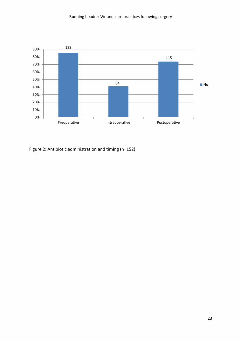
(Table 2). Intraoperative antibiotic prophylaxis was reportedly given to 88/152 (57.9%)
patients (Figure 2), however, the precise timing of antibiotic administration relative to the
start of procedure was not consistently documented. Of the 152 patients where data were
available, 10 (6.6%) had a documented unplanned return to the OR during their surgical
admission. Bleeding and washout for SSI were main contributors to unplanned return to OR
(Table 2).
Postoperative Wound Care Practices
Completion rates for charting wound assessment information varied according to
surgical specialty, type of surgery, ward unit and HLOS. Wound assessment was charted
either using the patient’s clinical pathway form, a daily care plan, or progress notes. Figure 3
illustrates the completion rates. Of the 152 charts with available data, 63 (41.4%) included a
specific wound assessment tool that was fully completed. In 28 (18.4%) charts,
postoperative wound assessments were documented in the progress notes rather than with
a specific tool. These 28 surgeries were coded as ‘Day Cases’ and included cataract
extractions, laparoscopy, removal of teeth, and tonsils and adenoid removal. Occasions of
wound assessment also varied with surgical specialty, location of wound/incision, and HLOS.
Figure 4 details the frequency of wound assessment occasions during the patient’s
admission, through to their follow up in the Out-Patients’ Department (OPD).
Approximately one fifth (32/152, 28.3%) of patients had their dressing changed at least once
during their surgical admission. Less than half (59/152, 38.8%) of the patients in this sample

had documented assessments made upon a change of their condition as an inpatient. Figure
5 graphs postoperative wound complication/event rates across the sample.
Of the 15/152 (9.9%) patients with charted postoperative complications, 7/152
(4.7%) developed clinical signs of SSI (redness, swelling, tenderness, warmth, fever or pain).
Slightly over half (8/15; 53.3%) of this subset of patients were males. Five of these 7 (71.4%)
patients were documented as having a superficial SSI. Diagnosis of SSI occurred between
postoperative days 3 and 5. Table 3 details the clinical characteristics of the subset of the 15
(9.95%) patients that had developed one or more wound complication/event (i.e.,
mechanical debridement and/or surgical debridement and/or SSI). Notably, these 13/15
(86.7%) patients had undergone orthopaedic procedures, and their HLOS ranged from 1 to
44 days. Nearly half (6/15; 40%) of the patients included in this subgroup had surgical
wounds that were classified as ‘clean-contaminated.’
Interrater Agreement
To assess interrater reliability, two authors independently reviewed 16 randomly
selected charts. Data across 17 variables including documentation of co-morbidities, current
prescribed medications, intraoperative care (antibiotics, ASA status, length of surgery), and
postoperative wound management (antibiotics, dressing assessments/changes) were cross-
checked. An ICC coefficient of 0.75 (p = 0.005) was achieved and indicated acceptable
interrater agreement.

DISCUSSION
This retrospective clinical audit adds to the limited body of literature that examines
postoperative wound management practices, specifically in relation to assessment and
documentation. While this study is exploratory, the findings are useful in describing current
clinical practice across a health services district and may have wider applicability beyond the
district. Additionally, this audit is one of the largest undertaken in the field of surgical
wound management, as opposed to including wounds that heal by secondary intention (i.e.,
where the wound edges are left open and not sutured together). Previous studies in this
field have included single hospital sites with sample sizes ranging from 49 to 80 patient
records (Birchall & Taylor 2003, Gartlan et al. 2010). The results from the current audit will
inform future interventional research which will target suboptimal wound care practices.
Of the 124 surgical patients where a specific wound assessment tool was used,
nearly 50% (61) were either partially completed or not completed at all. Documenting
practices and/or treatments acts as a risk-management strategy (Brown 2006). Inadequate
or incomplete documentation has patient safety implications in relation to continuity of
care, as well as legal and health services ramifications. Wound documentation provides
verification of the care provided, and as such, can be subpoenaed for use in legal
proceedings (Bachand & McNicholas 1999, Brown 2006). While the importance of concise,
accurate and contemporaneous documentation is highlighted when scrutinised in any legal
context, its most important function comes from increasing the likelihood of delivering high
quality, cost effective and safe patient care. Ensuring that documentation accurately and
contemporaneously reflects that care provided is particularly imperative in those
circumstances where an allegation that certain treatment was not provided, or not provided
to an appropriate standard has been raised such as in civil proceedings for negligence. The
failure to make any note of the care that was provided during delivery, represents a major
departure from acceptable practice and means that the healthcare professional may be
forced to rely on his/her memory when giving evidence. As there is often a significant delay
between the time that care has been provided, and when a healthcare professional may be
called upon to provide evidence of that care, reliance on what has previously been
documented is paramount.
Our audit results show some inconsistency and variation in the occasions when
wound assessment was documented. For instance, 52% of patients had wound assessments
documented on ‘any occasion’ (i.e., any available opportunity) while 65.1% of patients had
wound assessments documented ‘on discharge’. Common problems identified by others in
wound documentation include; inconsistency in the types of documents used (e.g., specific
tool versus progress notes) (Gartlan et al. 2010, Keast et al. 2004), the different time
intervals that wounds are assessed (Gartlan et al. 2010), the inconsistent use of terminology
(Bachand & McNicholas 1999, Keast et al. 2004), the way in which notes were made
(Bachand & McNicholas 1999) and positioned throughout the chart (Gartlan et al. 2010),
limited space available for multiple assessments (Bachand & McNicholas 1999, Gartlan et al.
2010), and the inconsistency in completion rates (Gartlan et al. 2010, Keast et al. 2004).
Specifically in this study, varying time intervals of wound assessment, space constraints,
different document formats and their positioning in the chart and incomplete
documentation were encountered throughout the conduct of our audit. This discrepancy in
findings suggests that these aspects of wound assessment and documentation warrant
further research.

Of the 152 patient charts included in our audit, 15 patients (9.9%) had developed
some type of postoperative complication, while 11 of these patients had a documented
return to OR. Notably, 7/10 of these patients had undergone an orthopaedic procedure.
Published rates of SSI following orthopaedic surgery range from 0.68% for low risk patients
undergoing hip or knee replacement to as high as 7.9% for high risk patients having spinal
fusion surgery (CDC 1996, HISWA 2011, Ridgeway et al. 2005). Surprisingly, the majority
(86.7%) of the patients that had developed a postoperative wound complication had
undergone orthopaedic surgery. Of these patients, just under half 40%) had wounds that
were classified as ‘clean-contaminated.’ According to the CDC guidelines, clean-
contaminated wounds include those in which respiratory, alimentary, genital, or urinary
tracts are entered under controlled conditions and without evidence of infection (Mangram
et al. 1999). Surgical orthopaedic wounds that heal by primary intention would almost
always be classified as ‘clean’ (Greene et al. 2010, Mangram et al. 1999). It is probable that
some of these orthopaedic wounds were misclassified during the coding process. As part of
routine EMR management in the OR, the task of coding the category of wounds is usually
left to the circulating or instrument nurse. In our study, the incorrectly entered wound
classifications suggest the need for additional education of scout/scrub nurses in the CDC
guidelines on wound classification. Although this clerical issue would not necessarily impact
on the direct care given to this group of patients, misclassification reduces the accuracy and
reliability of the data derived through EMR and chart audit
Limitations
The findings herein were based on a retrospective audit of medical records rather
than a prospective examination of postoperative wound management practices. Therefore,
there are caveats in the interpretation of these results. First, we relied on secondary source
data, which may be misclassified or incomplete. Wound management documentation may
not truly reflect how healthcare practitioners actually practice in real clinical environments.
Further, during the nominated chart audit period (2010-12), the district changed over from
the conventional hard copy medical chart to EMR, thus there was the chance that some
information may have been lost throughout this transition. It was not always possible to
identify from the charts or EMR whether postoperative wounds had been assessed as this
information may have been reported verbally. Therefore, the number of wound
assessments made may have conceivably been higher that indicated by the audit.
Observation of nurses’ wound assessment and management strategies may be more useful
to examine nurses’ actual practice. Second, the study was conducted across one heath
service district, which may in some way be idiosyncratic in respect to the case mix of surgical
patients, nurse-to-patient ratios and organisational culture. That said, the three hospital
sites included catered for a diverse mix of surgeries and patient populations, and our
findings are consistent with previous research. Where the findings are not generalisable to
other settings, this study will inform further work in this important area. Finally, only one
author categorised and entered information for data collection, so there was the potential
for misclassification. This was minimised through using a priori classifications and regular
meetings with two other study authors to discuss data management and analysis.
Additionally, 10% of the medical records included in this audit were independently cross-
checked by the lead author, which yielded acceptable interrater reliability levels.

CONCLUSIONS
Through the results of this study, important questions have raised around the timing
and completeness of documented wound assessments. Clearly, documentation of wound
assessment and management practices is integral to wound care. Provision of an integrated
electronic database and standardisation of wound care documentation that encompasses
wound assessment and wound management interventions may reduce inconsistencies
and/or omissions in the timing and detail of wound assessments, and the terminologies
used (Keast et al. 2004, Kinnunen et al. 2012). Ultimately, consistency in documentation will
go some way to enhancing multidisciplinary communication in wound care.
Relevance to Clinical Practice
Our findings suggest that wound assessment and documentation practices are
inconsistent with evidence-based guidelines. Nonetheless, we cannot assume that the gap
between evidence and practice is evident. While it is essential that practice reflects existing
evidence-based guidelines, the next crucial steps in addressing the issues raised here will
involve observing what is actually happening in clinical practice. Contextual nuances in the
clinical environment may potentially impact on clinicians’ ability to optimally perform
patient care activities—and hence fully utilise CPG. As part of this exploration, we will
engage stakeholders in identifying existing barriers and enablers that underpin the use of
evidence-based postoperative wound management strategies. Once these are known, we
will collaborate with stakeholders in developing translational interventions that are

contextually responsive to ensure greater success in using EBP (Michie et al. 2011) in
postoperative wound management.
References
ACHS (2011) Australian Clinical Indicator Report ACHS 2001-2009. Australian Commission of Healthcare Standards, Canberra. Available at: http://www.achs.org.au/ (accessed 12 August 2013).
Andersson AE, Bergh I, Karlsson J & Nilsson K (2010): Patients' experiences of acquiring a deep surgical site infection: an interview study. American Journal of Infection Control 38, 711-717.
Astagneau P, L'Hériteau F, Daniel F, Parneix P, Venier AG, Malavaud S, Jarno P, Lejeune B, Savey A, Metzger MH, Bernet C, Fabry J, Rabaud C, Tronel H, Thiolet JM & Coignard B (2009): Reducing surgical site infection incidence through a network: results from the French ISO-RAISIN surveillance system. Journal of Hospital Infection 72, 127-134.
Australian Institute of Health and Welfare (2010) Australia's hospitals 2008-09 - at a glance. AIHW, Canberra.
AWMA AWMA (2011) Standards for Wound Management. Cambridge Publishing, West Leederville W A, p. 5.
Bachand PM & McNicholas ME (1999): Creating a wound assessment record. Advances in Wound Care 12, 426.
Birchall L & Taylor S (2003): Surgical wound benchmark tool and best practice guidelines. British Journal of Nursing 12, 1013.
Brown G (2006): Wound Documentation: Managing Risk. Adv Skin Wound Care 19, 155-165. Carville K (2012) Wound care manual, 6th edn. Silver Chain Nursing Association, Perth. CDC (1996) NNISS Conference for Hospitals Participating in the National Infections Surveillance: SSI
Rates, by Operative Procedure and Risk Index Category, October 1986–July 1996., Atlanta, GA:.
Coello R, Charlett A, Wilson J & al e (2005): Adverse impact of surgical site infections in English hospitals. Journal of Hospital Infection 60, 93-103.
de Lissovoy G, Fraeman K, Hutchins V & et al (2009a): Cost analysis of surgical site infections. Surg Infect (Larchmt) 7, s19-s22.
de Lissovoy G, Fraeman K, Hutchins V, Murphy D, Song D & Vaughn BB (2009b): Surgical site infection: incidence and impact on hospital utilization and treatment costs. American Journal of Infection Control 37, 387-397.
Engemann J, Carmeli Y, Cosgrove S & al e (2003): Adverse clinical and economic outcomes attributable to methicillin resistance among patients with Staphylococcus aureus surgical site infection. Clin Infect Dis 36, 592-598.
Friedman D, Bull A, Russo P, Gurrin L & Richards M (2007): Performance of the National Nosocomial Infections Surveillance Risk Index in Predicting Surgical Site Infection in Australia. Infection Control and Hospital Epidemiology 28, 55-59.

Gartlan J, Smith A, Clennett S, Walshe D, Tomlinson-Smith A, Boas L & Robinson A (2010): An audit of the adequacy of acute wound care documentation of surgical inpatients. Journal of Clinical Nursing 19, 2207–2214.
Gillespie B, Chaboyer W, Allen P, Morley N & Niuewenhoven P (in press, 2013): Wound care practices: A survey of acute care nurses Journal of Clinical Nursing.
Gillespie BM, Chaboyer W, Niewenhoven P & Rickard CM (2012): Drivers and Barriers of Surgical Wound Management in a Large Healthcare Organisation: Results of an Environmental Scan. Journal of Wound Care Practice and Research 20, 90-102.
Greene L, Mills R, Moss R, Sposato K & Vignari M (2010) APIC Guide to the elimination of orthopaedic surgical site infections. APIC, Washington DC, pp. 1-80.
Health Do (2009) SQuIRe 2: CPI Guide to surgical site infection prevention. Office of Safety and Quality, WA Government, Perth, pp. 1-16.
HISWA (2011) Healthcare Infection Surveillance Western Australia. Department of Health, Perth, pp. 1-19.
Kassavin D, Pascarella L & Goldfab M (2011): Surgical site infections: incidence and trends at a community teaching hospital. The American Journal of Surgery 201, 749–753.
Keast DH, Bowering CK, Evans AW, Mackean GL, Burrows C & D'Souza L (2004): Contents. Wound Repair and Regeneration 12, s1-s17.
Kinnunen U, Saranto K, Ensio A, Iivanainen A & Dykes P (2012): Developing the Standardized Wound Care Documentation Model A Delphi S tudy to Improve the Quality of Patient Care Documentation. J Wound Ostomy Continence Nurs 39, 397-407.
Leape L, Brennan T, Laird N & al. e (1991): The nature of adverse events in hospitalized patients. Results of the Harvard Medical Practice Study II. New England Journal of Medicine 324, 377-384.
Leaper DJ, Van Goor H, Reilly J, Petrosillo N, Geiss HK, Torres AJ & Berger A (2004): Surgical site infection -- a European perspective of incidence and economic burden. International Wound Journal 1, 247-273.
Mangram AJ, Horan TC, Pearson ML, Silver LC & Jarvis WR (1999): Guideline for prevention of surgical site infection, 1999. Infection Control & Hospital Epidemiology 20, 247-280.
Michie S, van Stralen M & West R (2011): The behaviour change wheel: A new method for characterising and designing behaviour change interventions. Implementation Science 6, 1-12.
Nathens A, Cook C, Machiedo G, Moore E, Namias N & Nwariaku F (2006): Defining the research agenda for surgical infection: a consensus of experts using the Delphi approach. jounral of Surgical Infection 7, 101-110.
NICE NIfHaCE (2008) NICE Clinical Guideline 74: Surgical site infection: prevention and treatment of surgical site infection (National Collaborating Centre for Women's and Children's Health ed.). RCOG Press, London, pp. 4-28.
Plowman R, Graves N, Griffin M, Roberts J, Swan A, Cookson B & al. e (2001): The rate and cost of hospital-acquired infections occurring in patients admitted to selected specialties of a district general hospital in England and the national burden imposed. Journal of Hospital Infection 47, 198-209.
Polit D (2010) Statistics and data analysis for nursing research, Second edn. Pearson, Upper Saddle River.
Ridgeway S, Wilson J, Charlet A, Kafatos G, Pearson A & Coello R (2005): Infection of the surgical site after arthroplasty of the hip. The Journal of Bone and Joint Surgery 87, 844-850.
Smyth E, McIlvenny G, Enstone J & al e (2008): Four Country Healthcare Associated Infection Prevalence Survey 2006: overview of the results. Journal of Hospital Infection 69, 230-248.

Table 1: Recommendations for the prevention of SSI drawn from existing clinical guidelines,
practice standards, and position statements
Recommendation Guideline / Standard / Position Statement
Pre and Intraoperative phases:
A. Appropriate hair removal I. Use of clippers or depilatory cream
B. Antibiotic prophylaxis for: I. Clean surgery with implant
II. Clean-contaminate surgery III. Contaminated surgery IV. Single IV dose during induction
SQuIRe 2 CPI Guide: SSI Prevention (Health 2009)
AWMA Standard 4.6.11 (2011)
CDC Guideline for Prevention of SSI (1999)
EWMA Position Statement, Part 4 (2006)
NICE Clinical Guideline 74, Chapter 5/6 (NICE 2008)
C. Patient assessment I. Holistic patient assessment
II. Patient history III. Keep patients and caregivers informed
AWMA Standards 3.1 & 5.1 (2011)
EWMA Position Statement, Part 4 (2006)
NICE Clinical Guideline 74, Chapter 5/6 (NICE 2008)
Postoperative phase:
D. Wound Assessment I. Based on patient factors, determine wound
management strategy either using aseptic or clean technique
II. If SSI is suspected (i.e., cellulites), give antibiotics to cover infection
AWMA Standards 3.1 & 5.1 (2011)
EWMA Position Statement, Part 4 (2006)
NICE Clinical Guideline 74, Chapters 5, 6 & 7 (NICE 2008)
SQuIRe 2 CPI Guide: SSI Prevention (Health 2009)
E. Incision management / dressing selection & wound cleansing I. Assess the benefit & cost effectiveness of
dressings II. Leave dressing intact for at least 48hrs in
the postoperative period III. Use sterile normal saline to cleanse
wound up to 48hrs IV. Tap water may be used to cleanse surgical
site after first 48hrs
AWMA Standards 3.1 & 5.1 (2011)
CDC Guideline for Prevention of SSI (1999)
EWMA Position Statement, Part 4 (2006)
NICE Clinical Guideline 74, Chapter 6 (NICE 2008)
F. Wound Debridement I. Avoid eusol, saline soaked gauze,
dextranomer or enzymatic treatments to manage wound infections
II. Use appropriate interactive dressings
NICE Clinical Guideline 74, Chapter 7 (NICE 2008)
G. Documentation I. Initial and ongoing wound assessments
II. Environmental assessment III. Documented care plan IV. Integrated care pathway for management
of wound complications V. Collaborative multidisciplinary approach to
patient care
AWMA Standards 3.3 & 5.1 (2011)
NICE Clinical Guideline 74, Chapter 4 (NICE 2008)
SQuIRe 2 CPI Guide: SSI Prevention (Health 2009)

Table 2: Demographic and clinical characteristics of patients for the percentage of the total sample
(n=152)
Demographics n* %
Hospital Site
A – 450 bed hospital
B – 130 bed satellite hospital
C – Stand alone Day Surgery Unit
76
28
47
50.0
18.4
31.0
Surgical Specialty
General 26 17.1
Vascular 2 1.3
Plastics 4 2.6
Orthopaedics 50 32.9
Paediatric 2 1.3
Obstetrics and Gynaecology 13 8.6
Facial Maxillary 24 15.8
Ear, Nose and Throat 8 5.3
Neurosurgery 7 4.6
Ophthalmology 12 7.9
Urology 3 2.0
ASA Status
ASA 1&1E 110 72.4
ASA 2 & 2E 36 23.7
ASA 5 & 5E 6 4.0
Comorbidities
Chronic Heart Failure 2 1.3
IHD 8 5.3
COPD 23 15.1
Hypertension 26 17.1
Diabetes Mellitus 15 9.9
Hypercholesterolemia 15 9.9
Renal disease 8 5.3
Metastatic carcinoma/Malignancy 8 5.3
Stroke 5 3.3
Immuno-compromised 5 3.3
Reason for return to OR
Bleeding/ Haemorrhage at incision site 3 2.1
Wound Exploration 1 0.7
SSI ― Washout 3 2.1
Undergoing further surgery 1 0.7
Removal of prosthesis 1 0.7
Not stated 2 1.3
*Missing values not replaced

Table 3: Demographic and Clinical Characteristics of Patients with Postoperative Wound
Complications (*n=15)
Demographic / Clinical Characteristic Results
Age
Years
Median IQR
55.0
33.0
Length of Surgery
Minutes
70.5
77.0
HLOS
Days
2.0
5.0
Wound Assessment Occasions 4.7 0.0
n %
Number of Co-morbidities
≥ 3
> Medications
Yes
4
4
26.6
26.6
Surgical Specialty
Orthopaedic
13 86.6
General 1 6.6
Facial Maxillary 1 6.6
Wound Classification
Clean
9
60.0
Clean Contaminated 6 40.0
Antibiotic Administration
Preoperative 4 26.6
Intraoperative 12 80.0
Postoperative 4 46.6
†Postoperative Complications n %
Mechanical debridement 6 40.0
Surgical debridement 1 6.6
Mechanical Debridement for SSI 2 13.3
Surgical Debridement for SSI 1 6.6
Mechanical & Surgical Debridement 2 13.3
SSI 4 26.6
* 4 patients developed more than 1 postoperative wound complication
† Postoperative events: 1) Surgical debridement – used in surgery to extend healthy tissue;
2) Mechanical debridement – uses force (hydrosurgery, wet-to-dry gauze)

Running header: Wound care practices following surgery
22
FIGURES
Figure 1: Sample flow diagram of EMR/chart inclusion.
Iden
tifi
cati
on
Charts Screened
(n =200)
Initial charts (hard copy & EMR) 2010 to 2012
(n = 200)
Scre
enin
g
Charts excluded (n =48)
Miscoded
Did not involve a surgical incision
Elig
ibili
ty/e
xclu
ded
Charts eligible & included (n = 152)
Charts eligible for inclusion (n = 156)
Charts excluded (n = 4)
> 20% missing data on wound management
practices
Running header: Wound care practices following surgery
23
Figure 2: Antibiotic administration and timing (n=152)
133
64
115
0%
10%
20%
30%
40%
50%
60%
70%
80%
90%
Preoperative Intraoperative Postoperative
No

Running header: Wound care practices following surgery
24
Figure 3: Wound assessment documentation completion rates (n=152)
37
24
63
28
0%
5%
10%
15%
20%
25%
30%
35%
40%
45%
No tool Partially completed Fully completed Progress notes used

Running header: Wound care practices following surgery
25
Figure 4: Documented wound assessment occasions (n=152)
73
93
53
97
0%
10%
20%
30%
40%
50%
60%
70%
Wound assessment -any occasion
Wound assessment -change of condition
Wound assessment -discharge
Wound assessment -OPD
No

Running header: Wound care practices following surgery
26
Figure 5: Documented wound complication rates (n=15 patients*)
*Some patients were documented as having more than one wound complication.
6
1
4
11
0%
10%
20%
30%
40%
50%
60%
70%
80%
Medical debridement Surgical debridement Signs & symptoms of SSI Return to OR

Running header: Wound care practices following surgery
27