Embed Size (px)
Citation preview
Bahrain Medical Bulletin, Vol. 38, No. 3, September 2016
173
OVT is a rare but potentially serious complication which is most often diagnosed during the postpartum period. OVT occurs in 0.05% to 0.18% of pregnancies, but it is 1% to 2% after caesarean section1-4. The right ovarian vein is involved in 80% to 90% of cases5,6. It was suggested to be due to the length of the right ovarian vein, lack of retrograde flow and multiple incompetent valves1. To avoid serious maternal morbidity and mortality, early recognition and treatment is mandatory7. The symptoms could include lower abdominal quadrant tenderness and flank pain associated with fever, tachycardia, chills, nausea and vomiting. Differential diagnosis must be considered, which include acute appendicitis, endometritis, pyelonephritis, tubo-ovarian abscess or torsion of the ovarian ligament. OVT may progress to involve the inferior vena cava or the renal vein or may cause sepsis or septic pulmonary embolism, which are potentially life-threatening4,6,8.
The aim of this report is to present a case of ovarian vein thrombosis which was managed by anticoagulant treatment.
THE CASE
A thirty-three-year-old female Para 6 Abortion 1 presented eight days postpartum with acute lower abdominal pain on the right side, not associated with vaginal bleeding, nausea, vomiting, fever or urinary symptoms. In the immediate postpartum period, the patient developed mild postpartum hemorrhage, which was managed with uterine massage and uterotonic agents and IV antibiotics (Zinacef 750 mg 8 hourly and Flagyl 500 mg 8 hourly).
Abdominal examination revealed right lower quadrant tenderness. Ultrasonography showed well-defined, slightly heterogeneous hypoechoic mass (60x58x32mm) near the cornual region of the uterus, on the right side with moderate
Postpartum Ovarian Vein Thrombosis
Wassan Yassen, MBBchB, MRCOG PART I* Shaza Omer Mohammed Ali, MBBS, MRCOG PART I* Aysha Ebrahim Al Mutawa, MD, BSc**
Ovarian vein thrombosis (OVT) is a rare condition classically seen during the postpartum period. It typically presents with lower abdominal pain and fever which does not respond to broad-spectrum antibiotics.
A thirty-three-year-old female presented with Ovarian Vein Thrombosis. Ultrasonography revealed well-defined, slightly heterogeneous hypoechoic mass (60x58x32mm) near the cornual region of the uterus, on the right side. CT abdomen showed right gonadal vein thrombosis. MRI showed bulky uterus, thrombosed and bulky tortuous ovarian vein on the right. The patient was treated with anticoagulant of low molecular weight heparin and warfarin.
Bahrain Med Bull 2016; 38 (3): 173 - 175
* Department of Obstetrics and Gynecology ** Resident Department of Radiology Bahrain Defense Force Hospital The Kingdom of Bahrain Email: [email protected]
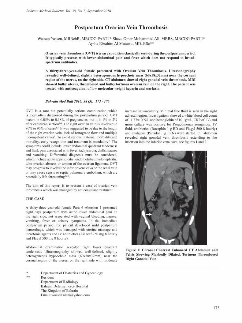
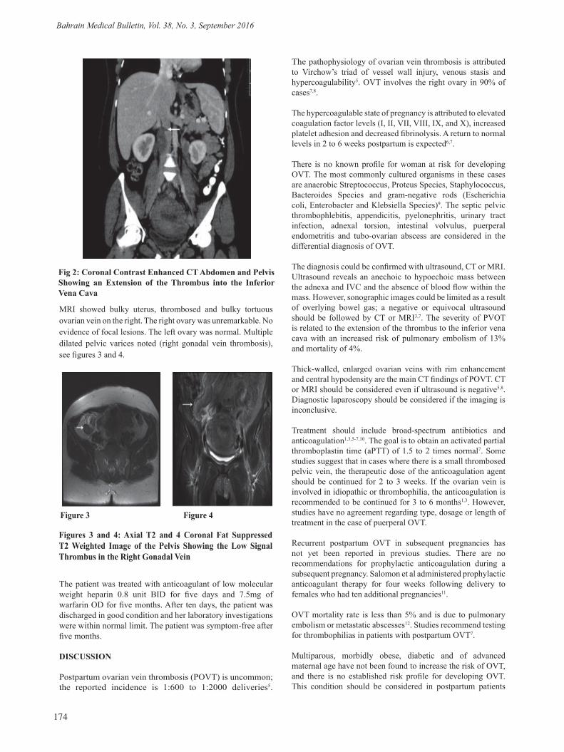
increase in vascularity. Minimal free fluid is seen in the right adnexal region. Investigations showed a white blood cell count of 11.37x10^9/L and hemoglobin of 10.1g/dL, CRP of 133 and urine culture was positive for Pseudomonas aeruginosa. IV fluid, antibiotics (Rocephin 1 g BD and Flagyl 500 8 hourly) and analgesia (Panadol 1 g PRN) were started. CT abdomen revealed right gonadal vein thrombosis extending to the insertion into the inferior vena cava, see figures 1 and 2.
Figure 1: Coronal Contrast Enhanced CT Abdomen and Pelvis Showing Markedly Dilated, Tortuous Thrombosed Right Gonadal Vein
Bahrain Medical Bulletin, Vol. 38, No. 3, September 2016
174
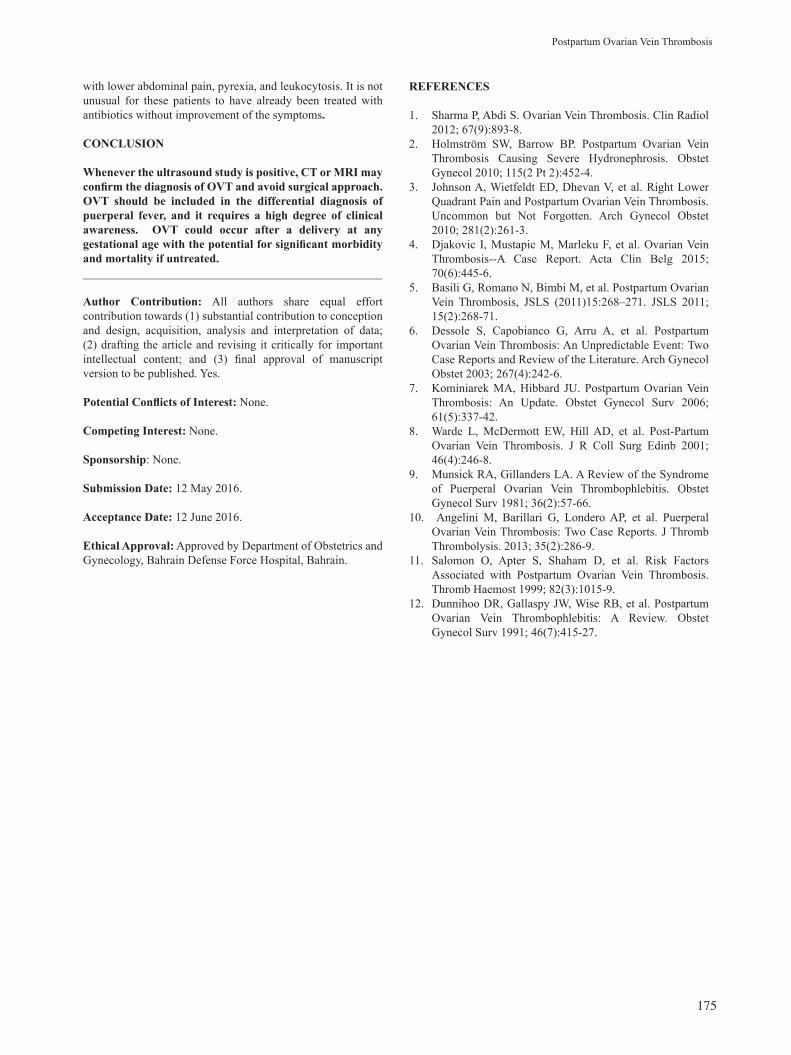
MRI showed bulky uterus, thrombosed and bulky tortuous ovarian vein on the right. The right ovary was unremarkable. No evidence of focal lesions. The left ovary was normal. Multiple dilated pelvic varices noted (right gonadal vein thrombosis), see figures 3 and 4.
The patient was treated with anticoagulant of low molecular weight heparin 0.8 unit BID for five days and 7.5mg of warfarin OD for five months. After ten days, the patient was discharged in good condition and her laboratory investigations were within normal limit. The patient was symptom-free after five months.
DISCUSSION
Postpartum ovarian vein thrombosis (POVT) is uncommon; the reported incidence is 1:600 to 1:2000 deliveries5.
The pathophysiology of ovarian vein thrombosis is attributed to Virchow’s triad of vessel wall injury, venous stasis and hypercoagulability5. OVT involves the right ovary in 90% of cases7,8.
The hypercoagulable state of pregnancy is attributed to elevated coagulation factor levels (I, II, VII, VIII, IX, and X), increased platelet adhesion and decreased fibrinolysis. A return to normal levels in 2 to 6 weeks postpartum is expected6,7.
There is no known profile for woman at risk for developing OVT. The most commonly cultured organisms in these cases are anaerobic Streptococcus, Proteus Species, Staphylococcus, Bacteroides Species and gram-negative rods (Escherichia coli, Enterobacter and Klebsiella Species)9. The septic pelvic thrombophlebitis, appendicitis, pyelonephritis, urinary tract infection, adnexal torsion, intestinal volvulus, puerperal endometritis and tubo-ovarian abscess are considered in the differential diagnosis of OVT.
The diagnosis could be confirmed with ultrasound, CT or MRI. Ultrasound reveals an anechoic to hypoechoic mass between the adnexa and IVC and the absence of blood flow within the mass. However, sonographic images could be limited as a result of overlying bowel gas; a negative or equivocal ultrasound should be followed by CT or MRI3,7. The severity of PVOT is related to the extension of the thrombus to the inferior vena cava with an increased risk of pulmonary embolism of 13% and mortality of 4%.
Thick-walled, enlarged ovarian veins with rim enhancement and central hypodensity are the main CT findings of POVT. CT or MRI should be considered even if ultrasound is negative5,8. Diagnostic laparoscopy should be considered if the imaging is inconclusive.
Treatment should include broad-spectrum antibiotics and anticoagulation1,3,5-7,10. The goal is to obtain an activated partial thromboplastin time (aPTT) of 1.5 to 2 times normal7. Some studies suggest that in cases where there is a small thrombosed pelvic vein, the therapeutic dose of the anticoagulation agent should be continued for 2 to 3 weeks. If the ovarian vein is involved in idiopathic or thrombophilia, the anticoagulation is recommended to be continued for 3 to 6 months1,3. However, studies have no agreement regarding type, dosage or length of treatment in the case of puerperal OVT.
Recurrent postpartum OVT in subsequent pregnancies has not yet been reported in previous studies. There are no recommendations for prophylactic anticoagulation during a subsequent pregnancy. Salomon et al administered prophylactic anticoagulant therapy for four weeks following delivery to females who had ten additional pregnancies11.
OVT mortality rate is less than 5% and is due to pulmonary embolism or metastatic abscesses12. Studies recommend testing for thrombophilias in patients with postpartum OVT7.
Multiparous, morbidly obese, diabetic and of advanced maternal age have not been found to increase the risk of OVT, and there is no established risk profile for developing OVT. This condition should be considered in postpartum patients
Fig 2: Coronal Contrast Enhanced CT Abdomen and Pelvis Showing an Extension of the Thrombus into the Inferior Vena Cava
Figures 3 and 4: Axial T2 and 4 Coronal Fat Suppressed T2 Weighted Image of the Pelvis Showing the Low Signal Thrombus in the Right Gonadal Vein
Figure 3 Figure 4
Bahrain Medical Bulletin, Vol. 38, No. 3, September 2016
175
with lower abdominal pain, pyrexia, and leukocytosis. It is not unusual for these patients to have already been treated with antibiotics without improvement of the symptoms.
CONCLUSION
Whenever the ultrasound study is positive, CT or MRI may confirm the diagnosis of OVT and avoid surgical approach. OVT should be included in the differential diagnosis of puerperal fever, and it requires a high degree of clinical awareness. OVT could occur after a delivery at any gestational age with the potential for significant morbidity and mortality if untreated.__________________________________________________
Author Contribution: All authors share equal effort contribution towards (1) substantial contribution to conception and design, acquisition, analysis and interpretation of data; (2) drafting the article and revising it critically for important intellectual content; and (3) final approval of manuscript version to be published. Yes.
Potential Conflicts of Interest: None.
Competing Interest: None.
Sponsorship: None.
Submission Date: 12 May 2016.
Acceptance Date: 12 June 2016.
Ethical Approval: Approved by Department of Obstetrics and Gynecology, Bahrain Defense Force Hospital, Bahrain.
REFERENCES
1. Sharma P, Abdi S. Ovarian Vein Thrombosis. Clin Radiol 2012; 67(9):893-8.
2. Holmström SW, Barrow BP. Postpartum Ovarian Vein Thrombosis Causing Severe Hydronephrosis. Obstet Gynecol 2010; 115(2 Pt 2):452-4.
3. Johnson A, Wietfeldt ED, Dhevan V, et al. Right Lower Quadrant Pain and Postpartum Ovarian Vein Thrombosis. Uncommon but Not Forgotten. Arch Gynecol Obstet 2010; 281(2):261-3.
4. Djakovic I, Mustapic M, Marleku F, et al. Ovarian Vein Thrombosis--A Case Report. Acta Clin Belg 2015; 70(6):445-6.
5. Basili G, Romano N, Bimbi M, et al. Postpartum Ovarian Vein Thrombosis, JSLS (2011)15:268–271. JSLS 2011; 15(2):268-71.
6. Dessole S, Capobianco G, Arru A, et al. Postpartum Ovarian Vein Thrombosis: An Unpredictable Event: Two Case Reports and Review of the Literature. Arch Gynecol Obstet 2003; 267(4):242-6.
7. Kominiarek MA, Hibbard JU. Postpartum Ovarian Vein Thrombosis: An Update. Obstet Gynecol Surv 2006; 61(5):337-42.
8. Warde L, McDermott EW, Hill AD, et al. Post-Partum Ovarian Vein Thrombosis. J R Coll Surg Edinb 2001; 46(4):246-8.
9. Munsick RA, Gillanders LA. A Review of the Syndrome of Puerperal Ovarian Vein Thrombophlebitis. Obstet Gynecol Surv 1981; 36(2):57-66.
10. Angelini M, Barillari G, Londero AP, et al. Puerperal Ovarian Vein Thrombosis: Two Case Reports. J Thromb Thrombolysis. 2013; 35(2):286-9.
11. Salomon O, Apter S, Shaham D, et al. Risk Factors Associated with Postpartum Ovarian Vein Thrombosis. Thromb Haemost 1999; 82(3):1015-9.
12. Dunnihoo DR, Gallaspy JW, Wise RB, et al. Postpartum Ovarian Vein Thrombophlebitis: A Review. Obstet Gynecol Surv 1991; 46(7):415-27.
Postpartum Ovarian Vein Thrombosis


![Welcome [applications.emro.who.int]applications.emro.who.int/docs/RC64_2017_bulletin_1_20097_en.pdf · their talents.This year, almost 50 drawings from across the Region were awarded](https://img.pdfslide.us/doc/110x75/5f31da622bb02e749c290170/welcome-their-talentsthis-year-almost-50-drawings-from-across-the-region-were.jpg)