Embed Size (px)
Citation preview

Original Article
Posterior Cervical Transfacet Fusion with Facetal Spacer for the Treatment of Single-Level
Cervical Radiculopathy: A Randomized, Controlled Prospective Study
Jacopo Lenzi1, Antonio Nardone1, Emiliano Passacantilli1, Alessandro Caporlingua2, Gennaro Lapadula2,Federico Caporlingua2
-BACKGROUND: Single-level cervical radiculopathy maybe treated conservatively with cervical tractions. Posteriorcervical transfacet fusion with a facetal spacer is a viableoption. The aim of the present study is to compare posteriorcervical transfacet fusion with conservative physicaltreatment in single-level cervical radiculopathy.
-METHODS: A total of 80 patients were randomized in 2groups, a surgical group in which patients were givenposterior cervical transfacet fusion and a traction group inwhich patients were treated conservatively with mechan-ical cervical tractions. Visual analog scale for arm andneck, Neck Disability Index, and Short Form-36 (SF-36)questionnaires were administered preoperatively and aftertreatment up to 12 months.
-RESULTS: After treatment, visual analog scale arm scoreswere greater in traction group (4.7 vs. 1.5 the day aftertreatment) and at follow-up controls (traction group vs.surgical group: 5.3 vs. 0.6 at 1 month, 3.6 vs. 0.3 at 6 months,1.8 vs. 0.2 at 12 months). Neck Disability Index scores werelower in the surgical group (surgical group vs. tractiongroup: 4.4 vs. 20.3 at 1 month, 1.3 vs. 10.5 at 6 months). SF-36scoreswere greater in the surgical group (surgical group vs.traction group: 96 vs. 70 at 1 month, 96.5 vs. 82.6 at 6 months).Neck disability index and SF-36 scores were superimpos-able between the groups at 12-month follow-up. No
Key words- Cervical disc herniation- Cervical manipulation- Cervical radiculopathy- Cervical stenosis- Cervical transfacet fusion- Mechanical cervical traction- Minimally invasive surgery- Percutaneous cervical fusion- Posterior cervical fusion- Randomized controlled study
Abbreviations and AcronymsACDF: Anterior cervical disc fusionAP: AnteroposteriorCT: Computed tomographyNDI: Neck Disability Index
WORLD NEUROSURGERY 100: 7-14, APRIL 2017
adjacent-segment arthrosis or late complications were re-ported at 1-year follow-up in the surgical group.
-CONCLUSIONS: posterior cervical transfacet fusion is asafe and effective procedure to treat single-level cervicalradiculopathy.
INTRODUCTION
urgical management of single-level cervical spondyloticstenosis with concomitant myelopathy entails the use of
Santerior cervical disc fusion (ACDF) or total disc replace-ment (TDR). ACDF and TDR represent the conventionally adoptedsurgical options in these cases. When the stenosis, either bony ordiscal, involves predominantly the foraminal region, there is nospinal cord compression and cervical radiculopathy may be theonly symptom. Cervical radiculopathy has an incidence of 1.79 per1000 person-years.1 The course of symptomatic cervical discherniation with radiculopathy is benign. Improvement can beexpected 4e6 months after the onset of symptoms.2,3 It is ex-pected that approximately 1e5 patients have a recurrence afterconservative treatment.2
Although there is no general consensus about treatment choicebetween physical, infiltrative (epidural injections), and operative,4
surgery is indicated when pain does not reduce after conservativetherapy or if progressive motor weakness is present. In thissetting, ACDF may be considered too invasive, and posterior
PCTF: Posterior cervical transfacet fusionSF-36: Short Form-36TDR: Total disc replacementVAS: Visual analog scale
From the 1Neurological Center of Latium, Neurosurgery, Rome; and 2Department ofNeurology and Psychiatry, Neurosurgery, “Sapienza” University of Rome, Rome, Italy
To whom correspondence should be addressed: Federico Caporlingua, M.D.[E-mail: [email protected]]
Citation: World Neurosurg. (2017) 100:7-14.http://dx.doi.org/10.1016/j.wneu.2016.12.125
Journal homepage: www.WORLDNEUROSURGERY.org
Available online: www.sciencedirect.com
1878-8750/$ - see front matter ª 2017 Elsevier Inc. All rights reserved.
www.WORLDNEUROSURGERY.org 7

ORIGINAL ARTICLE
JACOPO LENZI ET AL. PCTF FOR CERVICAL RADICULOPATHY
approaches may come in handy. Posterior foraminotomy is aconsolidated technique, but it has a few drawbacks, such aschronic neck pain originating from the stripping of the muscleto expose the articular facets.5
Posterior cervical transfacet fusion (PCTF) with indirectforaminal decompression is a relatively new treatment modalityfor single- and/or multiple-level cervical spondylotic foraminalstenosis.6,7 A titanium expandable washer with an internal screwcomposes the DTRAX expandable cages (Providence MedicalTechnology, Lafayette, California, USA). Once deployed andexpanded between the 2 facets, it indirectly increases the foram-inal volume, decompressing the exiting root. A rasp and adecorticator along with synthetic bone are used to promote fusion.The aim of this study is to assess the efficacy of PCTF comparedwith conservative therapy for the treatment of single-level symp-tomatic foraminal cervical stenosis without cervical myelopathy.
MATERIALS AND METHODS
Study DesignThe study was approved by the local ethical committee. A total of119 patients were enrolled in the study. The study was concludedat the moment we had the first 40 patients from each group(surgical and tractions groups) (Figure 1). Overall mean age was45.5 (standard deviation 12.7). Patients enrolled in the studywere predominantly male, with a male/female sex ratio of1.35. Demographic and preoperative data were substantiallycomparable between the 2 groups (Table 1). All patients had aphysical examination documenting reduction or loss of reflex,sensory deficit, and motor weakness. Magnetic resonance
Figure 1. Flow chart of s
8 www.SCIENCEDIRECT.com WORLD NEU
imaging scan to confirm a single-level foraminal stenosis and anelectromyogram that could confirm the compression of the cer-vical root were performed in all patients. If the anatomy of thefacet was unclear due to spondyloarthrosis, a computed tomog-raphy (CT) scan was performed.Inclusion criteria were age >18 and <75 years, single-level
cervical foraminal stenosis involving the segment C3-C7 docu-mented by magnetic resonance imaging and/or CT scan.Patients with multiple level radiculopathy, cervical instability or
kyphosis, who were pregnant, were affected by rheumatoid orconnective tissue diseases, osteopenia or osteoporosis, cervicalfractures, cervical column curve inversion at the level of the ste-nosis, and complete stenosis of the neuroforamen were excluded.All patients were treated conservatively with steroids and nonste-roidal anti-inflammatory drugs for 6 weeks. They were dividedrandomly in 2 groups: the surgical group was offered PCTF, andphysical therapy with mechanical cervical tractions was offered tothe traction group (Figure 1).An online program was used for the purposes of randomization
(www.randomization.com). Accordingly, the corresponding authorprepared a randomization scheme that was sent to the other au-thors. According to the treatment followed by each patient, thecorresponding author received information about when to call thepatients for the telephone interview at 3, 6, and 12 months. Allpatients gave their informed consent. Initial evaluation included thevisual analog scale (VAS) for both neck and arm pain, the NeckDisability Index (NDI), and Short Form-36 (SF-36). The VAS scoresonly were collected the day after surgery (surgical group) and after 10sessions of cervical tractions (first 5 weeks of conservative treat-ment). Patients in the traction group repeated the mechanical
tudy progression.
ROSURGERY, http://dx.doi.org/10.1016/j.wneu.2016.12.125

Table 1. Demographic and Clinical Data
Surgery Group Traction Group P Value
Male/female 1.1 1.6
Age, years (SD) 46.1 (12.52) 45.02 (12.87) 0.7
BMI, kg/m2 23.5 25 0.5933
Level, n
C3-C4 5 4
C4-C5 6 9
C5-C6 14 14
C6-C7 15 13
SD, standard deviation; BMI, body mass index.
ORIGINAL ARTICLE
JACOPO LENZI ET AL. PCTF FOR CERVICAL RADICULOPATHY
tractions once a week after the first pain and disability assessment.Regression of radiculopathy in the traction group was a primaryendpoint for treatment but not for assessment. Nevertheless,treatment and assessment were terminated if patient requestedsurgery. Patients in the traction group could decide to interrupt themechanical tractions; nevertheless, they were assessed for pain anddisability until the 12th month if they did not ask for surgery. Pa-tients in the traction group who asked for surgery did not enter thesurgical group. Because this was an intention-to-treat study, it wasconsidered finished when the first 40 patients from each group hadterminated the programmed follow-up.All patients were evaluated with VAS, NDI, and SF-36 scores at
1, 6, and 12 months after treatment. Pain and disability assessmentwas performed by a telephone interview by the correspondingauthor, who did not participate to the surgeries or to the physicaltherapy sections and was therefore blinded to the treatment.
Surgical TechniqueAfter intubation, the patient was positioned prone with the headin a neutral position and slightly flexed. The Mayfield clamp wasunnecessary. The shoulders were pulled down with tape. By theuse of intraoperative anteroposterior (AP) fluoroscopic guidance,the surgeon drew 3 lines corresponding to the cutaneous projec-tion of spinosus processes and medial and lateral facets lines onthe dorsal cervicodorsal skin. To designate the adequate skin entrypoint, under laterolateral fluoroscopic control, a spinal needle waspositioned aligned with the intended level facet orientation andentry point between the medial and lateral facet lines on one side.The side treated first was that of the radiculopathy or that wherethe symptoms were more intense.After adequate local anesthesia was administered to the patient,
a 1-cm long horizontal skin incision was made by advancing thescalpel deep through the muscular fascia. The chisel was theninserted through the fascia into the facet under laterolateralfluoroscopic control and advanced up to the pedicle. In case ofdegenerative arthrosis of the facet, hand pressure may not besufficient for the chisel to penetrate the articular capsule and/orbone osteophytes and therefore a small hammer was used topenetrate the interfacetal space. The position of the chisel was
WORLD NEUROSURGERY 100: 7-14, APRIL 2017
controlled in AP projection. The chisel should be positioned onthe lateral half of the facet to prevent any damage to the root.A decorticator was used to remove some of the superficial bone
to promote arthrodesis. Then, a guide was inserted and the chiselremoved from the facet. The guide tube has radiologic markers toalign the implant. A rasp was passed inside the facet to remove thecartilaginous endplates. The DTRAX implant was inserted insidethe facet to the pedicle. The position was controlled in AP andlateral projections before the screw was inserted. If the positionwas correct, the screw was advanced until it sprang (meaning thatit could not be removed). The facet distraction was controlled inlateral projection. The washer was removed and synthetic bonewas inserted inside the working cannula and pushed inside thefacet. The same procedure was repeated contralaterally. Figure 2shows a surgical case. Figure 3 shows the implant kit andprosthesis.Patients in the surgical group were given a soft cervical collar to
wear for 7 days after surgery. They were discharged on the firstpostoperative day. Control cervical radiographs were performed 1,6, and 12 months after surgery.
Cervical Traction TechniquePatients in the traction group were treated with mechanical trac-tion. They were positioned supine and mechanical tractionequipment was adjusted manually. A pulling force was appliedequal to 10% of patient’s weight for 10 seconds followed by 5seconds rest for a total of 15 minutes. The force was appliedparallel to the cervical spine. The sessions were repeated biweeklyfor 5 weeks before the first pain and disability assessment.
Statistical AnalysisThe sample size calculation was performed with the aid of an onlinecalculator (http://powerandsamplesize.com/Calculators/Compare-2-Means/2-Sample-Equality). To have a 99% chance of detecting asignificant (at 2-sided 5% level) difference of 3 points between the 2groups at VAS scores, with an assumed standard deviation of 3, aminimum of 37 patients were required in each group. The Studentunpaired t test was used for to compare the preoperative andpostoperative global results. A positive significance level was set at P< 0.05. Data were analyzed with the aid of SPSS v21 (IBM Inc.,Armonk, New York, USA).
RESULTS
VAS (arm and neck), NDI, and SF-36 scores are shown inFigures 4e6, respectively. Fourteen patients (35%) from tractiongroup asked to be operated on between the first and sixth month offollow-up. Four patients (10%) from traction group asked to beoperated on between the 6-month and 12-month follow-up control.They were all operated on with PCTF, although they reached theirendpoint once out of the traction group andwere not included in thesurgical group. In total, 18 patients (45%) from the traction groupreached their endpoint by asking for the surgical treatment.Although the 1-year overall outcomes documented good clinical
results in both groups, patients belonging to the surgical groupshowed better results than patients from the traction group after thefirst and sixth month from surgery or beginning of the physicaltherapy sessions, as clearly demonstrated in Figures 4e6. VAS neck
www.WORLDNEUROSURGERY.org 9

Figure 2. Illustrative case showing (A) C6-C7 lateral right disc herniationwith C7 root compression. Postoperative radiograph control shows the
position of the DTRAX implant inside the articular space at C6-C7bilaterally, in (B) anteroposterior and (C) laterolateral projection.
ORIGINAL ARTICLE
JACOPO LENZI ET AL. PCTF FOR CERVICAL RADICULOPATHY
scores were greater the first day after treatment in the surgical groupdue to the surgery. Pain was treated with conventional analgesictherapy and regressed 3e4 days after surgery. Control radiographsdid not show any sign of adjacent-segment arthrosis a follow-up.Fusion rate at one year in the operated patients was 89.5%.
ComplicationsOne patient from the surgical group suffered from persistentpostoperative radicular pain on the symptomatic side without anynew-onset neurologic signs. A cervical CT scan showed a radicularimpingement resulting from malpositioning of the implant. Hewas reoperated on the third postoperative day. Although theimplant could be removed percutaneously, given the high numberof intraoperative fluoroscopy controls needed to do so, we decidedto replace it with an open technique. Through a median posteriorcervical skin incision, open foraminal decompression was per-formed, which allowed repositioning the implant more laterally.
Figure 3. Implant kit an
10 www.SCIENCEDIRECT.com WORLD NEU
After surgery, the pain resolved, and he did not develop any formof cervical instability during the following 6 months of follow-up.One patient was involved in a motor vehicle accident on the
fourth postoperative day. Although he did not describe any focalneurologic sign, a cervical radiograph documented dislocationof one implant and a partially pulled-out screw on the bra-chialgia side. We decided not to perform any revision surgerybecause he did not complain of any pain; 6 months afterwards,no further implant migration was documented on control cer-vical radiographs. No complications were recorded in the trac-tion group.
DISCUSSION
The invasiveness and potential complications of currentlyavailable surgical options for single-level cervical radiculopathyhave seen increased interest toward conservative management
d the prosthesis.
ROSURGERY, http://dx.doi.org/10.1016/j.wneu.2016.12.125
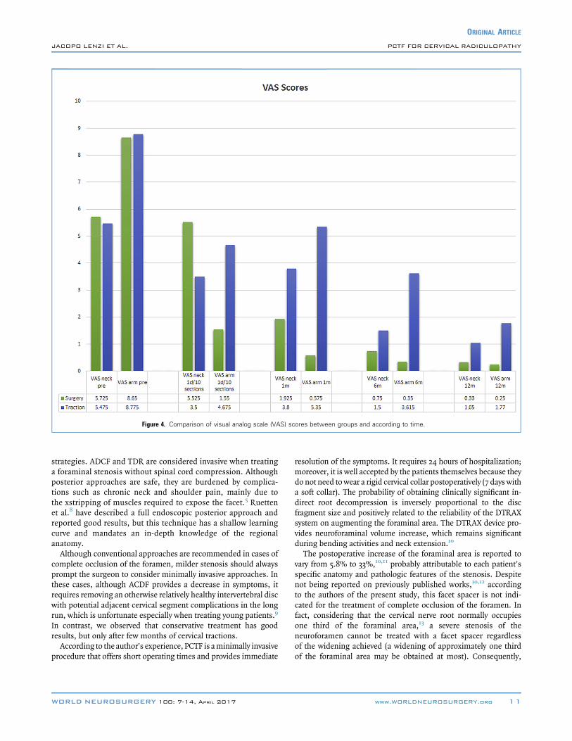
Figure 4. Comparison of visual analog scale (VAS) scores between groups and according to time.
ORIGINAL ARTICLE
JACOPO LENZI ET AL. PCTF FOR CERVICAL RADICULOPATHY
strategies. ADCF and TDR are considered invasive when treatinga foraminal stenosis without spinal cord compression. Althoughposterior approaches are safe, they are burdened by complica-tions such as chronic neck and shoulder pain, mainly due tothe xstripping of muscles required to expose the facet.5 Ruettenet al.8 have described a full endoscopic posterior approach andreported good results, but this technique has a shallow learningcurve and mandates an in-depth knowledge of the regionalanatomy.Although conventional approaches are recommended in cases of
complete occlusion of the foramen, milder stenosis should alwaysprompt the surgeon to consider minimally invasive approaches. Inthese cases, although ACDF provides a decrease in symptoms, itrequires removing an otherwise relatively healthy intervertebral discwith potential adjacent cervical segment complications in the longrun, which is unfortunate especially when treating young patients.9
In contrast, we observed that conservative treatment has goodresults, but only after few months of cervical tractions.According to the author’s experience, PCTF is aminimally invasive
procedure that offers short operating times and provides immediate
WORLD NEUROSURGERY 100: 7-14, APRIL 2017
resolution of the symptoms. It requires 24 hours of hospitalization;moreover, it is well accepted by the patients themselves because theydo not need towear a rigid cervical collar postoperatively (7 days witha soft collar). The probability of obtaining clinically significant in-direct root decompression is inversely proportional to the discfragment size and positively related to the reliability of the DTRAXsystem on augmenting the foraminal area. The DTRAX device pro-vides neuroforaminal volume increase, which remains significantduring bending activities and neck extension.10
The postoperative increase of the foraminal area is reported tovary from 5.8% to 33%,10,11 probably attributable to each patient’sspecific anatomy and pathologic features of the stenosis. Despitenot being reported on previously published works,10,12 accordingto the authors of the present study, this facet spacer is not indi-cated for the treatment of complete occlusion of the foramen. Infact, considering that the cervical nerve root normally occupiesone third of the foraminal area,13 a severe stenosis of theneuroforamen cannot be treated with a facet spacer regardlessof the widening achieved (a widening of approximately one thirdof the foraminal area may be obtained at most). Consequently,
www.WORLDNEUROSURGERY.org 11

Figure 5. Comparison of Neck Disability Index (NDI) scores between groups and according to time.
ORIGINAL ARTICLE
JACOPO LENZI ET AL. PCTF FOR CERVICAL RADICULOPATHY
these patients were excluded from the study and currentlymanaged with ACDF or TDR.Thirty-five percent of patients from the traction group chose
surgery. A subanalysis of this population revealed that most ofthese patients were young (<50 years old) and could not face aperiod of inactivity, especially in relation to occupational issuesand therefore needed to return to work and/or physical activity assoon as possible. Conservative treatment is documented to givethe best results on cervical single-level radiculopathy in the longrun14; moreover, the results of our study also showed this trend: at12 months’ follow-up, the pain scales were statistically superim-posable. One may speculate that older patients are much morekeen to postpone surgery hoping for conservative treatment toshow its effects rather than younger patients, who preferred to beoperated on when facing a relatively longer period of inactivity toachieve a faster relieve from pain and a relatively more immediatefunctional improvement, especially in the setting of a sooner re-turn to work. When a decision to be operated on was taken duringthe conservative treatment protocol, the patient was automaticallyexcluded from the study.
12 www.SCIENCEDIRECT.com WORLD NEU
Stability of the PCTFLeasure et al.10 compared the stiffness of the DTRAX system withthat of transarticular screws in cadaver specimens and concludedthat the former is more stable in flexion, axial rotation, and lateralbending but not in extension. Moreover, there was no arthrosisdevelopment in the adjacent segments at follow-up on cervicalradiographs. No changes were also reported at 1-year follow up byauthors from the international literature.6 Two-year follow-uprevealed adjacent segment arthrosis in 17.6% of a previouslypublished series.12 A 5-year follow-up will provide new insights onthe possible long-term development of adjacent segment arthrosisand instability or on recurrences.PCTF in patients affected by cervical kyphosis is not contra-
indicated in the current literature. Even so, cervical kyphosis was acontraindication to surgical treatment in our series. The possibleaggravation of cervical curve inversion after PCTF already has beentaken into consideration by previous reports,6,15 in which multiplelevels up to 4 were treated. Range of motion was not significantlyreduced,6 nor did the Ishihara index change after treatment.15 Thepresent series did not include patients treated on multiple levels.
ROSURGERY, http://dx.doi.org/10.1016/j.wneu.2016.12.125

Figure 6. Comparison of Short Form-36 (SF-36) scores between groups and according to time.
ORIGINAL ARTICLE
JACOPO LENZI ET AL. PCTF FOR CERVICAL RADICULOPATHY
Given the data already present in the literature and for the fact thatour study primarily analyzed the clinical outcome, we chose not toassess this particular issue.
CONCLUSIONS
The PCTF, through a minimally invasive surgical procedure,provides good results in adequately selected patients harboring
WORLD NEUROSURGERY 100: 7-14, APRIL 2017
single-level cervical radiculopathy due to foraminal stenosisresistant to pharmacologic treatment. Conventional surgicalapproaches such as ADCF or open posterior surgery should beconsidered in case of complete occlusion of the foramen.Conservative physical therapy does not provide comparable re-sults in the short term. The technique is therefore effective andsafe.
REFERENCES
1. Schoenfeld AJ, George AA, Bader JO,Caram PM Jr. Incidence and epidemiology ofcervical radiculopathy in the United States mili-tary: 2000 to 2009. J Spinal Disord Tech. 2012;25:17-22.
2. Bahadir C, Onal B, Yaman V, Yigit S. Relationshipbetween clinical and needle electromyographyfindings in patients with myotomal muscleweakness caused by cervical disk herniation: along-term follow-up study. Trakya Univ Tip FakDerg. 2008;25:214-220.
3. Cesaroni A, Nardi PV. Plasma disc decompres-sion for contained cervical disc herniation: arandomized controlled trial. Eur Spine J. 2010;19:477-486.
4. Iyer S, Kim HJ. Cervical radiculopathy. Curr RevMusculoskelet Med. 2016;9:272.
5. Chang JC, Park HK, Choi SK. Posterior cervicalinclinatory foraminotomy for spondylotic radicul-opathy preliminary. J Korean Neurosurg Soc. 2011;49:308-313.
6. McCormack BM, Bundoc RC, Ver MR, Ignacio JM,Berven SH, Eyster EF. Percutaneous posteriorcervical fusion with the DTRAX Facet System forsingle-level radiculopathy: results in 60 patients.J Neurosurg Spine. 2013;18:245-254.
7. Goel A, Shaha A. Facetal distraction as treat-ment for single- and multilevel cervical spon-dylotic radiculopathy and myelopathy: apreliminary report. J Neurosurg Spine. 2011;14:689-696.
8. Ruetten S, Komp M, Merk H, Godolias G. Full-endoscopic cervical posterior foraminotomy forthe operation of lateral disc herniations using 5.9-mm endoscopes: a prospective, randomized,controlled study. Spine (Phila Pa 1976). 2008;33:940-948.
9. Phillips FM, Geisler FH, Gilder KM, Reah C,Howell KM, McAfee PC. Long-term outcomes ofthe US FDA IDE prospective, randomizedcontrolled clinical trial comparing PCM cervicaldisc arthroplasty with anterior cervical dis-cectomy and fusion. Spine (Phila Pa 1976). 2015;40:674-683.
10. Leasure JM, Buckley J. Biomechanical evalu-ation of an interfacet joint decompressionand stabilization system. J Biomech Eng. 2014;136:1-8.
www.WORLDNEUROSURGERY.org 13

ORIGINAL ARTICLE
JACOPO LENZI ET AL. PCTF FOR CERVICAL RADICULOPATHY
11. Siemionow K, Janusz P, Glowka P. Cervical cagesplaced bilaterally in the facet joints from a poste-rior approach significantly increase foraminalarea. Eur Spine J. 2016;25:2279-2285.
12. Siemionow K, Monsef JB, Janusz P. Preliminaryanalysis of adjacent segment degeneration inpatients treated with posterior cervical cages: 2-year follow-up. World Neurosurg. 2016;89:730.e1-730.e7.
13. Ugbo JL, Pedlow FX Jr, Heller JG. Anatomy ofthe cervical spine. In: Benzel EC, ed. The CervicalSpine. Philadelphia: Lippincott Williams & Wil-kins; 2012:1-33.
14 www.SCIENCEDIRECT.com
DisclaimerDTRAX® Expandable Cage is CE mark only. It is Technology, Inc. plan on making it available in the
DTRAX® Expandable Cage differs from CAVUX®
• DTRAX® Expandable Cage is a variable-heigwhereas CAVUX® Cervical Cage-B and CAV
• DTRAX® Expandable Cage has a greater len• DTRAX® Expandable Cage does not have a
graft windows.• The cage release and bone screw deployme
Cervical Cage-B and CAVUX® Cervical Cag
14. Moustafa IM, Diac AA. Multimodal treatmentprogram comparing 2 different traction ap-proaches for patients with discogenic cervicalradiculopathy: a randomized controlled trial.J Chiropr Med. 2014;13:157-167.
15. Tan LA, Straus DC, Traynelis VC. Cervical inter-facet spacers and maintenance of cervical lordosis.J Neurosurg Spine. 2015;22:466-469.
Conflict of interest statement: The authors declare that thearticle content was composed in the absence of any
Jim Bean, MD. “Dewey Monument, Union Square, San Fran
WORLD NEUROSURGERY, http://
not FDA approved and is not available in the United United States.
Cervical Cage-B and CAVUX® Cervical Cage-T in
ht, expandable implant consisting of a titanium alloUX® Cervical Cage-T are fixed height implants magth and footprint relative to CAVUX® Cervical Cag graft window whereas CAVUX® Cervical Cage-B
nt mechanisms of DTRAX® Expandable Cage are e-T.
commercial or financial relationships that could be construedas a potential conflict of interest.
Received 5 October 2016; accepted 27 December 2016
Citation: World Neurosurg. (2017) 100:7-14.http://dx.doi.org/10.1016/j.wneu.2016.12.125
Journal homepage: www.WORLDNEUROSURGERY.org
Available online: www.sciencedirect.com
1878-8750/$ - see front matter ª 2017 Elsevier Inc. Allrights reserved.
cisco.”
dx.doi.org/10.1016/j.wneu.2016.12.125
States, nor does Providence Medical
the following ways:
y screw and expandable washer chined from titanium alloy.e-B and CAVUX® Cervical Cage-T.and CAVUX® Cervical Cage-T feature
different than those used for CAVUX®