Embed Size (px)
Citation preview
Position Sense Changes with Guidelines
J N E R JOURNAL OF NEUROENGINEERINGAND REHABILITATION
Herter et al. Journal of NeuroEngineering and Rehabilitation 2014, 11:43http://www.jneuroengrehab.com/content/11/1/43
RESEARCH Open Access
Systematic changes in position sense accompanynormal aging across adulthoodTroy M Herter1,2,3,4,6, Stephen H Scott3,4,5 and Sean P Dukelow1,2*
Abstract
Background: Development of clinical neurological assessments aimed at separating normal from abnormalcapabilities requires a comprehensive understanding of how basic neurological functions change (or do notchange) with increasing age across adulthood. In the case of proprioception, the research literature has failed toconclusively determine whether or not position sense in the upper limb deteriorates in elderly individuals. Thepresent study was conducted a) to quantify whether upper limb position sense deteriorates with increasing age,and b) to generate a set of normative data that can be used for future comparisons with clinical populations.
Methods: We examined position sense in 209 healthy males and females between the ages of 18 and 90 using arobotic arm position-matching task that is both objective and reliable. In this task, the robot moved an arm to oneof nine positions and subjects attempted to mirror-match that position with the opposite limb. Measures of positionsense were recorded by the robotic apparatus in hand-and joint-based coordinates, and linear regressions wereused to quantify age-related changes and percentile boundaries of normal behaviour. For clinical comparisons,we also examined influences of sex (male versus female) and test-hand (dominant versus non-dominant) on allmeasures of position sense.
Results: Analyses of hand-based parameters identified several measures of position sense (Variability, Shift, SpatialContraction, Absolute Error) with significant effects of age, sex, and test-hand. Joint-based parameters at theshoulder (Absolute Error) and elbow (Variability, Shift, Absolute Error) also exhibited significant effects of age andtest-hand.
Conclusions: The present study provides strong evidence that several measures of upper extremity position senseexhibit declines with age. Furthermore, this data provides a basis for quantifying when changes in position senseare related to normal aging or alternatively, pathology.
Keywords: Proprioception, Position sense, Upper limb, Robotics, Assessment, Aging
BackgroundProprioception refers to the ability to perceive the locationof one’s body in space and has been classically divided intotwo subcomponents: position sense and kinesthesia [1].Position sense is the ability of an individual to identify thestatic location of a body part, whereas kinesthesia is theability to identify body motion. Although muscle spindleafferents are considered to provide the dominant sourceof information for position sense, cutaneous afferents are
* Correspondence: [email protected] Brain Institute, University of Calgary, Calgary, Alberta, Canada2Department of Clinical Neurosciences, University of Calgary, Calgary, Alberta,CanadaFull list of author information is available at the end of the article
© 2014 Herter et al.; licensee BioMed Central LCommons Attribution License (http://creativecreproduction in any medium, provided the or
also an important source of information, particularly forthe more distal joints [2-6].In healthy individuals, the suggestion that position sense
may decline with age across adulthood is not entirely sur-prising because a number of physiological changes occurin the proprioceptive system with increasing age. Studieshave shown that muscle spindles show lower sensitivity[7-9], intrafusal fibres decline in number [10,11], spindlediameters decrease in size [12] and capsular thickness in-creases [11]. Cutaneous mechanoreceptors also decreasein number with age [13,14]. Each of these natural changesmay contribute to declines in position sense.In addition to peripheral changes that occur with in-
creasing age, modifications to the central nervous system
td. This is an Open Access article distributed under the terms of the Creativeommons.org/licenses/by/2.0), which permits unrestricted use, distribution, andiginal work is properly credited.
Herter et al. Journal of NeuroEngineering and Rehabilitation 2014, 11:43 Page 2 of 12http://www.jneuroengrehab.com/content/11/1/43
may also contribute to age-related declines in propriocep-tion. For example, decreased grey matter in the post-central gyrus [15,16] and reduced activity in proprioceptiveregions of the basal ganglia [17] may contribute to declinesin position sense across adulthood. Position sense is alsoinfluenced in a task-dependent manner by attentionand possibly other cognitive factors [18], thus age-relateddeclines in attention and cognition may contribute todeclines in proprioception with increasing age.In spite of these changes in the peripheral and central
aspects of somatosensory processing, there remains dis-agreement whether position sense in the upper limbactually declines with age (for a review see Goble et al.2009 [19]). In the upper limb, several small studies andone large study have found that position sense is slightlyworse in subjects in their seventh or eighth decade of lifeas compared to performance in their third [20-23]. How-ever, these studies contradict the findings of a largestudy by Kokmen et al. [24] that found no effects of ageon the sense of joint motion (kinesthesia) at the meta-carpophalangeal (MCP) joint. The discrepancy betweenthese studies may reflect differences between positionsense and kinesthesia. However, both functions aresusceptible to peripheral and central influences of aging,thus the effects of aging on position sense and kinesthesiaremain unclear.A clear understanding of the effects of increasing age
on position sense is important clinically for identifyinghow neurological disorders impact proprioception.Notably, stroke and traumatic brain injury often lead todeficits in position sense [25,26]. Poor position sensealso correlates with poor functional outcomes followingstroke [27], whereas individuals with intact positionsense following stroke have significantly better motorrecovery [28-32]. By characterizing the effects of agingon position sense, it is possible to differentiate deficitsthat are caused by stroke or TBI from those reflectingnormal declines due to aging.We conducted the present study to, a) provide a better
understanding of age-related declines in upper limbposition sense and b) develop a normative data set thatcould be used for comparison to clinical populations. Toachieve these goals, we have used an objective andreliable assessment of position sense based on robotictechnology to quantify arm position sense in a large co-hort of 209 healthy individuals between 18 and 90 yearsof age. To examine age-related declines in upper limbposition sense, regression models were developed toquantify the influence of age on measures of positionsense. Because neurological disorders, such as stroke,can affect both sexes and both sides of the body, we alsoexamined influences of sex (male versus female) and test-hand (dominant versus non-dominant) to control forthese factors in the normative data sets. We considered
the potential influence of sex given that age-relatedchanges are often thought to be more prominent in themale brain [15,33-35]. We examined differences betweenusing the dominant and non-dominant arms to probeposition sense because of their distinct contributions tovarious aspects of proprioceptive function [36-41].
MethodsParticipantsMale and female participants between the ages of 18 and90 were recruited from the communities of Kingston,Ontario and Calgary, Alberta, Canada. Contact wasmade through posted flyers, advertisements in localnewspapers, and direct communication with families ofstroke inpatients at St. Mary’s of the Lake Hospital(Kingston, Ontario) and Foothills Medical Centre (Calgary,Alberta). Participants were excluded from the study if they:1) had any history of neurological impairments, 2) had anyongoing musculoskeletal problems of the shoulder and/orelbow, and 3) were unable to understand the instructionsfor the testing procedure. To ensure eligibility for thestudy, all participants completed a clinical examination,which included a detailed medical history and physicalexaminations of strength of the upper extremity using theMRC grading system [42], range of motion, and motorcontrol using the Purdue Pegboard [43]. Subjects who didnot obtain a normal score on any of these tests wereexcluded from the study. We also excluded any subjectwho was unable to understand the instructions for any ofthe clinical assessments or the robotic assessment. A phys-ician or physical therapist with expertise in stroke rehabili-tation performed all clinical examinations. Subjects alsocompleted the 10 item modified version of EdinburghHandedness Inventory (writing, drawing, throwing, scis-sors, toothbrush, knife, spoon, broom, match, and lid) todetermine their hand dominance [44]. Subjects were con-sidered right-handed, left-handed, and ambidextrous if theyobtained scores of 50 to 100, −100 to −50, or −49 to 49, re-spectively [45]. Before entering the study, all participantsprovided informed consent. Data from some participants(n = 65) had been collected as control data for a previousstudy of position sense following stroke [26]. All methodsfor data collection used in the previous study were identi-cal to those used in the current study. Ethics approval wasobtained from the ethics boards of Queen’s University,Providence Care, and the University of Calgary.
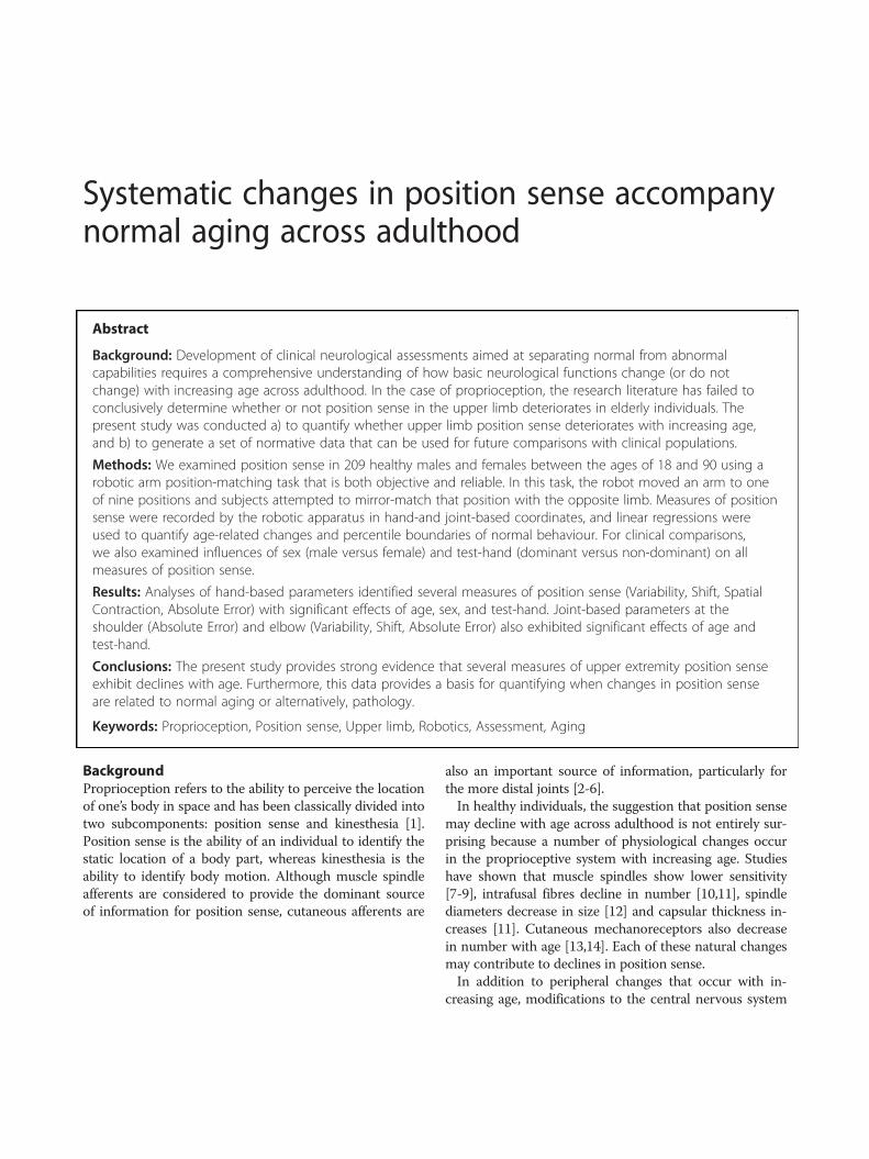
Robotic apparatusThe current study used the KINARM exoskeleton robot(BKIN Technologies Ltd., Kingston, Ontario), which is ajoint-based robotic apparatus that can be used to ma-nipulate, monitor and record joint- (shoulder and elbow)and hand-based kinematic data (Figure 1). The roboticapparatus has been described in a number of previous
Figure 1 Robotic apparatus. A, Frontal view of a participant sitting in the robotic apparatus. The participant sits comfortably in the modifiedwheelchair base with his arms and hands supported by troughs. B, Side view of a participant with the robotic apparatus docked at theaugmented reality workstation. Shields mounted under the glass and a soft, black cover hanging between the glass and the participant’s neckocclude all vision of the arms and hands. Written, informed consent for the publication of pictures was obtained from the participant.
Herter et al. Journal of NeuroEngineering and Rehabilitation 2014, 11:43 Page 3 of 12http://www.jneuroengrehab.com/content/11/1/43
publications [25-27,46-48]. Participants sit in a modifiedwheelchair seat with both arms comfortably placed inarm troughs that are adjusted to the dimensions of eachindividual (Figure 1A). The exoskeleton provides fullgravitational support of the arms and hands, permitsarm movements in the horizontal plane, and can applymechanical loads to the shoulder and/or elbow. Anglesof the shoulder and elbow are obtained directly fromencoders within the robotic motors, and a calibrationprocess is used to compute joint- and hand-based kine-matics, including the position of each index fingerwithin the horizontal plane. The robot is docked to anaugmented reality system that can display targets withinthe same plane as the arms and hands. For purposes ofthe current study, vision of the arms and hands wasoccluded and the augmented reality display was notemployed (Figure 1B).Although other studies have used instruments such as
dynamometers [49], motion-capture systems [50], incli-nometers [51], and light exoskeleton systems [52] tomeasure position sense, our robotic device offers a num-ber of advantages. Notably, our robotic apparatus allowsus to rapidly set-up and calibrate subjects, passivelymove the arms through smooth trajectories, and couplemovements at multiple joints (shoulder and elbow) injoint- or hand- coordinates. In addition, unloaded tasks,such as the arm position matching task, require negli-gible amounts of strength because the arms are fullysupported against gravity. Perhaps most important, how-ever, we are able to objectively obtain valid, reliable, andsensitive measures of sensory, motor, and cognitive func-tion with a single platform. Benefits of using robotic
technology for assessment of sensory and motor deficitshave also been described in a recent review [53].
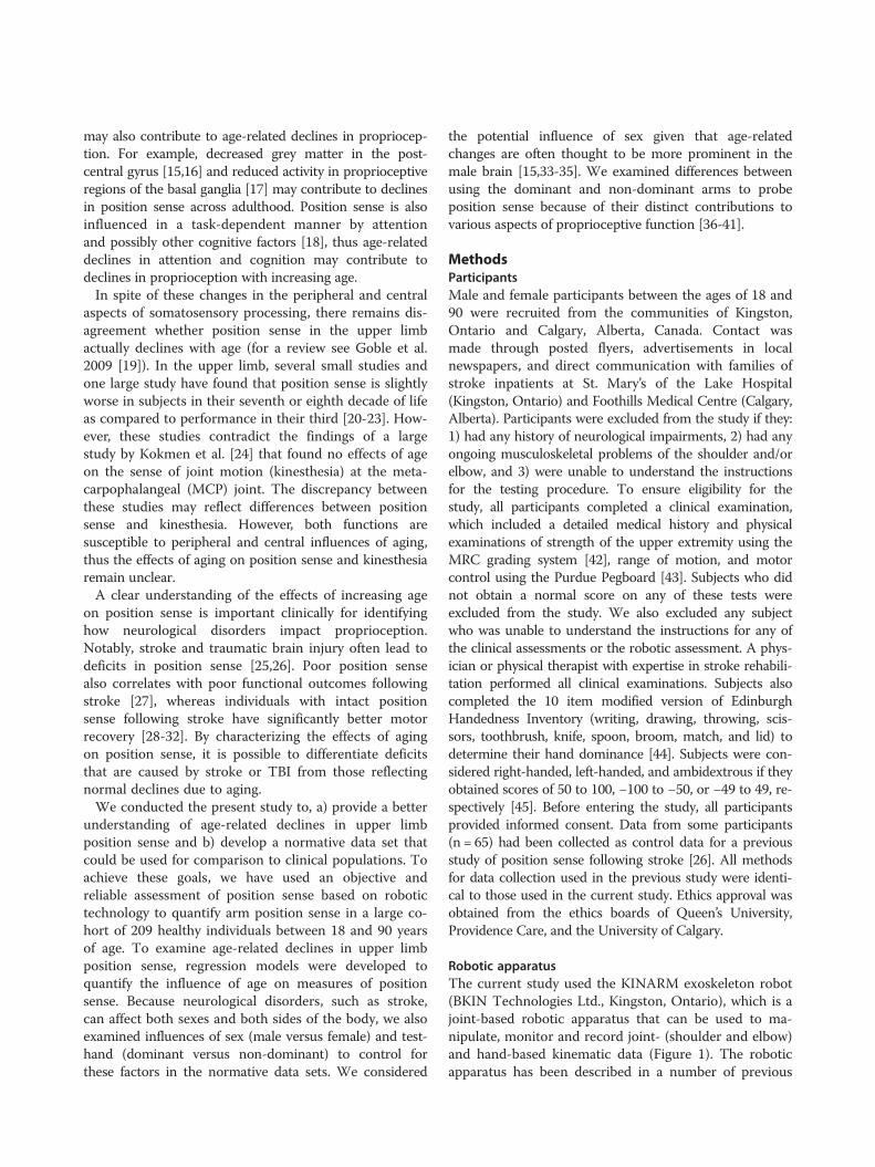
Arm position matching taskPosition sense of the upper extremity was assessed withan arm position matching task [25-27]. With the armsand hands occluded from vision, the robot passivelymoved one hand (passive hand) to one of nine differ-ent target locations (fingertip positions) organized ina 3×3 matrix with 10 cm separation between targets(Figure 2A). The participants were instructed to ac-tively move the opposite hand (active hand) to themirror location in space. To help ensure that match-ing performance reflected sensory perception ratherthan motor control, participants were given as longas needed to complete each trial; the examiner thentriggered the next trial when the subjects verballyinstructed to the examiner that they felt their activehand was mirror matching their passive hand. By re-quiring a verbal response before triggering the nexttrial, we were also able to control for momentary lapses ofattention. Each of the nine target locations was presentedonce in a randomized block. Six different blocks were ob-tained for a total of 54 trials. All participants completedthe task twice, once using each hand as the active hand.
AnalysesAll analyses were performed using MATLAB (MathworksInc., Massachusetts, USA). Matching performance wasexamined in both hand- and joint-coordinates to permitcomparisons with both research studies and clinical prac-tice, which commonly use single-joint or whole-limb tasks

Figure 2 Position matching task. A, Top down workspace view of a typical participant. The robot passively moved the left hand to nine spatiallocations (filled symbols) and the right arm was actively moved by the participant to mirror-match each spatial location (open symbols). Ellipsesaround each of open symbols represent one standard deviation. Areas enclosed by the solid and dashed grey lines show the matching areas ofpassive and active arms, respectively. B, Overlap of the passive and active arms after mirror transforming data from the left arm to the right side of theworkspace. C–E, The data has been modified to illustrate examples of increased variability (C), systematic shift (D) and spatial contraction (E).
Herter et al. Journal of NeuroEngineering and Rehabilitation 2014, 11:43 Page 4 of 12http://www.jneuroengrehab.com/content/11/1/43
to measure position sense. Parameters in hand-coordi-nates were quantified using Cartesian (x, y) positions ofthe index fingertips obtained from the robotic apparatusat the end of each trial. Figure 2A illustrates a workspaceview of the mean fingertip positions of the passive andactive hands of an exemplar participant for each target. Inorder to visually compare the positions of the passive andactive hands, the position of the passive hand was mir-rored across the x coordinate (Figure 2B).In a previous study [26], we developed three parame-
ters (Variability (Var), Systematic Shift (Shift), SpatialContraction/Expansion (C/E) to characterize subjectperformance based on hand position (See Table 1 fordefinitions). These parameters showed good to excellent
Table 1 Attributes and parameters of the arm position match
Coordinate frame Parameter Abrv Units Definition
Hand-coordinates Variability Var cm Mean trial-by-tria
Systematic shift Shift cm Systematic errors
Absolute error AE cm Absolute errors b
Spatial Cont/Exp C/E — Ratio of: i) meanenclosed by the
Joint-coordinates Variability Var deg Mean trial-by-tria
Systematic shift Shift deg Systematic errors
Absolute error AE deg Absolute errors b
reliability (Var: r = 0.81; Shift: r = 0.70; C/E: r = 0.86). Theformulae used to compute Var, Shift, and C/E have beenpreviously described in detail [26]. Separate values forVar and Shift were obtained for the x, y, and xy (lineardistance) dimensions. For didactic purposes, Figures 2C,D and E highlight representative patterns of errors, illus-trating large variability, a large systematic shift in handposition across the workspace, and a reduction in theoverall spatial area of the workspace used with the activehand, respectively. Other studies have generally quan-tified the absolute errors in position sense [54,55]. Theseabsolute errors should increase due to any of the pat-terns depicted in Figure 2C-E. For comparison with pre-vious studies, we also computed absolute error (AE).
ing task
l variability of the active hand in x, y, and xy coordinates.
between the mean x and y positions of the active and passive hands.
etween the mean x and y positions of the active and passive hands.
spatial area enclosed by the active hand to ii) mean spatial area ofpassive hand.
l variability of the shoulder and elbow angles of the active arm.
between the shoulder and elbow angles of the active and passive arms.
etween the shoulder and elbow angles of the active and passive arms.
Herter et al. Journal of NeuroEngineering and Rehabilitation 2014, 11:43 Page 5 of 12http://www.jneuroengrehab.com/content/11/1/43
Parameters in joint-coordinates were quantified usingshoulder and elbow angles obtained directly from therobotic apparatus at the end of each trial (See Table 1).In joint-space, separate values were obtained for theshoulder and elbow joints. Each parameter was calcu-lated separately for each target and then averaged acrossall targets.Linear regression was used to quantify age-related
changes for each parameter. If the grouped data exhib-ited a significant regression fit (F-test, P < 0.05), residualvalues were computed by subtracting out the regressionmodel from the original values. If the grouped data didnot exhibit a significant regression fit (F-tests, P ≥ 0.05),we continued with the original data values.For those parameters with a significant regression fit, we
tested whether the residual values were normally distrib-uted. If the residual values were not normally distributed(Lilliefors test, P < 0.05), the original data was transformedwith a: 1) logarithmic, 2) square root, or 3) inverse trans-form. In cases when a transform was needed, linearregression was repeated on the transformed data and, if asignificant fit was found (F-test, P < 0.05), residuals werere-calculated from the transformed data. The residualvalues were then retested for normality until a normal dis-tribution was obtained from one of the transforms.After a normal distribution of residual values was
obtained, Kolmogorov-Smirnov tests were used to quan-tify effects of sex (males versus females) and test-hand(dominant versus non-dominant). Note that our examin-ation of test-hand assessed whether subjects performeddifferently when matching with the dominant hand (leftor right) as the active hand compared to using theirnon-dominant hand as the active hand. Specifically, thisdid not examine whether right-dominant subjects differfrom left-dominant subjects. If a parameter exhibited asignificant effect of sex and/or test-hand (P < 0.05), theoriginal data was separated by sex (males and females)and/or test-hand (dominant and non-dominant) and themethods described above were repeated to quantify effectsof age for each separate group. Data from the separategroups were then used to compute normative statistics.
Table 2 Subject demographics (n= 209 subjects, 96 male and
Age # Subjects Median age Sex
Male
18 – 29 41 24 16
30 – 39 37 34 18
40 – 49 34 46 12
50 – 59 30 55 8
60 – 69 35 63 20
70 – 79 23 72 16
80 – 90 9 82 6
In order to establish normative statistics for each par-ameter, we obtained a number of percentiles (1, 2.5, 5,25, 50, 75, 95, 97.5, 99) from the residual values with asignificant effect of age (F-test, P < 0.05) and the originalvalues without a significant effect of age (F-test, P ≥ 0.05).Percentiles were obtained using the Matlab percentilefunction (prctile.m), which uses rank ordering with linearinterpolation to find percentiles. Percentiles obtained froma parameter with a significant regression fit could betransformed back into its native units to obtain a uniquestatistical distribution for any given age, sex, and test-hand. The formulae used for the different inverse trans-forms include:
No Transform: y ¼ age � slopeð Þ þ biasþ percentile
ð1Þ
Log Transform: y ¼ e age � slopeð Þþ biasþ percentileð Þ ð2ÞSquareRootTransform: y ¼ age � slopeð Þ þ biasþ percentileð Þ2
ð3Þ
Inverse Transform: y ¼ 1age � slopeð Þ þ biasþ percentileð Þ
ð4ÞPercent changes in parameters were computed using
the regression fits to find the median values of parame-ters for 18 and 90 year-old subjects, then calculating thepercent change:
Percent Change: Δy ¼ y90–y18ð Þy18
� 100% ð5Þ
ResultsA total of 209 participants (96 male, 113 female) com-pleted the arm position matching task with both arms.Demographic data describing the age, sex, and handed-ness of the participants are provided in Table 2. Scoresfrom the Modified Edinburgh Handedness Inventory wereused to classify participants as right-handed (handedness
113 female)
Handedness
Female Right (M/F) Left (M/F) Mixed (M/F)
25 14/22 1/1 1/2
19 17/18 1/1 0/0
22 10/17 1/4 1/1
22 6/21 0/1 2/0
15 18/15 0/0 2/0
7 15/6 1/1 0/0
3 6/3 0/0 0/0
Herter et al. Journal of NeuroEngineering and Rehabilitation 2014, 11:43 Page 6 of 12http://www.jneuroengrehab.com/content/11/1/43
score ≥ 50; n = 188), left-handed (handedness score ≤ −50;n = 12), or mixed handedness (−50 < handedness score < 50;n = 9) [44,45]. All nine participants who scored in themixed handedness range performed more tasks with theirright-hand and were, therefore, treated as right-handedparticipants in our analysis.
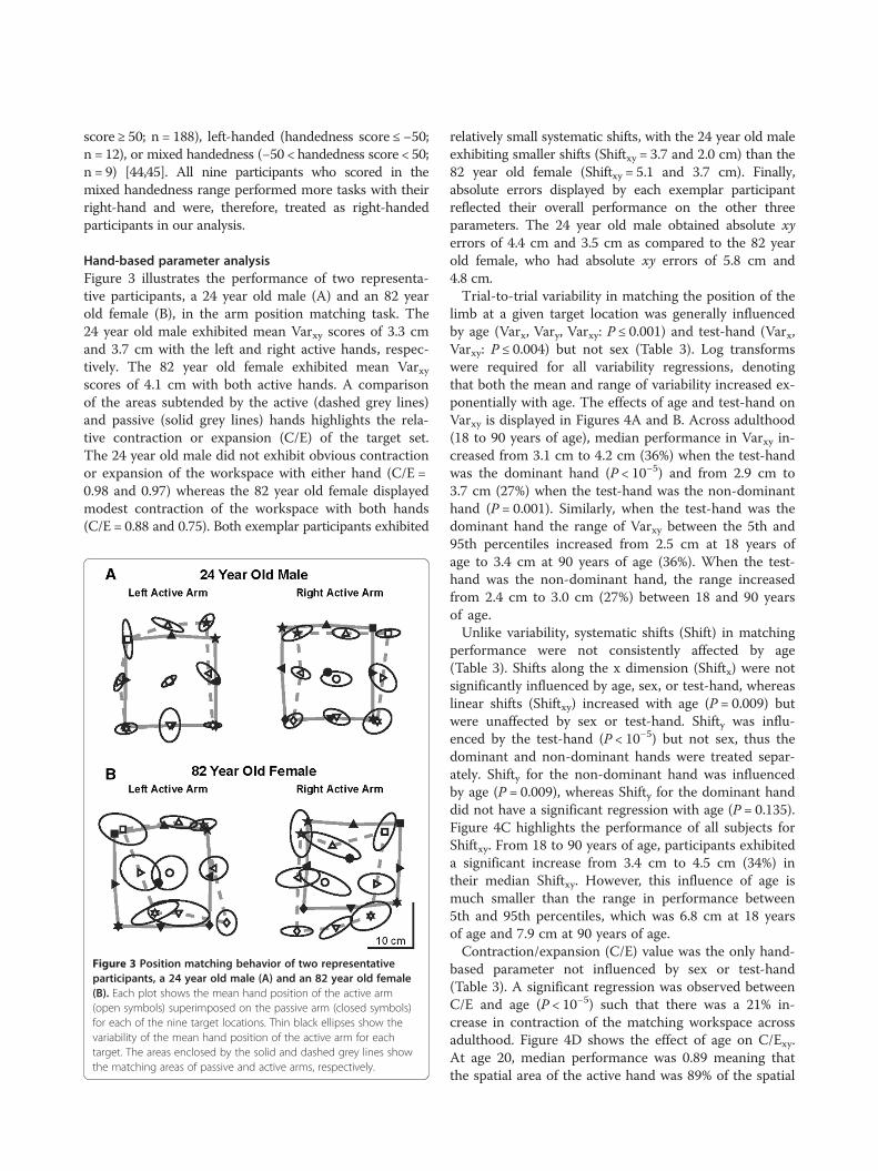
Hand-based parameter analysisFigure 3 illustrates the performance of two representa-tive participants, a 24 year old male (A) and an 82 yearold female (B), in the arm position matching task. The24 year old male exhibited mean Varxy scores of 3.3 cmand 3.7 cm with the left and right active hands, respec-tively. The 82 year old female exhibited mean Varxyscores of 4.1 cm with both active hands. A comparisonof the areas subtended by the active (dashed grey lines)and passive (solid grey lines) hands highlights the rela-tive contraction or expansion (C/E) of the target set.The 24 year old male did not exhibit obvious contractionor expansion of the workspace with either hand (C/E =0.98 and 0.97) whereas the 82 year old female displayedmodest contraction of the workspace with both hands(C/E = 0.88 and 0.75). Both exemplar participants exhibited
Figure 3 Position matching behavior of two representativeparticipants, a 24 year old male (A) and an 82 year old female(B). Each plot shows the mean hand position of the active arm(open symbols) superimposed on the passive arm (closed symbols)for each of the nine target locations. Thin black ellipses show thevariability of the mean hand position of the active arm for eachtarget. The areas enclosed by the solid and dashed grey lines showthe matching areas of passive and active arms, respectively.
relatively small systematic shifts, with the 24 year old maleexhibiting smaller shifts (Shiftxy = 3.7 and 2.0 cm) than the82 year old female (Shiftxy = 5.1 and 3.7 cm). Finally,absolute errors displayed by each exemplar participantreflected their overall performance on the other threeparameters. The 24 year old male obtained absolute xyerrors of 4.4 cm and 3.5 cm as compared to the 82 yearold female, who had absolute xy errors of 5.8 cm and4.8 cm.Trial-to-trial variability in matching the position of the
limb at a given target location was generally influencedby age (Varx, Vary, Varxy: P ≤ 0.001) and test-hand (Varx,Varxy: P ≤ 0.004) but not sex (Table 3). Log transformswere required for all variability regressions, denotingthat both the mean and range of variability increased ex-ponentially with age. The effects of age and test-hand onVarxy is displayed in Figures 4A and B. Across adulthood(18 to 90 years of age), median performance in Varxy in-creased from 3.1 cm to 4.2 cm (36%) when the test-handwas the dominant hand (P < 10−5) and from 2.9 cm to3.7 cm (27%) when the test-hand was the non-dominanthand (P = 0.001). Similarly, when the test-hand was thedominant hand the range of Varxy between the 5th and95th percentiles increased from 2.5 cm at 18 years ofage to 3.4 cm at 90 years of age (36%). When the test-hand was the non-dominant hand, the range increasedfrom 2.4 cm to 3.0 cm (27%) between 18 and 90 yearsof age.Unlike variability, systematic shifts (Shift) in matching
performance were not consistently affected by age(Table 3). Shifts along the x dimension (Shiftx) were notsignificantly influenced by age, sex, or test-hand, whereaslinear shifts (Shiftxy) increased with age (P = 0.009) butwere unaffected by sex or test-hand. Shifty was influ-enced by the test-hand (P < 10−5) but not sex, thus thedominant and non-dominant hands were treated separ-ately. Shifty for the non-dominant hand was influencedby age (P = 0.009), whereas Shifty for the dominant handdid not have a significant regression with age (P = 0.135).Figure 4C highlights the performance of all subjects forShiftxy. From 18 to 90 years of age, participants exhibiteda significant increase from 3.4 cm to 4.5 cm (34%) intheir median Shiftxy. However, this influence of age ismuch smaller than the range in performance between5th and 95th percentiles, which was 6.8 cm at 18 yearsof age and 7.9 cm at 90 years of age.Contraction/expansion (C/E) value was the only hand-
based parameter not influenced by sex or test-hand(Table 3). A significant regression was observed betweenC/E and age (P < 10−5) such that there was a 21% in-crease in contraction of the matching workspace acrossadulthood. Figure 4D shows the effect of age on C/Exy.At age 20, median performance was 0.89 meaning thatthe spatial area of the active hand was 89% of the spatial
Table 3 Model fits and percentiles for hand-based parameters of position sense
Param Group Trans Model fit Percentiles
P Slope Bias 1 2.5 5 25 50 75 95 97.5 99
Varx Dom log <10-5 0.0044 0.935 –0.566 –0.479 –0.406 –0.151 –0.006 0.169 0.418 0.458 0.489
Varx N-Dom log 0.001 0.0036 0.873 –0.729 –0.528 –0.462 –0.191 –0.005 0.178 0.456 0.529 0.652
Vary All log <10-4 0.0030 0.268 –0.604 –0.472 –0.400 –0.175 –0.011 0.164 0.425 0.505 0.642
Varxy Dom log <10-5 0.0042 1.056 –0.492 –0.410 –0.375 –0.146 –0.001 0.150 0.400 0.430 0.459
Varxy N-Dom log 0.001 0.0033 1.023 –0.616 –0.463 –0.432 –0.157 –0.007 0.152 0.414 0.519 0.595
Shiftx All — 0.673 — — –9.184 –7.765 –6.501 –3.221 –0.550 2.534 7.242 8.098 9.425
Shifty Dom — 0.135 — — –5.716 –4.684 –4.172 –1.464 0.015 1.627 3.614 5.143 5.610
Shifty N-Dom — 0.009 –0.0241 –0.093 –4.973 –4.058 –3.641 –1.622 –0.034 1.567 3.979 4.928 6.699
Shiftxy All sqrt 0.009 0.0040 1.784 –1.241 –1.066 –0.926 –0.399 –0.022 0.397 0.917 1.087 1.303
C/Exy All — <10-5 –0.0026 0.954 –0.416 –0.362 –0.318 –0.148 –0.009 0.146 0.350 0.425 0.513
AExy Male log 0.001 0.0038 1.479 –0.639 –0.569 –0.488 –0.265 0.030 0.229 0.483 0.593 0.661
AExy Female inv 0.012 –0.0007 0.247 0.146 0.141 0.113 0.042 –0.003 –0.049 –0.097 –0.106 –0.119
Abbreviations: Var, variability; C/E, spatial contraction/expansion; AE, absolute error. For parameters with a significant effect of sex or active-hand, model fits andpercentiles are given for each group. None of the parameters had a significant effect of both sex and active-hand.
Herter et al. Journal of NeuroEngineering and Rehabilitation 2014, 11:43 Page 7 of 12http://www.jneuroengrehab.com/content/11/1/43
area of the passive hand moved by the robot. In contrast,median performance was 0.73 at age 80 years old. Theinfluence of age on C/E was again much smaller thanthe difference between 5th to 95th percentile values,which was equal to 0.67 at a given age.Absolute errors in the xy dimension (AExy) exhibited a
significant effect of sex (P = 0.003) but not test-hand,thus males and females were separated for the regressionanalyses (Table 3). Figure 4E illustrates that male parti-cipants displayed a significant increase from 4.8 cm to6.3 cm (31%) in their median AExy across adulthood(P = 0.001). Similarly, Figure 4F shows that femaleparticipants exhibited a significant increase 3.8 cm to4.6 cm (22%) in their median AExy across adulthood(P = 0.012). Again, the influence of age on AE was muchsmaller than the range of values for males and females ata given age.
Joint-based parameter analysisFigure 5 and Table 4 show the statistical influence ofincreasing age on joint-based parameters. We did notfind a significant effect of sex or test-hand for variabilityat the shoulder and elbow. Furthermore, variability atthe shoulder did not show a significant change with age(Figure 5A), whereas we observed a 0.5° (13%) increasein variability at the elbow across adulthood (P = 0.030;Figure 5B). Age-related changes in variability at theelbow were much smaller than inter-subject differences,which could be as small as 2° for some subjects and aslarge as 7° for other subjects.Systematic shifts at the shoulder did not exhibit a
significant effect of sex or test-hand, nor did they changesignificantly with age (Figure 5C). Systematic shifts atthe elbow, however, displayed a significant effect of test-hand (P < 10−5). Although systematic shifts at the elbow
when the test-hand was the dominant hand did notexhibit a significant effect of aging (data not shown), sys-tematic shifts at the elbow for the non-dominant handincreased by 4.0° across adulthood (P = 0.015; Figure 5D).The range of shifts at any given age (approximately20 degrees) was much larger than the change that oc-curred with increases in age across adulthood.Absolute errors at the shoulder and elbow did not
display significant effects of sex or test-hand. Regressionanalyses showed a significant increase of 1.0° (18%) acrossadulthood for median absolute errors at the shoulder (P =0.024; Figure 5E). Regression analysis of absolute errorsat the elbow showed a significant increase of 1.0° (19%)across adulthood (P = 0.028; Figure 5F). Changes in abso-lute error at both the shoulder and elbow were muchsmaller than the inter-subject variability at any age.
DiscussionThe present study quantified position sense of the upperarm in a cohort of subjects spanning many decades oflife. Our findings demonstrate that position sense tendsto diminish with increasing age across adulthood. Thevast majority of the parameters changed with age (hand-based: Varx, Vary, Varxy, Shifty, Shiftxy, C/Exy, AEx; joint-based: Vare, Shifte, AEs, AEe), with the largest relativeeffect for the hand-based parameter Varxy (36%). Severalparameters also exhibited effects of sex (hand-based:AExy) and test-hand (hand-based: Varx, Varxy, Shifty,Varx; joint-based: Shifte).The effect of age on position sense is consistent with
most previous reports in the literature[20-23], though itis not clear why the large cohort of subjects in a study ofposition sense of the metacarpophalangeal joint did notshow an effect of age [24]. Theoretically, aging shouldhave similar influences on sensory receptors (muscle
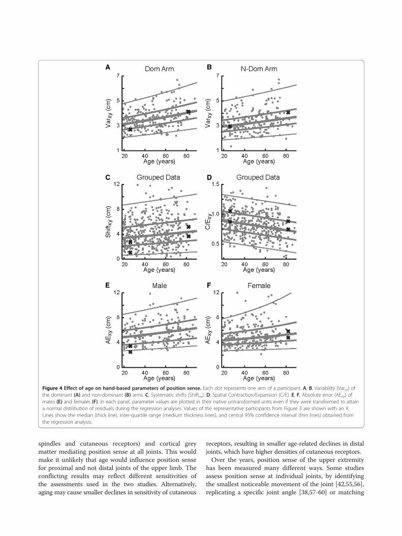
Figure 4 Effect of age on hand-based parameters of position sense. Each dot represents one arm of a participant. A, B, Variability (Varxy) ofthe dominant (A) and non-dominant (B) arms. C, Systematic shifts (Shiftxy). D, Spatial Contraction/Expansion (C/E). E, F, Absolute error (AExy) ofmales (E) and females (F). In each panel, parameter values are plotted in their native untransformed units even if they were transformed to attaina normal distribution of residuals during the regression analyses. Values of the representative participants from Figure 3 are shown with an X.Lines show the median (thick line), inter-quartile range (medium thickness lines), and central 95% confidence interval (thin lines) obtained fromthe regression analysis.
Herter et al. Journal of NeuroEngineering and Rehabilitation 2014, 11:43 Page 8 of 12http://www.jneuroengrehab.com/content/11/1/43
spindles and cutaneous receptors) and cortical greymatter mediating position sense at all joints. This wouldmake it unlikely that age would influence position sensefor proximal and not distal joints of the upper limb. Theconflicting results may reflect different sensitivities ofthe assessments used in the two studies. Alternatively,aging may cause smaller declines in sensitivity of cutaneous
receptors, resulting in smaller age-related declines in distaljoints, which have higher densities of cutaneous receptors.Over the years, position sense of the upper extremity
has been measured many different ways. Some studiesassess position sense at individual joints, by identifyingthe smallest noticeable movement of the joint [42,55,56],replicating a specific joint angle [38,57-60] or matching
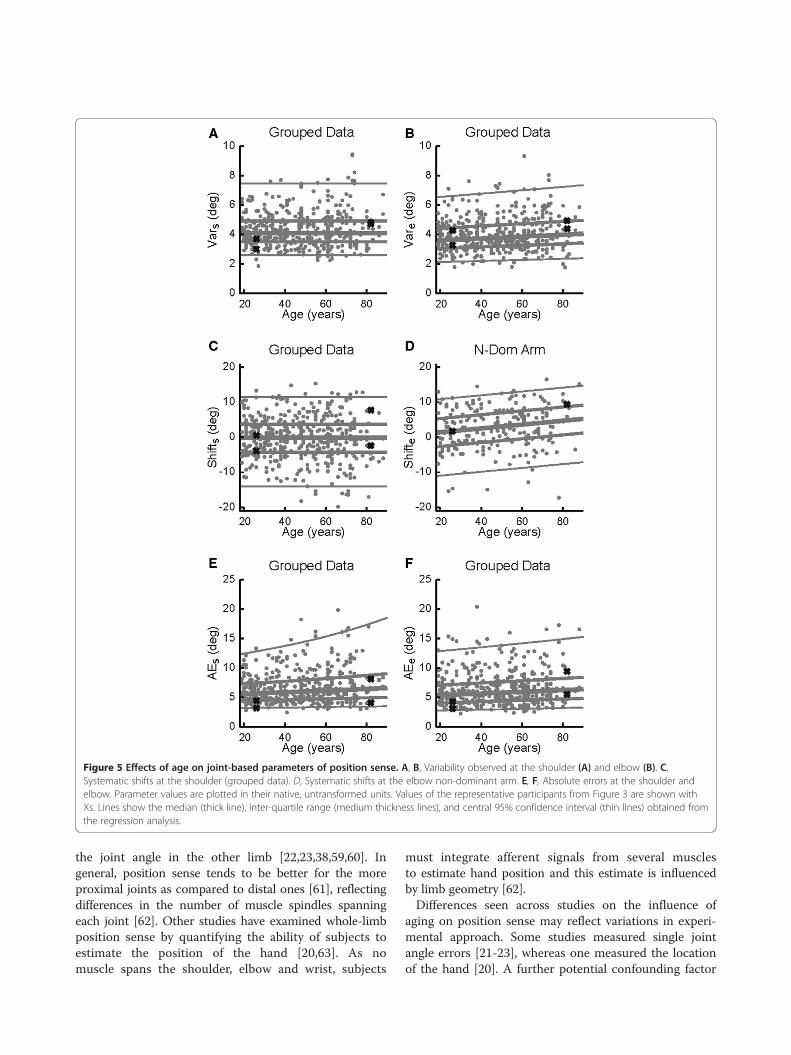
Figure 5 Effects of age on joint-based parameters of position sense. A, B, Variability observed at the shoulder (A) and elbow (B). C,Systematic shifts at the shoulder (grouped data). D, Systematic shifts at the elbow non-dominant arm. E, F, Absolute errors at the shoulder andelbow. Parameter values are plotted in their native, untransformed units. Values of the representative participants from Figure 3 are shown withXs. Lines show the median (thick line), inter-quartile range (medium thickness lines), and central 95% confidence interval (thin lines) obtained fromthe regression analysis.
Herter et al. Journal of NeuroEngineering and Rehabilitation 2014, 11:43 Page 9 of 12http://www.jneuroengrehab.com/content/11/1/43
the joint angle in the other limb [22,23,38,59,60]. Ingeneral, position sense tends to be better for the moreproximal joints as compared to distal ones [61], reflectingdifferences in the number of muscle spindles spanningeach joint [62]. Other studies have examined whole-limbposition sense by quantifying the ability of subjects toestimate the position of the hand [20,63]. As nomuscle spans the shoulder, elbow and wrist, subjects
must integrate afferent signals from several musclesto estimate hand position and this estimate is influencedby limb geometry [62].Differences seen across studies on the influence of
aging on position sense may reflect variations in experi-mental approach. Some studies measured single jointangle errors [21-23], whereas one measured the locationof the hand [20]. A further potential confounding factor
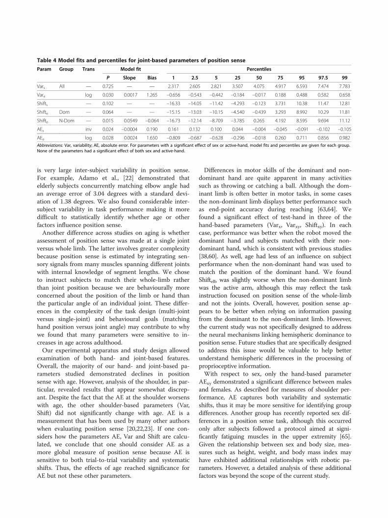
Table 4 Model fits and percentiles for joint-based parameters of position sense
Param Group Trans Model fit Percentiles
P Slope Bias 1 2.5 5 25 50 75 95 97.5 99
Vars All — 0.725 — — 2.317 2.605 2.821 3.507 4.075 4.917 6.593 7.474 7.783
Vare log 0.030 0.0017 1.265 –0.656 –0.543 –0.442 –0.184 –0.017 0.188 0.488 0.582 0.658
Shifts — 0.102 — — –16.33 –14.05 –11.42 –4.293 –0.123 3.731 10.38 11.47 12.81
Shifte Dom — 0.064 — — –15.15 –13.03 –10.15 –4.540 –0.439 3.293 8.992 10.29 11.81
Shifte N-Dom — 0.015 0.0549 –0.064 –16.73 –12.14 –8.709 –3.785 0.265 4.192 8.595 9.694 11.12
AEs inv 0.024 –0.0004 0.190 0.161 0.132 0.100 0.044 –0.004 –0.045 –0.091 –0.102 –0.105
AEe log 0.028 0.0024 1.650 –0.809 –0.687 –0.628 –0.296 –0.018 0.260 0.711 0.856 0.982Abbreviations: Var, variability; AE, absolute error. For parameters with a significant effect of sex or active-hand, model fits and percentiles are given for each group.None of the parameters had a significant effect of both sex and active-hand.
Herter et al. Journal of NeuroEngineering and Rehabilitation 2014, 11:43 Page 10 of 12http://www.jneuroengrehab.com/content/11/1/43
is very large inter-subject variability in position sense.For example, Adamo et al., [22] demonstrated thatelderly subjects concurrently matching elbow angle hadan average error of 3.04 degrees with a standard devi-ation of 1.38 degrees. We also found considerable inter-subject variability in task performance making it moredifficult to statistically identify whether age or otherfactors influence position sense.Another difference across studies on aging is whether
assessment of position sense was made at a single jointversus whole limb. The latter involves greater complexitybecause position sense is estimated by integrating sen-sory signals from many muscles spanning different jointswith internal knowledge of segment lengths. We choseto instruct subjects to match their whole-limb ratherthan joint position because we are behaviourally moreconcerned about the position of the limb or hand thanthe particular angle of an individual joint. These differ-ences in the complexity of the task design (multi-jointversus single-joint) and behavioural goals (matchinghand position versus joint angle) may contribute to whywe found that many parameters were sensitive to in-creases in age across adulthood.Our experimental apparatus and study design allowed
examination of both hand- and joint-based features.Overall, the majority of our hand- and joint-based pa-rameters studied demonstrated declines in positionsense with age. However, analysis of the shoulder, in par-ticular, revealed results that appear somewhat discrep-ant. Despite the fact that the AE at the shoulder worsenswith age, the other shoulder-based parameters (Var,Shift) did not significantly change with age. AE is ameasurement that has been used by many other authorswhen evaluating position sense [20,22,23]. If one con-siders how the parameters AE, Var and Shift are calcu-lated, we conclude that one should consider AE as amore global measure of position sense because AE issensitive to both trial-to-trial variability and systematicshifts. Thus, the effects of age reached significance forAE but not these other parameters.
Differences in motor skills of the dominant and non-dominant hand are quite apparent in many activitiessuch as throwing or catching a ball. Although the dom-inant limb is often better in motor tasks, in some casesthe non-dominant limb displays better performance suchas end-point accuracy during reaching [63,64]. Wefound a significant effect of test-hand in three of thehand-based parameters (Varx, Varxy, Shiftxy). In eachcase, performance was better when the robot moved thedominant hand and subjects matched with their non-dominant hand, which is consistent with previous studies[38,60]. As well, age had less of an influence on subjectperformance when the non-dominant hand was used tomatch the position of the dominant hand. We foundShiftelb was slightly worse when the non-dominant limbwas the active arm, although this may reflect the taskinstruction focused on position sense of the whole-limband not the joints. Overall, however, position sense ap-pears to be better when relying on information passingfrom the dominant to the non-dominant limb. However,the current study was not specifically designed to addressthe neural mechanisms linking hemispheric dominance toposition sense. Future studies that are specifically designedto address this issue would be valuable to help betterunderstand hemispheric differences in the processing ofproprioceptive information.With respect to sex, only the hand-based parameter
AExy demonstrated a significant difference between malesand females. As described for measures of shoulder per-formance, AE captures both variability and systematicshifts, thus it may be more sensitive for identifying groupdifferences. Another group has recently reported sex dif-ferences in a position sense task, although this occurredonly after subjects followed a protocol aimed at signi-ficantly fatiguing muscles in the upper extremity [65].Given the relationship between sex and body size, mea-sures such as height, weight, and body mass index mayhave exhibited additional relationships with robotic pa-rameters. However, a detailed analysis of these additionalfactors was beyond the scope of the current study.
Herter et al. Journal of NeuroEngineering and Rehabilitation 2014, 11:43 Page 11 of 12http://www.jneuroengrehab.com/content/11/1/43
Although we found age, sex and test-hand influencedposition sense, it is clear that these factors only explain asmall proportion of inter-subject variability. For example,changes in position sense across adulthood tended to beonly 10 to 30% of the range observed for the 5th to 95thpercentile performance at a given age. Inter-rater relia-bility of the hand-based parameters was generally goodalthough that analysis included both healthy controls andsubjects with stroke [26]. Further work is required to iden-tify how much of the inter-subject variability reflectsactual differences in position sense across subjects andwhether such differences impact or correlate with sensori-motor performance such as throwing accuracy or finemotor skills.
ConclusionThe present study identified how age, sex and test-handimpacts position sense and provides new knowledge onthis sensory process. Most hand- and joint-based para-meters examined in this study indicated that subjectperformance generally declined with increasing ageacross adulthood. There also appears to be effects oftest-hand and sex (to a lesser extent) on some attributesof position sense. Such information provides a basis forunderstanding impairments in position sense due toneurological disorders. Our previous research identifiedwhether individual subjects with stroke had deficits inposition sense [26,27]. The present regression modelswill improve these analyses, creating patient-specific es-timates of healthy performance based on age, sex andtest-hand.
Competing interestsSHS is the Co-founder and Chief Scientific Officer of BKIN Technologies thatcommercializes the robotic technology used in this study.
Authors’ contributionsTMH helped conceive of the concept for the manuscript, performed dataanalysis and participated in the writing of the manuscript. SHS assisted withdata analysis and participated in writing of the manuscript. SPD helpedconceive of the concept for the manuscript, assisted with data collectionand participated in the writing of the manuscript. All authors approved thefinal version of the manuscript.
AcknowledgmentsThe authors would like to thank H Bretzke, AM Coderre, MJ Demers, MMetzler, K Moore, J Peterson and J Yajure for data collection and technicalassistance. This work was supported by CIHR Operating Grants (MOP 81366and NSP-104015), a NSERC Strategic grant (2451–06), a Heart and StrokeFoundation of Alberta, NWT and Nunavut Grant-in-Aid, an Ontario ResearchFoundation-Research Excellence grant, and a Heart and Stroke Foundation ofAlberta, NWT and Nunavut Investigatorship in Stroke Rehabilitation Researchto author SPD. SHS is also supported by a GSK-CIHR Chair in Neuroscience.The funders had no role in study design, data collection and analysis,decision to publish, or preparation of the manuscript.
Author details1Hotchkiss Brain Institute, University of Calgary, Calgary, Alberta, Canada.2Department of Clinical Neurosciences, University of Calgary, Calgary, Alberta,Canada. 3Centre for Neuroscience Studies, Queen’s University, Kingston,Ontario, Canada. 4Department of Anatomy and Cell Biology, Queen’s
University, Kingston, Ontario, Canada. 5School of Medicine, Queen’sUniversity, Kingston, Ontario, Canada. 6Deprtment of Exercise Science,University of South Carolina, Columbia, South Carolina, USA.
Received: 2 January 2013 Accepted: 17 March 2014Published: 25 March 2014
References1. Sherrington CS: On the proprio-ceptive system, especially in its reflex
aspect. Brain 1907, 29:467–482.2. Proske U, Gandevia SC: The kinaesthetic senses. J Physiol 2009,
587:4139–4146.3. Proske U: Kinesthesia: the role of muscle receptors. Muscle Nerve 2006,
34:545–558.4. Proske U: What is the role of muscle receptors in proprioception?
Muscle Nerve 2005, 31:780–787.5. Matthews PB: Where does Sherrington's "muscular sense" originate?
Muscles, joints, corollary discharges? Annu Rev Neurosci 1982, 5:189–218.6. McCloskey DI: Kinesthetic Sensibility. Physiol Rev 1978, 58:763–820.7. Burke JR, Schutten MC, Koceja DM, Kamen G: Age-dependent effects of
muscle vibration and the Jendrassik maneuver on the patellar tendonreflex response. Arch Phys Med Rehabil 1996, 77:600–604.
8. Kim GH, Suzuki S, Kanda K: Age-related physiological and morphologicalchanges of muscle spindles in rats. J Physiol 2007, 582:525–538.
9. Miwa T, Miwa Y, Kanda K: Dynamic and static sensitivities of musclespindle primary endings in aged rats to ramp stretch. Neurosci Lett 1995,201:179–182.
10. Liu JX, Eriksson PO, Thornell LE, Pedrosa-Domellof F: Fiber content andmyosin heavy chain composition of muscle spindles in aged humanbiceps brachii. J Histochem Cytochem 2005, 53:445–454.
11. Swash M, Fox KP: The effect of age on human skeletal muscle. Studies ofthe morphology and innervation of muscle spindles. J Neurol Sci 1972,16:417–432.
12. Kararizou E, Manta P, Kalfakis N, Vassilopoulos D: Morphometric study ofthe human muscle spindle. Anal Quant Cytol Histol 2005, 27:1–4.
13. Aydog ST, Korkusuz P, Doral MN, Tetik O, Demirel HA: Decrease in thenumbers of mechanoreceptors in rabbit ACL: the effects of ageing.Knee Surg Sports Traumatol Arthrosc 2006, 14:325–329.
14. Morisawa Y: Morphological study of mechanoreceptors on thecoracoacromial ligament. J Orthop Sci 1998, 3:102–110.
15. Good CD, Johnsrude IS, Ashburner J, Henson RN, Friston KJ, Frackowiak RS:A voxel-based morphometric study of ageing in 465 normal adulthuman brains. Neuroimage 2001, 14:21–36.
16. Quiton RL, Roys SR, Zhuo J, Keaser ML, Gullapalli RP, Greenspan JD:Age-related changes in nociceptive processing in the human brain.Ann N Y Acad Sci 2007, 1097:175–178.
17. Goble DJ, Coxon JP, Van Impe A, Geurts M, Van Hecke W, Sunaert S,Wenderoth N, Swinnen SP: The neural basis of central proprioceptiveprocessing in older versus younger adults: an important sensory role forright putamen. Hum Brain Mapp 2012, 33:895–908.
18. Goble DJ, Mousigian MA, Brown SH: Compromised encoding ofproprioceptively determined joint angles in older adults: the role ofworking memory and attentional load. Exp Brain Res 2012, 216:35–40.
19. Goble DJ, Coxon JP, Wenderoth N, Van Impe A, Swinnen SP: Proprioceptivesensibility in the elderly: degeneration, functional consequences andplastic-adaptive processes. Neurosci Biobehav Rev 2009, 33:271–278.
20. Stelmach GE, Sirica A: Aging and Proprioception. Age 1986, 9:99–103.21. Ferrell WR, Crighton A, Sturrock RD: Age-dependent changes in position
sense in human proximal interphalangeal joints. Neuroreport 1992,3:259–261.
22. Adamo DE, Martin BJ, Brown SH: Age-related differences in upper limbproprioceptive acuity. Percept Mot Skills 2007, 104:1297–1309.
23. Adamo DE, Alexander NB, Brown SH: The influence of age and physicalactivity on upper limb proprioceptive ability. J Aging Phys Act 2009,17:272–293.
24. Kokmen E, Bossemeyer RW, Williams WJ: Quantitative Evaluation of JointMotion Sensation in an Aging Population. J Gerontol 1978, 33:62–67.
25. Debert CT, Herter TM, Scott SH, Dukelow S: Robotic assessment ofsensorimotor deficits after traumatic brain injury. J Neurol Phys Ther 2012,36:58–67.
“This course was developed and edited from the document: Systematic Changes in Position Sense
Accompany Normal Aging across Adulthood - Herter et al.: Journal of NeuroEngineering and Rehabilitation
(2014 11:43. doi: 10.1186/1743-0003-11-43), used under the Creative Commons Attribution License.”