Embed Size (px)
Citation preview
Pompe’s diseaseThe mouse as model in the development of enzyme therapy
Joep H.J. Kamphoven
These studies were performed at the department of Clinical Genetics, Erasmus MC Rotterdam, The Netherlands.The project was funded by the Sophia Foundation for scientifi c research (SSWO), and Genzyme Pharming LLC.Printing costs are covered by the Acid Maltase Defi ciency Association.
ISBN 90-9017696-9
© J. Kamphoven, 2004
No part of this book may be reproduced, stored in a retrieval system or transmitted in any form or by any means without permission of the author. The copyright of the publications remains with the publishers.
Layout: Tom de Vries LentschPrinted by: Ridderprint Offsetdrukkerij B.V., Ridderkerk, the Netherlands
Pompe’s diseaseThe mouse as model in the development of enzyme therapy
De ziekte van PompeDe ontwikkeling van enzymtherapie: de muis als model
Proefschrift
ter verkrijging van de graad van doctoraan de Erasmus Universiteit Rotterdam
op gezag van de Rector Magnifi cusProf. dr. S.W.J. Lamberts
en volgens besluit van het College voor Promoties.
De openbare verdediging zal plaatsvinden opwoensdag 18 februari 2004 om 13.45 uur
door
Jozef Hubertus Johannes Kamphovengeboren te ‘s-Hertogenbosch

Promotiecommissie:
Promotor: Prof. dr. B.A. Oostra
Overige leden: Prof. dr. H.A. Buller Prof. dr. J.G.G. Borst Prof. dr. D.J.G.M. Duncker
Co-promotoren: Dr. A.J.J. Reuser Dr. A.T. van der Ploeg
1 Introduction 11
1.1 Lysosomal storage diseases 11
1.2 Cell biology of lysosomes 12
1.3 Therapy for lysosomal storage disorders 15
1.4 Pompe’s disease 16
1.5 Animal models 19
1.6 Therapy for Pompe’s disease 21
1.7 Scope 24
2 Cardiac Remodeling and Contractile Function in Acid α-glucosidase Knock-out Mice
33
3 Long-term Intravenous Treatment of Pompe’s Disease With recombinant Human Alpha Glucosidase From Milk
49
4 Morphologican Changes in Muscle Tissue of Patients with Infantile Pompe’s Disease receiving Enzyme Replacement Therapy
65
5 Hearing loss in Infantile Pompe’s Disease and the Determination of Underlying pathology in the Knockout mouse
79
6 Both Low and High Uptake forms of acid α-glucosidase Target to Muscle of Mice with Pompe’s Disease
91
7 Discussion 107
7.1 The heart in Pompe’s disease 107
7.2 The muscle in Pompe’s disease 108
7.3 Hearing loss in Pompe’s disease 110
7.4 Concluding remarks about the mouse model of Pompe’s disease 111
7.5 Development of enzyme replacement therapy 111
7.6 Future prospects 114
Summary 117
Samenvatting 119
Curriculum vitae 121
List of abbreviations 123
Publications 124
In memoriam 125
Dankwoord 126
Contents
Chapter

Wijzen zijn niet geleerd;Geleerden zijn niet wijs Lao Tse/Poeh
Voor Hoepie (1898-1974)


1Introduction

Chapter 1
11
1.1 Lysosomal storage diseasesPompe’s disease or Glycogen Storage Disease type II (Pompe’s, disease OMIM 232300) is a lysosomal storage disorder with an autosomal recessive mode of inheritance. The disease is caused by the defi ciency of the lysosomal enzyme acid α-glucosidase (acid maltase).
Lysosomes are cytoplasmic organelles with an acidic interior containing a great variety of enzymes capable of hydrolysing most biologic materials (1, 2). Primary lysosomes pinch off from the Golgi apparatus and fuse with other membrane bound vesicles containing the lysosomal substrates, to form secondary lysosomes. These substrates are either derived from the extra cellular space through endocytosis or from within the cell through autophagy. A major function of the lysosome is degradation of intra- and extra cellular macromolecules in the process of normal cellular recycling and tissue remodeling. Other functions of the lysosome are more cell type specifi c. For instance, lysosomes play a special role in erythrocyte maturation, in macrophage activity and in bone resorption by osteoclasts (3). At present, approximately 40 lysosomal enzymes have been identifi ed (3, 4). They are glycoproteins that are synthesised in the endoplasmic reticulum in a precursor form. The initial protein undergoes extensive modifi cation including proteolytic cleavage, glycosylation and addition of recognition markers for compartmentalisation into the primary lysosomes.
The concept of lysosomal storage diseases arose from studies of glycogen storage disease type II. The demonstration of lysosomal accumulation of glycogen as a result of acid α-glucosidase defi ciency led Hers et al. to defi ne a lysosomal storage disease as an inborn error of metabolism in which a single lysosomal enzyme defi cit causes abnormal deposition of a substrate in vacuoles (lysosomes) (2). In time this defi nition was expanded to encompass diseases related to defi ciencies of other proteins necessary for lysosomal function such as activator proteins and lysosomal membrane proteins.
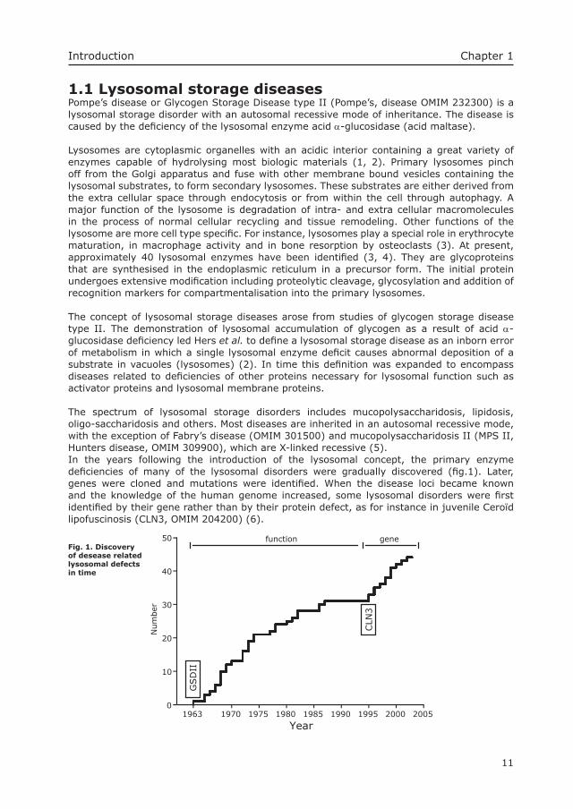
The spectrum of lysosomal storage disorders includes mucopolysaccharidosis, lipidosis, oligo-saccharidosis and others. Most diseases are inherited in an autosomal recessive mode, with the exception of Fabry’s disease (OMIM 301500) and mucopolysaccharidosis II (MPS II, Hunters disease, OMIM 309900), which are X-linked recessive (5). In the years following the introduction of the lysosomal concept, the primary enzyme defi ciencies of many of the lysosomal disorders were gradually discovered (fi g.1). Later, genes were cloned and mutations were identifi ed. When the disease loci became known and the knowledge of the human genome increased, some lysosomal disorders were fi rst identifi ed by their gene rather than by their protein defect, as for instance in juvenile Ceroïd lipofuscinosis (CLN3, OMIM 204200) (6).
0
50
Num
ber
Year
40
30
20
10
1985 20052000199519901980197519701963
CLN
3
GSD
II
function geneFig. 1. Discovery of desease related lysosomal defects in time
Introduction
12
Chapter 1
In lysosomal storage disorders, the affected cell type usually plays a major role in the degradation of the (accumulated) substrate. For example, cerebral white matter is affected in patients with defects in the degradation of sphingolipids. Connective tissue is involved in mucopolysaccharide storage disorders. The symptoms are obviously related to the site of accumulated undegradable materials, but the exact cause of cell death or dysfunction is often unclear. All the disorders are progressive and can be fatal. Defi nitive diagnosis is accomplished by measuring specifi c enzyme activities or function in leukocytes or cultured skin fi broblasts or other tissue specimens, selected on the basis of clinical symptoms. Light or electron microscopy, and DNA analysis can confi rm the diagnosis. There is extensive heterogeneity within each of the disorders, exemplifi ed by infantile, juvenile or adult onset of symptoms, mostly related to residual (enzyme) function. The different types of mutations within the same gene totally eliminating or partially reducing the enzyme activity or lysosomal protein function explain this. In addition, expression of modifying genes also infl uences the phenotype (7, 8).
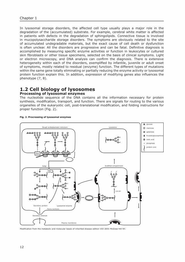
1.2 Cell biology of lysosomesProcessing of lysosomal enzymesThe nucleotide sequence of the DNA contains all the information necessary for protein synthesis, modifi cation, transport, and function. There are signals for routing to the various organelles of the eukaryotic cell, post-translational modifi cation, and folding instructions for proper function (Fig. 2).
Fig. 2. Procressing of lysosomal enzymes
P
Secretory protein
Secretory Granule
Lysosomal enzyme
Low pH
Acidified compartment
Plasma membrane
P
P
P
P
P
P
P
P
P P
P
P P
Dol
UDP
Golgi
Rough endoplasmatic reticulum Lysosome
Plasma membrane
UMP
P
glucose
mannose
galactose
N-acetylglucosamine
sialic acid
phosphate
protein core
Modification from the metabolic and molecular bases of inherited disease edition VIII 2001 McGraw-Hill NY.
13
Introduction
Glycoproteins, such as lysosomal enzymes undergo several modifying steps before transport to their functional location. An amino-terminal signal peptide (signal recognition sequence) leads to co-translational segregation into the lumen of the Endoplasmic Reticulum (ER). There are other peptide sequences that guide newly synthesised proteins to the nucleus, the peroxisomes, or the mitochondria. Proteins without a signal sequence remain in the cytoplasm (9-11). The glycoproteins receive their carbohydrate moieties co-translationally in the ER and are further modifi ed en route to the Golgi complex, the Trans Golgi Reticulum, and from there to either the lysosome, the plasma membrane, or the extracellular space (secretory proteins).As soon as the signal sequence extends from the ribosome it is recognised by a protein complex known as the signal recognition particle (SRP), and translation is transiently arrested (9-11). The protein complex targets to the ER where it is attached to the SRP-receptor at the ER membrane. By docking at this translocation site at the ER membrane the SRP is released and ribosomal activity is restored.
In the rough ER, co-translational modifi cations occur. An oligosaccharide complex of N-acetyl glucosamine, mannose and glucose is transferred from a lipid carrier to the NH2 group of an asparagine residue of the protein in the context of Asp-Xxx-Asn (12). This process is called N-linked glycosylation (13, 14). In contrast to N-linked glycosylation, oligosaccharides can also be linked to the hydroxyl-group of a serine or threonine residue (O-linked glycosylation). No lipid intermediate is required. All post-translational modifi cations have an effect on the three-dimensional structure of the protein and thereby on the stability, function and interaction with other proteins. The process of protein folding is controlled by a group of proteins functioning as chaperones, whereby incomplete or miss-folded proteins are detected and eliminated (15). When glycoproteins have the correct glycosylation and three-dimensional structure, they are allowed to leave the rough ER and enter the cis-Golgi cisternae of the Golgi complex.Lysosomal enzymes contain Asparagine-linked oligosaccharide chains that can be divided into three groups: high mannose, hybrid and complex. All three have a common core structure. The outer branches determine the differences. The common carbohydrate precursor contains three outer glucose residues linked to dolichol pyrophosphate, and is transferred en bloc to an asparagine residue of the protein. Subsequently, the fi rst glucose is removed by glucosidase I. Glucosidase II removes the remaining other two glucose molecules. A high mannose type of chain remains when processing stops at this point.A complex type of chain is formed when four mannose residues are cleaved off by mannosidase I (12) and N-acetylglucosamine is added by N-acetylglucosamine-transferase (16). Mannosidase II then cleaves off two other mannose residues, and a second N-acetylglucosamine is transferred to the mannose residues. Sequential addition of galactose, fucose, and sialic acid completes the synthesis of these complex types of oligosaccharide side chains. When an N-acetylglucosamine is transferred to the β-linked mannose residue, the two outer mannose residues cannot be removed by mannosidase II, and a hybrid type of oligosaccharide chain is created after linkage of galactose and sialic acid residues. Most lysosomal glycoproteins have at least one phosphorylated high mannose type of carbohydrate chain. The phosphorylation is a two-step process. First, N-acetyl- glucosamine 1-phosphate is transferred from UDP-N-acetyl glucosamine. Second, the N-acetyl glucosamine residue is cleaved off exposing the so-called mannose 6-phosphate recognition marker (17).
Routing of lysosomal enzymes to the lysosomeNewly formed lysosomal proteins with the M6P marker are bound by the mannose 6-phosphate receptor and gather as complexes in vesicles that pinch off from the Trans Golgi Network. These vesicles have clathrin as their major structural membrane protein and are therefore called Clathrin Coated Vesicles (CCV’s). There are different types of CCV’s, distinguishable by their assembly proteins (AP’s). The ones that pinch off from the Trans Golgi
14
Chapter 1
Network are associated with AP-1 complex assembly proteins. The ones that are involved in transport from the cell surface to the lysosomes are associated with AP-2 (18, 19). The mannose 6-phosphate receptor involved in the further routing of the lysosomal enzymes is a major constituent of the CCV’s (20).In mammalian cells, mannose 6-phosphate receptors (MPR) are involved in transport of lysosomal enzymes (21). Two distinct MPR’s have been isolated and characterized: (i) the 46 kD cation dependent MPR (CD-MPR) and the 300 kD cation independent MPR (CI-MPR), Which is also the receptor for insulin-like growth factor II (IGFII) (17). The two MPR’s recycle constitutively between the Trans Golgi Network and the endosomal compartment as well as between the endosomal compartment and the cell surface. Only the CI-MPR binds and mediates endocytosis of extra cellular ligands (22). The CD-MPR is known to contain binding sites for AP’s and plays a role in the formation of a lysosomal enzyme specifi c CCV (20). The role of MPR’s in lysosomal targeting of newly synthesised lysosomal enzymes is illustrated in cells from patients with I-cell disease. These patients lack the phosphotransferase needed for the transfer of the N-acetylglucosamine 1-phosphate to the high mannose carbohydrate chain of the lysosomal enzyme. As a consequence, the phosphotransferase defi cient cells secrete the non-phosphorylated lysosomal enzymes excessively and lack them inside so that undigested materials accumulate in the lysosomal system. However, in some tissues from I-cell patients, lysosomal storage is absent and lysosomal activities are normal (23). Further insight in the MPR system was provided by studies in CI-MPR (300 kD) defi cient cells containing normal levels of CD-MPR’s (46 kD). These transfection studies show that re-expression of the CI-MPR can fully restore lysosomal routing while over-expression of the CD-MPR only partially compensates for the loss of the CI-MPR (24-27). Experiments with cells defi cient for both receptors show that sorting can be partially restored by expressing the CD-MPR while it can be fully restored by expressing the CI-MPR.
Receptor mediated endocytosisReceptor mediated endocytosis is defi ned as the uptake of material into a cell by invagination of the plasma membrane, containing ligand-specifi c receptors, followed by the intra-cellular formation of a membrane-bound vesicle. The internalised material is delivered to the lysosomes for breakdown or transported to another destination. The mechanism of receptor mediated endocytosis was nicely demonstrated by Goldstein & Brown in their studies on the cause of atherosclerosis in humans with a strong genetic predisposition. It was shown that cholesterol, needed for membrane synthesis, was internalised via the Low-Density Lipoprotein receptor (28). At present more than 25 receptors are known to be involved in receptor mediated endocytosis, and they all apparently use the same clathrin coated pit pathway.
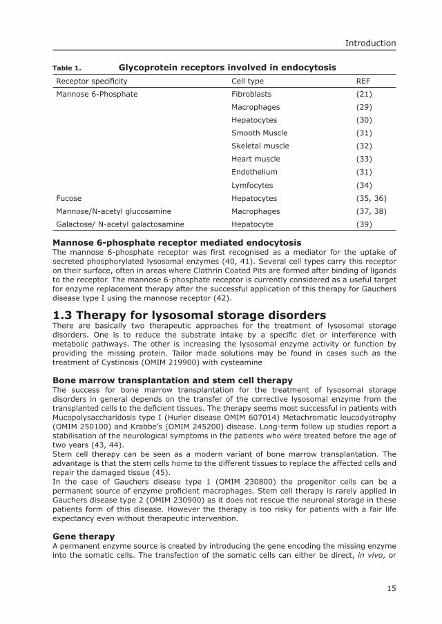
Endocytosis of glycoproteinsSeveral glycoprotein receptors have been identifi ed as mediators of glycoprotein endocytosis (note all lysosomal enzymes are glycoproteins). Glycoprotein receptors involved in endocytosis are shown in Table 1. Most of these receptors do not have an intracellular function as the M6P/ IGFII receptor. Circulating glycoproteins have a complex type of oligosaccharide chain ending with sialic acid residues. These molecules remain in the circulation for a rather long time. When the sialic acid is removed, the exposed galactose is available for the asialoglycoprotein receptor, and these glycoproteins are cleared from the circulation by the hepatocytes.
15
Introduction
Table 1. Glycoprotein receptors involved in endocytosis
Receptor specifi city Cell type REF
Mannose 6-Phosphate Fibroblasts (21)
Macrophages (29)
Hepatocytes (30)
Smooth Muscle (31)
Skeletal muscle (32)
Heart muscle (33)
Endothelium (31)
Lymfocytes (34)
Fucose Hepatocytes (35, 36)
Mannose/N-acetyl glucosamine Macrophages (37, 38)
Galactose/ N-acetyl galactosamine Hepatocyte (39)
Mannose 6-phosphate receptor mediated endocytosisThe mannose 6-phosphate receptor was fi rst recognised as a mediator for the uptake of secreted phosphorylated lysosomal enzymes (40, 41). Several cell types carry this receptor on their surface, often in areas where Clathrin Coated Pits are formed after binding of ligands to the receptor. The mannose 6-phosphate receptor is currently considered as a useful target for enzyme replacement therapy after the successful application of this therapy for Gauchers disease type I using the mannose receptor (42).
1.3 Therapy for lysosomal storage disordersThere are basically two therapeutic approaches for the treatment of lysosomal storage disorders. One is to reduce the substrate intake by a specifi c diet or interference with metabolic pathways. The other is increasing the lysosomal enzyme activity or function by providing the missing protein. Tailor made solutions may be found in cases such as the treatment of Cystinosis (OMIM 219900) with cysteamine
Bone marrow transplantation and stem cell therapyThe success for bone marrow transplantation for the treatment of lysosomal storage disorders in general depends on the transfer of the corrective lysosomal enzyme from the transplanted cells to the defi cient tissues. The therapy seems most successful in patients with Mucopolysaccharidosis type I (Hurler disease OMIM 607014) Metachromatic leucodystrophy (OMIM 250100) and Krabbe’s (OMIM 245200) disease. Long-term follow up studies report a stabilisation of the neurological symptoms in the patients who were treated before the age of two years (43, 44).Stem cell therapy can be seen as a modern variant of bone marrow transplantation. The advantage is that the stem cells home to the different tissues to replace the affected cells and repair the damaged tissue (45). In the case of Gauchers disease type 1 (OMIM 230800) the progenitor cells can be a permanent source of enzyme profi cient macrophages. Stem cell therapy is rarely applied in Gauchers disease type 2 (OMIM 230900) as it does not rescue the neuronal storage in these patients form of this disease. However the therapy is too risky for patients with a fair life expectancy even without therapeutic intervention.
Gene therapyA permanent enzyme source is created by introducing the gene encoding the missing enzyme into the somatic cells. The transfection of the somatic cells can either be direct, in vivo, or
16
Chapter 1
the indirect approach can be taken, that is to transfect the cells ex vivo and transplant them back into the body. These are the theoretical options. The widespread practical application is hampered by the lack of versatile vectors and problems encountered in clinical studies. For instance, the recent treatment of patients with the X-linked form of SCID (OMIM 300400) was successful for some patients but others developed leukemia (46). A patient with Ornithine transcarbamylase (OTC, OMIM 311250) defi ciency succumbed by exposure to an overdose of adeno virus. The ideal vector for gene therapy has not yet been found. All viral vectors have their limitations. Some elicit a restrictive immunologic response when they are used for the second time. Others are potentially harmful when they incorporate into the host-cell DNA. For these reasons non-viral vectors are also developed for the delivery of genes, and transfection effi ciency enhancers are studied. But, this is still in an experimental stage. The HIV-1 TAT protein regulating the transcriptional activation of HIV-1 has been shown to function as a protein transduction domain, penetrating the cell membrane in a manner different from the classical endocytotic route. The TAT peptide presents a nuclear localisation signal, which enables nuclear targeting (47).
Enzyme replacement therapyThe idea to treat lysosomal storage disorders with intravenous enzyme administration was brought up by de Duve shortly after the historic discovery of acid α-glucosidase defi ciency in Pompe’s disease. He suggested that the therapeutic enzyme could possibly reach the lysosome through heterophagy. The fi rst clinical attempt dates from 1964 and was performed in a case of infantile Pompe’s disease with acid α-glucosidase from the fungus Apergilles Niger (48, 49). The longest study by Hug et al. lasted 116 days (50). Initially there was an increase in acid α-glucosidase activity in the liver of the patients but not in the skeletal muscle. The patient then developed an immune nephritis as reaction to the foreign enzyme and eventually died. Several studies followed whereby acid α-glucosidases of different sources were used. Similar pioneering studies were also undertaken in other lysosomal storage disorders in the years 1965-1980 (51-54) but none was successful enough to proof the feasibility of enzyme replacement therapy. On the contrary it became clear that the therapy could not be used in lysosomal storage disorders involving the brain as the corrective enzyme did not cross the blood brain barrier (53). But, new insights in the role of cell surface receptors as effective mediators of endocytosis kept the concept of enzyme replacement therapy alive. Especially the discovery of the mannose and the mannose 6-phosphate receptors (21, 55) and the exchange of lysosomal enzymes between cells in culture were important events (41, 56, 57). In 1989 a patient with Gauchers disease type I was the fi rst to benefi t from enzyme replacement therapy. The therapeutic enzyme (glucocerebrosidase) was extracted from human placenta and modifi ed so as to expose mannose residues (58, 59). They could serve as a ligand for the mannose receptor to target the enzyme to the Kupfer cells and tissue macrophages that are the major sites of storage in Gaucher’s disease.In the same period, enzyme replacement for Fabry disease was developed. Single dose studies proved that intravenously infused enzyme could decrease the level of accumulated plasma Globotraiosylceramide. Phase I and II clinical trials proved the safety and effi cacy of enzyme replacement therapy for both Gauchers and Fabry disease but further research has to be conducted to optimize the therapy. In collaboration with the pharmaceutical industry both medicines are now registered for the treatment of these two lysosomal storage disorders. The development of ERT for other LSD’s like Niemann-Pick, Mucopolysaccharidosis type I and Pompe’s disease is well on its way (60, 61).
1.4 Pompe’s diseasePompe’s disease or Glycogen storage disease type II (GSDII, OMIM 232300) is an autosomal recessive myopathy caused by mutations in the acid α-glucosidase gene. The resulting total or partial defi ciency of lysosomal acid α-glucosidase causes glycogen to accumulate in lysosomes (62). In 1932 the most severe phenotype was described by the Dutch pathologist, Dr. J. C. Pompe, who reported about a girl of 7 months old with idiopathic hypertrophy of the
17
Introduction
heart (63). Massive vacuolar glycogen accumulation was histologically demonstrated in many organs and tissues. Later, milder phenotypes were also described, and early infantile, and late onset (juvenile and adult) forms were distinguished (64-68).
Infantile Pompe’s diseasePatients with infantile Pompe’s disease are the most severely affected. They have a rapidly progressive generalised hypotonia with severe muscle weakness and wasting resulting in a poor motor development. Symptoms develop within the fi rst months of life. Cardiac enlargement is characteristically manifested on an X-ray of the thorax, and hepatomegaly and macroglossia are often found on further physical examination. Echocardiography reveals a hypertrophy of both the ventricular (posterior) walls as well as the intra-ventricular septum. In addition, a left ventricular outfl ow tract obstruction may be present (69-72). Electrocardiographic fi ndings confi rm the ventricular hypertrophy (large QRS-complexes), and a shortening of the PR intervals is often found (73) Cardio-respiratory problems are common and the usual cause of death in the fi rst year of life.Patients with the infantile form of Pompe’s disease have virtually no acid α-glucosidase activity. Glycogen accumulation is found in skeletal muscle, heart, liver, spleen, kidneys, vascular smooth muscle, skin, endothelium, and Schwann-cells surrounding the peripheral nerves (62, 74-81). In addition, glycogen storage was found in post-mortem brain (74, 80). Lysosomal glycogen storage was most prominently present in the anterior horns, the motor nuclei of the brainstem and the spinal ganglia. Some thalamic nuclei were more involved in the disease process than others and contained balloon shaped neurones while others were spared. Cortical neurones showed very slight to no storage at all, and myelinisation appeared to be normal.
Late onset Pompe’s diseasePatients with late onset Pompe’s disease develop a proximal myopathy. Children with the disease usually present a delayed motor development or Gower’s sign. Older patients typically complain of diffi culties in climbing stairs and rising from a chair (82, 83). Some patients have respiratory diffi culties as initial symptom. There are no clinical signs of cardiac involvement. The symptoms may resemble those of a limb girdle muscular dystrophy or polymyositis, which may lead to misdiagnosis (82, 84). The age of onset of late onset Pompe disease varies greatly. In general, patients with an early onset of disease are more severely affected and decline more rapidly than patients with a later manifestation.When the disease progresses, patients may become wheelchair dependent and require artifi cial ventilation. Respiratory failure is the major cause of death.
The acid α-glucosidase defi ciency in late onset Pompe’s disease is usually less profound than in infants. Nevertheless, glycogen storage has been reported in many organs. Histological examination revealed vacuolisation of cardiac muscle cells, (not leading to clinical symptoms), nerve bundles (Schwann cells and perineurium), endothelial cells and smooth muscle cells. Lysosomal accumulation in the latter cell types may attribute to the occurrence of aneurysms of the basilar artery, as was described by Matsui and co-workers (85-89). Enlargement of the tongue has been reported in some cases of late onset Pompe’s disease (67, 90).The heterogeneous course of late onset Pompe’s disease has led to a further sub-typing of adult, juvenile, childhood, and non-classical infantile forms of Pompe’s disease (91). The latter classifi cation is somewhat arbitrary as Pompe’s disease encompasses in fact a clinical spectrum of subtypes with the classic infantile form on the one side and the adult phenotype at the other.
18
Chapter 1
DiagnosisClinical DiagnosisA thorough clinical examination may raise the suspicion of Pompe’s disease, but is not suffi cient to establish the diagnosis. Additional laboratory tests, such as the measurement of serum CK, ALAT, ASAT and LDH, which levels are usually increased, may be helpful in setting the diagnosis (82, 92, 93). An EMG may reveal abnormal patterns with repetitive discharges and positive waves as a sign of muscle pathology (94-96).Microscopic sections of the muscle show “purple” membrane bound deposits of glycogen within the muscle fi bres if the material is properly processed, fi xed with glutaraldehyde and stained with Periodic Acid Schiff (PAS). The glycogen is washed out if microscopic sections are not properly handled and a lacework pattern appears (97, 98). Glycogen storage is more pronounced in infantile than in the late onset forms of the disease, as is the rate of progression. In particular, in patients with an adult onset of disease the glycogen accumulation may vary between fi bers and muscle groups. Some muscle biopsies of mildly affected patients may show no microscopical abnormalities, and the diagnosis can be missed. A defi nite diagnosis for Pompe’s disease can be made by demonstrating a defi ciency of α-glucosidase activity or occurrence of a pathogenic mutation in each of the two acid a-glucosidase alleles. Enzyme diagnosisAcid α-glucosidase hydrolyses the α-1,4 and α-1,6 glycosidic linkages of glycogen to release glucose. The activity of the enzyme can be measured with the natural substrate glycogen or the synthetic substrate 4-Methylumbelliferyl-α-D-glucopyranoside. For diagnostic purposes the enzyme activity is preferably measured in fi broblasts or muscle tissue.Determination of acid α-glucosidase activity in leukocytes is not recommended because neutral α-glucosidases interfere with the measurement causing an overlapof the enzyme activity levels between patients and controls (99, 100). Patients with infantile Pompe’s disease have a complete enzyme defi ciency whereas patients with the late onset form of the disease have residual activities up to 25% of normal. In general the clinical subtype correlates well with the level of residual enzyme activity, but unusually low acid α-glucosidase activities have been reported in a few cases of adult onset disease. Also non-classical infantile and childhood Pompe’s disease cannot always be distinguished from the classic infantile type of disease on the basis of enzyme activity. In addition, there are mutations which affect the activity for the natural substrate more than for the artifi cial substrate (62, 101). Carrier detection is unreliable via enzyme activity measurement.
DNA diagnosisThe acid α-glucosidase gene (GAA) was mapped on chromosome 17, region q25.3 (102). It is approximately 20 kb long and contains 20 exons. The gene is transcribed and spliced into an m-RNA of 3.6 kb with 2856 bp of coding sequence (103). The acid α-glucosidase polypeptide contains 952 amino acids (104, 105). The mutations are equally distributed over all the coding exons (2-20). Deletions, insertions, missense and splice site mutations were found. More than 100 mutations are presently known in the public domain (www.Pompecenter.nl).The most common mutation world-wide is IVS1(-13T−>G). It causes aberrant splicing of the fi rst coding exon (exon 2) in 80-90% of the splicing events. Some mutations specifi cally occur in certain ethnic groups. For example, the deletion of exon 18 is quite common in the Caucasian Dutch sub-population and in the southern part of Italy, while the delT525 mutation in exon 2 is common in Western Europe and in the French-Canadian population (106). Both mutations lead to a total defi ciency of acid α-glucosidase. The C1935A (exon 14) transition is a frequent mutation in Taiwanese and Chinese populations and also leads to a total defi ciency of enzyme activity (107). Different strategies can be taken for mutation analysis. Certain subgroups of patients can be screened fi rst for the presence of frequent mutations, but otherwise it is quicker to sequence the whole gene. The fi nding of a known mutation is immediately informative, but new
19
Introduction
mutations have to be judged for their effect. Non-sense and frame-shift mutations have a predictable fully deleterious effect. Missense mutations can be introduced in the normal c-DNA construct and expressed in vitro to estimate their effect. It is most informative to measure not only the enzyme activity of the acid α-glucosidase that is generated by the transfection of the cDNA construct but also to analyse the biosynthesis of the enzyme by Western blotting or pulse-chase labeling. The more than 100 mutations were seen in a variety of allelic combinations in the clinically heterogeneous group of patients with Pompe’ s disease. Combining all information, there appears to be a correlation between the genotype and the phenotype, which makes DNA analysis useful for diagnositic and prognostic purposes. For instance, patients who are heterozygous for the common IVS1 (-13T->G) mutation have late onset Pompe’s disease and a slowly progressive myopathy. Only patients with two fully deleterious mutations have the classic infantile phenotype.DNA analysis is usually not necessary to confi rm the diagnosis, but is mandatory for carrier detection and genetic counseling of family members. If diagnostic material of a deceased patient is unavailable, DNA analysis in the parents may be required for setting a diagnosis in retrospect.
1.5 Animal modelsThere are several animal models of Pompe’s disease. They were used to study pathological features and therapeutic options. Lysosomal glycogen storage was found in Lapland dogs, Corriedale sheep, Nicholas turkey, cat, Japanese Quail and Brahman and Shorthorn cattle (108-119). Not all natural animal models parallel the human condition with respect to the clinical parameters. Cardiac enlargement for example was reported in a sub-population of Shorthorn cattle, dogs and turkeys but not in cats, sheep and quails. The Japanese quail and the artifi cially made mouse models were used most extensively. The quail model, mimicking the late onset form of Pompe’s disease, has a residual activity of approximately 10-16 % and manifests overt clinical symptoms at 6-12 weeks after hatching (116, 117). Importantly, the quail has reached its reproductive age by approximately 12 weeks. Shortly after hatching, affected quails still can return to a standing position after being placed in supine position with their wings extended. Six months after hatching their wing extension is impaired due to joint contractures. The average life span is reported to be 30 months. Affected animals may have a great variation of glycogen storage in their skeletal muscle, like in patients with late onset GSDII.
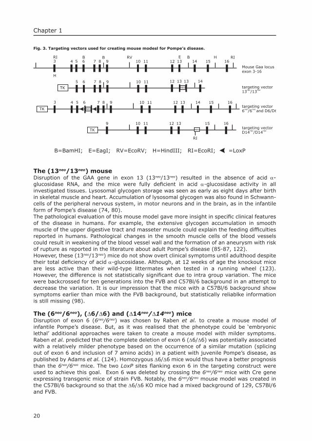
Mouse modelsTo have an easy to handle animal model for the infantile form of Pompe’s disease, knockout mouse models were made by disrupting the murine acid α-glucosidase gene in embryonic stem cells of mouse strain 129 (97, 120, 121). Different targeting vectors were used (Fig.3). Neo cassettes were inserted in exon 13 (13neo/13neo) (97) and in exon 6 with fl anking loxP sites (6neo/6neo and ∆6/∆6), and exon 14 was replaced by a neo cassette (∆14neo/∆14neo) (120).
20
Chapter 1
Fig. 3. Targeting vectors used for creating mouse modesl for Pompe’s disease.
43
5 6 7 8 9 10 11
12 13BRI
H
RIH14 15 16
43 5 6 7 8 9 10 11 12 13 14 15 16targeting vector6neo/6neo and D6/D6
targeting vector13neo/13neo
12 13 14
9 10 11 12 13 15 16
TK
NEOTK
NEO
RI
NEO
13
TK
5 6 7 8 9 10 11RVB B
B=BamHI; E=EagI; RV=EcoRV; H=HindIII; RI=EcoRI; =LoxP
E
targeting vectorD14neo/D14neo
Mouse Gaa locusexon 3-16
The (13neo/13neo) mouseDisruption of the GAA gene in exon 13 (13neo/13neo) resulted in the absence of acid α-glucosidase RNA, and the mice were fully defi cient in acid α-glucosidase activity in all investigated tissues. Lysosomal glycogen storage was seen as early as eight days after birth in skeletal muscle and heart. Accumulation of lysosomal glycogen was also found in Schwann-cells of the peripheral nervous system, in motor neurons and in the brain, as in the infantile form of Pompe’s disease (74, 80). The pathological evaluation of this mouse model gave more insight in specifi c clinical features of the disease in humans. For example, the extensive glycogen accumulation in smooth muscle of the upper digestive tract and masseter muscle could explain the feeding diffi culties reported in humans. Pathological changes in the smooth muscle cells of the blood vessels could result in weakening of the blood vessel wall and the formation of an aneurysm with risk of rupture as reported in the literature about adult Pompe’s disease (85-87, 122). However, these (13neo/13neo) mice do not show overt clinical symptoms until adulthood despite their total defi ciency of acid α-glucosidase. Although, at 12 weeks of age the knockout mice are less active than their wild-type littermates when tested in a running wheel (123). However, the difference is not statistically signifi cant due to intra group variation. The mice were backcrossed for ten generations into the FVB and C57Bl/6 background in an attempt to decrease the variation. It is our impression that the mice with a C57Bl/6 background show symptoms earlier than mice with the FVB background, but statistically reliablke information is still missing (98).
The (6neo/6neo), (∆6/∆6) and (∆14neo/∆14neo) miceDisruption of exon 6 (6neo/6neo) was chosen by Raben et al. to create a mouse model of infantile Pompe’s disease. But, as it was realised that the phenotype could be ‘embryonic lethal’ additional approaches were taken to create a mouse model with milder symptoms. Raben et al. predicted that the complete deletion of exon 6 (∆6/∆6) was potentially associated with a relatively milder phenotype based on the occurrence of a similar mutation (splicing out of exon 6 and inclusion of 7 amino acids) in a patient with juvenile Pompe’s disease, as published by Adams et al. (124). Homozygous ∆6/∆6 mice would thus have a better prognosis than the 6neo/6neo mice. The two LoxP sites fl anking exon 6 in the targeting construct were used to achieve this goal. Exon 6 was deleted by crossing the 6neo/6neo mice with Cre gene expressing transgenic mice of strain FVB. Notably, the 6neo/6neo mouse model was created in the C57Bl/6 background so that the ∆6/∆6 KO mice had a mixed background of 129, C57Bl/6 and FVB.
21
Introduction
The 6neo/6neo and ∆6/∆6 models turned out to be similar with respect to the acid α-glucosidase activity in skin, liver, and heart, and the lysosomal glycogen storage in skeletal muscle, heart and diaphragm. In an open-fi eld setting, the 6neo/6neo mice were less active than the ∆6/∆6 mice (36% and 91 %, respectively, compared to heterozygous littermates; ∆6/+ mice). The heterozygous 6neo/+ mice were reported to perform at the 50-70 % level of the heterozygous ∆6/+ mice(120). Based on the combination of all fi ndings it was concluded that the two strategies for knocking out exon 6 had been equally deleterious. The difference in performance was ascribed to the different genetic background of the mice.
To further investigate the infl uence of the genetic background on the phenotype of KO mice with Pompe’s disease, yet another model was created (121). Exon 14 was replaced by a neo cassette (∆14neo/∆14neo). The resulting mice with a 129/C57Bl/6 background were in all aspects very similar to the 6neo/6neo mice. Unfortunately the inbred C57Bl/6 were not available for testing.Table 2 summarises the age of onset of clinical symptoms in the various models. Notably, there does not seem to be a signifi cant difference in age of onset when comparing the 6neo/6neo, ∆6/∆6 and ∆14neo/∆14neo mice with the earlier published 13neo/13neo mice.
Table 2. Onset of clinical symptoms in three mouse models of Pompe’s disease
Strain Background Males (months) Females (months)
13neo/13neo FVB inbred 7.4 (n = 8) 7.4 (n=8)
6neo/6neo 129xC57Bl/6 8.1 ± 0.3 (n = 19) 7.1 ± 0.3 (n = 29)
∆6/∆6 129xC57Bl/6xFVB 9.8 ± 0.4 (n = 11) 9.0 ± 0.6 (n = 13)
∆14neo/∆14neo 129xC57Bl/6 8.3 ± 0.8 (n = 14) 6.6 ± 0.4 (n = 14)
The relatively slow progression of clinical signs in these four mouse models is not ideal for experimental purposes. Therefore, it was attempted to improve the model by crossing the 6neo/6neo KO mice with transgenic mice overexpressing either the human GlutI glucose transporter or the enzyme glycogen synthase (GS). These transgenic mice have a signifi cantly elevated level of cytoplasmic glucose (GlutI) or glycogen (GS) compared to wild type mice (125, 126). The knockout mice with either the GS or GlutI transgene have an increased lysosomal glycogen storage and develop clinical symptoms at the age of 4-5 months. By 9 months of age their phenotype is clinically indistinguishable from the knockout mice without the Glut I or GS transgene (127).
Another potential improvement of the KO mouse model of Pompe’s disease was the introduction of an α-glucosidase transgene with very low expression, exclusively in the liver (128). The purpose of this modifi cation was to prevent an antibody response against recombinant human acid α-glucosidase in experiments related to the application of enzyme replacement therapy. As anticipated, the response was markedly diminished.
1.6 Therapy for Pompe’s diseaseWith present day knowledge, the therapeutic options for Pompe’s disease are limited. Bone marrow transplantation was attempted but the results of the experiments were disappointing. No increase of acid α-glucosidase activity was detected in the muscles of treated patients, and the method is no longer applied for the treatment of Pompe’s disease (129, 130). There are several reports in which a high protein diet with branched chain amino acids or a supplement of L-alanine is advised to help restore the net protein balance and thereby prevent muscle breakdown. The studies report improvement of function of respiratory and skeletal
22
Chapter 1
muscles in approximately 25% of cases (93, 131-133). Daily exercise may also improve the condition of the patient but should not be exhaustive. The variation of disease severity and clinical course of patients with Pompe’s disease complicates the evaluation of the effect of any form of therapy.At present ERT is the most promising as the preliminary results of the clinical trials suggest that patients with both the early as well as the late onset form of disease can benefi t from this type of therapy. The application of gene therapy is still in the pre-clinical phase and dependent on the production of safe and effective vectors for gene delivery.
Gene therapyAmalfi tano et al. and Pauly et al. (134, 135) showed that a single intravenous injection of a modifi ed adenoviral vector, expressing human acid α-glucosidase, resulted in effi cient hepatic transduction and expression of acid α-glucosidase. High levels of acid α-glucosisdase activity were also found in the plasma of the treated animals. The plasma enzyme secreted by the hepatocytes can potentially restore the enzyme defi ciency in other tissues via endocytosis. Local administration of adenoviral gene constructs by intra-muscular injection in three days old mice led to a 50-fold elevation of acid α-glucosidase activity in the muscles at the site of injection. Importantly the vector RNA was also detected in the hind limb muscle, the heart and the liver for as long as 6 months (136). A similar experiment was performed in acid maltase defi cient quails (137) and the results were more or less the same.
Intra-muscular and intra-cardial delivery of acid α-glucosidase gene constructs with a recombinant adeno-associated vector resulted in near normal enzyme activities and at least a partial restoration of muscle strength in the soleus muscle of KO mice up to 6 months after treatment (138). Martin-Touaux et al. performed a similar experiment injecting an acid α-glucosidase expressing adenoviral vector in the gastrocnemius muscle of KO mouse neonates. Strong expression was detected at the site of injection but activity was also found in heart and more distant skeletal muscles.All gene therapy studies use a single injection while expression of the gene is still transient. The problem of immune response arising with repeated dosing, especially when using adenovirus-derived vectors, has to be overcome.
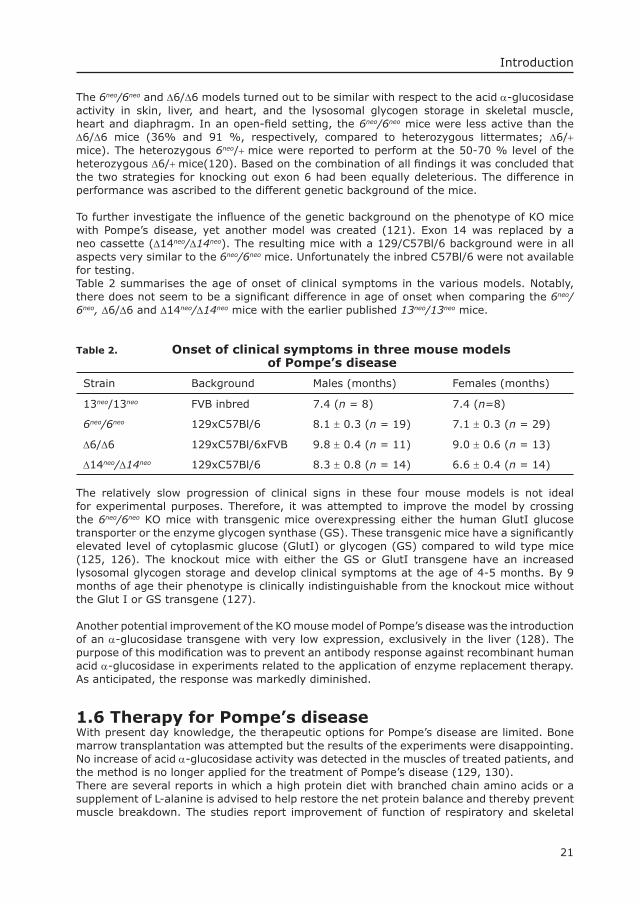
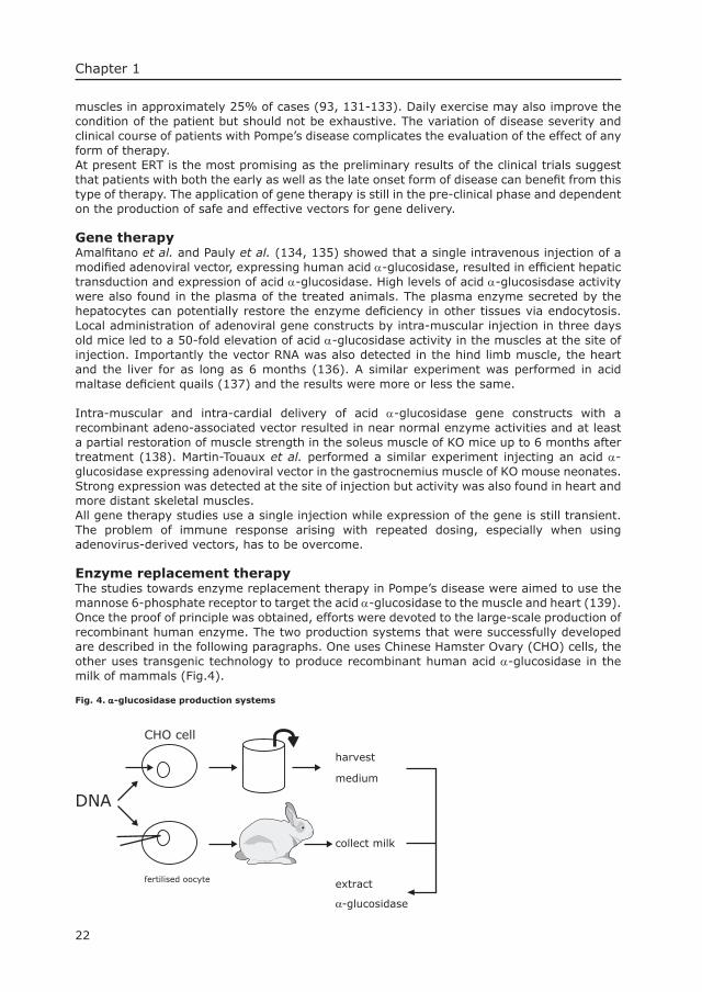
Enzyme replacement therapyThe studies towards enzyme replacement therapy in Pompe’s disease were aimed to use the mannose 6-phosphate receptor to target the acid α-glucosidase to the muscle and heart (139). Once the proof of principle was obtained, efforts were devoted to the large-scale production of recombinant human enzyme. The two production systems that were successfully developed are described in the following paragraphs. One uses Chinese Hamster Ovary (CHO) cells, the other uses transgenic technology to produce recombinant human acid α-glucosidase in the milk of mammals (Fig.4).
Fig. 4. α-glucosidase production systems
DNA
CHO cell
fertilised oocyte
harvest
medium
collect milk
extract
α-glucosidase
23
Introduction
Acid α-glucosidase production in CHO cellsChinese Hamster Ovary cells (CHO) are commonly used for the production of recombinant enzymes and are therefore a logical option for the production of human recombinant acid α-glucosidase. Several gene promoters can be used for this purpose. They are usually linked to the cDNA in a suitable vector system. Fuller et al. used the promotor of the human elongation factor 1α gene (140). A CMV promoter driven expression vector was used by van Hove et al. containing a DHFR gene in tandem with the acid α-glucosidase cDNA (141). This type of vector allows for metrotrexate-induced amplifi cation of the construct. Under maximal stimulation and optimal circumstances, a production capacity of 91.4 µgr/ml secreted acid α-glucosidase over a period of 24 hours was reached in a laboratory setting. The enzyme collected was in all aspects very similar to the recombinant human acid α-glucosidase reported by Fuller et al. (140). The uptake in fi broblasts was mannose 6-phosphate dependent (141). The enzyme was also tested in the quail model for Pompe’s disease. Normal levels of acid α-glucosidase activity were reached in the spleen with either a high (14 mg/kg) or a low (4.2 mg/kg) dose (142). Martiniuk et al. also reported the production of recombinant human acid α-glucosidase in CHO cells but did not harvest the enzyme from the medium but from the cells (143). Fibroblasts of patients with Pompe’s disease were corrected by the administration of the enzyme to the culture medium. Intra-peritoneal administration of the enzyme preparation to KO mice (6neo/6neo) resulted in an increase of acid α-glucosidase activity to heterozygous levels in skeletal-muscle, heart, and diaphragm after two intra-peritoneal infusions over 10 days in a dose of approximately 1 mg/kg. After three infusions improvement of motor activity was reported (143).
Production in transgenic animalsInvestigations into the large-scale production of acid α-glucosidase in transgenic animals started with the expression of cDNA encoded acid α-glucosidase in the milk of transgenic mice (144). To regulate expression of the acid α-glucosidase gene constructs, they were placed under control of a bovine casein gene promoter. This way a high expression was expected in the epithelial cells of the mammary gland. The gene constructs were injected into the pronucleus of fertilised oocytes and the embryos were transferred to the uterus of foster mothers to further develop into transgenic animals. The transgenic females produce acid α-glucosidase in the mammary gland during lactation and the product is secreted in the milk from which it can be extracted. The expression was demonstrated to be mammary gland specifi c. The mouse milk contained two acid α-glucosidase isoforms of 110 and 76 kD, and the therapeutic potential was tested in fi broblasts and KO mice (144, 145). The 110 kD precursor was taken up in a mannose 6-phosphate dependent manner and cleared lysosomal glycogen from cultured fi broblasts of patients with Pompe’s disease.
On the way to large scale production and clinical testing, the recombinant human acid α-glucosidase was produced in the milk of transgenic rabbits using the same bovine αS1 casein expression cassette as in the mouse model (146). The procedure was very successful. The rabbits produced up to 8-gr recombinant human acid α-glucosidase per litre milk. The 110 kD product was effective in the long-term treatment of KO mice with Pompe’s disease (13neo/13neo). The stored glycogen was degraded in a variety of tissues including the target organs (heart, skeletal muscle and smooth muscle). But the therapeutic enzyme did not reach the brain (146). While these pre-clinical studies in the mouse model were ongoing, the Dutch biotechnology company Pharming B.V. developed a safe product for clinical testing. After completion of the toxicology studies in animals and phase I studies in healthy volunteers, the fi rst clinical trial of enzyme replacement therapy for Pompe’s disease started in 1999 in the Sophia Children’s hospital.
24
Chapter 1
1.7 ScopeThe studies described in this thesis were aimed to further develop enzyme replacement therapy for Pompe’s disease. The potential effi cacy of this therapy had been demonstrated in knockout mice but not yet in patients. Four patients with the infantile form of Pompe’s disease were included in the fi rst clinical trial that started in 1999. The purpose of this trial was to study the long-term safety and effi cacy of repeated intravenous administrations of recombinant human acid α-glucosidase from rabbit milk. Survival was chosen as the primary clinical end point and several clinical and morphological investigations were done at regular intervals to monitor the effect of treatment. Chapter 3 describes the three-year follow up of the patients and Chapter 4 presents in more detail the morphological changes that occurred in muscle tissue in the fi rst 72 weeks of treatment. The mouse model of Pompe’s disease was employed to investigate the cause of hearing loss (Chapter 5) that was detected serendipitously when the infants were clinically examined before inclusion. Chapter 2 gives a detailed description of the cardiac pathophysiology in knock-out mice. These latter investigations were undertaken to develop a non-invasive method to follow the effect of therapeutic interventions in mice.During the clinical studies in humans questions arose about the height of the dose and the optimal dosing regimen. These molecular aspects of enzyme replacement therapy were also studied in the knockout mouse model and are described in Chapter 6.The combined studies described in this thesis encourage the further development of enzyme replacement therapy for Pompe’s disease and illustrate how the mouse model can be used to explore ways to improve the effect of treatment.
References1. De Duve, C., et al., Tissue fractionation studies. 6. Intracellular distribution patterns of enzymes in rat-liver
tissue. Biochem. J., 1955. 60: p. 604-617.2. Hers, H.G., α-Glucosidase defi ciency in generalized glycogen storage disease (Pompe’s disease). Biochem.
J., 1963. 86: p. 11-16.3. Walkley, S.U., Cellular pathology of lysosomal storage disorders. Brain Pathol., 1998. 8: p. 175-193.4. Gieselmann, V., Lysosomal storage diseases. Biochim. Biophys. Acta, 1995. 1270: p. 103-136.5. d’Azzo, A.A., G;Strisciuglio, P.; Galjaard, H., Galactosialidosis in The Metabolic & Inherited Bases of Inherited
Disease, ed. C.R.B. Scriver, A.L.; Valle, D.; Sly, W. 2001, New York: McGraw-Hill.6. Isolation of a novel gene underlying Batten disease, CLN3. The International Batten Disease Consortium. Cell,
1995. 82(6): p. 949-57.7. Huie, M.L., et al., Glycogen storage disease type II: identifi cation of four novel missense mutations (D645N,
G648S, R672W, R672Q) and two insertions/deletions in the acid alpha-glucosidase locus of patients of differing phenotype. Biochem Biophys Res Commun, 1998. 244(3): p. 921-7.
8. Huie, M.L., et al., Increased occurrence of cleft lip in glycogen storage disease type II (GSDII): exclusion of a contiguous gene syndrome in two patients by presence of intragenic mutations including a novel nonsense mutation Gln58Stop. Am J Med Genet, 1999. 85(1): p. 5-8.
9. Walter, P., I. Ibrahimi, and G. Blobel, Translocation of proteins across the endoplasmic reticulum. I. Signal recognition protein (SRP) binds to in-vitro-assembled polysomes synthesizing secretory protein. J Cell Biol, 1981. 91(2 Pt 1): p. 545-50.
10. Walter, P. and G. Blobel, Translocation of proteins across the endoplasmic reticulum. II. Signal recognition protein (SRP) mediates the selective binding to microsomal membranes of in-vitro-assembled polysomes synthesizing secretory protein. J Cell Biol, 1981. 91(2 Pt 1): p. 551-6.
11. Walter, P. and G. Blobel, Translocation of proteins across the endoplasmic reticulum III. Signal recognition protein (SRP) causes signal sequence-dependent and site-specifi c arrest of chain elongation that is released by microsomal membranes. J Cell Biol, 1981. 91(2 Pt 1): p. 557-61.
12. Kornfeld, R. and S. Kornfeld, Assembly of asparagine-linked oligosaccharides. Annu Rev Biochem, 1985. 54: p. 631-64.
13. Elbein, A.D., Inhibitors of the biosynthesis and processing of N-linked oligosaccharides. CRC Crit Rev Biochem, 1984. 16(1): p. 21-49.
14. Fuhrmann, U., E. Bause, and H. Ploegh, Inhibitors of oligosaccharide processing. Biochim Biophys Acta, 1985. 825(2): p. 95-110.
15. Ellis, R.J. and S.M. Hemmingsen, Molecular chaperones: proteins essential for the biogenesis of some macromolecular structures. Trends Biochem Sci, 1989. 14(8): p. 339-42.
25
Introduction
16. Reitman, M.L. and S. Kornfeld, UDP-N-acetylglucosamine:glycoprotein N-acetylglucosamine-1-phosphotransferase. Proposed enzyme for the phosphorylation of the high mannose oligosaccharide units of lysosomal enzymes. J. Biol. Chem., 1981. 256: p. 4275-4281.
17. Kornfeld, S., Structure and function of the mannose 6-phosphate/insulinlike growth factor II receptors. Annu Rev Biochem, 1992. 61: p. 307-330.
18. Hirst, J. and M.S. Robinson, Clathrin and adaptors. Biochim Biophys Acta, 1998. 1404(1-2): p. 173-93.19. Le Borgne, R. and B. Hofl ack, Protein transport from the secretory to the endocytic pathway in mammalian
cells. Biochim Biophys Acta, 1998. 1404(1-2): p. 195-209.20. Waheed, A., A. Hasilik, and K. von Figura, Processing of the phosphorylated recognition marker in
lysosomal enzymes. Characterization and partial purifi cation of a microsomal alpha-N-acetylglucosaminyl phosphodiesterase. J Biol Chem, 1981. 256(11): p. 5717-21.
21. Kaplan, A., D.T. Achord, and W.S. Sly, Phosphohexosyl components of a lysosomal enzyme are recognized by pinocytosis receptors on human fi broblasts. Proc Natl Acad Sci U S A, 1977. 74(5): p. 2026-30.
22. Dahms, N.M., P. Lobel, and S. Kornfeld, Mannose 6-phosphate receptors and lysosomal enzyme targeting. J Biol Chem, 1989. 264(21): p. 12115-8.
23. Owada, M. and E.F. Neufeld, Is there a mechanism for introducing acid hydrolases into liver lysosomes that is independent of mannose 6-phosphate recognition? Evidence from I-cell disease. Biochem Biophys Res Commun, 1982. 105(3): p. 814-20.
24. Lobel, P., et al., Mutations in the cytoplasmic domain of the 275 kd mannose 6-phosphate receptor differentially alter lysosomal enzyme sorting and endocytosis. Cell, 1989. 57(5): p. 787-96.
25. Johnson, K.F. and S. Kornfeld, The cytoplasmic tail of the mannose 6-phosphate/insulin-like growth factor-II receptor has two signals for lysosomal enzyme sorting in the Golgi. J Cell Biol, 1992. 119(2): p. 249-57.
26. Ma, Z.M., J.H. Grubb, and W.S. Sly, Cloning, sequencing, and functional characterization of the murine 46-kDa mannose 6-phosphate receptor. J Biol Chem, 1991. 266(16): p. 10589-95.
27. Watanabe, H., J.H. Grubb, and W.S. Sly, The overexpressed human 46-kDa mannose 6-phosphate receptor mediates endocytosis and sorting of β-glucuronidase. Proc Natl Acad Sci USA, 1990. 87(20): p. 8036-40.
28. Brown, M.S., Y.K. Ho, and J.L. Goldstein, The low-density lipoprotein pathway in human fi broblasts: relation between cell surface receptor binding and endocytosis of low-density lipoprotein. Ann N Y Acad Sci, 1976. 275: p. 244-57.
29. Shepherd, V.L., Macrophage receptors for lysosomal enzymes. Lysosomes in biology and pathology, ed. W. Sly. Vol. 7. 1984, Amsterdam: Elsevier. 83-98.
30. Ullrich, K., et al., Recognition of human urine alpha-N-acetylglucosaminidase by rat hepatocytes. Involvement of receptors specifi c for galactose, mannose 6-phosphate and mannose. Biochem J, 1979. 180(2): p. 413-9.
31. Hasilik, A., B. Voss, and K. Von Figura, Transport and processing of lysosomal enzymes by smooth muscle cells and endothelial cells. Exp Cell Res, 1981. 133(1): p. 23-30.
32. Reuser, A.J., et al., Uptake and stability of human and bovine acid alpha-glucosidase in cultured fi broblasts and skeletal muscle cells from glycogenosis type II patients. Exp Cell Res, 1984. 155(1): p. 178-89.
33. Marjomaki, V.S. and A. Salminen, Characteristics of lysosomal phosphomannosyl-enzyme receptors in the rat heart. Basic Res Cardiol, 1987. 82(3): p. 252-60.
34. Griffi ths, G.M. and Y. Argon, Structure and biogenesis of lytic granules. Curr Top Microbiol Immunol, 1995. 198: p. 39-58.
35. Prieels, J.P., et al., Hepatic receptor that specifi cally binds oligosaccharides containing fucosyl alpha1 leads to 3 N-acetylglucosamine linkages. Proc Natl Acad Sci U S A, 1978. 75(5): p. 2215-9.
36. Furbish, F.S., et al., Fucose plays a role in the clearance and uptake of glucocerebrosidase by rat liver cells. Biochem Biophys Res Commun, 1980. 95(4): p. 1768-74.
37. Stahl, P., et al., Evidence for specifi c recognition sites mediating clearance of lysosomal enzymes in vivo. Proc Natl Acad Sci U S A, 1976. 73(11): p. 4045-9.
38. Achord, D.T., et al., Human beta-glucuronidase: in vivo clearance and in vitro uptake by a glycoprotein recognition system on reticuloendothelial cells. Cell, 1978. 15(1): p. 269-78.
39. Ashwell, G. and A.G. Morell, The role of surface carbohydrates in the hepatic recognition and transport of circulating glycoproteins. Adv Enzymol Relat Areas Mol Biol, 1974. 41(0): p. 99-128.
40. Hickman, S., L.J. Shapiro, and E.F. Neufeld, A recognition marker required for uptake of a lysosomal enzyme by cultured fi broblasts. Biochem Biophys Res Commun, 1974. 57(1): p. 55-61.
41. Sly, W.S., et al., Receptor-mediated uptake of lysosomal enzymes. Prog Clin Biol Res, 1978. 23: p. 547-51.42. Grabowski, G.A., N. Leslie, and R. Wenstrup, Enzyme therapy for Gaucher disease: the fi rst fi ve years. Blood
Rev, 1998. 12: p. 115-133.43. Hoogerbrugge, P.M., et al., Allogeneic bone marrow transplantation for lysosomal storage diseases. The
European Group for Bone Marrow Transplantation. Lancet, 1995. 345(8962): p. 1398-402.44. Hoogerbrugge, P.M. and D. Valerio, Bone marrow transplantatioin and gene therapy for lysosomal storage
diseases. Bone Marrow Transplant., 1998. 21: p. S34-S36.45. Peters, C. and C.G. Steward, Hematopoietic cell transplantation for inherited metabolic diseases: an overview
of outcomes and practice guidelines. Bone Marrow Transplant, 2003. 31(4): p. 229-39.46. Kohn, D.B., M. Sadelain, and J.C. Glorioso, Occurrence of leukaemia following gene therapy of X-linked SCID.
Nat Rev Cancer, 2003. 3(7): p. 477-88.
26
Chapter 1
47. Rudolph, C., et al., Oligomers of the arginine-rich motif of the HIV-1 TAT protein are capable of transferring plasmid DNA into cells. J Biol Chem, 2003. 278(13): p. 11411-8.
48. Hug, G., W.K. Schubert, and G. Chuck. Type II glycogenosis: Treatment with extract of Aspergillus Niger. Abstr. in Clin Res. 1968.
49. Baudhuin, P., H.G. Hers, and H. Loeb, An electron microscopic and biochemical study of type II glycogenosis. Lab. Invest., 1964. 13: p. 1139-1152.
50. Hug, G. and W.K. Schubert, Lysosomes in type II glycogenosis. Changes during administration of extract from Aspergillus niger. J Cell Biol, 1967. 35(1): p. C1-6.
51. de Barsy, T., et al., Enzyme replacement in Pompe disease: an attempt with purifi ed human acid alpha-glucosidase. Birth Defects Orig Artic Ser, 1973. 9(2): p. 184-90.
52. Pentchev, P.G., Enzyme replacement therapy in Gaucher’s and Fabry’s disease. Ann Clin Lab Sci, 1977. 7(3): p. 251-3.
53. Godel, V., Visual functions in Tay Sachs diseased patients following enzyme replacement therapy. Metabolic Ophtalmology, 1978. 2: p. 27-32.
54. Greene, H.L., G. Hug, and W.K. Schubert, Metachromatic leukodystrophy. Treatment with arylsulfatase-A. Arch Neurol, 1969. 20(2): p. 147-53.
55. Stahl, P.D., et al., Evidence for receptor-mediated binding of glycoproteins, glycoconjugates, and lysosomal glycosidases by alveolar macrophages. Proc Natl Acad Sci U S A, 1978. 75(3): p. 1399-403.
56. Sando, G.N. and E.F. Neufeld, Recognition and receptor-mediated uptake of a lysosomal enzyme, α-L-iduronidase, by cultured human fi broblasts. Cell, 1977. 12: p. 619-627.
57. Kornfeld, S., et al., Steps in the phosphorylation of the high mannose oligosaccharides of lysosomal enzymes. Ciba Found Symp, 1982. 92: p. 138-56.
58. Barton, N.W., et al., Replacement therapy for inherited enzyme defi ciency. Macrophage-targeted glucocerebrosidase for Gaucher’s disease. N. Engl. J. Med., 1991. 324(21): p. 1464-1470.
59. Barton, N.W., et al., Therapeutic response to intravenous infusions of glucocerebrosidase in a patient with Gaucher disease. Proc. Natl. Acad. Sci. USA, 1990. 87: p. 1913-1916.
60. Kakkis, E.D., et al., Enzyme replacement therapy in feline mucopolysaccharidosis I. Mol Genet Metab, 2001. 72(3): p. 199-208.
61. Miranda, S.R., et al., Infusion of recombinant human acid sphingomyelinase into niemann-pick disease mice leads to visceral, but not neurological, correction of the pathophysiology. Faseb J, 2000. 14(13): p. 1988-95.
62. Hirschhorn, R. and A.J.J. Reuser, Glycogen Storage Disease type II; acid α-Glucosidase (Acid Maltase) defi ciency, in The Metabolic and Molecular Bases of Inhertited Disease, D.V. M.D., Editor. 2001, Mc Graw-Hill: New York. p. 3389-3420.
63. Pompe, J.C., Over idiopathische hypertrofi e van het hart. Ned Tijdsch Geneesk, 1932. 76: p. 304-311.64. Mekanik, G., R.L. Smith, and R.M. MacLeod, Enzyme patterns in glycogen storage disease type II (Pompe’s
disease). Metabolism, 1966. 15(7): p. 641-8.65. Smith, J., H. Zellweger, and A.K. Afi fi , Muscular form of glycogenosis, type II (Pompe). Neurology, 1967. 17(6):
p. 537-549.66. Swaiman, K.F., W.R. Kennedy, and H.S. Sauls, Late infantile acid maltase defi ciency. Arch Neurol, 1968. 18(6):
p. 642-648.67. Hudgson, P. and J.J. Fulthorpe, The pathology of type II skeletal muscle glycogenosis. A light and electron-
microscopic study. J Pathol, 1975. 116(3): p. 139-47.68. Engel, A.G., Acid maltase defi ciency in adults: studies in four cases of a syndrome which may mimic muscular
dystrophy or other myopathies. Brain, 1970. 93(3): p. 599-616.69. Gussenhoven, W.J., et al., Echocardiographic features in the cardiac type of glycogen storage disease II. Eur
Heart J, 1983. 4(1): p. 41-3.70. Lorber, A. and A.S. Luder, Very early presentation of Pompe’s disease and its cross-sectional echocardiographic
features. Int J Cardiol, 1987. 16(3): p. 311-4.71. Seifert, B.L., et al., Development of obstruction to ventricular outfl ow and impairment of infl ow in glycogen
storage disease of the heart: serial echocardiographic studies from birth to death at 6 months. Am Heart J, 1992. 123(1): p. 239-42.
72. Shapir, Y. and N. Roguin, Echocardiographic fi ndings in Pompe’s disease with left ventricular obstruction. Clin Cardiol, 1985. 8(3): p. 181-5.
73. Hwang, B., et al., Clinical analysis of fi ve infants with glycogen storage disease of the heart--Pompe’s disease. Jpn Heart J, 1986. 27(1): p. 25-34.
74. Mancall, E.L., G.E. Aponte, and R.G. Berry, Pompe’s disease (diffuse glycogenosis) with neuronal storage. J Neuropath Exp Neurol, 1965. 24: p. 85-96.
75. Cardiff, R.D., A histochemical and electron microscopic study of skeletal muscle in a case of Pompe’s disease (glycogenosis II). Pediatrics, 1966. 37(2): p. 249-59.
76. Hug, G. and W.K. Schubert, Glycogenosis type II. Glycogen distribution in tissues. Arch Path, 1967. 84: p. 141-152.77. Garancis, J.C., Type II Glycogenosis; Biochemical and electron microscopic study. Americ J Med, 1968. 44: p.
289-299.78. Gambetti, P., S. DiMauro, and L. Baker, Nervous system in Pompe’s disease. Ultrastructure and biochemistry.
J Neuropathol Exp Neurol, 1971. 30(3): p. 412-30.
27
Introduction
79. Hers, H.G. and T. De Barsy, Type II glycogenosis (acid maltase defi ciency), in Lysosomes and storage diseases, F. Van Hoof, Editor. 1973, Academic Press: New York. p. 197-216.
80. Martin, J.J., et al., Pompe’s disease: an inborn lysosomal disorder with storage of glycogen. A study of brain and striated muscle. Acta Neuropathol, 1973. 23(3): p. 229-244.
81. Sakurai, I., et al., Glycogenosis type II (Pompe). The fourth autopsy case in Japan. Acta Pathol Jpn, 1974. 24(6): p. 829-46.
82. Moufarrej, N.A. and T.E. Bertorini, Respiratory insuffi ciency in adult-type acid maltase defi ciency. South Med J, 1993. 86(5): p. 560-7.
83. Van der Walt, J.D., et al., The pattern of involvement of adult-onset acid maltase defi ciency at autopsy. Muscle Nerve, 1987. 10(3): p. 272-281.
84. Kren, B.T., P. Bandyopadhyay, and C.J. Steer, In vivo site-directed mutagenesis of the factor IX gene by chimeric RNA/DNA oligonucleotides. Nat. Med., 1998. 4(3): p. 285-290.
85. Makos, M.M., et al., Acid maltase defi ciency and basilar artery aneurysms: a report of a sibship. Neurology, 1985. 35 Suppl 1: p. 193-194.
86. Braunsdorf, W.E., Fusiform aneurysm of basilar artery and ectatic internal carotid arteries associated with glycogenosis type 2 (Pompe’s disease). Neurosurg, 1987. 21(5): p. 748-749.
87. Matsuoka, Y., et al., Late-onset acid maltase defi ciency associated with intracranial aneurysm. J Neurol, 1988. 235(6): p. 371-373.
88. Kretzschmar, H.A., et al., Aneurysms and vacuolar degeneration of cerebral arteries in late-onset acid maltase defi ciency. J Neurol Sci, 1990. 98(2-3): p. 169-83.
89. Toda, G., et al., Glycogen storage disease associated with left ventricular aneurysm in an elderly patient. Jpn Circ J, 2001. 65(5): p. 462-4.
90. Margolis, M.L., et al., Obstructive sleep apnea syndrome in acid maltase defi ciency. Chest, 1994. 105(3): p. 947-9.
91. Slonim, A.E., et al., Identifi cation of two subtypes of infantile acid maltase defi ciency. J Pediatr, 2000. 137(2): p. 283-5.
92. Keunen, R.W., et al., Respiratory failure as initial symptom of acid maltase defi ciency. J. Neurol. Neurosurg. Psychiatry, 1984. 47: p. 549-552.
93. Padberg, G.W., et al., Effects of a high-protein diet in acid maltase defi ciency. J Neurol Sci, 1989. 90(1): p. 111-7.94. Roth, J.C. and H.E. Williams, The muscular variant of Pompe’s disease. J Pediatr, 1967. 71(4): p. 567-73.95. Tanaka, K., et al., Muscular form of glycogenosis type II (Pompe’s disease). Pediatrics, 1979. 63(1): p. 124-9.96. Matsuishi, T., et al., Vacuolar myopathy with type 2 A fi ber atrophy and type 2 B fi ber defi ciency. A case of
childhood form acid alpha-1,4-glucosidase defi ciency. Neuropediatrics, 1982. 13(4): p. 173-6.97. Bijvoet, A.G., et al., Generalized glycogen storage and cardiomegaly in a knockout mouse model of Pompe
disease. Hum Mol Genet, 1998. 7(1): p. 53-62.98. Bijvoet, A.G., et al., Pathological features of glycogen storage disease type II highlighted in the knockout
mouse model. J Pathol, 1999. 189(3): p. 416-24.99. Bertagnolio, B., et al., Acid maltase defi ciency in adults. Clinical, morphological and biochemical study of three
patients. Eur Neurol, 1978. 17: p. 193-204.100. Carrier, H., et al., Late familial pseudo-myopathic muscular glycogenosis with α-1,4 glucosidase defi ciency.
Path Europ, 1975. 10: p. 51-59.101. Swallow, D.M., et al., An investigation of the properties and possible clinical signifi cance of the lysosomal
alpha-glucosidase GAA*2 allele. Ann Hum Genet, 1989. 53(Pt 2)(8): p. 177-84.102. Kuo, W.L., et al., Localization and ordering of acid α-glucosidase (GAA) and thymidine kinase (TK1) by
fl uorescence in situ hybridization. Hum Genet, 1996. 97(3): p. 404-6.103. Hoefsloot, L.H., et al., Characterization of the human lysosomal α-glucosidase gene. Biochem. J., 1990.
272(2): p. 493-497.104. Hoefsloot, L.H., et al., Primary structure and processing of lysosomal α-glucosidase; homology with the
intestinal sucrase-isomaltase complex. Embo J, 1988. 7(6): p. 1697-1704.105. Hermans, M.M., et al., The conservative substitution Asp-645-->Glu in lysosomal alpha-glucosidase affects
transport and phosphorylation of the enzyme in an adult patient with glycogen-storage disease type II. Biochem J, 1993. 289(Pt 3)(5): p. 687-93.
106. Ausems, M.G., et al., Frequency of glycogen storage disease type II in The Netherlands: implications for diagnosis and genetic counselling. Eur J Hum Genet, 1999. 7(6): p. 713-6.
107. Shieh, J.J. and C.Y. Lin, Frequent mutation in Chinese patients with infantile type of GSD II in Taiwan: evidence for a founder effect. Hum Mutat, 1998. 11(4): p. 306-12.
108. Mostafa, I.E., A case of glycogenic cardiomegaly in a dog. Acta Vet Scand, 1970. 11(2): p. 197-208.109. Walvoort, H.C., R.G. Slee, and J.F. Koster, Canine glycogen storage disease type II. A biochemical study of an
acid alpha-glucosidase-defi cient Lapland dog. Biochim Biophys Acta, 1982. 715(1): p. 63-9.110. Walvoort, H.C., et al., Biochemical genetics of the Lapland dog model of glycogen storage disease type II
(acid α-glucosidase defi ciency). Am J Med Genet, 1984. 19(3): p. 589-98.111. Di Marco, P.N., J.M. Howell, and P.R. Dorling, Bovine glycogenosis type II. Biochemical and morphological
characteristics of skeletal muscle in culture. Neuropathol Appl Neurobiol, 1984. 10(5): p. 379-95.
28
Chapter 1
112. Dorling, P.R., J.M. Howell, and J.M. Gawthorne, Skeletal-muscle alpha-glucosidases in bovine generalized glycogenosis type II. Biochem J, 1981. 198(2): p. 409-12.
113. Oya, Y., et al., [Adult form of acid maltase defi ciency presenting with pattern of muscle weakness resembling facioscapulohumeral dystrophy]. Rinsho Shinkeigaku, 2001. 41(7): p. 390-6.
114. O’Sullivan, B.M., et al., Generalised glycogenosis in Brahman cattle. Aust Vet J, 1981. 57(5): p. 227-9.115. Healy, P.J., Diagnosis of genotypes for generalized glycogenosis in cattle. Biochem Med, 1982. 28(2): p. 224-8.116. Miyagawa-Tomita, S., et al., Pathological study of Japanese quail embryo with acid α-glucosidase defi ciency
during early development. Acta Neuropathol, 1996. 92(3): p. 249-54.117. Fujita, T., I. Nonaka, and H. Sugita, Japanese quail and human acid maltase defi ciency: a comparative study.
Brain Dev, 1991. 13(4): p. 247-55.118. Czarnecki, C.M., J.K. Feneau, and E.F. Jankus, Blood glucose and tissue glycogen levels in turkey poults with
spontaneous round heart disease and furazolicone-induced cardiomyopathy. Avian Dis, 1975. 19: p. 773-780.119. Czarnecki, C.M., A. Jegers, and E.F. Jankus, Characterization of glycogen in selected tissues of turkey poults
with spontaneous round heart disease and furazolidone-induced cardiomyopathy. Acta Anat (Basel), 1978. 102(1): p. 33-9.
120. Raben, N., et al., Targeted disruption of the acid alpha-glucosidase gene in mice causes an illness with critical features of both infantile and adult human glycogen storage disease type II. J Biol Chem, 1998. 273(30): p. 19086-92.
121. Raben, N., et al., Modulation of disease severity in mice with targeted disruption of the acid alpha-glucosidase gene. Neuromuscul Disord, 2000. 10(4-5): p. 283-91.
122. Miyamoto, Y., et al., Adult-onset acid maltase defi ciency in siblings. Acta Pathol Jpn, 1985. 35(6): p. 1533-42.123. Van Leenen, D., et al., A Low -Cost Computerized System to Monitor Running Performance and Circadian
Rythms of Twenty Mice Simultanaeously. Contemporary Topics, 1999. 38(6): p. 29-32.124. Adams, E.M., et al., Glycogenosis type II: a juvenile-specifi c mutation with an unusual splicing pattern and a
shared mutation in African Americans. Hum Mutat, 1997. 10(2): p. 128-34.125. Manchester, J., et al., Increased glycogen accumulation in transgenic mice overexpressing glycogen synthase
in skeletal muscle. Proc Natl Acad Sci U S A, 1996. 93(20): p. 10707-11.126. Marshall, B.A., et al., Germline manipulation of glucose homeostasis via alteration of glucose transporter
levels in skeletal muscle. J Biol Chem, 1993. 268(25): p. 18442-5.127. Raben, N., et al., Surprises of genetic engineering: a possible model of polyglucosan body disease. Neurology,
2001. 56(12): p. 1739-45.128. Raben, N., et al., Induction of tolerance to a recombinant human enzyme, acid alpha- glucosidase, in enzyme
defi cient knockout mice. Transgenic Res, 2003. 12(2): p. 171-8.129. Hoogerbrugge, P.M., et al., Bone marrow transplantation for Pompe’s disease. N Engl J Med, 1986. 315(1): p. 65-6.130. Howell, J.M., et al., Natural bone marrow transplantation in cattle with Pompe’s disease. Neuromuscul Disord,
1991. 1(6): p. 449-54.131. Bodamer, O.A., J.V. Leonard, and D. Halliday, Dietary treatment in late-onset acid maltase defi ciency. Eur J
Pediatr, 1997. 156(Suppl 1): p. S39-42.132. Finegold, D.N. and I. Bergman, High-protein feeding in an infant with Pompe’s disease. Neurology, 1988.
38(5): p. 824-5.133. Slonim, A.E., et al., Improvement of muscle function in acid maltase defi ciency by high-protein therapy.
Neurology, 1983. 33(1): p. 34-38.134. Amalfi tano, A., et al., Systemic correction of the muscle disorder glycogen storage disease type II after hepatic
targeting of a modifi ed adenovirus vector encoding human acid-alpha-glucosidase. Proc Natl Acad Sci U S A, 1999. 96(16): p. 8861-6.
135. Pauly, D.F., et al., Intercellular transfer of the virally derived precursor form of acid alpha-glucosidase corrects the enzyme defi ciency in inherited cardioskeletal myopathy Pompe disease. Hum Gene Ther, 2001. 12(5): p. 527-38.
136. Sun, B., et al., Long-term correction of glycogen storage disease type II with a hybrid Ad-AAV vector. Mol Ther, 2003. 7(2): p. 193-201.
137. McVie-Wylie, A.J., et al., Multiple muscles in the AMD quail can be “cross-corrected” of pathologic glycogen accumulation after intravenous injection of an [E1-, polymerase-] adenovirus vector encoding human acid-alpha-glucosidase. J Gene Med, 2003. 5(5): p. 399-406.
138. Fraites, T.J., Jr., et al., Correction of the Enzymatic and Functional Defi cits in a Model of Pompe Disease Using Adeno-associated Virus Vectors. Mol Ther, 2002. 5(5 Pt 1): p. 571-8.
139. Van der Ploeg, A.T., et al., Intravenous administration of phosphorylated acid alpha-glucosidase leads to uptake of enzyme in heart and skeletal muscle of mice. J Clin Invest, 1991. 87(2): p. 513-8.
140. Fuller, M., et al., Isolation and characterisation of a recombinant, precursor form of lysosomal acid alpha-glucosidase. Eur J Biochem, 1995. 234(3): p. 903-9.
141. Van Hove, J.L., et al., High-level production of recombinant human lysosomal acid alpha- glucosidase in Chinese hamster ovary cells which targets to heart muscle and corrects glycogen accumulation in fi broblasts from patients with Pompe disease. Proc Natl Acad Sci U S A, 1996. 93(1): p. 65-70.
142. Kikuchi, T., et al., Clinical and metabolic correction of Pompe disease by enzyme therapy in acid maltase-defi cient quail. J. Clin. Invest., 1998. 101(4): p. 827-833.
29
Introduction
143. Martiniuk, F., et al., Correction of glycogen storage disease type II by enzyme replacement with a recombinant human acid maltase produced by over-expression in a CHO-DHFR(neg) cell line. Biochem Biophys Res Commun, 2000. 276(3): p. 917-23.
144. Bijvoet, A.G., et al., Recombinant human acid alpha-glucosidase: high level production in mouse milk, biochemical characteristics, correction of enzyme defi ciency in GSDII KO mice. Hum Mol Genet, 1998. 7(11): p. 1815-24.
145. Bijvoet, A.G.A., et al., Expression of cDNA-encoded human acid α-glucosidase in milk of transgenic mice. Biochim. Biophys. Acta, 1996. 1308(2): p. 93-96.
146. Bijvoet, A.G., et al., Human acid alpha-glucosidase from rabbit milk has therapeutic effect in mice with glycogen storage disease type II. Hum Mol Genet, 1999. 8(12): p. 2145-53.

2Cardiac Remodeling and ContractileFunction in Acid α-glucosidase
Knock-out MiceJoep H.J. Kamphoven,1 Rene Stubenitsky,2 Arnold J.J. Reuser,
1Ans T. Van der Ploeg,3 Pieter D. Verdouw2 and Dirk J. Duncker2
1Department of Clinical Genetics and 2Experimental Cardiology, Thoraxcenter, Erasmus University Rotterdam, and 3Department of Pediatrics, Sophia Children’s Hospital Rotterdam, The Netherlands
Physiological Genomics 2001: 5:171-9

Chapter 2
33
SummaryPompe’s disease is an autosomal recessive and often fatal condition, caused by mutations in the acid α-glucosidase gene, leading to lysosomal glycogen storage in heart and skeletal muscle. We investigated the cardiac phenotype of an acid α-glucosidase knock-out (KO) mouse model. Left ventricular weight to body weight ratio were increased 6.3±0.8 mg/g in 7 KO compared to 3.2±0.2 mg/g in 8 wild-type (WT) mice (P<0.05). Echocardiography under ketamine-xylazine anesthesia revealed an increased LV wall thickness (2.17±0.16 mm in KO vs 1.18±0.10 mm in WT mice, P<0.05) and a decreased LV lumen diameter (2.50±0.32 mm in KO vs 3.21±0.14 mm in WT mice, P<0.05), but LV diameter shortening was not different between KO and WT mice. LV dP/dtmax was lower in KO than in WT mice under basal conditions (2720±580 vs 4440±440 mmHg/s) and during dobutamine infusion (6220±800 vs 8730±790 mmHg/s, both P<0.05). Similarly, during isofl urane anesthesia LV dP/dtmax was lower in KO than in WT mice under basal conditions (5400±670 vs 8250±710 mmHg/s) and during norepinephrine infusion (10010±1320 vs 14710±220 mmHg/s, both P<0.05). In conclusion, the markedly increased LV weight and wall thickness, the encroachment of the LV lumen and LV dysfunction refl ect cardiac abnormalities, although not as overt, of human infantile Pompe’s disease and make these mice a suitable model for further investigation of pathophysiology and of novel therapies of Pompe’s disease.
IntroductionPompe’s disease or glycogen storage disease type II (GSD II) is an autosomal recessive and often fatal disease caused by mutations in the acid α-glucosidase gene (16). The resultant defi ciency of acid α-glucosidase activity prohibits degradation, and causes storage, of lysosomal glycogen in predominantly cardiac and skeletal muscle. Early and late onset forms of GSD II can be distinguished, which has been attributed to varying degrees of residual acid α-glucosidase activity (16,24). The infantile form shows no residual activity and patients suffering from this condition present themselves with hypotonia and hypertrophic cardiomegaly within a few months after birth. They usually die of cardio-respiratory failure before they reach the age of one year (13,24). Milder forms have residual activities up to 25% of normal values, and are typically characterized by skeletal muscle weakness without involvement of the heart. Ultimately, these patients become wheelchair-bound and dependent on artifi cial ventilation.Recently, knock-out (KO) mouse models of GSD II have been developed (5-7, 22). In the present study we investigated the cardiac phenotype of our mouse model, in terms of cardiac mass, left ventricular (LV) geometry, and LV contractile function and reserve in order to assess its usefulness for investigations into the pathophysiology of this disease and assessment of novel therapies.
Methods All experiments were conducted in accordance with the “Guiding Principles in the Care and Use of Laboratory Animals” as approved by the American Physiological Society and with prior approval of the Animal Care Committee of the Erasmus University Rotterdam.
Experimental designIn the fi rst part of the study we investigated the LV morphological, geometrical, and contractile functional consequences of α-glucosidase defi ciency in ketamine-xylazine anesthetized mice. To determine whether the low heart rates (250-300 beats/min) that were produced by ketamine-xylazine anesthesia infl uenced our results on LV contractile function, we repeated the hemodynamic measurements in the second part of the study in isofl urane-anesthetized mice, in which heart rates (450-600 beats/min) approximated the values we (450-550 beats/min; unpublished data from our laboratory) and others (478±12 beats/min; 21) observe under awake conditions.
Cardiac remodeling and contractile function in KO-mice
34
Chapter 2
Experimental groupsThe acid α-glucosidase defi cient knock-out mice in this study were obtained by targeted disruption of the acid α-glucosidase gene in embryonic stem cells as previously described (5). For the fi rst part of the study we used seven KO mice of which three had been crossed back into FVB background for six generations and four into C57Bl/6 background for three generations. Eight wild-type (WT) littermates served as controls. Five of these were FVB and three were C57Bl/6. FVB mice were between 10 and 14 months of age and C57Bl/6 mice were between 20 and 24 months of age. For the second part of the study we used three KO mice, which had been crossed back into C57Bl/6 background for ten generations, while 5 WT littermates served as controls. Mice were between 16 and 20 months of age. The night before the experiment, mice were starved to deplete cytoplasmic glycogen (5,7).
Experimental protocolEchocardiographyIn the fi rst part of the study, mice were weighed, anesthetized with ketamine (25 mg/kg, intraperitoneally) and xylazine (5 mg/kg, intraperitoneally), and hair from the left side of the thorax was shaved off. A tracheotomy was performed, a polyethylene tube (inner/outer diameter: 0.58/0.80 mm) was inserted and secured with a suture to prevent leakage of air, and the mouse was artifi cially ventilated at a rate of 60-70 strokes/min (Harvard rodent ventilator, Hilliston, MA). ECG leads were connected, and the mouse was submerged in a water bath of 37o C, approximately 2 cm below the water surface. Echocardiograms were made with the HP sonos 5500 echo device (Hewlett Packard, Andover, MA) using a 12 MHz probe. Images of the short axis were obtained in 2D- and M-mode settings, with simultaneous recordings of ECG. All measurements were obtained while artifi cial ventilation was temporarily turned off.
Hemodynamic measurementsFollowing echocardiography mice were removed from the water basin and placed on a heating pad. Body temperature was monitored via a rectal thermometer and maintained at 37o C. After receiving a second intraperitoneal bolus of anesthetics (25 mg/kg ketamine and 5 mg/kg xylazine) mice were instrumented for hemodynamic measurements. For this purpose, a polyethylene catheter (PE 10) was inserted into the right carotid artery and advanced into the aortic arch to measure aortic blood pressure. An identical catheter was introduced into the right external jugular vein and advanced into the superior caval vein for infusion of dobutamine. After a thoracotomy was performed through the 4th left intercostal space, the pericardium was opened and the heart exposed. Subsequently, a 2 French Millar microtipped manometer (calibrated prior to each experiment with a mercury manometer) was inserted into the left ventricle via the apex to measure left ventricular (LV) pressure and its fi rst derivative LV dP/dt (obtained via electronic differentiation).Following a 15 min stabilization period, baseline recordings were obtained of aortic blood pressure, heart rate, LV pressure and LV dP/dt. A continuous intravenous infusion of dobutamine (concentration 10 µg/ml) was started at 2 µg/kg/min and the rate of infusion was increased at 3 min intervals until LV dP/dtmax did not further increase. At peak responses to dobutamine all measurements were repeated.
Hemodynamic measurements under isofl urane anesthesiaIn the second part of the study, mice were ventilated with a mixture of O2 and N2O (1/2, vol/vol) to which isofl urane (1.5-2.0 vol%) was added. The surgical instrumentation and experimental protocol were identical to that in the ketamine-xylazine anesthetized mice of part one, but we now used a 1.4 French Millar catheter to measure LV pressure. We observed in two pilot experiments in C57Bl/6 WT mice under isofl urane anesthesia, dobutamine produced a decrease in blood pressure while it had no effect on heart rate, confi rming previous observations by Hoit et al. (17). Therefore, we chose to infuse norepinephrine (concentration 20 µg/ml) starting at 2 µg/kg/min iv and doubled the infusion rate every 3 min until LV dP/dtmax did not increase further.
35
Cardiac remodeling and contractile function in KO-mice
Biochemistry and morphologyAt the conclusion of each experiment, the heart was excised and the atria were removed. The right and left ventricle were separated, weighed and freeze dried for the determination of dry weights and, in the fi rst part of the study, for determination of acid α-glucosidase activity and glycogen content. In addition, samples were also obtained from the musculus quadriceps femoris and the liver to measure acid α-glucosidase activity and glycogen content. The atria and another sample from the musculus quadriceps femoris were fi xed in glutaraldehyde (4%), embedded in glycolmethacrylate and histological sections of 4 µm were stained with haematoxylin azofl oxin and periodic acid schiff. Tail DNA was extracted to verify the genotype by PCR (5).Atria, muscle and liver samples were homogenized in phosphate buffered saline, using an Ultra Turax (TP18-20, 20000 UPM; 170W; see reference 5). Large debris was removed after centrifugation at 10,000 g for 15 min. The supernatant was sonicated on ice for two times 10 s at an amplitude of 10 µm, and analysed for total protein concentration with the Bichionic Acid Protein Assay (Pierce, Rockford, IL) and acid α-glucosidase activity with 4MU (4-methylumbelliferyl-α-D-glucopyranoside) as substrate at a pH of 4. The glycogen content was measured after dialysis of the sample to remove cytoplasmic glucose, as previously described (5,28).
Data analysis Echocardiographic data were collected on an optical disk and stored for off-line analysis. Left ventricular (LV) diameters at end-diastole (EDD) and end-systole (ESD) as well as the ED and ES thickness of the LV posterior wall were measured from the M-mode images using Clemex Vision software package (Clemex Technologies, Quebec, Canada). Five consecutive beats were analyzed in triplicate and averaged. LV absolute shortening (EDD - ESD) and fractional shortening ((EDD - ESD)/EDD*100%) were calculated. Hemodynamic data were recorded and digitized using an on-line 4-channel data acquisition program (ATCODAS, Dataq Instruments, Akron, OH) for post-acquisition off-line analysis with a program written in MATLAB (Mathworks, Natick, MA). Fifteen consecutive beats were selected for determination of heart rate, LV peak systolic and end-diastolic pressures, diastolic aortic pressure and the maximum rate of rise of LV pressure (LV dP/dtmax). In addition, the time constant (τ) of LV pressure decay, an index of early LV relaxation, was computed as described earlier (11).Statistical analysis of anatomical and echocardographic data was performed using unpaired t-test or McNemar test as appropriate. Analysis of hemodynamic data was performed using two way analysis of variance followed by post-hoc testing with paired and unpaired t-test or signed rank test and McNemar test, as appropriate. Statistical signifi cance was accepted when P<0.05 (two-tailed).
ResultsBiochemistryThe acid α-glucosidase activity in the heart of WT mice that were studied under ketamine-xylazine anesthesia was 12.6±2.6 nmol 4MU/h per mg protein, but only 1.0±0.1 nmol 4MU/h per mg protein in KO mice. Consequently, the glycogen content in the heart was high in KO mice (330±30 mg per mg protein in 10-14 months old FVB KO mice, and 3260±980 mg per mg protein in 20-24 months old C57Bl/6 KO mice) and very low in WT mice (1.8±0.9 mg per mg protein; Fig. 1).
Microscopy. Atria and skeletal muscle samples from all KO mice had periodic acid schiff positive arrays of glycogen fi lled lysosomes and lace work patterns resulting from lysosomal rupture and loss of content, as previously reported (7). Some of the WT mice showed little cytoplasmic glycogen content.
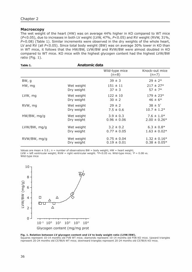
36
Chapter 2
Macroscopy The wet weight of the heart (HW) was on average 44% higher in KO compared to WT mice (P<0.05), due to increases in both LV weight (LVW, 47%, P<0.05) and RV weight (RVW, 31%, P=0.08) (Table 1). Similar increments were observed in the dry weights of the whole heart, LV and RV (all P<0.05). Since total body weight (BW) was on average 30% lower in KO than in WT mice, it follows that the HW/BW, LVW/BW and RVW/BW were almost doubled in KO compared to WT mice. KO mice with the highest glycogen content had the highest LVW/BW ratio (Fig. 1).
Table 1. Anatomic data
Wild-type mice (n=8)
Knock-out mice (n=7)
39 ± 3 29 ± 2*
Wet weight 151 ± 11 217 ± 27* Dry weight 37 ± 3 57 ± 7*
Wet weight 122 ± 10 179 ± 23* Dry weight 30 ± 2 46 ± 6*
Wet weight 29 ± 2 38 ± 5† Dry weight 7.5 ± 0.6 10.7 ± 1.2*
Wet weight 3.9 ± 0.3 7.6 ± 1.0* Dry weight 0.96 ± 0.06 2.00 ± 0.26*
Wet weight 3.2 ± 0.2 6.3 ± 0.8* Dry weight 0.77 ± 0.05 1.63 ± 0.02*
BW, g
HW, mg
LVW, mg
RVW, mg
HW/BW, mg/g
LVW/BW, mg/g
RVW/BW, mg/g Wet weight 0.75 ± 0.04 1.32 ± 0.16* Dry weight 0.19 ± 0.01 0.38 ± 0.05*
Values are mean ± S.E.; n = number of observations BW = body weight; HW = heart weight; LVW = left ventricular weight; RVW = right ventricular weight. *P<0.05 vs. Wild-type mice; †P = 0.08 vs. Wild-type mice
Glycogen content (mg/mg prot
10-1 100 101 102 103 104
LVW
/BW
(m
g/g
)
0
2
4
6
8
10
Fig. 1. Relation between LV glycogen content and LV to body weight ratio (LVW/BW). Squares represent 10-14 months old FVB WT mice; diamonds represent 10-14 months old FVB KO mice. Upward triangles represent 20-24 months old C57Bl/6 WT mice; downward triangles represent 20-24 months old C57Bl/6 KO mice.
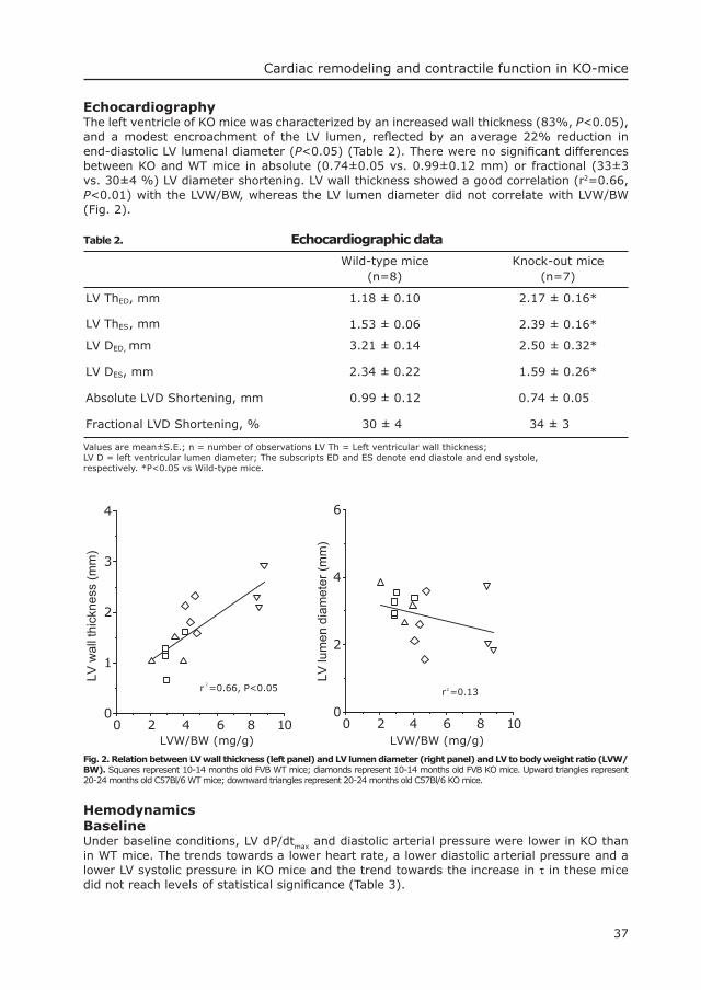
37
Cardiac remodeling and contractile function in KO-mice
EchocardiographyThe left ventricle of KO mice was characterized by an increased wall thickness (83%, P<0.05), and a modest encroachment of the LV lumen, refl ected by an average 22% reduction in end-diastolic LV lumenal diameter (P<0.05) (Table 2). There were no signifi cant differences between KO and WT mice in absolute (0.74±0.05 vs. 0.99±0.12 mm) or fractional (33±3 vs. 30±4 %) LV diameter shortening. LV wall thickness showed a good correlation (r2=0.66, P<0.01) with the LVW/BW, whereas the LV lumen diameter did not correlate with LVW/BW (Fig. 2).
Table 2. Echocardiographic data
Wild-type mice (n=8)
Knock-out mice (n=7)
LV ThED, mm
LV ThES, mm
1.18 ± 0.10 2.17 ± 0.16*
1.53 ± 0.06 2.39 ± 0.16*
LV DED, mm 3.21 ± 0.14 2.50 ± 0.32*
LV DES, mm 2.34 ± 0.22 1.59 ± 0.26*
Absolute LVD Shortening, mm 0.99 ± 0.12 0.74 ± 0.05
Fractional LVD Shortening, % 30 ± 4 34 ± 3
Values are mean±S.E.; n = number of observations LV Th = Left ventricular wall thickness;LV D = left ventricular lumen diameter; The subscripts ED and ES denote end diastole and end systole, respectively. *P<0.05 vs Wild-type mice.
LVW/BW (mg/g)LVW/BW (mg/g)0 2 4 6 8 10
LV lu
men
dia
met
er (m
m)
0
2
4
6
0 2 4 6 8 10
LV w
all t
hick
ness
(mm
)
0
1
2
3
4
r2=0.13r 2=0.66, P<0.05
Fig. 2. Relation between LV wall thickness (left panel) and LV lumen diameter (right panel) and LV to body weight ratio (LVW/BW). Squares represent 10-14 months old FVB WT mice; diamonds represent 10-14 months old FVB KO mice. Upward triangles represent 20-24 months old C57Bl/6 WT mice; downward triangles represent 20-24 months old C57Bl/6 KO mice.
HemodynamicsBaseline Under baseline conditions, LV dP/dtmax and diastolic arterial pressure were lower in KO than in WT mice. The trends towards a lower heart rate, a lower diastolic arterial pressure and a lower LV systolic pressure in KO mice and the trend towards the increase in τ in these mice did not reach levels of statistical signifi cance (Table 3).
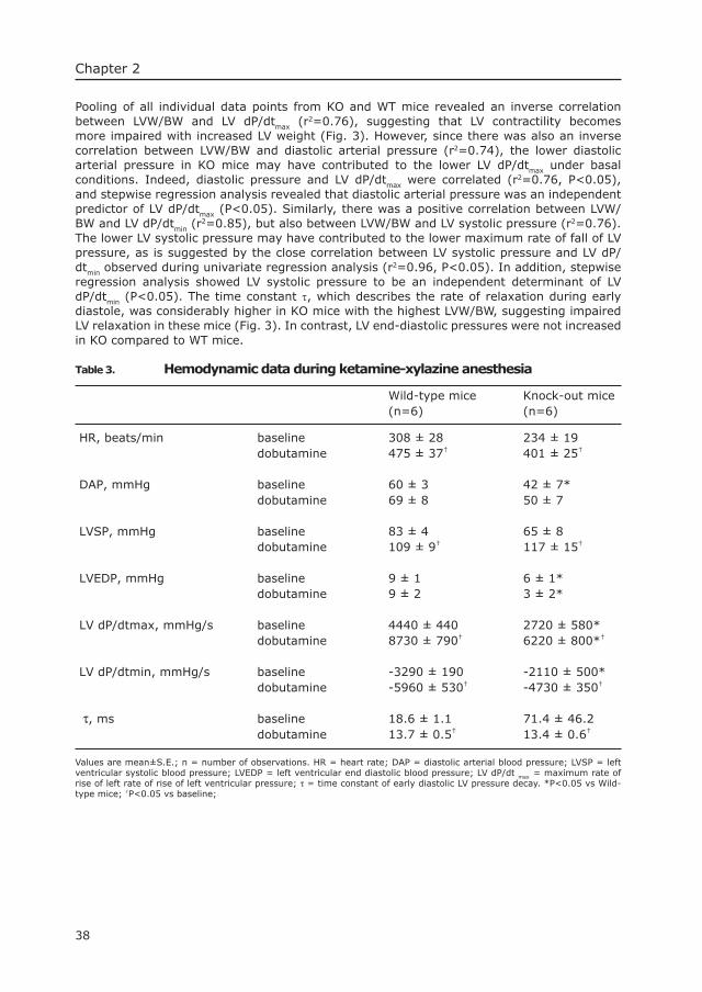
38
Chapter 2
Pooling of all individual data points from KO and WT mice revealed an inverse correlation between LVW/BW and LV dP/dtmax (r2=0.76), suggesting that LV contractility becomes more impaired with increased LV weight (Fig. 3). However, since there was also an inverse correlation between LVW/BW and diastolic arterial pressure (r2=0.74), the lower diastolic arterial pressure in KO mice may have contributed to the lower LV dP/dtmax under basal conditions. Indeed, diastolic pressure and LV dP/dtmax were correlated (r2=0.76, P<0.05), and stepwise regression analysis revealed that diastolic arterial pressure was an independent predictor of LV dP/dtmax (P<0.05). Similarly, there was a positive correlation between LVW/BW and LV dP/dtmin (r
2=0.85), but also between LVW/BW and LV systolic pressure (r2=0.76). The lower LV systolic pressure may have contributed to the lower maximum rate of fall of LV pressure, as is suggested by the close correlation between LV systolic pressure and LV dP/dtmin observed during univariate regression analysis (r2=0.96, P<0.05). In addition, stepwise regression analysis showed LV systolic pressure to be an independent determinant of LV dP/dtmin (P<0.05). The time constant τ, which describes the rate of relaxation during early diastole, was considerably higher in KO mice with the highest LVW/BW, suggesting impaired LV relaxation in these mice (Fig. 3). In contrast, LV end-diastolic pressures were not increased in KO compared to WT mice.
Table 3. Hemodynamic data during ketamine-xylazine anesthesia
Wild-type mice (n=6)
Knock-out mice (n=6)
HR, beats/min baseline 308 ± 28 234 ± 19 dobutamine 475 ± 37† 401 ± 25†
DAP, mmHg baseline 60 ± 3 42 ± 7* dobutamine 69 ± 8 50 ± 7
LVSP, mmHg baseline 83 ± 4 65 ± 8 dobutamine 117 ± 15†
LVEDP, mmHg baseline
109 ± 9†
9 ± 1 6 ± 1* dobutamine 9 ± 2 3 ± 2*
LV dP/dtmax, mmHg/s baseline 4440 ± 440 2720 ± 580* dobutamine 8730 ± 790† 6220 ± 800*†
LV dP/dtmin, mmHg/s baseline -3290 ± 190 -2110 ± 500* dobutamine -5960 ± 530† -4730 ± 350†
, mτ s baseline 18.6 ± 1.1 71.4 ± 46.2 dobutamine 13.7 ± 0.5† 13.4 ± 0.6†
Values are mean±S.E.; n = number of observations. HR = heart rate; DAP = diastolic arterial blood pressure; LVSP = left ventricular systolic blood pressure; LVEDP = left ventricular end diastolic blood pressure; LV dP/dt max = maximum rate of rise of left rate of rise of left ventricular pressure; τ = time constant of early diastolic LV pressure decay. *P<0.05 vs Wild-type mice; †P<0.05 vs baseline;
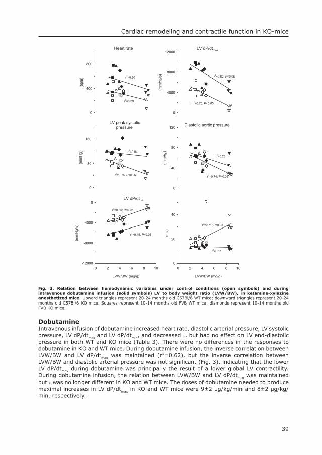
39
Cardiac remodeling and contractile function in KO-mice
Fig. 3. Relation between hemodynamic variables under control conditions (open symbols) and during intravenous dobutamine infusion (solid symbols) LV to body weight ratio (LVW/BW), in ketamine-xylazine anesthetized mice. Upward triangles represent 20-24 months old C57Bl/6 WT mice; downward triangles represent 20-24 months old C57Bl/6 KO mice. Squares represent 10-14 months old FVB WT mice; diamonds represent 10-14 months old FVB KO mice.
Dobutamine Intravenous infusion of dobutamine increased heart rate, diastolic arterial pressure, LV systolic pressure, LV dP/dtmax and LV dP/dtmin, and decreased τ, but had no effect on LV end-diastolic pressure in both WT and KO mice (Table 3). There were no differences in the responses to dobutamine in KO and WT mice. During dobutamine infusion, the inverse correlation between LVW/BW and LV dP/dtmax was maintained (r2=0.62), but the inverse correlation between LVW/BW and diastolic arterial pressure was not signifi cant (Fig. 3), indicating that the lower LV dP/dtmax during dobutamine was principally the result of a lower global LV contractility. During dobutamine infusion, the relation between LVW/BW and LV dP/dtmin was maintained but τ was no longer different in KO and WT mice. The doses of dobutamine needed to produce maximal increases in LV dP/dtmax in KO and WT mice were 9±2 µg/kg/min and 8±2 µg/kg/min, respectively.
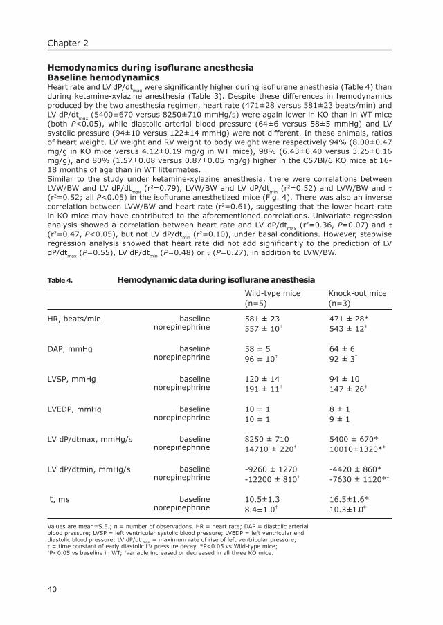
40
Chapter 2
Hemodynamics during isofl urane anesthesiaBaseline hemodynamics Heart rate and LV dP/dtmax were signifi cantly higher during isofl urane anesthesia (Table 4) than during ketamine-xylazine anesthesia (Table 3). Despite these differences in hemodynamics produced by the two anesthesia regimen, heart rate (471±28 versus 581±23 beats/min) and LV dP/dtmax (5400±670 versus 8250±710 mmHg/s) were again lower in KO than in WT mice (both P<0.05), while diastolic arterial blood pressure (64±6 versus 58±5 mmHg) and LV systolic pressure (94±10 versus 122±14 mmHg) were not different. In these animals, ratios of heart weight, LV weight and RV weight to body weight were respectively 94% (8.00±0.47 mg/g in KO mice versus 4.12±0.19 mg/g in WT mice), 98% (6.43±0.40 versus 3.25±0.16 mg/g), and 80% (1.57±0.08 versus 0.87±0.05 mg/g) higher in the C57Bl/6 KO mice at 16-18 months of age than in WT littermates.Similar to the study under ketamine-xylazine anesthesia, there were correlations between LVW/BW and LV dP/dtmax (r
2=0.79), LVW/BW and LV dP/dtmin (r2=0.52) and LVW/BW and τ
(r2=0.52; all P<0.05) in the isofl urane anesthetized mice (Fig. 4). There was also an inverse correlation between LVW/BW and heart rate (r2=0.61), suggesting that the lower heart rate in KO mice may have contributed to the aforementioned correlations. Univariate regression analysis showed a correlation between heart rate and LV dP/dtmax (r
2=0.36, P=0.07) and τ (r2=0.47, P<0.05), but not LV dP/dtmin (r
2=0.10), under basal conditions. However, stepwise regression analysis showed that heart rate did not add signifi cantly to the prediction of LV dP/dtmax (P=0.55), LV dP/dtmin (P=0.48) or τ (P=0.27), in addition to LVW/BW.
Table 4. Hemodynamic data during isofl urane anesthesia
Values are mean±S.E.; n = number of observations. HR = heart rate; DAP = diastolic arterial blood pressure; LVSP = left ventricular systolic blood pressure; LVEDP = left ventricular end diastolic blood pressure; LV dP/dt max = maximum rate of rise of left ventricular pressure; τ = time constant of early diastolic LV pressure decay. *P<0.05 vs Wild-type mice;†P<0.05 vs baseline in WT; ‡variable increased or decreased in all three KO mice.
Wild-type mice (n=5)
Knock-out mice (n=3)
HR, beats/min 581 ± 23 471 ± 28* 557 ± 10† 543 ± 12‡
DAP, mmHg 58 ± 5 64 ± 6 ‡92 ± 3
LVSP, mmHg
96 ± 10†
120 ± 14 94 ± 10 191 ± 11† 147 ± 26‡
LVEDP, mmHg 10 ± 1 8 ± 1 10 ± 1 9 ± 1
LV dP/dtmax, mmHg/snorepinephrine
baseline
norepinephrinebaseline
norepinephrinebaseline
norepinephrinebaseline
norepinephrinebaseline
norepinephrinebaseline
norepinephrinebaseline
8250 ± 710 5400 ± 670* 14710 ± 220† 10010±1320*‡
LV dP/dtmin, mmHg/s -9260 ± 1270 -4420 ± 860* -12200 ± 810† -7630 ± 1120*‡
t, ms 10.5±1.3 16.5±1.6* 8.4±1.0† 10.3±1.0‡
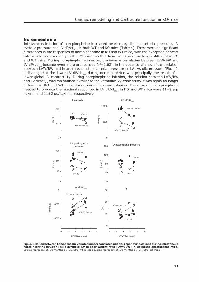
41
Cardiac remodeling and contractile function in KO-mice
Norepinephrine Intravenous infusion of norepinephrine increased heart rate, diastolic arterial pressure, LV systolic pressure and LV dP/dtmax in both WT and KO mice (Table 4). There were no signifi cant differences in the responses to norepinephrine in KO and WT mice, with the exception of heart rate which increased only in the KO mice, so that heart rates were no longer different in KO and WT mice. During norepinephrine infusion, the inverse correlation between LVW/BW and LV dP/dtmax became even more pronounced (r2=0.62), in the absence of a signifi cant relation between LVW/BW and heart rate, diastolic arterial pressure or LV systolic pressure (Fig. 4), indicating that the lower LV dP/dtmax during norepinephrine was principally the result of a lower global LV contractility. During norepinephrine infusion, the relation between LVW/BW and LV dP/dtmin was maintained. Similar to the ketamine-xylazine study, τ was again no longer different in KO and WT mice during norepinephrine infusion. The doses of norepinephrine needed to produce the maximal responses in LV dP/dtmax in KO and WT mice were 11±3 µg/kg/min and 11±2 µg/kg/min, respectively.
Fig. 4. Relation between hemodynamic variables under control conditions (open symbols) and during intravenous norepinephrine infusion (solid symbols) LV to body weight ratio (LVW/BW) in isofl urane-anesthetized mice. Circles represent 16-20 months old C57Bl/6 WT mice; squares represent 16-20 months old C57Bl/6 KO mice.
42
Chapter 2
DiscussionThe major fi ndings of the present study are that (i) mice lacking acid α-glucosidase activity show an increase in cardiac mass involving both left and right ventricles, (ii) the increase in LV weight is associated with a marked increase in LV wall thickness and encroachment of the LV lumen, and (iii) the degree of LV mass increase is inversely correlated with the loss of maximal attainable LV global contractility during ß-adrenergic stimulation.
Cardiac massQualitative descriptions of enlarged hearts have been previously reported in naturally occurring GSDII in turkey (9), sheep (19), dog (29), and a sub-population of shorthorn cattle (18). Heart, LV and RV weights were reported in only one study in two calves, at an age of 3-7 months, in which the heart weight to body weight ratio had tripled compared to normal, due to more than a doubling of LV weight and a quadrupling of RV weight (25). In the present murine study we observed an increase in relative heart weight of up to 160%, due to a 166% increase in relative LV weight and a 130% increase in relative RV weight. We observed a strong correlation between glycogen storage in the heart and the relative LV weight. In approximately 10-14 months old FVB KO mice the average glycogen content was around 300 mg per mg protein, while in 20-24 months old C57Bl/6 KO mice glycogen content was beween 2200 and 5200 mg per mg protein, resulting in a 40% increase in relative LV weight in 10-14 months old FVB and a 166 % increase in 20-24 months old C57Bl/6 KO mice. In the three additional 16-18 months old C57Bl/6 KO mice relative LV weight was 98% higher than in littermate WT mice. Together, these fi ndings suggest that glycogen continues to accumulate in the murine heart up to two years of age. The increase in relative heart weight in mice (up to 160% at 24 months + 3 weeks gestation time) approximates that in calves (up to 200% at 3.7 months + 6 months gestation time), but is signifi cantly less than the increase in humans (up to 350% at 3-10 months + 9 months gestation time). Interestingly, the increase in relative heart weight in humans appears to level off after approximately 3 months after birth, as heart weights are approximately 3 times normal at two weeks of age (n=8) and 4.5 times normal at 3 months (n=8), but with no signifi cant further increase between 3 and 10 months (n=40) (1,8,12,20). In contrast, glycogen continues to accumulate in the murine heart between 10 months and 24 months of age.
LV wall thickness and lumen diameterData on alterations in LV geometry in infants with GSDII have been obtained during autopsy and revealed increases in wall thickness of 6-10 mm in the left ventricle (normal range 2-4 mm) a few weeks after birth, whereas in children who died at approximately two-years of age, wall thickness had increased up to a maximum of 25 mm the left ventricle (normal range 4-6 mm) (12). More recent echocardiography studies have generally reported severe cardiomegaly with extremely thick ventricular walls and relatively normal atria (4). In most cases hypertrophy of the LV free wall and septum is more pronounced than in the right ventricle (14). Another typical echocardiographic fi nding is the encroachment of the LV lumen as was found in two girls of 6-7 months of age (14,23). Encroachment of the lumen may become so severe that outfl ow tract obstruction and infl ow impairment develop (12,23,27).The present study is the fi rst to describe the echocardiographic abnormalities in an animal model of GSDII. The main feature of the echocardiographic fi ndings was an increased LV wall thickness up to 200% of WT mice together with a modest encroachment of the LV lumen, which corresponds well with the echocardiographic fi ndings in infantile GSD II in humans. The degree of encroachment was somewhat variable and did not occur in all KO animals (Fig. 1), but could refl ect the onset of LV failure with resulting LV dilation.
LV contractile function and reserveIn contrast to the well-described anatomic and echocardiographic abnormalities in patients with GSDII, there is surprisingly little information on LV contractile function in GSDII. De Dominicis et al (10) reported normal ejection fractions (70% and 68%) measured with
43
Cardiac remodeling and contractile function in KO-mice
echocardiography in two 6 months old patients. Bulkley and Hutchins (4) also reported a normal ejection fraction (>70%), measured with LV angiography, in a 5-month-old girl. In two other case reports (children up to 9 months of age), invasive assessment of LV function using cardiac catherization demonstrated normal LV fi lling pressures (2-3 mmHg), in the presence of severe cardiomegaly (12,15). In another case report (4 months of age) normal LV hemodynamics and cardiac index were observed, although early diastolic pressure was elevated with a restrictive LV fi lling pattern (2). In contrast, markedly elevated LV fi lling pressures (up to 20 mmHg) and a depressed cardiac index (2.44 l/min/m2) were found in a 4-month-old child (26). In all these case studies, children invariably developed progressive clinical heart failure and died within 2 months after catherization. Unfortunately, in none of these studies LV contractility and LV contractile reserve were determined.The present murine study also shows a normal fractional LV diameter shortening in KO compared to WT mice under basal conditions. However, LV function was not normal as we observed decreases in LV dP/dtmax, under basal conditions and, in particular, during ß-adrenergic stimulation with either dobutamine or norepinephrine infusion. The additional experiments with norepinephrine were performed during isofl urane anesthesia to exclude that the low heart rates and LV dP/dtmax values observed during ketamine-xylazine anesthesia infl uenced the results (21). In addition, we used a 1.4 Fr microtip pressure transducer in these experiments to further minimize mechanical perturbation of LV volume and contractile performance. The maximal attainable LV dP/dtmax correlated well with the increase in LV weight in both series of experiments, thus independent of the anesthesia regimen or size of the LV catheter, suggesting that at more severe stages of the disease LV function becomes impaired. However, from the present study it also becomes clear that the KO animals are not yet in severe cardiac failure. This is suggested by the normal fractional LV diameter shortening, and by the comparable doses of both dobutamine and norepinephrine needed to elicit maximal responses of LV dP/dtmax in KO compared to WT mice, suggesting normal ß-adrenergic receptor sensitivity (3). Moreover, although the less negative LV dP/dtmin and increase in τ in KO compared to WT mice indicate a slight impairment of early diastolic function, which is in close agreement with human data (2), the normal levels of LV fi lling pressure in KO compared to WT mice also suggest the absence of overt heart failure.
ConclusionsThe present study demonstrates that the acid α-glucosidase KO mouse model is characterized by increased cardiac mass, which is associated with an increased LV free wall thickness and with encroachment of the LV lumen, as well as a lower global LV contractility. These manifestations of cardiomegaly and left ventricular dysfunction, although not as overt, parallel the human conditions of early onset GSD II and make these mice a suitable model for further investigation of pathophysiology and novel therapies of infantile Pompe’s disease.
AcknowledgementsThe authors gratefully acknowledge the technical assistance of Elza D. van Deel and Monique C. de Waard. The study was in part fi nanced by a grant from the Sophia Foundation for Medical Research. Dr. Duncker’s research was supported by a Research Fellowship of the Royal Netherlands Academy of Arts and Sciences and by an Established Investigator Grant of the Netherlands Heart Foundation
44
Chapter 2
References1. Bharati S, Serratto M, DuBrow I, Paul MH, Swiryn S, Miller RA, Rosen K, and Lev M. The conduction
system in Pompe’s disease. Pediatr Cardiol 2: 25-32, 1982.2. Bonnici F, Shapiro R, Joffe HS, and Petersen EM. Angiocardiographic and enzyme studies in a patient
with type II glycogenosis (Pompe’s disease). A case report. S Afr Med J 58: 860-862, 1980. 3. Bristow MR, Ginsburg R, Minobe W, Cubicciotti RS, Sageman WS, Lurie K, Billingham ME, Harrison C,
and Stinson EB. Decreased catecholamine sensitivity and beta-adrenergic-receptor density in failing human hearts. N Engl Med 307: 205-211, 1982.
4. Bulkley BH and Hutchins GM. Pompe’s disease presenting as hypertrophic myocardiopathy with Wolff-Parkinson-White syndrome. Am Heart J 96: 246-252, 1978.
5. Bijvoet AG, Van de Kamp EH, Kroos MA, Ding JH, Yang BZ, Visser P, Bakker CE, Verbeet MP, Oostra BA, Reuser AJ and Van der Ploeg AT. Generalized glycogen storage and cardiomegaly in a knockout mouse model of Pompe disease. Hum Mol Genet 7: 53-62, 1998.
6. Bijvoet AG, Van Hirtum H, Kroos MA, Van de Kamp EH, Schoneveld O, Visser P,. Brakenhoff JP,Weggeman M. ,Van Corven EJ, Van der Ploeg AT, and Reuser AJJ. Human acid alpha-glucosidase from rabbit milk has therapeutic effect in mice with glycogen storage disease type II. Hum Mol Genet 8: 2145-2153, 1999.
7. Bijvoet AG, Van Hirtum H, Vermey M, Van Leenen D, Van der Ploeg AT, Mooi WJ, and Reuser AJJ. Pathological features of glycogen storage disease type II highlighted in the knockout mouse model. J Pathol189: 416-424, 1999.
8. Childs AW, Grose RF and Henderson PH. Glycogen disease of the heart. Report of two cases occurring in siblings. Pediatrics 10: 208-217, 1952.
9. Czarnecki CM, Reneau JK, and Jankus EF. Blood glucose and tissue glycogen levels in turkey poults with spontaneous round heart disease and furazolidone-induced cardiomyopathy. Avian Dis 19: 773-780, 1975.
10. De Dominicis E, Finocchi G, Vincenzi M, Calvelli M, Ronconi G, Angelini C, and Vergani L. Echocardiographic and pulsed Doppler features in glycogen storage disease type II of the heart (Pompe’s disease). Acta Cardiol 46: 107-114, 1991.
11. De Zeeuw S, Trines SA, Krams R, Verdouw PD, and Duncker DJ. Cardiovascular profi le of the calcium sensitizer EMD 57033 in open-chest anaesthetized pigs with regionally stunned myocardium. Br J Pharmacol 129: 1413-1422, 2000.
12. Ehlers KH, Hagstrom JWC, Lukas DS, Redo SF, and Engele MA. Glycogen-storage disease of the myocardium with obstruction to the left ventricular outfl ow. Circulation XXV: 96-108, 1962.
13. Engel AG, Gomez MR, Seybold ME, and Lambert EH. The spectrum and diagnosis of acid maltase defi ciency. Neurology 23: 95-106, 1973.
14. Gussenhoven WJ, Busch HF, Kleijer WJ, and De Villeneuve VH. Echocardiographic features in the cardiac type of glycogen storage disease II. Eur Heart J 4: 41-43, 1983.
15. Hernandez A Jr, Marchesi V, Goldring D, Kissane J, and Hartmann AF Jr. Cardiac glycogenosis. Hemodynamic, angiocardiographic, and electron microscopic fi ndings-report of a case. J Pediatr 68: 400-412, 1966.
16. Hirschhorn R and Reuser AJJ. Glycogen storage disease type II: acid α-glucosidase (acid maltase) defi ciency. In: Scriver C.R., Beaudet A.L., Valle D. and Sly W.S., Eds. The Metabolic and Molecular Bases of Inherited Disease. New York: McGraw-Hill; 2001. p. 3389-3420.
17. Hoit BD, Khan ZU, Pawloski-Dahm CM, and Walsh RA. In vivo determination of left ventricular wall stress-shortening relationship in normal mice. Am J Physiol 272:H1047-H1052, 1997.
18. Howell JM, Dorling PR, Cook RD, Robinson WF, Bradley S, and Gawthorne JM. Infantile and late onset form of generalised glycogenosis type II in cattle. J Pathol 134: 267-277, 1981.
19. Manktelow BW and Hartley WJ. Generalized glycogen storage disease in sheep. J Comp Pathol 85: 139-145, 1975.
20. Nihill MR, Wilson DS, and Hugh-Jones K. Generalized glycogenosis type II (Pompe’s disease). Arch Dis Child 45:122-129, 1970.
21. Palakodeti V, Oh S, Oh BH, Mao L, Hongo M, Peterson KL, and Ross J. Force-frequency effect is a powerful determinant of myocardial contractility in the mouse. Am J Physiol 273:H1283-H1290, 1997.
22. Raben, N., K. Nagaraju, E. Lee, P. Kessler P, Byrne B, Lee L, LaMarca M, King C, Ward J, Sauer B, and Plotz P. Targeted disruption of the acid alpha-glucosidase gene in mice causes an illness with critical features of both infantile and adult human glycogen storage disease type II. J. Biol. Chem. 273: 19086-19092, 1998.
23. Rees, A., F. Elbl, K. Minhas, and R. Solinger. Echocardiographic evidence of outfl ow tract obstruction in Pompe’s disease (glycogen storage disease of the heart). Am. J. Cardiol. 37: 1103-1106, 1976.
24. Reuser, A. J., M. A. Kroos, M. M. Hermans, A. G. Bijvoet, M. P. Verbeet, O. P. van Diggelen, W.J. Kleijer, and A.T. Van der Ploeg. Glycogenosis type II (acid maltase defi ciency). Muscle Nerve 3: S61-69, 1995.
45
Cardiac remodeling and contractile function in KO-mice
25. Robinson, W. F., J. M. Howell, and P. R. Dorling. Cardiomyopathy in generalised glycogenosis type II in cattle. Cardiovasc. Res. 17: 238-242, 1983.
26. Ruttenberg, H. D., R. M. Steidl, L. S. Carey, and J. E. Edwards. Glycogen-storage disease of the heart. Hemodynamic and angiocardiographic features in 2 cases. Am. Heart J. 67: 469-480, 1964.
27. Seifert, B. L., M. S. Snyder, A. A. Klein, J. E. O’Loughlin, M. S. Magid, and M. A. Engle. Development of obstruction to ventricular outfl ow and impairment of infl ow in glycogen storage disease of the heart: serial echocardiographic studies from birth to death at 6 months. Am. Heart J. 123: 239-242, 1992.
28. Van der Ploeg A. T., M. Kroos, J. M. van Dongen, W. J. Visser, P. A. Bolhuis, M. C. Loonen, A. J .J. Reuser. Breakdown of lysosomal glycogen in cultured fi broblasts from glycogenosis type II patients after uptake of acid alpha-glucosidase. J. Neurol. Sci.79: 327-336, 1987.
29. Walvoort, H. C., R. G. Slee, and J. F. Koster. Canine glycogen storage disease type II. A biochemical study of an acid alpha-glucosidase-defi cient Lapland dog. Biochim. Biophys. Acta 715: 63-69, 1982.

3Long Term Intravenous Treatment of Pompe’s Disease With Recombinant Human Alpha Glucosidase From Milk
Hannerieke M.P. Van den Hout MD PhD1, Joep H.J. Kamphoven MD1,2, Léon P.F. Winkel MD1, Willem F.M. Arts MD PhD3, Johannes B.C. De Klerk MD1, M.Christa B. Loonen MD PhD3, Arnold G. Vulto PharmD PhD4, Adri Cromme-Dijkhuis MD PhD5, Nynke Weisglas-Kuperus 6, Wim Hop PhD7, Hans Van Hirtum MSc2, Otto P. Van Diggelen PhD2, Marijke Boer MSc2, Marian A. Kroos MSc2, Pieter A. Van Doorn MD PhD8, Edwin Van der Voort MD9, Barbara Sibbles MD PhD10, Emiel J.J.M. Van Corven PhD11, Just P.J. Brakenhoff PhD11, Johan Van Hove MD PhD12, Jan A.M. Smeitink MD PhD13, Gerard de Jong MD PhD14, Arnold J.J. Reuser PhD2,Ans T. Van der Ploeg MD PhD1*
1Department of Pediatrics, Division of Metabolic Diseases and Genetics, 5Division of Pediatric Cardiology, 6Division of Neonatology, 9Division of Pediatric Intensive Care,10Dept. of Pediatrics, 3Department of Pediatric Neurology, Erasmus MC-Sophia, Rotterdam, Dr. Molewaterplein 60, 3015 GJ, Rotterdam, The Netherlands.2Department of Clinical Genetics, 7Department of Epidemiology and Biostatistics, Erasmus MC, Rotterdam, Dr. Molewaterplein 50, 3015 GE Rotterdam, The Netherlands.14Department of Internal Medicine, 4Hospital Pharmacy, 8Department of Neurology, Erasmus MC, Rotterdam, Dr. Molewaterplein 40, 3015 GD Rotterdam, The Netherlands. 11Pharming-Genzyme LLC, Archimedesweg 4, 2333 CN, Leiden, The Netherlands.12 Department of Pediatrics, University Hospital Gasthuisberg, Herestraat 49, B-3000 Leuven, Belgium. 13Department of Pediatrics, Division of Metabolic Diseases, University Medical Center Nijmegen, Geert Grooteplein 10, P O Box 9101, 6500 HB Nijmegen, The Netherlands.
-in press, Pediatrics 2004-

Chapter 3
49
AbstractObjectiveRecent reports warn that the worldwide cell culture capacity is insuffi cient to fulfi ll the increasing demand for human protein drugs. Production in milk of transgenic animals is an attractive alternative. We tested the long-term safety and effi cacy of recombinant human alpha-glucosidase from rabbit milk for the treatment of the lysosomal storage disorder Pompe’s disease. Methods: In the beginning of 1999 four critically ill patients with infantile Pompe’s disease (2.5-8 months old) were enrolled in a single-center open-label study and treated intravenously with recombinant human alpha-glucosidase, 15-40 mg/kg/week.
ResultsGenotypes of patients were consistent with the most severe form of Pompe’s disease. Transgenic alpha-glucosidase was tolerated well during more than 3 years of treatment. Clinical effects were signifi cant. All patients survived beyond the age of 4 years, whereas untreated patients succumb at a median age of 6 to 8 months. Alpha-glucosidase activity normalized. Muscle morphology improved markedly in 1 and cardiac hypertrophy in all patients. Two infants achieved motor milestones that are unmet in infantile Pompe’s disease, but only 1 patient learned to walk.
ConclusionOur study shows that a safe and effective medicine can be produced in the milk of mammals and encourages further development of enzyme replacement therapy for the several forms of Pompe’s disease. Restoration of skeletal muscle function and prevention of pulmonary insuffi ciency require dosing in the range of 20-40 mg/kg. The effect depends on residual muscle function at the start of treatment.
IntroductionProduction of therapeutic proteins in milk of transgenic animals is a promising new technology in light of the rapidly expanding market for protein drugs and the ever-increasing costs of healthcare (1). As the transgenic protein concentration in milk is very high, kilogram quantities of product per year can be obtained at relatively low costs, even in small animals like rabbits (2).Since the fi rst report in 1988 on alpha-1-antitrypsin production in sheep milk, (3) a variety of transgenic proteins has been produced. These range from small unstable peptides (salmon calcitonin peptide (4)) to large proteins with complex post-translational modifi cations (factor VIII (5, 6) and lactoferrin (7)). However, despite the great promises of this novel production platform, there is not yet a single transgenic medicine on the market. Three recombinant proteins from milk are being tested in clinical trials: antithrombin III from goats, alpha-1-antitrypsin from sheep, and alpha-glucosidase from rabbits (8-10).We have focused on the production of recombinant human acid alpha-glucosidase for the treatment of Pompe’s disease, an autosomal recessive muscle disorder. The classic infantile form of the disease leads to death at a median age of 6 to 8 months (11) and is diagnosed by absence of alpha-glucosidase activity and presence of fully deleterious mutations in the alpha-glucosidase gene (12). Cardiac hypertrophy is characteristically present (11). Loss of muscle strength prevents infants from achieving developmental milestones like sitting, standing, and walking (11-13). Milder forms of the disease caused by less severe mutations and partial defi ciency of alpha-glucosidase are also known. Pompe’s disease is a lysosomal glycogen storage disorder with an estimated frequency of 1:40,000 births (14, 15) and is designated an orphan disease.We have successfully explored the feasibility of enzyme therapy for Pompe’s disease in preclinical studies (16-19). The principle of treatment is based on the capacity of lysosomes to engulf exogenous proteins via endocytosis (20). After cloning the human alpha-glucosidase
ERT in infantile Pompe’s disease
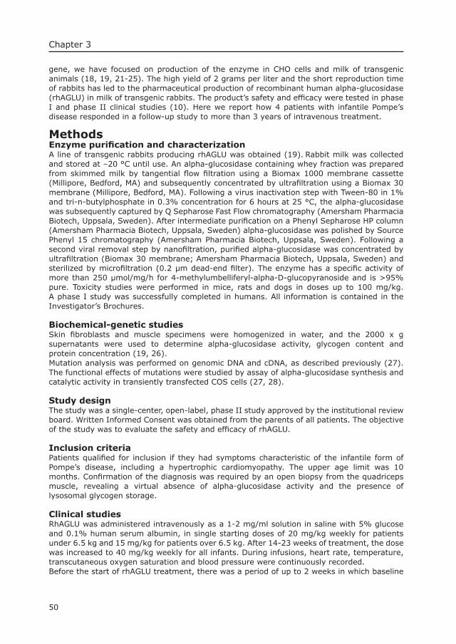
50
Chapter 3
gene, we have focused on production of the enzyme in CHO cells and milk of transgenic animals (18, 19, 21-25). The high yield of 2 grams per liter and the short reproduction time of rabbits has led to the pharmaceutical production of recombinant human alpha-glucosidase (rhAGLU) in milk of transgenic rabbits. The product’s safety and effi cacy were tested in phase I and phase II clinical studies (10). Here we report how 4 patients with infantile Pompe’s disease responded in a follow-up study to more than 3 years of intravenous treatment.
MethodsEnzyme purifi cation and characterizationA line of transgenic rabbits producing rhAGLU was obtained (19). Rabbit milk was collected and stored at –20 °C until use. An alpha-glucosidase containing whey fraction was prepared from skimmed milk by tangential fl ow fi ltration using a Biomax 1000 membrane cassette (Millipore, Bedford, MA) and subsequently concentrated by ultrafi ltration using a Biomax 30 membrane (Millipore, Bedford, MA). Following a virus inactivation step with Tween-80 in 1% and tri-n-butylphosphate in 0.3% concentration for 6 hours at 25 °C, the alpha-glucosidase was subsequently captured by Q Sepharose Fast Flow chromatography (Amersham Pharmacia Biotech, Uppsala, Sweden). After intermediate purifi cation on a Phenyl Sepharose HP column (Amersham Pharmacia Biotech, Uppsala, Sweden) alpha-glucosidase was polished by Source Phenyl 15 chromatography (Amersham Pharmacia Biotech, Uppsala, Sweden). Following a second viral removal step by nanofi ltration, purifi ed alpha-glucosidase was concentrated by ultrafi ltration (Biomax 30 membrane; Amersham Pharmacia Biotech, Uppsala, Sweden) and sterilized by microfi ltration (0.2 µm dead-end fi lter). The enzyme has a specifi c activity of more than 250 µmol/mg/h for 4-methylumbelliferyl-alpha-D-glucopyranoside and is >95% pure. Toxicity studies were performed in mice, rats and dogs in doses up to 100 mg/kg. A phase I study was successfully completed in humans. All information is contained in the Investigator’s Brochures.
Biochemical-genetic studiesSkin fi broblasts and muscle specimens were homogenized in water, and the 2000 x g supernatants were used to determine alpha-glucosidase activity, glycogen content and protein concentration (19, 26).Mutation analysis was performed on genomic DNA and cDNA, as described previously (27). The functional effects of mutations were studied by assay of alpha-glucosidase synthesis and catalytic activity in transiently transfected COS cells (27, 28).
Study designThe study was a single-center, open-label, phase II study approved by the institutional review board. Written Informed Consent was obtained from the parents of all patients. The objective of the study was to evaluate the safety and effi cacy of rhAGLU.
Inclusion criteriaPatients qualifi ed for inclusion if they had symptoms characteristic of the infantile form of Pompe’s disease, including a hypertrophic cardiomyopathy. The upper age limit was 10 months. Confi rmation of the diagnosis was required by an open biopsy from the quadriceps muscle, revealing a virtual absence of alpha-glucosidase activity and the presence of lysosomal glycogen storage.
Clinical studiesRhAGLU was administered intravenously as a 1-2 mg/ml solution in saline with 5% glucose and 0.1% human serum albumin, in single starting doses of 20 mg/kg weekly for patients under 6.5 kg and 15 mg/kg for patients over 6.5 kg. After 14-23 weeks of treatment, the dose was increased to 40 mg/kg weekly for all infants. During infusions, heart rate, temperature, transcutaneous oxygen saturation and blood pressure were continuously recorded. Before the start of rhAGLU treatment, there was a period of up to 2 weeks in which baseline
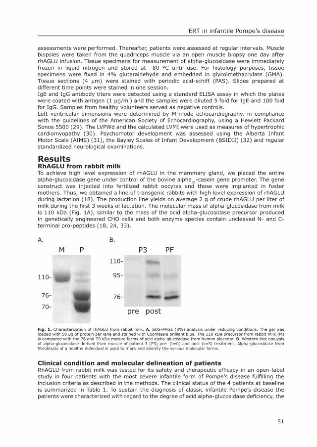
51
ERT in infantile Pompe’s disease
assessments were performed. Thereafter, patients were assessed at regular intervals. Muscle biopsies were taken from the quadriceps muscle via an open muscle biopsy one day after rhAGLU infusion. Tissue specimens for measurement of alpha-glucosidase were immediately frozen in liquid nitrogen and stored at –80 °C until use. For histology purposes, tissue specimens were fi xed in 4% glutaraldehyde and embedded in glycolmethacrylate (GMA). Tissue sections (4 µm) were stained with periodic acid-schiff (PAS). Slides prepared at different time points were stained in one session.IgE and IgG antibody titers were detected using a standard ELISA assay in which the plates were coated with antigen (1 µg/ml) and the samples were diluted 5 fold for IgE and 100 fold for IgG. Samples from healthy volunteers served as negative controls.Left ventricular dimensions were determined by M-mode echocardiography, in compliance with the guidelines of the American Society of Echocardiography, using a Hewlett Packard Sonos 5500 (29). The LVPWd and the calculated LVMI were used as measures of hypertrophic cardiomyopathy (30). Psychomotor development was assessed using the Alberta Infant Motor Scale (AIMS) (31), the Bayley Scales of Infant Development (BSIDII) (32) and regular standardized neurological examinations.
ResultsRhAGLU from rabbit milkTo achieve high level expression of rhAGLU in the mammary gland, we placed the entire alpha-glucosidase gene under control of the bovine alphaS1-casein gene promoter. The gene construct was injected into fertilized rabbit oocytes and these were implanted in foster mothers. Thus, we obtained a line of transgenic rabbits with high level expression of rhAGLU during lactation (18). The production line yields on average 2 g of crude rhAGLU per liter of milk during the fi rst 3 weeks of lactation. The molecular mass of alpha-glucosidase from milk is 110 kDa (Fig. 1A), similar to the mass of the acid alpha-glucosidase precursor produced in genetically engineered CHO cells and both enzyme species contain uncleaved N- and C-terminal pro-peptides (18, 24, 33).
PA.
110-
76-
70-
M P3 PF
110-
95-
76-
pre post
B.
Fig. 1. Characterization of rhAGLU from rabbit milk. A. SDS-PAGE (8%) analysis under reducing conditions. The gel was loaded with 50 µg of protein per lane and stained with Coomassie brilliant blue. The 110 kDa precursor from rabbit milk (M) is compared with the 76 and 70 kDa mature forms of acid alpha-glucosidase from human placenta. B. Western blot analysis of alpha-glucosidase derived from muscle of patient 3 (P3) pre- (t=0) and post (t=3) treatment. Alpha-glucosidase from fi broblasts of a healthy individual is used to mark and identify the various molecular forms.
Clinical condition and molecular delineation of patientsRhAGLU from rabbit milk was tested for its safety and therapeutic effi cacy in an open-label study in four patients with the most severe infantile form of Pompe’s disease fulfi lling the inclusion criteria as described in the methods. The clinical status of the 4 patients at baseline is summarized in Table 1. To sustain the diagnosis of classic infantile Pompe’s disease the patients were characterized with regard to the degree of acid alpha-glucosidase defi ciency, the
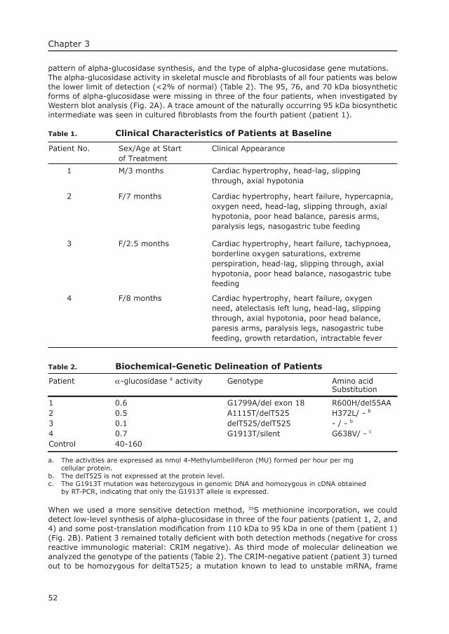
52
Chapter 3
pattern of alpha-glucosidase synthesis, and the type of alpha-glucosidase gene mutations.The alpha-glucosidase activity in skeletal muscle and fi broblasts of all four patients was below the lower limit of detection (<2% of normal) (Table 2). The 95, 76, and 70 kDa biosynthetic forms of alpha-glucosidase were missing in three of the four patients, when investigated by Western blot analysis (Fig. 2A). A trace amount of the naturally occurring 95 kDa biosynthetic intermediate was seen in cultured fi broblasts from the fourth patient (patient 1).
Table 1. Clinical Characteristics of Patients at Baseline
Patient No. Sex/Age at Start of Treatment
Clinical Appearance
1 M/3 months Cardiac hypertrophy, head-lag, slipping through, axial hypotonia
2 moF/7 nths Cardiac hypertrophy, heart failure, hypercapnia, oxygen need, head-lag, slipping through, axial hypotonia, poor head balance, paresis arms, paralysis legs, nasogastric tube feeding
3 F/2.5 months Cardiac hypertrophy, heart failure, tachypnoea, borderline oxygen saturations, extreme perspiration, head-lag, slipping through, axial hypotonia, poor head balance, nasogastric tube feeding
4 F/8 months Cardiac hypertrophy, heart failure, oxygen need, atelectasis left lung, head-lag, slipping through, axial hypotonia, poor head balance, paresis arms, paralysis legs, nasogastric tube feeding, growth retardation, intractable fever
Table 2. Biochemical-Genetic Delineation of Patients
Patient α-glucosidase a activity Genotype Amino acidSubstitution
1 0.6 G1799A/del exon 18 R600H/del55AA 2 0.5 A1115T/delT525 H372L/ - b
3 0.1 delT525/delT525 - / - b 4 0.7 G1913T/silent G638V/ - c Control 40-160
a. The activities are expressed as nmol 4-Methylumbelliferon (MU) formed per hour per mg cellular protein.b. The delT525 is not expressed at the protein level.c. The G1913T mutation was heterozygous in genomic DNA and homozygous in cDNA obtained by RT-PCR, indicating that only the G1913T allele is expressed.
When we used a more sensitive detection method, 35S methionine incorporation, we could detect low-level synthesis of alpha-glucosidase in three of the four patients (patient 1, 2, and 4) and some post-translation modifi cation from 110 kDa to 95 kDa in one of them (patient 1) (Fig. 2B). Patient 3 remained totally defi cient with both detection methods (negative for cross reactive immunologic material: CRIM negative). As third mode of molecular delineation we analyzed the genotype of the patients (Table 2). The CRIM-negative patient (patient 3) turned out to be homozygous for deltaT525; a mutation known to lead to unstable mRNA, frame
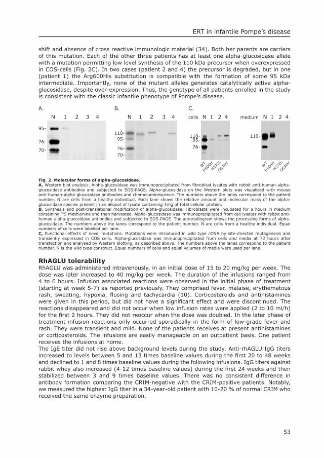
53
ERT in infantile Pompe’s disease
shift and absence of cross reactive immunologic material (34). Both her parents are carriers of this mutation. Each of the other three patients has at least one alpha-glucosidase allele with a mutation permitting low level synthesis of the 110 kDa precursor when overexpressed in COS-cells (Fig. 2C). In two cases (patient 2 and 4) the precursor is degraded, but in one (patient 1) the Arg600His substitution is compatible with the formation of some 95 kDa intermediate. Importantly, none of the mutant alleles generates catalytically active alpha-glucosidase, despite over-expression. Thus, the genotype of all patients enrolled in the study is consistent with the classic infantile phenotype of Pompe’s disease.
cells N 1 2
110-95-
76-
110-
WT
R600
H
H372L
G638V
C.
WT
R600
H
H372L
G638V
A.
95-
76-
70-
N 1 2 3 4
110-95-
76-70-
B.
N 1 2 3 4 N 1 2 4 medium 4
Fig. 2. Molecular forms of alpha-glucosidase.A, Western blot analysis. Alpha-glucosidase was immunoprecipitated from fi broblast lysates with rabbit anti-human-alpha-glucosidase antibodies and subjected to SDS-PAGE. Alpha-glucosidase on the Western blots was visualized with mouse anti-human-alpha-glucosidase antibodies and chemoluminescence. The numbers above the lanes correspond to the patient number. N are cells from a healthy individual. Each lane shows the relative amount and molecular mass of the alpha-glucosidase species present in an aliquot of lysate containing 1mg of total cellular protein. B, Synthesis and post-translational modifi cation of alpha-glucosidase. Fibroblasts were incubated for 8 hours in medium containing 35S methionine and then harvested. Alpha-glucosidase was immunoprecipitated from cell lysates with rabbit anti-human alpha-glucosidase antibodies and subjected to SDS-PAGE. The autoradiogram shows the processing forms of alpha-glucosidase. The numbers above the lanes correspond to the patient number. N are cells from a healthy individual. Equal numbers of cells were labelled per lane. C, Functional effects of novel mutations. Mutations were introduced in wild type cDNA by site-directed mutagenesis and transiently expressed in COS cells. Alpha-glucosidase was immunoprecipitated from cells and media at 72 hours after transfection and analysed by Western blotting, as described above. The numbers above the lanes correspond to the patient number. N is the wild type construct. Equal numbers of cells and equal volumes of media were used per lane.
RhAGLU tolerabilityRhAGLU was administered intravenously, in an initial dose of 15 to 20 mg/kg per week. The dose was later increased to 40 mg/kg per week. The duration of the infusions ranged from 4 to 6 hours. Infusion associated reactions were observed in the initial phase of treatment (starting at week 5-7) as reported previously. They comprised fever, malaise, erythematous rash, sweating, hypoxia, fl using and tachycardia (10). Corticosteroids and antihistamines were given in this period, but did not have a signifi cant effect and were discontinued. The reactions disappeared and did not occur when low infusion rates were applied (2 to 10 ml/h) for the fi rst 2 hours. They did not reoccur when the dose was doubled. In the later phase of treatment infusion reactions only occurred sporadically in the form of low-grade fever and rash. They were transient and mild. None of the patients receives at present antihistamines or corticosteroids. The infusions are easily manageable on an outpatient basis. One patient receives the infusions at home. The IgE titer did not rise above background levels during the study. Anti-rhAGLU IgG titers increased to levels between 5 and 13 times baseline values during the fi rst 20 to 48 weeks and declined to 1 and 8 times baseline values during the following infusions. IgG titers against rabbit whey also increased (4-12 times baseline values) during the fi rst 24 weeks and then stabilized between 3 and 9 times baseline values. There was no consistent difference in antibody formation comparing the CRIM-negative with the CRIM-positive patients. Notably, we measured the highest IgG titer in a 34-year-old patient with 10-20 % of normal CRIM who received the same enzyme preparation.
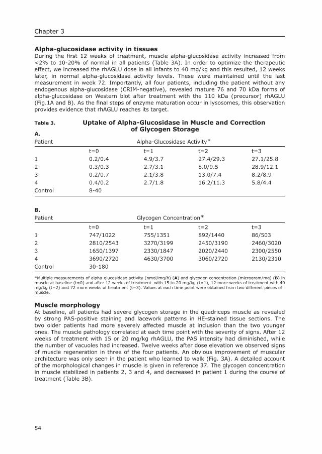
54
Chapter 3
Alpha-glucosidase activity in tissuesDuring the fi rst 12 weeks of treatment, muscle alpha-glucosidase activity increased from <2% to 10-20% of normal in all patients (Table 3A). In order to optimize the therapeutic effect, we increased the rhAGLU dose in all infants to 40 mg/kg and this resulted, 12 weeks later, in normal alpha-glucosidase activity levels. These were maintained until the last measurement in week 72. Importantly, all four patients, including the patient without any endogenous alpha-glucosidase (CRIM-negative), revealed mature 76 and 70 kDa forms of alpha-glucosidase on Western blot after treatment with the 110 kDa (precursor) rhAGLU (Fig.1A and B). As the fi nal steps of enzyme maturation occur in lysosomes, this observation provides evidence that rhAGLU reaches its target.
Table 3. Uptake of Alpha-Glucosidase in Muscle and Correction of Glycogen StorageA.Patient Alpha-Glucosidase Activity*
t=0 t=1 t=2 t=3 1 0.2/0.4 4.9/3.7 27.4/29.3 27.1/25.8 2 0.3/0.3 2.7/3.1 8.0/9.5 28.9/12.1 3 0.2/0.7 2.1/3.8 13.0/7.4 8.2/8.9 4 0.4/0.2 2.7/1.8 16.2/11.3 5.8/4.4 Control 8-40
B.Patient Glycogen Concentration*
t=0 t=1 t=2 t=3 1 747/1022 755/1351 892/1440 86/503 2 2810/2543 3270/3199 2450/3190 2460/30203 1650/1397 2330/1847 2020/2440 2300/25504 3690/2720 4630/3700 3060/2720 2130/2310Control 30-180
*Multiple measurements of alpha-glucosidase activity (nmol/mg/h) (A) and glycogen concentration (microgram/mg) (B) in muscle at baseline (t=0) and after 12 weeks of treatment with 15 to 20 mg/kg (t=1), 12 more weeks of treatment with 40 mg/kg (t=2) and 72 more weeks of treatment (t=3). Values at each time point were obtained from two different pieces ofmuscle.
Muscle morphologyAt baseline, all patients had severe glycogen storage in the quadriceps muscle as revealed by strong PAS-positive staining and lacework patterns in HE-stained tissue sections. The two older patients had more severely affected muscle at inclusion than the two younger ones. The muscle pathology correlated at each time point with the severity of signs. After 12 weeks of treatment with 15 or 20 mg/kg rhAGLU, the PAS intensity had diminished, while the number of vacuoles had increased. Twelve weeks after dose elevation we observed signs of muscle regeneration in three of the four patients. An obvious improvement of muscular architecture was only seen in the patient who learned to walk (Fig. 3A). A detailed account of the morphological changes in muscle is given in reference 37. The glycogen concentration in muscle stabilized in patients 2, 3 and 4, and decreased in patient 1 during the course of treatment (Table 3B).
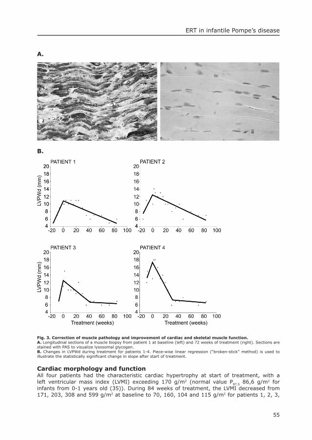
55
ERT in infantile Pompe’s disease
A.
B.
Fig. 3. Correction of muscle pathology and improvement of cardiac and skeletal muscle function. A. Longitudinal sections of a muscle biopsy from patient 1 at baseline (left) and 72 weeks of treatment (right). Sections are stained with PAS to visualize lysosomal glycogen.B. Changes in LVPWd during treatment for patients 1-4. Piece-wise linear regression (“broken-stick” method) is used to illustrate the statistically signifi cant change in slope after start of treatment.
Cardiac morphology and functionAll four patients had the characteristic cardiac hypertrophy at start of treatment, with a left ventricular mass index (LVMI) exceeding 170 g/m2 (normal value P97.5 86,6 g/m2 for infants from 0-1 years old (35)). During 84 weeks of treatment, the LVMI decreased from 171, 203, 308 and 599 g/m2 at baseline to 70, 160, 104 and 115 g/m2 for patients 1, 2, 3,
56
Chapter 3
and 4, respectively. The decrease of the LVMI along time appeared linear (r = -0.86, -0.91, -0.89 and -0.80 for the four respective patients; all P <0.01). The diastolic thickness of the left ventricular posterior wall (LVPWd) was increased before start of rhAGLU treatment. We found a signifi cant change of slope for LVPWd against time at t=0 for each separate patient (P <0.01) (Fig. 3B). The decrease in LVPWd after the start of treatment leveled out in two patients (patients 3 and 4) when the P95 of normal was approached, as evidenced by a second signifi cant change of slope using the “broken stick” method (P <0.05). The systolic function normalized and the diastolic function improved in all patients.
Respiratory conditionDuring the fi rst 2 years of life, the two younger patients (patients 1 and 3) remarkably showed no signifi cant respiratory problems. Patient 3 recovered from a life-threatening bronchiolitis at the age of 1 year without sequelae, despite borderline oxygen saturation at inclusion. At the age of two she became ventilator-dependent after surgical removal of an infected Port-A-Cath. The respiratory course of patient 1, now 4 years old, remained uneventful. Patients 2 and 4, who were older and more severely affected at inclusion, had a marginal respiratory condition from the start of treatment, and both required oxygen. Patient 2 (pCO2 10.6 kPa equivalent to 80 mm Hg at start; normal range 4.5-6.8 kPa) became ventilator-dependent before the fi rst rhAGLU infusion and remained fully ventilated. Patient 4 (pCO2 9.8 kPa equivalent to 75 mm Hg at start) became ventilator-dependent during a bout of pneumonia after 10 weeks of low-dose treatment. She could gradually be weaned from the ventilator after one year of high dose treatment and was eventually completely ventilator free for 5 days, but this situation could not be maintained. At the age of 4 her ventilator needs are 22-23 hours per day.
Motor developmentThe most remarkable progress in motor development was seen in the younger patients (patients 1 and 3, Fig. 4). Patient 1 learned to crawl (12 months), walk (16 months), squat (18 months) and climb stairs (22 months). Patient 3 learned to sit unsupported (Fig 4), and her condition further improved until the age of 2, when she became ventilator dependent. At the age of 4 years and 3 months she died quite suddenly after a short period of intractable fever of more than 42 0C, unstable blood pressure and coma. At baseline, the older patients (patients 2 and 4) could hardly lift their arms while in a supine position, and their legs lay immobile on the surface. During treatment, the muscle function of the arms improved signifi cantly. Patient 2, however, lost her regained muscle strength after a series of airway infections. In her case, restoration of function appears to be a diffi cult process. Patient 4 maintained the strength in her arms. When she was 24 months old, she could roll over onto her side and play for longer periods while seated in a wheel chair. Her condition has not improved since then. The AIMS score for patients 2, 3 and 4 remained far below the p5. Patient 1 followed the p5 of normal (Fig 5). For patient 1 the psychomotor development on the BSID II, based on raw score equivalents, progressed from 2 to 37 months developmental ages at chronological ages of 3 to 37 months. The psychomotor developmental index (PDI) progressed from 79 to 97. In patients 2, 3 and 4 the PDI was below 50, and a motor developmental age could not be calculated.
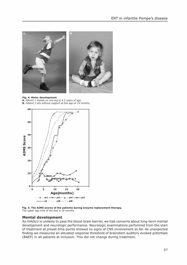
57
ERT in infantile Pompe’s disease
Fig. 4. Motor development.A. Patient 1 stands on one leg at 4.5 years of age.B. Patient 3 sits without support at the age of 19 months.
0
10
20
30
40
50
60
0 5 10 15 20
Age(months)
AIM
S S
core
pt1 pt2 pt4 pt3
p5 p50 p90
Fig. 5. The AIMS scores of the patients during enzyme replacement therapy.The upper age limit of the test is 18 months.
Mental developmentAs rhAGLU is unlikely to pass the blood-brain barrier, we had concerns about long-term mental development and neurologic performance. Neurologic examinations performed from the start of treatment at preset time points showed no signs of CNS involvement so far. As unexpected fi nding we measured an elevated response threshold of brainstem auditory evoked potentials (BAEP) in all patients at inclusion. This did not change during treatment.
58
Chapter 3
For patient 1 the mental development on the BSID II, based on raw score equivalents, progressed from 3 to 31 months developmental ages at chronological ages of 3 to 37 months. The mental developmental index (MDI) was 101 and 79 at 3 months and 37 months, respectively. The BSID II was inadequate to test the mental development of patient 2 because of her severe hypotonia. For patient 3 the mental development progressed from 2 to 18 months at chronological ages of 3 to 22 months. The MDI was 81 and 72, respectively. The mental development of patient 4 progressed from 5 to 27 months at chronological ages of 8 to 37 months. The MDI of patient 4 was 76 and 67, respectively. In practice, the BSID II scores are not a true refl ection of mental development. Items were missed through motor handicaps and hearing problems rather than through mental delay. All patients were interested in their environment, interacted with their parents and were able to attend school.
SurvivalThree of the 4 patients are alive. All 4 patients reached the age of 4 years, whereas the life expectancy of untreated patients with the classic infantile form of Pompe’s disease is typically less than 1 year (10-13).
DiscussionOur study demonstrates that a safe and effective medicine for IV treatment of human disease can be produced in the milk of transgenic animals, in this case rhAGLU from rabbit milk for patients with Pompe’s disease. The safety of the product is proven by the fact that more than 700 infusions were well tolerated by critically ill patients. The effi cacy of the therapy is evident from several observations. First, all 4 patients have reached the age of 4 years, whereas babies with classic infantile Pompe’s disease typically succumb before they are one year old (10-13). Second, the cardiac hypertrophy diminished and cardiac function improved, whereas cardiac failure is a major cause of death in infantile Pompe’s disease. Third, patients gained muscle strength, whereas there normally is a progressive loss of muscle function. Two patients achieved developmental milestones that are unmet by untreated patients. Notably, the motor score on the Bayley Scale of Infant Development of one patient normalized at the age of two years.
The effi cacy of treatment is further supported by biochemical fi ndings. Correction of alpha-glucosidase defi ciency occurred in skeletal muscle of all four patients. After three months of single weekly doses of 15 to 20 mg/kg the correction was partial. A 100% correction was achieved after three additional months on a weekly dose of 40 mg/kg. Conversion of the 110 kDa precursor from milk to mature 76/70 kDa alpha-glucosidase provides evidence that the enzyme is targeted to lysosomes, where this proteolytic processing occurs (36). Moreover, we observed a decrease of the PAS-staining intensity and the appearance of empty vacuoles in the quadriceps muscle of all patients, suggestive for an effect on the lysosomal glycogen pool. In the best performing patient this resulted in greatly improved muscle morphology and glycogen content (37).
It is evident that the 4 patients respond differently to the treatment, and we have tried to fi nd an explanation. All patients appeared to have molecular-genetic defects and clinical characteristics consistent with classic infantile Pompe’s disease (11-13). Notably, the best performing patient had the most severe hearing defi cit (50-80dB). This makes it very unlikely that the difference in response is explained by inclusion of patients with milder non-classic infantile phenotypes.
We fi nd it equally unlikely that an inhibitory immune response, as described in a study by Amalfi tano et al. (38) with alpha-glucosidase from CHO-cells, explains the difference in effi cacy. The latter study claims, that neutralizing antibodies caused the loss of treatment benefi t at the age of 8 months in two CRIM-negative patients. This counter-effect did not
59
ERT in infantile Pompe’s disease
occur in our study. On the contrary, our truly CRIM–negative patient was the second best responder. Enzyme therapy studies for Gaucher’s disease, Fabry’s disease and MPS I do not indicate either that a CRIM-negative status per se or antibody formation interferes with the effi cacy of enzyme replacement therapy (39-43).
In our view, the degree of impairment of skeletal muscle function and the age of the patient at start of treatment play the decisive roles in outcome. The two patients, included at 2.5 and 3 months of age, had milder symptoms at inclusion and responded clearly better than the 7 and 8 months old patients who were in an end stage of the disease at the time of inclusion. The patient who had the best muscle function at start of treatment learned to walk. This may indicate that active muscle movement is required to correct the lysosomal glycogen storage and restore the muscle morphology (37). Despite the signifi cant effects of our therapy, we warn that patients with infantile Pompe’s disease are at risk of developing residual disease with contractures, scoliosis, or respiratory insuffi ciency if treatment is started too late or with a too low a dose. The therapeutic window is small. We may have lost precious time by treating the patients for 3 to 6 months with a sub optimal dose.
The results of our study are encouraging for the further exploration of enzyme replacement therapy. This is true for infants but even more so for patients with milder forms of Pompe’s disease. The therapeutic window is larger as we experienced in our pilot study with 3 patients aged 12 to 33 years. One of them, who was wheelchair-bound for 4 years, started to walk again after 2 years of treatment with rhAGLU, 20 mg/kg/week (44). The effective dose of rhAGLU is high compared with the 1 mg/kg dose of glucocerebrosidase used for the treatment of type 1 Gaucher’s disease and the 0.2-1 mg/kg dose of alpha-galactosidase used for treatment of Fabry’s disease (39-41, 43, 45). These differences are explainable by the different target tissues. In Pompe’s disease, the target tissue (skeletal muscle) is shielded from the enzyme by the capillary endothelium and the interstitial tissue. In contrast, the targets in Gaucher’s disease (liver and spleen macrophages) and Fabry’s disease (endothelial cells) are directly exposed to circulating enzyme. Notably, in Fabry’s disease 3 mg/kg alpha-galactosidase is suffi cient to reach the endocardium but insuffi cient to reach the cardiomyocytes (40). The higher dose required to correct enzyme activity in skeletal muscle is supported by studies in animal models and was demonstrated for various enzyme species among which recombinant human alpha-glucosidase from both transgenic mammals as well as genetically engineered CHO cells (19, 33, 46, 47).
ConclusionThe results of our study demonstrate both the safety and effi cacy of enzyme replacement therapy in Pompe’s disease as well as the feasibility of producing medicines in the milk of transgenic animals. This novel production platform can potentially reduce the costs of therapeutics, but scale-up persists as major challenge in the rhAGLU production process. The currently used transgenic rabbit production line supplies approximately 10 g of rhAGLU per animal per year. At a dose of 10-40 mg/kg per patient per week, the production of rhAGLU in rabbit milk is feasible in the initial phase of product development, but falls short of supplying patients with Pompe’s disease world-wide. The more conventional production system in genetically modifi ed CHO cells can be used as alternative, but also has a capacity limitation (1). Thus, timely investments need to be made in the development of alternative production platforms. Our studies show that production in milk of sheep, goat, or cow is the option of choice.
AcknowledgmentsWe thank the patients and parents for participating in this study and all who have given their support: A. Franken, M. Etzi, L. Vendrig, G. Kreek, L. van der Giessen, M. Bruning, P. Merkus,
60
Chapter 3
R.M. Koppenol, T. de Vries Lentsch, L-A. Severijnen, G.A. van Zanten, J. Ponsen, J. Huijmans, F. Bel, H. Versprille, the staff of the pharmacy, the clinical genetic and neurophysiological laboratory, H. Galjaard, and H.A. Büller.The study was sponsored by Pharming-Genzyme LLC and fi nanced in part by a grant from the Sophia Foundation for Medical Research (J.H.J. Kamphoven and L.P.F. Winkel).
References1. Mynahan R, Anderson R. The protein production challenge. In vivo; The business & Medicine Report.
2001;72.2. Janne J, Alhonen L, Hyttinen JM, Peura T, Tolvanen M, Korhonen VP. Transgenic bioreactors.
Biotechnol Annu Rev. 1998;4:55-74.3. Simons JP, Wilmut I, Clark AJ, Archibald AL, Bishof JO, Lathe R. Gene transfer into sheep.
Biotechnology. 1988;6:113-120.4. McKee C, Gibson A, Dalrymple M, Emslie L, Garner I, Cottingham I. Production of biologically active
salmon calcitonin in the milk of transgenic rabbits. Nat Biotechnol. 1998;16:647-651.5. Niemann H, Halter R, Carnwath JW, Herrmann D, Lemme E, Paul D. Expression of human blood
clotting factor VIII in the mammary gland of transgenic sheep. Transgenic Res. 1999;8:237-247.6. Paleyanda RK, Velander WH, Lee TK et al. Transgenic pigs produce functional human factor VIII in
milk. Nat Biotechnol. 1997;15:971-975.7. Van Berkel PH, Welling MM, Geerts M, et al. Large scale production of recombinant human lactoferrin
in the milk of transgenic cows. Nat Biotechnol. 2002;20:484-487.8. Lu W, Mant T, Levy JH, Bailey JM. Pharmacokinetics of recombinant transgenic antithrombin in
volunteers. Anesth Analg. 2000;90:531-534.9. Carver AS, Dalrymple MA, Wright G, et al. Transgenic livestock as bioreactors: stable expression of
human alpha-1- antitrypsin by a fl ock of sheep. Biotechnology. 1993;11:1263-1270.10. Van den Hout H, Reuser AJ, Vulto AG, Loonen MC, Cromme-Dijkhuis A, Van der Ploeg AT. Recombinant
human alpha-glucosidase from rabbit milk in Pompe patients. Lancet. 2000;356:397-398.11. Van den Hout JMP, Hop, W., van Diggelen, O.P. et al. The natural course of infantile Pompe’s disease;
20 original cases compared with 133 cases from the literature. Pediatrics 2003; 112, in press.12. Hirschhorn R, Reuser AJJ. Glycogen storage disease type II: acid alpha-glucosidase (acid maltase)
defi ciency. In: Scriver CR, Beaudet A, Sly WS, Valle D, editors. The Metabolic and Molecular Bases of Inherited Disease. 8 th ed. NY: McGraw-Hill; 2001. 3389-3420.
13. Slonim AE, Bulone L, Ritz S, Goldberg T, Chen A, Martiniuk F. Identifi cation of two subtypes of infantile acid maltase defi ciency. J Pediatr. 2000;137:283-285.
14. Ausems MG, Verbiest J, Hermans MP, et al. Frequency of glycogen storage disease type II in The Netherlands: implications for diagnosis and genetic counselling. Eur J Hum Genet. 1999;7:713-716.
15. Martiniuk F, Chen A, Mack A, et al. Carrier frequency for glycogen storage disease type II in New York and estimates of affected individuals born with the disease. Am J Med Genet. 1998;79:69-72.
16. Van der Ploeg AT, Van der Kraaij AM, Willemsen R et al. Rat heart perfusion as model system for enzyme replacement therapy in glycogenosis type II. Pediatr Res. 1990;28:344-347.
17. Van der Ploeg AT, Kroos MA, Willemsen R, Brons NH, Reuser AJ. Intravenous administration of phosphorylated acid alpha-glucosidase leads to uptake of enzyme in heart and skeletal muscle of mice. J Clin Invest. 1991;87:513-518.
18. Bijvoet AG, Kroos MA, Pieper FR, et al. Recombinant human acid alpha-glucosidase: high level production in mouse milk, biochemical characteristics, correction of enzyme defi ciency in GSDII KO mice. Hum Mol Genet. 1998;7:1815-1824.
19. Bijvoet AG, Van Hirtum H, Kroos MA, et al. Human acid alpha-glucosidase from rabbit milk has therapeutic effect in mice with glycogen storage disease type II. Hum Mol Genet. 1999;8:2145-2153.
20. De Duve C, Wattiaux R. Functions of lysosomes. Annu Rev Physiol. 1966;28:435-492.21. Hoefsloot LH, Hoogeveen-Westerveld M, Kroos MA, Van Beeumen J, Reuser AJ, Oostra BA. Primary
structure and processing of lysosomal alpha-glucosidase; homology with the intestinal sucrase-isomaltase complex. Embo J. 1988;7:1697-1704.
22. Martiniuk F, Mehler M, Tzall S, Meredith G, Hirschhorn R. Sequence of the cDNA and 5’-fl anking region for human acid alpha- glucosidase, detection of an intron in the 5’ untranslated leader sequence, defi nition of 18-bp polymorphisms, and differences with previous cDNA and amino acid sequences. DNA Cell Biol. 1990;9:85-94.
23. Hoefsloot LH, Hoogeveen-Westerveld M, Reuser AJ, Oostra BA. Characterization of the human lysosomal alpha-glucosidase gene. Biochem J. 1990;272:493-497.
24. Fuller M, Van der Ploeg A, Reuser AJ, Anson DS, Hopwood JJ. Isolation and characterisation of a recombinant, precursor form of lysosomal acid alpha-glucosidase. Eur J Biochem. 1995;234:903-909.
61
ERT in infantile Pompe’s disease
25. Bijvoet AG, Kroos MA, Pieper FR, et al. Expression of cDNA-encoded human acid alpha-glucosidase in milk of transgenic mice. Biochim Biophys Acta. 1996;1308:93-96.
26. Reuser AJ, Koster JF, Hoogeveen A, Galjaard H. Biochemical, immunological, and cell genetic studies in glycogenosis type II. Am J Hum Genet. 1978;30:132-143.
27. Hermans MM, Kroos MA, Smeitink JA, Van der Ploeg AT, Kleijer WJ, Reuser AJ. Glycogen storage disease type II: genetic and biochemical analysis of novel mutations in infantile patients from Turkish ancestry. Hum Mutat. 1998;11:209-215.
28. Reuser AJ, Kroos M, Oude Elferink RP, Tager JM. Defects in synthesis, phosphorylation, and maturation of acid alpha- glucosidase in glycogenosis type II. J Biol Chem. 1985;260:8336-8341.
29. Sahn DJ, DeMaria A, Kisslo J, Weyman A. Recommendations regarding quantitation in M-mode echocardiography: results of a survey of echocardiographic measurements. Circulation. 1978;58:1072-1083.
30. Devereux RB, Casale PN, Kligfi eld P, et al. Performance of primary and derived M-mode echocardiographic measurements for detection of left ventricular hypertrophy in necropsied subjects and in patients with systemic hypertension, mitral regurgitation and dilated cardiomyopathy. Am J Cardiol. 1986;57:1388-1393.
31. Piper MC, Darrah, J. Motor Assessment of the Developing Infant. Philadelphia: W. B. Saunders Company; 1994.
32. Bayley N. Bayley scales of infant develoment. Second edition ed. San Antonio: Harcourt Brace & Company; 1993.
33. Van Hove JL, Yang HW, Wu JY, Brady RO, Chen YT. High-level production of recombinant human lysosomal acid alpha- glucosidase in Chinese hamster ovary cells which targets to heart muscle and corrects glycogen accumulation in fi broblasts from patients with Pompe disease. Proc Natl Acad Sci USA. 1996;93:65-70.
34. Hermans MM, De Graaff E, Kroos MA, et al. The effect of a single base pair deletion (delta T525) and a C1634T missense mutation (pro545leu) on the expression of lysosomal alpha- glucosidase in patients with glycogen storage disease type II. Hum Mol Genet.1994;3:2213-2218.
35. Vogel M, Staller W, Buhlmeyer K. Left ventricular myocardial mass determined by cross-sectional echocardiograpy in normal newborns, infants and children. Pediatr Cardiol. 1991;12:143-149.
36. Wisselaar HA, Kroos MA, Hermans MM, Van Beeumen J, Reuser AJ. Structural and functional changes of lysosomal acid alpha-glucosidase during intracellular transport and maturation. J Biol Chem. 1993;268:2223-2231.
37. Winkel LPF, Kamphoven JHJ, Van den Hout JMP, et al. Morphological changes in muscle tissue of patients with infantile Pompe’s disease receiving enzyme replacement therapy. Muscle Nerve. 2003;.27:743-751.
38. Amalfi tano A, Bengur AR, Morse RP, et al. Recombinant human acid alpha-glucosidase enzyme therapy for infantile glycogen storage disease type II: results of a phase I/II clinical trial. Genet Med. 2001;3:132-138.
39. Eng C, Guffon N, Wilcox W, et al. Safety and effi cacy of recombinant human alpha-galactosidase a replacement therapy in Fabry’s Disease. New Engl J Med. 2001;345:9-16.
40. Eng CM, Banikazemi M, Gordon RE, et al. A phase 1/2 clinical trial of enzyme replacement in fabry disease: pharmacokinetic, substrate clearance, and safety studies. Am J Hum Genet. 2001;68:711-722.
41. Grabowski GA, Leslie N, Wenstrup R. Enzyme therapy for Gaucher disease: the fi rst 5 years. Blood Rev. 1998;12:115-133.
42. Kakkis ED, Muenzer J, Tiller GE, et al. Enzyme-replacement therapy in mucopolysaccharidosis I. N Engl J Med. 2001;344:182-188.
43. Schiffmann R, Kopp JB, Austin HA, et al. Enzyme Replacement therapy in Fabry Disease. A randomized controlled trial. JAMA 2001;285:2743-2749.
44. Winkel LPF, Van den Hout JMP, Kamphoven JHJ, et al. Preliminary fi ndings in patients with juvenile Pompe’s disease treated with recombinant human alpha-glucosidase from rabbit milk. Am J Hum Genet. 2001;S96:674.
45. Schiffmann R, Murray GJ, Treco D, et al. Infusion of alpha-galactosidase A reduces tissue globotriaosylceramide storage in patients with Fabry disease. Proc Natl Acad Sci USA. 2000;97:365-370.
46. Sands MS, Vogler, CA, Ohlemiller KK, et al. Biodistribution, kinetics, and effi cacy of highly phosphorylated and non-phosphorylated beta-glucuronidase in the murine model of mucopolysaccharidosis VII. J Biol Chem. 2001;276(46):43160-43165.
47. Raben N, Jatkar T, Lee A, et al. Glycogen stored in skeletal but not in cardiac muscle in acid alpha-glucosidase mutant (Pompe) mice is highly resistant to transgene-encoded human enzyme. Mol Ther. 2002;6:601-608.

4Morphological Changes in Muscle Tissue of Patients with Infantile Pompe’s Disease receiving Enzyme Replacement TherapyLéon P.F. Winkel, MD1, Joep H.J. Kamphoven, MD2, Hannerieke J.M.P. Van den
Hout, MD1, Lies A. Severijnen, MSc2, Pieter A. Van Doorn, MD, PhD³,Arnold J.J. Reuser, PhD2, Ans T. Van der Ploeg, MD, PhD1
¹ Department of Pediatrics, Erasmus MC-Sophia, Rotterdam, The Netherlands² Department of Clinical Genetics, Erasmus MC, Rotterdam, The Netherlands
³ Department of Neurology, Erasmus MC, Rotterdam, The Netherlands
Muscle Nerve 27: 743-751, 2003

Chapter 4
65
AbstractPompe’s disease (glycogen storage disease type II; GSDII) is an autosomal recessive myopathy caused by lysosomal α-glucosidase defi ciency. Enzyme replacement therapy (ERT) is currently under development for this disease. We evaluated the morphological changes in muscle tissue of four children with infantile Pompe’s disease who received recombinant human α-glucosidase (rhAGLU) from rabbit milk for 72 weeks. The patients were aged 2.5 – 8 months at entry. Prior to treatment, all patients showed lysosomal glycogen storage in skeletal and smooth muscle cells, vascular endothelium, Schwann cells, and perineurium. The fi rst response to treatment was noticed in vascular endothelium and in peripheral nerves after 12 weeks of treatment at an enzyme dose of 15-20 mg/kg. Increasing the dose to 40 mg/kg led, after 72 weeks of treatment, to a reduction of glycogen storage and substantial improvement of muscle architecture in the least affected patient. Not all patients responded equally well, possibly due to differences in degree of glycogen storage and concomitant muscle pathology at the start of treatment. We conclude that intravenous administration of rhAGLU from rabbit milk can improve muscle morphology in classic infantile Pompe’s disease when treatment is started before irreversible damage has occurred.
Introduction Pompe’s disease (glycogen storage disease type II) is an autosomal recessive lysosomal storage disorder, caused by acid α-glucosidase defi ciency 13.Classic infantile Pompe’s disease presents in the fi rst months of life. Affected infants have respiratory and feeding diffi culties, severe hypotonia, and a hypertrophic cardiomyopathy. Other frequent symptoms are macroglossia and hepatomegaly. Major milestones are typically not achieved, and most infants die within their fi rst year of life (median survival, 6-8 months) 13, 23.Enzyme replacement therapy (ERT) is presently under development and appears to be a realistic option for treatment of Pompe’s disease 13, 24. The method was fi rst investigated in vitro and later in animal models before it was tested in patients 2, 10, 14, 26-28. Seven patients (4 infants, 2 teenagers and 1 adult) started experimental treatment with recombinant human α-glucosidase from rabbit milk in our hospital in 1999. Based on the preliminary fi ndings in the infantile group we concluded that ERT with recombinant human α-glucosidase is safe and effective 24. During the clinical trial of ERT multiple muscle biopsies were performed at different intervals after the treatment was started. The purpose of this paper is to describe the fi ndings in these biopsies, particularly with regard to how ERT may modify the storage of glycogen in skeletal muscle, intramuscular nerves, and blood vessels.
Methods PatientsFour patients with classic infantile Pompe’s disease were included in our open-label study to assess the safety and effi cacy of ERT with recombinant human α-glucosidase from rabbit milk 24, 25. The diagnosis was based on clinical symptoms and defi ciency of acid α-glucosidase activity, and confi rmed by mutation analysis. The study was approved by the medical ethics committee of our hospital. At the time of entry to the study, the patients were 2.5 to 8 months old and all four had complete α-glucosidase defi ciency 24, 25. The least affected patient (patient 1) then was a 3-month-old boy in relatively good condition. The other three patients were girls. Patient 3 (2.5 months old) was more affected than patient 1, but less than patients 2 and 4 who were then 7 and 8 months old, respectively, and in an end stage of their disease 24,
25. Clinically, patient 1 responded well to treatment. At the time of each consecutive biopsy, he was able to move his legs against gravity and showed close to normal motor development. Patient 3 showed delayed motor development; she could move her legs, but not against gravity. Patients 2 and 4 were unable to move their legs.
Chapter 4Morphologigal changes in muscle tissue during ERT
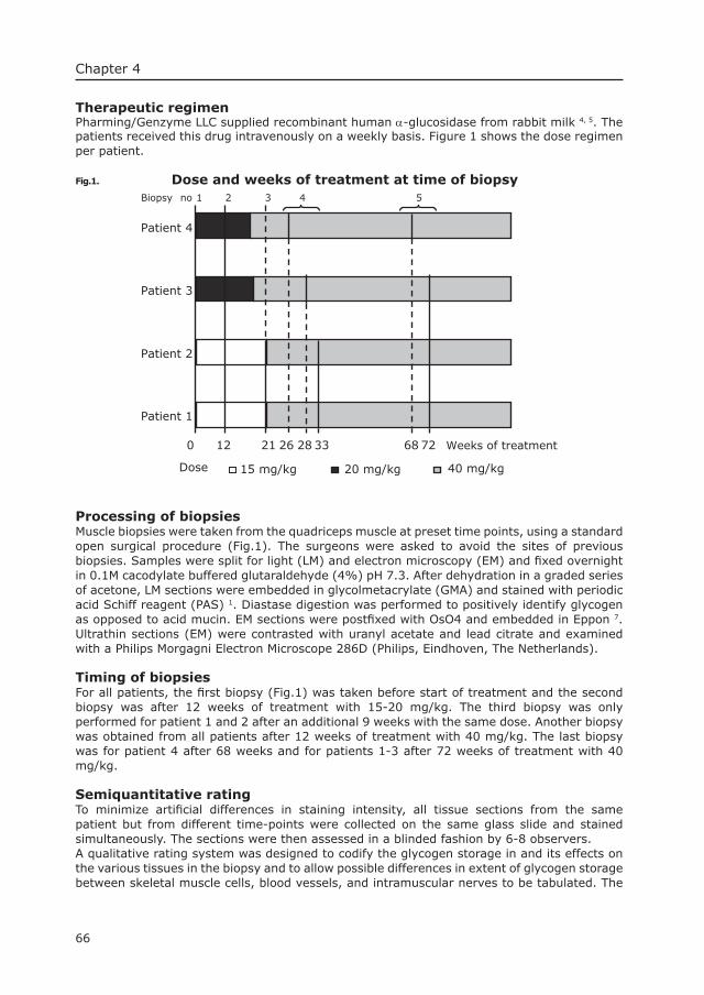
66
Chapter 4
Therapeutic regimenPharming/Genzyme LLC supplied recombinant human α-glucosidase from rabbit milk 4, 5. The patients received this drug intravenously on a weekly basis. Figure 1 shows the dose regimen per patient.
Fig.1. Dose and weeks of treatment at time of biopsy
Patient 1
Patient 2
Patient 3
Patient 4
Dose 20 mg/kg15 mg/kg 40 mg/kg
12 21 33 726828260
Biopsy no 1 2 3 4 5
Weeks of treatment
Processing of biopsiesMuscle biopsies were taken from the quadriceps muscle at preset time points, using a standard open surgical procedure (Fig.1). The surgeons were asked to avoid the sites of previous biopsies. Samples were split for light (LM) and electron microscopy (EM) and fi xed overnight in 0.1M cacodylate buffered glutaraldehyde (4%) pH 7.3. After dehydration in a graded series of acetone, LM sections were embedded in glycolmetacrylate (GMA) and stained with periodic acid Schiff reagent (PAS) 1. Diastase digestion was performed to positively identify glycogen as opposed to acid mucin. EM sections were postfi xed with OsO4 and embedded in Eppon 7. Ultrathin sections (EM) were contrasted with uranyl acetate and lead citrate and examined with a Philips Morgagni Electron Microscope 286D (Philips, Eindhoven, The Netherlands).
Timing of biopsiesFor all patients, the fi rst biopsy (Fig.1) was taken before start of treatment and the second biopsy was after 12 weeks of treatment with 15-20 mg/kg. The third biopsy was only performed for patient 1 and 2 after an additional 9 weeks with the same dose. Another biopsy was obtained from all patients after 12 weeks of treatment with 40 mg/kg. The last biopsy was for patient 4 after 68 weeks and for patients 1-3 after 72 weeks of treatment with 40 mg/kg.
Semiquantitative ratingTo minimize artifi cial differences in staining intensity, all tissue sections from the same patient but from different time-points were collected on the same glass slide and stained simultaneously. The sections were then assessed in a blinded fashion by 6-8 observers. A qualitative rating system was designed to codify the glycogen storage in and its effects on the various tissues in the biopsy and to allow possible differences in extent of glycogen storage between skeletal muscle cells, blood vessels, and intramuscular nerves to be tabulated. The
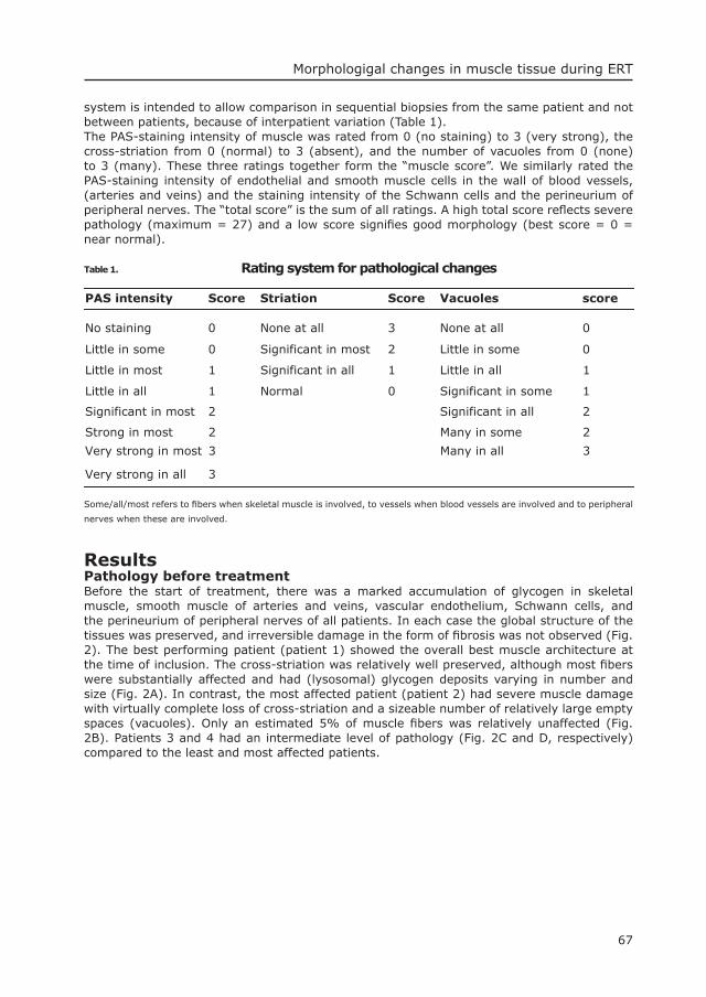
67
Morphologigal changes in muscle tissue during ERT
system is intended to allow comparison in sequential biopsies from the same patient and not between patients, because of interpatient variation (Table 1). The PAS-staining intensity of muscle was rated from 0 (no staining) to 3 (very strong), the cross-striation from 0 (normal) to 3 (absent), and the number of vacuoles from 0 (none) to 3 (many). These three ratings together form the “muscle score”. We similarly rated the PAS-staining intensity of endothelial and smooth muscle cells in the wall of blood vessels, (arteries and veins) and the staining intensity of the Schwann cells and the perineurium of peripheral nerves. The “total score” is the sum of all ratings. A high total score refl ects severe pathology (maximum = 27) and a low score signifi es good morphology (best score = 0 = near normal).
Table 1. Rating system for pathological changes
PAS intensity Score Striation Score Vacuoles score
No staining 0 None at all 3 None at all 0
Little in some 0 Significant in most 2 Little in some 0
Little in most 1 Significant in all 1 Little in all 1
Little in all 1 Normal 0 Significant in some 1
Significant in most 2 Significant in all 2
Strong in most 2 Many in some 2
Very strong in most 3 Many in all 3
Very strong in all 3
Some/all/most refers to fi bers when skeletal muscle is involved, to vessels when blood vessels are involved and to peripheral
nerves when these are involved.
ResultsPathology before treatment Before the start of treatment, there was a marked accumulation of glycogen in skeletal muscle, smooth muscle of arteries and veins, vascular endothelium, Schwann cells, and the perineurium of peripheral nerves of all patients. In each case the global structure of the tissues was preserved, and irreversible damage in the form of fi brosis was not observed (Fig. 2). The best performing patient (patient 1) showed the overall best muscle architecture at the time of inclusion. The cross-striation was relatively well preserved, although most fi bers were substantially affected and had (lysosomal) glycogen deposits varying in number and size (Fig. 2A). In contrast, the most affected patient (patient 2) had severe muscle damage with virtually complete loss of cross-striation and a sizeable number of relatively large empty spaces (vacuoles). Only an estimated 5% of muscle fi bers was relatively unaffected (Fig. 2B). Patients 3 and 4 had an intermediate level of pathology (Fig. 2C and D, respectively) compared to the least and most affected patients.
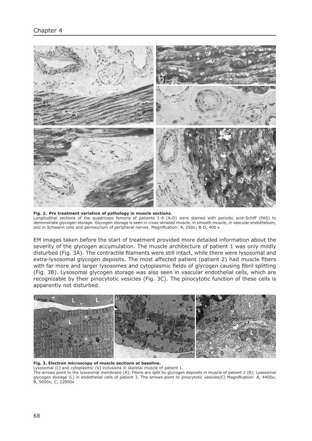
68
Chapter 4
Fig. 2. Pre treatment variation of pathology in muscle sections.Longitudinal sections of the quadriceps femoris of patients 1-4 (A-D) were stained with periodic acid-Schiff (PAS) to demonstrate glycogen storage. Glycogen storage is seen in cross-striated muscle, in smooth muscle, in vascular endothelium, and in Schwann cells and perineurium of peripheral nerves. Magnifi cation: A, 250x; B-D, 400 x
EM images taken before the start of treatment provided more detailed information about the severity of the glycogen accumulation. The muscle architecture of patient 1 was only mildly disturbed (Fig. 3A). The contractile fi laments were still intact, while there were lysosomal and extra-lysosomal glycogen deposits. The most affected patient (patient 2) had muscle fi bers with far more and larger lysosomes and cytoplasmic fi elds of glycogen causing fi bril splitting (Fig. 3B). Lysosomal glycogen storage was also seen in vascular endothelial cells, which are recognizable by their pinocytotic vesicles (Fig. 3C). The pinocytotic function of these cells is apparently not disturbed.
Fig. 3. Electron microscopy of muscle sections at baseline. Lysosomal (L) and cytoplasmic (V) inclusions in skeletal muscle of patient 1.The arrows point to the lysosomal membrane (A); Fibrils are split by glycogen deposits in muscle of patient 2 (B); Lysosomal glycogen storage (L) in endothelial cells of patient 3. The arrows point to pinocytotic vesicles(C) Magnifi cation: A, 4400x; B, 5600x; C, 22000x
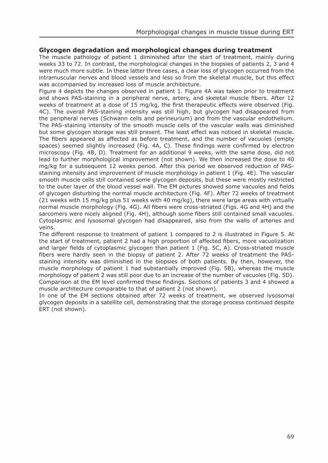
69
Morphologigal changes in muscle tissue during ERT
Glycogen degradation and morphological changes during treatmentThe muscle pathology of patient 1 diminished after the start of treatment, mainly during weeks 33 to 72. In contrast, the morphological changes in the biopsies of patients 2, 3 and 4 were much more subtle. In these latter three cases, a clear loss of glycogen occurred from the intramuscular nerves and blood vessels and less so from the skeletal muscle, but this effect was accompanied by increased loss of muscle architecture.Figure 4 depicts the changes observed in patient 1. Figure 4A was taken prior to treatment and shows PAS-staining in a peripheral nerve, artery, and skeletal muscle fi bers. After 12 weeks of treatment at a dose of 15 mg/kg, the fi rst therapeutic effects were observed (Fig. 4C). The overall PAS-staining intensity was still high, but glycogen had disappeared from the peripheral nerves (Schwann cells and perineurium) and from the vascular endothelium. The PAS-staining intensity of the smooth muscle cells of the vascular walls was diminished but some glycogen storage was still present. The least effect was noticed in skeletal muscle. The fi bers appeared as affected as before treatment, and the number of vacuoles (empty spaces) seemed slightly increased (Fig. 4A, C). These fi ndings were confi rmed by electron microscopy (Fig. 4B, D). Treatment for an additional 9 weeks, with the same dose, did not lead to further morphological improvement (not shown). We then increased the dose to 40 mg/kg for a subsequent 12 weeks period. After this period we observed reduction of PAS-staining intensity and improvement of muscle morphology in patient 1 (Fig. 4E). The vascular smooth muscle cells still contained some glycogen deposits, but these were mostly restricted to the outer layer of the blood vessel wall. The EM pictures showed some vacuoles and fi elds of glycogen disturbing the normal muscle architecture (Fig. 4F). After 72 weeks of treatment (21 weeks with 15 mg/kg plus 51 weeks with 40 mg/kg), there were large areas with virtually normal muscle morphology (Fig. 4G). All fi bers were cross-striated (Figs. 4G and 4H) and the sarcomers were nicely aligned (Fig. 4H), although some fi bers still contained small vacuoles. Cytoplasmic and lysosomal glycogen had disappeared, also from the walls of arteries and veins.The different response to treatment of patient 1 compared to 2 is illustrated in Figure 5. At the start of treatment, patient 2 had a high proportion of affected fi bers, more vacuolization and larger fi elds of cytoplasmic glycogen than patient 1 (Fig. 5C, A). Cross-striated muscle fi bers were hardly seen in the biopsy of patient 2. After 72 weeks of treatment the PAS-staining intensity was diminished in the biopsies of both patients. By then, however, the muscle morphology of patient 1 had substantially improved (Fig. 5B), whereas the muscle morphology of patient 2 was still poor due to an increase of the number of vacuoles (Fig. 5D). Comparison at the EM level confi rmed these fi ndings. Sections of patients 3 and 4 showed a muscle architecture comparable to that of patient 2 (not shown).In one of the EM sections obtained after 72 weeks of treatment, we observed lysosomal glycogen deposits in a satellite cell, demonstrating that the storage process continued despite ERT (not shown).
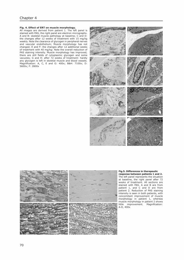
70
Chapter 4
Fig. 4. Effect of ERT on muscle morphology. All images are derived from patient 1. The left panel is stained with PAS, the right panel are electron micrographs. A and B: skeletal muscle pathology at baseline; C and D: the changes after 12 weeks of treatment with 15 mg/kg weekly. Note the clearance of glycogen in peripheral nerves and vascular endothelium; Muscle morphology has not changed; E and F: the changes after 12 additional weeks of treatment with 40 mg/kg: Note the overall reduction of PAS staining intensity. Muscle morphology has improved; there are still fi elds of cytoplasmic glycogen and some vacuoles; G and H: after 72 weeks of treatment: hardly any glycogen is left in skeletal muscle and blood vessels. Magnifi cation: A, C, E and G: 400x; B&H: 7100x; D: 5600x; F: 2800x
Fig.5. Differences in therapeutic response between patients 1 and 2. The left panel represents the situation at baseline, the right panel after 72 weeks of treatment. All sections are stained with PAS. A and B are from patient 1, and C and D are from patient 2. Reduction of PAS staining intensity is seen in both patients, with concomitant improvement of muscle morphology in patient 1, whereas muscle morphology in patient 2 shows little improvement. Magnifi cation: A-D, 400x
71
Morphologigal changes in muscle tissue during ERT
Clinical response to treatmentAfter 72 weeks of treatment, all patients had normal muscle α-glucosidase activity. The glycogen concentration in the muscle of patients 2, 3 and 4 had not changed signifi cantly, but was decreased to normal in patient 1 (unpublished results). Patient 1 also showed signifi cant clinical improvement. He achieved normal major milestones of motor development (rolling, sitting, standing and walking) as established by AIMS scores. Patient 2 initially showed some improvement of skeletal muscle function. She learned to move her arms and had improved head balance, but lost these abilities again after a series of pulmonary infections. Patient 4 learned to sit with support, but did not reach the milestones of standing and walking. Her arm function improved signifi cantly. Patient 3 learned to sit without support, could move her legs, but could not stand. All four patients were in a better clinical condition after 72 weeks of treatment than they were at entry into the trial 24, 25.
Semiquantitative rating of muscle pathologyPAS-stained muscle tissue sections were microscopically inspected and ratings were given for PAS-staining intensity, number and size of vacuoles and cross-striation as outlined in the Methods section (Table 1). The muscle score at inclusion was 8 for the most affected patient (patient 2) and 7 for the others (Table 2). The total score was 23 for patient 2 and 25 for the other three patients. After 12 weeks of treatment none of the patients had an improved muscle score, but the total score had fallen to 13-14. This was mainly due to a lower rating of the PAS-staining intensity of the endothelium, peripheral nerves, and smooth muscle cells of the blood vessels. After the following 12 weeks of treatment with 40 mg/kg, the total score was further diminished by a lower rating of the PAS-staining intensity of the muscle. After 72 weeks of treatment, patient 1 had a near-normal total score. Treatment of the other three patients led to a lowering of the total score but their muscle score stayed the same because the diminished PAS-staining intensity was counterbalanced by an increase of vacuolization.
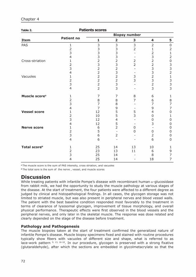
72
Chapter 4
Table 2. Patients scores
Patient noBiopsy number
Item 1 2 3 4 5
PAS 1 3 3 3 2 02 3 3 2 1 23 3 3 - 2 14 3 3 - 3 2
Cross-striation 1 2 2 2 2 02 3 3 2 2 33 2 2 - 3 34 2 3 - 3 2
Vacuoles 1 2 2 3 2 12 2 2 3 3 33 2 3 - 2 34 2 3 - 3 3
Muscle scorea 1 7 7 8 6 12 8 8 7 6 83 7 8 - 7 74 7 9 - 9 7
Vessel score 1 12 5 5 4 02 10 5 3 0 13 12 4 - 0 04 12 5 - 3 0
Nerve score 1 6 2 0 - 02 5 - 0 0 03 6 2 - 2 04 6 - - 6 0
Total scoreb 1 25 14 13 10 12 23 13 11 6 93 25 14 - 8 74 25 14 - 18 7
a The muscle score is the sum of PAS intensity, cross striation, and vacuolesb The total sore is the sum of the nerve , vessel, and muscle scores
Discussion While treating patients with infantile Pompe’s disease with recombinant human α-glucosidase from rabbit milk, we had the opportunity to study the muscle pathology at various stages of the disease. At the start of treatment, the four patients were affected to a different degree as judged by clinical and histopathological fi ndings. In all cases, the glycogen storage was not limited to striated muscle, but was also present in peripheral nerves and blood vessel walls. The patient with the best baseline condition responded most favorably to the treatment in terms of clearance of lysosomal glycogen, improvement of tissue morphology, and overall physical performance. Therapeutic effects were fi rst observed in the blood vessels and the peripheral nerves, and only later in the skeletal muscle. The response was dose related and clearly depended on the stage of the disease before treatment.
Pathology and PathogenesisThe muscle biopsies taken at the start of treatment confi rmed the generalized nature of infantile Pompe’s disease. Muscle biopsy specimens fi xed and stained with routine procedures typically show fi bers with vacuoles of different size and shape, what is referred to as lace-work pattern 9, 13, 16-18. In our procedure, glycogen is preserved with a strong fi xative (glutaraldehyde), after which the sections are embedded in glycolmetacrylate so that the
73
Morphologigal changes in muscle tissue during ERT
extent of glycogen storage is dramatically exhibited. Before treatment, all four patients had severely affected skeletal muscle fi bers. However, not all muscle fi bers in the same section of the same patient were equally affected, and there was interpatient variation. Moreover, we noticed that structural damage can vary substantially along the fi ber length, as if the storage process spreads from discrete foci.The biopsies taken prior to and during treatment are informative about the possible course of the pathogenic process, which starts in lysosomes. As the lysosomal compartment expands, it can be envisioned that the contractile machinery of the muscle is hampered by fi bril splitting resulting in diminished vectorial strength 12. Reduced lysosomal function and rupture of the lysosomal membrane by contractile forces will lead to an increase of the cytoplasmic glycogen concentration. Increased autophagy results in even more lysosomal storage and dysfunction. Release of lysosomal enzymes into the cytoplasm contributes to the cascade of damage.The glycogen storage in smooth muscle cells of the arteries and veins is quite dramatic. It is conceivable that the integrity of the blood vessel wall is lost as the disease progresses. This may explain the occurrence of aneurysms in the basal arteries of patients with Pompe’s disease as described in some case reports 6, 15, 19. We have also seen vascular pathology in the knockout mouse model of Pompe’s disease 3, and storage of glycogen in smooth muscle along the digestive tract and in other organs with a smooth muscle component. Storage in Schwann cells and epineurium of peripheral nerves also was noted earlier, but is not known to have functional consequences 17, 18.
Therapeutic response and dosingThe vascular endothelium and the peripheral nerves are the fi rst sites where a response to treatment is noted. After 12 weeks of low-dose treatment (15 or 20 mg/kg), the glycogen had disappeared from the endothelial and Schwann cells, but it was still present in skeletal muscle fi bers and vascular smooth muscle cells. This is not unexpected as the endothelial cells are directly exposed to the circulating enzyme, whereas Schwann cells and perineurium are optimally supplied by the anastomosing vascular system that feeds the nerve bundles. We consider it likely that cell- and tissue-specifi c differences in accessibility explain in part the much lower dose of α-galactosidase needed for the treatment of Fabry’s disease (0.2-1 mg/kg) 8, 20 compared to the dose needed to correct the muscle pathology in Pompe’s disease. In Pompe’s disease the administered α-glucosidase must pass the endothelial barrier and the endomysium before it reaches the muscle fi bers.Judging from the changes in muscle morphology over time, we believe that the dose of 15-20 mg/kg of α-glucosidase, during the fi rst 21 weeks of treatment, was insuffi cient to achieve correction of smooth and skeletal muscle. The higher dose of 40 mg/kg seems necessary because the PAS-staining intensity became less, and the vascular smooth muscle started to be cleared after 12 weeks of additional treatment at this dose. The skeletal muscle morphology of the least affected patient (patient 1) had clearly improved after 72 weeks of treatment.The rating system that we used to evaluate the morphological changes during the course of treatment proved to be helpful in obtaining objective measures. The total scores and muscle scores correlated well with the clinical observations.
Therapeutic mechanism and differential responseEven at the high dose of 40 mg/kg per week, not all patients responded suffi ciently well. This may relate to the different glycogen load and the associated muscle immobility of the patients prior to and during treatment. From both aspects, patient 1 had the best starting point with the least glycogen accumulation and the best muscle function.The various muscle sections revealed that fi bers with solely lysosomal glycogen storage maintained a relatively normal sarcomeric organization and were cross-striated. It is to be expected that normal function is regained after clearance of lysosomal glycogen by ERT. Likewise, it is conceivable that massive accumulation of cytoplasmic glycogen complicates the repair process. An immobile volume of cytoplasmic glycogen mechanically blocks the intracellular/vesicular transport pathways required for lysosomal targeting of the therapeutic
74
Chapter 4
enzyme. Moreover, when the lysosomal compartment is emptied, autophagic activity is needed to clear the cytoplasmic glycogen. This process requires a proper intracellular organization, which is completely lost at a severe stage of the disease.Muscle movement is required for activation, proliferation, and fusion of myogenic satellite cells with existing muscle fi bers 11, 21, 22, 29. Patient 1 had substantial muscle strength in his legs at the start of treatment and he currently walks, whereas patients 2 and 4 could not move their legs, and their muscle function is still extremely compromised. Patient 3 could move her legs, but could not stand.
ConclusionWe have shown that intravenous administration of recombinant human α-glucosidase from rabbit milk can improve muscle morphology in classic infantile Pompe’s disease. Preservation of muscle architecture and residual muscle function at start of treatment seems a prerequisite for the successful outcome of treatment. A high dose of enzyme is required to obtain an effect on skeletal muscle.
AcknowledgementsWe are grateful to Hans van Hirtum for help with the processing and analysis of the light microscopic sections, to Piet van der Heul for analysis of the electron microscopy sections, to Marian Kroos for expert advice, and to Ruud Koppenol for the photography. Financial support was obtained from the Sophia Foundation for Medical Research, and recombinant human α-glucosidase was supplied by Pharming /Genzyme LLC.
References1. Bijvoet AG, van de Kamp EH, Kroos MA, Ding JH, Yang BZ, Visser P, Bakker CE, Verbeet MP,
Oostra BA, Reuser AJ, Van der Ploeg AT. Generalized glycogen storage and cardiomegaly in a knockout mouse model of Pompe disease. Hum Mol Genet 1998;7:53-62.
2. Bijvoet AG, Van Hirtum H, Kroos MA, Van de Kamp EH, Schoneveld O, Visser P, Brakenhoff JP, Weggeman M, Van Corven EJ, Van der Ploeg AT, Reuser AJ. Human acid alpha-glucosidase from rabbit milk has therapeutic effect in mice with glycogen storage disease type II. Hum Mol Genet 1999;8:2145-2153.
3. Bijvoet AG, Van Hirtum H, Vermey M, Van Leenen D, Van der Ploeg AT, Mooi WJ, Reuser AJ. Pathological features of glycogen storage disease type II highlighted in the knockout mouse model. J Pathol 1999;189:416-424.
4. Bijvoet AGA, Kroos MA, Pieper FR, de Boer HA, Reuser AJJ, Van der Ploeg AT, Verbeet MP. Expression of cDNA-encoded human acid α-glucosidase in milk of transgenic mice. Biochim Biophys Acta
1996;1308:93-96.5. Bijvoet AGA, Kroos MA, Pieper FR, Van der Vliet M, De Boer HA, Van der Ploeg AT, Verbeet MP, Reuser
AJJ. Recombinant human acid α-glucosidase: high level production in mouse milk, biochemical characteristics, correction of enzyme defi ciency in GSDII KO mice. Hum Mol Genet 1998;7:1815-
1824.6. Braunsdorf WE. Fusiform aneurysm of basilar artery and ectatic internal carotid arteries associated
with glycogenosis type 2 (Pompe’s disease). Neurosurgery 1987;21:748-749.7. De Bruijn WC. Glycogen, its chemistry and morphologic appearance in the electron microscope. I. A
modifi ed OsO4 fi xative which selectively contrasts glycogen. J Ultrastruct Res 1973;42:29-50.8. Eng CM, Guffon N, Wilcox WR, Germain DP, Lee P, Waldek S, Caplan L, Linthorst GE, Desnick RJ.
Safety and effi cacy of recombinant human alpha-galactosidase A replacement therapy in Fabry’s disease. N Engl J Med 2001;345:9-16.
9. Engel AG, Gomez MR, Seybold ME, Lambert EH. The spectrum and diagnosis of acid maltase defi ciency. Neurology 1973;23:95-106.
10. Fuller M, Van der Ploeg A, Reuser AJJ, Anson DS, Hopwood JJ. Isolation and characterisation of a recombinant, precursor form of lysosomal acid α-glucosidase. Eur J Biochem 1995;234:903-909.
11. Hawke TJ, Garry DJ. Myogenic satellite cells: physiology to molecular biology. J Appl Physiol 2001;91:534-551.
12. Hesselink RP, Gorselink M, Schaart G, Wagenmakers AJ, Kamphoven J, Reuser AJ, Van der Vusse G J, Drost MR. Impaired performance of skeletal muscle in alpha-glucosidase knockout mice. Muscle Nerve 2002;25:873-883.
75
Morphologigal changes in muscle tissue during ERT
13. Hirschhorn R, Reuser AJJ. Glycogen Storage Disease type II; acid α-glucosidase (acid maltase) defi ciency. In: Scriver CR, Beaudet AL, Valle D, Sly W.S., editors. The metabolic & molecular bases of inherited diseases. New York: McGraw-Hill; 2000. p. 3389-3420.
14. Kikuchi T, Yang HW, Pennybacker M, Ichihara N, Mizutani M, Van Hove JLK, Chen YT. Clinical and metabolic correction of Pompe disease by enzyme therapy in acid maltase-defi cient quail. J Clin Invest 1998;101:827-833.
15. Makos MM, McComb RD, Adickes ED, Bennett DR. Acid maltase defi ciency and basilar artery aneurysms: a report of a sibship. Neurology 1985;35 (Suppl 1):193-194.
16. Mancall EL, Aponte GE, Berry RG. Pompe’s disease (diffuse glycogenosis) with neuronal storage. J Neuropathol Exp Neurol 1965;24:85-96.17. Martin JJ, De Barsy T, De Schrijver LJG, Palladini G. Acid maltase defi ciency (type II glycogenosis).
Morphological and biochemical study of a childhood phenotype. J Neurol Sci 1976;30:155-166.18. Martin JJ, De Barsy T, Van Hoof F, Palladini G. Pompe’s disease: an inborn lysosomal disorder with
storage of glycogen. A study of brain and striated muscle. Acta Neuropathol 1973;23:229-244.19. Matsuoka Y, Senda Y, Hirayama M, Matsui T, Takahashi A. Late-onset acid maltase defi ciency
associated with intracranial aneurysm. J Neurol 1988;235:371-373.20. Schiffmann R, Kopp JB, Austin HA, 3rd, Sabnis S, Moore DF, Weibel T, Balow JE, Brady RO. Enzyme
replacement therapy in Fabry disease: a randomized controlled trial. JAMA 2001;285:2743-2749.21. Schultz E, Jaryszak DL, Gibson MC, Albright DJ. Absence of exogenous satellite cell contribution to
regeneration of frozen skeletal muscle. J Muscle Res Cell Motil 1986;7:361-367.22. Schultz E, McCormick KM. Skeletal muscle satellite cells. Rev Physiol Biochem Pharmacol 1994;123:
213-257.23. Slonim AE, Bulone L, Ritz S, Goldberg T, Chen A, Martiniuk F. Identifi cation of two subtypes of
infantile acid maltase defi ciency. J Pediatr 2000;137:283-285.24. Van den Hout H, Reuser AJ, Vulto AG, Loonen MC, Cromme-Dijkhuis A, Van der Ploeg AT. Recombinant
human alpha-glucosidase from rabbit milk in Pompe patients. Lancet 2000;356:397-398.25. Van den Hout JM, Reuser AJ, de Klerk JB, Arts WF, Smeitink JA, Van der Ploeg AT. Enzyme therapy for
Pompe disease with recombinant human alpha- glucosidase from rabbit milk. J Inherit Metab Dis 2001;24:266-274.26. Van der Ploeg AT, Bolhuis PA, Wolterman RA, Visser JW, Loonen MCB, Busch HFM, Reuser AJJ.
Prospect for enzyme therapy in glycogenosis II variants: a study on cultured muscle cells. J Neurol 1988;235:392-396.
27. Van der Ploeg AT, Kroos M, Van Dongen JM, Visser WJ, Bolhuis PA, Loonen MC, Reuser AJJ. Breakdown of lysosomal glycogen in cultured fi broblasts from glycogenosis type II patients after uptake of acid α-glucosidase. J Neurol Sci 1987;79:327-336.
28. Van Hove JL, Yang HW, Wu JY, Brady RO, Chen YT. High-level production of recombinant human lysosomal acid alpha-glucosidase in Chinese hamster ovary cells which targets to heart muscle and corrects glycogen accumulation in fi broblasts from patients with Pompe disease.
Proc Natl Acad Sci U S A 1996;93:65-70.29. Watt DJ, Morgan JE, Clifford MA, Partridge TA. The movement of muscle precursor cells between
adjacent regenerating muscles in the mouse. Anat Embryol 1987;175:527-536.

5Hearing loss in Infantile Pompe’s Disease and Determination of Underlying
Pathology in the Knockout MouseJoep H. J. Kamphoven1,2, Martijn M. de Ruiter3, Leon P. F. Winkel2, Hannerieke M.
P. Van den Hout2, Jan Bijman3, Chris I. de Zeeuw3, Hans L. Hoeve4,Bert A. Van Zanten4, Ans T. Van der Ploeg2, and Arnold J.J. Reuser1
From the departments of: ¹Clinical Genetics, ²Pediatrics, Division of Metabolic Diseases and Genetics,3Neuroscience, and 4Pediatric-rhino-otolaryngeology, Erasmus MC, Rotterdam, The Netherlands
-in press, Neurobiology of Disease 2004-

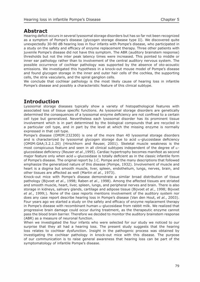
Chapter 5
79
AbstractHearing defi cit occurs in several lysosomal storage disorders but has so far not been recognized as a symptom of Pompe’s disease (glycogen storage disease type II). We discovered quite unexpectedly 30-90 dB hearing loss in four infants with Pompe’s disease, who participated in a study on the safety and effi cacy of enzyme replacement therapy. Three other patients with juvenile Pompe’s disease did not have this symptom. The ABR (auditory brainstem response) thresholds but not the inter peak latency times were increased. This pointed to middle or inner ear pathology rather than to involvement of the central auditory nervous system. The possible occurrence of cochlear pathology was supported by the absence of oto-acoustic emissions. We investigated this hypothesis in a knock-out mouse model of Pompe’s disease and found glycogen storage in the inner and outer hair cells of the cochlea, the supporting cells, the stria vascularis, and the spiral ganglion cells. We conclude that cochlear pathology is the most likely cause of hearing loss in infantile Pompe’s disease and possibly a characteristic feature of this clinical subtype.
IntroductionLysosomal storage diseases typically show a variety of histopathological features with associated loss of tissue specifi c functions. As lysosomal storage disorders are genetically determined the consequences of a lysosomal enzyme defi ciency are not confi ned to a certain cell type but generalized. Nevertheless each lysosomal disorder has its prominent tissue involvement which is in part determined by the biological compounds that are recycled in a particular cell type, and in part by the level at which the missing enzyme is normally expressed in that cell type.Pompe’s disease (OMIM:232300) is one of the more than 40 lysosomal storage disorders and is characterized by lysosomal glycogen storage due to acid α-glucosidase defi ciency (OMIM:GAA;3.2.1.20) (Hirschhorn and Reuser, 2001). Skeletal muscle weakness is the most conspicuous feature and seen in all clinical subtypes independent of the degree of α-glucosidase defi ciency (Reuser et al.,1995). Cardiac hypertrophy becomes manifest as second major feature only when acid α-glucosidase is totally defi cient as in the classic infantile form of Pompe’s disease. The original report by J.C. Pompe and the many descriptions that followed emphasize the generalized nature of this disease (Pompe, 1932). Involvement of muscle and heart is a dogma but smooth muscle, liver, spleen, endothelium, lungs, nerves, brain, and other tissues are affected as well (Martin et al., 1973).Knock-out mice with Pompe’s disease demonstrate a similar broad distribution of tissue pathology (Bijvoet et al., 1998; Raben et al., 1998). Among the affected tissues are striated and smooth muscle, heart, liver, spleen, lungs, and peripheral nerves and brain. There is also storage in kidneys, salivary glands, cartilage and adipose tissue (Bijvoet et al., 1998; Bijvoet et al., 1999;). None of the case reports mentions involvement of the auditory system nor does any case report describe hearing loss in Pompe’s disease (Van den Hout, et al., 2003). Four years ago we started a study on the safety and effi cacy of enzyme replacement therapy in Pompe’s disease with recombinant human α-glucosidase from rabbit milk. We realized that progressive brain damage could occur during treatment, as the therapeutic enzyme cannot pass the blood brain barrier. Therefore we decided to monitor the auditory brainstem response (ABR) as a measure of neuronal function. When we investigated the four infants who were selected for our study we noticed to our surprise that they all had a hearing loss. The present study suggests that the hearing loss relates to cochlear dysfunction. Insight in the pathogenic process was obtained by investigating the cochlear pathology in knock-out mice with this disease. The purpose of our communication is to raise general awareness that hearing loss can be part of the symptomatology of infantile Pompe’s disease.
Hearing loss in infantile Pompe’s Disease
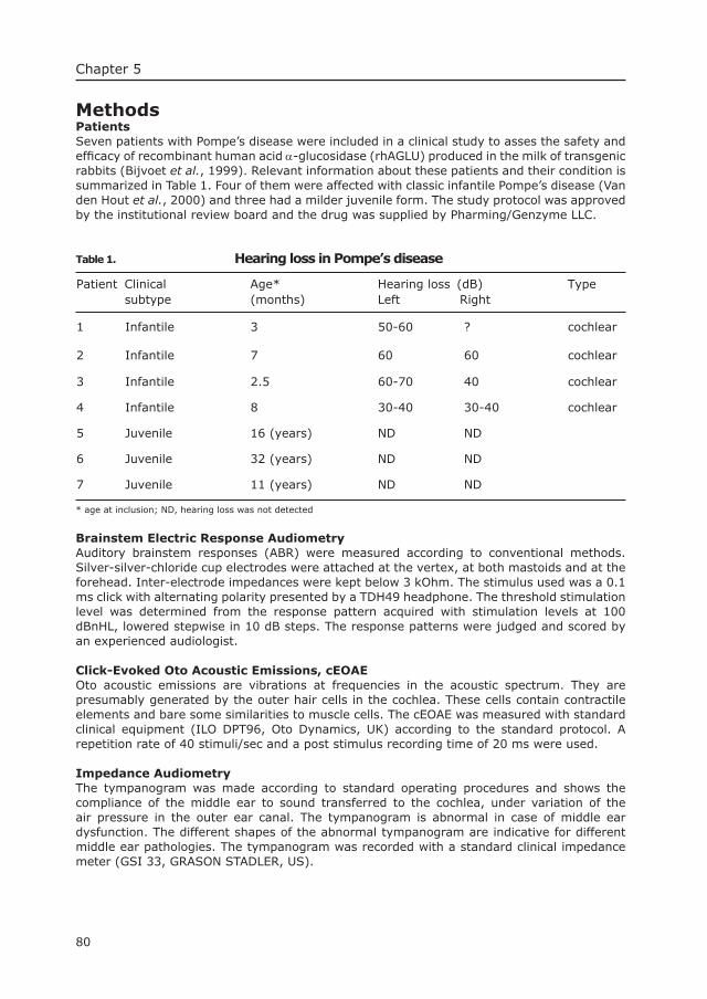
80
Chapter 5
MethodsPatientsSeven patients with Pompe’s disease were included in a clinical study to asses the safety and effi cacy of recombinant human acid α-glucosidase (rhAGLU) produced in the milk of transgenic rabbits (Bijvoet et al., 1999). Relevant information about these patients and their condition is summarized in Table 1. Four of them were affected with classic infantile Pompe’s disease (Van den Hout et al., 2000) and three had a milder juvenile form. The study protocol was approved by the institutional review board and the drug was supplied by Pharming/Genzyme LLC.
Table 1. Hearing loss in Pompe’s disease
Patient Clinical subtype
Age* (months)
Hearing loss (dB)Left Right
Type
1 Infantile 3 50-60 ? cochlear
2 Infantile 7 60 60 cochlear
3 Infantile 2.5 60-70 40 cochlear
4 Infantile 8 30-40 30-40 cochlear
5 Juvenile 16 (years) ND ND
6 Juvenile 32 (years) ND ND
7 Juvenile 11 (years) ND ND
* age at inclusion; ND, hearing loss was not detected
Brainstem Electric Response AudiometryAuditory brainstem responses (ABR) were measured according to conventional methods. Silver-silver-chloride cup electrodes were attached at the vertex, at both mastoids and at the forehead. Inter-electrode impedances were kept below 3 kOhm. The stimulus used was a 0.1 ms click with alternating polarity presented by a TDH49 headphone. The threshold stimulation level was determined from the response pattern acquired with stimulation levels at 100 dBnHL, lowered stepwise in 10 dB steps. The response patterns were judged and scored by an experienced audiologist.
Click-Evoked Oto Acoustic Emissions, cEOAEOto acoustic emissions are vibrations at frequencies in the acoustic spectrum. They are presumably generated by the outer hair cells in the cochlea. These cells contain contractile elements and bare some similarities to muscle cells. The cEOAE was measured with standard clinical equipment (ILO DPT96, Oto Dynamics, UK) according to the standard protocol. A repetition rate of 40 stimuli/sec and a post stimulus recording time of 20 ms were used.
Impedance AudiometryThe tympanogram was made according to standard operating procedures and shows the compliance of the middle ear to sound transferred to the cochlea, under variation of the air pressure in the outer ear canal. The tympanogram is abnormal in case of middle ear dysfunction. The different shapes of the abnormal tympanogram are indicative for different middle ear pathologies. The tympanogram was recorded with a standard clinical impedance meter (GSI 33, GRASON STADLER, US).
81
Hearing loss in infantile Pompe’s Disease
Pure tone AudiogramThe pure tone audiogram was made with standard equipment (OB822, Madsen, Denmark). Pure tone threshold levels indicate the ear’s sensitivity to sound of frequencies between 0.25 and 8 kHz. The hearing loss is expressed in dB. The normal range is -10 to +10 dB.
MiceThe mice used in this study are completely defi cient in acid α-glucosidase and were obtained by targeted disruption of exon 13 of the GAA gene. The line was crossed back for over 10 generations in the FVB background (Bijvoet et al., 1998). The mouse model mimics the infantile form of Pompe’s disease both biochemically and pathologically and has generalized glycogen storage. Mice in different age groups were selected for ABR measurement and investigation of inner ear pathology. All experiments were conducted in accordance with the “Guiding Principles in the care and use of Laboratory animals” as approved by the animal care committee of the Erasmus MC Rotterdam, the Netherlands
Brainstem Electric Response Audiometry in MiceTo enable ABR measurements in alert mice, platinum electrodes were mounted in an acrylic pedestal and fi xed to the skull. To do so the mice were anaesthetized with a mixture of O2, N2O and 2% halothane. The two recording electrodes were placed in previously localized spots in the left and right occipital bone (Crus 2 level of the Cerebellum). The ground electrode was located in the frontal bone plate. The central reference electrode was fi xed between the two parietal bones as close to the sagital suture as possible. Both recording electrodes and the reference electrode were wired to a three-poled connector and fi xed to the skull by use of dental cement. At the time of measurement, the mice were placed in a fi xed position inside an insulated chamber so that sound stimuli could be presented from a constant distance (40 mm from the external auditory canals). Thresholds were obtained in alert mice at frequencies of 4, 8, 16 and 32 kHz (duration 1 ms and tone pip repetition rate of 80/s) and clicks (repetition rate: 23 and 80/s). Responses were averaged to 500 pips of alternating polarity. Several age groups of mice (Table 2) were included to study the progress of hearing loss.
HistologyMice were anaesthetized with ketamine and xylazine (25 and 5 mg/kg, respectively), and the systemic circulation was perfused with a physiological salt solution (saline) to expel the circulating blood. The brain was taken out with forceps, the temporal bones were removed from the skull and the bony labyrinth was separated from the middle ear along a suture line (Bohne and Harding, 1997). Specimens were fi xed in glutaraldehyde and paraformaldehyde in a concentration of 1% and 4%, respectively, dehydrated in a graded series of alcohol and embedded in Durcupan (Fluka) or paraffi n. The unstained total cochlea’s were contrasted with OsO4 before dehydration and embedding in Durcupan, or decalcifi ed using EDTA before dehydration and embedding in paraffi n. Periodic acid Schiff (PAS) staining was used to visualize lysosomal glycogen storage in light microscopy. For electron microscopy (EM), the ultrathin sections were contrasted with uranylacetate and lead citrate (De Bruijn and Den Beejen, 1975)
ResultsPatients at baseline and during treatmentSeven patients with Pompe’s disease were enrolled in a study on the safety and effi cacy of Enzyme Replacement Therapy (ERT) with recombinant human acid α-glucosidase from rabbit milk. Four of these patients had the classic infantile form of Pompe’s disease and three the late onset juvenile form (Table 1). All seven patients received weekly enzyme infusions and underwent diagnostic tests before and during treatment.Brainstem audiometry (ABR) was included in the clinical protocol as one of the tools to monitor Central Nervous System (CNS) involvement in the infants with classic Pompe’s disease. The fi rst ABR measurement was performed in the week before the fi rst enzyme infusion and
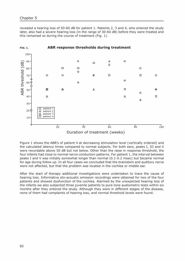
82
Chapter 5
revealed a hearing loss of 50-60 dB for patient 1. Patients 2, 3 and 4, who entered the study later, also had a severe hearing loss (in the range of 30-60 dB) before they were treated and this remained so during the course of treatment (Fig. 1).
FIG. 1. ABR response thresholds during treatment
10
20
30
40
50
60
70
80
90
100
20 40 60 80 100
Duration of treatment (weeks)
ABR t
hre
shol
d (d
B)
patient 1patient 2patient 3patient 4
Figure 1 shows the ABR’s of patient 4 at decreasing stimulation level (vertically ordered) and the calculated latency times compared to normal subjects. For both ears, peaks I, III and V were recordable above 50 dB but not below. Other than the raise in response threshold, the four infants had close to normal nerve conduction patterns. For patient 1, the interval between peaks I and V was initially somewhat longer than normal (0.1-0.2 msec) but became normal for age during follow up. In all four cases we concluded that the brainstem and auditory nerve were not affected, but that the problem was located in the cochlea or middle ear.
After the start of therapy additional investigations were undertaken to trace the cause of hearing loss. Informative oto-acoustic emission recordings were obtained for two of the four patients and showed dysfunction of the cochlea. Alarmed by the unexpected hearing loss of the infants we also subjected three juvenile patients to pure tone audiometric tests within six months after they entered the study. Although they were in different stages of the disease, none of them had complaints of hearing loss, and normal threshold levels were found.
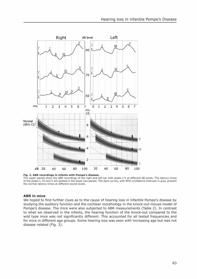
83
Hearing loss in infantile Pompe’s Disease
Fig. 2. ABR recordings in infants with Pompe’s disease.The upper panels show the ABR recordings of the right and left ear with peaks I-V at different dB levels. The latency times of the peaks I, III and V are plotted in the lower two panels. The dark curves, with 95% confi dence intervals in gray, present the normal latency times at different sound levels.
ABR in miceWe hoped to fi nd further clues as to the cause of hearing loss in infantile Pompe’s disease by studying the auditory function and the cochlear morphology in the knock-out mouse model of Pompe’s disease. The mice were also subjected to ABR measurements (Table 2). In contrast to what we observed in the infants, the hearing function of the knock-out compared to the wild type mice was not signifi cantly different. This accounted for all tested frequencies and for mice in different age groups. Some hearing loss was seen with increasing age but was not disease related (Fig. 3).
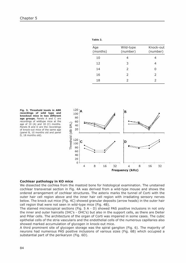
84
Chapter 5
Table 2.
Age (months)
Wild-type (number)
Knock-out(number)
10 4
12 4
14 0
16 2
18
4
3
2
2
2 2
Cochlear pathology in KO miceWe dissected the cochlea from the mastoid bone for histological examination. The unstained cochlear transversal section in Fig. 4A was derived from a wild-type mouse and shows the ordered arrangement of cochlear structures. The asterix marks the tunnel of Corti with the outer hair cell region above and the inner hair cell region with irradiating sensory nerves below. The knock out mice (Fig. 4C) showed granular deposits (arrow heads) in the outer hair cell region that were not seen in wild-type mice (Fig. 4B).The stained microscopical sections (Fig. 5 A - D) showed PAS positive inclusions in not only the inner and outer haircells (IHC’s - OHC’s) but also in the support cells, as there are Deiter and Pillar cells. The architecture of the organ of Corti was impaired in some cases. The cubic epithelial cells of the stria vascularis and the endothelial cells of the numerous capillaries also showed marked accumulation of glycogen in knock-out mice. A third prominent site of glycogen storage was the spiral ganglion (Fig. 6). The majority of neurons had numerous PAS positive inclusions of various sizes (Fig. 6B) which occupied a substantial part of the perikaryon (Fig. 6D).
Fig. 3. Threshold levels in ABR recordings of wild type and knockout mice in two different age groups. Panels A and C are recordings of wildtype mice at the age of 10 (A) and 18 (C) months. Panels B and D are the recordings of knock-out mice of the same age (panel B, 10 months old and panel D, 18 months old).
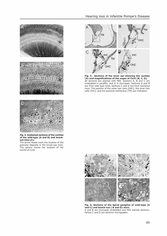
85
Hearing loss in infantile Pompe’s Disease
Fig. 4. Unstained sections of the cochlea of the wild-type (A and B) and knock-out mice (C).The arrow heads mark the location of the granular deposits in the knock-out mice. The asterix marks the location of the tunnel of Corti.
A
B
C
Fig. 5. Sections of the inner ear showing the cochlea (A) and magnifi cations of the organ of Corti (B, C, D). All sections are stained with PAS. Sections A, B and C are embedded in paraffi n, and D in Durcupan. Sections A and B are from wild type mice, sections C and D are from knockout mice. The position of the outer hair cells (OHC), the inner hair cells (IHC), and the tectorial membrane (TM) are indicated.
Fig. 6. Sections of the Spiral ganglion of wild-type (A and C) and knock-out ( B and D) mice.A and B are Durcupan embedded and PAS stained sections. Panels C and D are electron micrographs.
86
Chapter 5
DiscussionHearing loss in Pompe’s DiseaseHearing-loss occurs in several lysosomal storage disorders (Hayes et al., 1980; Brown et al., 1981; Lacey and Terplan, 1984; Peck, 1984; Morgan et al., 1990; d’Azzo et al., 2001; Thomas, 2001) and has different causes. The conductive hearing loss in the Mucopolysaccharide storage diseases results from ossicles articular alterations and occurs in this group of diseases together with sensorineural hearing loss (Peck, 1984; Hayes et al., 1980). The sensorineural hearing loss in α-mannosidosis (Thomas, 2001), metachromatic leucodystrophy (Brown et al., 1981), Fabry’s disease (Morgan et al., 1990), Galactosialidosis (d’Azzo et al., 2001), and Gauchers disease type 2 (Lacey and Terplan, 1984) relates to a combination of cochlear and central nervous system dysfunction. The hearing defi cit that we serendipitously discovered in four infants with classic infantile Pompe’s disease is of cochlear nature as demonstrated by the normal nerve conduction patterns and normal inter-peak latency times combined with the disappearance of peak I in the lower dB levels of stimulation. It is surprising that this symptom was not reported earlier. It may have been missed because the medical attention is drawn to the cardio-pulmonary complications that lead to death in the fi rst year of life, before adequate speech develops. Hearing defi cits seem not to be present in patients with the milder forms of Pompe’s disease who survive longer. The three patients with juvenile Pompe’s disease that took part in our study had an uneventful history of speech and language development which is incompatible with an existing hearing defi cit. Moreover, their hearing ability was found normal, and a recent survey among 54 patients with late-onset Pompe’s disease showed that the frequency of hearing defi cit in this group of patients was the same as in the general population.Sensorineural hearing loss in infantile Pompe’s disease could have been anticipated considering the well documented glycogen storage in brain and other parts of the central nervous system (Martin et al., 1973). For instance, nuclei of the brain-stem, as well as the neurones of the spinal ganglia and the anterior horn contain lysosomal glycogen deposits. Balloon shaped neurones were also seen in the thalamic nuclei. The cortical neurones showed minimal glycogen storage and normal myelination (Martin et al., 1973). Based on the normal nerve conduction patterns combined with the normal development of the cognitive functions of the infants in our study we conclude that the CNS storage does not aggravate to a level that it interferes with function. Our best performing patient on enzyme therapy is at present 4 years old and has reached normal developmental milestones. The other patients did not show any signs of central nervous system involvement either. It is important to continue monitoring the CNS function of patients receiving life prolonging enzyme replacement therapy, because the therapeutic enzyme does not pass the blood brain barrier. As the hearing loss of the patients did not improve during treatment we have prescribed hearing devices as supportive communication tool.
The cause of hearing lossThe cause of hearing loss in infants with Pompe’s disease remains uncertain by lack of means to investigate the inner ear pathology. In other lysosomal storage disorders, such as MPS VII, knock-out mice were shown to have the same type of inner ear pathology as patients. If such similarity holds for Pompe’s disease there is a strong suggestion from the mouse model that the cochlear dysfunction is related to storage of glycogen in the cells forming the organ of Corti. Both the inner and outer hair cells as well as the supporting cells were clearly affected. Not only did they have PAS positive inclusions but they had also lost their ordered array. A similar but more severe pathology in humans can explain the loss of oto-acoustic emissions. Glycogen storage in the spiral ganglia may add to the increased response threshold in the ABR measurements. Considering the pathologic changes in the cochlea of knock-out mice it is surprising that the mice did not have a recordable hearing defi cit. It is known however that knock-out mice develop symptoms at a later age than infants with Pompe’s disease, and that hearing defi cit in mice can be masked by the age related hearing loss.When comparing our observations in mice and infants it is evident that inner ear pathology
87
Hearing loss in infantile Pompe’s Disease
can lead to hearing loss in the classic infantile form of Pompe’ s disease and may even be a typical feature. We therefore recommend regular evaluation of the auditory function and early implementation of hearing support and speech or sign-language lessons for infants receiving enzyme replacement therapy.
AcknowledgementsThe authors like to thank M. Kroos for help with data interpretation, and E. Haasdijk for technical assistance and processing of material for the histochemical analysis in the mouse studies. Financial support was obtained from the Sophia Foundation for Medical Research.
1. Bijvoet, A. G., Van de Kamp, E. H., Kroos, M. A., Ding, J. H., Yang, B. Z., Visser, P., Bakker, C. E., Verbeet, M. P., Oostra, B. A., Reuser, A. J., Van der Ploeg, A. T., 1998. Generalized glycogen storage and cardiomegaly in a knockout mouse model of Pompe disease. Hum. Mol. Genet. 7, 53-62.
2. Bijvoet, A. G., Van Hirtum, H., Vermey, M., Van Leenen, D., Van Der Ploeg, A. T., Mooi, W. J., Reuser, A. J., 1999. Pathological features of glycogen storage disease type II highlighted in the knockout mouse model. J. Pathol. 189, 416-24.
3. Bohne, B. A., Harding, G. W. , 1997. Processing and analyzing the mouse temporal bone to identify gross, cellular and subcellular pathology. Hear. Res. 109, 34-45.
4. Brown, F. R., 3rd, Shimizu, H., McDonald, J. M., Moser, A. B., Marquis, P., Chen, W. W., Moser, H. W., 1981. Auditory evoked brainstem response and high-performance liquid chromatography sulfatide assay as early indices of metachromatic leukodystrophy. Neurology 31, 980-5.
5. d’Azzo, A., Andria, G., Strisciuglio, P., Galjaard, H., 2001. Galactosialidosis in: Scriver, C. R., Beaudet, A. L., Sly, W. S., Valle, D. (Eds.), The Metabolic & Molecular Bases of Inherited Disease, Mc Graw-Hill, New York, pp. 3811-3826
6. De Bruijn, W. C., Den Beejen, P., 1975. Glycogen, its chemistry and morphological appearance in the electron microscope II. The complex formed in the selective contrast staining of glycogen. Histochem. J. 73, 205-29.
7. Hayes, E., Babin, R., Platz, C., 1980. The otologic manifestations of mucopolysaccharidoses. Am. J. Otol. 2, 65-9.
8. Hirschhorn, R., Reuser, A. J. J., 2001. Glycogen Storage Disease type II; acid α-Glucosidase (Acid Maltase) defi ciency, in: Scriver, C. R., Beaudet, A. L., Sly, W. S., Valle, D. (Eds.), The Metabolic & Molecular Bases of Inherited Disease, Mc Graw-Hill, New York, pp. 3389-3420.
9. Lacey, D. J., Terplan, K., 1984. Correlating auditory evoked and brainstem histologic abnormalities in infantile Gaucher’s disease. Neurology 34, 539-41.
10. Martin, J. J., De Barsy, T., Van Hoof, F., Palladini, G., 1973. Pompe’s disease: an inborn lysosomal disorder with storage of glycogen. A study of brain and striated muscle. Acta Neuropathol.23, 229-244.
11. Morgan, S. H., Rudge, P., Smith, S. J., Bronstein, A. M., Kendall, B. E., Holly, E., Young, E. P., Crawfurd, M. D., Bannister, R., 1990. The neurological complications of Anderson-Fabry disease (alpha-galactosidase A defi ciency)--investigation of symptomatic and presymptomatic patients. Q. J. Med. 75, 491-507.
12. Peck, J. E., 1984. Hearing loss in Hunter’s syndrome-mucopolysaccharidosis II. Ear Hear. 5, 243-6.13. Pompe, J. C., 1932. Over idiopathische hypertrofi e van het hart. Ned. Tijdsch. Geneesk. 76, 304-311.14. Raben, N., Nagaralu, K., Lee, E., Kessler, P., Byrne, B., Lee, L., LaMarca, M., King, C., Ward, J., Sauer,
B., Plotz, P., 1998. Targeted disruption of the acid α-glucosidase gene in mice causes an illness with critical features of both infantile and adult human glycogen storage disease type II. J. Biol. Chem. 273, 19086-19092.
15. Reuser, A. J., Kroos, M. A., Hermans, M. M., Bijvoet, A. G., Verbeet, M. P., Van Diggelen, O. P., Kleijer, W. J., Van der Ploeg, A. T., 1995. Glycogenosis type II (acid maltase defi ciency). Muscle Nerve 3: S61-9.
16. Thomas, G., 2001. Disorders of glycoprotein degradation, in: Scriver, C. R., Beaudet, A. L., Sly, W. S., Valle, D. (Eds.), The Metabolic & Molecular Bases of Inherited Disease, Mc Graw-Hill, New York, pp. 3507-3534.
17. Van den Hout, H., Reuser, A. J., Vulto, A. G., Loonen, M. C., Cromme-Dijkhuis, A., Van der Ploeg, A. T., 2000. Recombinant human α-glucosidase from rabbit milk in Pompe patients. Lancet 356, 397-8.
18. Van den Hout, J. M. P., Hop, W., van Diggelen, O.P., Smeitink, J.A.M., Smit, G.P.A., Poll-The, B.T., Bakker, H.D., Loonen, M.C.B., de Klerk, J.B.C., Reuser, A.J.J., Van der Ploeg, A.T., 2003. The natural course of infantile Pompe’s disease; 20 original cases compared with 133 cases from the literature. Pediatrics 112, 332-40.
Literature

6Both Low and High Uptake Forms of Acid α-glucosidase Target to Muscle of KO
Mice with Pompe’s Disease
Joep H.J. Kamphoven1, Laura van den Dool1, Marian A. Kroos1, Ans T. Van der Ploeg2, Dirk J. Duncker³, Arnold J.J. Reuser1
From the Departments of 1Clinical Genetics, 2Pediatrics, and 3Experimental Cardiology,Erasmus MC, PO Box 1738, 3000 DR Rotterdam, the Netherlands
-submitted-

Chapter 6
91
Summary The introduction of enzyme replacement therapy for the treatment of lysosomal storage diseases is vividly pursued. Utilization of the mannose receptor for enzyme targeting was the key to the fi rst successful application of this therapy in Gaucher’s disease type I. The mannose 6-phosphate receptor is seen as suitable tool for enzyme targeting in several other lysosomal storage disorders, as it is exposed on the surface of many cell types. We investigated the role of mannose 6-phosphate receptor mediated endocytosis in enzyme replacement therapy for Pompe’s disease. The uptake of phosphorylated, non-phosphorylated and de-phosphorylated forms of acid α-glucosidase was studied in vitro and in vivo using a knock-out mouse model of Pompe’s disease.Our data demonstrate that the mannose 6-phosphate recognition marker has a great impact on the uptake of acid α-glucosidase by cultured fi broblasts but less so on the uptake by heart and skeletal muscle, the target organs in Pompe’s disease. The half-life of acid α-glucosidase in liver, heart and skeletal muscle was, 9, 4.7, and 3.3 days, respectively. Our fi ndings are discussed in relation to the ongoing studies of safety and effi cacy of enzyme replacement therapy for Pompe’s disease and indicate what the optimal dosing regimen could be for infantile, juvenile and adult patients.
IntroductionThe mannose 6-phosphate (M6P) receptor was discovered through its role in the binding and internalization of lysosomal enzymes by cultured fi broblasts. Low and high uptake forms of these enzymes had been identifi ed and shown to have a different number of exposed M6P groups (free-M6P) (1-5). Later, the M6P receptor was recognized for its function in the intracellular targeting of lysosomal proteins (6-8). At that time, enzyme replacement therapy (ERT) for the treatment of lysosomal storage disorders had already been investigated for more than 10 years without decisive success (9) and the focus was shifted towards bone marrow transplantation as therapeutic approach (10). The clinical application of receptor mediated enzyme replacement therapy awaited the development of procedures for large-scale production of native and recombinant human lysosomal enzymes. The fi rst success was obtained with glucocerebrosidase for the treatment of Gaucher’s disease type I (11). The enzyme was initially obtained from human placental tissue, and is presently produced in genetically engineered chinese hamster ovary cells (CHO). In both cases it is modifi ed to expose mannose residues in order to bind to the mannose receptors of the glycolipid-storing macrophages. At present enzyme replacement therapy is approved for two lysosomal storage disorders, and clinical studies in four others are in progress (12-17).We investigated the benefi t of M6P receptor mediated endocytosis for enzyme replacement therapy in Pompe’s disease, one of the more than 40 lysosomal storage disorders. A defi ciency of acid α-glucosidase in this condition results in lysosomal accumulation of glycogen, affecting primarily the skeletal muscle function (18,19). Cultured fi broblasts and muscle cells from patients were corrected with M6P containing acid α-glucosidases from bovine testis and human urine, and with recombinant human isoforms from genetically engineered mammalian cells and mouse milk (20-26). In vivo studies were performed as well and showed that the uptake of phosphorylated bovine testis acid α-glucosidase in muscle and heart of normal mice was twice as effi cient as the uptake of non-phosphorylated human placental acid α-glucosidase (27). The acid α-glucosidase from mouse milk manifested, both in vitro and in vivo, the same uptake characteristics as the phosphorylated acid α-glucosidases from bovine testis and human urine, and the recombinant acid α-glucosidases from genetically engineered CHO and BHK cells. Alternative studies were performed in quail with Pompe’s disease whereby recombinant human acid α-glucosidase from CHO cells was used (28).A second-generation recombinant product, meant to be used for enzyme replacement therapy in humans, was successfully produced in transgenic rabbits. Repeated doses of this product led to correction of the lysosomal glycogen storage in almost all tissues of KO mice (29), although in vitro experiments indicated that the free-M6P content of this rabbit milk product was low (A.J.J.R. unpublished data). After completion of a phase 1 safety study in
Enzyme replacement therapy and the M6P receptor
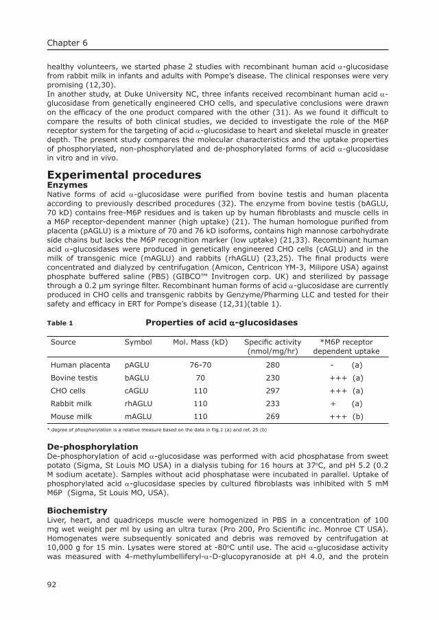
92
Chapter 6
healthy volunteers, we started phase 2 studies with recombinant human acid α-glucosidase from rabbit milk in infants and adults with Pompe’s disease. The clinical responses were very promising (12,30). In another study, at Duke University NC, three infants received recombinant human acid α-glucosidase from genetically engineered CHO cells, and speculative conclusions were drawn on the effi cacy of the one product compared with the other (31). As we found it diffi cult to compare the results of both clinical studies, we decided to investigate the role of the M6P receptor system for the targeting of acid α-glucosidase to heart and skeletal muscle in greater depth. The present study compares the molecular characteristics and the uptake properties of phosphorylated, non-phosphorylated and de-phosphorylated forms of acid α-glucosidase in vitro and in vivo.
Experimental proceduresEnzymesNative forms of acid α-glucosidase were purifi ed from bovine testis and human placenta according to previously described procedures (32). The enzyme from bovine testis (bAGLU, 70 kD) contains free-M6P residues and is taken up by human fi broblasts and muscle cells in a M6P receptor-dependent manner (high uptake) (21). The human homologue purifi ed from placenta (pAGLU) is a mixture of 70 and 76 kD isoforms, contains high mannose carbohydrate side chains but lacks the M6P recognition marker (low uptake) (21,33). Recombinant human acid α-glucosidases were produced in genetically engineered CHO cells (cAGLU) and in the milk of transgenic mice (mAGLU) and rabbits (rhAGLU) (23,25). The fi nal products were concentrated and dialyzed by centrifugation (Amicon, Centricon YM-3, Milipore USA) against phosphate buffered saline (PBS) (GIBCO™ Invitrogen corp. UK) and sterilized by passage through a 0.2 µm syringe fi lter. Recombinant human forms of acid α-glucosidase are currently produced in CHO cells and transgenic rabbits by Genzyme/Pharming LLC and tested for their safety and effi cacy in ERT for Pompe’s disease (12,31)(table 1).
Table 1 Properties of acid α-glucosidases
Source Symbol Mol. Mass (kD) Specifi c activity(nmol/mg/hr)
*M6P receptordependent uptake
Human placenta pAGLU 76-70 280 - (a)
Bovine testis bAGLU 70 230 +++ (a)
CHO cells cAGLU 110 297 +++ (a)
Rabbit milk rhAGLU 110 233 + (a)
Mouse milk mAGLU 110 269 +++ (b)
* degree of phosphorylation is a relative measure based on the data in Fig.1 (a) and ref. 25 (b)
De-phosphorylationDe-phosphorylation of acid α-glucosidase was performed with acid phosphatase from sweet potato (Sigma, St Louis MO USA) in a dialysis tubing for 16 hours at 37oC, and pH 5.2 (0.2 M sodium acetate). Samples without acid phosphatase were incubated in parallel. Uptake of phosphorylated acid α-glucosidase species by cultured fi broblasts was inhibited with 5 mM M6P (Sigma, St Louis MO, USA).
BiochemistryLiver, heart, and quadriceps muscle were homogenized in PBS in a concentration of 100 mg wet weight per ml by using an ultra turax (Pro 200, Pro Scientifi c inc. Monroe CT USA). Homogenates were subsequently sonicated and debris was removed by centrifugation at 10,000 g for 15 min. Lysates were stored at -80oC until use. The acid α-glucosidase activity was measured with 4-methylumbelliferyl-α-D-glucopyranoside at pH 4.0, and the protein
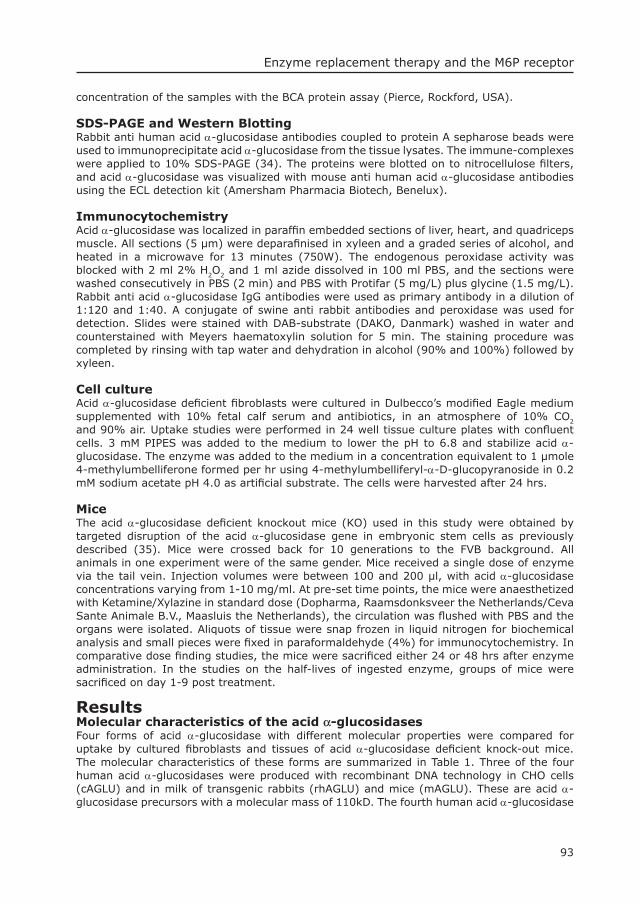
93
Enzyme replacement therapy and the M6P receptor
concentration of the samples with the BCA protein assay (Pierce, Rockford, USA).
SDS-PAGE and Western BlottingRabbit anti human acid α-glucosidase antibodies coupled to protein A sepharose beads were used to immunoprecipitate acid α-glucosidase from the tissue lysates. The immune-complexes were applied to 10% SDS-PAGE (34). The proteins were blotted on to nitrocellulose fi lters, and acid α-glucosidase was visualized with mouse anti human acid α-glucosidase antibodies using the ECL detection kit (Amersham Pharmacia Biotech, Benelux).
ImmunocytochemistryAcid α-glucosidase was localized in paraffi n embedded sections of liver, heart, and quadriceps muscle. All sections (5 µm) were deparafi nised in xyleen and a graded series of alcohol, and heated in a microwave for 13 minutes (750W). The endogenous peroxidase activity was blocked with 2 ml 2% H2O2 and 1 ml azide dissolved in 100 ml PBS, and the sections were washed consecutively in PBS (2 min) and PBS with Protifar (5 mg/L) plus glycine (1.5 mg/L). Rabbit anti acid α-glucosidase IgG antibodies were used as primary antibody in a dilution of 1:120 and 1:40. A conjugate of swine anti rabbit antibodies and peroxidase was used for detection. Slides were stained with DAB-substrate (DAKO, Danmark) washed in water and counterstained with Meyers haematoxylin solution for 5 min. The staining procedure was completed by rinsing with tap water and dehydration in alcohol (90% and 100%) followed by xyleen.
Cell cultureAcid α-glucosidase defi cient fi broblasts were cultured in Dulbecco’s modifi ed Eagle medium supplemented with 10% fetal calf serum and antibiotics, in an atmosphere of 10% CO2 and 90% air. Uptake studies were performed in 24 well tissue culture plates with confl uent cells. 3 mM PIPES was added to the medium to lower the pH to 6.8 and stabilize acid α-glucosidase. The enzyme was added to the medium in a concentration equivalent to 1 µmole 4-methylumbelliferone formed per hr using 4-methylumbelliferyl-α-D-glucopyranoside in 0.2 mM sodium acetate pH 4.0 as artifi cial substrate. The cells were harvested after 24 hrs.
MiceThe acid α-glucosidase defi cient knockout mice (KO) used in this study were obtained by targeted disruption of the acid α-glucosidase gene in embryonic stem cells as previously described (35). Mice were crossed back for 10 generations to the FVB background. All animals in one experiment were of the same gender. Mice received a single dose of enzyme via the tail vein. Injection volumes were between 100 and 200 µl, with acid α-glucosidase concentrations varying from 1-10 mg/ml. At pre-set time points, the mice were anaesthetized with Ketamine/Xylazine in standard dose (Dopharma, Raamsdonksveer the Netherlands/Ceva Sante Animale B.V., Maasluis the Netherlands), the circulation was fl ushed with PBS and the organs were isolated. Aliquots of tissue were snap frozen in liquid nitrogen for biochemical analysis and small pieces were fi xed in paraformaldehyde (4%) for immunocytochemistry. In comparative dose fi nding studies, the mice were sacrifi ced either 24 or 48 hrs after enzyme administration. In the studies on the half-lives of ingested enzyme, groups of mice were sacrifi ced on day 1-9 post treatment.
ResultsMolecular characteristics of the acid α-glucosidasesFour forms of acid α-glucosidase with different molecular properties were compared for uptake by cultured fi broblasts and tissues of acid α-glucosidase defi cient knock-out mice. The molecular characteristics of these forms are summarized in Table 1. Three of the four human acid α-glucosidases were produced with recombinant DNA technology in CHO cells (cAGLU) and in milk of transgenic rabbits (rhAGLU) and mice (mAGLU). These are acid α-glucosidase precursors with a molecular mass of 110kD. The fourth human acid α-glucosidase
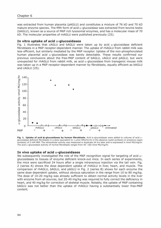
94
Chapter 6
was extracted from human placenta (pAGLU) and constitutes a mixture of 76 kD and 70 kD mature enzyme species. The fi fth form of acid α-glucosidase was extracted from bovine testis (bAGLU), known as a source of M6P rich lysosomal enzymes, and has a molecular mass of 70 kD. The molecular properties of mAGLU were published previously (25).
In vitro uptake of acid α-glucosidasesFig. 1 illustrates that cAGLU and bAGLU were taken up by acid α-glucosidase defi cient fi broblasts in a M6P receptor-dependent manner. The uptake of rhAGLU from rabbit milk was less effi cient, but similarly mediated by the M6P receptor. Uptake of the non-phosphorylated human placental acid α-glucosidase was barely detectable. These results confi rmed our previous conclusions about the free-M6P content of bAGLU, cAGLU and pAGLU, but were unexpected for rhAGLU from rabbit milk, as acid α-glucosidase from transgenic mouse milk was taken up in a M6P receptor-dependent manner by fi broblasts, equally effi cient as bAGLU and cAGLU (25).
RrhAGLU CcAGLU BbAGLU PpAGLU Untreated
20
40
60
100
120
0
80
140
α-glu
cosi
das
e ac
tivi
ty n
mol M
U/m
g/h
r
M6P +- +- +- +-
Fig. 1. Uptake of acid α-glucosidases by human fi broblasts. Acid α-glucosidases were added to cultures of acid α-glucosidase defi cient fi broblasts in a dose equivalent to 1 µmol 4MU/hr/ml in the absence (solid symbols) or presence (open symbols) of 5 mM M6P. The intracellular activity was measured in duplicate 24 hrs later and is expressed in nmol MU/mg/hr. The acid α-glucosidase activity of normal fi broblasts ranges from 40 -160 nmol MU/mg/hr.
In vivo uptake of acid α-glucosidasesWe subsequently investigated the role of the M6P recognition signal for targeting of acid α-glucosidases to tissues of enzyme defi cient knock-out mice. In each series of experiments, the mice were sacrifi ced 24 hours after a single intravenous injection via the tail vein. Fig. 2 (series A) shows the dose dependent uptake of rhAGLU in liver, heart, and muscle. The comparison of rhAGLU, bAGLU, and pAGLU in Fig. 2 (series B) shows for each enzyme the same dose dependent uptake, without obvious saturation in the range from 10 to 80 mg/kg. The dose of 10-20 mg/kg was already suffi cient to obtain normal activity levels in the liver with enzyme from all sources, but 20-40 mg/kg was required to fully correct the defi ciency in heart, and 40 mg/kg for correction of skeletal muscle. Notably, the uptake of M6P containing bAGLU was not better than the uptake of rhAGLU having a substantially lower free-M6P content.
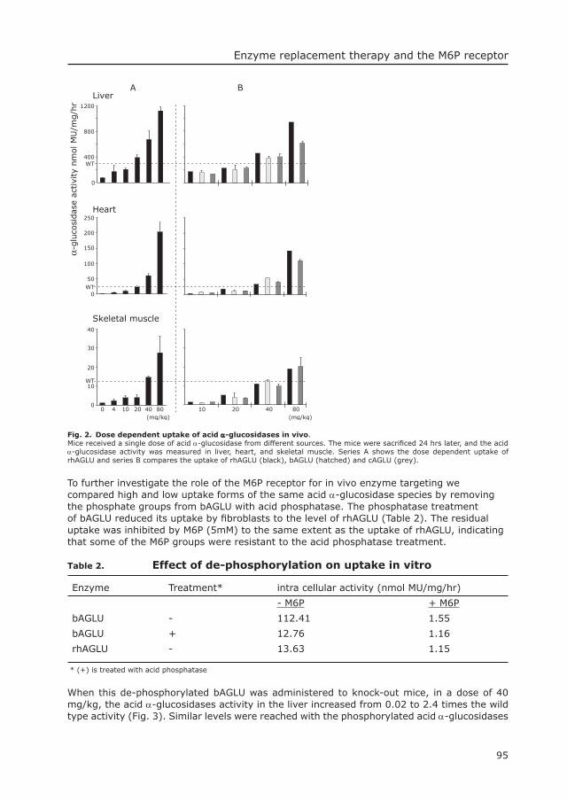
95
Enzyme replacement therapy and the M6P receptorα-
glu
cosi
das
e ac
tivi
ty n
mol M
U/m
g/h
r 1200
800
400
0
250
200
150
100
50
0
40
30
20
10
0
WT
WT
WT
Skeletal muscle
(mg/kg)0 4 10 20 40 80 10 20 40 80
(mg/kg)
Heart
LiverA B
Fig. 2. Dose dependent uptake of acid α-glucosidases in vivo. Mice received a single dose of acid α-glucosidase from different sources. The mice were sacrifi ced 24 hrs later, and the acid α-glucosidase activity was measured in liver, heart, and skeletal muscle. Series A shows the dose dependent uptake of rhAGLU and series B compares the uptake of rhAGLU (black), bAGLU (hatched) and cAGLU (grey).
To further investigate the role of the M6P receptor for in vivo enzyme targeting we compared high and low uptake forms of the same acid α-glucosidase species by removing the phosphate groups from bAGLU with acid phosphatase. The phosphatase treatment of bAGLU reduced its uptake by fi broblasts to the level of rhAGLU (Table 2). The residual uptake was inhibited by M6P (5mM) to the same extent as the uptake of rhAGLU, indicating that some of the M6P groups were resistant to the acid phosphatase treatment.
Table 2. Effect of de-phosphorylation on uptake in vitro
Enzyme Treatment* intra cellular activity (nmol MU/mg/hr)
- M6P + M6P
bAGLU - 112.41 1.55
bAGLU + 12.76 1.16
rhAGLU - 13.63 1.15 * (+) is treated with acid phosphatase
When this de-phosphorylated bAGLU was administered to knock-out mice, in a dose of 40 mg/kg, the acid α-glucosidases activity in the liver increased from 0.02 to 2.4 times the wild type activity (Fig. 3). Similar levels were reached with the phosphorylated acid α-glucosidases
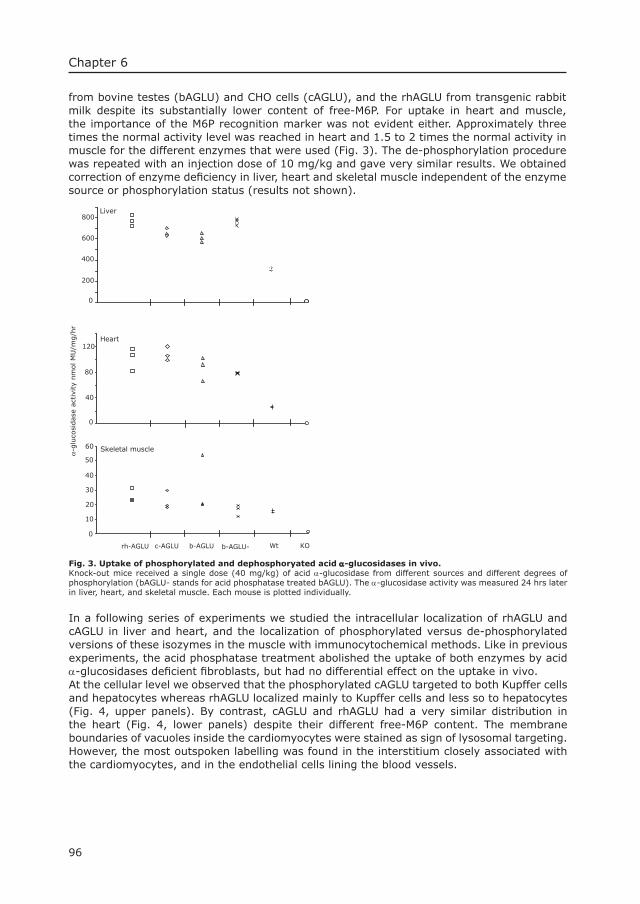
96
Chapter 6
from bovine testes (bAGLU) and CHO cells (cAGLU), and the rhAGLU from transgenic rabbit milk despite its substantially lower content of free-M6P. For uptake in heart and muscle, the importance of the M6P recognition marker was not evident either. Approximately three times the normal activity level was reached in heart and 1.5 to 2 times the normal activity in muscle for the different enzymes that were used (Fig. 3). The de-phosphorylation procedure was repeated with an injection dose of 10 mg/kg and gave very similar results. We obtained correction of enzyme defi ciency in liver, heart and skeletal muscle independent of the enzyme source or phosphorylation status (results not shown).
Bb-AGLU-
10
20
30
40
50
60
0
40
80
120
0
Heart
Liver
0
200
400
600
800
Rrh-AGLU Bb-AGLUcc-AGLU Wt KO
Skeletal muscle
α-glu
cosi
dase
act
ivity
nm
ol M
U/m
g/h
r
Fig. 3. Uptake of phosphorylated and dephosphoryated acid α-glucosidases in vivo.Knock-out mice received a single dose (40 mg/kg) of acid α-glucosidase from different sources and different degrees of phosphorylation (bAGLU- stands for acid phosphatase treated bAGLU). The α-glucosidase activity was measured 24 hrs later in liver, heart, and skeletal muscle. Each mouse is plotted individually.
In a following series of experiments we studied the intracellular localization of rhAGLU and cAGLU in liver and heart, and the localization of phosphorylated versus de-phosphorylated versions of these isozymes in the muscle with immunocytochemical methods. Like in previous experiments, the acid phosphatase treatment abolished the uptake of both enzymes by acid α-glucosidases defi cient fi broblasts, but had no differential effect on the uptake in vivo. At the cellular level we observed that the phosphorylated cAGLU targeted to both Kupffer cells and hepatocytes whereas rhAGLU localized mainly to Kupffer cells and less so to hepatocytes (Fig. 4, upper panels). By contrast, cAGLU and rhAGLU had a very similar distribution in the heart (Fig. 4, lower panels) despite their different free-M6P content. The membrane boundaries of vacuoles inside the cardiomyocytes were stained as sign of lysosomal targeting. However, the most outspoken labelling was found in the interstitium closely associated with the cardiomyocytes, and in the endothelial cells lining the blood vessels.
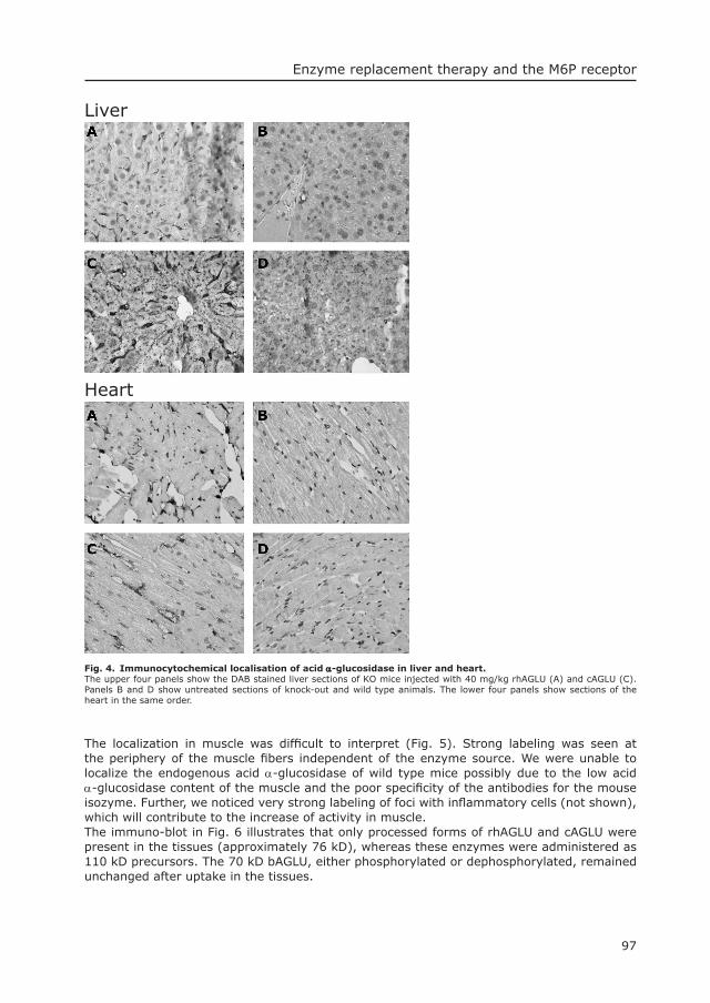
97
Enzyme replacement therapy and the M6P receptor
Fig. 4. Immunocytochemical localisation of acid α-glucosidase in liver and heart.The upper four panels show the DAB stained liver sections of KO mice injected with 40 mg/kg rhAGLU (A) and cAGLU (C). Panels B and D show untreated sections of knock-out and wild type animals. The lower four panels show sections of the heart in the same order.
The localization in muscle was diffi cult to interpret (Fig. 5). Strong labeling was seen at the periphery of the muscle fi bers independent of the enzyme source. We were unable to localize the endogenous acid α-glucosidase of wild type mice possibly due to the low acid α-glucosidase content of the muscle and the poor specifi city of the antibodies for the mouse isozyme. Further, we noticed very strong labeling of foci with infl ammatory cells (not shown), which will contribute to the increase of activity in muscle.The immuno-blot in Fig. 6 illustrates that only processed forms of rhAGLU and cAGLU were present in the tissues (approximately 76 kD), whereas these enzymes were administered as 110 kD precursors. The 70 kD bAGLU, either phosphorylated or dephosphorylated, remained unchanged after uptake in the tissues.
Liver
Heart
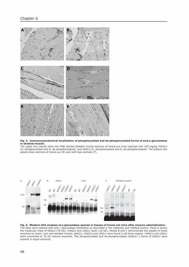
98
Chapter 6
Fig. 5. Immunocytochemical localisation of phosphorylated and de-phosphorylated forms of acid α-glucosidase in skeletal muscle. The upper four panels show the DAB stained skeletal muscle sections of knock-out mice injected with 100 mg/kg rhAGLU (A, phosphorylated and B, de-phosphorylated), and cAGLU (C, phosphorylated and D, de-phosphorylated). The bottom two panels show sections of knock-out (E) and wild type animals (F).
Heart LiverB
76-
70-
bAGLU
--bA
GLU
cAGLU
rhAGLU
WT
KO
bAGLU
--bA
GLU
rhAGLU
cAGLU
WT
KO
110-
A
76-
70-
C Skeletal muscle
cAGLU
rhAGLU
bAGLU
bAGLU
--
WT
KO
70-
bAGLU
cAGLU
rhAGLU
Fig. 6. Western blot analysis of α-glucosidase species in tissues of knock-out mice after enzyme administration.The blots were stained with anti α-glucosidase antibodies as described in the materials and method section. Panel A shows the molecular mass of bAGLU (70 kD), rhAGLU and cAGLU (both 110 kD). Panels B and C demonstrate the uptake of these enzymes by heart, liver and skeletal muscle. bAGLU, rhAGLU and cAGLU were found in all three organs. rhAGLU and cAGLU were converted to 76 kD mature enzymes. The phosphorylated and de-phosphorylated (bAGLU--) forms of bAGLU were present in equal amounts.
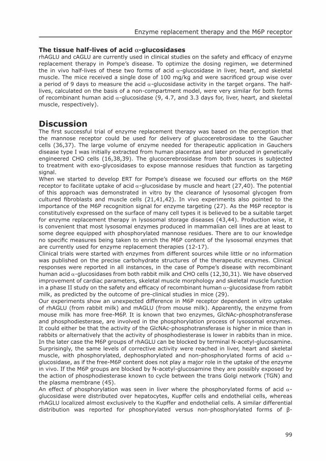
99
Enzyme replacement therapy and the M6P receptor
The tissue half-lives of acid α-glucosidasesrhAGLU and cAGLU are currently used in clinical studies on the safety and effi cacy of enzyme replacement therapy in Pompe’s disease. To optimize the dosing regimen, we determined the in vivo half-lives of these two forms of acid α-glucosidase in liver, heart, and skeletal muscle. The mice received a single dose of 100 mg/kg and were sacrifi ced group wise over a period of 9 days to measure the acid α-glucosidase activity in the target organs. The half-lives, calculated on the basis of a non-compartment model, were very similar for both forms of recombinant human acid α-glucosidase (9, 4.7, and 3.3 days for, liver, heart, and skeletal muscle, respectively).
DiscussionThe fi rst successful trial of enzyme replacement therapy was based on the perception that the mannose receptor could be used for delivery of glucocerebrosidase to the Gaucher cells (36,37). The large volume of enzyme needed for therapeutic application in Gauchers disease type I was initially extracted from human placentas and later produced in genetically engineered CHO cells (16,38,39). The glucocerebrosidase from both sources is subjected to treatment with exo-glycosidases to expose mannose residues that function as targeting signal. When we started to develop ERT for Pompe’s disease we focused our efforts on the M6P receptor to facilitate uptake of acid α-glucosidase by muscle and heart (27,40). The potential of this approach was demonstrated in vitro by the clearance of lysosomal glycogen from cultured fi broblasts and muscle cells (21,41,42). In vivo experiments also pointed to the importance of the M6P recognition signal for enzyme targeting (27). As the M6P receptor is constitutively expressed on the surface of many cell types it is believed to be a suitable target for enzyme replacement therapy in lysosomal storage diseases (43,44). Production wise, it is convenient that most lysosomal enzymes produced in mammalian cell lines are at least to some degree equipped with phosphorylated mannose residues. There are to our knowledge no specifi c measures being taken to enrich the M6P content of the lysosomal enzymes that are currently used for enzyme replacement therapies (12-17).Clinical trials were started with enzymes from different sources while little or no information was published on the precise carbohydrate structures of the therapeutic enzymes. Clinical responses were reported in all instances, in the case of Pompe’s disease with recombinant human acid α-glucosidases from both rabbit milk and CHO cells (12,30,31). We have observed improvement of cardiac parameters, skeletal muscle morphology and skeletal muscle function in a phase II study on the safety and effi cacy of recombinant human α-glucosidase from rabbit milk, as predicted by the outcome of pre-clinical studies in mice (29). Our experiments show an unexpected difference in M6P receptor dependent in vitro uptake of rhAGLU (from rabbit milk) and mAGLU (from mouse milk). Apparently, the enzyme from mouse milk has more free-M6P. It is known that two enzymes, GlcNAc-phosphotransferase and phosphodiesterase, are involved in the phosphorylation process of lysosomal enzymes. It could either be that the activity of the GlcNAc-phosphotransferase is higher in mice than in rabbits or alternatively that the activity of phosphodiesterase is lower in rabbits than in mice. In the later case the M6P groups of rhAGLU can be blocked by terminal N-acetyl-glucosamine. Surprisingly, the same levels of corrective activity were reached in liver, heart and skeletal muscle, with phosphorylated, dephosphorylated and non-phosphorylated forms of acid α-glucosidase, as if the free-M6P content does not play a major role in the uptake of the enzyme in vivo. If the M6P groups are blocked by N-acetyl-glucosamine they are possibly exposed by the action of phosphodiesterase known to cycle between the trans Golgi network (TGN) and the plasma membrane (45).An effect of phosphorylation was seen in liver where the phosphorylated forms of acid α-glucosidase were distributed over hepatocytes, Kupffer cells and endothelial cells, whereas rhAGLU localized almost exclusively to the Kupffer and endothelial cells. A similar differential distribution was reported for phosphorylated versus non-phosphorylated forms of β-
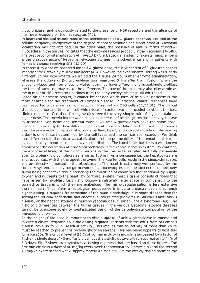
100
Chapter 6
glucuronidase, and is obviously related to the presence of M6P receptors and the absence of mannose receptors on the hepatocytes (46).In heart and skeletal muscle most of the administered acid α-glucosidase was localized at the cellular periphery, irrespective of the degree of phosphorylation and direct proof of lysosomal localization was not obtained. On the other hand, the presence of mature forms of acid α-glucosidase in the tissues indicates that the enzyme resides probably intra-lysosomal (47,48). The best proof of internalization of rhAGLU by the lysosomal system of skeletal muscle fi bers is the disappearance of lysosomal glycogen storage in knockout mice and in patients with Pompe’s disease receiving ERT (12,29).In contrast to what we observed for acid α-glucosidase, the M6P content of β-glucuronidase is important for uptake by muscle and heart (46). However, the experimental setting was slightly different. In our experiments we isolated the tissues 24 hours after enzyme administration, whereas the uptake of β-glucuronidase was measured 5 hrs after the infusion. When the phosphorylated and non-phosphorylated isozymes have different pharmacokinetic profi les, the time of sampling may make the difference. The age of the mice may also play a role as the number of M6P receptors declines from the early embryonic stage till adulthood. Based on our present results, it cannot be decided which form of acid α-glucosidase is the most desirable for the treatment of Pompe’s disease. In practice, clinical responses have been reported with enzymes from rabbit milk as well as CHO cells (12,30,31). The clinical studies continue and time will tell which dose of each enzyme is needed to obtain the best clinical response. Our dose fi nding studies reveal the very simple rule of higher uptake at higher dose. The correlation between dose and increase of acid α-glucosidase activity is close to linear for liver, heart and skeletal muscle. All acid α-glucosidases gave the same dose-response curve despite their different degrees of phosphorylation and maturation. It seems that the preference for uptake of enzyme by liver, heart, and skeletal muscle -in decreasing order- is only in part determined by the cell types and the cell surface receptors. We think that differences in the micro-vascularization and the permeability of the endothelial barrier play an equally important role in enzyme distribution. The blood brain barrier is a well-known problem for the correction of lysosomal pathology in the central nervous system. By contrast, the endothelial lining of the blood sinusoids in the liver is fenestrated and fully permeable even to protein-lipid complexes as large as 150 nm. As a consequence, the hepatocytes are in direct contact with the therapeutic enzyme. The Kupffer cells reside in the sinusoidal spaces and are directly immersed in the bloodstream. The heart is extremely well perfused by the coronary system. The physiologic network of cardiomyocytes is embedded in the immediately surrounding connective tissue harboring the multitude of capillaries that continuously supply oxygen and nutrients to the heart. By contrast, skeletal muscle tissue consists of fi bers that have arisen by myoblast fusion and occupy a relatively large space in comparison to the connective tissue in which they are embedded. The micro-vascularisation is less extensive than in heart. Thus, from a histological perspective it is quite understandable that much higher dosing is required for correction of the muscle pathology in Pompe’s disease than for solving the reticulo-endothelial and endothelial cell related problems in Gaucher’s and Fabry’s disease, or the hepatic storage of mucopolysaccharides in Hurler-Scheie syndrome (49). The histologic differences between the target tissues in the various lysosomal storage diseases cannot be overcome solely by sophisticated design of the carbohydrate composition of the therapeutic enzymes. As the height of the dose is important to obtain uptake of acid α-glucosidase in muscle and to elicit a clinical response so is the dosing regimen. Patients with the adult form of Pompe’s disease have up to 25 % residual activity. This implies that an activity of more than 25 % must be reached to prevent or reverse glycogen storage. This reasoning appears to hold also for mice (50). The critical level of 25 % of normal activity in muscle is surpassed by a factor of 4 when a single dose of 40 mg/kg is given but the activity decays with an estimated half-life of 3.3 days. Fig. 7 shows two hypothetical dosing regimens that are based on these fi gures. The fi rst one employs a dose of 40 mg/kg every week (approximately 2 times t ½) and the second 40 mg/kg every second week (approximately 4 times t ½). In the weekly dosing regimen the
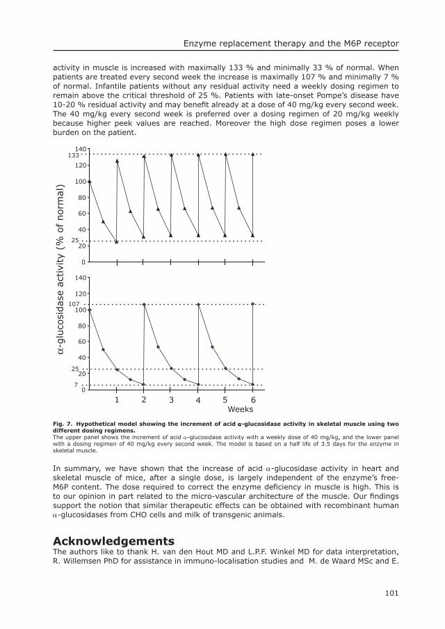
101
Enzyme replacement therapy and the M6P receptor
activity in muscle is increased with maximally 133 % and minimally 33 % of normal. When patients are treated every second week the increase is maximally 107 % and minimally 7 % of normal. Infantile patients without any residual activity need a weekly dosing regimen to remain above the critical threshold of 25 %. Patients with late-onset Pompe’s disease have 10-20 % residual activity and may benefi t already at a dose of 40 mg/kg every second week. The 40 mg/kg every second week is preferred over a dosing regimen of 20 mg/kg weekly because higher peek values are reached. Moreover the high dose regimen poses a lower burden on the patient.
0
20
40
60
80
100
120
140
0
20
40
60
80
100
120
140
1 2 3 4 5 6Weeks
25
7
107
25
133
α-glu
cosi
das
e ac
tivi
ty (
% o
f norm
al)
Fig. 7. Hypothetical model showing the increment of acid α-glucosidase activity in skeletal muscle using two different dosing regimens.The upper panel shows the increment of acid α-glucosidase activity with a weekly dose of 40 mg/kg, and the lower panel with a dosing regimen of 40 mg/kg every second week. The model is based on a half life of 3.5 days for the enzyme in skeletal muscle.
In summary, we have shown that the increase of acid α-glucosidase activity in heart and skeletal muscle of mice, after a single dose, is largely independent of the enzyme’s free-M6P content. The dose required to correct the enzyme defi ciency in muscle is high. This is to our opinion in part related to the micro-vascular architecture of the muscle. Our fi ndings support the notion that similar therapeutic effects can be obtained with recombinant human α-glucosidases from CHO cells and milk of transgenic animals.
AcknowledgementsThe authors like to thank H. van den Hout MD and L.P.F. Winkel MD for data interpretation, R. Willemsen PhD for assistance in immuno-localisation studies and M. de Waard MSc and E.
102
Chapter 6
van Deel MSc for technical assistance in the mouse studies. Financial support was obtained from the Sophia Foundation for Medical Research.
References1. Hickman, S., Shapiro, L. J., and Neufeld, E. F. (1974) Biochem Biophys Res Commun 57(1), 55-61.2. Glaser, J. H., Roozen, K. J., Brot, F. E., and Sly, W. S. (1975) Arch Biochem Biophys 166(2), 536-42.3. Kaplan, A., Achord, D. T., and Sly, W. S. (1977) Proc Natl Acad Sci U S A 74(5), 2026-30.4. Sando, G. N., and Neufeld, E. F. (1977) Cell 12, 619-6275. Kornfeld, S. (1992) Annu Rev Biochem 61, 307-3306. Hasilik, A., and Neufeld, E. F. (1980) J Biol Chem 255(10), 4937-49457. Reitman, M. L., Varki, A., and Kornfeld, S. (1981) J Clin Invest 67(5), 1574-9.8. Hasilik, A., Waheed, A., and von Figura, K. (1981) Biochem Biophys Res Commun 98(3), 761-7.9. Tager J.M, P. H., PhD, Andre W. Schram, PhD. (1980) An apraisal of human trails in
enzyme replacement theapy of Genetic diaseases. Enzyme therapy in genetic diseases:2 (Desnick, R. J., Ed.), Alan R. Liss, New York
10. Hobbs, J. R., Hugh-Jones, K., Barrett, A. J., Byrom, N., Chambers, D., Henry, K., James, D. C., Lucas, C. F., Rogers, T. R., Benson, P. F., Tansley, L. R., Patrick, A. D., Mossman, J., and Young, E. P. (1981) Lancet 2(8249), 709-12.
11. Barton, N. W., Furbish, F. S., Murray, G. J., Garfi eld, M., and Brady, R. O. (1990) Proc Natl Acad Sci USA 87, 1913-1916
12. Van den Hout, H., Reuser, A. J., Vulto, A. G., Loonen, M. C., Cromme-Dijkhuis, A., and Van der Ploeg, A. T. (2000) Lancet 356(9227), 397-8.
13. Eng, C. M., Guffon, N., Wilcox, W. R., Germain, D. P., Lee, P., Waldek, S., Caplan, L., Linthorst, G. E., and Desnick, R. J. (2001) N Engl J Med 345(1), 9-16.
14. Schiffmann, R., Kopp, J. B., Austin, H. A., 3rd, Sabnis, S., Moore, D. F., Weibel, T., Balow, J. E., and Brady, R. O. (2001) Jama 285(21), 2743-9.
15. Kakkis, E. D., Schuchman, E., He, X., Wan, Q., Kania, S., Wiemelt, S., Hasson, C. W., O’Malley, T., Weil, M. A., Aguirre, G. A., Brown, D. E., and Haskins, M. E. (2001) Mol Genet Metab 72(3), 199-208.
16. Weinreb, N. J., Charrow, J., Andersson, H. C., Kaplan, P., Kolodny, E. H., Mistry, P., Pastores, G., Rosenbloom, B. E., Scott, C. R., Wappner, R. S., and Zimran, A. (2002) Am J Med 113(2), 112-9.
17. Muenzer, J., Lamsa, J. C., Garcia, A., Dacosta, J., Garcia, J., and Treco, D. A. (2002) Acta Paediatr Suppl 91(439), 98-9
18. Hers, H. G. (1963) Biochem J 86, 11-1619. Hirschhorn, R., and Reuser, A. J. J. (2000) (Scriver, C. R., Beaudet, A. L., Valle, D., and
W. S. Sly, eds) Vol. III, pp. 3389-3420, Mc Graw-Hill, New York20. Oude Elferink, R. P. J., Brouwer-Kelder, E. M., Surya, I., Strijland, A., Kroos, M., Reuser, A. J. J.,
and Tager, J. M. (1984) Eur J Biochem 139(3), 489-49521. Reuser, A. J. J., Kroos, M. A., Ponne, N. J., Wolterman, R. A., Loonen, M. C. B., Busch, H. F. M.,
Visser, W. J., and Bolhuis, P. A. (1984) Exp Cell Res 155(1), 178-18922. Van der Ploeg, A. T., Loonen, M. C. B., Bolhuis, P. A., Busch, H. M. F., Reuser, A. J. J., and Galjaard,
H. (1988) Pediatr Res 24(1), 90-9423. Fuller, M., Van der Ploeg, A., Reuser, A. J., Anson, D. S., and Hopwood, J. J. (1995) Eur J Biochem
234(3), 903-9.24. Van Hove, J. L., Yang, H. W., Wu, J. Y., Brady, R. O., and Chen, Y. T. (1996) Proc Natl Acad Sci U
S A 93(1), 65-70.25. Bijvoet, A. G. A., Kroos, M. A., Pieper, F. R., Van der Vliet, M., De Boer, H. A., Van der Ploeg, A. T.,
Verbeet, M. P., and Reuser, A. J. J. (1998) Hum Mol Genet 7(11), 1815-182426. Bijvoet, A. G., Van Hirtum, H., Vermey, M., Van Leenen, D., Van der Ploeg, A. T., Mooi, W. J., and
Reuser, A. J. (1999) J Pathol 189(3), 416-24.27. Van der Ploeg, A. T., Kroos, M. A., Willemsen, R., Brons, N. H. C., and Reuser, A. J. J. (1991)
J Clin Invest 87(2), 513-51828. Kikuchi, T., Yang, H. W., Pennybacker, M., Ichihara, N., Mizutani, M., Van Hove, J. L., and
Chen, Y. T. (1998) J Clin Invest 101(4), 827-33.29. Bijvoet, A. G., Van Hirtum, H., Kroos, M. A., Van de Kamp, E. H., Schoneveld, O., Visser,
P., Brakenhoff, J. P., Weggeman, M., van Corven, E. J., Van der Ploeg, A. T., and Reuser, A. J. (1999) Hum Mol Genet 8(12), 2145-53.
30. Van den Hout, J. M., Reuser, A. J., de Klerk, J. B., Arts, W. F., Smeitink, J. A., and Van der Ploeg, A. T. (2001) J Inherit Metab Dis 24(2), 266-74.
31. Amalfi tano, A., Bengur, A. R., Morse, R. P., Majure, J. M., Case, L. E., Veerling, D. L., Mackey, J., Kishnani, P., Smith, W., McVie-Wylie, A., Sullivan, J. A., Hoganson, G. E., Phillips, J. A., 3rd, Schaefer, G. B., Charrow, J., Ware, R. E., Bossen, E. H., and Chen, Y. T. (2001) Genet Med 3(2), 132-8.
103
Enzyme replacement therapy and the M6P receptor
32. Reuser, A. J. J., Kroos, M., Oude Elferink, R. P. J., and Tager, J. M. (1985) J Biol Chem 260(14), 8336-8341
33. Mutsaers, J. H., Van Halbeek, H., Vliegenthart, J. F., Tager, J. M., Reuser, A. J., Kroos, M., and Galjaard, H. (1987) Biochim Biophys Acta 911(2), 244-51.
34. Reuser, A. J., Kroos, M., Willemsen, R., Swallow, D., Tager, J. M., and Galjaard, H. (1987) J Clin Invest 79(6), 1689-99.
35. Bijvoet, A. G. A., Van de Kamp, E. H. M., Kroos, M. A., Ding, J. H., Yang, B. Z., Visser, P., Bakker, C. E., Verbeet, M. P., Oostra, B., Reuser, A. J. J., and Van der Ploeg, A. T. (1998) Hum Mol Genet 7(1), 53-62
36. Murray, G. J., Doebber, T. W., Shen, T. Y., Wu, M. S., Ponpipom, M. M., Bugianesi, R. L., Brady, R. O., and Barranger, J. A. (1985) Biochem Med 34(2), 241-6.
37. Stahl, P., and Schwartz, A. L. (1986) J Clin Invest 77(3), 657-62.38. Barton, N. W., Brady, R. O., Dambrosia, J. M., Di Bisgeglie, A. M., Doppelt, S. H., Hill, S. C.,
Mankin, H. J., Murray, G. J., Parker, R. I., Argoff, C. E., Grewal, R. P., Yu, K. T., and collaborators. (1991) N Engl J Med 324(21), 1464-1470
39. Grabowski, G. A., Leslie, N., and Wenstrup, R. (1998) Blood Rev 12, 115-13340. Van der Ploeg, A. T., Van der Kraaij, A. M. M., Willemsen, R., Kroos, M. A., Loonen, M. C. B.,
Koster, J. F., and Reuser, A. J. J. (1990) Pediatr Res 28(4), 344-34741. Van der Ploeg, A. T., Kroos, M., Van Dongen, J. M., Visser, W. J., Bolhuis, P. A., Loonen, M. C., and
Reuser, A. J. J. (1987) J Neurol Sci 79(3), 327-33642. Van der Ploeg, A. T., Bolhuis, P. A., Wolterman, R. A., Visser, J. W., Loonen, M. C. B., Busch, H. F.
M., and Reuser, A. J. J. (1988) J Neurol 235(7), 392-39643. Matzner, U., von Figura, K., and Pohlmann, R. (1992) Development 114(4), 965-72.44. Takahashi, K., Donovan, M. J., Rogers, R. A., and Ezekowitz, R. A. (1998) Cell Tissue Res 292(2),
311-23.45. Rohrer, J., and Kornfeld, R. (2001) Mol Biol Cell 12(6), 1623-31.46. Sands, M. S., Vogler, C. A., Ohlemiller, K. K., Roberts, M. S., Grubb, J. H., Levy, B., and Sly, W. S.
(2001) J Biol Chem 276(46), 43160-43165.47. Oude Elferink, R. P., Strijland, A., Surya, I., Brouwer-Kelder, E. M., Kroos, M., Hilkens, J., Hilgers,
J., Reuser, A. J., and Tager, J. M. (1984) Eur J Biochem 139(3), 497-50248. Wisselaar, H. A., Kroos, M. A., Hermans, M. M. P., van Beeumen, J., and Reuser, A. J. J. (1993) J
Biol Chem 268(3), 2223-223149. Schiffmann, R., and Brady, R. O. (2002) Drugs 62(5), 733-4250. Raben, N., Jatkar, T., Lee, A., Lu, N., Dwivedi, S., Nagaraju, K., and Plotz, P. H. (2002) Mol Ther
6(5), 601-8.


7Discussion

Chapter 7
107
7 DiscussionAnimal models can be very helpful for understanding the pathophysiology of human diseases. They may shed light on clinical features that were so far not assigned to the disease. Further they may be instrumental for the design of therapeutic strategies like bone marrow transplantation (Haematopoietic Cell Transplantation), enzyme replacement therapy and gene therapy. For Pompe’s disease animal models also have proven their value. Dogs with Pompe’s disease (1) were originally diagnosed because they could not retain their food. A dilation of the oesophagus was noted upon X-ray examination and the discovery underscored the generalised nature of the muscle weakness in Pompe’s disease. When specifi cally asked for, patients with Pompe’s disease also have chewing and swallowing diffi culties. This bares the risk of a low calorie intake leading to a catabolic state of the patient, increased protein turnover, and further decline of muscle structure and function. The smooth muscle cell pathology was very striking in the genetically modifi ed mouse model of Pompe’s disease. This fi nding shed new light on the causative relation between smooth muscle cell pathology and the occurrence of aneurysms in Pompe’s disease (2, 3). Chapter 5 of this thesis demonstrates the usefulness of the mouse model to investigate the cause of the newly discovered hearing defi cit in infantile Pompe’ s disease. In other collaborative studies the emphasis was laid on understanding the pathogenesis of muscle dysfunction in humans and mice with Pompe’s disease (4). Affected mice and quail have been particularly useful for demonstrating the feasibility of enzyme replacement therapy for this otherwise incurable disease (5).
This thesis covers the period that the mouse model was established but not yet fully characterised with respect to its clinical, cardiac, and skeletal muscle dysfunction. At the same time the fi rst clinical trial of enzyme replacement therapy started at the Erasmus MC-Sophia and the lessons learned from the mouse model were applied in clinical practice. The mouse model was used to approach questions that came up during the clinical study. This discussion focuses on the value of the mouse model for investigating in particular the pathophysiology of the heart, skeletal muscle, and the hearing organ, and for answering questions related to dosing and mechanisms of enzyme replacement therapy.
7.1 The heart in Pompe’s diseaseCardiac dimensionsThe echocardiography study in the knockout (KO) mouse with Pompe’s disease (Chapter 2) shows that KO mice have a similar progressive cardiomyopathy leading to cardiac hypertrophy as patients with infantile Pompe’s disease. The heart weight of the KO mice (FVB background) is 160% of normal at the age of 25 months due to hypertrophy of both the left and right ventricle (6). The heart weight of C57Bl/6 KO mice, in the same age group as the FVB KO mice, increases to approximately 200% (both FVB and C57Bl/6 mice were back-crossed for 6 generations. Studies in humans show an increase of the left ventricular (LV) mass from close to 1.5 times normal at birth to 5 times normal at the end stage of disease. The encroachment of the LV lumen can ultimately lead to outfl ow tract obstruction (7, 8). Hyperthrophic cardiomyopathy has also been described in a sub-population of Shorthorn cattle (200% at the age of 6.7 months), Turkey (round heart disease), sheep, and dog, but is not a feature in Japanese quail (9-15). In general, the cardiac hypertrophy in the known animal models is less than in humans.The abnormal LV geometry in the KO mouse parallels the human situation. The increase of LV wall thickness is most prominent, and the encroachment of the lumen is variable. The progressive hyperthrophic cardiomyopathy in Pompe’s disease is very much the same as seen in Anderson Fabry’s disease (16, 17). In both these diseases the lysosomal storage leads to a disruption of the cardiac muscle architecture and triggers the heart’s compensatory mechanisms, which may lead to proliferation of contractile proteins and cellular hypertrophy (18-20). The cardiomyopathy in infantile Pompe’s disease is more conspicuous than in Fabry’s disease.
108
Chapter 7
Cardiac functionThe mouse model allows to study the cardiac pathophysiology in more detail than in humans (Chapter 2). Monitoring of the intra-cardiac blood pressure in the left ventricle and aorta during infusion of a β-adrenergic stimulant showed that the LV contractility reduces when the disease progresses. Nevertheless even one-year-old mice do not suffer from overt heart failure as patients with infantile Pompe’s disease. At that age, the KO mice still have a normal fractional shortening of the LV diameter, and the dose of a β-adrenergic stimulant needed to elicit maximal response in LV contractility is the same as in normal mice. There is little information available in the literature about the cardio-dynamics of Pompe’s disease in humans. Ejection fractions of approximately 70% were reported for humans, and normal LV fi lling pressures were found using cardiac catheterisation in patients from 6-9 months of age (7, 21).
Response to therapyWhen infants with Pompe’s disease were treated with weekly infusions of recombinant human acid α-glucosidase from rabbit milk, the cardiac dimensions decreased signifi cantly. The left ventricular mass index decreased up to 16% of pre-treatment value after 9 months of treatment (unpublished data). Similar results were obtained in patients with Fabry’s disease who received enzyme replacement therapy (16, 17).The cardiac hypertrophy is believed to arise from the interference of the glycogen fi lled lysosomes with the normal muscle architecture leading to a diminished force transmission. Cellular stress induced by the lysosomal deposits may also play a role in the increase in cardiac muscle cell volume. The three year follow-up study of the safety and effi cacy of ERT in four patients with infantile Pompe’s disease (Chapter 3) shows that the cardiac response to lysosomal glycogen storage is largely a reversible process (22). The heart remodels to approximately normal dimensions and the systolic function improves signifi cantly whereas cardiorespiratory failure is the major cause of death of untreated patients (23). When treated in time patients with classic infantile Pompe’s disease are no longer at risk to succumb from cardiac failure. The therapeutic effect must be brought about by the uptake of the rabbit milk acid α-glucosidase by the cardio-myocytes as fi rst demonstrated in the Langendorf perfusion system and later in knock-out mice with Pompe’s disease (24, 25).
7.2 The muscle in Pompe’s disease PathologyThe KO mouse model parallels the human infantile form of Pompe’s disease by its complete defi ciency of acid α-glucosidase (26). However, the rate of glycogen storage and the ensuing pathological effects seem to progress slower in mice than in humans. The mice reveal only limited glycogen storage in muscle and other tissues at the time they are born. They show absolutely no clinical signs, whereas almost all infants with classic infantile Pompe’s disease present as fl oppy baby’s with cardiomegaly and massive lysosomal glycogen storage in many cell types and tissues (27, 28).When the mice are eight months old they have developed substantial muscle pathology and start to exhibit weakness of the hind limbs. The average life span of affected animals is only slightly shortened whereas affected infants succumb in their fi rst year of life (29). Otherwise, the disease develops similar in both species in that the glycogen storage is progressive. Besides the heart and skeletal muscle, smooth muscle is typically involved. The glycogen deposits in the wall of blood vessels of mice and humans is very striking. One can envisage that the integrity of the tissue structure is lost when the disease progresses. This may ultimately lead to formation of an aneurysm. The occurrence of aneurysms in the basal arteries of patients with Pompe’s disease are mentioned in some case reports (30). A minor intriguing difference between the pathology in mice and humans is the storage in endothelial cells. It was not observed in mice but is obvious in patients. Notably clearance of
109
Discussion
glycogen from the endothelial cells was one of the fi rst signs of therapeutic effect of enzyme replacement therapy (Chapter 4) (3).
Muscle functionProgressive skeletal muscle weakness is one of the most prominent features in infantile and late onset Pompe’s disease. The skeletal muscle function is assessed by performance tests. In infants, physical capabilities are assessed with validated tests like the AIMS and BSIDII. In infantile Pompe patient’s milestones of normal development are not reached. Symptoms of muscle weakness start shortly after birth (median age 1.6 months), hospitalisation follows at an age of approximately 3-4 months (28). Most patients die younger than 1 year old (median age 7.7 and 6.0 months for the Dutch and world-wide population, respectively) (28).The muscle function of mice with Pompe’s disease was analysed using the ∆6/∆6 And the 13neo/13neo strain (31, 32). At fi rst in was suggested that the ∆6/∆6 mice had an earlier onset of disease than the13neo/13neo mice. But, more recent publications revealed the same disease progression in both mouse models (33). As a matter of fact it was very diffi cult to judge and compare the phenotypes of the different mouse models because of the different methodologies that were used to measure muscle function. The ∆6/∆6 mice were tested in open-fi eld studies and had lower scores than their healthy littermates (8-11 months of age). This test was not available for the 13neo/13neo mice. Their performance was tested in running wheels and signifi cant differences were not noted before 8 months of age. Other tests that were implemented later such as a hanging wire and vertical pole also revealed the start of clinical symptoms around eight months of age an age of 30 weeks (data not shown). The variable test results obtained with the ∆6/∆6 mice are in part explained by differences in genetic constitution. The 13neo/13neo mice are back-crossed to the FVB and C57Bl/6 background for ten generations whereas the ∆6/∆6 model has mixed 129, C57Bl/6, and FVB background. The most sophisticated assessment of muscle performance in the C57Bl/6 13neo/13neo mice (4, 34) shows that the absolute maximum torque of the muscles of 18-month old mice is two fold reduced compared to wild-type littermates. These 18-month old mice were severely affected and showed a splayed limb posture, a waddling gait, and they suffered from thoracic kyphosis. The reduction in muscle strength was associated with a 1.3 fold decrease in muscle mass. When tested in a fatigue contraction protocol, the muscles of both the KO mice and their control littermates responded in a similar fashion indicating that the mechanisms involved in muscle fatigue are not affected in mice with Pompe’s disease. It is speculated that the lysosomal glycogen storage leads to interruption of the myofi brils whereby longitudinal force transmission is hampered, leading to muscle weakness (35). Apparently the muscle impairment is not due to metabolic disturbances but mainly due to a decrease of muscle mass and interference of glycogen fi lled vacuoles with the contractile structures. This hypothesis is not yet confi rmed in patients with Pompe’s disease.
Response to therapyBy lack of reliable methods for clinical assessment, the effect of enzyme replacement therapy in mice was evaluated by measuring the increase of acid α-glucosidase activity in the tissues and the reduction of tissue pathology. Correction of enzyme defi ciency and improvement of tissue morphology was demonstrated after a six-month treatment period. The patients in the clinical study were evaluated for muscle function using the AIMS test and the BSIDII. One patient had normal scores at the age of two. This best responding patient is at present 5 years old, ventilator free and fully mobile without support. Two patients of the same age are ventilator dependent and wheelchair bound. They initially gained muscle strength in the upper extremities, but further improvement halted. The fourth patient who participated in the study manifested steady progress in the fi rst two years that she was treated. She could use her arms and sit upright unsupported. In the third year of the study she became ventilator dependent during a portacath infection and her muscular condition gradually decelined. She died quite suddenly at the age of 4 years and 3 months after a period of intractable fever.
110
Chapter 7
After the fi rst three months of treatment with a dose of 15-20 mg/kg the acid α-glucosidase defi ciency in muscle was partly corrected (36). An increase of the dose to 40 mg/kg resulted in correction of the activity to a normal level (Chapter 3) (3). Several muscle biopsies were taken during the course of treatment to monitor the therapeutic effect. The fi ndings are described in Chapter 4, and are informative for understanding the process of muscle wasting and for estimating the chance of successful treatment. Three of the four patients included in the study had advanced muscle pathology at the start of treatment. Glycogen loaded lysosomes were seen over the entire length of virtually all muscle fi bres. The fourth patient was also clearly affected but the pattern of muscle damage was less severe. All fi bres showed lysosomal glycogen storage but not always over the entire length of the fi bre. The glycogen load per fi bre also varied. After the start of treatment the glycogen load of the muscle decreased in all patients when judged by the diminished PAS staining intensity although this was not confi rmed by the quantitative measurement of the glycogen content of the whole muscle biopsy. The disappearance of glycogen in the muscles of the three most affected patients resulted in a lace work pattern of empty spaces that were previously occupied by glycogen, but there was little left of the muscular architecture. Clearance of glycogen from the muscle of the least affected patient also resulted in empty spaces but these were interspersed between the still ordered arrays of contractile elements. As we observed, the muscle morphology of this patient slowly improved during the 72 weeks of continuous treatment, using the still intact muscle structure as a matrix. The residual contractile function of the muscle may have helped to feed and oxygenate the muscle during the treatment and deliver acid α-glucosidase to the muscle. The myogenic satellite cells may have assisted in the repair of the muscle damage. When muscle fi bres have lost all architecture and function, as in the biopsied muscle of the three severely affected infants in our study, they are probably beyond repair. It is therefore that the muscle morphology and the residual function can be used as an indicator for the outcome of enzyme replacement therapy. In the line of this reasoning it is important to keep the muscles in optimal condition before and during enzyme replacement therapy by a proper diet and moderate exercise. But surprisingly little has been done to investigate the role of diet and exercise in the treatment of Pompe’s disease (37, 38). For muscle as for heart, the KO mouse is a suitable model to study pathogenesis, function and response to therapy. The impact of diet and exercise on the therapy has to be investigated in the patient population because of differences in metabolism and feeding behaviour between mice and humans.
7.3 Hearing loss in Pompe’s diseaseBefore the start of enzyme replacement therapy, hearing loss was not known as a feature of classic infantile Pompe’s disease. It had not been reported either in any of the animal models. The discovery was serendipitous, as we had included the measurement of brainstem auditory evoked potentials in the study protocol as a tool to monitor brain function. The latter was important because the blood brain barrier prevents the therapeutic enzyme to enter the brain. The four infants in our study all had severe hearing loss. When we communicated this fi nding to colleagues, they specifi cally tested their patients and confi rmed our fi ndings. So far we have not discovered any hearing loss in our patients with late onset Pompe’s disease. Further investigation into the basis of hearing loss was limited to non-invasive tests like BERA, OAE and tympanogram, and this pointed to cochlear dysfunction rather than central nervous system involvement (Chapter 5).The mouse model was again used as a reference. The mice manifested cochlear pathology as suspected from the investigations in humans, but did not have hearing loss. The latter is explainable by the slower course of the disease in mice compared to humans. The cochlear pathology consisted of lysosomal glycogen storage in the inner- and outer hair cells, the support cells of the cochlea and the neurons of the spiral ganglion. Hearing loss has been observed in various other lysosomal storage disorders but the direct causes differ (39-45).
111
Discussion
Response to therapyDespite the improvement of the cardiac and muscular parameters in the infantile patients participating in our clinical trial, there was no effect on the hearing loss, although threshold values did not increase either during therapy. The lack of response may be inherent to the cochlear structure wherein the hair cells and support cells are not positioned directly adjacent to the blood supply.
7.4 Concluding remarks about the mouse model ofPompe’s diseaseThe mouse model of Pompe’s disease has proven its value both for understanding the pathology of the disease as well as for the development of therapy. The parallel between mice and humans is close but species-specifi c differences do exist and fi ndings need to be interpreted with caution. The most striking difference between the KO mice and patients with classic infantile Pompe’s disease is the slower disease progression and the longer survival of mice compared to humans. It was reasoned previously that lysosomal glycogen storage starts in the single cell embryonic state and that the total length of the pathogenic process counts more than the relative age. The following paragraphs specifi cally discuss the value of the mouse model for studying the safety and effi cacy of enzyme replacement therapy with a focus on enzyme targeting and dosing.
7.5 Development of enzyme replacement therapy Gene therapy and enzyme replacement therapy have in common that the basic principles of these approaches are easy to conceive, but the problems associated with clinical application are far greater than originally anticipated. Taken the state of present day technology and the actual therapeutic achievements, the fi rst clinical trial of enzyme replacement therapy, 40 years ago, was futile and far from realistic (46). In retrospect, the development of enzyme replacement therapy for Pompe’s disease has also been a painstakingly slow process. One to two decades of unsuccessful studies in humans were followed by a series of in vitro experiments to re-explore the feasibility of enzyme replacement therapy, using the then newly acquired knowledge on the role of the mannose 6-phosphate (M6P) receptor for lysosomal targeting of exogenous ligands (24, 25, 47-53). The basic criteria were met in that the lysosomal glycogen storage in cultured fi broblasts and muscle cells of Pompe patients was corrected by enzyme supplementation. Subsequent studies indicated that the principles of in vitro correction also applied in the experimental set-up of the ex vivo rat heart perfusion and the in vivo administration of enzyme via the tail vain of mice (24, 52). The dose used in the latter experiment was used as a guide for the design of all following studies on the feasibility of enzyme replacement therapy in patients with Pompe’s disease. In a study with affected Japanese quail 4.2 to 14 mg/kg of recombinant human α-glucosidase manufactured in genetically modifi ed CHO cells was administered. KO mice were treated with weekly doses of 17 mg/kg recombinant human α-glucosidase from rabbit milk (25, 53). Under these conditions the α-glucosidase activity of the mice was corrected to normal levels in all tissues except the brain, and the glycogen was cleared from the target tissues. When protocols were designed for a clinical trial with recombinant human acid α-glucosidase from rabbit milk, the starting dose for adult patients was set at 10 mg/kg per week and for infants at 15-20 mg/kg based on their higher metabolic (Chapter 3).The fi rst patient was enrolled in our study in January 1999. Since then several other studies with CHO enzyme were started, some have already ended, and yet others are planned for testing the safety and effi cacy of enzyme replacement therapy in infantile and late onset Pompe’s disease.Genzyme Corp., from Boston, USA is engaged in the commercial development of enzyme replacement therapy for Pompe’s disease and intends to fi le for approval of the therapy
112
Chapter 7
in 2004. This is 40 years after the fi rst historical study of enzyme replacement therapy in Pompe’s disease was published.
Upcoming questions about the optimal dose, dosing regimen, and the mechanism of enzyme targeting marked the most recent investigations into the feasibility of ERT for Pompe’s disease. The following paragraphs are devoted specifi cally to these two issues.
The mechanism of enzyme replacement therapyThe choice of the mannose 6-phosphate (M6P) receptor as target for enzyme therapy in Pompe’s disease was born out of the fi nding that this receptor was involved in the exchange of enzymes between cultured fi broblasts. The 300 KD M6P receptor appeared to be exposed on the surface of many cell types including cultured myoblasts derived from a muscle biopsy and differentiating in vitro into myotubes. A fi rst indication for the in vivo importance of the M6P receptor for enzyme targeting was obtained in a rat heart perfusion system and in normal mice. A higher uptake was obtained with phosphorylated compared to non-phosphorylated acid α-glucosidase, in several organs of normal mice (24, 52). When larger supplies of recombinant human acid α-glucosidase became gradually available from production in transgenic mouse milk, rabbit milk, and genetically engineered CHO cells, these experiments were repeated and extended in KO mice (Chapter 6).As demonstrated in this thesis the M6P content of acid α-glucosidase appeared less crucial than anticipated, and the dose more critical. The impact of the precise carbohydrate structure varies by the cell type and the localisation of the cell type within the tissue. As discussed in Chapter 4, the correction of the reticulo-endothelial cells in Gaucher’s disease by Cerezyme approaches the in vitro experimental situation. Glycoproteins containing carbohydrate side chains with exposed mannose residues bind to cells with mannose receptors and are internalised. Free access of enzyme to the target cells is guaranteed in vivo because the glycolipid-storing macrophages (such as the Kupffer cells in the liver) reside in the blood sinusoids. A similar comparison between the in vivo and in vitro situation exists in Fabry’s disease, presently treated with Replagal (TKT) and Fabrazyme (Genzyme) (54, 55). The endothelial cells have free access to the therapeutic enzyme, and highly effi cient binding of ligands via the M6P receptor leads to the described effect seen in these cells. The hepatocytes also respond as expected. These cells have virtually free access to administered enzymes, so that the ligand-receptor functions are directly operative. The more effi cient uptake of phosphorylated versus non-phosphorylated α-glucosidase measured in vitro (in fi broblasts) is similarly seen in vivo (in hepatocytes) (Chapter 6). The recombinant α-glucosidase from rabbit milk with a lower number of freely exposed M6P residues will be less effective in clearing glycogen from the lysosomes of hepatocytes than the α-glucosidase from CHO cells with a higher free M6P content. But, liver is not a target organ in Pompe’s disease.
There is no question that the intravenously administered acid α-glucosidase reaches many different cell types in the body including the cardiomyocytes and the skeletal muscle cells that are shielded off from the bloodstream by the endothelium of the capillary wall. The evidence is provided by the clearance of glycogen from the tissues of KO mice and children with Pompe’s disease after 72 weeks of treatment (22, 25) (Chapters 3 and 4). The clinical improvement of the patients in our study is the best evidence that the target cells are reached (56). But, after the many years of advocating the importance of M6P receptor mediated endocytosis in the therapeutic process we are left with questions about the underlying mechanisms. Certainly, one of the most intriguing questions is how acid α-glucosidase passes the endothelial barrier. Ceredase and Cerezyme using the mannose receptor for entry into cells are localised in both Kupffer and endothelial cells of the liver after intravenous administration. Fabrazyme and Replagal, thought to utilize the M6P receptor for cell entry, are also routed to the lysosomal system of the endothelial cells where they degrade the stored globotriosylceramide (54, 55).
113
Discussion
Notably, α-glucosidase follows the same route because the removal of lysosomal glycogen from the vascular endothelium is one of the fi rst events after start of ERT. To cross the endothelial barrier a substantial proportion of enzyme has to escape the lysosomal route of no return and alternatively enter the pathway of transcytosis.Studies on the molecular and cellular aspects of transcytosis indicate that the plasmalemmal vesicle transport system is a quite import mode to transport molecules across endothelial cells. Two fundamentally distinct mechanisms have been identifi ed. Receptor mediated and non-receptor (or fl uid phase) mediated transcytosis. Receptor mediated transcytosis is believed to be mediated by clathrin coated vesicles or caveolae. It is thought to occur for plasma molecules like insulin, transferrin, ceruloplasmin, and albumin. Co-localisation of acid α-glucosidase with albumin suggests a similar pathway for the transcytosis of this lysosomal enzyme (57). Further studies are obviously needed to resolve the pending questions about the nature of trans-endothelial transport of recombinant human acid α-glucosidase.
DosingAs mentioned in the previous paragraph about enzyme targeting, the initial weekly dose used in the clinical study was set at 15-20 mg/kg for the four infants and at 10 mg/kg for the three older patients. This dosing was based on pre-clinical studies in mice and quail (25, 53). The study protocol did not foresee in dose fi nding but allowed for dose adaptation when indicated by study results. It was realized beforehand that the evaluation of dose related clinical effects would be diffi cult in the short time span that patients with classic infantile Pompe’s disease live. As it happened, the condition of one of the patients became extremely critical after 12 weeks of treatment and it was decided, after long debate, to double the dose to 40 mg/kg/week. All patients remained on this dose for more than 3 years. There was no reason to lower the dose for safety reasons, and the response to treatment was still not optimal for all infants. Moreover, there were no reference studies indicating what the optimal dose for the treatment of classic infantile Pompe’s disease should be. There was just one other study that could serve as a guide (58). Three patients received recombinant human acid α-glucosidase from CHO cells in a reported dose of 2 times 5 mg/kg/week. The fi rst results were publicly available in April 2001. All three patients had an improved muscle function after the fi rst fi ve weeks of treatment but two of the patients deteriorated in the period thereafter. Both these patients became ventilator dependent at the age of age of 8 months (5 months of treatment), and the dose of one patient was raised to 5 times 10 mg/kg/week (59). This higher dosing regimen did not improve the clinical condition of the patient and resulted in nephrotic syndrome, as also occurred in the pioneering study of Hug et al. using acid α-glucosidase from Aspergillus niger (60). The third patient responded very well to the low dose treatment of 2 times 5 mg/kg/wk. Recently, it was communicated that this patient did not have the classic-infantile form of Pompe’s disease, which makes it diffi cult to estimate the effect of treatment (dr P. Kishnani, personal comunication).A new study with α-glucosidase from CHO cells has started. Eight patients with infantile Pompe’s disease received weekly doses of 10 mg/kg. The preliminary results indicate that this dose was suffi cient for 4 of the 8 patients (61).
The patients receiving acid α glucosidase from rabbit milk were kept on a weekly dose of 40 mg/kg. Meanwhile we tried to get more insight in the optimal dose and dosing regimen using the mouse model (Chapter 6).
The results obtained in the mouse model cannot be directly translated to the human situation, as the mode of enzyme administration is quite different. The mice received a bolus injection whereas the patients received the enzyme over a period of several hours. The mouse model shows that uptake of acid α-glucosidase by liver, heart, and skeletal muscle is dose dependent and linear up to 80 mg/kg. Further, it is evident that muscle and heart (to a lesser extent) are diffi cult to correct compared to liver. The minimal dose to obtain normal enzyme activities in skeletal muscle is 40 mg/kg but lower doses starting from 10 mg/kg are suffi cient to reach
114
Chapter 7
the skeletal muscle. The important question is how much activity is needed to correct the lysosomal glycogen storage. Mother Nature provides the best answer. Patients with late onset Pompe’s disease have rarely more than 25% of normal activity (23). Higher activity levels do apparently not lead to lysosomal glycogen storage. Based on this fi gure and the estimated half-life of recombinant human acid α-glucosidase from rabbit milk in the skeletal muscle of the knockout mice, a tentative dosing scheme was proposed in Chapter 6 as a guideline for future studies on enzyme replacement therapy in Pompe’s disease. Only after completion of the clinical studies in patients, the optimal dose and the limits of the possibilities to change the natural course of Pompe’s disease into the better will be known.
7.6 Future prospectsSeveral hurdles on the road to enzyme replacement therapy for Pompe’s disease have been taken successfully. The safety of the procedure has been proven and the benefi cial effects cannot be ignored, but pivotal clinical studies are still needed for ultimate proof of effi cacy and drug approval. The innovative process of producing therapeutic enzymes in the milk of transgenic animals has demonstrated its clinical safety and potential. The more conventional production system using genetically engineered CHO cells is currently under development.The challenge of the future is to solve the problems related to the high dose of enzyme that is required to obtain clinical effects. Problems related to production capacity and high costs may be solved by further development of the transgenic production platform. Molecular modifi cations of acid α-glucosidase may result in an enzyme species with improved uptake by the target organs.
References1. Walvoort, H.C., et al., Canine glycogen storage disease type II (GSDII): a clinical study of four
affected Lapland dogs. J Am Anim Hosp Assoc, 1984. 20: p. 279-286.2. Makos, M.M., et al., Acid maltase defi ciency and basilar artery aneurysms: a report of a sibship.
Neurology, 1985. 35 Suppl 1: p. 193-194.3. Winkel, L.P., et al., Morphological changes in muscle tissue of patients with infantile Pompe’s
disease receiving enzyme replacement therapy. Muscle Nerve, 2003. 27(6): p. 743-51.4. Hesselink, R.P., et al., Impaired performance of skeletal muscle in alpha-glucosidase knockout
mice. Muscle Nerve, 2002. 25(6): p. 873-83.5. Kikuchi, T., et al., Clinical and metabolic correction of Pompe disease by enzyme therapy in acid
maltase-defi cient quail. J. Clin. Invest., 1998. 101(4): p. 827-833.6. Kamphoven, J.H., et al., Cardiac remodeling and contractile function in acid alpha-glucosidase
knockout mice. Physiol Genomics, 2001. 5(4): p. 171-9.7. Gussenhoven, W.J., et al., Echocardiographic features in the cardiac type of glycogen storage
disease II. Eur Heart J, 1983. 4(1): p. 41-3.8. Rees, A., et al., Echocardiographic evidence of outfl ow tract obstruction in Pompe’s disease
(glycogen storage disease of the heart). Am J Cardiol, 1976. 37(7): p. 1103-6.9. Czarnecki, C.M., J.K. Feneau, and E.F. Jankus, Blood glucose and tissue glycogen levels in turkey
poults with spontaneous round heart disease and furazolicone-induced cardiomyopathy. Avian Dis, 1975. 19: p. 773-780.
10. Czarnecki, C.M., A. Jegers, and E.F. Jankus, Characterization of glycogen in selected tissues of turkey poults with spontaneous round heart disease and furazolidone-induced cardiomyopathy. Acta Anat (Basel), 1978. 102(1): p. 33-9.
11. Walvoort, H.C., J.A.M.A. Dormans, and T.S.G.A.M. Van den Ingh, Comparative pathology of the canine model of glycogen storage disease type II (Pompe’s disease). J Inherit Metab Dis, 1985. 8(1): p. 38-46.
12. Nihill, M.R., D.S. Wilson, and K. Hugh-Jones, Generalized glycogenosis type II (Pompe’s disease). Arch Dis Child, 1970. 45(239): p. 122-9.
13. Manktelow, B.W. and W.J. Hartley, Generalized glycogen storage disease in sheep. J Comp Pathol, 1975. 85(1): p. 139-145.
14. Howell, J.M., et al., Infantile and late onset form of generalised glycogenosis type II in cattle. J Pathol, 1981. 134(4): p. 267-77.
15. Robinson, W.F., J.M. Howell, and P.R. Dorling, Cardiomyopathy in generalised glycogenosis type II in cattle. Cardiovasc Res, 1983. 17(4): p. 238-42.
115
Discussion
16. Linhart, A., et al., Cardiac involvement in Fabry disease. Acta Paediatr Suppl, 2002. 91(439): p. 15-20.17. Linhart, A., et al., Cardiac manifestations in Fabry disease. J Inherit Metab Dis, 2001. 24 Suppl
2: p. 75-83; discussion 65.18. De Dominicis, E., et al., Echocardiographic and pulsed Doppler features in glycogen storage
disease type II of the heart (Pompe’s disease). Acta Cardiol, 1991. 46(1): p. 107-14.19. Gillette, P.C., M.R. Nihill, and D.B. Singer, Electrophysiological mechanism of the short PR interval
in Pompe disease. Am J Dis Child, 1974. 128(5): p. 622-6.20. Hwang, B., et al., Clinical analysis of fi ve infants with glycogen storage disease of the heart-
Pompe’s disease. Jpn Heart J, 1986. 27(1): p. 25-34.21. Bulkley, B.H. and G.M. Hutchins, Pompe’s disease presenting as hypertrophic myocardiopathy
with Wolff-Parkinson-White syndrome. Am Heart J, 1978. 96(2): p. 246-52.22. Van den Hout, J.M., et al., Enzyme therapy for pompe disease with recombinant human alpha-
glucosidase from rabbit milk. J Inherit Metab Dis, 2001. 24(2): p. 266-74.23. Hirschhorn, R. and A.J.J. Reuser, Glycogen Storage Disease type II; acid α-Glucosidase (Acid
Maltase) defi ciency, in The Metabolic and Molecular Bases of Inhertited Disease, C.R.S. M.D.C.M., et al., Editors. 2000, Mc Graw-Hill: New York. p. 3389-3420.
24. Van der Ploeg, A.T., et al., Rat heart perfusion as model system for enzyme replacement therapy in glycogenosis type II. Pediatr Res, 1990. 28(4): p. 344-7.
25. Bijvoet, A.G., et al., Human acid alpha-glucosidase from rabbit milk has therapeutic effect in mice with glycogen storage disease type II. Hum Mol Genet, 1999. 8(12): p. 2145-53.
26. Bijvoet, A.G., et al., Pathological features of glycogen storage disease type II highlighted in the knockout mouse model. J Pathol, 1999. 189(3): p. 416-24.
27. Reuser, A.J., et al., Glycogenosis type II (acid maltase defi ciency). Muscle Nerve, 1995. 3: p. S61-9.
28. van den Hout, H.M., et al., The natural course of infantile Pompe’s disease: 20 original cases compared with 133 cases from the literature. Pediatrics, 2003. 112(2): p. 332-40.
29. Van den Hout, J.M.P., Hop, W., van Diggelen, O.P., Smeitink, J.A.M., Smit, G.P.A., Poll-The, B.T., Bakker, H.D., Loonen, M.C.B., de Klerk, J.B.C., Reuser, A.J.J, Van der Ploeg, A.T., The natural course of infantile Pompe’s disease; 20 original cases compared with 133 cases from the literature. Pediatrics, 2003. 112(2): p. 332-340.
30. Makos, M.M., et al., Alpha-glucosidase defi ciency and basilar artery aneurysm: report of a sibship. Ann Neurol, 1987. 22(5): p. 629-33.
31. Raben, N., et al., Targeted disruption of the acid alpha-glucosidase gene in mice causes an illness with critical features of both infantile and adult human glycogen storage disease type II. J Biol Chem, 1998. 273(30): p. 19086-92.
32. Bijvoet, A.G., et al., Generalized glycogen storage and cardiomegaly in a knockout mouse model of Pompe disease. Hum Mol Genet, 1998. 7(1): p. 53-62.
33. Raben, N., et al., Modulation of disease severity in mice with targeted disruption of the acid alpha-glucosidase gene. Neuromuscul Disord, 2000. 10(4-5): p. 283-91.
34. Gorselink, M., et al., Accurate assessment of in situ isometric contractile properties of hindlimb plantar and dorsal fl exor muscle complex of intact mice. Pfl ugers Arch, 2000. 439(5): p. 665-70.
35. Hesselink, R.P., et al., Lysosomal dysfunction in muscle with special reference to glycogen storage disease type II. Biochim Biophys Acta, 2003. 1637(2): p. 164-70.
36. Van den Hout, H., et al., Recombinant human alpha-glucosidase from rabbit milk in Pompe patients. Lancet, 2000. 356(9227): p. 397-8.
37. Slonim, A.E., et al., Improvement of muscle function in acid maltase defi ciency by high-protein therapy. Neurology, 1983. 33(1): p. 34-38.
38. Demey, H.E., et al., Respiratory insuffi ciency in acid maltase defi ciency: the effect of high protein diet. J Parenter Enteral Nutr, 1989. 13(3): p. 321-3.
39. Brown, F.R., 3rd, et al., Auditory evoked brainstem response and high-performance liquid chromatography sulfatide assay as early indices of metachromatic leukodystrophy. Neurology, 1981. 31(8): p. 980-5.
40. d’Azzo, A.A., G;Strisciuglio, P.; Galjaard, H., Galactosialidosis in The Metabolic & Inherited Bases of Inherited Disease, ed. C.R.B. Scriver, A.L.; Valle, D.; Sly, W. 2001, New York: McGraw-Hill.
41. Hayes, E., R. Babin, and C. Platz, The otologic manifestations of mucopolysaccharidoses. Am J Otol, 1980. 2(2): p. 65-9.
42. Lacey, D.J. and K. Terplan, Correlating auditory evoked and brainstem histologic abnormalities in infantile Gaucher’s disease. Neurology, 1984. 34(4): p. 539-41.
43. Morgan, S.H., et al., The neurological complications of Anderson-Fabry disease (alpha- galactosidase A defi ciency)--investigation of symptomatic and presymptomatic patients. Q J Med, 1990. 75(277): p. 491-507.
44. Peck, J.E., Hearing loss in Hunter’s syndrome--mucopolysaccharidosis II. Ear Hear, 1984. 5(4): p. 243-6.
45. Thomas, G., Dissorders of glycoprotein degradation, in The Metabolic & Molecular Bases of Inherited Disease, C.R.B. Scriver, A.L.; Sly, W.;Valle, D., Editor. 2001, McGraw-Hill: New York. p. 3507-3534.
116
Chapter 7
46. Baudhuin, P., H.G. Hers, and H. Loeb, An electron microscopic and biochemical study of type II glycogenosis. Lab. Invest., 1964. 13: p. 1139-1152.
47. Hug, G., W.K. Schubert, and G. Chuck. Type II glycogenosis: Treatment with extract of Aspergillus Niger. Abstr. in Clin Res. 1968.
48. Brady, R.O., et al., Replacement therapy for inherited enzyme defi ciency. Use of purifi ed glucocerebrosidase in Gaucher’s disease. N Engl J Med, 1974. 291(19): p. 989-93.
49. Van der Ploeg, A.T., et al., Receptor-mediated uptake of acid alpha-glucosidase corrects lysosomal glycogen storage in cultured skeletal muscle. Pediatr Res, 1988. 24(1): p. 90-4.
50. de Barsy, T. and F. Van Hoof, Enzyme replacement therapy with purifi ed human acid a-glucosidase in type II glycogenosis, in Enzyme therapy in lysosomal storage diseases, J.M. Tager, G.J.M. Hooghwinkel, and W.T. Daems, Editors. 1974, North-Holland Publishing Company: Amsterdam. p. 277-279.
51. Van der Ploeg, A.T., et al., Breakdown of lysosomal glycogen in cultured fi broblasts from glycogenosis type II patients after uptake of acid alpha-glucosidase. J Neurol Sci, 1987. 79(3): p. 327-36.
52. Van der Ploeg, A.T., et al., Intravenous administration of phosphorylated acid alpha-glucosidase leads to uptake of enzyme in heart and skeletal muscle of mice. J Clin Invest, 1991. 87(2): p. 513-8.
53. Yang, H.W., et al., Recombinant human acid alpha-glucosidase corrects acid alpha- glucosidase-defi cient human fi broblasts, quail fi broblasts, and quail myoblasts. Pediatr Res, 1998. 43(3): p. 374-80.
54. Schiffmann, R., et al., Enzyme replacement therapy in Fabry disease: a randomized controlled trial. Jama, 2001. 285(21): p. 2743-9.
55. Eng, C.M., et al., Safety and effi cacy of recombinant human alpha-galactosidase A-- replacement therapy in Fabry’s disease. N Engl J Med, 2001. 345(1): p. 9-16.
56. Van den Hout, J.M.P., et al., Recombinant human alpha-glucosidase from rabbit milk safe and effective in long term iv treatment of Pompe’s disease. Submitted, 2003.
57. Willemsen, R., H.A. Wisselaar, and A.T. van der Ploeg, Plasmalemmal vesicles are involved in transendothelial transport of albumin, lysosomal enzymes and mannose 6-phosphate receptor fragments in capillary endothelium. Eur J Cell Biol, 1990. 51(2): p. 235-41.
58. Amalfi tano, A., et al., Recombinant human acid alpha-glucosidase enzyme therapy for infantile glycogen storage disease type II: results of a phase I/II clinical trial. Genet Med, 2001. 3(2): p. 132-8.
59. Hunley, T., et al., Nephrotic syndrome complicating recombinant human acid alpha glucosidase replacement therapy in a patient with infantile Pompe disease. Genetics in medicine, 2003. 5(3): p. abstract 38.
60. Hug, G. and W.K. Schubert, Lysosomes in type II glycogenosis. Changes during administration of extract from Aspergillus niger. J Cell Biol, 1967. 35(1): p. C1-6.
61. Kishnani, P., Enzyme replacement therapy with recombinant human acid alpha glucosidase (RHGAA) in infantile Pompe’s disease (IPD); results from a phase 2 study. Genetics in Medicine, 2003. 5: p. 209.
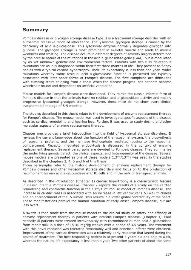
117
Summary
Pompe’s disease or glycogen storage disease type II is a lysosomal storage disorder with an autosomal recessive mode of inheritance. The lysosomal glycogen storage is caused by the defi ciency of acid α-glucosidase. This lysosomal enzyme normally degrades glycogen into glucose. The glycogen storage is most prominent in skeletal muscle and leads to muscle weakness and wasting. The disease occurs in different degrees of severity largely determined by the precise nature of the mutations in the acid a-glucosidase gene (GAA), but is modulated by as yet unknown genetic and environmental factors. Patients with two fully deleterious mutations are usually diagnosed within their fi rst three months of life. They present as fl oppy babies with a typical cardiac hypertrophy. Their life expectancy is less than one year. Milder mutations whereby some residual acid a-glucosidase function is preserved are typically associated with later onset forms of Pompe’s disease. The fi rst complains are diffi culties with climbing stairs or rising from a chair. When the disease progres ses patients become wheelchair bound and dependent on artifi cial ventilation.
Mouse models for Pompe’s disease were developed. They mimic the classic infantile form of Pompe’s disease in that the animals have no residual acid a-glucosidase activity and rapidly progressive lysosomal glycogen storage. However, these mice do not show overt clinical symptoms till the age of 8-9 months.
The studies described in this thesis relate to the development of enzyme replacement therapy for Pompe’s disease. The mouse model was used to investigate specifi c aspects of the disease such as cardiac remodeling and hearing loss. Further, it was used to study dosing and other molecular aspects of enzyme replacement therapy.
Chapter one provides a brief introduction into the fi eld of lysosomal storage disorders. It reviews the current knowledge about the function of the lysosomal system, the biosynthesis of lysosomal proteins, and the mannose 6-phosphate mediated routing to the lysosomal compartment. Receptor mediated endocytosis is discussed in the context of enzyme replacement therapy. Several paragraphs are devoted to Pompe’s disease. They summarise the under lying genetic defect, the clinical aspects, and heterogeneity of the disease. Several mouse models are presented as one of these models (13neo/13neo) was used in the studies described in the chapters 2, 4, 5 and 6 of this thesis.Three paragraphs refer to the historic development of enzyme replacement therapy for Pompe’s disease and other lysosomal storage disorders and focus on to the production of recombinant human acid a-glucosidase in CHO cells and in the milk of transgenic animals.
As described in the introduction (Chapter 1) cardiac hypertrophy is a characteristic feature in classic infantile Pompe’s disease. Chapter 2 reports the results of a study on the cardiac remodeling and contractile function in the 13neo/13neo mouse model of Pompe’s disease. The increase in cardiac mass is associated with an increase in left ventricular (LV) wall thickness and an encroachment of the LV lumen. This results in a lower global contractility of the heart. These manifestations parallel the human condition of early onset Pompe’s disease, but are less overt.
A switch is then made from the mouse model to the clinical study on safety and effi cacy of enzyme replacement therapy in patients with infantile Pompe’s disease. (Chapter 3). Four critically ill patients were treated intravenously with recombinant human acid a-glucosidase from rabbit milk in a dose of 15-40 mg/kg weekly over a period of 3.5 years. The treatment with this novel medicine was tolerated remarkably well and benefi cial effects were obtained. Improvement of the cardiac dimensions was a relatively early response that lasted during the course of treatment. The best responding patient is at present 5 years old and able to walk, whereas the natural life expectancy is less than a year. Two other patients of about the same
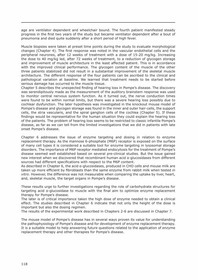
118
age are ventilator dependent and wheelchair bound. The fourth patient manifested steady progress in the fi rst two years of the study but became ventilator dependent after a bout of pneumonia and died quite suddenly after a short period of high fever.
Muscle biopsies were taken at preset time points during the study to evaluate morphological changes (Chapter 4). The fi rst response was noted in the vascular endothelial cells and the peripheral neurones, after 12 weeks of treatment with a dose of 15-20 mg/kg. Increasing the dose to 40 mg/kg led, after 72 weeks of treatment, to a reduction of glycogen storage and improvement of muscle architecture in the least affected patient. This is in accordance with the improved clinical performance. The glycogen content of the muscle of the other three patients stabilized did not result in a substantial improvement of the skeletal muscle architecture. The different response of the four patients can be ascribed to the clinical and pathological variation at baseline. We learned that treatment needs to be started before serious damage has occurred to the muscle tissue.Chapter 5 describes the unexpected fi nding of hearing loss in Pompe’s disease. The discovery was serendipitously made as the measurement of the auditory brainstem response was used to monitor central nervous system function. As it turned out, the nerve conduction times were found to be within normal limits, but there was a severe hearing loss possibly due to cochlear dysfunction. The later hypothesis was investigated in the knockout mouse model of Pompe’s disease and glycogen storage was found in the inner and outer hair-cells, the support cells, the stria vascularis, and the spiral ganglion cells of the cochlea (Chapter 5). If these fi ndings would be representative for the human situation they could explain the hearing loss of the patients. The problem of hearing loss seems to be restricted to classic infantile Pompe’s disease, as far as we can tell from the limited investigations that we did in patients with late onset Pompe’s disease.
Chapter 6 addresses the issue of enzyme targeting and dosing in relation to enzyme replacement therapy. As the mannose 6-phosphate (M6P) receptor is exposed on the surface of many cell types it is considered a suitable tool for enzyme targeting in lysosomal storage disorders. The importance of M6P receptor mediated endocytosis for the treatment of Pompe’s disease seemed well established based on several pre-clinical studies. But the issue gained new interest when we discovered that recombinant human acid α-glucosidases from different sources had different specifi cations with respect to the M6P content.As described in Chapter 6, the acid α-glucosidases, produced in CHO cells and mouse milk are taken up more effi cient by fi broblasts than the same enzyme from rabbit milk when tested in vitro. However, the difference was not measurable when comparing the uptake by liver, heart, and, skeletal muscle, the target organs in Pompe’s disease.
These results urge to further investigations regarding the role of carbohydrate structures for targeting acid α-glucosidase to muscle with the fi nal aim to optimize enzyme replacement therapy for Pompe’s disease.The later is of critical importance taken the high dose of enzyme needed to obtain a clinical effect. The studies described in Chapter 6 indicate that not only the height of the dose is important but also the dosing regimen.The results of the experimental work described in Chapters 2-6 are discussed in Chapter 7.
The mouse model of Pompe’s disease has in several ways proven its value for understanding the pathophysiology of Pompe’s disease and for development of enzyme replacement therapy. It is a suitable model to help answering future questions related to the application of enzyme replacement therapy and other therapies for Pompe’s disease.
119
Samenvatting
De ziekte van Pompe, ook wel glycogeen stapelingsziekte type II genoemd, is een metabole ziekte die autosomaal reccesief overerft. De aandoening wordt veroorzaakt door een defi ciëntie van het enzym “zure alfa-glucosidase”. Dit enzym breekt normaliter glycogeen af tot glucose in een specifi ek compartiment van de cel, het lysosoom.
De ziekte van Pompe manifesteert zich door spierzwakte. Het klinisch beeld is variabel. De “infantiele” vorm van de ziekte leidt al kort na de geboorte tot ernstige en progressieve spierzwakte. De wand van de hartspier is verdikt wat ten koste gaat van de hartfunctie. De meeste patiëntjes overlijden voor het eerste levensjaar aan hart- en ademhalingsproblemen. Presenteert de ziekte zich op de kinder- of volwassen leeftijd dan is er wel progressieve spierzwakte maar geen betrokkenheid van het hart. De ziekte begint vaak met problemen bij het traplopen of opstaan uit een stoel. Op latere leeftijd worden deze patiënten afhankelijk van een rolstoel en hebben soms ademhalingsondersteuning nodig.
In 1998 werd een muis model voor de ziekte van Pompe ontwikkeld. De muizen hebben net als de “infantiele” patiëntjes een totale defi ciëntie van het enzym zure α-glucosidase en ook zij lijden aan lysosomale glycogeen stapeling. Zij hebben een progressieve spierzwakte en een vergroting van het hart. Desondanks vertonen deze muizen pas klinische symptomen op de leeftijd van 8 maanden.
In dit proefschrift zijn een aantal studies opgenomen die betrekking hebben op de ontwikkeling van enzym vervangingstherapie voor de ziekte van Pompe.
Het eerste hoofdstuk dient als algemene inleiding in het veld van de lysosomale stapelingsziekten. Het behandelt in het kort de functie van de lysosomen, de productie en het transport van lysosomale eiwitten en het proces van receptor gemedieerde opname van macro-moleculen in het lysosoom. Het hoofdstuk gaat verder in op de ziekte van Pompe, met een speciale aandacht voor de klinische heterogeniteit, de onderliggende genetische defecten en de eigenschappen van de diverse muis modellen die er voor deze ziekte zijn ontwikkeld. Tot slot worden in dit hoofdstuk de mogelijkheden voor therapie besproken met nadruk op de ontwikkeling van enzym vervangingstherapie en de productie van recombinant humaan zure α-glucosidase in de melk van zoogdieren en CHO cellen (Chinese Hamster Ovary).
Verdikking van de hartspier is een van de karakteristieke gebeurtenissen bij de “infantiele” vorm van de ziekte van Pompe. Hoofdstuk twee beschrijft dit fenomeen in het muismodel van de ziekte van Pompe. De massa van het hart neemt toe en het volume van de kamers neemt af. Dit resulteert in een globale afname van de hartspierfunctie, hoewel de muizen, in tegenstelling tot de patiënten, geen uitdrukkelijke hartfunctiestoornis hebben.
In hoofdstuk drie worden de resultaten beschreven van enzym vervangingstherapy.Vier patiëntjes met de infantiele vorm van de ziekte van Pompe kregen eens per week recombinant humaan α-glucosidase intraveneus toegediend gedurende een periode van 3,5 jaar. Het enzym werd gewonnen uit de melk van transgene konijnen en toegediend in een dosis van 15-40 mg/kg. De behandeling werd goed verdragen en herstel van de hartspier was een van de eerste klinische effecten die bij alle patiënten werd gezien. De patiënt die het beste reageerde is nu 5 jaar oud en kan lopen terwijl de levensverwachting zonder behandeling niet langer dan een jaar is. Twee andere patiënten van ongeveer dezelfde leeftijd zijn afhankelijk van een rolstoel en kunstmatige beademing. De vierde patiënt maakte in de eerste twee jaren een periode van klinische verbetering door maar werd afhankelijk van kunstmatige beademing na een long ontsteking. De patiënt overleed na een korte periode van onbegrepen hoge koorts.Tijdens de behandeling van de patiënten werden op geregelde tijden spier biopten genomen
120
om de morfologie van de spieren te bestuderen (Hoofdstuk 4). Na 12 weken werd een afname van de glycogeenstapeling in de perifere zenuwen en in de endotheel cellen van de bloedvaten gezien bij een dosis van 15-20 mg/kg. Na verhoging van de dosis tot 40 mg/kg werd ook een vermindering van de glycogeenstapeling en een verbetering van de spierarchitectuur van de best reagerende patiënt gezien. Bij de andere patiënten werd wel een vermindering van het glycogeengehalte waargenomen maar bleef een substantiële verbetering van de spiermorfologie uit. Het is dan ook sterk aan te bevelen om zo vroeg mogelijk met de behandeling te beginnen.Bij het includeren van de patiënten, voor aanvang van de behandeling, werd onverwacht bij alle patiënten een ernstig gehoorsverlies vastgesteld (Hoofdstuk 5). Aanvullend onderzoek wees uit dat de gehoorszenuw en de hersenstam normaal functioneerden. Een verminderde functie van de cochlea werd gezien als meest voor hand liggende verklaring voor het gehoorsverlies. Omdat pathologisch onderzoek bij de mens niet mogelijk was werd voor verder onderzoek naar de oorzaak van het gehoorsverlies gebruik gemaakt van het muis model. De studies laten zien dat er glycogeen stapeling optreedt in zowel de binnenste- als de buitenste haarcellen en de steun cellen van de cochlea. Deze bevindingen ondersteunen de resultaten van het klinisch onderzoek. Het gehoorsverlies komt alleen voor bij patiënten met de infantiele vorm van de ziekte van Pompe.
Hoofdstuk 6 beschrijft experimenten die betrekking hebben op de mannose 6-fosfaat receptor gemedieerde opname van recombinant humaan zure α-glucosidase in die organen die voor de behandeling van de ziekte van Pompe van belang zijn. Er wordt een vergelijk gemaakt tussen de opname van verschillende vormen van recombinant humaan zure α-glucosidase in vitro (fi broblasten ) en in vivo (muis model). Het recombinant humaan α-glucosidase geproduceerd in CHO cellen en in de melk van transgene muizen wordt effi ciënter opgenomen door fi broblasten dan het zure α-glucosidase geproduceerd in de melk van transgene konijnen. Maar bij toediening van de verschillende enzymen aan muizen met de ziekte van Pompe is dit verschil niet meetbaar.
Op grond van deze resultaten blijven er vragen over de rol van de mannose 6-fosfaat receptor bij enzym vervangingstherapie voor de ziekte van Pompe. De effi cientie van enzym therapie kan mogelijk verhoogd worden door de rol van de suiker ketens bij de opname van zure α-glucosidase verder te bestuderen; dit in relatie tot de hoge dosis die nodig is voor het bereiken van klinische effect.
Het muis model voor de ziekte van Pompe heeft een waardevolle bijdrage geleverd aan het begrip van het ziektebeeld en is uitermate geschikt voor de verdere ontwikkeling van enzym vervangingstherapie en onderzoek naar andere therapeutische mogelijkheden.
121
Curriculum vitae (Door R. Kamphoven)
Joep Kamphoven kwam, overigens zonder dat zelf te weten, al vroeg in aanraking met de medische stand. Hij werd in 1970 in deze wereld opgevangen in de handen van gynaecoloog Joep Vos in het oude Carolusziekenhuis in ‘s-Hertogenbosch. Dat feit, plus de geboortedatum 19 maart, feest van St.-Jozef, bepaalde zijn naam. In zijn kleuterjaren had hij een ‘abonnement’ bij de KNO-specialist dr. Verhaegh. Dat beviel hem kennelijk zo goed dat hij toen zelfs ‘assistent van de dokter’ wilde worden. Verder groeide hij voorspoedig op, praatte al vroeg als Brugman en had behoudens de normale kinderziekten, nergens meer last van. Behalve dan die ene keer, op oudejaarsavond, dat zijn blinde darm eruit moest. Hij was toen 17. Altijd iets bijzonders dus.Als heel klein jongetje stond Joep op en ging naar bed met een pet of hoedje op z’n bol. Hier werd een trend in gang gezet: nu hoort een pet bij standaarduitrusting van jongeren. Bij Joep groeide deze tic uit tot een verzameling van zeker honderd hoeden en petten.De basisschool in Boxtel was een Jenaplan-school. Dit onderwijs probeert kinderen al zo vroeg mogelijk heel zelfstandig, sociaal en creatief te maken. Niets is gek, alles moet kunnen, meende zijn moeder. Zelfs het dragen van een knickerbocker. Dat leverde hem een enorme frustratie op die nog lang heeft doorgewerkt. Want toen al was een knickerbocker helemaal fout.Joep werd een jongen dus die ‘altijd iets bijzonders’ had. Bij het afzwemmen voor het diploma B bij voorbeeld was er één jongetje dat iets toch niet goed had gedaan. Joepje dus. Die moest voor een volle zwemzaal opnieuw een aantal meters onder water zwemmen… en hij redde het. Met gemak. Applaus dus. Joep ging ook op muziekles, en koos natuurlijk een bijzonder instrument: de accordeon… Dat instrument ruilde hij overigens na zijn afstuderen in voor een elektrische basgitaar. Verder ging hij met zijn zus Aukje in zijn havo-tijd op naailes, want hij wilde per se zijn eigen rits in zijn eigen broek kunnen zetten. Als enige jongen kwam hij op de dansschool terecht bij het tapdansen. Dat ging prima. Zo goed dat hij bij het lezen van een oproep in de krant voor audities bij het Scapino-ballet, dacht: ‘dat kan ik ook’. Hij schreef erop, en kreeg een uitnodiging die hij op het laatste moment toch maar afzegde. De studie havo ging goed, in ieder geval zo goed dat hij daarna het atheneum kon halen. Maar eerst probeerde hij op de opleiding tot piloot te komen bij de Rijksluchtvaartdienst. Daar kon men brildragers minder goed gebruiken, en bovendien: qua postuur was hij aan de kleine kant. Na het atheneum ging de militaire dienst aan hem voorbij: na een paar keer uitstel hoefde het niet meer van hare majesteit. Als cadeautje voor het behalen van zijn atheneumdiploma kreeg hij een ‘enkele reis’ met een vliegtuigje aangeboden: eenmaal boven moest hij gewoon ‘uitstappen’. Het werd met de instructeur een duo-parachutesprong om nooit te vergeten. Op het atheneum had hij al het idee verder te studeren. Geneeskunde leek hem wel wat. Bezig zijn met mensen. Niet zo gek, want Joep had zich tot een echt sociaal mens ontwikkeld. Hij koos de Universiteit van Maastricht, vooral om het onderwijssysteem. En hij werd meteen de eerste keer nog ingeloot ook, de bofkont. Het was een goede keuze, zo bleek. Hij haalde in het eerste jaar zijn propedeuse, en heeft daarna zijn studiejaren wel volledig opgesoupeerd. Natuurlijk werd Joep onmiddellijk betrokken bij het befaamde dispuut Ormètikos, waar hij later de feuten sokken liet breien, en een uniforme bloes liet naaien voor de inauguratie. Er werd zelfs een keer een tuinbroek voorgeschreven… Over een knickerbocker werd later een veto uitgesproken: iemand vond dat zijn kuiten niet goed uitkwamen. Joep ritselde verder bij de Maastrichtse brandweer een aantal afgeschreven brandweerjassen, en zo was het Ormètikos-uniform compleet. Aanvankelijk was het zijn bedoeling kinderarts te worden. Tijdens zijn co-schappen kwam hij met diverse specialisaties in aanraking maar kindergeneeskunde bleef bovenaan staan; zijn laatste stage was een wetenschapsstage bij kinderoncologie in Rotterdam waar hij met onderzoek geconfronteerd werd. Een boeiende kant van het vak, vond hij. Joep kwam in gesprek met dr. Ans van der Ploeg van de Erasmus-Universiteit over het lopende onderzoek
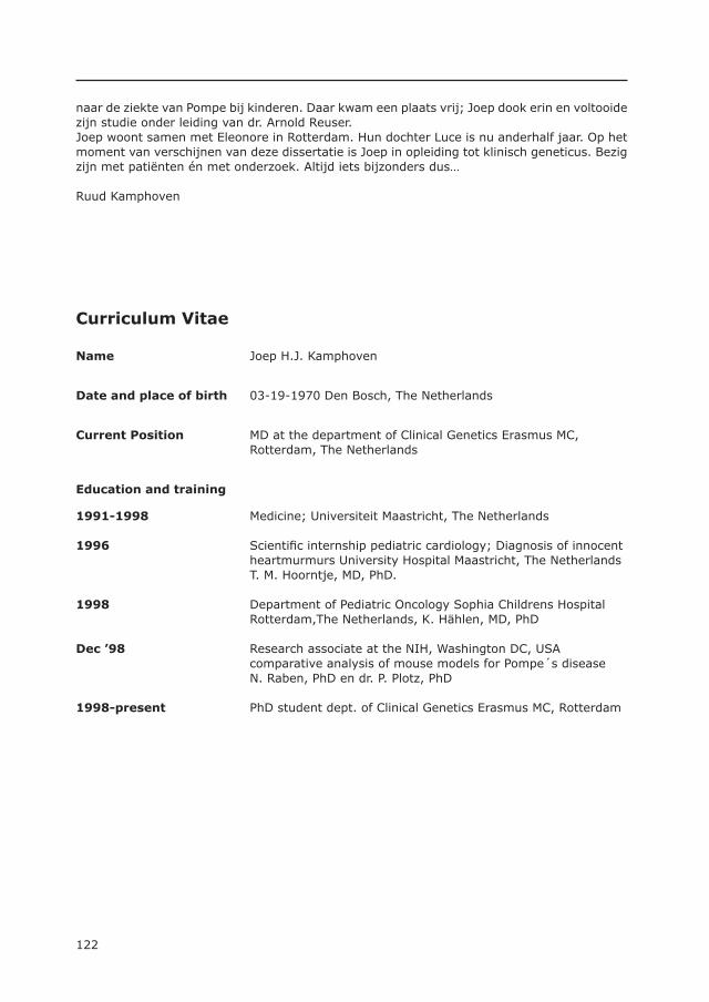
122
naar de ziekte van Pompe bij kinderen. Daar kwam een plaats vrij; Joep dook erin en voltooide zijn studie onder leiding van dr. Arnold Reuser.Joep woont samen met Eleonore in Rotterdam. Hun dochter Luce is nu anderhalf jaar. Op het moment van verschijnen van deze dissertatie is Joep in opleiding tot klinisch geneticus. Bezig zijn met patiënten én met onderzoek. Altijd iets bijzonders dus…
Ruud Kamphoven
Curriculum Vitae
Name Joep H.J. Kamphoven
Date and place of birth 03-19-1970 Den Bosch, The Netherlands
Current Position MD at the department of Clinical Genetics Erasmus MC, Rotterdam, The Netherlands
Education and training
1991-1998
1996
1998
Dec ’98
1998-present
Medicine; Universiteit Maastricht, The Netherlands
Scientifi c internship pediatric cardiology; Diagnosis of innocent heartmurmurs University Hospital Maastricht, The NetherlandsT. M. Hoorntje, MD, PhD.
Department of Pediatric Oncology Sophia Childrens Hospital Rotterdam,The Netherlands, K. Hählen, MD, PhD
Research associate at the NIH, Washington DC, USAcomparative analysis of mouse models for Pompe´s disease N. Raben, PhD en dr. P. Plotz, PhD
PhD student dept. of Clinical Genetics Erasmus MC, Rotterdam
123
List of abbreviations
bAGLU acid α-glucosidase from bovine testis;
BW body weight
cAGLU acid α-glucosidase from CHO cells;
DAB 3’3’diaminobenzidine
EDD end-diastolic diameter
EM Electron microscopy
ERT Enzyme replacement therapy
ESD end-systolic diameter
free M6P exposed (uncapped) M6P groups;
GMA Glycolmetacrylate
GSDII Glycogen storage disease type II
HW heart weight
KO knock-out
LM Light microscopy
LV left ventricular
LV dP/dtmax maximum rate of rise of left ventricular pressure
LVW left ventricular weight
M6P mannose 6-phosphate;
mAGLU acid α-glucosidase from mouse milk;
pAGLU acid α-glucosidase from human placenta;
PAS Periodic acid Schiff reagent
rhAGLU Recombinant human a-glucosidase
RV right ventricular
RVW right ventricular weight
τ time constant of early diastolic decay of LV pressure
WT wild-type
124
Publications
Cardiac remodeling and contractile function in acid α-glucosidase knock-out miceJoep H.J. Kamphoven, Rene Stubenitsky, Arnold J.J. Reuser, Ans T. Van der Ploeg, Pieter D. Verdouw and Dirk J. Duncker
Physiological Genomics 2001: 5:171-9
Impaired Performance of skeletal muscle in Alpha glucosidase knockout miceHesselink R.P., M. Gorselink, G. Schaart, A.J. Wagemakers, J. kamphoven, A.J. Reuser, G.J. Van der Vusse en M.R. DrostMuscle Nerve 25; 873-83, 2002.
Morphological Changes in Muscle Tissue of Patients with Infantile Pompe’s Disease receiving Enzyme Replacement TherapyLéon P.F. Winkel, MD, Joep H.J. Kamphoven, MD, Hannerieke J.M.P. Van den Hout, MD,Lies A. Severijnen, MSc, Pieter A. Van Doorn, MD, PhD, Arnold J.J. Reuser, PhD, Ans T. Van der Ploeg, MD, PhDMuscle Nerve 27: 743-751, 2003
Long Term Intravenous Treatment of Pompe’s Disease With Recombinant Human Alpha Glucosidase From MilkHannerieke M.P. Van den Hout MD PhD, Joep H.J. Kamphoven MD, Léon P.F. Winkel MD, Willem F.M. Arts MD PhD, Johannes B.C. De Klerk MD, M.Christa B. Loonen MD PhD, Arnold G. Vulto PharmD PhD, Adri Cromme-Dijkhuis MD PhD, Nynke Weisglas-Kuperus, Wim Hop PhD, Hans Van Hirtum MSc, Otto P. Van Diggelen PhD, Marijke Boer MSc, Marian A. Kroos MSc, Pieter A. Van Doorn MD , Edwin Van der Voort MD, Barbara Sibbles MD PhD, Emiel J.J.M. Van Corven PhD, Just P.J. Brakenhoff PhD, Johan Van Hove MD PhD, Jan A.M. Smeitink MD PhD, Gerard de Jong MD PhD, Arnold J.J. Reuser PhD, Ans T. Van der Ploeg MD PhDin press, Pediatrics 2004
Hearing loss in Infantile Pompe’s Disease and Determination of Underlying Pathology in the Knockout MouseJoep H. J. Kamphoven, Martijn M. de Ruiter, Hannerieke M. P. Van den Hout, Leon P. F. Winkel, Jan Bijman, Chris I. de Zeeuw, Hans L. Hoeve, Bert A. Van, Ans T. Van der Ploeg, and Arnold J. J. Reuser.in press, Neurobiology of Disease 2004
Both Low and High Uptake Forms of Acid α-glucosidase Target to Muscle of KO Mice with Pompe’s DiseaseJoep H.J. Kamphoven, Laura van den Dool, Marian A. Kroos, Ans T. Van der Ploeg, Dirk J. Duncker, Arnold J.J. Reuser-submitted-
Enzyme Replacement Therapy in Late Onset Pompe’s Disease: a Three Year Follow upLéon P.F. Winkel, MD, Hannerieke M.P. Van den Hout, MD, Joep H.J. Kamphoven, MD, Janus A.M. Disseldorp, MSc, Maaike Remmerswaal, MSc, Willem F.M. Arts, MD, PhD, M. Christa B. Loonen MD, PhD, Arnold G. Vulto, PhD, Pieter A. Van Doorn, MD, PhD, Gerard De Jong, MD, PhD, Wim Hop, PhD, G.P.A Smit, MD, PhD, S.K.Shapira MD, PhD, Marijke Boer, MSc, Arnold J.J.Reuser, PhD, Ans T. Van der Ploeg, MD, PhD.in press, Annals of neurology 2004
125
In memoriam
Dr. Johannes Cassianus Pompe(9 september, 1901 – 15 april, 1945)
Door R. Kamphoven en drs. G.A.M. Bak, arts
De ziekte van Pompe is genoemd naar dr. J.C. Pompe die de oorzaak van deze erfelijke ziekte voor het eerst beschreef in zijn proefschrift ‘Cardiomegalia glycogenica’, Amsterdam, 1936.Jan Pompe werd in Utrecht geboren 09 september 1901 in Utrecht. Na de lagere school gaat hij naar het gymnasium, daarna studeert hij medicijnen aan de Universiteit van Utrecht, thans Rijksuniversiteit Utrecht. Hij krijgt na zijn afstuderen een baan in het Wilhelminagasthuis in Amsterdam waar hij zich gaat specialiseren in de pathologische anatomie. Tijdens die specialisatie verdiept hij zich al in de glucose-stofwisseling.In zijn arikel ‘Over idiopatische hypertrophy van het hart’ (1932) beschrijft hij de sterke vergroting van het hart als aangeboren erfelijke aandoening. Onderzoek naar de oorzaak daarvan leidt tot zijn proefschrift ‘Cardiomegalia glycogenica’ waarop hij in 1936 promoveert. Het gaat hier om het ontbreken van het enzym alfa 1,4-glucosidase, nu bekend als de type II-vorm, een van de acht inmiddels bekende typen van deze ziekte.Dr. Pompe begint na zijn promotie zijn carrière als patholoog-anatoom in het Canisius-ziekenhuis in Nijmegen maar verruilt dit ziekenhuis in 1939 voor het Onze-Lieve-Vrouwe-Gasthuis in Amsterdam. Dan krijgen de voorbereidingen op de Tweede Wereldoorlog hem in de greep. Hij wordt opgeroepen in militaire dienst als de mobilisatie wordt afgekondigd, en krijgt een benoeming als vestinghygiënist bij de Vesting Holland. Maar de capitulatie van Nederland laat niet lang op zich wachten: dr. Jan Pompe neemt zijn baan in het Onze-Lieve-Vrouwe-Gasthuis weer op in mei 1940. In 1942 weigert hij het verplichte lidmaatschap van de Nationaal-Socialistische Artsenkamer. Pompe zit dan al in het verzet. Hij biedt in het ziekenhuis plaats aan studenten die ook in het verzet zitten, hij neemt later een geheime radio-zender over die eind 1944 in zijn laboratorium in het ziekenhuis verborgen wordt. Dat kost hem uiteindelijk het leven.Waarschijnlijk door verraad ‘ontdekken’ de Duitse bezetters in februari 1945 de zender in het ziekenhuislaboratorium. Tijdens de inval is dr. Pompe zelf niet aanwezig, maar hij wordt even later thuis gearresteerd. Als een Nederlandse verzetsgroep op 15 april 1945 een spoorlijn in St.-Pancras opblaast, nemen de Duitsers harde represaillemaatregelen: twintig gevangenen worden gefusilleerd. Onder hen dr. Jan Pompe.Ter herinnering aan hem en aan andere verzetshelden uit het Onze-Lieve-Vrouwe-Gasthuis wordt in dit ziekenhuis in 1947 een gebrandschilderd raam onthuld. De naam van Jan Pompe wordt verder opgenomen in de Erelijst van Gevallenen die in het gebouw van de Tweede Kamer in Den Haag ligt.
Noot:Het Pompe-instituut in Utrecht en de Pompe-kliniek in Nijmegen zijn vernoemd naar de oudere broer van dr. Jan Pompe, mr. Willem Pompe, hoogleraar strafrecht.
Bronnen: 1 Marten Dooper; ‘Joannes (Jan) Cassianus Pompe’ rubriek ‘Onsterfelijke namen’, bijlage
Scala, De Limburger 01-05-2002. 2 Rik Nijland; ‘Pluizige medicijnfabriekjes’, bijlage wetenschap, De Volkskrant 04-07-
1998.
126
Dankwoord
Of ‘Mice and men’ zou een ondertitel van dit proefschrift kunnen zijn. Zonder de mus musculus zou het zover niet gekomen zijn en dus wil ik van mijn dankwoord gebruik maken om ook daar even bij stil te staan.
Binnen het Erasmus MC werken vele mensen die een bijdrage hebben geleverd; van medewerkers van het EDC tot aan de collega’s van de Klinische Genetica op de 24e verdieping, en in de kliniek. Mijn dank gaat uit naar iedereen die een bijdrage leverde.
Mijn promotor, professor B.A. Oostra, ben ik erkentelijk voor de gelegenheid die hij mij geboden heeft om te promoveren aan de vakgroep Klinische Genetica te Rotterdam, voor het vertrouwen in het Pompe-onderzoek in het algemeen en de kritische blik op dit proefschrift.Ik dank de commissieleden prof. dr. H.A. Bűller, prof. dr. J.G.G. Borst en prof. dr. D.J. Duncker voor het vlotte lezen van dit werk.
Mijn co-promotor dr. Arnold Reuser wil ik bedanken voor de begeleiding van het gehele promotie-traject. Een promotie bestaat uit ‘leermomenten’ en: wat zijn dat er veel geweest. Zuivere wetenschap, dankzij jouw inspanning ben ik ermee in aanraking gekomen, op velerlei manieren, en vaak ook buiten het Pompe-project om. Het is een andere manier van naar de wereld kijken en je verbazen over waarom de dingen zijn, zoals ze zijn; daar dan geen genoegen mee nemen, maar willen weten waarom het is, zoals het is.Mijn tweede co-promotor dr. Ans van der Ploeg representeert de klinische visie binnen dit project. Wegens drukte in de kliniek hebben we elkaar te weinig gesproken maar je bijdrage was van wezenlijk belang. Je inzet is onbetaalbaar. Als duo zijn jullie uniek en in staat tot grote hoogte te komen. In dit project waar kliniek en fundament elkaar ontmoeten, wetenswaardigheden uitwisselen en vragen proberen te beantwoorden is nog een hoop te doen. Ik hoop dan ook van harte dat jullie daar weer meer tijd voor vinden.
Voor de onwetende/ontketende dokter die in het lab strandt, zijn rotsen in de branding bittere noodzaak. Discussie over PCR’s, lab-bepalingen, wat er alweer mis is gegaan, waarom en wat die grap kostte. SOP’s en protocollen tot je er gek van werd en altijd je troep opruimen!!!! Marian, we hebben wel eens ‘gevochten’ maar ik had je o, zo nodig; bedankt voor de vruchtbare samenwerking, je scherpe en brede visie (blijf studeren!!).De Pompe Club wisselde in zijn aantal deelnemers maar bleef een gebalanceerd geheel. Ik ben iedereen (Monique, Dik, Hans, Agnes) dankbaar voor de goede sfeer en samenwerking. Jammer dat ik niet bij dat uitje kon zijn.Laura, je begon als student en liet vele muizen door je handen gaan. We hebben daarna??? goed samengewerkt en je hebt bergen werk verzet; dank voor de goede en prettige samenwerking.
Maandag-ochtend telefonisch contact met dokter Winkel. Gelukkig zijn we beiden rasoptimisten van ‘de copy paste generatie’ tot ‘Is Goed’… ‘Fokke en Sukke ronden het af’; beginnen bij versie 13 en direct de ‘nu echt allerlaatste versie’ er achteraan. ‘Het was een bloempje van een artikel maar het leest nog niet als een Boek!!’ ‘Wacht maar tot er een nietje doorzit’. Hou vol, en goed dat je mijn Paranimf wilt zijn.Doktor vd Hout, (Hm mooi niet, dr.) soms lijkt het H’tje op z’n baas al is het alleen maar in de kordate pas hoewel de wervelwind wel binnenkomt als jij de deur opendoet; dank voor je energie, samenwerking en het afronden van jouw boekwerk waardoor ik aan de beurt was. Drs. Hagemans, Marloes, met jou komt alles goed, degelijke onderzoekster, het is een kwestie van volhouden en….. af en toe met de mannen in de auto zitten (10 uur heen en 12 uur terug).
127
Hollen of stilstaan, het lijkt alsof het niet anders kan. Ventileren bij je mede-Aio’s/benchgenoten (arts-onderzoekers enz.) is broodnodig, af en toe een Frustootje (cappuccino in een gefrustreerde bui), en hop daar gaat-ie weer. (Of course thanks to all from abroad, working in the lab.)
Gesprekken over onderwerpen die zeker niet geschikt zijn voor in een proefschrift, maar die wel sfeerbepalend zijn. Dank voor vier jaar ‘Lab en Leed’ gaat uit naar Surya, Stefano, creative minds behind ‘LAB, the movie; Elly: is dat Perzische bed er nog gekomen?? Rachel: schippersverhalen; Miriam: genealogie, of hoeveel gespuis komt er voor in je familie; Ozgur: I speak fl uently Turkish now, sorry, it is only three words: Naber and I jijim; en de vele studenten in het lab.Mieke en Leontine: we blijven koffi e ‘doen’, met af en toe een broodje.
Dr. André Hoogeveen: er moet meer gezongen worden op het lab; dr. Frans Verheijen: altijd in voor de scherpe opmerking. Kijk uit voor de hiërarchische apoptosis en sla af en toe een balletje. Dr. Mark Nellist: je Engelse scheldkanonnades, bescheidenheid en gedrevenheid op het lab en in de sport; nog bedankt voor de squashlessen. Violeta, I still practice on your christmas song, (Chervenite Boetoeski……).
De medewerkers van de enzymdiagnostiek wil ik bedanken voor hun vele werk in de klinische trial en inzichten in de interpretatie van de enzymwaarden die we vonden. Tevens dank voor de fl exibiliteit; het lenen van een peukje substraat, even de incubator gebruiken, pipetteren op jullie lab; de voorzet van de SOP’s en de gezelligheid. Memorabele momenten in het slachthuis en bij het gevecht met de stierentestikels. (Marijke, Joke, Otto, Wim, bedankt voor jullie geduld.)
De collega’s van de andere laboratoria: Pre-en Postnataal, DNA-diagnostiek, het metabole lab, de beide secretariaten. Dank voor de goede sfeer en het geklets aan de ‘Dorpspomp’ die we koffi e-automaat noemen. De praatjes waren nooit slapper dan de koffi e die eruit kwam!!!
Mederwerkers van de experimentele cardiologie onder de bezielende leiding van (toen) prof. dr. ir. Pieter Verdouw en (nu) prof. dr. Dirk Duncker, altijd in voor de discussie over sport en cardiologie. Het opstarten van de echocardiografi e bij muizen was moeizaam. Van het onderwater-echoën via de tube ontharingscrème naar een echt non-invasieve methode. René, Mirella dank voor de ontwikkeling en het meten. Rob, zonder jouw computergave was de analyse in het waterbad blijven steken.Het meten van intra-cardiale, en intra-vasculaire drukken in de muis blijft priegelwerk dat Monique en Elza tot in de fi nesses beheersen. (Blijf reizen; gitaar spelen enz.)
Zonder de afdeling pathologie wordt het met die spierbiopten nooit wat. Rob Willemsen bedankt voor je adviezen (blijf met twee wielen aan de grond) en Lies-Anne bedankt voor je geduld en de hoge kwaliteit van de coupes.
Bij het werken met muizen is niets zo cruciaal als de verzorging van het dier. Gaandeweg dit project is er, wat dit betreft, veel veranderd. De overgang naar een nieuwe faciliteit met een nieuw drinkwatersysteem ging niet zonder slag of stoot. De verzorging van grote aantallen muizen is uiterst complex. Zeker als bepaalde groepen ook nog van verschillende onderzoekers (moeilijke mensen) zijn. Toch hebben we in deze periode prettig samengewerkt. Dank aan iedereen voor en achter de barrière.
Aan alles komt een eind maar dan steeds is er weer een nieuw begin. Collega’s en bazen van ‘de overkant’, bedankt voor jullie geduld. Ik was vast wel eens sjacherijnig of vervelend, hopelijk wordt dat minder.
128
Dit proefschrift had er niet zo uit gezien zonder de gedrevenheid waarmee Tom de Vries Lentsch de Layout voor elkaar gebokst heeft; Tom bedankt en … de deadline gehaald. Ruud bedankt voor je ontwerp adviezen, koffi e en fotografi sche visie. Jullie beiden bedankt voor de fi losofi sche en politieke debatten tijdens het werk.
In de turbulentie van het promotie-leven is stabiliteit onontbeerlijk. Verwaarlozing van familie en vrienden, het zit me niet lekker. Bedankt voor het blijven vragen: ‘hoe is het met je boekje???’ Het boekje is af, dus nu kan het weer…….
Naarmate het einde van een dankwoord nadert blijven er steeds minder mensen over. Je ouders en schoonouders, het is toch mooi dat ze onvoorwaardelijk in je blijven geloven. Pap & Mam: opvoeden is een vak apart en de dingen die jullie me meegegeven hebben……… ik heb ze goed gebruikt. Bedankt voor jullie vertrouwen en ondersteuning. Geen mens kan zonder. Pap leuk dat je mijn cv wilde schrijven.Gijs en Henny, bedankt voor de fi losofi sche momenten over de telefoon, aan tafel, op de bank en te voet. Trrriing, en weer een stelling; we hebben het er nog over!!
Eleonore, na alles wat we door of dankzij dit proefschrift hebben meegemaakt, kan ik alleen maar héél véél van je houden. Promoveren doe je niet alleen. Eerst waren we met zijn tweeën en nu met zijn drieën. Druk op de ketel komt de sfeer niet altijd ten goede maar we hielden vol, nog even. Ik heb het wel gemist: samen dingen doen; plannetjes maken en uitvoeren; lekker naar buiten. De balans vinden is moeilijk als je iedereen tevreden wilt houden. Ik heb wel geleerd dat dat idealistisch is.Luce, vrolijkheid van binnenuit en dat vleugje temperament, ik ga meer tijd met jou en Eleonore doorbrengen. Op naar de bergen en de koeien (Hiiiiiiiiih) en de paardjes (Boehoehoe), lekker rommelen met ons ouwe Bakkie.
Groet
Joep
P.S. Antwoord op de vraag wie is Hoepie (Hoepie = Hub Kamphoven)