Embed Size (px)
Citation preview
P O L Y S A C C H A R I D E I N D E L A Y E D H Y P E R S E N S I T I V I T Y
I. PNEUMOCOCCAL POLYSACCHARIDE AS INDUCER AND ELICITOR OF DELAYED P~EACTIVITY IN GUINEA PIGS
BY ROBERT J. GERETY, RODOLFO W. FERRARESI, M.D., AND SIDNEY RAFFEL, M.D.
(From the Department of Medical Microbiology, School of Medicine, Stanford University, California 94305)
(Received for publication 8 September 1969)
The role of polysaccharides as an t ibody inducers has been appreciated for many years, bu t there has been some doubt as to their capacities as immunogens for delayed hypersensi t ivi ty . If they are active in this respect, the capaci ty is less clearly evident than is tha t of proteins or of contact inducing agents.
For example, in the test devised by Tiller and Francis (1) for patients with lobar pneumonia, the intradermal injection of purified pneumococcal polysaccharide derived from the infecting type of organism gave rise to reactions of the immediate type, although the pneumococcus is known to induce delayed reactivity to its protein constituents (1, 2). In work with blood group substances, Holborow and Loewi (3) found antibodies to be produced against the sugar components, while delayed reac- tivity, which was also seen, was attributed to the peptide moiety of these substances. Similar conclusions were reached using chondromucoprotein as antigen (4). Jankovic and Waksman (5) also concluded that delayed hypersensitivity induced by the sac- charide-peptides of human erythrocytes may have been in part directed against the peptide structure. Barker and coworkers (6) with polysaccharide from Trichophyton concluded that it participated in antibody-mediated reactions, the protein of the organism in delayed ones.
In 1963 it was demonstrated that mono- and disaccharides coupled to protein carrier induced delayed reactivity elicitable by the same hapten attached to different carriers, although homologous conjugates were best for the purpose (7). Several reports de- scribe the induction of delayed reactivity to polysaccharides, while presumably obvi- ating the possible role of associated peptides. Thus, Knight et al. (8-10) described the induction of delayed reactivity in guinea pigs to polysaccharides of Histoplasma capsulatum and Blastomyces dermatitidis by sensitization with whole fungi and testing with appropriate polysaccharide preparations, but these contained 4-5% nitrogen. Freund et al. (11) induced aspermatogenesis in guinea pigs with testicular extract
* This investigation was supported by a training grant AI-82 and a research grant AI-01178 from the National Institute of Allergy and Infectious Diseases, United States Public Health Services; and by a research grant to Dr. Raffel from the San Francisco Foundation.
189
Dow
nloaded from http://rupress.org/jem
/article-pdf/131/1/189/1083491/189.pdf by guest on 08 January 2022
190 POLYSACCHARIDE IN DELAYED H Y P E R S E N S I T I V I T Y . I
composed largely of polysaccharide, but containing about 4% N and giving positive biuret and ninhydrin reactions. Rowlands, Crowle and Russe (12) found positive reac- tions to a polysaccharide derived from Candida albicans in mice immunized with heat- killed fungi, and Campbell earlier (13) made similar observations in rabbits sensitized with Ascaris and tested with a highly purified polysaccharide derived from this worm. In the case of Mycobacterium tuberculosis, despite a long history of evidence indicating that tuberculin reactivity is directed against its protein constituents, Baer and co- workers (14-17) have more recently detailed experiments implicating a polysaccharide extract in this respect, also, in guinea pigs sensitized with whole organisms. However, some protein nitrogen was present in this preparation, and there was cross reactivity between the polysaccharide and protein fractions used. Azuma and coworkers (18) with a nitrogen-free polysaccharide isolated from culture filtrates of the tubercle bacillus have been able to find only anaphylactic reactivity elicitable in guinea pigs sensitized by bacilli.
Recently, Crowle and Hu (19) have described induction and elicitation of delayed reactions to dextran in the mouse, and Battisto et al. (20) have established such reac- tivity to this polymer in certain strains of guinea pigs. These last works in fact come closest to the only evidence extant that pure polysaccharides may be able to induce the delayed hypersensitive state. The evidence on the whole is very scant that poly- saccharides can induce delayed reactivity, and sufficiently irregular in respect to elicitation of reactions as to have led to the suggestion (21) that saccharides may be relatively ineffective in delayed reactions, possibly because they lack sufficient bind- ing energy to interact effectively with the antibody (or antibody-like substance) re- sponsible for such reactions.
In the present report, a pneumococcal capsular polysaccharide (type II) was used to induce and elicit delayed hypersensitive reactivity in guinea pigs. This was employed in the face of several discouraging reports. Freund and Bonanto (22) in 1944 had in- corporated pneumococcal polysaccharide (type not stated) in adjuvant containing mycobacteria in efforts to induce antibodies or hypersensitivity in rabbits. Their failure may have reflected the relative inability of this species to respond immuno- logically to such polysaccharides (23-25). Failure with type I I I pneumococcal poly- saccharide in guinea pigs was reported by Maurer and Mansmann (26) with small inducing doses (10 ttg) which could have been an insufficient quantity for sensitiza- tion. However, in the work reported here, we also failed with this polysaccharide in amounts of 100 t~g. Tremaine (2) could not detect delayed reactivity to capsular poly- saccharide in the corneas of rabbits passively sensitized with lymphoid cells from donors previously immunized with intact pneumococci.
The present work was instigated primarily by an interest in the question of mechanistic basis for delayed hypersensitive reactivity. We think that delayed reactivity of the "tuberculin type" eventuates under certain circumstances of exposure of an animal to antigen, while another expression of cellular reactivity (termed "Jones-Mote" type) comes about under other circumstances of expo- sure to antigen as an earl)" phase of antibody synthesis (27-31). The bases for this distinction have been observational, including the time of appearance of the reactive state, its duration, and the gross and microscopic characteristics
Dow
nloaded from http://rupress.org/jem
/article-pdf/131/1/189/1083491/189.pdf by guest on 08 January 2022
R. J. GERETY, R. W. FERRARESI, AND S. RAFFEL 191
of dermal reactions (30-34). I t was thought tha t if a polysaccharide were capable of inducing antibodies bu t lacked the capacity to induce persisting delayed reactivity of the tuberculin type, it would be interesting to observe its possible role in the induction and elicitation of early-appearing Jones-Mote reactivity. If the latter were in fact an aspect of the evolution of the conven- tional ant ibody response, the polysaccharide should participate in this. We turned to Pn I IS as a saccharide known to be free of nitrogen-containing sugars (35, 36) and hence amenable to testing for possible protein contaminants. We found that this substance per se is immunogenic for guinea pigs, inducing anti- bodies in low but detectable quantit ies in most of them, and giving rise to striking levels of delayed reactivity and to excellent instances of Jones-Mote reactivity. This report deals with studies of the homogeneity and composition of the polysaccharide employed, and with the results of its use as a stimulus to the induction of these various reactivities as determined by skin tests, by tests for inhibition of macrophage migration, by serum and cell transfers, by sero- logic tests, and by histological correlations.
Materials and Methods
Animals.--Random-bred guinea pigs, 250-350 g, were used for sensitization and skin test- ing, and as the source of cells for macrophage migration tests. Animals of 250-300 g were used for passive cutaneous anaphylactic (]?CA) tests. Albino Wistar rats, 200-250 g, were used as recipients of guinea pig serum for heterologous PCA tests.
Antigens.--Pneumococcus type II capsular polysaceharide (PnlIS) was obtained from Lederle Laboratories, Division of American Cyanamid, Pearl River, N.Y., courtesy of Dr. W. S. Hammond. The sample received had an N content of 0.10%. In an effort to reduce this, the sugar was repeatedly precipitated with 95% ethanol and propanol, followed by shakings in chloroform and butanol (35-37). It was then precipitated with barium acetate, reprecipi- tated with sodium acetate and isopropanol, centrifuged at 10,000 rpm and retreated with 95% ethanol (35, 36). The eventual product contained 0.04% nitrogen as an irreducible mini- mum. In view of the fact that PnIIS does not contain amino sugars, it was important to deter- mine whether this might represent peptide nitrogen, which in turn could be responsible for some of the biologic activities to be described. The polysaccharide was subjected to the fol- lowing tests and procedures.
Antigenic homogeneity was determined by double diffusion tests in agar against rabbit anti- PnII sera from animals vaccinated with killed organisms; these showed single lines of pre- cipitate. The polysaceharide was also subjected to electrophoresis on slides in Noble agar at pH 8.6, 30 ma, for 2-5 hr. Against rabbit anti-pneumococcal type II serum, only one precipi- tation arc formed.
The composition of the sample of PnlIS was assayed by several methods: Methyl Pentose Analysis.--Rhamnose accounts for about 50% of the PnIIS molecule (38),
which is composed of L-rhamnose, D-glucose, and D-glucuronic acid in the ratio 3:1 : 2. Quan- titative tests for this sugar (39) were carried out simultaneously with control samples of rham- nose dried to constant weight. 10 mg samples of PnlIS and rhamnose were hydrolyzed with HzSO4, treated with freshly prepared cysteine HC1 reagent, and measured for absorption at 396 m/z, correcting for glucose absorption on the basis of its equivalent absorption peaks at 396 and 430 mtt. Similar untreated samples served as blank controls for reading in the Beck- man DB spectrophotometer. Rhamnose was found to comprise 50% of the total weight.
Dow
nloaded from http://rupress.org/jem
/article-pdf/131/1/189/1083491/189.pdf by guest on 08 January 2022
192 POLYSACCHARIDE IN DELAYED HYPERSENSITIVITY. I
Thin Layer Chromatography in Silica Gd.--lO mg of PnIIS was treated with 2 N H2SO4, heated at 100°C. for 3 hr, and neutralized to pH 7 with saturated Ba(OH)~. The salts were centrifuged off, the samples evaporated to 0.5 ml, and 10 ~ quantities were spotted on gel plates (2 parts 0.I ~ boric acid and i part silica gel G. mixed in a blender), with solutions of glucose, rhamnose, and glucuronic acid as controls. The spotted plates were placed in butanol- acetic acid (4:1) solvent for 5 hr, then sprayed with a solution of aniline (1%) and diphenyl- amine (l%) in acetone, and dried with a heat gun. Only the appropriate sugars were found.
The nitrogen content of the polysaccharide had been determined by micro-Kjeldahl analyses of three 10 mg samples to be 0.04%. The following analyses were made to determine its source:
Flnorometry.--The possible presence of aromatic amino acids was sought with a Turner Model Ii1 Fluorometer, using 0.01 -10 mg samples in 1 ml. Reference proteins were included as controls. This method is sensitive to 0.001 #g of tyrosine. There was no detectable result above background.
Silica Gel Chromatography for Amino Aeids.--Three 10 mg samples of polysaccharide were hydrolyzed with 2 N H~SO4 at 100°C. The hydrolysates along with solutions of known amino acids and amino sugars were spotted on Eastman Chromogram No. 6060 sheets in volumes ranging from 20-40 microliters of solution containing 40 mg PnIIS/ml (up to 1.6 mg polysac- charide per test, of which the N could represent about 4 #g of peptide). The chromatograms were developed with a mixture of butanol, acetic acid, and water (4:1:1) for 3 hr, then dried and sprayed with ninhydrin reagent. This method can detect as little as 0.01/zg glycine and 0.1 #g proline. When a mixture of constituent sugars of PnIIS and ammonium salts (NH4C1, NH4NO3, (NH4)2SO4) were chromatographed, color developed in the same region as in the experimental analysis of the PnIIS, suggesting that the N in this preparation derived from ammonia absorbed from the air by the acidic polysaccharide.
Tests for Free Amino Groups were made on 10 mg hydrolyzed samples of the polysaccha- ride with 2,4,6-trinitrobenzene 1-sulfonic acid (40). Readings for absorbance at 340 nap showed values corresponding to a possible average content of 1.4 #g of amino acids.
Enzymatic treatment of the PnIIS with peptidases will be described under experimental results.
Pneumococeal type I I I capstdar polysaccharide was obtained from Dr. Michael Heidelberger. It was used in skin and serological tests as a control for the possible presence of non-type- specific bacterial substances in the PnIIS preparation which might have escaped the purifica- tion procedures.
Vaccination Procedures.--PnIIS was administered to guinea pigs intradermally in saline or subcutaneously in incomplete or complete Freund's adjuvant (Difco). Varying concentra- tions of polysaccharide in 0.1 ml of saline were mixed with 0.1 ml of adjuvant.
Skin tests were performed at 7, 14, or 21 days after the single sensitizing injection, using usually 100 #g of polysaccharide in 0.l ml of saline in the flank. Readings were made at 4, 12, 24, and 48 hr.
Antlsera.--Several rabbit anti-PnII sara were used for immunodiffusion tests. One lyophi- lized sample I was selected as a standard positive control in carrying out various serological tests with experimental guinea pig sara.
Serological Tests.--Guinea pigs were bled at various intervals just before, or in some cases after, skin testing. Antibodies were detected by these procedures:
(a) Double diffusion in agar, using wells of 0.01 ml capacity spaced 2 mm between circum- ferences. Concentrations of PnIIS used varied between 0.1 and 0.0001 rag.
(b) Passive cutaneous anaphylaxis (PCA) tests were carried out in guinea pigs (for detection of % antibodies (41-43)), and in heterologous animals (Wistar albino rats) for the detection of % and T'a antibodies (42, 43) with 0.1 ml quantities of undiluted serum. In the same recipi-
1 Obtained from the Communicable Disease Center, Atlanta, Ga.
Dow
nloaded from http://rupress.org/jem
/article-pdf/131/1/189/1083491/189.pdf by guest on 08 January 2022
R. J. GERETY~ R. W. FERRARESI~ AND S. RAFFEL 193
ents, a nonspecific rabbit serum (anti-ribonuclease) and a positive control rabbit serum (anti- PnII) were used. After 4 hr, Evans blue dye was injected intravenously; if no dye leakage occurred by 5 min, 5 or 10 mg of PnIIS in 1 ml of saline was injected intravenously. Reactions were read at ~ hr.
(c) Micro-complement fxation tests were done with sheep red cells labeled with radioactive chromium (44). Lysis was measured by the release of radioactivity in mixtures of test serum, rabbit hemolysin, and complement.
Tests were done with 0.05 ml samples of heat inactivated serum added to 1 #g PnIIS in 0.025 ml in an ice bath. To this was added 0.05 ml of guinea pig serum containing two 50% hemolytic doses of complement for the sheep cell system employed. The mixture was incu- bated at 37°C for 1 hr with occasional shaking, and left overnight at 4°C. Control tubes con- tained known positive (rabbit) serum; normal inactivated guinea pig serum; antigen plus complement; test serum plus complement; and serum alone. Sheep cells collected in citrated Alsever's solution were washed 3 times in Veronal-buffered saline (pH 7.3) containing 1% gelatin. To 0.1 ml packed cells, about 50 #Ci of 51Cr were added (~tsodium chromate, Abbott Laboratories, North Chicago, Ill.). The mixture was incubated at 37°C for 1 hr with continu- ous gentle agitation, and left overnight at 4°C. The cells were then washed 5 times with buffer and made up to a 5% suspension.
The incubated test mixtures each received 0.025 ml of the red cell suspension mixed with rabbit hemolysin. Tubes were reincubated at 37°C for 1 hr; 1 ml of cold buffer was added, and the tubes were centrifuged at 2000 rpm under refrigeration. Sedimens and supernatants Were counted in a well-type gamma sensitive scintillation counter. This method can detect proba- bly 0.003 gg antibody N (44).
Macrophage inhibition tests were carried out by a modification of the procedure of David et al. (45). Guinea pigs received 10 ml heavy mineral oil i.p. 4 days before cell removal. The peritoneal cavities were washed with 20 ml Hanks' solution containing 1% normal guinea pig serum and in each ml 10 units of preservative-free heparin, 50/zg streptomycin, and 50 units of penicillin. Cells were washed 3 times in this solution without heparin in a refrigerated centrifuge at 500 rpm for 5 rain. Differential counts were made; these usually showed about 60-68% macrophages, 20% (+ 10%) lymphocytes and some polymorphonuclear cells. Cells were suspended 0.05 ml packed volume per 1 ml medium. Approximately 2.5 X 10⁶ cells were taken up in capillary tubes (1.3-1,5 mm inside diameter, X 75 ram). The filled tubes were sealed at one end with paraffm and centrifuged at 400 rpm for 5 min, providing packed cell columns about 1--2 mm long. The capillaries were cut at the cell-fluid interface, and six such tubes were laid in a Falcon disposable tissue culture dish (60 mm diameter, 12 mm deep) and attached with stopcock grease. These were overlaid with 4 ml of medium consisting of 199 Earle's salt base plus 15% normal guinea pig serum, 200/zg bovine serum albumin (BSA)/ml with antibiotics (penicillin, 50 units; streptomycin and neomycin, 50/zg of each), and con- taining 25/zg PnIIS/ml. The plates were incubated in 95% alr-S°~ CO2 for 24 hr at 37°C. The supernatant was then removed and the dish allowed to dry. The areas of cell migrations were projected to 7 M onto paper of constant weight with a photographic enlarger, the out- lines of the migrating cell borders were drawn, and these areas were cut out and weighed. The percentage of inhibition of migration was calculated for each cell suspension by comparing the mean of the migration in the presence of antigen to that in the absence of antigen.
Cell and Serum Tranffers.--Sensitized guinea pigs were treated as follows: On the 7th, 14th, or 21st day after sensitization, 10-12 animals of each group were bled, and mineral oil-induced peritoneal exudates were harvested. The cells were pooled and washed 3 times, counts were made, and 3-4.3 X l0 s cells in 2.5 ml of Hank's solution containing 1% normal guinea pig serum were injected intravenously into each of 2 or 3 normal recipients. 15 rain-1 hr later, the animals were given skin tests with the usual dose of PnIIS.
Serum of each group were also pooled and within 4-6 hr of bleeding, 20 ml were injected
Dow
nloaded from http://rupress.org/jem
/article-pdf/131/1/189/1083491/189.pdf by guest on 08 January 2022

194 POLYSACCHARIDE IN DELAYED HYPERSENSITIVITY. I
i.o. into each of 2-4 normal recipients. Again skin tests were done at 1/~-I hr after the injec- tions had been made.
RESULTS
Immunologic Responses of the Guinea Pig to P n I I S
100 Ng2 of PnIIS was given to guinea pigs in incomplete or complete Freund's adjuvant, subcutaneously in the groin. Skin tests were made at 7, 14, and 21 days in separate animals with the same dose of polysaccharide. Readings were at 4, 24, and 48 hr. The average of reac- tions from several experiments are shown in Table I and Figs. 1 and 4.
These resul ts indicate t ha t react ions ear ly (7 days) af ter sensi t iza t ion appea r
essent ial ly the same in bo th groups, wi th no indicat ions of i m m e d i a t e (ana-
phy lac t i c or Ar thus) responses up to 4 hr, wi th zeni ths at 24 hr and decl ine by
TABLE I
Skin Reactions in Guinea Pigs at 7, 14, and 21 Days after Subcutaneous Sensitization with 100 #g of PnlIS in incomplete or Complele Freund's Adjuvant, and Tested* with
100 #g of PnlIS
Number Days of animals 4 hr 24 hr 48 hr
Incomplete Freund's adjuvant 7 33 0.2, 0~ 9.4, 0.4 3.9, 0.03
14 14 6.3, 0.33 10.8, 0.6 0.7, 0 21 28 17.5, 1.3 8.9, 0.3 0.5, 0
Complete Freund's adjuvant 7 34 0.6, 0 10.2, 0.6 5.4, 0.2
14 15 7.5, 0.57 17.1, 1.07 4.0, 0.18 21 27 12.9, 0.8 21.8, 1.4 10.6, 0.6
* One test per animal. :~ First number, mm diameter; second number, estimated mm thickness. § Control readings in normal guinea pigs given skin tests at the same times have been
subtracted. For 42 animals, the means were: 4 hr. : 6.9, 0.4; 24 hr. : 1.4, 0.1 ; 48 hr. : 0.1, 0.02.
48. As shown in Tab le I I , an t ibodies were no t de tec tab le in e i ther a d j u v a n t
group by homologous and hetero logous P C A tests (p resumably de tec t ing 3'1
and 3"2 ant ibodies) bu t m i c r o c o m p l e m e n t f ixations tests were posi t ive, usual ly
at low levels, in 8 of 14 sera in the incomple te F r e u n d ' s group, and in 5 of 14
in the comple te F r e u n d ' s group. On the basis of serologic behavior , we judge
this to be I g M . Thus , guinea pigs sensi t ized wi th P n I I S in incomple te or com-
plete F r e u n d ' s a d j u v a n t showed delayed r eac t i v i t y in the absence of de tec tab le
i m m e d i a t e skin responses and, in 15 of 28 cases, in the absence of ant ibodies
de tec tab le by P C A or m i c r o c o m p l e m e n t f ixat ion tests. T h e skin sites in bo th
groups had the appearance of J o n e s - M o t e react ions in t h a t t hey showed con-
-0 Quantities as small as 20 vg were found to elicit responses; 10 ug were insufficient.
Dow
nloaded from http://rupress.org/jem
/article-pdf/131/1/189/1083491/189.pdf by guest on 08 January 2022

25
z 2 o
o- 15 Z
to
5
o
20
5
" 15 7
~o
5
0
2 5 -
E E
I00 25 - I00
c o r f ,
N z N 5 o g ~ 5o,=,.. 4
°
I ° - N ~
0 0 0 4 24 48 4 24 48
HOURS HOURS
f
25
20
Q: 15 z
N 5
0
HOURS
i 100 2 5
I
I00
4 24 48 4 24 48
HOURS
20 l -
t5 z
Jo F.- I ,~[
~' 5- z
0
eo ~) 20
~5
z 5 o ~ ,~
,.=, io
,(I
24 48 ~ 24
HOURS HOURS
O
.50
48
FIO. 1. PnlIS in water/oil emulsion--7 day reactions; hatched lines, antibodies determined by PCA (71 and 72); solid bar, antibodies determined by C.F. (~,2 or 7M). FIG. 2. PnIIS in water/oil emulsion--14 day reactions. FIO. 3. PnIIS in water/oil emulsion--21 day reactions. FIG. 4. PnIIS in complete Freund's adjuvant - -7 day reactions. FIC. 5. PnlIS in complete Freund's adjuvant--14 day reactions. FIo. 6. PnIIS in complete Freund's adjuvant--21 day reactions.
195
36 V - z
o ~ r ~ W O .
Dow
nloaded from http://rupress.org/jem
/article-pdf/131/1/189/1083491/189.pdf by guest on 08 January 2022
196 POLYSACCHARIDE IN DELAYED HYPERSENSITIVITY. I
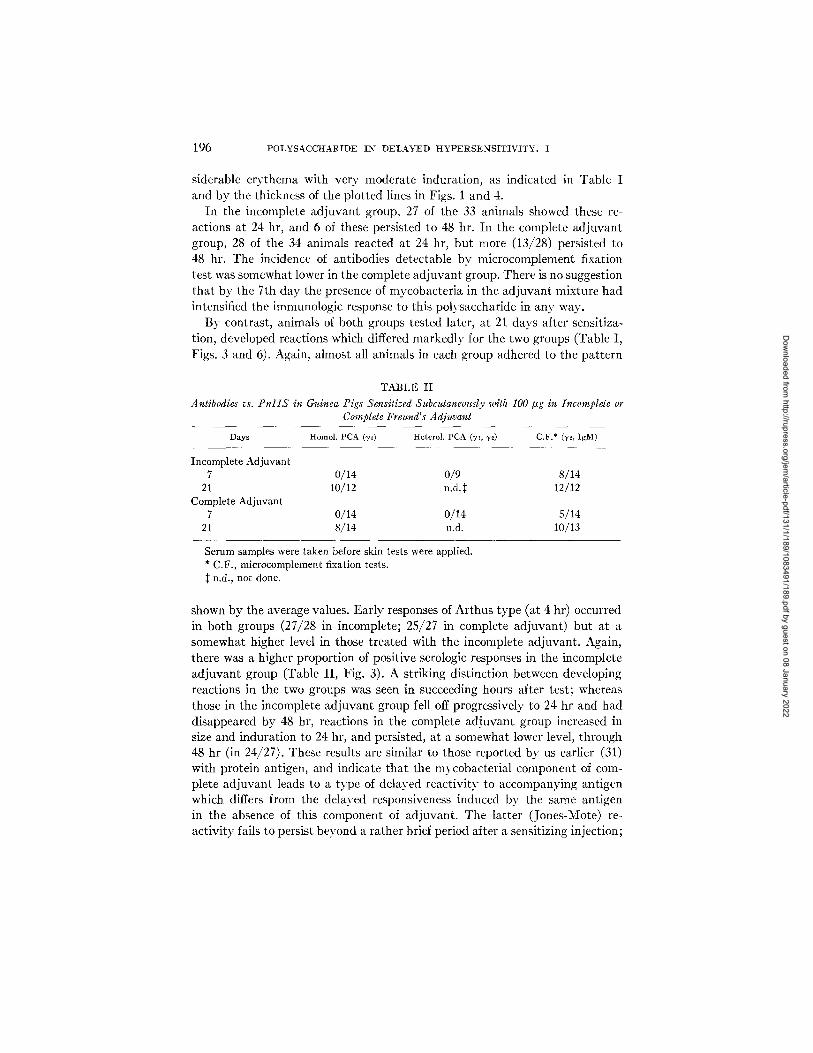
siderable erythema with very moderate induration, as indicated in Table I and by the thickness of the plotted lines in Figs. 1 and 4.
In the incomplete adjuvant group, 27 of the 33 animals showed these re- actions at 24 hr, and 6 of these persisted to 48 hr. In the complete adjuvant group, 28 of the 34 animals reacted at 24 hr, but more (13/28) persisted to 48 hr. The incidence of antibodies detectable by microcomplement fixation test was somewhat lower in the complete adjuvant group. There is no suggestion that by the 7th day the presence of mycobacteria in the adjuvant mixture had intensified the immunologic response to this polysaccharide in any way.
By contrast, animals of both groups tested later, at 21 days after sensitiza- tion, developed reactions which differed markedly for the two groups (Table I, Figs. 3 and 6). Again, almost all animals in each group adhered to the pattern
TABLE II Antibodies vs. P n l I S in Guinea Pigs Sensitized Subcutaneously with 100 t~g in Incomplete or
Complete Frez~nd' s Adjuvant
Days Homol. PCA (71) Heterol. PCA (~q, -/:) C.F.* (-y~, IgM)
Incomplete Adjuvant 7 0/14 0/9 8/14
21 10/12 n.d.$ 12/12 Complete Adjuvant
7 0/14 0/14 5/14 21 8/14 n.d. 10/13
Serum samples were taken before skin tests were applied. * C.F., microeomplement fixation tests. ~C n.d., not done.
shown by the average values. Early responses of Arthus type (at 4 hr) occurred in both groups (27/28 in incomplete; 25/27 in complete adjuvant) but at a somewhat higher level in those treated with the incomplete adjuvant. Again, there was a higher proportion of positive serologic responses in the incomplete adjuvant group (Table II , Fig. 3). A striking distinction between developing reactions in the two groups was seen in succeeding hours after test; whereas those in the incomplete adjuvant group fell off progressively to 24 hr and had disappeared by 48 hr, reactions in the complete adjuvant group increased in size and induration to 24 hr, and persisted, at a somewhat lower level, through 48 hr (in 24/27). These results are similar to those reported by us earlier (31) with protein antigen, and indicate that the mycobacterial component of com- plete adjuvant leads to a type of delayed reactivity to accompanying antigen which differs from the delayed responsiveness induced by the same antigen in the absence of this component of adjuvant. The latter (Jones-Mote) re- activity fails to persist beyond a rather brief period after a sensitizing injection;
Dow
nloaded from http://rupress.org/jem
/article-pdf/131/1/189/1083491/189.pdf by guest on 08 January 2022

R. J. GERETY, R. W. FERRARESI, AND S. RAFFEL 197
depending upon the magni tude of the stimulus, i t is followed by Arthus reac- t iv i ty as seen here, or if the st imulus has been a small one, it m a y be succeeded by loss of all detectable dermal react ivi ty.
I t might be inferred tha t the continuing and increasing delayed react iv i ty seen in animals sensitized with immunogen in complete mycobacter ia l ad juvan t depends upon some kind of potent ia t ion of the antigenic st imulus by the myco-
TABLE III Skin Reactions in Guinea Pigs at 14 Days after Subcutaneous Sensitization with 100 #g of
P n l I S in IncomNete or Complete Freund's Adjuvant and Tested with 100 #g of PnI1S
Incomplete adjuvant Complete adjuvant
Type of reaction 4 hr 24 hr 48 hr 4 hr 24 hr 48 hr
Negative 0 0 0 0 1.5, 0.1 0
Arthus* 5.1, 0 0 0 21.1, 1.6 1.5, 0.1 0
Delayed 1.0, 0 7.5, 0 0 2.0, 0.1 12.5, 1.4 10.0, 0.5 0 3.5, 0.1 0 3.0, 0.1 12.5, 0.4 5.0, 0.5
1.0, 0 6.5, 0.1 0 2.0, 0 13.5, 0.4 0 2.0, 0 13.5, 0.4 0 1.0, 0 11.5, 0.4 10.0, 0.2 2.0, 0 19.5, 0.9 0 0 12.5, 0.4 7.0, 0.5 2.0, 0 21.5, 1.1 0 4.0, 0.6 16.5, 1.4 10.0, 0.5
4.0, 0.1 12.5, 0.4 8.0, 0.2
Arthus*and 15.0, 1.6 17.5, 0.1 0 delayed 14.0, 1.1 21.5, 1.9 7.0, 0
12.0, 0.6 19.5, 1.4 5.0, 0 13.0, 1.1 19.5, 1.9 0
6.0, 0.6 14.5, 0.9 0 6.0, 0.1 18.5, 0.9 0 7.0, 0 18.5, 0.4 0
11.0, 0.1 19.5, 1.4 0 14.0, 1.6 21.5, 1.9 0 16.0, 2.1 27.5, 2.4 12.0, 0.5 16.0, 1.6 21.5, 1.9 0 20.0, 2.1 22.5, 1.4 0
Notes as in Table I. * Arthus reactivity at 4 hr arbitrarily considered to be 5 mm or greater.
bacteria, bu t such an inference is not supported by these results, for in the present case immedia te (Arthus) react iv i ty was more str iking at 21 days in the incomplete Freund ' s group, and there was a more general induction of antibodies in tha t group also.
The foregoing in terpre ta t ion of unfolding events at 1 and 3 wk is supported by the results of tests done at 14 days after sensitization. Unlike the uniformity of profiles of developing reactions at 7 and 21 days, a t this point after sensi- t izat ion reaction types were mixed. The averages of reactions in the two groups
Dow
nloaded from http://rupress.org/jem
/article-pdf/131/1/189/1083491/189.pdf by guest on 08 January 2022
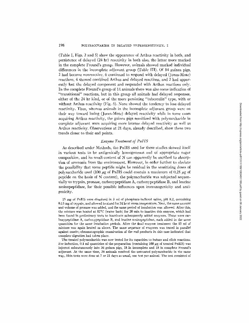
198 POLYSACCHARIDE IN DELAYED H Y P E R S E N S I T I V I T Y . I
(Table I, Figs. 2 and 5) show the appearance of Arthus react iv i ty in both, and persistence of delayed (24 hr) react iv i ty in both also, the la t ter more marked in the complete Freund ' s group. However, animals showed marked individual differences in the incomplete ad juvan t group (Table I I I ) . Of 14 guinea pigs, 2 had become nonreactive, 6 continued to respond with delayed (Jones-Mote) reactions, 4 showed combined Arthus and delayed reactions, and 2 had appar- ent ly lost the delayed component and responded with Arthus reactions only. In the complete Freund ' s group of 14 animals there was also some indication of " t rans i t iona l" reactions, bu t in this group all animals had delayed responses, either of the 24 hr kind, or of the more persisting " tubercul in" type, with or wi thout Arthus react iv i ty (Fig. 5). None showed the tendency to lose delayed reactivi ty. Thus, whereas animals in the incomplete ad juvan t group were on their way toward losing (Jones-Mote) delayed react iv i ty while in some cases acquiring Arthus react ivi ty, the guinea pigs sensitized with polysaccharide in complete ad juvan t were acquiring more intense delayed reac t iv i ty as well as Arthus reactivi ty. Observations at 21 days, a l ready described, show these two trends closer to their end points.
Enzyme Treatment of PnlIS
As described under Methods, the P n I I S used for these studies showed itself in various tests to be antigenically homogeneous and of appropr ia te sugar composition, and its small content of N can apparen t ly be ascribed to absorp- t ion of ammonia from the environment. However, in order further to obvia te the possibi l i ty tha t some pept ide might be residual in the sensitizing doses of polysaccharide used (100/zg of P n l I S could contain a max imum of 0.25/~g of pepfide on the basis of N content) , the polysaccharide was subjected sequen- t ia l ly to t rypsin, pronase, carboxypepfidase A, carboxypepfidase B, and leucine aminopeptidase, for their possible influences upon immunogenici ty and anti- genicity.
15 mg of PnIIS were dissolved in 3 ml of phosphate-buffered saline, pH 8.2, containing 0.15 mg of trypsin, and allowed to stand for 24 hr at room temperature. Next, the same amount and volume of pronase was added, and the same period of incubation was allowed. After this, the mixture was heated at 82°C (water bath) for 20 rain to inactive this enzyme, which had been found in preliminary tests to inactivate subsequently added enzymes. These were car- boxypeptidase A, carboxypeptidase B, and leucine aminopeptidase, each added in the same quantities for the same incubation periods. After the final enzyme treatment the 15 ml of mixture was again heated as above. The same sequence of enzymes was tested in parallel against casein; chromatographic examination of the end products in this case indicated that complete digestion had taken place.
The treated polysaccharide was now tested for its capacities to induce and elicit reactions. For induction, 0.1 ml quantities of the preparation (containing 100 #g of treated PnIIS) was injected subcutaneously into 36 guinea pigs, 18 in incomplete and 18 in complete Freund's adjuvant. At the same time, 36 animals received the untreated polysaccharide in the same way. Skin tests were done at 7 or 21 days as usual, one test per animal. The test consisted of
Dow
nloaded from http://rupress.org/jem
/article-pdf/131/1/189/1083491/189.pdf by guest on 08 January 2022

R. J. GERETY, R. W. FERRARESI, AND S. RAFFEL 199
100 #g of native PnIIS in one flank and the same amount of enzyme treated polysaccharide in the other flank of animals sensitized with native PnIIS. Those sensitized with enzyme treated material were tested only with native polysaccharide, in order to avoid complication.~ introduced by possible sensitivity to the enzymes themselves.
TABLE IV
Skin Reactivities in Guinea Pigs Induced with 100 #g of Enzyme Treated P n l I S and Elicited with 100 #g of Native P n l I S
Days 4 hr 24 hr 48 hr
Incomplete adjuvant 7* 0 13.0, 0.5 7.7, 0.03
21:~ 13.3, 1.1 9.8, 0.3 1.0, 0.03 Complete adjuvant§
7* 0 10.9, 0.6 2.8, 0.1 21~ 11.9, 1.2 16.8, 1.1 10.3, 0.45
Notes as in Table I. * 9 animals.
8 animals. § 9 animals for each testing time.
TABLE V
Skin Reactivities in Guinea Pigs Induced with 100 #g of Native Pn l IS , and Elicited with 100 #g of Native and Enzyme-Treated P n [ I S
Days Tested with 4 hr 24 hr 48 hr
Incomplete adjuvant 7~ PnIIS 0 11.2, 0.45 3.5, 0
PnIIE* 0 7.5, 0.35 1.7, 0.05 21§ PnIIS 8.4, 0.65 6.8, 0.3 1.0, 0
PnIIE* 8.8, 1.4 2.8, 0.1 0 Complete adjuvant
7:~ PnIIS 0 7.3, 0.35 4.6, 0.03 PnIIE* 0 3.0, 0.25 1.0, 0
2111 PnIIS 8.7, 1.0 21.7, 1.3 12.0, 0.3 PnIIE* 8.3, 0.75 19.5, 1.1 12.2, 0.3
Notes as in Table I. * Enzyme treated. :~'8 animals. §'6 animals. II 4 animals.
T h e resul ts (Tables IV and V) show t h a t the enzyme t r e a t m e n t did n o t
s ignif icant ly de t r ac t f rom the polysacchar ide as a sensi t iz ing or el ici t ing agent .
Sensi t iz ing ac t i v i t y is shown in Tab l e IV; groups of gu inea pigs sensi t ized wi th
enzyme- t r ea t ed P n I I S in incomple te or comple te F r e u n d ' s a d j u v a n t were
tes ted 7 or 21 days la ter wi th na t ive polysacchar ide . T h e chronologic response
Dow
nloaded from http://rupress.org/jem
/article-pdf/131/1/189/1083491/189.pdf by guest on 08 January 2022

200 POLYSACCItARIDE IN DELAYED H Y P E R S E N S I T I V I T Y . I
curves were of the types and degrees seen in animals sensitized with native polysaccharide. The obverse experiment is shown in Table V, where sensitiza- tion was done with native polysaccharide, and the animals were simultaneously tested on opposite flanks with native and enzyme-treated material. Again, the responses were parallel in kind; some tendency for apparently smaller responses to the enzyme-treated material at 24 hr after testing may depend upon the fact that these values were arrived at by subtracting average readings made in 10 control animals tested at the same time. Whereas native polysaccharide never produced local irritation persisting for 24 hr, the enzyme treated material showed a degree of this.
Again related to the question of possible responses to a nonpolysaccharide contaminant of the PnIIS preparation, skin tests were made with PnlIIS at
TABLE VI Macrophage Inhibition Tests, with PnlIS* As Antigen
Test after Number Average % Sensitization sensitization days of tests inhibition{
Normal controls -- 12 2
PnIIS, 100 #g in w/o§, subcutane- 7 20 17.5 ously 21 8 9
PnIIS, 100 #g in Freund's adjuvant, 7 15 22 subcutaneously 21 13 32
* 25 #g PnIIS/ml medium. ~: Compared with cells not exposed to antigen. § w/o, water/oil emulsion.
7 and 21 days in groups of guinea pigs sensitized with PnlIS in incomplete and complete adjuvant. The thought was that if a non-type-specific proteinace- ous substance of the pneumococcus accounted for the reactions seen, this might accompany the polysaccharide obtained from an organism of another antigenic type. 11 animals tested at 7 days, and 9 at 21 days, responded well in the usual patterns to PnIIS, but showed no reactions above control levels to PnIIIS.
Macrophage Inhibition Tests
The basis for the reaction of inhibition of macrophage motility by appropri- ate antigen appears to be either a mediator of unknown nature produced by lymphoid ceils (46) or a cytophilic antibody (47, 48). Despite the still cryptic significance of this reaction in respect to elucidating the nature of delayed reactivity, we think that it has been helpful for present purposes. The results shown in Table VI are recorded as average degrees of inhibition of migration of 24 hr packed cell preparations made from the peritoneal exudates of guinea
Dow
nloaded from http://rupress.org/jem
/article-pdf/131/1/189/1083491/189.pdf by guest on 08 January 2022
R . J . G E R E T Y , R . W . F E R R A R E S I ~ A N D S. R A F ~ ' E L 201
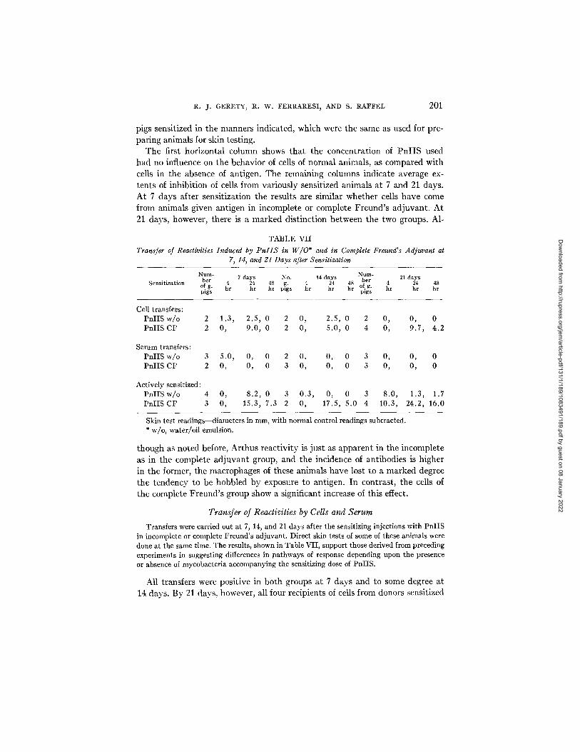
pigs sensitized in the manners indicated, which were the same as used for pre- paring animals for skin testing.
The first horizontal column shows tha t the concentrat ion of P n I I S used had no influence on the behavior of cells of normal animals, as compared with cells in the absence of antigen. The remaining columns indicate average ex- tents of inhibit ion of cells from variously sensitized animals at 7 and 21 days. At 7 days after sensit ization the results are similar whether cells have come from animals given antigen in incomplete or complete Freund ' s ad juvant . A t 21 days, however, there is a marked dist inct ion between the two groups. A1-
TABLE VII Transfer of Reaetivities Induced by PnIIS in W/O* and in Complete Freund's Adjuvant at
7, 14, and 21 Days after Sensitization
Num- 7 days No. 14 days Nurn- 21 days her 4 24 48 g. 4 24 48 ber 4 24 48
of g. hr hr hr Sensitization of g. hr hr hr pigs hr hr hr pigs Digs
Cell transfers: PnIISw/o 2 1.3, 2.5, 0 2 0, 2.5, 0 2 0, 0, 0 PnIIS CF 2 0, 9.0, 0 2 0, 5.0, 0 4 0, 9.7, 4.2
Serum transfers: PnIIS w/o 3 5.0, 0, 0 2 0, 0, 0 3 0, 0, 0 PnIIS CF 2 0, 0, 0 3 0, 0, 0 3 0, 0, 0
Actively sensitized: PnIIS w/o 4 0, 8.2, 0 3 0.3, 0, 0 3 8.0, 1.3, 1.7 PnIISCF 3 0, 15.3, 7.3 2 0, 17.5, 5.0 4 10.3, 24.2, 16.0
Skin test readings--diameters in ram, with normal control readings subtracted. * w/o, water/oil emulsion.
though as noted before, Ar thus react iv i ty is jus t as apparen t in the incomplete as in the complete ad juvan t group, and the incidence of ant ibodies is higher in the former, the macrophages of these animals have lost to a marked degree the tendency to be hobbled by exposure to antigen. In contrast , the cells of the complete Freund ' s group show a significant increase of this effect.
Transfer of Reactivities by Cells and Serum
Transfers were carried out at 7, 14, and 21 days after the sensitizing injections with PnIIS in incomplete or complete Freund's adjuvant. Direct skin tests of some of these animals were done at the same time. The results, shown in Table VII, support those derived from preceding experiments in suggesting differences in pathways of response depending upon the presence or absence of mycobacteria accompanying the sensitizing dose of PnIIS.
All transfers were posit ive in both groups at 7 days and to some degree at 14 days. By 21 days, however, all four recipients of cells from donors sensitized
Dow
nloaded from http://rupress.org/jem
/article-pdf/131/1/189/1083491/189.pdf by guest on 08 January 2022

202 POLYSACCItARIDE IN DELAYED HYPERSENSITIVITY. I
with antigen in mycobacterial adjuvant showed good delayed reactions at levels somewhat higher than at the earlier times after sensitization. In contrast, no significant reactivity was transferred to the three recipients from the water/ oil emulsion sensitized group. This provides evidence again of increasing cellu- lar reactivity in the mycobacterial adjuvant group at 3 wk, when that in the water-in-oil group had vanished. I t bears repeating that this distinction occurs even though the latter animals show a higher incidence and titer of antibodies than the former at this time.
Serum in large quantities (20 ml per recipient) failed to transfer the 7 day or subsequent delayed reactivities from either group. I t was surprising that the immediate (4 hr) reactivity so apparent by direct tests in the water-in-oil group at 21 days was not passively transferred. However, circulating antibodies, as indicated in a preceding section, seemed not to attain good concentrations in either group at best.
Histologic Observations Skin test sites were examined after 4, 12, 24, and 48 hr, from guinea pigs sensitized with
PnIIS in incomplete and complete Freund's adjuvants and tested at 7 and 21 days.
The sites from the incomplete adjuvant group at 7 days showed mononuclear cell infiltration restricted to the dermis; at 21 days, many of the cells appear- ing at the test site had developed into plasmacytes by 48 hr. By contrast, the 2l day reactions in the Freund's group revealed a larger component of small lymphocytes among the infiltrating mononnclears, epidermal invasion by such cells was common, and there were relatively few plasmacytes in the site even by 48 hr. These findings were consistent with those that we have described previously with another antigenic system (30, 32).
DISCUSSION
This report deals with two questions concerning delayed hypersensitive reactivity. One has to do with the role of polysaccharide immunogens as in- ducers and elicitors of such reactivity; the second with the question whether the delayed reactive state may be heterogeneous from the standpoint of reasons for occurrence, in one instance depending upon a cellular responsiveness which represents an early phase of antibody synthesis (termed Jones-Mote reactivity) ; in another, determined by a cell-bound effector of unknown kind, and revealing itself as the persisting reactivity of "tuberculin type."
I t has become apparent from several recent studies that polysaccharides may act as elicitors of delayed hypersensitive reactivity when this has been induced by immunogen of which the sugar is a moiety. The evidence that a pure sac- charide may be an inducer of the delayed reactive state is more restricted, although this has been shown to occur (e.g., 20). In the present case, the type specific polysaccharide of Diplococcus pneumoniae type II, freed of nitrogen that could be a constituent of peptides, was found to be an excellent inducer
Dow
nloaded from http://rupress.org/jem
/article-pdf/131/1/189/1083491/189.pdf by guest on 08 January 2022
R. J. GERETY, R. W. FERRARESI, AND S. RAFFEL 203
of delayed hypersensitivity in random-bred guinea pigs, when administered in complete Freund's adjuvant. Its capacity to induce antibodies under these circumstances was very modest, as judged by homologous and heterologous PCA tests and microcomplement fixation tests.
This immunogen was then found to lend itself well to a study of the second point; the existence of Jones-Mote reactivity as a distinct type of cellular hypersensitivity, different from that of tuberculin type. I t was soon seen that administration of PnlIS without the mycobacterial component of Freund's adjuvant led to the early (7 day) advent of delayed reactivity which manifested itself by fiat erythematous reactions with minor induration, reaching a maxi- mum at 24 hr and usually disappearing by 48 hr, and with histologic charac- teristics somewhat different from those of reactions of the tuberculin type (30, 32). This reactive state, as has been described before (30-32) tended to disap- pear by 2 or 3 wk, to be succeeded by Arthus reactivity.
Skin tests at 21 days revealed striking gross and histologic differences in the reactions elic;.ted in the two groups. These differences were seen also in macro- phage inhibition tests, which showed, at 21 days, a disappearance of cellular reactivity from the Jones-Mote group, and an intensification of this reactivity in the complete adjuvant-sensitized group with developing reactivity of tuber- culin type. Analogously, attempted transfers of reactivities by serum and cells from animals of the two groups at 7 and 21 days revealed that by the latter time, only cells from the complete Freund's group successfully transmitted reactivity, despite evidence that both groups had received an equally intense stimulus as judged by occurrence of antibodies and Arthus reactivity.
SUMMARY
A highly purified pneumococcal polysaccharide (Type II SSS) is a very effi- cient inducer of delayed hypersensitivity in random-bred guinea pigs. The cellular reactivity induced by this polysaccharide administered subcutaneously in complete Freund's adjuvant is of "tuberculin type"; it increases in intensity with time after the sensitizing injection, as judged by skin tests, the macro- phage inhibition reaction and transfer of reactivity by peritoneal exudate cells.
By contrast, the cellular reactivity induced by this immunogen in the absence of mycobacterial adjuvant has the characteristics of "Jones-Mote" reactivity. I t is best seen at about 1 wk after sensitization; the reactions are character- istically little indurated and show histologic differences from tuberculin type responses; and the reactive state begins to disappear by 2-3 wk, with the ac- cession of Arthus reactivity. This type of delayed reactivity may be related to an early phase of antibody synthesis.
BIBLIOGRAPHY
1. Tillet, W. S., and T. Francis. 1929. Cutaneous reactions to the polysaccharides and proteins of pneumococci. J. Exp. Med. 50:687.
Dow
nloaded from http://rupress.org/jem
/article-pdf/131/1/189/1083491/189.pdf by guest on 08 January 2022

204 POLYSACCHARIDE IN DELAYED H Y P E R S E N S I T I V I T Y . I
2. Tremaine, M. M. 1963. Study of delayed type sensitization to pneumococcal polysaccharides. Fed. Proc. 22:617.
3. Holborow, E. J., and G. Loewi. 1962. The immune response to blood group sub- stances. Immunology. 5:278.
4. Holborow, E. J., and G. Loewi. 1967. Polysaccharide-containing antigens. Brit. Med. Bull. 23:72.
5. Jankovic, B. D., and B. H. Waksman. 1962. Delayed sensitization to purified blood group substances in the guinea pig. J. Immunol. 89:598.
6. Barker, S. A., C. N. D. Cruickshank, J. H. Morris, and S. R. Wood. 1962. The isolation of Trichophylon glycopeptide and its structure in relation to the im- mediate and delayed reactions. Immunology. 5:627.
7. Borek, F., A. M. Silverstein, and P. G. H. Gell. 1963. Delayed hypersensitivity to hapten-protein conjugates. III . Saccharides as haptens. Proc. Soc. Exp. Biol. Med. 114:266.
8. Knight, R. A., and S. Marcus. 1958. Polysaccharide skin test antigens derived from HislopIasma capsulalum and Blastomyces dermatitidis. Amer. Rev. Tuberc. Pulmonary Dis. 77:983.
9. Knight, R. A., S. Coray, and S. Marcus. 1959. Histoplasma capsulatum and Blas- tomyces dermalitidis polysaccharide skin tests in humans. Amer. Rev. Resp. Dis. 80:246.
10. Marcus, S., R. A. Knight, and P. Q. Edwards. 1961. Skin sensitivity of human beings to H. capsulatum and B. dermatitidis polysaccharide antigens. Amer. Rev. Resp. Dis. 83:528.
11. Freund, J., G. E. Thompson, and M. M. Lipton. 1955. Aspermatogenesis, ana- phylaxis, and cutaneous sensitization induced in the guinea pig by homologous testicular extract. J. Ea:p. Med. 101:591.
12. Rowlands, D. T., A. J. Crowle, and H. P. Russe. 1965. Passively transferred delayed hypersensitivity reactions in mice and guinea pigs. int. Arch. Allergy A ppl. Immunol. 28:328.
13. Campbell, D. H. 1936. An antigenic polysaccharide fraction of Ascaris lumbricoides (from hog). J. [~fec. Dis. 59:266.
14. Baer, H., and S. D. Chaparas. 1964. Tuberculin reactivity of a carbohydrate component of unheated BCG culture filtrate. Science (Washington). 146:245.
15. Baer, H., and S. D. Chaparas. 1963. The immunology and chemistry of tuberculin. 1. The isolation of dialyzable and non-dialyzable tuberculin-active compo- nents from unheated BCG culture filtrates. Amer. Rev. Resp. Dis. 88:28.
16. Chaparas, S. D., and H. Baer. 1964. The immunology and chemistry of tuberculin. II . Chromatography with Sephadex of the non-dialyzable tuberculin-active constituents of BCG culture filtrate. Amer. Rev. Resp. Dis. 89:41.
17. Baer, H., and S. D. Chaparas. 1966. Delayed skin reactivity to carbohydrate and protein fractions of Mycobacterium bovis (BCG) culture filtrate in the absence and presence of serum antibody. J. Immunol. 96:353.
18. Azuma, I., H. Kimura, and Y. Yamamura. 1967. Further purification of poly- saccharides having anapbylactic activity from culture filtrates of human tu- bercle bacilli. Amer. Rev. Resp. Dis. 96:536.
19. Crowle, A. J., and C. C. Hu. 1967. Delayed hypersensitivity in mice to dextran. Int. Arch. Allergy A ppl. hnmunol. 31:123.
Dow
nloaded from http://rupress.org/jem
/article-pdf/131/1/189/1083491/189.pdf by guest on 08 January 2022
R. J. GERETY, R. W. FERRARESI~ AND S. RAFFEL 205
20. Battisto, J. R., G. Chappetta, and R. Hixon. 1968. Immunologic responses of guinea pigs to dextran. J. Immunol. 101:203.
21. Karush, F., and H. N. Eisen. 1962. A theory of delayed hypersensitivity. Science (Washington). 136:1032.
22, Freund, H., and M. Bonanto. 1944. The effect of paraffin oil, lanolin type sub- stances and killed tubercle baccilli on immunization with diphtheria toxoid and B. typhosum. J. Immunol. 48:325.
23. Avery, O. T., and H. J. Morgan. 1925. Immunological reactions of the isolated carbohydrate and protein of pneumococcus. J. Exp. Med. 42:347.
24. Avery, O. T. and W. F. Goebel. 1933. Chemoimmunological studies on the soluble specific substance of pneumococcus. I. The isolation and properties of the acetyl polysaccharide of pneumocoecus type I. J. Exp. Med. 58:731.
25. Morgan, P., D. Watson, and W. Cromartie. 1952. Immunization of rabbits with type I I pneumococcal polysaccharide. Proc. Soc. Exp. Biol. Med. 80:512.
26. Maurer, P. H., and H. C. Mansmann, Jr. 1958. On the non-antigenicity of poly- saccharides in guinea pigs. Proc. Soc. Exp. Biol. Med. 99:378.
27. Jones, T. D., and J. R. Mote. 1934. The phases of foreign protein sensitization in human beings. N. Engl. J. Med. 30:149.
28. Mote, J. R., and T. D. Jones. 1936. The development of foreign protein sensitiza- tion in human beings. J. Immunol. 30:149.
29. Raffel, S. 1962. The immune response in hypersensitivity. In Allergology. Proc. 4th Int. Cong. E. A. Brown, editor. Pergamon Press, Ltd., Oxford. 139.
30. Martins, A. B., and Raffel, S. 1964. Cellular activities in hypersensitive reac- tions. II. Plasmopoiesis in delayed and "Jones-Mote" reaction sites. J. Immunol. 93:948.
31. Raffel, S., and J. M. Newell. 1956. The "delayed hypersensitivity" induced by antigen-antibody complexes, J. Exp. Med. 108:823.
32. Martins, A. B., and S. Raffel. 1964. Cellular activities in hypersensitive reactions. I. Comparative cytology of delayed, "Jones-Mote" and Arthus reactions. J. Immunol. 93:937.
33. Dedrick, C. T. 1966. The definition of hypersensitive reactivity. Unpublished Ph.D. thesis, Stanford University.
34. Coe, J. E., and S. B. Salvin. 1963. The specificity of allergic reactions. VI. Unre- sponsiveness to simple chemicals. J. Exp. Med. 117:401.
35. Heidelberger, M. 1960. Immunological specificity of type II pneumococcus and its separation into partial specificities. II. J. Exp. Med. 111:33.
36. Heidelberger, M., C. M. MacLeod, H. Markowitz, and M. M. DiLapi. 1951. Absence of a prosthetic group in a type-specific polysaccharide of pneumococcus. J. Exp. Med. 94:359.
37. Sevag, M. G. 1934. Eine neue physikalische Enteiweibungsmethode zur darstel- lung biologish wirksamer Substanzen. Isolierung von Kohlenhydraten aus Htihnereiweib und Pneumococcen. Biochem. Z. 9.73:419.
38. Butler, K., and M. Stacey. 1955. Immunopolysaccharides. IV. Structural studies on the type I I pneumococcus specific polysaccharide. J. Chem. Soc. (London). 1537.
39. Kabat, E. A., and M. M. Mayer. 1961. Kabat and Mayer's Experimental Im- munochemistry. Charles C Thomas, Springfield, Illinois. 2nd edition, 538.
Dow
nloaded from http://rupress.org/jem
/article-pdf/131/1/189/1083491/189.pdf by guest on 08 January 2022
206 P O L Y S A C C H A R I D E I N D E L A Y E D H Y P E R S E N S I T I V I T Y . I
40. Satake, K., T. Okuyama, M. Ohashi, and T. Shinoda. 1960. The spectrophoto- metric determination of amine, amino acid, and peptide with 2,4,6-trinitro- benzene 1-sulfonic acid. J. Biochem. (Tokyo). 47:654.
41. Benacerraf, B., Z. Ovary, K. J. Bloch, and E. Franklin. 1963. Properties of guinea pig 7S antibodies. I. Electrophoretic separation of two types of guinea pig 7S antibodies. J. Exp. Med. 117:937.
42. Ovary, B., G. Benacerraf, and K. J. Bloch. 1963. Properties of guinea pig 7S antibodies. II. Identification of antibodies involved in passive cutaneous and systemic anaphylaxis. J. Exp. Med. 117:951.
43. Bloch, K. J. 1967. The anaphylactic antibodies of mammals including man. Progr. Allergy. 10:84.
44. Rosenberg, L. T., and D. K. Tachibana. 1962. Activity of mouse complement. J. Immunol. 89:861.
45. David, J. E., S. A1-Askari, H. S. Lawrence, and L. Thomas. 1964. Delayed hyper- sensitivity in vitro. I. The specificity of inhibition of cell migration by antigens~ J. Immunol. 93:264.
46. Bennett, B., and B. R. Bloom. 1968. Reactions in vivo and in vitro produced by a soluble substance associated with delayed-type hypersensitivity. Proe. Nat. Acad. Sci. U. S. A. 59:756.
47. Amos, H. E., B. W. Gurner, R. J. Olds, and R. R. A. Coombs. 1967. Passive sensitization of tissue cells. II . Ability of cytophilic antibody to render the migration of guinea pig peritoneal exudate cells inhibitable by antigen. Int. Arch. Allergy Appl. Immunol. 32:496.
48. Heise, E. R., S. Han, and R. S. Weiser. 1968. In vitro studies on the mechanism of macrophage migration inhibition in tuberculin sensitivity. J. Immunol. 101:1004.
Dow
nloaded from http://rupress.org/jem
/article-pdf/131/1/189/1083491/189.pdf by guest on 08 January 2022