Embed Size (px)
Citation preview

Practice Parameter
Contact Dermatitis: A Practice ParametereUpdate 2015
Luz Fonacier, MD, David I. Bernstein, MD, Karin Pacheco, MD, D. Linn Holness, MD, Joann Blessing-Moore, MD,
David Khan, MD, David Lang, MD, Richard Nicklas, MD, John Oppenheimer, MD, Jay Portnoy, MD,
Christopher Randolph, MD, Diane Schuller, MD, Sheldon Spector, MD, Stephen Tilles, MD, and Dana Wallace, MD
This parameter was developed by the Joint Task Force onPractice Parameters, which represents the American Academy ofAllergy, Asthma & Immunology (AAAAI); the American Collegeof Allergy, Asthma & Immunology (ACAAI); and the JointCouncil of Allergy, Asthma & Immunology. The AAAAI and theACAAI have jointly accepted responsibility for establishing“Contact Dermatitis: A Practice ParametereUpdate 2015.” Thisis a complete and comprehensive document at the current time.The medical environment is changing and not allrecommendations will be appropriate or applicable to allpatients. Because this document incorporated the efforts of manyparticipants, no single individual, including members serving onthe Joint Task Force, are authorized to provide an official AAAAIor ACAAI interpretation of these practice parameters. Anyrequest for information or interpretation of this practiceparameter by the AAAAI or ACAAI should be directed to theExecutive Offices of the AAAAI, the ACAAI, and the JointCouncil of Allergy, Asthma & Immunology. These parametersare not designed for use by the pharmaceutical industry in drugdevelopment or promotion. Previously published practiceparameters of the Joint Task Force on Practice Parameters for
See Appendix A for members of the Joint Task Force Contact Dermatitis ParameterWorkgroup, reviewers of this Practice Parameter, and members of the Joint TaskForce on Practice Parameters.
Disclosure of potential conflict of interest: L. Fonacier has received research andeducational grants (made to Winthrop University Hospital) from Genentech,Merck, and Baxter; is in the Speaker’s Bureau/Honoraria of Baxter; and is on theBoard of Directors, Joint Council of Allergy, Asthma and Immunology (JCAAI)2012-2015. D. Bernstein is the consultant in Merck, Genentech, Proctor andGamble, Sanofi, and TEVA; and has received research grants from Amgen,GlaxoSmithKline, Greer, Johnson & Johnson, Merck, Teva, Pfizer, Genentech,Array, Cephalon, Novartis, Boeringer Ingelheim, and Medimmune. The rest of theauthors declare that they have no relevant conflicts of interest.
The Joint Task Force recognizes that experts in a field are likely to have interests thatcould come into conflict with the development of a completely unbiased andobjective practice parameter. To take advantage of that expertise, a process hasbeen developed to prevent potential conflicts from influencing the final documentin a negative way.
At the workgroup level, members who have a potential conflict of interest either donot participate in discussions concerning topics related to the potential conflict orif they do write a section on that topic, the workgroup completely rewrites itwithout their involvement to remove potential bias. In addition, the entire docu-ment is then reviewed by the Joint Task Force and any apparent bias is removed atthat level. Finally, the practice parameter is sent for review both by invited re-viewers and by anyone with an interest in the topic by posting the document on theweb sites of the ACAAI and the AAAAI.
Corresponding author: Joint Task Force on Practice Parameters, 59 N Brockway St,#304, Palatine, IL 60067. E-mail: [email protected].
Received for publication February 25, 2015; accepted for publication February 26,2015.
2213-2198� 2015 American Academy of Allergy, Asthma & Immunologyhttp://dx.doi.org/10.1016/j.jaip.2015.02.009
Allergy & Immunology are available at http://www.JCAAI.org orhttp://www.allergyparameters.org. � 2015 American Academyof Allergy, Asthma & Immunology (J Allergy Clin ImmunolPract 2015;3:S1-S39)
Key words: Allergic contact dermatitis; patch testing; allergen;parameter; guideline; contact dermatitis; occupational; sensitizer
PREFACEThe Practice Parameter on Contact Dermatitis (CD) was last
updated in 2006, and focused primarily on the basics of CD andpatch testing for the allergist. In the ensuing years, there has beenconsiderable interest by the allergist in allergic skin diseases dueto increasing numbers of referrals for CD. With the ease ofapplication, the use of the preloaded commercially availableT.R.U.E. Test patch testing method has increased among aller-gists, as has the use of patch testing with individually loadedchambers. The T.R.U.E. Test has also been expanded to include35 antigens and a negative control, improving their sensitivity todetect inclusive allergens. There have also been advances in thefield in many areas including our basic understanding of type IVhypersensitivity reactions, emerging contact allergens, irritantcontact dermatitis (ICD), systemic contact dermatitis (SCD),patch testing in children, occupational dermatitis, and reactionsto biomedical devices. Improved diagnosis and management ofCD and availability of more comprehensive databases of causa-tive contact allergens enable physicians to manage allergic contactdermatitis (ACD) with avoidance of allergens the patient issensitized to and availability of lists of safe products that do notcontain these allergens. Given the many advances in the field, theJoint Task Force on Practice Parameters (JTF) appointed aworking group to review and update the standing practiceparameters.
The Contact Dermatitis: A Practice ParametereUpdate 2015workgroup was commissioned by the JTF to develop a practiceparameter that addresses recent advances in the field of CD andthe optimal methods of diagnosis and management based on anassessment of the most current literature. The Chair (LuzFonacier, MD) invited workgroup members to participate in theparameter development who are considered to be experts in thefield of CD. Workgroup members have been vetted for conflictof interest (COI) by the JTF and their COIs have been listed inthis document and are posted on the JTF web site at http://www.allergyparameters.org.
The charge of the workgroup was to develop current practiceguidelines based on an up-to-date systematic literature review.Consensus expert opinion and workgroup-identified supplementarydocuments were utilized when published evidence was lacking.
A search of the medical literature on PubMed was performedfor a variety of terms that were considered to be relevant to this
S1

J ALLERGY CLIN IMMUNOL PRACTMAY/JUNE 2015
S2 FONACIER ETAL
Abbreviations used
AA- A midoamineAAAAI- A
merican Academy of Allergy, Asthma and Immunology ACAAI- A merican College of Allergy, Asthma and Immunology ACC- A llergic contact cheilitis ACD- A llergic contact dermatitis ACDS- A merican Contact Dermatitis SocietyAD- A
topic dermatitis AGEP- A cute generalized exanthematous pustulosis APT- A topy patch test BOP- B alsam of Peru BTM- B etamethasoneCAMP- C
ontact Allergen Management Program CAPB- C ocoamidopropyl betaine CARD- C ontact Allergen Replacement DatabaseCD- C
ontact dermatitis CLO- C lobetasol COI- C onflict of interest CS- C orticosteroid CU- C ontact urticariaDMAPA-D
imethylaminopropylamine DRESS- D rug rash with eosinophilia and systemic symptoms ELISA- E nzyme-linked immunosorbent assayEliSPOT- E
nzyme-linked immunospot ENDA- E uropean Network on Drug Allergy ESCD- E uropean Society of Contact Dermatitis FDA- F ood and Drug Administration FM- F ragrance mixFM I- F
ragrance mix I FM II- F ragrance mix II GCDG-G erman Contact Dermatitis GroupHC-H
ydrocortisone ICD- Ir ritant contact dermatitis IM- In tramuscularIPPD- Is
opropyl-para-phenylenediamine IUDs- In trauterine devicesIV- In
travenous LPTs- L ymphocyte proliferation tests MCI-M ethychloroisothiazolinoneMELISA-M
emory Lymphocyte Immuno Stimulation Assay MI-M ethylisothiazolinoneMPL-M
ethylprednisolone MSDS-M aterial safety data sheetsNACDG-N
orth American Contact Dermatitis Group NHIS- N ational Health Interview SurveyNS- N
asal spray NSAIDs- N onsteroidal anti-inflammatory drugsOCD-O
ccupational contact dermatitis OHS- O ccupational health supplement PABA- P ara-aminobenzoic acid PPD- P ara-phenylenediamine PT- P atch testPTDS- P
ara-toluenediamine sulfate ROAT- R epeated open application test SCD- S ystemic contact dermatitis SJS- S tevens Johnson syndrome TCI- T opical calcineurin inhibitors TCL- T riamcinolone TCS- T opical corticosteroids TEN- T oxic epidermal necrolysis UK-U nited KingdomUVA-U
ltraviolet A UVB-U ltraviolet Bpractice parameter. All reference types were included in the re-sults. References identified as being relevant were searched forother relevant references. Published clinical studies were rated bycategory of evidence and utilized to establish the strength of therecommendations (see Appendix B). The parameter was subse-quently appraised by reviewers designated by the AAAAI andACAAI. Based on this process, this parameter represents an ev-idence-based, broadly accepted consensus document.
Search terms include contact dermatitis, eczema, cosmeticallergy, contact allergen, patch testing, and each of the specificconditions reviewed in this parameter.
GLOSSARY“Angry back” syndrome or “excited skin” syndrome: defined
as false-positive patch test (PT) reactions usually adjacent to largetrue-positive reactions that induce contiguous skin inflammationand irritability.
Ectopic allergic contact dermatitis: contact allergy lesionsmanifested in locations distant from or indirectly in contact withthe original skin sites directly exposed to allergens due to inad-vertent transfer by the patient (eg, transfer of sensitizers in nailpolish to the eyelids) or others (eg, mother transferring allergento the child or a partner transferring the allergen by contact).
Contact sensitization: evidence of sensitization such as pos-itive PT reaction is not definitive of an “allergy” but simply aconfirmation of immunologic sensitization that must then beconfirmed as clinically relevant by history and clinical findingsanalysis.
Contact urticaria: defined as the development of a wheal-and-flare reaction at a site where an external agent contacts theskin or mucosa.
Late patch test reading: late PT reading is performed at orafter 7 days after application of a PT as opposed to the standardof care reading that is performed between day 3 and 7.
Photo-allergic contact dermatitis: it is a delayed contacthypersensitivity reaction to an allergen activated by exposure toUV radiation.
Repeated open application test (ROAT): several open PTtechniques have been used to test substances with the potential forirritation, and are especially suitable for cosmetics and other per-sonal care products such as makeup foundation and skin lotions.The more commonly used provocative open use test involves therepeated application of a suspected allergen to the antecubital fossatwice daily for up to 1 to 2 weeks, and observation for the localdevelopment of dermatitis at the application site.
Usage test: use of a product highly suspected of containing asensitizer under real world conditions to prove causation. Anexample is for a patient to use eye mascara daily on 1 eye and notthe other to observe for the development of local dermatitis at theexposed site. This is often used when PT with suspected com-mercial allergens is negative but the suspicion of ACD is high.
Systemic allergic contact dermatitis: a generalized ACD rashfrom systemic administration of a drug, chemical, or food towhich the patient previously experienced ACD.
INTRODUCTION
Contact dermatitis (CD) is defined as any skin disorder causedby contact with an exogenous substance that elicits an allergic

J ALLERGY CLIN IMMUNOL PRACTVOLUME 3, NUMBER 3S
FONACIER ETAL S3
and/or irritant response. The vast majority of cases are attribut-able to irritant ICD. CD is also a significant cause of workplacedisability.
Contact urticaria (CU) is defined as the development of a wheal-and-flare reaction, or hives, at a site where an external agent contactsthe skin or mucosa. CU can be divided in 2 broad categories:nonimmunologic CU and immunologic CU (caused by an IgE-mediated hypersensitivity reaction). Symptoms of CU range frompruritic, localized wheal-and-flare reactions to generalized urticariaand anaphylaxis. Aside from the need to differentiate between ACDand CU, this parameter will not discuss CU in detail.
This CD practice parameter, updated from the original docu-ment published in 2006, is intended as a useful guide for thepracticing allergist in the evaluation and management of ACD inadults and children. This updated parameter has been restructuredaround action-based and patient-centered summary statements thatprovide specific evidence-based recommendations for assessing andtreating ACD. In contrast to the original 2006 parameter, thepathophysiology, susceptibility, and clinical background are notreviewed here. The evidence-based summary statements in thisdocument provide specific recommendations pertaining to theapproach to medical history, physical examination, patch testing,and management of patients suspected of ACD.
As in the 2006 parameter, action-based summary statementsprovide guidance for identification of potential causative sensi-tizers based on clinical presentation in specific geographical skinlocations. Patch testing is emphasized in this updated parameter,with action-based statements that address selection of PT anti-gens; testing to personal products when necessary; different patchtesting devices; timing of readings; late PT reactions; false-posi-tive, false-negative, and true-negative responses; and photo-patchtesting. Lists of sensitizers encountered in different settings or inspecific types of products (eg, cosmetics, sunscreens, joint pros-theses) are presented as tables in the appendices.
Since the publication of the original parameter, new questionshave been addressed in summary statements related to emergingclinical problems including preoperative screening for and post-implantation patch testing for metal allergy in patients who haveundergone joint replacement surgery. In this updated practiceparameter, summary statements have been added that morecomprehensively address evaluation and management of occupa-tional contact dermatitis (OCD). The potential benefits and limi-tations of drug patch testing in patients with maculopapular rashes,erythroderma, and nonimmediate cutaneous reactions are addressedin a summary statement. New summary statements have beenincluded that make recommendations pertaining to the overallmanagement of CD, focusing on avoidance and prevention.
The majority of summary statements in this document arebased on descriptive and retrospective studies, representativeof the current published CD literature. Because the treat-ment of choice for CD is avoidance, there are limitednumbers of published placebo-controlled studies of othertherapeutic interventions (eg, drugs). The absence of a vali-dated positive control to confirm a diagnosis of ACD is amajor limitation of studies reporting patch testing data. Forthese reasons, the categories of evidence supporting thesummary statements in this document are relatively low.Therefore, the strength of recommendation for most of thestatements in this parameter is “Moderate” even if in someclearly identified circumstances, “Strong” recommendationsmay be made based on lesser evidence because high-quality
evidence is impossible to obtain, and the anticipated benefitsstrongly outweigh the harms.
Overall, this is a practical, clinically pertinent, and user-friendly parameter that has attempted to address importantclinical questions pertaining to the evaluation and managementof ACD. This document, although not intended to replace anauthoritative textbook, is a valuable updated evidence-basedresource for the practicing allergist.
COMPILATION OF SUMMARY STATEMENTSSummary Statement 1: Consider ACD in the differential
diagnosis of patients with chronic eczematous or noneczematousdermatitis. [Strength of Recommendation: Strong; C Evidence]
Summary Statement 2: In patients suspected of ACD, patchtesting is the gold standard to confirm the diagnosis. [Strength ofRecommendation: Strong; C Evidence]
Summary Statement 3: In addition to personal products usedby a patient suspected of ACD, review the home and workplacefor other sources of contact allergens. [Strength of Recommen-dation: Moderate; D Evidence]
Summary Statement 4: Evaluate patients for both irritant andallergic causes, especially in those presenting with hand derma-titis. [Strength of Recommendation: Strong; C Evidence]
Summary Statement 5: Allergic CD should be suspected andevaluated in the patient with both generalized and anatomicallylocalized skin eruptions (such as the hands, face, eyelids) thatcome in contact with the substances in the environment.[Strength of Recommendation: Moderate; C Evidence]
Summary Statement 6: In a patient with a facial rash involvingthe periorbital areas (eg, eyelids), evaluate for ACD caused bycomponents of cosmetics, such as fragrances, preservatives, andexcipients, because these are common sensitizers of the facialskin. [Strength of Recommendation: Moderate; C Evidence]
Summary Statement 7: Evaluate patients presenting with lipdermatitis (cheilitis) and perioral dermatitis for both irritant andallergic causes of contact dermatitis. [Strength of Recommen-dation: Moderate; C Evidence]
Summary Statement 8: Evaluate patients with chronic oralmucosal inflammatory conditions for disorders other than ACD.[Strength of Recommendation: Moderate; C Evidence]
Summary Statement 9: In patients presenting with dermatitisthat involves the scalp and neck, consider patch testing forcommon causative sensitizers in cosmetics, hair products, andjewelry. [Strength of Recommendation: Moderate; C Evidence]
Summary Statement 10: Consider irritant and ACD in allpatients presenting with acute or chronic hand eczema. All suchpatients suspected of CD should undergo patch testing.[Strength of Recommendation: Moderate; C Evidence]
Summary Statement 11: Evaluate patients with axillarydermatitis for ACD caused by local contact sensitivity to allergensin topically applied products found in deodorants and textiles. Insome cases, axillary dermatitis could be a manifestation of systemiccontact dermatitis (SCD) (ie, “the baboon syndrome”). [Strengthof Recommendation: Moderate; C Evidence]
Summary Statement 12: Evaluate patients presenting withanogenital dermatitis for possible ACD to antigens contained intopically applied products. [Strength of Recommendation:Moderate; C Evidence]
Summary Statement 13: Consider a diagnosis of SCDfollowing systemic exposure (eg, ingestion, infusion, or

J ALLERGY CLIN IMMUNOL PRACTMAY/JUNE 2015
S4 FONACIER ETAL
transcutaneous exposure) to a known contact sensitizer in a pa-tient who presents with generalized dermatitis, intertriginous andflexural exanthema (Baboon syndrome), and/or a flare at previ-ous cutaneous sites of exposure [Strength of Recommendation:Moderate; C Evidence].
Summary Statement 14: Consider PT to rubber chemicals,adhesives, and leather components of footwear in patients pre-senting with unexplained chronic dermatitis involving the lowerextremities, feet and/or soles. [Strength of Recommendation:Moderate; C Evidence]
Summary Statement 15: In addition to avoiding irritants inpatients with atopic dermatitis (AD), evaluate for ACD, if sus-pected, as the 2 dermatologic conditions often coexist in thesame patient. [Strength of Recommendation: Moderate; CEvidence]
Patch testing recommendationsSummary Statement 16: Avoid or reduce doses of immuno-
suppressant medications such as systemic corticosteroids (CS)and systemic immunosuppressants before patch testing. Avoidapplication of topical corticosteroids (TCS), topical calcineurininhibitors (TCI), or ultraviolet radiation to the PT site, becausethese may reduce allergic PT responses. [Strength of Recom-mendation: Moderate; C Evidence]
Summary Statement 17: In addition to using a core or base-line series of PT allergens in evaluating ACD, consider usingsupplemental series of PT allergens based on specific patientexposures, and the patient’s personal products to increase theprobability of identifying relevant sensitizers. [Strength ofRecommendation: Moderate; C Evidence]
Summary Statement 18: Patch testing can be performed eitherusing a preloaded thin-layer rapid use epicutaneous testing kit of36 chambers or with a panel of antigens loaded individually in achamber system recommended by the North American ContactDermatitis Group (NACDG) Research Group or the AmericanContact Dermatitis Society (ACDS). [Strength of Recommen-dation: Moderate; C Evidence]
Summary Statement 19: Read and interpret PT conforming tothe scoring system developed by the International ContactDermatitis Research Group. [Strength of Recommendation:Moderate; D Evidence]
Summary Statement 20: Remove and read PT at approxi-mately 48 hours after application. A second reading should bedone between 3 and 7 days after application. [Strength ofRecommendation: Moderate; C Evidence]
Summary Statement 21: Consider that a possible false-positivereaction can result with the use of irritants or allergic substancesat potentially irritating higher concentrations, pressure reactionfrom the filling chamber, an “angry back syndrome,” or patchtesting on skin with active dermatitis. [Strength of Recommen-dation: Moderate; D Evidence]
Summary statement 22: Recognize the possibility thatfalse-negative reactions could be due to inadequate allergenconcentration needed to elicit a response; inability of thevehicle to release sufficient allergen; reduced skin respon-siveness because of prior ultraviolet light exposure (ie, sun,tanning bed); concomitant immunosuppressive therapies; ormethodological testing errors such as insufficient occlusion,failure to perform delayed readings, and failure to perform aphoto PT. [Strength of Recommendation: Moderate; CEvidence]
Summary Statement 23: Determine the relevance of a PT resultbased on the clinical and exposure history when interpreting thePT. [Strength of Recommendation: Moderate; D Evidence]
Summary Statement 24: Consult physicians with expertise inpatch testing to household cleaning or industrial products iftesting to the actual product suspected of containing the relevantallergen(s) is necessary, because false-positive and severe irritantreactions can occur. [Strength of Recommendation: Moderate; CEvidence]
Summary Statement 25: Consult physicians with expertise inUV radiation and photo-patch testing to confirm a suspecteddiagnosis of photo-allergic CD. [Strength of Recommendation:Strong; C Evidence]
Summary Statement 26: Although in vitro tests for delayedhypersensitivity to contact allergens (ie, metals and bone cement)are available, routine use of such assays is not currently recom-mended as their sensitivity and specificity for diagnosing ACDhas not been determined and should be considered investiga-tional. [Strength of Recommendation: Moderate; C Evidence]
Summary Statement 27: Use the repeated open applicationtest (ROAT) to further evaluate a patient suspected of ACD whoexhibits doubtful or negative PT responses, to confirm that thepatient is reacting to that particular product or to determineclinical tolerability to new cosmetic products. [Strength ofRecommendation: Moderate; C Evidence]
Sources of exposure to clinically relevant allergens
Summary Statement 28: Evaluate patients who present withrecurrent dermatitis on exposed skin surfaces during airbornepollen seasons for contact sensitization to seasonal pollen aller-gens. [Strength of Recommendation: Moderate; C Evidence]
Summary Statement 29: The clinician should consider cos-metics and personal hygiene products that are directly applied toinvolved skin or ectopically transferred from uninvolved skin aspotential sources of allergens in patients with ACD. [Strength ofRecommendation: Strong; C Evidence]
Summary Statement 30: When evaluating ACD from cos-metics and personal care products that contain many differentchemical ingredients, consider that the most common causes aredue to a few important chemical classes, including fragrances,preservatives, excipients, nickel, and sun screening agents.[Strength of Recommendation: Moderate; C Evidence]
Summary Statement 31: Patients suspected to have allergy tohair products should be evaluated for PT reactions to cocoami-dopropyl betaine (CAPB), para-phenylenediamine (PPD), fra-grances, preservatives, and glycerol thioglycolate. [Strength ofRecommendation: Moderate; C Evidence]
Summary Statement 32: Suspect allergy to nail products whenthe dermatitis presents locally at the distal digit or ectopically onthe eyelids and face. [Strength of Recommendation: Moderate; CEvidence]
Summary Statement 33: Suspect the diagnosis of photo-allergic CD to cosmetics when eczema occurs in a light-exposeddistribution following the use of a skin care product or cosmetic,including sunscreens. [Strength of Recommendation: Strong; CEvidence]
Topical medicinal CD
Summary Statement 34: If an eruption worsens, rather thanimproves, after the topical application of certain medications, orfails to respond to TCS, PT should be performed to the

J ALLERGY CLIN IMMUNOL PRACTVOLUME 3, NUMBER 3S
FONACIER ETAL S5
suspected product and/or ingredients known to be contact sen-sitizers. [Strength of Recommendation: Moderate; C Evidence]
Summary Statement 35: The clinician may use the drug PTfor the diagnosis of some drug hypersensitivity reactions, recog-nizing that there is no standardized approach to define thepopulation, clinical manifestation, drug to PT, and PT materialsto make patch testing to drugs a standard of care. [Strength ofRecommendation: Weak; D Evidence]
Summary statement 36: Consider preoperative patch testing formetal sensitization in patients with a significant history of metalallergy. [Strength of Recommendation: Moderate; C Evidence]
Summary Statement 37: In patients with joint replacementfailure, patch testing to components of the implant may behelpful after infection and biomechanical causes have beenexcluded. [Strength of Recommendation: Moderate; C Evidence]
Special populations
Contact dermatitis in children. Summary Statement 38:ACD and ICD are significant clinical problems in children.Patch testing should be performed and remains the gold standardfor the diagnosis of ACD in children. [Strength of Recommen-dation: Strong; C Evidence]
Occupational contact dermatitis. Summary Statement39: In a patient who presents with dermatitis associated withworkplace exposures (ie, OCD), consider ICD as well as ACD.[Strength of Recommendation: Strong; C Evidence]
Summary Statement 40: In patients with suspected occupa-tion-related CD, the examining physician should verify thediagnosis by confirming that the dermatitis was caused oraggravated by workplace exposures. [Strength of Recommenda-tion: Moderate; C Evidence]
Summary Statement 41: Consider botanical-related ACD inoutdoor workers, or others exposed to plants, including florists,gardeners, landscapers, maintenance workers, park, and wildlifeofficials. [Strength of Recommendation: Moderate; C Evidence]
Treatment of contact dermatitis. Summary Statement42: Once the allergen or irritant has been identified, the patientshould be counseled on avoidance of contact with the offendingagent and informed of any cross-reactivity concerns. [Strength ofRecommendation: Strong; B Evidence]
Summary Statement 43: In addition to avoidance of exposure,the physician should prescribe appropriate adjunct medicaltreatment. [Strength of recommendation: Strong; B Evidence]
Summary Statement 44: To prevent CD, avoid exposure toirritants and allergens and use appropriate skin protection.[Strength of Recommendation: Strong; B Evidence]
Summary Statement 45: Education of the workers with ACDor ICD should include prognosis, and information that theirdisease may persist and need long-term management even aftertreatment and workplace modifications. [Strength of Recom-mendation: Moderate; C Evidence]
EXECUTIVE SUMMARY
Contact dermatitis may be suspected on the basis of theclinical appearance of the cutaneous lesions, the distribution ofthe dermatitis, and the absence of other etiologies. Acute CD ischaracterized by erythematous papules, vesicles, and crusted le-sions. There are other dermatological conditions that mayresemble the clinical and/or histological appearance of CD, and
these should be considered in the differential diagnosis. Thesuspicion of ACD is the first step in making the diagnosis. Patchtesting is indicated in any patient with acute or chronic, oftenpruritic, dermatitis if underlying or secondary ACD is suspected.
The history is important for the diagnosis and subsequentmanagement of this disease. Although medical history canstrongly suggest the cause of ACD, it has moderate sensitivity(76%) and specificity (76%) in establishing the diagnosis. Inaddition, the occupational, avocational, and environmental his-tory must all be carefully reviewed. Chronologic exposure his-tories that include hobbies and specific activities relative to onsetof the dermatitis should be obtained. Because the worker may beunaware of specific chemicals to which he or she is exposed,material safety data sheets (MSDS) obtained from the manu-facturer may be helpful. Hobbies and nonwork activity such asgardening, macramé, painting, ceramic work, carpentry, andphotography may be sources of exposure to culprit contactants.In addition to exposure to a single agent, simultaneous exposureto multiple irritants and contact allergens may produce additive,synergistic, or antagonistic responses. Simultaneous exposure toboth an irritant and a contact allergen or 2 contact allergens canreduce the clinical threshold concentration for elicitation ofresponse to a given allergen due to irritant disruption of the skinbarrier and immunologic activation of the skin.
There is conflicting evidence as to whether patients with ADare at heightened overall risk of contact sensitization comparedwith nonatopic individuals. Because AD is associated with animpaired skin barrier, it is plausible that this impairment is likelyto increase absorption of topically applied chemicals and enhancethe risk of subsequent sensitization, resulting in ACD andworsening of the underlying dermatitis. In children with severerecalcitrant AD and concomitant ACD, avoidance of offendingallergens in topically applied products can result in markedimprovement of eczema.
The latest NACDG lists the top 3 most common body lo-cations of contact dermatitis as scattered and/or generalizeddistribution, the hands, and the face. In addition, attentionshould be given to specific anatomical sites, particularly theeyelids, neck, scalp, axillae, lower extremities, and anogenitalarea. Facial ACD may present as a generalized facial eruption orin specific regions such as the forehead, periorbital, or perioralareas. Sensitizers in commercial facial products that are in directskin contact are the most common causes of facial ACD.
Patients presenting with acute or chronic hand eczema shouldundergo patch testing. Although most cases of CD involving thehands are caused by irritants, allergic contact sensitization is acommon cause of chronic hand dermatitis. The prevalence ofACD in patients presenting with hand dermatitis or hand eczemavaries according to exposure history and occupation. Thus, it isstrongly recommended to evaluate all patients with chronic handeczema for ACD by obtaining a medical history of contact allergyand performing patch testing.
Acute or chronic inflammation of the lips manifested aseczematous cheilitis can be characterized by itching, burning,redness, edema, and fissuring. This is most commonly caused byphysical (eg, cold, dryness, wind) or chemical irritants (eg, saliva,lip cosmetics, or other oral products). Fragrance mix (FM),balsam of Peru (BOP, Myroxylon pereirae), and nickel are themost common positive allergens on PT. Sources of fragrancesinclude oral hygiene products (eg, toothpastes, mouthwashes,flavorings, compounds used for dental impressions), cosmetics,

J ALLERGY CLIN IMMUNOL PRACTMAY/JUNE 2015
S6 FONACIER ETAL
and lip products (including lipsticks, glosses, and lip balms). Oralcontact sensitization is considered to be uncommon. Persistentoral complaints or gingivitis has been associated with positive PTreactions to allergens in dental components, including mercury,methacrylate, and beryllium. Chemical and traumatic injury maybe the most common causes of contact reactions involving mu-cous membranes. Other conditions that should be considered inpatients with oral mucosal inflammation include burning mouthsyndrome, lichenoid tissue reactions, stomatitis, gingivitis, oro-facial granulomatosis, recurrent aphthous stomatitis, precancer-ous and cancerous lesions, viral and fungal infections and lichenplanus.
In patients presenting for patch testing for evaluation of CD,nickel remains the most common contact sensitizer and is foundmore frequently in women than it is in men. The gender dif-ference is likely due to greater exposure of the neck, hands, andears to nickel in jewelry and body piercing practices. Females aretwice as likely as males to have ACD involving the head and neckdue to cosmetics. Among patients with cosmetic allergies, fra-grances, preservatives, and emulsifiers are the most commoncausative allergens. In addition to the most common hair dyesensitizer, PPD, there are sensitizers in shampoos, includingfragrances, CAPB, and preservatives. ACD involving the scalp isfrequently caused by allergens in personal hygiene and medicalproducts (eg, neomycin, benzocaine), hair tint and/or dyes, haircleansing products, and bleaches.
ACD involving the axillary region is often due to contactsensitivity to fragrance chemicals in deodorants; antiperspirantchemicals are uncommon causes of ACD. Allergic CD due todisperse dyes in clothing can elicit eczematous eruptions in theaxillae, feet, and groin. Axillary dermatitis may be a manifestationof SCD, specifically the “baboon syndrome,” a diffuse eruptioninvolving flexural and intertriginous areas following oral,intravenous, or transcutaneous exposure to the allergen in acontact-sensitized individual. Three groups of allergens are mostcommon causes of SCD: (i) metals such as mercury, nickel, andgold; (ii) medications including aminoglycoside antibacterials,CS, and aminophylline; and (iii) plants and herbal productsincluding Compositae and Anacardiaceae families and BOP (alsoknown as Myroxylon pereirae).
Patients presenting with anogenital dermatoses have beendiagnosed with confirmed ACD to allergens contained in topi-cally applied products such as cosmetics, medications, femininehygiene and contraceptive products. The most common sourcesof antigens were topical medications, including TCS, fragrances,BOP, nickel sulfate, cinnamic aldehyde, and neomycin sulfate.The preservative methylisothiazolinone (MI) and benzocainewere frequently identified as contact allergens in patients withanogenital complaints.
The pattern of foot dermatitis due to ACD varies according tothe type of footwear used. Para-tertiary butylphenol formaldehyderesin (in adhesives), potassium dichromate, cobalt chloride, andcarbamates are among the most common allergens. Allergic CDinvolving the feet is commonly caused by sensitization to commonrubber allergens (carbamates, thiurams, and mercaptobenzothia-zole). Children presenting with sole dermatitis should be evaluatedby patch testing to rule out ACD caused by rubber additives, ad-hesives, and/or chromates. The majority of patients with chronicleg ulcers and leg dermatitis have contact sensitization to chemicalsensitizers found in topically applied preparations including BOP,FMs, antibacterial agents, CS, and lanolin.
Patch testing is indicated in any patient suspected of ACD.Patch testing can be performed using either a preloaded thin-layer rapid use epicutaneous testing kit of 36 chambers or with apanel of antigens individually loaded in a chamber system rec-ommended by the NACDG Research Group or the ACDS. TheT.R.U.E. Test (panel of 35 antigens and a negative control) (seeAppendix H) is standardized across lot numbers and is highlyreproducible. Depending on the test antigen, the T.R.U.E. Testmethod has moderate concordance (62% to 63%) with indi-vidually loaded chamber systems (eg, Finn chamber system).Reliance on a core or baseline series of PT antigens such as thoseused by the NACDG Research Group or in the T.R.U.E. Testpanel for assessing all patients is likely to lead to underdiagnosesof ACD. Selection of allergens to be patch tested will be moreaccurate when selection is based on the clinical history. One canuse PT panels based on the specific industry or exposure group.Frequently, especially in the eyelid, lip, and facial dermatitis, itmay be necessary to include personal products and substancesspecific to the patient’s exposure history.
Commercially available panels of supplemental allergens thatare constituents of personal care products or encountered inspecific occupational environments are listed in the AppendicesB, C, and D.
The International Contact Dermatitis Research Group’sscoring system listed below is widely used:
(-) Negative reaction(?þ) Doubtful reaction with faint erythema only(1þ) Weak positive reaction with nonvesicular erythema,
infiltration, possibly papules(2þ) Strong positive reaction with vesicular erythema, infil-
tration, and papules(3þ) Extreme positive reaction with intense erythema and
infiltration, coalescing vesicles, bullous reaction(IR) Irritant reaction(NT) Not testedIn the evaluation of delayed hypersensitivity reactions, the
initial reading of PT should be done approximately 48 hoursafter their application following patch removal. Tests may needto be read 30 minutes after removal of the patches to allow er-ythema from the occluding pressure of the tape and/or chamberto resolve. A second reading must be done; this is often done atday 3 to 7 after the initial application. A collaborative studydemonstrated that 30% of relevant allergens were positive at 96hours and were negative at the 48-hour reading, which suggeststhat 96 hours may be optimal for a second reading. Occasionally,an additional late reading after 7 days may be needed for certaincontactants such as metals, some antibiotics, and TCS that mayyield late reactions. Oral CS exceeding 20 mg/day of prednisoneor its equivalent have been shown to diminish skin test reactivityto 5% nickel sulfate at 48 hours. There is minimal evidence toguide the duration of steroid reduction or withdrawal beforeperforming patch testing. If the clinical suspicion is high despitea negative PT in a patient receiving immunosuppressive medi-cations, consider repeat testing when the immunosuppressantdoses are lowered or discontinued. The test site where the PT areapplied should have no topical potent CS or TCI applied for 5 to7 days before testing. UV irradiation of PT sites before testingcan suppress PT responses.
Doubtful (?þ) or weakly positive (1þ) questionable or irre-producible reactions on PT can be easily misinterpreted. Thetiming of the response may also affect its clinical significance,

J ALLERGY CLIN IMMUNOL PRACTVOLUME 3, NUMBER 3S
FONACIER ETAL S7
with a weak reaction at day 7 more likely to be clinically relevantthan one at day 3. The inability to separate nonspecific from trueallergic responses may be due to the “angry back” or “excitedskin” syndrome, which is defined as false-positive reactionsadjacent to large true-positive reactions that induce contiguousskin inflammation and irritability. The frequency of false-nega-tive results is not known, but has been estimated to occur in upto 30% of patch-tested patients. The ROAT is used to furtherevaluate a patient suspected of ACD who exhibits doubtful orsuspected false-negative PT responses, to confirm that the patientis reacting to that particular product or to determine clinicaltolerability to new cosmetic products. The threshold concen-tration for a positive reaction for the ROAT is lower than thethreshold concentration for a positive PT, although the accu-mulated ROAT dose was very similar to the PT.
The clinical relevance of positive PT reactions to ACD canonly be established by carefully correlating the history, whichincludes exposure to the allergen, with the PT results. A positivePT may be clinically relevant depending on current or past ex-posures. Current relevance is defined as definite if the PT or usetest with the suspected material is positive; probable if the PT ispositive and the antigen is present in known skin contactants andthe clinical presentation is consistent with that exposure; orpossible if the PT is positive, and skin contact with materialsknown to contain the allergen was likely.
If photo-allergic CD is suspected, physicians should be con-sulted with expertise in UV radiation and photo-patch testing toconfirm a suspected diagnosis. Photo-allergic CD typically affectssun-exposed areas such as the face, the ‘‘V’’ of the anterior neck,the dorsal hands, and forearms. It typically spares the uppereyelids, upper lip, and submental and postauricular areas. Themore common cause of sunscreen sensitization is the chemicalsunscreens. Titanium dioxide and zinc oxide (physical UVblockers) have not been reported to cause ACD or photo-allergy,although there are a few reports of titanium in implants causingACD. Testing requires duplicate application of allergen withsubsequent occlusion, and irradiation of one side to compare tothe other, nonirradiated application.
Although in vitro tests for delayed hypersensitivity to contactallergens (ie, metals and bone cement) are available, routine use ofsuch assays is not currently recommended as their sensitivity andspecificity for diagnosing ACDhas not been determined and shouldbe considered investigational. In vitro tests for assessing antigenspecific sensitization are based on measuring lymphocyte prolifera-tion (lymphocyte proliferation tests—LPTs) or cytokine production(ELISA or EliSPOT) after incubation with antigens. Some in vitrotests have been validated against patch testing, whereas others havenot. The clinical relevance of in vitro testing to the diagnosis of CDhas not been established and is still investigational.
Identifying sources of exposure to clinically relevant allergensis challenging. Dermatitis present on the face, hands, andexposed chest may be triggered by airborne protein allergens suchas grass pollen, house dust mite, and cat dander; and diagnosedby the application of the allergen by patch testing. CD caused bycosmetics is noted predominantly at the site of application;however, occasionally personal care products and cosmeticsmanifest the contact allergy lesions in locations distant from theoriginal skin sites. This phenomenon termed ectopic CD can becaused by nickel transferred to the eyelid by fingers that havebeen exposed to a nickel source or toluene sulfonamide formal-dehyde resin in nail polish.
When evaluating ACD from cosmetics and personal careproducts that contain many different chemical ingredients,consider that the most common causes are due to a fewimportant chemical classes, including fragrances, preservatives,excipients, nickel, and sun blocks. Fragrances are complex sub-stances and are the most common cause of ACD from cosmeticin the United States. Previous studies suggest that the standardFM and BOP will detect approximately 60% to 70% offragrance-allergic individuals. The addition of other commonlyused fragrance ingredients (FM II, lyral, ylang ylang oil, narcissusoil, and sandalwood oil) may increase the yield up to 96%.However, it should be noted that fragrances in PT have marginalirritant potential and weak positive reactions may not be regar-ded as proof of contact sensitization (low specificity of the test).
Preservatives and antibacterials are used to prevent rancidityand microbial contamination. Preservatives tend to be groupedinto 2 broad categories: formaldehyde releasers (products thatemit formaldehyde) and nonformaldehyde releasers. It is rec-ommended that patients allergic to formaldehyde be advised toavoid stay-on cosmetics preserved with formaldehyde releasers.Among nonformaldehyde releaser preservatives, methlydibromogluteronitrile and methychloroisothiazolinone/methyl-isothiazolinone (MCI/MI) (trade name: Kathon CG) haveemerged as an important cosmetic and toiletry allergen withincreasing prevalence. The use of MI alone as a preservative inpersonal care and cosmetic products has increased in the past fewyears especially in rinse-off products such as shampoos, condi-tioners, baby soaps and detergents, and wet wipes. Althoughparabens formulated in cosmetics are infrequent causes of ACD,they can induce ACD when used as antibacterial in topicalmedications especially those used on damaged skin, such as inlong-standing dermatitis and stasis ulcers. The rate of sensitiza-tion to parabens in patients with chronic leg ulcers is higher thanthat of the general population.
“Botanicals” (such as tea tree oil, propolis, and other essentialoils) are plant extracts that are increasingly used as additives toskin care products and are potential causes of CD. It is importantthat patients who are allergic to fragrance also be made aware ofthe potential dangers of cosmetic products that may containplant extracts and patients should also be counseled that “naturalproducts” does not equate with safety.
In patients suspected to have allergy to hair products, CAPB,PPD, fragrances, preservatives and glycerol thioglycolate shouldbe considered. CAPB is an amphoteric surfactant that is oftenfound in shampoos, bath products, and cleaners. Allergy toCAPB typically presents as eyelid, facial, scalp, and/or neckdermatitis. Paraphenylenediamine is the active ingredient inmany hair dyes, and is a very common cause of CD in hair-dressers. Other routes of exposure include body painting andtemporary tattooing. ACD from PPD can be severe, sometimesmimicking angioedema. Cross-reactivity of PPD with otherpara-amino compounds, such as benzocaine, para-amino-benzoic acid (PABA), sulfa drugs, aminoazobenzene, isopropyl-para-phenylenediamine (IPPD), and azo dyes has been reportedand may require avoidance. Glycerol thioglycolate is the activeingredient in permanent wave solution and tends to cause moreoccupational dermatitis in hair dressers than consumers. Thi-oglycolates may remain allergenic in the hair long after it hasbeen rinsed out.
Allergy to nail products is suspected when dermatitis presentslocally at the distal digit or ectopically on the eyelids and face.
J ALLERGY CLIN IMMUNOL PRACTMAY/JUNE 2015
S8 FONACIER ETAL
Most allergic reactions to nail polish and artificial nail productsare to tosylamide and/or formaldehyde resin found in nail polishenamel, in addition to nail hardeners and setting lacquers. Up to80% of the reactions appear on the neck, face, lips, and eyelids.Alkyl polyester resin may be a suitable alternative for sensitivepatients.
Topical medicinal CD commonly develops after exposure totopical medications, including lanolin, para-aminobenzoic acid(in sunscreens), “caines” (anti-itch preparations), topical antibi-otics (neomycin, bacitracin), topical antihistamines, nonsteroidalanti-inflammatory drugs (NSAIDs), and/or TCS. Lanolin is usedas the base of many topical medications, including TCS andmoisturizers. Allergy to TCS affects 0.5% to 5.8% of patientssuspected of ACD. PT to CS is complicated by the inherent,anti-inflammatory nature of the drug itself, which results infrequent false-negative results if tested at too high concentrationor late PT readings (7-10 days following application) are notdone. Coopman et al classified 4 major groups of CS prepara-tions based on 2 immune recognition sites with considerablecross-reactivity within the groups. Testing should include tix-ocortol pivalate, budesonide, triamcinolone, the patient’s com-mercial steroid, the vehicle, and the preservatives in thepreparations. Although rare, patients sensitized to TCS candevelop SCD with administration of the CS by an oral, IV, IM,or inhalation route.
PT to drugs may have a role in delayed hypersensitivity drugreactions and have a higher positivity in patients presenting withmaculopapular rashes, erythroderma, and nonimmediate cuta-neous reactions including drug rash with eosinophilia and sys-temic symptoms (DRESS), acute generalized exanthematouspustulosis (AGEP), Stevens Johnson syndrome/toxic epidermalnecrolysis (SJS/TEN), and fixed drug eruptions. The utility ofthe PT depends on various factors including the type andformulation of the drug being tested, the vehicle used, as well asthe immunopathogenesis eliciting the eruption. Currently, thereis no standardized approach to define the population likely tobenefit and validated PT materials to make PT to drugs a stan-dard of care.
Indications for pre-operative patch testing in patients with ahistory of metal allergy are still being studied. However, pre-operative PT may help guide the selection of implant alloys inpatients with a high suspicion of metal allergy, and such patientsdemonstrate improved outcomes. This testing is not recom-mended for patients without such a history of metal sensitivity.There is no information regarding pre-operative PT in patientswith a prior history of methacrylate or antibiotic sensitivity.
The clinician should recognize that contact sensitization tometals or bone cement that are used in orthopedic, cardiac,dental, and gynecological implants have been associated withboth dermatitis and noncutaneous complications. These com-plications may include localized pain, swelling, erythema,warmth, implant loosening, decreased range of motion, stentstenosis, and pericardial effusions in the case of cardiac implants.Patch testing to implant or device components is recommendedto help determine the etiology of the postimplantation adversereaction.
Patients who experienced failed joint replacements and un-derwent revision using components dictated by a positive metalPT reported resolution of their joint symptoms, most frequentlyjoint pain, joint loosening, and localized dermatitis. Those pa-tients with a positive metal PT who were not revised continued
to experience the same symptoms. Similarly, a group of patientswith implant-related eczema who were metal sensitized, and thenunderwent revision with a different metal alloy implant, had ahigher incidence of eczema resolution. Anecdotal case reportssuggest that patients with skin or systemic manifestations ofsensitization to components of implantable defibrillators, pace-makers, arterial stents, dentures, and intrauterine devices (IUDs)appeared to improve once the sensitizing agent was replaced.
There are no current guidelines or recommendations forsymptomatic patients with positive PT to metals or bone cementcomponents. The decision regarding implant revision followingpositive PT results can only be made after a thorough discussionbetween the patient, the allergist or dermatologist, and the or-thopedic surgeon. In addition to the possibility of metal sensi-tization as a potential cause of joint replacement failure, there arealso reports of implant failure related to bone cement or itscomponents including benzoyl peroxide, hydroquinone, methylmethacrylate, and n,n-dimethyl para-toluidine.
In considering special populations, both ACD and ICD aresignificant clinical problems in children. Patch testing should beperformed and remains the gold standard for the diagnosis ofACD in children. In children, a careful, age-appropriate historyshould include exposure to diapers, hygiene products, personalcare products, cosmetics, sunscreens, textiles with dyes and fireretardant materials, medications, pets and pet products, schoolprojects, sports, and so on. A US-based study showed nickel,fragrance, cobalt, thimerosal, BOP, potassium dichromate,neomycin, lanolin, thiuram mix, and PPD to be common al-lergens in children. In addition, there are highly relevant aller-gens that have significant frequency in children because of theirunique exposure such as MCI/MI, dialkyl thiourea, p-tert-butylformaldehyde resin, CAPB, and disperse dyes.
Contact dermatitis is one of the most common types ofoccupational illness, with estimated annual costs exceeding $1billion. OCD is classically divided into ICD and ACD. ICDrepresents approximately 80% of all cases of OCD and mostcommonly involves the hands. Common irritant exposuresinclude wet work, solvents and alcohols, cutting oils, coolants,degreasers, soaps, detergents, and other cleaning agents anddisinfectants. The major chemical groups associated with ACDinclude metals, rubber-related materials, epoxies, resins andacrylics, organic dyes, plants, foods, medications, biocides, andgermicides. The most common causes of plant dermatitis inoutdoor workers include poison ivy, poison oak, and poisonsumac. Patch testing is not recommended to poison ivy becauseit can cause sensitization or large bullous reactions.
Accepted and validated criteria such as those proposed byMathias should be used to confirm the diagnosis of OCD. Theseinclude (1) the clinical appearance that is consistent with CD; (2)potential culprit cutaneous irritants and/or allergens are presentin the workplace; (3) the anatomic distribution of dermatitis isconsistent with workplace skin exposure; (4) the temporal rela-tionship between exposure and onset of symptoms is consistentwith CD; (5) nonoccupational exposures are excluded as prob-able causes of the dermatitis; (6) the dermatitis improves whenabsent from work exposure, and re-exposure results in exacer-bation; and (7) PT performed according to established guidelinesdemonstrates positive and relevant reactions.
Management of CD begins with avoidance of contact with theconfirmed offending agent and the patient is informed of anycross-reactivity concerns. The identification and avoidance of

J ALLERGY CLIN IMMUNOL PRACTVOLUME 3, NUMBER 3S
FONACIER ETAL S9
contact with the offending agent(s) is the key to successfultreatment of ICD and ACD. For cosmetic products, the patientsshould be given not only a list of what they are allergic to but alsoa list of products that they can use, that are free of the suspectedallergens. Several databases are currently available in the UnitedStates.
Components of medical management of ACD include TCSwith second line therapies including phototherapy, oral retinoids,and immunosuppression. TCS are widely accepted as the treat-ment of acute and chronic dermatitis, and selection of the TCSfor efficacy, potency, and acceptability is determined by manyfactors including the severity, the location, and the acuteness ofthe dermatitis. Key to the management of ACD is still theidentification and avoidance of the allergen. Several topical T-cellselective inhibitors (topical tacrolimus and pimecrolimus) havebeen used successfully in the treatment of AD, but their efficacyin ACD or ICD has not been established. Other treatmentsincluding cyclosporin, azathioprine and psoralen plus ultravioletA (UVA) have been used for steroid-resistant ACD such aschronic hand dermatitis.
Primary prevention of ICD and ACD involves avoidance ofexposure to possible irritants and allergens and appropriate skinprotection. Avoidance of exposure may be accomplished by severalmeans including elimination of an irritant or an allergen, substi-tution, training, and rotation of job task. The use of personalprotective equipment such as gloves, goggles and/or face shields,uniforms, and equipment to protect the skin from the exposure isimportant. The use of cotton liners under gloves can be useful. Skincare to protect the barrier function of the skin is important andinvolves the use of moisturizers, particularly lipid-rich moisturizers.
In a review of 15 studies reporting prognosis in OCD between1958 and 2002, the range of complete clearance of the dermatitiswas 18% to 72%. Atopic dermatitis is associated with pooreroutcomes. The longer the duration between the onset anddiagnosis of hand dermatitis, the poorer the outcome. There issignificant job disruption for workers with CD. There are a smallpercentage of individuals with occupational hand dermatitis whodo poorly even with removal from exposure.
CONTACT DERMATITIS: A PRACTICE
PARAMETEReUPDATE 2015
Clinical evaluationSummary Statement 1: Consider ACD in the differential
diagnosis of patients with chronic eczematous or non-eczematous dermatitis. [Strength of Recommendation:Strong; C Evidence]
Contact dermatitis may be suspected on the basis of theclinical appearance of the lesions, the distribution of thedermatitis, and the absence of other etiologies or lack of associ-ated systemic manifestations. Acute CD is characterized byerythematous papules, vesicles, and crusted lesions. Recurrent orpersistent episodes of CD will change over time from acute skininflammation to skin thickening, hardening, scaling, andfissuring, with exaggeration of the normal markings known aslichenification. Pruritus is characteristic of both acute andchronic CDs, and constant skin rubbing contributes to thelichenification. Histologically, CD demonstrates intercellularedema of the epidermis known as spongiosis, with varying de-grees of acanthosis (thickening of the epidermal stratum basaleand stratum spinosum) and superficial perivascular,
lymphohistiocytic infiltrates. Features on physical examination orhistological findings are unable to differentiate ACD from ICD.Patch testing and environmental history of exposure to contactallergens is required. There are other dermatological conditionsthat may resemble the clinical and/or histological appearance ofCD, and these should be considered in the differential diagnosis(Table I)1,2 that includes cutaneous T-cell lymphoma. Thecutaneous biopsy, if needed to differentiate CD from other formsof dermatitis, should be interpreted by a pathologist withexpertise in dermatopathology.
Summary Statement 2: In patients suspected of ACD,patch testing is the gold standard to confirm the diagnosis.[Strength of Recommendation: Strong; C Evidence]
The suspicion of ACD is the first step in making the diag-nosis. Patch testing is indicated in any patient with acute orchronic, often pruritic, dermatitis if underlying or secondaryACD is suspected. The history is important for the diagnosis andsubsequent management of this disease. Although medical his-tory can strongly suggest the cause of ACD, it has moderatesensitivity (76%) and specificity (76%) in establishing thediagnosis.3 Because the patient may be unaware of any relevantexposure, virtually any eczematous lesion could be aggravated bya contact sensitizer.4-8 Noneczematous eruptions such a prurigonodularis may also be associated with clinically relevant positivePT.9 Studies have demonstrated the utility of patch testing inchildren with chronic dermatitis.10
The sensitivity and specificity of patch testing varies accordingto the allergen. For example, it has been reported that a positivePT to nickel sulfate is demonstrable in only 60% of patients with apositive history of nickel allergy (ie, positive predictive value 60%),whereas 12.5% to 15% of persons reporting a negative history ofmetal allergy had a positive PT response to nickel sulfate.3,11
Patch testing identifies contact sensitizers in nearly 50% ofpatients presenting with scattered generalized dermatitis.12 Theexperienced clinician can misclassify ACD as nonspecific eczemaor IgE-mediated CU if the assessment is based solely on themedical history without patch testing.13,14
Although sensitization occurring after patch testing is rare, thishas been reported after testing to plant allergens such as poisonivy or poison oak, as well as to p-aminoazobenzene, p-phenyl-enediamine, diaminodiphenylmethane, cobalt, chromium,15 andberyllium.16 The possibility of active sensitization can be mini-mized by testing with dilute solutions.17
Patch testing has been shown to be cost effective if performedearly in the course of the disease in patients with chronic ACDby reducing prediagnosis costs of treatment. Treated patientswith CD confirmed by patch testing exhibit significantly greaterimprovement in dermatology-specific quality of life than thosepatients who were not patch tested.18 Skin prick testing has norole in the evaluation of ACD but is often useful in patientspresenting with allergic CU.
Summary Statement 3: In addition to personal productsused by a patient suspected of ACD, review the home andworkplace for other sources of contact allergens. [Strength ofRecommendation: Moderate; D Evidence]
Work and environmental history must be carefully reviewed.Chronologic exposure histories that include hobbies and spe-cific activities relative to onset of the dermatitis should beobtained.
The exact nature of the work duration of each activity andoccurrence of similar skin effects in coworkers may provide clues

TABLE I. Differential diagnosis of allergic contact dermatitis (ACD)
Dermatologic condition Differentiating features and clues to diagnosis
Irritant contact dermatitis � Glazed, parched, or scalded appearance� Sharply circumscribed dermatitis� Healing begins promptly on withdrawal of the offending agent� Patch testing negative
Atopic dermatitis � Personal or family history of atopy� Early age of onset� Chronic and recurrent� Dry, scaly very pruritic� Typical distribution
Facial in infancyExtensors in early childhoodFlexural areas in adolescence and adults
Seborrheic dermatitis � Distribution: areas with sebaceous glands� Scalp, periauricular, face (medial eyebrows, glabella, nasolabial folds), presternal trunk, interscapular� Blepharitis common� Dandruff appears to be a precursor� Distinctive morphology: dull, yellowish-red, sharply demarcated lesions covered with greasy-looking scales
Dyshidrotic eczema � Small (1-2 mm) vesicles, deep seated on nonerythematous base� Palms, soles, and/or lateral aspects of fingers, often symmetrical� Intensely pruritic and itching prodrome� Persists for 2-3 weeks and then resolves by involution and desquamation
Psoriasis � Plaques typically have dry, thin, silvery-white, or micaceous scale� Auspitz sign: removing scale reveals a smooth, red, glossy membrane with tiny punctate bleeding
Dermatitis herpetiformis � Genetic predisposition for gluten sensitivity� Intensely pruritic� Symmetrically grouped (herpetiform) papules and vesicles� Elbows, knees, buttocks, scapula, scalp� Direct immunofluorescence of the skin shows granular IgA at dermal papillae and occasionallyalong the dermo-epidermal border
Mycoses fungoides andcutaneous T-cell lymphoma
� Patches with thin, wrinkled quality, often with reticulated pigmentation� Pruritus varies from minimal or absent to common in premycotic phase and may precede MF by years� Often on lower trunk and buttocks� Cutaneous biopsy required for confirmation
J ALLERGY CLIN IMMUNOL PRACTMAY/JUNE 2015
S10 FONACIER ETAL
as to potential causes of work-related ICD or ACD.19,20 Relevantchanges in work environments that result in new direct chemicalexposures to the skin, including vapors and fumes, must beprobed. Certain occupations (eg, hospital workers) requirefrequent hand washing, and the use of cleansing agents maycompromise the skin barrier and cause irritant hand dermatitis.21
Because the worker may be unaware of specific chemicals towhich he or she is exposed, MSDS obtained from the manu-facturer may be helpful; however, key sensitizing ingredientsfound at low concentrations are often omitted from productdescriptions.22
Hobbies and nonwork activity such as gardening, macramé,painting, ceramic work, carpentry, and photography may besources of exposure to culprit contactants. Obtaining a detailedhistory of animal and animal product exposure is essential.
Summary Statement 4: Evaluate patients for both irritant andallergic causes, especially in those presenting with handdermatitis. [Strength of Recommendation: Strong; C Evidence]
In addition to exposure to a single agent, simultaneous exposure tomultiple irritants and contact allergens may produce additive, syn-ergistic, or antagonistic responses. Although most research relatedto irritant and allergic effects comes from studies of single agents,individuals are often exposed to multiple irritants and allergens. Insome situations, accepted threshold concentrations for elicitation ofan allergic cutaneous PT response to a specific contact allergen may
not apply. Simultaneous exposure to both an irritant and a contactallergen or 2 contact allergens can reduce the clinical thresholdconcentration for elicitation of response to a given allergen. The 2mechanisms have been suggested to explain the effect of exposure toan irritant on potentiation of contact sensitization, including effectson the immune response by upregulation of proinflammatorycytokines and/or enhanced penetration of the allergen.23
Detergents are common causes of hand dermatitis because oftheir disruption of the skin barrier and are frequently associatedwith ICD of the hand. Although there are some reports of ACDrelated to detergents, careful evaluation suggests that allergic re-sponses are rare.24 Irritants that disrupt the skin barrier may thenpenetrate into the epidermis resulting in injury to the keratino-cyte membranes and release of inflammatory cytokines, andcontribute to developing ICD. This disruption of the skin barrieralso allows for allergen penetration and resultant induction ofimmunological responses.25
Physical examination
Summary Statement 5: Allergic CD should be suspectedand evaluated in the patient with both generalized andanatomically localized skin eruptions (such as the hands,face, eyelids) that come in contact with the substances in theenvironment. [Strength of Recommendation: Moderate; CEvidence]
J ALLERGY CLIN IMMUNOL PRACTVOLUME 3, NUMBER 3S
FONACIER ETAL S11
The latest NACDG lists the top 3 most common body lo-cations of CD as scattered and/or generalized distribution, thehands and the face.26 In addition, attention should be given tospecific anatomical sites, particularly the face, eyelids, lips, oralmucosa, neck and scalp, hand, axillae, anogenital area, feet, andlower extremities. Each of these areas can be affected by ACDand will be described in greater detail in Summary statements 6through 14. A diagnosis of ACD based on the physical exami-nation and history alone, however, is not conclusive and shouldbe confirmed by PT.27
Summary Statement 6: In a patient with a facial rashinvolving the periorbital areas (eg, eyelids), evaluate for ACDcaused by components of cosmetics, such as fragrances, pre-servatives, and excipients, because these are common sensi-tizers of the facial skin. [Strength of Recommendation:Moderate; C Evidence]
Facial ACD may present as a generalized facial eruption or inspecific regions such as the forehead, periorbital, or perioral areas.Sensitizers in commercial facial products that are in direct skincontact are the most common causes of facial ACD.28 FacialACD may also occur when contact allergens are transferredectopically to the face by the hands from other regions of thebody. Skin exposure to airborne plant-derived aeroallergens (eg,tree, weed pollens) may cause an eczematous dermatitis of theexposed areas of the face, neck, and arms. These reactions typi-cally occur on a seasonal basis during the summer months.29
Compositae sensitizers are also found in many “natural” cosmeticproducts and may cause facial ACD.
Allergic CD is the most common cause of isolated periorbitaland eyelid dermatitis.28 Risk factors include female gender, AD,and age over 40 years. In one study, the most common sourcesof causative allergens were found in cosmetic products (eg,facial cream, eye shadow) and ophthalmic therapeutics. Themost commonly identified sensitizers were FM (19%), BOP(10%), thimerosal (10%), and neomycin sulfate (8%).28 Nickelhas also been identified as a very common sensitizer associatedwith periorbital CD.30 Although it has been suggested thatpreservatives in topical ophthalmic medications are importantsensitizers, benzalkonium chloride (the most frequently usedtoday) has not been found to be a common sensitizer in patientswith periorbital CD.31 Thimerosal, a possible sensitizer, is lesscommonly used in ophthalmic products. A recent retrospectiveNorth American study of patients evaluated for periorbitaldermatitis could not detect significant sensitizers related toophthalmic products, and found that nickel and fragrances werestill the most common sensitizers identified by PT.32 ACD isresponsible for 81% of cases of eyelid dermatitis. Commonsensitizers included nail product chemicals (tosylamide and/orformaldehyde resin, acyrlates), botanicals in personal careproducts, and nickel.33
Summary Statement 7: Evaluate patients presenting withlip dermatitis (cheilitis) and perioral dermatitis for bothirritant and allergic causes of contact dermatitis. [Strength ofRecommendation: Moderate; C Evidence]
Eczematous cheilitis is an acute or chronic inflammation ofthe lips and is characterized by itching, burning, redness, edema,and fissuring. This is most commonly caused by physical (eg,cold, dryness, wind) or chemical irritants (saliva, lip cosmetics, orother oral products). Other causes include atopic cheilitis that isobserved in patients with AD. In a series of more than 10,000patients reported by the NACDG, 2% of patients presented with
lip dermatitis and 85% of these cases were women.34 Allergiccontact cheilitis (ACC) often involves the lip vermillion borderand extends to contiguous skin presenting with concomitantperioral dermatitis; with adjacent oral mucosa typically spared. Inpatients presenting to dermatologists with cheilitis, historycombined with patch testing was able to confirm ACC in only34% to 38% of patients.34,35 FM, BOP, and nickel were themost common positive allergens on PT. Sources of fragrancesinclude oral hygiene products (eg, toothpastes, mouthwashes,flavorings, compounds used for dental impressions), cosmetics,and lip products (including lipsticks, glosses, and lip balms). Inanother study, lipsticks and lip balms were identified as the mostcommon sources of allergens for ACC in females and toothpastewas the most commonly implicated allergen35 in males. Intoothpastes, flavoring chemicals are most frequent relevant al-lergens, including mint derivatives such as spearmint, menthol,peppermint, carvone as well as cinnamal, and anethole.36 In lipbalms, propolis produced by bees, lanolin, coconut oil, almondoil, peppermint oil, and vitamin E are potential sensitizers.37 Lesscommon antigen sources of ACC are jewelry (ie, nickel byectopic transfer) and topical medications (eg, neomycin, bude-sonide, tetracaine). Interestingly, relevant positive PT to allergensthat were not part of the NACDG patch series have beenidentified in 36% of patients with ACC.34 This suggests that aselected panel should be used that is based on the patient’spersonal products.
Summary Statement 8: Evaluate patients with chronic oralmucosal inflammatory conditions for disorders other thanACD. [Strength of Recommendation: Moderate; C Evidence]
ACD is often considered in the differential diagnosis ofburning mouth syndrome, lichenoid tissue reactions, stomatitis,gingivitis, orofacial granulomatosis, recurrent aphthous stomati-tis, precancerous and cancerous lesions, viral and fungal in-fections, lichen planus, especially in human immunodeficiencyvirus-infected patients and those with Melkersson-Rosenthalsyndrome. Nevertheless, the oral mucosa is considered an im-mune privileged site and oral contact sensitization is consideredto be uncommon. Persistent oral complaints or gingivitis hasbeen associated with positive PT to allergens in dental compo-nents including mercury, methacrylate, and beryllium.38
In a large study of 331 patients presenting with oral symp-toms, PT was conducted to a comprehensive panel of flavorings,preservatives, acrylates, medications, and metals.39 The mean agein this study was 58 years and 81% were women. The mostfrequent positive PT was to potassium dicyanoaurate, nickel,gold sodium thiosulfate, FM, BOP, beryllium, cobalt, andacrylate. More than 50% of patients presenting with burningmouth syndrome, lichenoid tissue reaction, cheilitis, stomatitis,and gingivitis exhibited at least one positive reaction consideredto be relevant by the reporting physician. However, the term“relevant positive” PT used in large retrospective PT studies isseverely limited due to the lack of documentation of clinicalimprovement following avoidance to the suspected “relevant”allergens. Thus, based on available clinical data, there is insuffi-cient evidence to confirm a causative role of contact allergy in theaforementioned oral syndromes.
Chemical and traumatic injury may be the most commoncauses of contact reactions involving mucous membranes. Manyof these reactions are caused by caustic chemical agents inad-vertently applied during dental treatment. Lastly, one should beaware that oral erosions and blistering lesions may be the initial

J ALLERGY CLIN IMMUNOL PRACTMAY/JUNE 2015
S12 FONACIER ETAL
presenting symptoms of autoimmune blistering diseases such aspemphigus.
Summary Statement 9: In patients presenting withdermatitis that involves the scalp and neck, consider patchtesting for common causative sensitizers in cosmetics, hairproducts, and jewelry. [Strength of Recommendation: Mod-erate; C Evidence]
Nickel remains the most common contact sensitizer and isfound more frequently in women than it is in men. The genderdifference is likely due to greater exposure of the neck, hands andears to nickel in jewelry,40,41 as well as piercing practices.
Females are twice as likely as males to have ACD involving thehead and neck due to cosmetics.42 Among patients with cosmeticallergies, fragrances, preservatives, and emulsifiers are the mostcommon causative allergens. Specifically the most common inboth genders are quaternium-15, FM and BOP. PPD (hair dye),glyceryl thioglycolate (permanent wave solutions), tosylamideand/or formaldehyde resin (nail enamel products), and methylmethacrylate (nail product adhesive) were common sensitizers infemales. Sensitizers in hair care products affect 30% of femalesand 22% of male patients who were evaluated for CD.42 Inaddition to the most common hair dye sensitizer, PPD, morethan 20 other potential sensitizers have been identified in hairdye products.43 Frequent sensitizers contained in shampoosinclude fragrances, CAPB (a surfactant), preservatives such asMCI/MI, and preservatives that are formaldehyde releasers (eg,quaternium-15, imidazolidinyl urea). Other ingredients that arepotential sensitizers include propylene glycol, vitamin E, para-bens, benzophenones, iodopropynyl butylcarbamate, and meth-yldibromo glutaronitrile/phenoxyethanol.44 Allergic CDinvolving the scalp is most frequently caused by sensitization tomedical products (eg, neomycin, benzocaine), hair tint, dyes, haircleansing products, and bleaches.45
Summary Statement 10: Consider irritant and ACD in allpatients presenting with acute or chronic hand eczema. Allsuch patients suspected of CD should undergo patch testing.[Strength of Recommendation: Moderate; C Evidence]
Allergic contact sensitization is a common cause of chronichand dermatitis. The prevalence of ACD in patients pre-senting with hand dermatitis or hand eczema varies accordingto exposure history and occupation. Hair dressers presentingwith hand dermatitis had a high prevalence of ACD (75%)with 25% of the remaining cases being attributed to irri-tants.40 In a multicenter collaborative study in Denmark, 508consecutive patients who presented with hand eczema wereevaluated. In these patients, ICD was diagnosed in 38%, ACDin 24%, AD in 19%, and in 22%, nonspecific dermatitis wasthe diagnosis.46 Even in children, ACD is a common cause ofhand dermatitis with one study reporting as high as 36%prevalence. Sensitizers deemed relevant to ACD involving thehands included the preservative quaternium-15 (16.5%),formaldehyde (13.0%), nickel sulfate (12.2%), FM (11.3%),thiuram mix (10.2%), BOP (9.6%), carba mix (7.8%) used inrubber products, neomycin sulfate (7.7%), bacitracin (7.4%),and methyldibromo glutaronitrile/phenoxyethanol 2.0%(7.4%). Thus, it is strongly recommended to evaluate all pa-tients with chronic hand eczema for ACD by obtaining amedical history of contact allergy and performing patchtesting. In addition to ACD, chronic hand eczema may be apresenting symptom of psoriasis and should be considered inthe differential diagnosis.
Summary Statement 11: Evaluate patients with axillarydermatitis for ACD caused by local contact sensitivity to al-lergens in topically applied products found in deodorantsand textiles. In some cases, axillary dermatitis could be amanifestation of SCD (ie, “the baboon syndrome”).[Strength of Recommendation: Moderate; C Evidence]
ACD involving the axillary region is often due to contactsensitivity to fragrance chemicals in deodorants, includinghydroxyisohexyl-3-cyclohexene carboxaldehyde, isoeugenol,hydroxycitronellal, as well as cinnamic aldehyde and sensitizers innatural botanical deodorants.47-52 Although ICD is more com-mon, ACD has been rarely attributed to antiperspirants.53 Iso-lated case reports of ACD causing axillary dermatitis have beenattributed to propantheline bromide used as a treatment forhyperhidrosis.54 Pretesting with a ROAT on the flexor surface ofthe forearm and axilla is advised in any patient with a history of apre-existing axillary dermatitis before initiating use of a newproduct.
ACD due to disperse dyes in clothing can elicit eczematouseruptions in the axillae, feet, and groin.55 In Sweden, 1.5% of allpatients undergoing patch testing has positive reactions to atextile dye mix and the most common reactive dye was disperseorange 1, whereas a clinic in North America reported thatdisperse blue 106 and disperse blue 124 were the most frequentsensitizers.56 Patients reacting to a textile dye mix more oftenreported dermatitis involving the axillary folds, arms, face, andneck.57 In the axillae, the periphery is more often involved thanthe axillary vault due to greater contact of the garment to the skinin this area.
Axillary dermatitis may be a manifestation of SCD, specificallythe “baboon syndrome”, a diffuse eruption involving flexural andintertriginous areas following oral, intravenous, or trans-cutaneous exposure to the allergen in a contact-sensitized indi-vidual.58 Allergens associated with SCD are listed in Appendix C.
Summary Statement 12: Evaluate patients presenting withanogenital dermatitis for possible ACD to antigens containedin topically applied products. [Strength of Recommendation:Moderate; C Evidence]
Allergic CD can cause anogenital dermatitis. A total of 17% to74% of patients presenting with anogenital dermatoses have beendiagnosed with confirmed ACD to allergens contained in topi-cally applied products such as cosmetics, medications, andfeminine hygiene and contraceptive products. In a recent largeretrospective study, 44% of patients with anogenital dermatitis(including 41% of women and 50% of men) were identified withACD. The most common sources of antigens were topicalmedications, including TCS, fragrances, BOP, nickel sulfate,cinnamic aldehyde, and neomycin sulfate. Cinnamic aldehyde,dibucaine, benzocaine, hydrocortisone-17-butyrate, and bude-sonide were more common sensitizers in patients presentingexclusively with anogenital dermatitis. A total of 21% patientswere diagnosed with ICD; the most common irritants werecosmetics, soaps and cleansers, various health aides, and un-known agents.59 In another patient series, the preservative MIand benzocaine were frequently identified as contact allergens inpatients with anogenital complaints.60 Methylisothiazolinone,used as a preservative in wet baby wipes has been identified as asensitizer and cause of ACD involving the buttocks and perianalarea in children.61
Summary Statement 13: Consider a diagnosis of SCDfollowing systemic exposure (eg, ingestion, infusion, or
J ALLERGY CLIN IMMUNOL PRACTVOLUME 3, NUMBER 3S
FONACIER ETAL S13
transcutaneous exposure) to a known contact sensitizer in apatient who presents with generalized dermatitis, inter-triginous and flexural exanthema (Baboon syndrome), and/ora flare at previous cutaneous sites of exposure. [Strength ofRecommendation: Moderate; C Evidence]
The most common causes of SCD consist of 3 groups of al-lergens: (i) metals such as mercury, nickel, and gold; (ii) medica-tions including aminoglycoside antibacterials, CS, andaminophylline; and (iii) plants and herbal products including theCompositae and Anacardiaceae plant families and BOP.58 Nickelsulfate is ubiquitous in steel devices, jewelry, clothing, and food.Systemic CD can result from ingestion of trace amounts of nickelin soy, chocolate, nuts, green beans, peas, and canned foods.62
Other examples of systemic exposure to allergens that can triggerdiffuse SCD include systemic administration of aminoglycosideantibiotics in a patient sensitized to topical neomycin; hydroxyzineingestion or administration of IV aminophylline in patients withACD to ethylenediamine, which cross-reacts with both medica-tions; oral estrogen triggering a systemic dermatitis after sensiti-zation to estrogen patches63; or flare of previously positivebudesonide PT sites after inhalation of nebulized budesonide.64 Itis postulated that once the allergen has entered the blood stream, itencounters and reactivates specific memory T cells that then hometo the site of the previous dermatitis.
Patients may also experience SCD after oral challenges withfragrance-containing foods, Chinese herbs, or drugs. Patientswho are contact sensitive to BOP are prone to SCD withingestion of foods or flavoring agents that are constituents ofBOP (eg, citrus products, ice cream, cinnamon, chutney, cola,vanilla, curry, ketchup, or tomatoes) or cross-react with thoseconstituent allergens. In addition, various spices, garlic, cashewnuts, and proteinaceous substances handled by grocers, meat andfish handlers, and bakers have been cited as causes of SCD.
Summary Statement 14: Consider PT to rubber chemicals,adhesives, and leather components of footwear in patientspresenting with unexplained chronic dermatitis involving thelower extremities, feet and/or soles. [Strength of Recom-mendation: Moderate; C Evidence]
The pattern of foot dermatitis due to ACD varies according tothe type of footwear used. ACD rarely localizes between the toesand typical sole involvement spares the instep and the toe’sflexural creases. Patch testing studies have identified p-tertiarybutylphenol formaldehyde resin (in adhesives), potassium di-chromate, cobalt chloride, and carbamates as the most commonallergens.27,65-72 Allergic CD involving the feet is commonlycaused by sensitization to common rubber allergens (carbamates,thiurams, and mercaptobenzothiazole). Patients suspected ofrubber ACD should also be tested to mixed dialkyl thioureas(diethylthiourea and dibutylthiourea) because the majority ofthiourea-sensitized patients do not react on PT to the morecommon rubber allergens.67 Children presenting with soledermatitis should be evaluated by PT to rule out ACD caused byrubber additives or chromates (from leather tanning).68 All theaforementioned chemicals should be included in PT panels toevaluate patients with foot dermatitis.
The vast majority of patients with chronic leg ulcers havepositive PT to chemical sensitizers found in topically appliedpreparations. The most common of sensitizers were BOP, FM I,antibacterial agents, CS, and lanolin.69,70 In a recent prospectivestudy of patients with leg ulcers, the number of positive PTcorrelated with duration of the leg ulcers. This suggests that
topical preparations containing fragrances and antiseptics shouldbe avoided in patients with leg ulcers and that they have thepotential to become sensitized to components of products andmedications that are used to treat leg ulcers.70
Summary Statement 15: In addition to avoiding irritantsin patients with AD, evaluate for ACD if suspected, as the 2dermatologic conditions often coexist in the same patient.[Strength of Recommendation: Moderate; C Evidence]
There is conflicting evidence as to whether patients with ADare at heightened overall risk of contact sensitization comparedwith nonatopic individuals. One recent study showed an inverserelationship between contact sensitization and severe AD.73
Because AD is associated with an impaired skin barrier, it isplausible that this impairment is likely to increase absorption oftopically applied chemicals and enhance the risk of subsequentsensitization. Atopic dermatitis has been diagnosed in 34% ofchildren with clinically relevant PT reactions, although childrenwithout AD are equally as likely as those with AD to exhibitclinically relevant positive PT.74
In a large population-based study of Danish adults, contactsensitization to at least one allergen was observed in 14% ofpatients who self-reported AD, whereas in 10% of those withoutAD had ACD. This overall difference between atopics andnonatopics in this study was primarily attributed to a higherfrequency of allergy to fragrances that may reflect a greater cu-mulative skin exposure to topical treatments containing fra-grances.73 In this same study, the risks associated with filaggrinmutations were also evaluated. Self-reported hand dermatitis aswell as AD combined with hand dermatitis was significantlyassociated with contact sensitization in patients with a filaggringene mutation (R501X, 2282del4), whereas AD alone combinedwith filaggrin mutations but without hand dermatitis was notsignificantly associated with contact sensitization.75 In a NorthAmerican study, PT results compared between 300 patients withAD and approximately 3000 patients without AD found thatpatients with AD were significantly more likely to exhibit contactsensitization and this difference was attributable to sensitizationto metals.76
In a report by Jacob et al,77 comprehensive PT played a keyrole in the identification of relevant chemical allergens in per-sonal hygiene products and topical treatments used in manage-ment of 3 children with severe, recalcitrant AD. Avoidance ofoffending allergens resulted in marked improvement of eczema,which permitted reduction in TCS and subsequent discontinu-ation of systemic immunosuppressive therapy.
Patch testing recommendationsSummary Statement 16: Avoid or reduce doses of immu-
nosuppressant medications such as systemic CS and othersystemic immunosuppressants before patch testing. Avoidapplication of TCS, TCI, or ultraviolet radiation to the PTsite, because these may reduce allergic PT responses.[Strength of Recommendation: Moderate; C Evidence]
The majority of adult patients treated with oral CS exceeding20 mg/day of prednisone or its equivalent have been shown todiminish skin test reactivity at 48 hours to 5% nickel sulfate.78
The effect of systemic CS on the results of PT is less understoodfor children. Patch tests in patients on low doses of prednisoneand cyclosporine may still yield clinically relevant results.79
There are no supporting data that guide the duration of ste-roid reduction or withdrawal before performing PT. The

J ALLERGY CLIN IMMUNOL PRACTMAY/JUNE 2015
S14 FONACIER ETAL
suppression is not absolute, and if necessary, PT should beperformed while on the lowest possible dose of the immuno-suppressant medication.79,80 If the clinical suspicion is highdespite a negative PT, consider repeat testing when the immu-nosuppressant doses are lowered or discontinued.
High/medium potency TCS (ie, betamethasone dipropi-onate 0.05%) applied topically to PT sites for 3 successivedays suppress 48-hour responses to contact allergens.81 Thetest site where the PTs are applied should have no topicalpotent CS or TCI applied for 5 to 7 days beforetesting.82,83 Topical tacrolimus (0.1%) pre-applied to skintest sites for 48 hours suppressed 48-hour PT responses to5% nickel sulfate.84 Pretreatment of skin test sites with UVirradiation produced dose-related suppression of erythemameasured at 48 hours after application of nickel sulfate PTin nickel-sensitized subjects.85 Protection from UV-inducedimmunosuppression of allergic responses to nickel sulfatewas achieved by application of sunscreen products blockingUVA and UVB wavelengths.86
Systemic antihistamines are generally not believed to interferewith the PT readings. A study showed that treatment with 10 mgloratadine for 4 days before patch testing was associated with asignificant reduction in the size of the eczematous responses tonickel sulfate. Recently, desloratadine given for 4 days at twicethe normal daily dose (5 mg po bid) did not significantly impactinterpretation of positive patch responses to 10 contact aller-gens.87 This would indicate that antihistamines do not need tobe withheld for PT.
Summary Statement 17: In addition to using a core orbaseline series of PT allergens in evaluating ACD, considerusing supplemental series of PT allergens based on specificpatient exposures and the patient’s personal products, toincrease the probability of identifying relevant sensitizers.[Strength of Recommendation: Moderate; C Evidence]
Reliance on a core or baseline series of PT antigens such asthose used by the NACDG or in the T.R.U.E. Test panel forassessing all patients is likely to lead to underdiagnoses of ACD. Arecent multicenter North American study of over 4300 patientspublished in 2013 revealed that 25% of patients exhibited aclinically relevant positive test to an antigen not included in astandard 70-antigen panel, and 25% reacted to an allergen thatwas not part of the T.R.U.E. TEST panel.26 In 2009, theNACDG reported that 23% of 4454 patients in a multicenterstudy exhibited at least one relevant positive test to a supple-mentary allergen and 5% reacted to a clinically relevant occupa-tional allergen not part of the standardized panel of 65.88 ManyPT companies provide kits with allergen panels selected for aspecific industry such as machinists, cosmetologists, or dentalworkers (Appendix D). There are other standardized panels forexposure groups such as cosmetics, textiles, plastics, and glues(Appendix E), and medications and topical treatments (AppendixF). Currently, such kits can only be obtained from the manufac-turers listed in Appendix G. Frequently, especially in the eyelid,lip, and facial dermatitis, it may be necessary to include personalproducts and substances specific to the patient’s exposure history.
Summary Statement 18: Patch testing can be performedeither using a preloaded thin-layer rapid use epicutaneoustesting kit of 36 chambers or with a panel of antigens loadedindividually in a chamber system recommended by theNACDG Research Group or the ACDS. [Strength ofRecommendation: Moderate; C Evidence]
A study of the T.R.U.E. Test (panel of 35 antigens and anegative control) (Appendix H) showed that it is highly repro-ducible with only a 5% discordance between concomitantduplicate tests in individual patients.89 Depending on the testantigen, the T.R.U.E. Test method has moderate concordancewith individually loaded chamber systems. In separate studies,62% to 63% overall positive concordance rates were reportedbetween the Finn chamber system and T.R.U.E. Testmethods.90,91 The T.R.U.E. Test is widely used because of itsease of application. However, it lacks flexibility and has currentlya limited number of allergens available. The NACDG seriescomprises 65-70 allergens and is used as a screening research toolto track trends in delayed-type contact sensitization. It also testsestablished and newly marketed chemicals to determine preva-lence and relevance in causing ACD. Thus, the NACDG maycontain allergens in different vehicles and concentrations. TheACDS has outlined a Core Allergen Series of suggested 80 al-lergens that can be scaled up or down depending on the needs ofthe physician and the patient being tested. The allergens arearranged with more likely allergens being higher in the tray.
Appendix I is an example of a PT form listing the NACDGseries. Exclusive reliance on the T.R.U.E. Test antigen panel asopposed to an extended panel used by the NACDG or thestandard series outlined by the ACDS and personal products canmiss detection of sensitization to clinically relevant antigens.26
Currently, there are different loading chambers available; how-ever, none have shown superiority over another.
Summary Statement 19: Read and interpret PT conform-ing to the scoring system developed by the InternationalContact Dermatitis Research Group. [Strength of Recom-mendation: Moderate; D Evidence]
Patch testing techniques and scoring reactions by a gradingscale were first standardized in the 1930s. The InternationalContact Dermatitis Research Group published the followingnonlinear, descriptive grading scale in 1970,92 which continuesto be widely used.
(-) Negative reaction(?þ) Doubtful reaction with faint erythema only(1þ) Weak positive reaction with nonvesicular erythema,
infiltration, possibly papules(2þ) Strong positive reaction with vesicular erythema, infil-
tration, and papules(3þ) Extreme positive reaction with intense erythema and
infiltration, coalescing vesicles, bullous reaction(IR) Irritant reaction(NT) Not testedThe details of this rating system and corresponding clinical
interpretation with a visual key are given in Appendix H andFigure 1.
Summary Statement 20: Remove and read PT at approx-imately 48 hours after application. A second reading shouldbe done between 3 and 7 days following application.[Strength of Recommendation: Moderate; C Evidence]
In the evaluation of delayed hypersensitivity reactions, theinitial reading of PT should be done approximately 48 hoursafter their application following patch removal.93 However, ifCU is considered, the PT has to be checked at 20-30 minutesafter application. Tests may need to be read 30 minutes afterremoval of the patches to allow erythema from the occludingpressure or stripping of the tape and/or chamber to resolve. Asecond reading must be done, usually between day 3 and day 7
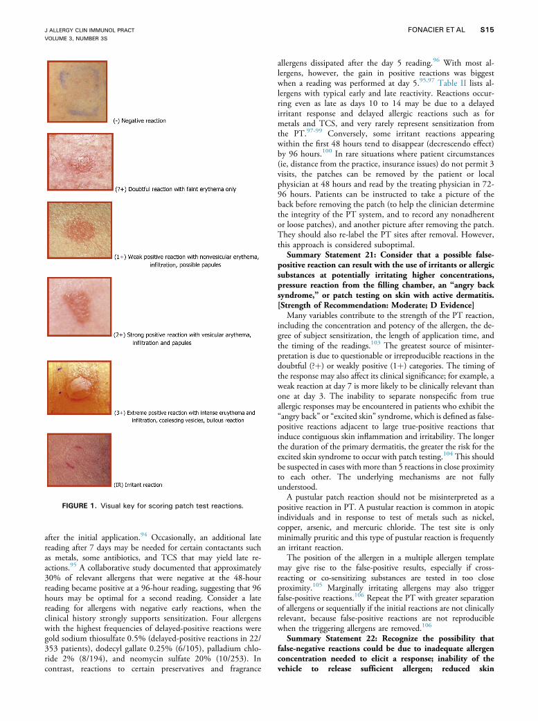
FIGURE 1. Visual key for scoring patch test reactions.
J ALLERGY CLIN IMMUNOL PRACTVOLUME 3, NUMBER 3S
FONACIER ETAL S15
after the initial application.94 Occasionally, an additional latereading after 7 days may be needed for certain contactants suchas metals, some antibiotics, and TCS that may yield late re-actions.95 A collaborative study documented that approximately30% of relevant allergens that were negative at the 48-hourreading became positive at a 96-hour reading, suggesting that 96hours may be optimal for a second reading. Consider a latereading for allergens with negative early reactions, when theclinical history strongly supports sensitization. Four allergenswith the highest frequencies of delayed-positive reactions weregold sodium thiosulfate 0.5% (delayed-positive reactions in 22/353 patients), dodecyl gallate 0.25% (6/105), palladium chlo-ride 2% (8/194), and neomycin sulfate 20% (10/253). Incontrast, reactions to certain preservatives and fragrance
allergens dissipated after the day 5 reading.96 With most al-lergens, however, the gain in positive reactions was biggestwhen a reading was performed at day 5.95,97 Table II lists al-lergens with typical early and late reactivity. Reactions occur-ring even as late as days 10 to 14 may be due to a delayedirritant response and delayed allergic reactions such as formetals and TCS, and very rarely represent sensitization fromthe PT.97-99 Conversely, some irritant reactions appearingwithin the first 48 hours tend to disappear (decrescendo effect)by 96 hours.100 In rare situations where patient circumstances(ie, distance from the practice, insurance issues) do not permit 3visits, the patches can be removed by the patient or localphysician at 48 hours and read by the treating physician in 72-96 hours. Patients can be instructed to take a picture of theback before removing the patch (to help the clinician determinethe integrity of the PT system, and to record any nonadherentor loose patches), and another picture after removing the patch.They should also re-label the PT sites after removal. However,this approach is considered suboptimal.
Summary Statement 21: Consider that a possible false-positive reaction can result with the use of irritants or allergicsubstances at potentially irritating higher concentrations,pressure reaction from the filling chamber, an “angry backsyndrome,” or patch testing on skin with active dermatitis.[Strength of Recommendation: Moderate; D Evidence]
Many variables contribute to the strength of the PT reaction,including the concentration and potency of the allergen, the de-gree of subject sensitization, the length of application time, andthe timing of the readings.103 The greatest source of misinter-pretation is due to questionable or irreproducible reactions in thedoubtful (?þ) or weakly positive (1þ) categories. The timing ofthe response may also affect its clinical significance; for example, aweak reaction at day 7 is more likely to be clinically relevant thanone at day 3. The inability to separate nonspecific from trueallergic responses may be encountered in patients who exhibit the“angry back” or “excited skin” syndrome, which is defined as false-positive reactions adjacent to large true-positive reactions thatinduce contiguous skin inflammation and irritability. The longerthe duration of the primary dermatitis, the greater the risk for theexcited skin syndrome to occur with patch testing.104 This shouldbe suspected in cases withmore than 5 reactions in close proximityto each other. The underlying mechanisms are not fullyunderstood.
A pustular patch reaction should not be misinterpreted as apositive reaction in PT. A pustular reaction is common in atopicindividuals and in response to test of metals such as nickel,copper, arsenic, and mercuric chloride. The test site is onlyminimally pruritic and this type of pustular reaction is frequentlyan irritant reaction.
The position of the allergen in a multiple allergen templatemay give rise to the false-positive results, especially if cross-reacting or co-sensitizing substances are tested in too closeproximity.105 Marginally irritating allergens may also triggerfalse-positive reactions.106 Repeat the PT with greater separationof allergens or sequentially if the initial reactions are not clinicallyrelevant, because false-positive reactions are not reproduciblewhen the triggering allergens are removed.106
Summary Statement 22: Recognize the possibility thatfalse-negative reactions could be due to inadequate allergenconcentration needed to elicit a response; inability of thevehicle to release sufficient allergen; reduced skin

TABLE II. Allergens associated with early and late reactions
Allergens associated with early peak reactions (at 48 h)
Balsam of Peru resin (Myroxylon pereirae)95,101,102
Benzoyl peroxide102
Carba mix102
Cinnamic alcohol102
Cocamidopropyl betaine95
Fragrance mix95
Imidazolidinyl urea102
Thiuram mix102
Wool alcohols102
Allergens associated with reaction on day 5, resolved day 7
Fragrance mix101
Methyl dibromo glutaronitrile phenoxy ethanol101
Octyl gallate101
Balsam of Peru101
Benzalkonium chloride101
Benzoic acid101
Disperse blue #124101
Allergens associated with late peak reactions (days 6-7)
DyesPara-phenylenediamine95,102
Medications
Neomycin95,101,102
Caine mix95
Topical corticosteroids
Tixocortol-21-pivalate95
Budesonide95,101
Metals
Nickel sulfate95,101
Gold sodium thiosulfate101
Palladium chloride101
Potassium dichromate95
Cobalt chloride95
Preservatives and glues
Dodecyl gallate101
p-Tert-butyl phenol formaldehyde resin95,102
Methylchloroisothiazolinone95
Epoxy resin95
Ethylenediamine dihydrochloride102
Mercapto mix95,102
Thimerosol95,101,102
J ALLERGY CLIN IMMUNOL PRACTMAY/JUNE 2015
S16 FONACIER ETAL
responsiveness because of prior ultraviolet light exposure (ie,sun, tanning bed); concomitant immunosuppressive thera-pies; or methodological testing errors such as insufficientocclusion, failure to perform delayed readings, and failure toperform a photo PT. [Strength of Recommendation: Mod-erate; C Evidence]
The strength of the reaction on the skin does not necessarilycorrelate with clinical relevance. For example, aminoglycosidesmay cause weak reactions on PT that are nonetheless clinicallyrelevant.107 The frequency of false-negative results is not known,but has been estimated to occur in up to 30% of patch-testedpatients.108 Potential causes of false-negative reactions includetoo low a concentration of the allergen in the extract, use of the
wrong carrier vehicle that resulted in insufficient penetration ofthe allergen, or inclusion of the wrong salt or version of theallergen. UV sunlight (eg, tanning), TCS, and TCI on the area ofPT and systemic CS (ie, >20 mg/day prednisone)78 and otherimmunosuppressives can all inhibit a positive patch response.Also, the patient may need photo-patch testing if photo-allergicCD is suspected.
Summary Statement 23: Determine the relevance of a PTresult based on the clinical and exposure history wheninterpreting the PT. [Strength of Recommendation: Moder-ate; D Evidence]
The clinical relevance of positive PT reactions to ACD can onlybe established by carefully correlating the history, which includesexposure to the allergen, with the PT test results. A positive PTmay be clinically relevant depending on current or past exposures.Current relevance is defined as definite if the PT or use test withthe suspected material is positive; probable if the antigen is presentin known skin contactants and the clinical presentation isconsistent with that exposure; or possible if skin contact withmaterials known to contain the allergen was likely. Past relevanceis considered if the PT is positive but the exposure was in the past,and not the present.109,110
Summary Statement 24: Consult physicians with expertisein patch testing to household cleaning or industrial productsif testing to the actual product suspected of containing therelevant allergen(s) is necessary, because false-positive andsevere irritant reactions can occur. [Strength of Recommen-dation: Moderate; C Evidence]
Household and industrial products should only be tested byphysicians with expertise on this type of testing after determinationof safety from MSDS information and using nonirritating PTbased on an authoritative text.111 Some of these chemicals can beextremely toxic to the skin and on rare occasions even producesystemic effects. The PT concentration of these products must bebased on established protocols when available. Nonirritant con-centrations are established by testing groups of unaffected volun-teer control subjects. Whenever possible, customized contactantsshould be incorporated into a petrolatum base, but in some in-stances, a different vehicle should be used to increase exposure tothe relevant antigen.112,113 It may be difficult to distinguish anirritant from an allergic reaction. Examples of direct PT to prod-ucts at nonirritating concentrations found in Patch Testing 3rdEd.,112 are bath products 1% aqua, shampoo 5% aqua, syntheticdetergents 2% aqua, soap 1% or 2% aqua, and glues 1% to 20%in aqua, acetone, alcohol, or petrolatum. Antiperspirant, eau decologne, cosmetics that are leave on, and insect spray may be PTwithout dilution. Agents that should not be patch tested includebenzene, toluene, and other solvents, such as gasoline, kerosene,lime, floor wax and polish, diesel oil, rust removers, and others.Furthermore, unknown substances should not be tested.
Summary Statement 25: Consult physicians with expertisein UV radiation and photo-patch testing to confirm a sus-pected diagnosis of photo-allergic CD. [Strength of Recom-mendation: Strong; C Evidence]
Photo-patch testing should be done in clinical settings with theexpertise, materials, and equipment to perform the procedure. Inbrief, duplicate applications of the suspected photo-sensitizer(s) areplaced on either side of the upper back, and occluded for 24 to 48hours. A recent study suggests that 2 days of occlusion beforeirradiation of allergens is more sensitive at detecting photo-allergy.114 After PT removal, one side of the back is then irradiated

J ALLERGY CLIN IMMUNOL PRACTVOLUME 3, NUMBER 3S
FONACIER ETAL S17
with 5 J cm�2 of UVA and the other side is left open but un-treated as the control. Both irradiated and unirradiated sides arethen measured 48 hours after irradiation for a response. If thepatient has persistent photosensitivity, the minimum erythemadose (MED) must be determined first and reduced to 1/2 of theMED for the photo-patch test. Readings are recorded pre-irradiation, immediately postirradiation, and 48 hours post-irradiation. Additional readings have been recommended.115
Summary Statement 26: Although in vitro tests for delayedhypersensitivity to contact allergens (ie, metals and bonecement) are available, routine use of such assays is notcurrently recommended as their sensitivity and specificity fordiagnosing ACD has not been determined and should beconsidered investigational. [Strength of Recommendation:Moderate; C Evidence]
In vitro tests for assessing antigen-specific sensitization arebased on measuring lymphocyte proliferation (LPTs) or cytokineproduction (ELISA or EliSPOT) after incubation with antigens.Some in vitro tests have been validated by patch testing tonickel,116,117 chromium,118 cobalt,119 and beryllium.120 Severalother in vitro tests are available, including the MELISA (MemoryLymphocyte Immuno Stimulation Assay), and LPTs from Or-thopedic Analysis, but have not been validated against patchtesting. The clinical relevance of in vitro testing in the diagnosisof contact dermatitis has not been established and is stillconsidered investigational.
Summary Statement 27: Use the ROAT to further evaluatea patient suspected of ACD who exhibits doubtful or negativePT responses, to confirm that the patient is reacting to thatparticular product or to determine clinical tolerability to newcosmetic products. [Strength of Recommendation: Moderate;C Evidence]
Several open PT techniques have been used to test substanceswith the potential for irritation, and are especially suitable forcosmetics and other personal care products such as make-up andskin lotions. The ROAT involves the repeated application of asuspected allergen to the antecubital fossa twice daily for up to 1to 2 weeks, and observing for the development of dermatitis. Toreplicate the reactivity of the eyelid skin, the ROAT can also beperformed on the back of the ear. Another provocative open usetest involves the application of the product to the skin of theforearm, which is then left untouched and observed for 5 to 10days for a reaction. A comparison of the ROAT with the PT fornickel demonstrated that although the threshold concentrationfor a positive reaction for the ROAT per application wassignificantly lower than the threshold concentration for a positivePT, the accumulated ROAT dose was very similar to the PT.121
A usage test involves the daily direct application, under real worldconditions, of an undiluted product highly suspected of con-taining a sensitizer, to prove causation. An example is for a pa-tient to apply mascara to one set of eyelashes and to leave theother eye bare, to observe for dermatitis. This is often used whenPT with suspected commercial allergens is negative but thesuspicion of contact allergy is high.122
Sources of exposure to clinically relevant allergensSummary Statement 28: Evaluate patients who present
with recurrent dermatitis on exposed skin surfaces duringairborne pollen seasons for contact sensitization to seasonalpollen allergens. [Strength of Recommendation: Moderate; CEvidence]
Patients with AD presenting in a pattern of airborne exposure(ie, present on face, hands, and exposed chest) may be triggeredby airborne protein allergens such as grass pollen, house dustmite, and cat dander. Although currently not standardized, thiscan be diagnosed by the atopy patch test (APT), involving theapplication of intact protein allergens by PT and reading the siteafter 24 to 72 hours. An APT reaction correlates frequently withthe skin prick test and serum IgE, but not always.123,124
For some plants, both plant parts and pollen may contain thesame allergen, and have been reported to cause airborne CD.These include the weed Parthenium hysterophorus L. (a memberof the Compositae family) in India and Australia,125,126 Japanesecedar pollen confirmed by a positive scratch-patch test,127 andAmbrosia deltoidea, or triangle-leaf bursage, confirmed by PTwith an oleoresinous extract of A. deltoidea leaves.128
Mulberry pollen129 and Compositae pollen from dandelions,blazing star, golden rod, yarrow, Aster ssp, chrysanthemums, ormarguerite130 are reported to cause airborne CU that can beconfirmed by prick skin testing.
Summary Statement 29: The clinician should considercosmetics and personal hygiene products that are directlyapplied to involved skin or ectopically transferred from un-involved skin as potential sources of allergens in patients withACD. [Strength of Recommendation: Strong; C Evidence]
The US Food and Drug Administration (FDA) defines“cosmetic” as articles intended to be rubbed, poured, sprinkled,or sprayed on, introduced into, or otherwise applied to the hu-man body or any part thereof for cleansing, beautifying, pro-moting attractiveness, or altering the appearance, and articlesintended for use as a component of any such articles except soap(US FDA. The Federal Food, Drug, and Cosmetic Act, Sec. 201[21 U.S.C. 321]; Chapter II, Definitions 1: www.fda.gov). Thusaccording to this broad definition, it is not unusual for in-dividuals to apply dozens of personal hygiene products to theirskin on a daily basis including a plethora of cosmetics, each witha unique formulation of synthetic or natural ingredients. Suchproducts can include emollients for day and night use, hair careproducts (shampoos, conditioners, pomade, relaxers, sprays, gels,mousses, foams), nail products (acrylic nails, polishes, hardeners,repair agents, extenders, wraps), traditional cosmetics (eye liners,mascara, eye shadow, foundation, lipstick, lip liners), concealers,shave creams and gels, antiperspirants and deodorants, tooth-pastes, dentifrices, hand creams, and barrier creams.
Although ACD caused by cosmetics is noted predominantly atthe site of application, occasionally personal care products andcosmetics will manifest the contact allergy lesions in locationsdistant from the original skin sites. This phenomenon is termedectopic CD. Typical causes of ectopic ACD are allergens such asnickel transferred to the eyelid by fingers, toluene sulfonamideformaldehyde resin in nail polish (which may cause eyelid derma-titis yet spare the periungual skin and distal fingers), and gold131
(where dermatitis is reported in women who wear facial cosmeticsthat contain titanium dioxide that may adsorb or abrade the goldreleased from jewelry and make occasional contact with facialskin).132 In addition, patients allergic to hair products that containCAPB, a surfactant in shampoo, can present with eyelid dermatitiswithout concurrent dermatitis on the scalp, neck, or ears.Consideration must also be given to dermatitis where the allergen istransferred between partners, parent or child.
Summary Statement 30: When evaluating ACD fromcosmetics and personal care products that contain many

J ALLERGY CLIN IMMUNOL PRACTMAY/JUNE 2015
S18 FONACIER ETAL
different chemical ingredients, consider that the most com-mon causes are due to a few important chemical classesincluding fragrances, preservatives, excipients, nickel, andsun screening agents. [Strength of Recommendation: Mod-erate; C Evidence]
In aggregate, the number of chemical contactants used by anindividual patient in a typical day can be more than 100. Despitethis extensive use, typical contact allergens contained in theseproducts tend to be clustered in a few important classes, includingfragrances, preservatives, formulation excipients, nickel, and sunblocks. The 15 most frequently positive allergens of the NACD2009-2010 PT were nickel sulfate (15.5%), neomycin (8.7%),FM I (8.5%), bacitracin (8.3%), BOP (7.2%), cobalt chloride(8.2%), formaldehyde (5.8%), quaternium-15 (5.8%), PPD(5.5%), FM II (4.7%), carba mix (4.6%), iodopropynyl butyl-carbamate (4.3%), methyldibromo glutaronitrile/phenoxyethanol(3.8%), propylene glycol (3.2%), and thiuram mix (3.1%).26
Fragrances are complex substances that contain hundreds ofdifferent chemicals and are the most common cause of ACDfrom cosmetic in the United States. Fragrances are regularlypresent in cosmetics and personal care products, householdproducts, and medicaments, either to achieve an appealing scentor to mask unpleasant odors. However, the labeling of productswith regard to fragrance can be confusing.133-137 The use of theterm unscented can erroneously suggest that a product does notcontain fragrance when, in fact, a masking fragrance is present.Fragrance-free products are typically free of classic fragrance in-gredients and are generally acceptable for the allergic patient.Caution should be exercised when substitute products, which arelabeled fragrance free, contain large numbers of botanical extractsused for the purpose of improving odor characteristics.138 Allergyto fragrances can be detected clinically when obvious contact sitesof perfume are involved. Clear demarcation of eczematousdermatitis on the neck where perfume is sprayed may be anobvious indication of fragrance allergy.
It is necessary to PT to appropriate screening chemicals fordetection of delayed hypersensitivity to this group of aller-gens.139-143 The fragrance antigens in the current T.R.U.E.Test include BOP (a fragrant resinous natural product con-taining a mixture of many substances), and FM I (cinnamylalcohol, cinnamaldehyde, a-amyl cinnamaldehyde [amyl cin-namal], hydroxycitronellal, geraniol, isoeugenol, eugenol, oakmoss). Although there is a strong association between thesefragrances, separated PT may still be warranted to identify thespecific offending fragrance so that not all fragrances need tobe avoided.
Previous studies suggest that the standard FM and BOP willdetect approximately 60% to 70% of fragrance-allergic in-dividuals. The addition of other commonly used fragrance in-gredients (ylang ylang oil, narcissus oil, and sandalwood oil) mayincrease the yield up to 96%.141 In a recent study of patientswith eyelid dermatitis, PT to fragrance markers within thestandard series (ie, FM I, FM II, Myroxylon pereirae, and cin-namic aldehyde) detected 73.2% of cases of fragrance allergy.144
The elucidation of fragrance allergy should result in advising anavoidance protocol that eliminates all culprit fragranced cos-metics and personal hygiene products. However, it should benoted that fragrances in PT have marginal irritant potential andweak positive reactions may not be regarded as proof of contactsensitization (low specificity of the test). The increased strengthof the test reaction, a positive reaction on retest to FM (repeated
positive reactions), and a positive PT to individual ingredientsadds significantly to the probability of a relevant test.
Current labeling laws do not always require manufacturers tolabel a specific fragrance present in a product and regulation offragrance ingredients in cosmetics exempts fragrance formulas as“trade secrets.” Therefore, some manufacturers do not list essentialoils that can also cause ACD such as tea tree oil (Melaleucaalternifolia), ylang-ylang oil (Cananga odorata), jasmine flower oil(Jasminum officinale), peppermint oil (Mentha piperita), lavenderoil (Lavandula angustifolia), and citrus oil (limonene). “Covertfragrances” that may be used for purposes other than for aroma, iepreservatives, may be added to “fragrance free” products (benzal-dehyde, benzyl alcohol, bisabolol, citrus oil, unspecified essentialoils) and may be problematic. In addition, new fragrance chem-icals are constantly introduced. Use testing and slow reintro-duction of some fragrance products may allow for the detection ofintolerance to specific cosmetic agents. It may be possible toidentify the presence of specific fragrance ingredients bycommunicating directly with product manufacturers.
Preservatives and antibacterials are present in most aqueous-based cosmetics and personal hygiene products to preventrancidity and microbial contamination. These preservatives areimportant cosmetic allergens. Preservatives tend to be groupedinto 2 broad categories: formaldehyde releasers (products thatemit formaldehyde) and nonformaldehyde releasers.145-148
Table III is a list of preservative systems commonly used incosmetic and personal care products.
In the United States, approximately 20% of cosmetics andpersonal care products (stay-on and rinse-off products) contain aformaldehyde releaser.149 The most recent data from the FDAVoluntary Cosmetic Registration Program Database150 approxi-mate that 1 in 6 stay-on cosmetics and 1 in 4 rinse-off productscontain a formaldehyde releaser, the most frequent of which isimidazolidinyl urea (7%), followed by DMDM hydantoin(5.4%), diazolidinyl urea (4.5%), and quaternium-15 (1.4%).
De Groot et al149 recommend that patients allergic to form-aldehyde be advised to avoid stay-on cosmetics preserved withquaternium-15, diazolidinyl urea, DMDM hydantoin, or imi-dazolidinyl urea. Provocation tests may also be performed todetermine relevance to this particular patient.
Among nonformaldehyde releaser preservatives, methlydi-bromo gluteronitrile (also known as 1,2-dibromo-2,4-dicyano-butane and is the sensitizing ingredient in Euxyl K 400) hasemerged as an important cosmetic allergen in recent years.151 InNorth America, the prevalence of positive PT reactions to EuxylK 400 increased from 1.5% between 1992 and 1994 to thecurrent rate of 5.5% for 2007 and 2008.109 A total of 11.8% ofhand dermatitis cases associated with Euxyl K 400 were occu-pation related and were linked to solvents, oils, lubricants, fuels,and cosmetics.152 In cosmetics, ACD from Euxyl K 400 or itscomponents is most commonly reported in hand and face lo-tions, hair products, and ultrasonic gels.152
Another nonformaldehyde releaser preservative MCI/MI(trade name: Kathon CG) is commonly used in cosmetics andtoiletries in the United States. The NACDG data from 2009to201026 show that MCI/MI had a 2.5% frequency of positivePT reactions, ranking it the fifth most commonly positive pre-servative. The combination of MCI/MI is tested at a 3:1 com-bination. Both MCI and MI can cause contact allergy with MCIas the more potent allergen in this combination.153 However, theuse of MI alone as a preservative in personal care and cosmetic

TABLE III. Cosmetic preservatives
Formaldehyde releaser Nonformaldehyde releaser
Formaldehyde Iodopropynyl butylcarbamate
Quaternium-15 Methychloroisothiazolinone/methylisothiazolinone (MCI/MI)
Diazolidinyl urea Parabens
Imidazolidinyl urea Methyldibromo glutaronitrile
Bromonitropropane Chloroxylenol
DMDM hydantoin Benzalkonium chloride
Thimerosal
Phenoxyethanol
J ALLERGY CLIN IMMUNOL PRACTVOLUME 3, NUMBER 3S
FONACIER ETAL S19
products has increased in the past few years. According to the USFood and Drug Administration Voluntary Cosmetic IngredientRegistration Program, MI was used in a total of 1125 cosmeticproducts in the United States in 2007.154 Of these, the majorityare in rinse-off products: 24% were shampoos, 18% were con-ditioners, and 10% were baby soaps and detergents. Wet wipes(baby wipes, moist towelettes, and moist toilet paper) are a well-identified sensitization source for MI.155
The MCI/MI mix misses approximately 40% of allergy to MIlikely because of the low concentration of MI in the MCI/MIcombination in the PT. In Europe, several groups have docu-mented frequency of allergy to this preservative of approximately1.5%.156 Patch testing to MI alone will likely diagnose morecases of MI contact allergy.
Although parabens formulated in cosmetics are infrequentcauses of ACD, they can induce ACD when used as antibacterialsin topical medications. ACD has most commonly been reportedwhen paraben-containing products are used on damaged skinsuch as in long-standing dermatitis and stasis ulcers. The rate ofsensitization to parabens in patients with chronic leg ulcers ishigher than that of the general population.157
“Botanicals” are plant extracts that are increasingly used asadditives to skin care products either for their medicinal prop-erties or as fragrances (such as essential oils). Unfortunately, incosmetics, product labeling may not list essential oils as fra-grances. These natural botanicals, plant extracts, and herbalremedies are potential causes of CD. One study showed asensitivity rate of 2.4% to testing with pure tea tree oil.158 Otherstudies showed that 1.2% to 6.6% of patients patch tested fordermatitis are sensitive to propolis,159 which is commonly usedin cosmetic and medicinal preparations because of its antiseptic,anti-inflammatory, and anesthetic properties. Propolis is found inmany “all natural’’ products, including lip balms, cosmetics, lo-tions and ointments, shampoos, conditioners, and toothpastes.Synonyms for propolis include bee glue, bee bread, hive doss,propolis balsam, propolis resin, and propolis wax.
Thus, PT should be considered for propolis, tea tree oil, andother essential oils in patients with cosmetic dermatitis. It isimportant that patients who are allergic to fragrance also be madeaware of the potential dangers of cosmetics containing plantextracts and patients should be counseled that “natural products”does not equate with safety.160
Summary Statement 31: Patients suspected to have allergyto hair products should be evaluated for PT reactions toCAPB, PPD, fragrances, preservatives, and glycerol thio-glycolate. [Strength of Recommendation: Moderate; CEvidence]
CAPB is an amphoteric surfactant that is often found inshampoos, bath products, eye and facial cleaners, liquiddetergents, surface cleaners, pet care products, and other skinand hair care products, and the incidence of sensitization isincreasing. Although it is less irritating than the older polarsurfactants such as sodium lauryl sulfate,161,162 it is moresensitizing. CAPB allergy typically presents as eyelid, facial,scalp, and/or neck dermatitis.163 Consumers were sensitizedmainly through shampoos (including baby shampoo) and othertoiletry products that include liquid shower gels, roll-ondeodorants, and facial cleansers.
According to the NACDG data for 2007-2008, 1.1% of pa-tients tested had a positive reaction to CAPB109 and positive PTreactions to this allergen are often clinically relevant.
Commercial bulk production of CAPB may result incontamination of the final product with 2 chemicals that are usedin the synthesis of CAPB, such as amidoamine anddimethylaminopropylamine.163
Paraphenylenediamine is the active ingredient in many hairdyes, both permanent and semipermanent, and is a very com-mon cause of CD in hairdressers. Although hair dye is the mainsource of exposure,164 other routes of exposure include bodypainting and temporary tattooing. ACD from PPD can be severe,sometimes mimicking angioedema. The “skin sensitivity test”recommended in the package insert of hair dyes has been vali-dated as an effective method to predict a type IV hypersensitivityreaction and should be used by hairdressers.165 Nevertheless, PTmay be needed to identify the active allergen in the consumerproduct.
It is difficult to find alternative hair dyes for PPD-allergicindividuals. Alternatives include henna (giving a reddish tintfor any hair color), lead oxide (which oxidizes to darken grayhair but has not been adequately evaluated for its toxicity),and temporary coloring agents (which only last for a fewwashes). Semipermanent hair dyes containing F, D & C andD & C dyes appear to have very low cross-reactivity withPPD (examples: Elumen Hair Color from Goldwell Cos-metics, Linthicum Heights, MD, and Clairol Basic Instincts-Loving Care from the Proctor & Gamble Company,Cincinnati, Ohio). However, semipermanent dyes may notbe as cosmetically elegant and require more frequent appli-cation. Scheman et al reported that PPD-sensitive individualswho test negative to para-toluenediamine sulfate (PTDS)will very likely tolerate the newer permanent and demi-permanent PPD-free hair-dye products.166 However, thisstudy suggests that patients be tested for PTDS before usingPPD contacting dyes. Examples of PPD-free hair dyesinclude Wella Koleston Perfect (permanent), Wella ColorCharm (demi-permanent), Schwarzkopf Igora Royal (per-manent), Goldwell Color Chic (permanent), GoldwellReShade for Men (demi-permanent), Sanotint Light (demi-permanent), and L’Oreal Paris Excellence To-Go 10-Min.Crème Colorant (demi-permanent).26,167 Both physicians andpatients should consult available databases like ContactAllergen Management Program (CAMP) and Contact AllergenReplacement Database (CARD) regularly for updates.
Other sources of exposure to PPD include leather, fur, textiles,industrial rubber products, and black henna tattoos.167 Cross-reactivity with other para-amino compounds, such as benzocaine,PABA, sulfa drugs, aminoazobenzene, IPPD, and azo dyes hasbeen reported and may require avoidance.168

J ALLERGY CLIN IMMUNOL PRACTMAY/JUNE 2015
S20 FONACIER ETAL
Glycerol thioglycolate is the active ingredient in permanentwave solution. ACD to this chemical tends to cause moreoccupational dermatitis in hair dressers than consumers. UnlikePPD, thioglycolates may remain allergenic in the hair long after ithas been rinsed out. Hence, those individuals who are allergic toit may continue to have skin eruptions weeks after application ofthe perm, and hairdressers allergic to it may be unable to cut orshape permanent waved hair.169
Summary Statement 32: Suspect allergy to nail productswhen the dermatitis presents locally at the distal digit orectopically on the eyelids and face. [Strength of Recom-mendation: Moderate; C Evidence]
Most allergic reactions to nail polish and artificial nailproducts are to tosylamide/formaldehyde resin105 found innail polish enamel, in addition to nail hardeners and settinglacquers. Up to 80% of the reactions appear on the neck,face, lips, and eyelids, although unusual locations includingthe gluteal, perianal, and genital areas have been reported.Only 27% of reactions were reported in the periungual re-gion of the hands and feet. Some patients react to the polishwhen it is still wet, but the majority of patients appear toreact to the water-soluble components (including monomersand dimers) of tosylamide/formaldehyde resin found in drypolish.160
As an alternative, some manufacturers may use an alkylpolyester resin and label their products as “hypoallergenic.”These products would be suitable alternatives for sensitivepatients.170
Artificial nails are increasingly used and are available assculptured nails, photobonded nails, and preformed nails. Re-actions to artificial nails have included paronychia, onychody-strophies, and dermatitis at contact areas and at distant sites.Acrylate monomers used for sculpting artificial nails are impor-tant sensitizers for contact and occupational dermatitis. Pre-formed plastic nails may be glued over the natural nail plate usingethyl cyanoacrylate, a potential sensitizer.
Certain guidelines for testing nail cosmetics are as follows: (1)Nail polish should be tested as is—undiluted. (2) Acrylate allergyshould be screened with an acrylate test panel, including 2%methyl methacrylate, 1% bisphenol A, 2% tetraethylene glycoldimethacrylate, 2% bisphenol A dimethacrylate, 2% ethyleneglycol dimethacrylate, 2% dimethyl-p-toluidine, and 1% benzoylperoxide has been advocated.171 (3) Patch testing for nail polishremovers should be an open PT, at a concentration of 10% inolive oil. (4) Cuticle removers are tested as an open PT at a 2%aqueous concentration.172
Summary Statement 33: Suspect the diagnosis of photo-allergic CD to cosmetics when eczema occurs in a light-exposed distribution following the use of a skin care productor cosmetic, including sunscreens. [Strength of Recommen-dation: Strong; C Evidence]
Some cosmetic ingredients may only cause an ACD afterexposure to UV radiation. Photo-allergic CD typically affectssun-exposed areas such as the face, the ‘‘V’’ of the anterior neck,the dorsal hands, and forearms. It typically spares the uppereyelids, upper lip, and submental and postauricular areas. Beforeevaluation for photo-allergic CD, one should rule out phototoxicdrug eruption, photo-allergic drug eruption, and systemic diseasesuch as lupus erythematosus.
The prevalence of allergic reactions to sunscreens maycontinue to increase as the use of sunscreen continues to become
more widespread. Allergic and photo-allergic reactions have beenreported with several chemical sunscreen families.173,174
Sunscreens have traditionally been divided into chemicalabsorbers (UVB [290-320 nm], UVA II [321-340 nm], andUVA I [341-400 nm]) and physical blockers.
Sunscreens are often overlooked as a cause of CD, becauseother excipients (fragrances, formaldehyde releasers, pre-servatives, vitamin E, and lanolin alcohol)175 are morefrequently implicated. Sunscreen sensitization is much higher inindividuals referred for evaluation of photosensitivity.176 Themost common cosmetic sunscreen agents used are listed inTable IV.
Physical ultraviolet light blockersTitanium dioxide and zinc oxide are the most common
physical UV blockers used today and have not been reported tocause contact dermatitis or photo-allergy.
Topical medicinal CD
Summary Statement 34: If an eruption worsens, ratherthan improves, after the topical application of certain med-ications, or fails to respond to TCS, patch testing should beperformed to the suspected product and/or ingredientsknown to be contact sensitizers. [Strength of Recommenda-tion: Moderate; C Evidence]
CD may develop after exposure to topical medications,including lanolin, para-aminobenzoic acid (in sunscreens),“caines” (anti-itch preparations), topical antibiotics, topical an-tihistamines, NSAIDs, and/or TCS.177-182 Neomycin, bacitra-cin, and iodochlorhydroxyquin are well-known sensitizers.Lanolin is used as the base of many topical medications includingTCS and moisturizers.
Allergy to TCS affects 0.5% to 5.8% of patients183 suspectedof ACD. Sensitization can occur by skin, airborne, oral, and IVroutes.184,185 Certain disorders predispose patients to anincreased risk of CS ACD. These include treatment of refractoryeczema, chronic venous leg ulcers, stasis dermatitis, and CD (inparticular, patients with a history of 2 or more positive PT resultsand multiple medicament sensitivities).
Patch testing to CS is complicated by the inherent, anti-in-flammatory nature of the drug itself, which results in frequentfalse-negative results if tested at too high concentration.Accordingly, PT readings should also be done 7-10 daysfollowing application because approximately 30% of ACD toTCS could be missed by conventional readings.186,187
Patch testing substances for TCS allergy that are commer-cially available include amcinonide, betamethasone-17,21-dipropionate, betamethasone-17-valerate, budesonide,clobetasol-17-propionate, desoximetasone, dexamethasone,hydrocortisone, hydrocortisone 17-butyrate, prednisolone,tixocortol-21-pivalate, and triamcinolone acetonide. The pa-tient’s own commercial steroid,184 as well as the vehicle andpreservatives in the preparations,188,189 must also be tested.Coopman et al190 suggested that 4 major groups of CS prep-arations should suffice, because there is considerable cross-reactivity within the groups and possible cross-reactivitybetween them. Ninety percent of ACD to CS should bedetected by using tixocortol pivalate, budesonide, triamcino-lone, and the patient’s commercial steroid.191,192 Althoughrare, patients sensitized to CS by skin contact can develop SCDwith administration of the CS by an oral, IV, IM, or inhalation

TABLE IV. The most common cosmetic sunscreen agentsCinnamates Octyl dimethyl
para-aminobenzoic acid
Salicylates Benzophenones
Titanium dioxide Anthranilates
Butyl methoxydibenzoylmethaneor avobenzone (Parsol 1789)
Zinc oxide
J ALLERGY CLIN IMMUNOL PRACTVOLUME 3, NUMBER 3S
FONACIER ETAL S21
route.193 Cross-reactivity based on 2 immune recognition siteshas been reported,194 and the avoidance of TCS within eachclass is recommended once allergy to TCS has been confirmedby PT (Appendix J).
Summary Statement 35: The clinician may use the drugPT for the diagnosis of some drug hypersensitivity re-actions, recognizing that there is no standardized approachto define the population, clinical manifestation, drug toPT, and PT materials to make patch testing to drugs astandard of care. [Strength of Recommendation: Weak; DEvidence]
Patch testing to drugs may have a role in delayed hypersen-sitivity drug reactions177 and have a higher positivity in patientspresenting with maculopapular rashes, erythroderma, and non-immediate cutaneous reactions,195 including DRESS,196
AGEP,197 SJS/TEN, and fixed drug eruptions. The utility of thePT depends on various factors including the type and formula-tion of the drug being tested, the vehicle used, as well as theimmunopathogenesis eliciting the eruption.
PT may be helpful with aromatic anticonvulsants and variousantibiotics, but it is not consistently helpful for a wide range ofdrugs. Patients with a history of drug exanthem from antibioticsare more likely to have a positive PT (10% to 46%) comparedwith those with a history of a drug exanthem from nonantibioticmedications (w10% to 11%).198-200
Within antibiotic classes, there are higher rates of positive PTreactions to aminopenicillins, cephalosporins, pristinamycin, andclindamycin compared with macrolides, tetracyclines, andquinolones.195
PT can be performed for a wide variety of medications inmultiple concentrations and vehicles.201 The European Societyof Contact Dermatitis (ESCD) and the European Network onDrug Allergy (ENDA) have guidelines for performing PT formedication-induced cutaneous eruptions.202,203 However, thelimitations of these studies include the lack of standardized testmaterials, the absence of information as to the ideal test con-centration and vehicle to use, and the differences in the inter-pretation of the tests.
Summary Statement 36: Consider pre-operative patchtesting for metal sensitization in patients with a significanthistory of metal allergy. [Strength of Recommendation:Moderate; C Evidence]
Indications for pre-operative PT in patients with a history ofmetal allergy are still being developed. However, pre-operativePT may help guide the selection of implant alloys in patientswith a high suspicion of metal allergy, and such patientsdemonstrate improved outcomes.204-207 This testing is not rec-ommended for patients without such a history of metal sensi-tivity. There is no information regarding pre-operative PT inpatients with a prior history of methacrylate or antibioticsensitivity.
Summary Statement 37: In patients with joint replacementfailure, patch testing to components of the implant may behelpful after infection and biomechanical causes have beenexcluded. [Strength of Recommendation:Moderate; C Evidence]
The clinician should recognize that contact sensitization tometalsor bone cement that is used in orthopedic, cardiac, dental, and gy-necological implants has been associated with both dermatitis andnoncutaneous complications. These complications may includelocalized pain, swelling, erythema, warmth, implant loosening,decreased range ofmotion, stent stenosis, and pericardial effusions inthe case of cardiac implants. Patch testing to implant or devicecomponents has been recommended to help determine the etiologyof the adverse reaction (Tables V and VI).208-210
In a meta-analysis, the rates of sensitization to metals weresignificantly higher in patients with a failed implant than in pa-tients with a well-functioning implant (P ¼ .002) or without animplant (P < .001).209 Patients who experienced failed joint re-placements and underwent revision using components dictated bya positive metal PT reported resolution of their joint symptoms,most frequently joint pain, joint loosening, and localized derma-titis. Those patients with a positive metal PT who were not revisedcontinued to experience the same symptoms.204 Similarly, a groupof patients with implant-related eczema who were metal sensitized,and then underwent revision with a different metal alloy implant,had a higher incidence of eczema resolution.211 Anecdotal casereports suggest that skin or systemic manifestations of sensitizationto components of implantable defibrillators,212 pacemakers,213
arterial stents,214 dentures,215 and IUDs,216 appeared to improveonce the sensitizing agent was replaced.
At present, the recommendation for implant removal remainscontroversial. Indeed there are reports of individual patients withdocumented metal allergy who have tolerated implants of thesame metal without adverse reactions. An older study reportedthat 18 patients with documented metal allergy did well for over6 years following a joint replacement that contained the aller-genic metal.217 Gawkrodger stated in 1993 that there was noevidence that nickel-sensitive patients, when given a plastic-to-stainless-steel hip implant, developed cutaneous reactions orloosening of their prostheses,218 although he has since identifiedan association between metal sensitization, peri-implant hyper-sensitivity reactions, and implant loosening and failure. Theoverall risk, however, was low.219 Other patients with docu-mented metal sensitization have tolerated cardiac implants withthe same metal without adverse reaction.220
As in all cases of PT, resultsmust be interpretedwithin the clinicalhistory and physical examination. If an implant is functioning well,then a positive PT to an implant component is not clinically rele-vant.210 The likelihood that an allergy to implant components is thecause of implant failure is higherwhen other causes of implant failure(infection and biomechanical issues) have been ruled out. There areno current guidelines or recommendations for symptomatic patientswith positive PT to metals or bone cement components. The de-cision regarding implant revision following positive relevant PTresults can only be made after a thorough discussion between thepatient, the allergist or dermatologist, and the orthopedic surgeon.
In addition to the possibility of metal sensitization as a po-tential therapeutic cause of joint replacement failure, there arealso reports of implant failure related to bone cement or itscomponents, including benzoyl peroxide, hydroquinone, methylmethacrylate, and n,n-dimethyl para-toluidine.221-223

TABLE V. Components of selected alloys used in metal implants
Alloy
316L Stainless
Steel w% content
Cobalt-chromium-molybdenum
steel (ASTM F75) w% content
Vitallium
w% content
Titanium
w% content
Nitinol
w% content
Oxinium
w% content
Nickel 8.3-35 <0.5 Trace 45 None
Chromium 20 27-30
Manganese 2 <1 0.5
Molybdenum 2-3 5-7 5.6
Nitrogen 0.1 <0.25
Carbon 0.003 <0.35 0.02
Sulfur 0.03 <0.01
Silicon 0.75 <1 0.5
Phosphorus 0.045 <0.02
Iron Balance <0.75 None
Tungsten <0.2
Aluminum <0.1 5.5-6.5
Titanium <0.1 89.9 55
Boron <0.01 0.1
Cobalt Balance 61
Chromium 32
Vanadium 3.5-4.5
Zirconium (oxidized) 97.5
Niobium 2.5
J ALLERGY CLIN IMMUNOL PRACTMAY/JUNE 2015
S22 FONACIER ETAL
Special population
ACD in children. Summary Statement 38: ACD and ICDare significant clinical problems in children. Patch testingshould be performed and remains the gold standard for thediagnosis of ACD in children. [Strength of Recommendation:Strong; C Evidence]
Although ACD was historically considered to occur lessfrequently in children, recent studies show that positive PT re-actions range from 14% to 70% of children patch tested.224
ACD is considered rare in the first few months of life withincreased reports suggesting an early peak around age 3, and anincreasing rate of occurrence through the teen years, attainingand even exceeding that observed in adults.225-227
In children, a careful, age-appropriate history should includeexposure to diapers, hygiene products, cosmetics, sun blocks,textiles with dyes and fire retardant materials, medications, petsand pet products, school projects, sports, and so on. The influ-ence of fashion trends, hobbies, and lifestyle activity such as bodypiercing, decorative skin paintings (eg, PPD-laced black hennatattoos), natural remedies, and cosmetics (eg, tea tree oil), orproducts with fragrances and herbal ingredients have all beenassociated with ACD in this population.
Perioral dermatitis in children is associated with lip licking, lipchewing, thumb sucking, or excessive drooling. Metals includingmercury, chromate, nickel, gold, cobalt, beryllium, and palladiumare important allergens in patients with dental implants, ortho-dontic devices, or who play an instrument. ICD is the mostcommon cause of diaper dermatitis in infancy because of friction,occlusion, maceration, and increased exposure to water, moisture,urine, and feces. Allergic CD to rubber chemicals (mercapto-benzothiazole, cyclohexyl thiophathalimide) or glues (p-tertiary-butylphenol-formaldehyde resin) has been reported to causeCD,228 a dermatitis that is predominantly on the outer buttocksand on the hips in toddlers. This is frequently caused by the elasticbands that hold tightly on the thighs to prevent leaking.
The NACDG compared results of PT in children and adultsand found no significant difference in the overall frequency of atleast one relevant positive PT reaction in children (51.2%)compared with adults (54.1%).34 Additionally, there are highlyrelevant allergens that have significant frequency in childrenbecause of their unique exposure such as MCI/MI, a preservativein infant wet wipes, liquid soaps, and shampoos. Also, exposuresto dialkyl thiourea and p-tert-butyl formaldehyde resin in rubberproducts are seen in shin guards, wet suits, and protective pads.
A US-based study showed nickel, fragrance, cobalt, thimer-osal, BOP, potassium dichromate, neomycin, lanolin, thiurammix, and PPD to be common allergens in children.77 Eight ofthese are also in the top 10 allergens in adults suggesting that thesensitization profile for children does not differ significantly fromthat of adults. An allergen found in higher frequency in childrenthan in adults is lanolin/wool alcohols that can be found inhealing ointments, aftershave, baby and bath oil, hand sanitizers,and creams, reflecting the frequency of use of the productscontaining this contactant. Thimerosal positive PT has beenreported in both adults and children, with the main source ofsensitization likely due to previous vaccination and may not be aclinically relevant allergen. There are additional highly relevantallergens in children that correlate with unique exposures such as(1) MCI/MI, a preservative found in infant products (wet wipes,liquid soaps, shampoos); (2) CAPB, a surfactant in cleansingproducts (eg, No More Tears formulations); (3) disperse dyes indiaper material and colored garments (school and athletic uni-forms); (4) carbamates and thiuram used in rubber (gloves,garments, shoes, and toys); (5) dialkyl thioureas; and (6) p-tert-butyl formaldehyde resin found in rubber and neoprene (shinguards, protective pads, and wetsuits).
The same test concentrations used in adults can be used inchildren.229 However, it has been suggested that in very youngchildren (<6 years of age), allergens such as formaldehyde,formaldehyde releasing preservatives, mercaptobenzothiazole, and

TABLE VI. Substances that may be present in different types of implant or device and that potentially should be considered for diagnosticpatch testing
Substances or alloy3
Implant or device
Dental
Orthopedic
Intravascular Pacemaker and ICD GynecologicalPre-implant Post-implant
Aluminum X X X - X -
Beryllium X - - - - -
Cadmium X - - - - -
Chromium X X X X X X
Cobalt X X X X X -
Copper X - - - - X
Gold X - - X - -
Indium X - - - - -
Iridium - - - - X X
Iron X X X X - -
Manganese X X X X - X
Mercury X - - - X -
Molybdenum X X X X X -
Nickel X X X X X X
Niobium X X X - - -
Palladium X - - - - -
Phosphorus X X X - - -
Platinum X - - - X X
Rhodium X - - - - -
Ruthenium X - - - - -
Silicon - X X - - -
Silver - - - - X X
Tantalum - X X - X -
Tin X - - - - X
Titanium X X X X X X
Tungsten - X - X - -
Vanadium X X X - X -
Zinc X - - - - X
Zirconium X X X - - -
Custom-made disk of relevant alloy X X X X X -
ICD, irritant contact dermatitis.Used with permission from Contact Dermatitis 2011;66:4-19.
J ALLERGY CLIN IMMUNOL PRACTVOLUME 3, NUMBER 3S
FONACIER ETAL S23
thiuram be diluted 50%, and potassium dichromate diluted 25%in petrolatum, to avoid irritant false-positive reactions.230,231
The German Contact Dermatitis Group (GCDG)232 rec-ommends that children under 6 years should only be PT if thereis a high degree of clinical suspicion and only to the suspectedallergens. Children over the age of 12 can be tested in the samemanner as adults.
The ideal number of PT to be applied depends on the patientand could be limited by the surface available for testing and thepotential risk of active sensitization. Thus, Jacob et al.233
recommend a basic North American Standard Series for childrenaged 6-12 years to include 20 selected allergens that are the mostprevalent in the pediatric population with the highest clinicalrelevance and therefore would be the highest yield as a basicseries. These are bacitracin, budesonide, carba mix, cobaltchloride, cocamidopropyl betaine, colophonium, Compositaemix/dandelion extract, disperse blue, ethylenediamine, formal-dehyde, FM I, FM II, lanolin alcohol, MCI/MI, BOP, neomycinsulfate, nickel sulfate, potassium dichromate, quaternium-15,and tixocortol-21-pivalate. Additional allergens can also be tested
for if there is a relevant exposure history, for example, blackrubber mix, dialkyl thioureas, mercaptobenzothiazole, PPD, andp-tert-butylphenol formaldehyde resin. In conclusion, PT canand should be performed in children and remains the goldstandard for the diagnosis of ACD with appropriate parentalinformed consent.
Occupational contact dermatitis. Summary Statement39: In a patient who presents with dermatitis associated withworkplace exposures (ie, OCD), consider ICD as well asACD. [Strength of Recommendation: Strong; C Evidence]
Contact dermatitis is one of the most common types ofoccupational illness, with estimated annual costs exceeding $1billion (http://www.cdc.gov/niosh/topics/skin). An occupationalhealth supplement (OHS) to the 2010 National Health Inter-view Survey (NHIS) noted that 10%, or approximately 15.2million US current or recent workers reported the presence ofdermatitis. There was a higher prevalence rate in women (11.2%;95% CI 10.4-12.0) than in men (8.5%; 95% CI 7.8-9.3). Theestimate of work-related dermatitis was 7.4% or 1.12 million.234

J ALLERGY CLIN IMMUNOL PRACTMAY/JUNE 2015
S24 FONACIER ETAL
Occupational CD is classically divided into ICD and ACD.Although the mechanisms differ between the two, the clinicaland histologic appearance may be similar.
Irritant CD represents approximately 80% of all cases ofOCD. Common irritant exposures include wet work, solventsand alcohols, cutting oils, coolants, degreasers, soaps, detergents,and other cleaning agents and disinfectants. The major chemicalgroups associated with ACD include metals, rubber-related ma-terials, epoxies, resins and acrylics, organic dyes, plants, foods,medications, biocides, and germicides.235
A worker’s skin may be exposed through direct contact withcontaminated surfaces, deposition of aerosols or vapors, skin im-mersion, or splashes. The hands are most commonly affected byOCD and followed by the wrists, arms, and face. OCD can presentat any stage in a worker’s career, including apprenticeship.236 Thecommon agents that cause ICD and ACD in OCD as reported bythe NACDG in the United States include carba mix, cobalt chlo-ride, epoxy resin, formaldehyde, glutaraldehyde, glyceryl thio-glycolate, mercaptobenzothiazole, nickel sulfate, potassiumdichromate, quaternium-15, and thiuram. In addition, The Healthand Occupation Reporting System, the European PreventionInitiative for Dermatological Malignancies, and the OccupationalPhysicians Reporting Activity in the UK reported chromes and/orchromates, foods, latex, rubber chemicals, PPD, preservatives,resins and acrylics, soaps and cleansers, wet work, cutting oils andcoolants, petroleum products, solvents, and alcohols.
Summary Statement 40: In patients with suspected occu-pation-related CD, the examining physician should verify thediagnosis by confirming that the dermatitis was caused oraggravated by workplace exposures. [Strength of Recom-mendation: Moderate; C Evidence]
Accepted and validated criteria should be used to establishcausation and aggravation of OCD. Mathias237 proposed 7criteria as a practical guideline for confirming this diagnosis: (1)the clinical appearance that is consistent with CD; (2) potentialculprit cutaneous irritants and/or allergens are present in theworkplace; (3) the anatomic distribution of dermatitis isconsistent with workplace skin exposure; (4) the temporal rela-tionship between exposure and onset of symptoms is consistentwith CD; (5) nonoccupational exposures are excluded as prob-able causes of the dermatitis; (6) the dermatitis improves whenabsent from work exposure, and re-exposure results in exacer-bation; and (7) PT performed according to established guidelinesdemonstrates positive and relevant reactions.237 Of these 7criteria, 4 must be present to conclude that the dermatitis isOCD. The validity of the Mathias criteria for establishingoccupational causation and aggravation of CD was recentlyconfirmed in a 2- to 5-year prospective study.238
Industries and jobs that pose a high risk for development ofOCD are as follows:
1. Food service and food processing (cooks and caterers)2. Cosmetology (beauticians and hairdressers)3. Health care (personnel)4. Agriculture, forestry, and fishing5. Cleaning6. Painting7. Mechanics, metal working, and vehicle assembly8. Electronics industry9. Printing and/or lithography10. Construction.
In food processing workers with OCD, the prevailing factorsare exposure to food ingredients, even intact proteins, and handwashing. A review of NACDG results for hairdressers andcosmetologists demonstrated that glyceryl thioglycolate inpermanent wave solutions, PPD in hair dyes, nickel sulfate, 2-hydroxyethyl methacrylate, and quaternium-15 are commonsources of allergens.239
Among health professionals, hand dermatitis may be due toICD, ACD, and IgE-mediated CU. With the advent of increasedbarrier control recommended for health professionals, the rapidlyincreased need for latex gloves resulted in a spike in the prevalenceof both immune-mediated and irritant skin reactions. IgE-medi-ated responses include CU, rhinitis, asthma, and/or anaphylaxis,and sensitization can be confirmed by specific prick or in vitro tests.Health care workers may also develop ACD to rubber accelerantsand other chemicals in gloves, which include bisphenol A in vinylgloves. In one study of 3448 patients (1058 health care workers)with occupational dermatitis due to suspected glove allergy, 13%were sensitized to thiurams, 3.5% to dithiocarbamates, 3% tomercaptobenzothiazole and/or its derivatives, 0.4% to thioureas,and 3% to 1,3-diphenylguanidine.240 Patch testing to rubberchemical mix or the suspected article itself is appropriate.
In 132 farmers with OCD, metals, disinfectants, rubber, andpesticides were the most important allergens. Less commonly,they reacted to colophony, lanolin, and propolis (especially beekeepers). Contact dermatitis lesions in farmers are frequentlyaggravated by irritant chemicals in fertilizers and pesticides.
A survey of Danish cleaners and/or housekeepers who hadOCD showed significantly increased rates of sensitization toformaldehyde and rubber additives such as thiurams, zincdiethyldithiocarbamate and mercaptobenzothiazole comparedwith controls.241
In the military, common causes of ACD include exposure toplants and insects, formaldehyde resins, disperse dyes, and chro-mate-containing dyes in uniforms, methylchloroisothiazolinone/methylisothiazolinone in coolants and cutting oils, metal allergy toembedded shrapnel, and phenoxyethanol, formaldehyde,neomycin, aluminum, and thimerosal in vaccines.242
Summary Statement 41: Consider botanical-related ACDin outdoor workers, or others exposed to plants, includingflorists, gardeners, landscapers, maintenance workers, andpark and wildlife officials. [Strength of Recommendation:Moderate; C Evidence]
The most common causes of plant dermatitis in outdoorworkers are plants within the genus Toxicodendron, still identifiedas Rhus in the dermatological literature. These include poisonivy, poison oak, and poison sumac. The allergenic substance,urushiol, derives its name from the Japanese word for the sapfound in the Japanese lacquer tree. It contains a mixture ofcatechols (1,2-dihydroxybenzenes) and resorcinols (1,3-dihy-droxybenzenes). Urushiol avidly binds to skin, but it is readilydegraded in the presence of water. Therefore, soak exposed skinwith cool water as soon as contact is suspected. Interestingly, thenonleaf portions of the plant can also induce dermatitis, even inthe winter (http://telemedicine.org/botanica/bot6.htm). Thediagnosis is made on the basis of the history. Patch testing toToxicodendron is generally not recommended because it cancause sensitization in an otherwise nonsensitized person and alsolarge bullous reactions.
Alstroemeria (Peruvian lily) is the most frequent cause of handdermatitis in floral workers. Lily and tulip sensitivity is caused by

J ALLERGY CLIN IMMUNOL PRACTVOLUME 3, NUMBER 3S
FONACIER ETAL S25
sensitivity to alpha-methylene-gamma-butyrolactone or tulipalinA, which is derived from the glycoside tuliposide A. The aller-genic chemical is found in several of the lily florae, includingAlstroemeria (Peruvian or Inca lily), Bomarea (restios or grassesfrom South Africa), Erythronium (dog tooth violet, trout lily,adder’s tongue), and Tulipa. It is present in less amount inDioscorea hispida (water yam), Fritillaria (snake’s head, chessflower, frog-cup, guinea-hen flower, checkered lily), and Gagea(Yellow Star-of-Bethlehem), and in at least one species of onion,Allium triquetrum. The allergen is present in both the flower andthe bulb. Because the allergen penetrates latex and vinyl gloves,nitrile protective gloves should be used by allergic individualswhen handling tulips and Alstroemeria.243 Calcium oxalatecrystals in the plant sap may also cause an irritant dermatitis.244
There are few standardized testing extracts available for plantallergens, although some companies do offer a limited plant seriesthat includes alpha-methylene-gamma-butyrolactone and a fewother flower allergens. In the absence of commercially availableextracts, PT may be performed with caution by using smallamounts of the fresh plant or bulb, as severe bullous reactionsmay result from their high allergy content.245,246 Because of thepotential for severe bullous reactions, it is recommended that anopen test without occlusion be done.
Treatment of contact dermatitis. Summary Statement42: Once the allergen or irritant has been identified, thepatient should be counseled on avoidance of contact with theoffending agent and informed of any cross-reactivity con-cerns. [Strength of Recommendation: Strong; B Evidence]
The identification and avoidance of contact with the offend-ing agent(s) is the key to successful treatment of ICD and ACD.Recovery is possible if the agent is identified and avoided.
For cosmetic products, if PT identifies specific allergens, thepatient should be informed of these allergens and counseledregarding avoidance. However, typical allergen names are long,difficult to spell, commonly have numerous complex synonyms,and are often intimidating for patients making compliance withallergen avoidance difficult. To improve compliance,247,248 thereare currently 2 computer-generated databases available in theUnited States. These databases list of products that are free of thesuspected allergens. One database is the CAMP that is availablefor members of the American Contact Dermatitis Society (www.contactderm.org) and the other is Mayo Clinic’s SkinSAFEDatabase (www.SkinSAFEapp.com) that is available for physi-cians for purchase, and patients of enrolled providers.
The dimethyl-glyoxime test (nickel spot test) can be used todetect nickel released from metal objects. It has a limit ofdetection of 0.13 mcg/cm2.249 The sensitivity of the test hasbeen estimated at 59%, and a specificity of 97%.250 The cobaltspot test is based on disodium-1-nitroso-2-naphthol-3,6-disul-fonate251 and may serve to detect dermal exposure.252 There arewipe tests that can detect nickel, cobalt, and chromium on theskin.253,254 Detection of these metals can aid in avoidance ofexposure.
If contact with the culprit allergen or irritant continues, thedermatitis may become chronic, more generalized, disabling, andbecome a problemwith continued employment and quality of life.There is some evidence that the use of conditioning creams mayimprove the skin condition. However, even with removal fromexposure and avoidance of contact, the dermatitis may persist in
some patients. On occasion, partnering with an occupationalhealth professional may help with patient management.
Summary Statement 43: In addition to avoidance ofexposure, the physician should prescribe appropriate adjunctmedical treatment. [Strength of recommendation: Strong; BEvidence]
A number of professional organizations provide guidelines andconsensus statements related to medical treatment. Several recentreviews provide guidelines for the medical management of handdermatitis.255,256 Key components of medical managementinclude TCS with second line therapies including phototherapy,oral retinoids, and immunosuppression.
TCS are widely accepted as the treatment of acute andchronic dermatitis.257 The selection of TCS for efficacy, po-tency, and acceptability is determined by many factorsincluding the severity, the location, and the acuteness of thedermatitis. TCS may be sufficient for localized lesions, butacute extensive and severe dermatitis such as extensive Tox-icodendron dermatitis may need systemic therapy. The clinicianshould avoid the prolonged use of systemic steroids for man-agement of chronic dermatitis. Ointments and potent fluori-nated CS should be avoided on areas of thinner skin such as theintertrigenous areas, eyelids and face, and in young children.The use of TCS over prolonged periods of time should beavoided and should not be a substitute for defining the etiologyof the dermatitis. If symptoms worsen, the possibility of contactsensitization to the CS itself, the vehicle, or other ingredients inthe TCS should be considered.258-260
Several topical T-cell selective inhibitors have been used suc-cessfully in the treatment of AD, but their efficacy in ACD orICD has not been established. Topical tacrolimus has beenshown to be effective in the murine model of nickel ACD.261
However, there are no published randomized, double-blindstudies to verify these preliminary results. Pimecrolimus has beenshown to inhibit the elicitation phase but has no demonstrableeffect on the sensitization phase of ACD in the murine model.Several preliminary studies suggest that pimecrolimus may beeffective in the treatment of ACD.262,263
Other treatments including cyclosporin, azathioprine, andpsoralen plus UVA have been used for steroid-resistant ACDsuch as chronic hand dermatitis.255,264-266 The risks and benefitsof these treatment options need to be considered; informedconsent before use is necessary.
Prevention. Summary Statement 44: To prevent CD, avoidexposure to irritants and allergens and use appropriate skinprotection. [Strength of Recommendation: Strong; B Evidence]
Primary prevention of ICD and ACD involves avoidance ofexposure to possible irritants and allergens and appropriate skinprotection.
Avoidance of exposure may be accomplished by several means.Elimination of an irritant or an allergen from exposure may notalways be possible. Nevertheless, removal of chromium fromcement in Europe is an example of successful elimination.236
Substitution of a potential allergen with another agent in theworkplace that is less allergenic may be effective.236 Training isan important component of avoiding exposure in the work-place.236,267 Rotation of job task may also reduce irritant expo-sure but may not eliminate the risk of sensitization. Examples ofmethods of reducing exposure include using long handled

J ALLERGY CLIN IMMUNOL PRACTMAY/JUNE 2015
S26 FONACIER ETAL
cleaning tools (brush with a handle), vacuuming, or wetsweeping.
Skin protection remains the primary goal of prevention ofoccupational dermatitis. This should include the use of personalprotective equipment such as gloves, goggles and/or face shields,uniforms, and equipment to protect the skin from the exposure.The use of cotton liners under gloves can be useful.236 In someinstances, this may also involve the use of specialized skin creamssuch as barrier creams containing quaternium-18 bentonite(organoclay) to prevent Rhus dermatitis or creams containingchelators such as pentaacetic acid to prevent nickel, chrome, orcopper dermatitis.268 In general, pre-work creams have not beendemonstrated to be useful, but skin care to protect the barrierfunction of the skin is important. This involves the use ofmoisturizers, particularly lipid-rich moisturizers.236,268
Screening, to detect disease at an early stage when the diseaseis still reversible, is used in the occupational setting. Althoughscreening for early detection appears to be feasible, there is littleinformation available on its effectiveness.
Given the visual nature of dermatitis, screening for handdermatitis seems feasible and has been recommended in theoccupational setting.269 Other than a program in Germanyfocused on dermatologists270 and several research studies focusedon the intervention,271,272 there are no reports of its general usein workplaces with a high risk of OCD. As such, there is noevidence of the effectiveness of surveillance programs or partic-ular methods for screening.236
Prognosis. Summary Statement 45: Education of theworkers with ACD or ICD should include prognosis andinformation that their disease may persist and need long-term management even after treatment and workplace mod-ifications. [Strength of Recommendation: Moderate; CEvidence]
In a review of 15 studies reporting prognosis in OCD between1958 and 2002, the range of complete clearance of the dermatitiswas 18% to 72%.273 Two Australian studies from the 1980sdocumented ongoing problems in a significant proportion ofaffected workers. In one study, 55% had ongoing problems frombetween 6 months and 8 years following diagnosis, and the otherstudy documented that 29% were unchanged or worse onaverage of between 1 and 5 years postdiagnosis.274,275 A Torontostudy that evaluated outcomes at a minimum of 2 years post-diagnosis found that 63% were clear of disease, 28% had milddisease, 15% had moderate disease, and 5% had severe dis-ease.108 Seventy-eight percent of the patients noted improve-ment, 17% were unchanged, and 5% reported it to be worsethan at diagnosis. Two recent studies provide prognostic infor-mation in workers with occupational hand dermatitis. A 1-yearfollow-up study found that 41% had improved, but 25% hadpersistent, aggravated, or severe disease.276 A longer term studywith a follow-up between 7 and 14 years found that 40% hadnot experienced any dermatitis in the past year.277 Atopicdermatitis was associated with poorer outcomes, whereas contactallergy was not. The longer the duration of the hand dermatitisbefore diagnosis, the poorer the outcome.
A number of studies have examined work outcomes inworkers with OCD. These studies demonstrate that there issignificant job disruption for workers with CD. Some studiesreport work absence at the time of assessment and others reportthe results of a follow-up study. Status at the time of assessment
from the British reporting system EPIDERM found that 7% hadbeen unemployed and 17% had taken time off work.278
A Danish study found similar results with prolonged sick leavereported by 20% of patients.276 A recent study reported workstatus at 6 months postdiagnosis found 38% unemployedbecause of their skin disease.279 Another Toronto follow-upstudy—at least 2 years postdiagnosis—found that 78% wereworking, but 57% had changed jobs and 35% had lost time of atleast 1 month.280 Two recent studies have also reported on jobchange many years after the diagnosis of OCD. Meding et al, in a12-year follow-up, found that 82% had some change in theirwork, with 44% changing jobs.281 In a Finish follow-up study at7-14 years postdiagnosis, 54% had job modifications, 34% hadchanged jobs, 20% were re-trained, and 25% were not work-ing.277 Only 8% had no change in their work.
There are a small percentage of individuals with occupationalhand dermatitis who do poorly even with removal from expo-sure. In a recent Australian study,282 18% of those with OCDdermatitis had persistent dermatitis.
REFERENCES
1. Velykoredko Y, Houle MC, Skotnicki S. Contact dermatitis mimickers: atertiary care center’s experience. J Cutan Med Surg 2013;17:429-32 (III).
2. Rietschel RL. Clues to an accurate diagnosis of contact dermatitis. DermatolTher 2004;17:224-30 (IV).
3. Josefson A, Farm G, Meding B. Validity of self-reported nickel allergy.Contact Dermatitis 2010;62:289-93 (IIb).
4. Marks JG, Belsito DV, DeLeo VA, Fowler JF Jr, Fransway AF, Maibach HI,et al. North American Contact Dermatitis Group patch test results for thedetection of delayed-type hypersensitivity to topical allergens. J Am AcadDermatol 1998;38(Pt 1):911-8 (III).
5. Marks JG Jr, Belsito DV, DeLeo VA, Fowler JF Jr, Fransway AF, Maibach HI,et al. North American Contact Dermatitis Group patch-test results, 1998 to2000. Am J Contact Dermat 2003;14:59-62 (III).
6. Pratt MD, Belsito DV, DeLeo VA, Fowler JF Jr, Fransway AF, Maibach HI,et al. North American Contact Dermatitis Group patch-test results, 2001-2002study period. Dermatitis 2004;15:176-83 (III).
7. Britton JE, Wilkinson SM, English JS, Gawkrodger DJ, Ormerod AD,Sansom JE, et al. The British standard series of contact dermatitis allergens:validation in clinical practice and value for clinical governance. Br J Dermatol2003;148:259-64 (III).
8. Jain VK, Aggarwal K, Passi S, Gupta S. Role of contact allergens in pom-pholyx. J Dermatol 2004;31:188-93 (III).
9. Zelickson BD, McEvoy MT, Fransway AF. Patch testing in prurigo nodularis.Contact Dermatitis 1989;20:321-5 (III).
10. Clayton TH, Wilkinson SM, Rawcliffe C, Pollock B, Clark SM. Allergiccontact dermatitis in children: should pattern of dermatitis determine referral?A retrospective study of 500 children tested between 1995 and 2004 in oneU.K. centre. Br J Dermatol 2006;154:114-7 (III).
11. Bohm I, Brody M, Bauer R. Comparison of personal history with patch testresults in metal allergy. J Dermatol 1997;24:510-3 (III).
12. Zug KA, Rietschel RL, Warshaw EM, Belsito DV, Taylor JS, Maibach HI,et al. The value of patch testing patients with a scattered generalized distri-bution of dermatitis: retrospective cross-sectional analyses of North AmericanContact Dermatitis Group data, 2001 to 2004. J Am Acad Dermatol 2008;59:426-31 (III).
13. Lee TY, Lam TH. Patch testing of 490 patients in Hong Kong. ContactDermatitis 1996;35:23-6 (III).
14. Usmani N, Wilkinson SM. Allergic skin disease: investigation of both im-mediate- and delayed-type hypersensitivity is essential. Clin Exp Allergy 2007;37:1541-6 (III).
15. Agrup G. Sensitization induced by patch testing. Br J Dermatol 1968;80:631-4(III).
16. Yoshifuku A, Kawai K, Saruwatari H, Higashi Y, Kanekura T. Active sensiti-zation to beryllium by diagnostic patch tests. J Dermatol 2012;39:1084-5 (IV).
17. Devos SA, Van Der Valk PG. Epicutaneous patch testing. Eur J Dermatol2002;12:506-13 (IV).
18. Rajagopalan R, Anderson R. Impact of patch testing on dermatology-specificquality of life in patients with allergic contact dermatitis. Am J Contact Dermat1997;8:215-21 (III).

J ALLERGY CLIN IMMUNOL PRACTVOLUME 3, NUMBER 3S
FONACIER ETAL S27
19. Mirabelli MC, Vizcaya D, Marti Margarit A, Anto JM, Arjona L, Barreiro E,et al. Occupational risk factors for hand dermatitis among professional cleanersin Spain. Contact Dermatitis 2012;66:188-96 (III).
20. Lazzarini R, Duarte IA, Sumita JM, Minnicelli R. Allergic contact dermatitisamong construction workers detected in a clinic that did not specialize inoccupational dermatitis. An Bras Dermatol 2012;87:567-71 (III).
21. Lan CC, Tu HP, Lee CH, Wu CS, Ko YC, Yu HS, et al. Hand dermatitisamong university hospital nursing staff with or without atopic eczema:assessment of risk factors. Contact Dermatitis 2011;64:73-9 (III).
22. Keegel T, Saunders H, LaMontagne AD, Nixon R. Are material safety datasheets (MSDS) useful in the diagnosis and management of occupationalcontact dermatitis? Contact Dermatitis 2007;57:331-6.
23. Pedersen LK, Johansen JD, Held E, Agner T. Augmentation of skin responseby exposure to a combination of allergens and irritants—a review. ContactDermatitis 2004;50:265-73 (III).
24. Belsito DV, Fransway AF, Fowler JF Jr, Sherertz EF, Maibach HI, Mark JG Jr,et al. Allergic contact dermatitis to detergents: a multicenter study to assessprevalence. J Am Acad Dermatol 2002;46:200-6.
25. Proksch E, Brasch J. Abnormal epidermal barrier in the pathogenesis ofcontact dermatitis. Clin Dermatol 2012;30:335-44 (IV).
26. Warshaw EM, Belsito DV, Taylor JS, Sasseville D, Dekoven JG, Zirwas MJ,et al. North American Contact Dermatitis Group Patch Test Results: 2009 to2010. Dermatitis 2013;24:50-9 (III).
27. Toledo F, Garcia-Bravo B, Fernandez-Redondo V, De la Cuadra J, Gimenez-Arnau AM, Borrego L, et al. Patch testing in children with hand eczema. A5-year multicentre study in Spain. Contact Dermatitis 2011;65:213-9.
28. Feser A, Plaza T, Vogelgsang L, Mahler V. Periorbital dermatitis—a recalci-trant disease: causes and differential diagnoses. Br J Dermatol 2008;159:858-63 (III).
29. Gordon LA. Compositae dermatitis. Aust J Dermatol 1999;40:123-8 (IV).30. Temesvari E, Ponyai G, Nemeth I, Hidvegi B, Sas A, Karpati S. Periocular
dermatitis: a report of 401 patients. J Eur Acad Dermatol Venereol 2009;23:124-8.
31. Novitskaya ES, Dean SJ, Craig JP, Alexandroff AB. Current dilemmas andcontroversies in allergic contact dermatitis to ophthalmic medications. ClinDermatol 2011;29:295-9 (IV).
32. Landeck L, Schalock PC, Baden LA, Gonzalez E. Periorbital contact sensiti-zation. Am J Ophthalmol 2010;150:366-370 e2 (III).
33. Guin JD. Eyelid dermatitis: a report of 215 patients. Contact Dermatitis 2004;50:87-90 (III).
34. Zug KA, Kornik R, Belsito DV, DeLeo VA, Fowler JF Jr, Maibach HI, et al.Patch-testing North American lip dermatitis patients: data from theNorth American Contact Dermatitis Group, 2001 to 2004. Dermatitis 2008;19:202-8 (III).
35. Lim SW, Goh CL. Epidemiology of eczematous cheilitis at a tertiary derma-tological referral centre in Singapore. Contact Dermatitis 2000;43:322-6 (III).
36. Francalanci S, Sertoli A, Giorgini S, Pigatto P, Santucci B, Valsecchi R.Multicentre study of allergic contact cheilitis from toothpastes. ContactDermatitis 2000;43:216-22 (III).
37. Tran A, Pratt M, DeKoven J. Acute allergic contact dermatitis of the lips frompeppermint oil in a lip balm. Dermatitis 2010;21:111-5 (III).
38. Kirshen C, Pratt M. Dental allergic contact dermatitis: an interesting case seriesand review of the literature. Dermatitis 2012;23:222-6 (III).
39. Torgerson RR, Davis MD, Bruce AJ, Farmer SA, Rogers RS 3rd. Contactallergy in oral disease. J Am Acad Dermatol 2007;57:315-21 (III).
40. Carlsen BC, Menne T, Johansen JD. 20 Years of standard patch testing in aneczema population with focus on patients with multiple contact allergies.Contact Dermatitis 2007;57:76-83 (III).
41. Dotterud LK. The prevalence of allergic contact sensitization in a generalpopulation in Tromso, Norway. Int J Circumpolar Health 2007;66:328-34 (III).
42. Warshaw EM, Buchholz HJ, Belsito DV, Maibach HI, Fowler JF Jr,Rietschel RL, et al. Allergic patch test reactions associated with cosmetics:retrospective analysis of cross-sectional data from the North American ContactDermatitis Group, 2001-2004. J Am Acad Dermatol 2009;60:23-38 (III).
43. Yazar K, Boman A, Liden C. p-Phenylenediamine and other hair dye sensi-tizers in Spain. Contact Dermatitis 2012;66:27-32 (III).
44. Zirwas M, Moennich J. Shampoos. Dermatitis 2009;20:106-10.45. Hillen U, Grabbe S, Uter W. Patch test results in patients with scalp dermatitis:
analysis of data of the Information Network of Departments of Dermatology.Contact Dermatitis 2007;56:87-93 (III).
46. Johansen JD, Hald M, Andersen BL, Laurberg G, Danielsen A, Avnstorp C,et al. Classification of hand eczema: clinical and aetiological types. Based onthe guideline of the Danish Contact Dermatitis Group. Contact Dermatitis2011;65:13-21.
47. Klaschka U. Contact allergens for armpits—allergenic fragrances specified ondeodorants. Int J Hyg Environ Health 2012;215:584-91 (III).
48. Jorgensen PH, Jensen CD, Rastogi S, Andersen KE, Johansen JD. Experi-mental elicitation with hydroxyisohexyl-3-cyclohexene carboxaldehyde-containing deodorants. Contact Dermatitis 2007;56:146-50 (IIa).
49. Svedman C, Bruze M, Johansen JD, Andersen KE, Goossens A, Frosch PJ,et al. Deodorants: an experimental provocation study with hydroxycitronellal.Contact Dermatitis 2003;48:217-23 (IIa).
50. Sheu M, Simpson EL, Law SV, Storrs FJ. Allergic contact dermatitis from anatural deodorant: a report of 4 cases associated with lichen acid mix allergy.J Am Acad Dermatol 2006;55:332-7 (III).
51. Bruze M, Johansen JD, Andersen KE, Frosch P, Goossens A, Lepoittevin JP,et al. Deodorants: an experimental provocation study with isoeugenol. ContactDermatitis 2005;52:260-7.
52. Bruze M, Johansen JD, Andersen KE, Frosch P, Lepoittevin JP, Rastogi S,et al. Deodorants: an experimental provocation study with cinnamic aldehyde.J Am Acad Dermatol 2003;48:194-200 (IIb).
53. Gall H, Kempf E. Contact allergy due to the topical antiperspirant prop-antheline bromide (author’s transl). Derm Beruf Umwelt 1982;30:55-6. 7 (III).
54. Przybilla B, Schwab U, Holzle E, Ring J. Contact sensitization to anantiperspirant with the active ingredient propantheline bromide. Hautarzt 1983;34:459-62.
55. Giusti F, Massone F, Bertoni L, Pellacani G, Seidenari S. Contact sensitizationto disperse dyes in children. Pediatr Dermatol 2003;20:393-7 (III).
56. Wentworth AB, Richardson DM, Davis MD. Patch testing with textileallergens: the mayo clinic experience. Dermatitis 2012;23:269-74 (III).
57. Ryberg K, Isaksson M, Gruvberger B, Hindsen M, Zimerson E, Bruze M.Contact allergy to textile dyes in southern Sweden. Contact Dermatitis 2006;54:313-21 (III).
58. Winnicki M, Shear NH. A systematic approach to systemic contact dermatitisand symmetric drug-related intertriginous and flexural exanthema (SDRIFE): acloser look at these conditions and an approach to intertriginous eruptions. AmJ Clin Dermatol 2011;12:171-80 (IV).
59. Warshaw EM, Furda LM, Maibach HI, Rietschel RL, Fowler JF Jr,Belsito DV, et al. Anogenital dermatitis in patients referred for patch testing:retrospective analysis of cross-sectional data from the North American ContactDermatitis Group, 1994-2004. ArchDermatol 2008;144:749-55.
60. Bauer A, Geier J, Elsner P. Allergic contact dermatitis in patients with ano-genital complaints. J Reproduct Med 2000;45:649-54 (III).
61. Chang MW, Nakrani R. Six children with allergic contact dermatitis tomethylisothiazolinone in wet wipes (baby wipes). Pediatrics 2014;133:e434-8(III).
62. Theler B, Bucher C, French LE, Ballmer Weber B, Hofbauer GF. Clinicalexpression of nickel contact dermatitis primed by diagnostic patch test.Dermatology 2009;219:73-6 (IIb).
63. Corazza M, Mantovani L, Montanari A, Virgili A. Allergic contact dermatitisfrom transdermal estradiol and systemic contact dermatitis from oral estradiol.A case report. J Reproduct Med 2002;47:507-9 (III).
64. Isaksson M, Bruze M. Allergic contact dermatitis in response to budesonidereactivated by inhalation of the allergen. J Am Acad Dermatol 2002;46:880-5(III).
65. Landeck L, Uter W, John SM. Patch test characteristics of patients referred forsuspected contact allergy of the feet-retrospective 10-year cross-sectional studyof the IVDK data. Contact Dermatitis 2012;66:271-8.
66. Warshaw EM, Schram SE, Belsito DV, DeLeo VA, Fowler JF Jr, Maibach HI,et al. Shoe allergens: retrospective analysis of cross-sectional data from theNorth American contact dermatitis group, 2001-2004. Dermatitis 2007;18:191-202 (III).
67. Warshaw EM, Cook JW, Belsito DV, DeLeo VA, Fowler JF Jr, Maibach HI,et al. Positive patch-test reactions to mixed dialkyl thioureas: cross-sectionaldata from the North American Contact Dermatitis Group, 1994 to 2004.Dermatitis 2008;19:190-201 (III).
68. Darling MI, Horn HM, McCormack SK, Schofield OM. Sole dermatitis inchildren: patch testing revisited. Pediatric Dermatol 2012;29:254-7 (III).
69. Smart V, Alavi A, Coutts P, Fierheller M, Coelho S, Linn Holness D, et al.Contact allergens in persons with leg ulcers: a Canadian study in contactsensitization. Int J Low Extrem Wounds 2008;7:120-5 (III).
70. Barbaud A, Collet E, Le Coz CJ, Meaume S, Gillois P. Contact allergy inchronic leg ulcers: results of a multicentre study carried out in 423 patients andproposal for an updated series of patch tests. Contact Dermatitis 2009;60:279-87 (III).
71. Carlsen BC, Andersen KE, Menne T, Johansen JD. Sites of dermatitis in apatch test population: hand dermatitis is associated with polysensitization. Br JDermatol 2009;161:808-13 (IIa).

J ALLERGY CLIN IMMUNOL PRACTMAY/JUNE 2015
S28 FONACIER ETAL
72. Renner R, Simon JC, Treudler R. Contact sensitization to modern wounddressings in 70 patients with chronic leg ulcers. Dermatitis 2013;24:60-3 (IIb).
73. Thyssen JP, Linneberg A, Engkilde K, Menne T, Johansen JD. Contactsensitization to common haptens is associated with atopic dermatitis: newinsight. Br J Dermatol 2012;166:1255-61 (III).
74. Zug KA, McGinley-Smith D, Warshaw EM, Taylor JS, Rietschel RL,Maibach HI, et al. Contact allergy in children referred for patch testing: NorthAmerican Contact Dermatitis Group data, 2001-2004. Arch Dermatol 2008;144:1329-36 (III).
75. Thyssen JP, Linneberg A, Ross-Hansen K, Carlsen BC, Meldgaard M,Szecsi PB, et al. Filaggrin mutations are strongly associated withcontact sensitization in individuals with dermatitis. Contact Dermatitis 2013;68:273-6 (III).
76. Malajian D, Belsito DV. Cutaneous delayed-type hypersensitivity in patientswith atopic dermatitis. J Am Acad Dermatol 2013;69:232-7.
77. Jacob SE, Brod B, Crawford GH. Clinically relevant patch test reactions inchildren—a United States based study. Pediatric Dermatol 2008;25:520-7 (III).
78. Anveden I, Lindberg M, Andersen KE, Bruze M, Isaksson M, Liden C, et al.Oral prednisone suppresses allergic but not irritant patch test reactions in in-dividuals hypersensitive to nickel. Contact Dermatitis 2004;50:298-303 (Ib).
79. Rosmarin D, Gottlieb AB, Asarch A, Scheinman PL. Patch-testing while onsystemic immunosuppressants. Dermatitis 2009;20:265-70 (III).
80. Wee JS, White JM, McFadden JP, White IR. Patch testing in patients treatedwith systemic immunosuppression and cytokine inhibitors. Contact Dermatitis2010;62:165-9 (III).
81. Green C. The effect of topically applied corticosteroid on irritant and allergicpatch test reactions. Contact Dermatitis 1996;35:331-3 (IIb).
82. Prens EP, Benne K, Geursen-Reitsma AM, van Dijk G, Benner R, van Joost T.Effects of topically applied glucocorticosteroids on patch test responses andrecruitment of inflammatory cells in allergic contact dermatitis. Agents Actions1989;26:125-7 (IIb).
83. Aldridge RD, Sewell HF, King G, Thomson AW. Topical cyclosporin A innickel contact hypersensitivity: results of a preliminary clinical and immuno-histochemical investigation. Clin Exp Immunol 1986;66:582-9 (IIb).
84. Alomar A, Puig L, Gallardo CM, Valenzuela N. Topical tacrolimus 0.1%ointment (protopic) reverses nickel contact dermatitis elicited by allergenchallenge to a similar degree to mometasone furoate 0.1% with greater sup-pression of late erythema. Contact Dermatitis 2003;49:185-8 (Ib).
85. Damian DL, Halliday GM. Measurement of ultraviolet radiation-inducedsuppression of recall contact and delayed-type hypersensitivity in humans.Methods 2002;28:34-45 (IV).
86. Damian DL, Halliday GM, Barnetson RS. Broad-spectrum sunscreens providegreater protection against ultraviolet-radiation-induced suppression of contacthypersensitivity to a recall antigen in humans. J Invest Dermatol 1997;109:146-51 (IIa).
87. Chen XJ, Chen LL, Shi X, Xie LX, Leng H, Ji J, et al. The effect of deslor-atadine on patch test reactions in Chinese patients. Asian Pac J AllergyImmunol 2012;30:209-13 (IIb).
88. Zug KA, Warshaw EM, Fowler JF Jr, Maibach HI, Belsito DL, Pratt MD, et al.Patch-test results of the North American Contact Dermatitis Group 2005-2006.Dermatitis 2009;20:149-60 (III).
89. Ale SI, Maibach HI. Reproducibility of patch test results: a concurrent right-versus-left study using TRUE Test. Contact Dermatitis 2004;50:304-12 (IIb).
90. Goh CL. Comparative study of TRUE Test and Finn Chamber patch testtechniques in Singapore. Contact Dermatitis 1992;27:84-9. 1992.
91. Wilkinson JD, Bruynzeel DP, Ducombs G, Frosch PJ, Gunnarsson Y,Hannuksela M, et al. European multicenter study of TRUE Test, Panel 2.Contact Dermatitis 1990;22:218-25 (III).
92. Wilkinson DS, Fregert S, Magnusson B, Bandmann HJ, Calnan CD,Cronin E, et al. Terminology of contact dermatitis. Acta Derm Venereol1970;50:287-92 (IV).
93. Kalimo K, Lammintausta K. 24 and 48 h allergen exposure in patch testing.Comparative study with 11 common contact allergens and NiCl2. ContactDermatitis 1984;10:25-9 (III).
94. Davis MD, Bhate K, Rohlinger AL, Farmer SA, Richardson DM, Weaver AL.Delayed patch test reading after 5 days: the Mayo Clinic experience. J AmAcad Dermatol 2008;59:225-33 (III).
95. Jonker MJ, Bruynzeel DP. The outcome of an additional patch-test reading ondays 6 or 7. Contact Dermatitis 2000;42:330-5.
96. Geier J, Gefeller O, Wiechmann K, Fuchs T. Patch test reactions at D4, D5 andD6. Contact Dermatitis 1999;40:119-26 (III).
97. Gawkrodger DJ, Paul L. Late patch test reactions: delayed immune responseappears to be more common than active sensitization. Contact Dermatitis 2008;59:185-7 (III).
98. Hillen U, Jappe U, Frosch PJ, Becker D, Brasch J, Lilie M, et al. Late reactionsto the patch-test preparations para-phenylenediamine and epoxy resin: a pro-spective multicentre investigation of the German Contact Dermatitis ResearchGroup. Br J Dermatol 2006;154:665-70 (IIb).
99. Aalto-Korte K, Alanko K, Kuuliala O, Jolanki R. Late reactions in patch tests:a 4-year review from a clinic of occupational dermatology. Contact Dermatitis2007;56:81-6 (III).
100. Flannigan SA, Smolensky MH, Harrist R, Machinski G, McGovern JP. Timeconsiderations in scoring contact irritant patch test sites. Contact Dermatitis1983;9:519-20 (IIb).
101. Davis MD, Scalf LA, Yiannias JA, Cheng JF, El-Azhary RA, Rohlinger AL,et al. Changing trends and allergens in the patch test standard series: a mayoclinic 5-year retrospective review, January 1, 2001, through December 31,2005. Arch Dermatol 2008;144:67-72.
102. Dickel H, Taylor JS, Evey P, Merk HF. Delayed readings of a standardscreening patch test tray: frequency of “lost,” “found,” and “persistent” re-actions. Am J Contact Dermatitis 2000;11:213-7.
103. Carlson S, Gipson K, Nedorost S. Relevance of doubtful (“equivocal”) latepatch-test readings. Dermatitis 2010;21:102-8 (III).
104. Duarte I, Lazzarini R, Bedrikow R. Excited skin syndrome: study of 39 pa-tients. Am J Contact Dermat 2002;13:59-65 (IIb).
105. Lazzarini R, Duarte I, de Farias DC, Santos CA, Tsai AI. Frequency and mainsites of allergic contact dermatitis caused by nail varnish. Dermatitis 2008;19:319-22.
106. Cockayne SE, Gawkrodger DJ. Angry back syndrome is often due to marginalirritants: a study of 17 cases seen over 4 years. Contact Dermatitis 2000;43:280-2.
107. White JM. Patch testing: what allergists should know. Clin Exp Allergy 2012;42:180-5 (IV).
108. Nethercott JR, Holness DL. Disease outcome in workers with occupationalskin disease. J Am Acad Dermatol 1994;30:569-74 (III).
109. Fransway AF, Zug KA, Belsito DV, Deleo VA, Fowler JF Jr, Maibach HI,et al. North American Contact Dermatitis Group patch test results for 2007-2008. Dermatitis 2013;24:10-21 (III).
110. van der Valk PG, Devos SA, Coenraads PJ. Evidence-based diagnosis in patchtesting. Contact Dermatitis 2003;48:121-5 (IV).
111. De Groot A. Test Concentrations and Vehicles for 4350 Chemicals. 3rd ed.Wapserveen, The Netherlands: A.C. DeGroot Publishing; 2008 (IV).
112. Mendelow AY, Forsyth A, Florence AT, Baillie AJ. Patch testing for nickelallergy. The influence of the vehicle on the response rate to topical nickelsulphate. Contact Dermatitis 1985;13:29-33 (IIb).
113. Tanglertsampan C, Maibach HI. The role of vehicles in diagnostic patchtesting. A reappraisal. Contact Dermatitis 1993;29:169-74 (III).
114. Batchelor RJ, Wilkinson SM. Photopatch testing—a retrospective review usingthe 1 day and 2 day irradiation protocols. Contact Dermatitis 2006;54:75-8(III).
115. Lachapelle JM, Maibach HI. Patch Testing and Prick Testing. A PracticalGuide. 2nd ed. Berlin, Heidelberg, Springer Verlag: 2009 (IV).
116. Pacheco K, Barker L, Maier L, Erb S, Sills M, Knight V. Development of avalidated blood test for nickel sensitization. J Allergy Clin Immunol 2013;132:767-9 (IIb).
117. Spiewak R, Moed H, von Blomberg BM, Bruynzeel DP, Scheper RJ,Gibbs S, et al. Allergic contact dermatitis to nickel: modified in vitro testprotocols for better detection of allergen-specific response. Contact Derma-titis 2007;56:63-9 (IIb).
118. Lindemann M, Rietschel F, Zabel M, Grosse-Wilde H. Detection of chromiumallergy by cellular in vitro methods. Clin Exp Allergy 2008;38:1468-75 (IIb).
119. Moed H, von Blomberg M, Bruynzeel DP, Scheper R, Gibbs S, Rustemeyer T.Improved detection of allergen-specific T-cell responses in allergic contactdermatitis through the addition of ’cytokine cocktails’. Exp Dermatol 2005;14:634-40 (IIb).
120. Kreiss K, Newman LS, Mroz MM, Campbell PA. Screening blood test iden-tifies subclinical beryllium disease. J Occup Med 1989;31:603-8 (IIb).
121. Fischer LA, Johansen JD, Menne T. Nickel allergy: relationship between patchtest and repeated open application test thresholds. Br J Dermatol 2007;157:723-9 (IIb).
122. Zaghi D, Maibach HI. Quantitative relationships between patch test reactivityand use test reactivity: an overview. Cutan Ocular Toxicol 2008;27:241-8 (III).
123. Kerschenlohr K, Darsow U, Burgdorf WH, Ring J, Wollenberg A. Lessonsfrom atopy patch testing in atopic dermatitis. Curr Allergy Asthma Reports2004;4:285-9 (IV).
124. Darsow U, Vieluf D, Ring J. The atopy patch test: an increased rate of reac-tivity in patients who have an air-exposed pattern of atopic eczema. Br JDermatol 1996;135:182-6 (IIb).

J ALLERGY CLIN IMMUNOL PRACTVOLUME 3, NUMBER 3S
FONACIER ETAL S29
125. Towers GH, Mitchell JC. The current status of the weed Parthenium hyster-ophorus L. as a cause of allergic contact dermatitis. Contact Dermatitis 1983;9:465-9 (IV).
126. Sharma VK, Sethuraman G. Parthenium dermatitis. Dermatitis 2007;18:183-90 (IV).
127. Yokozeki H, Satoh T, Katayama I, Nishioka K. Airborne contact dermatitisdue to Japanese cedar pollen. Contact Dermatitis 2007;56:224-8 (III).
128. Schumacher MJ, Silvis NG. Airborne contact dermatitis from Ambrosia del-toidea (triangle-leaf bursage). Contact Dermatitis 2003;48:212-6 (III).
129. Munoz FJ, Delgado J, Palma JL, Gimenez MJ, Monteseirin FJ, Conde J.Airborne contact urticaria due to mulberry (Morus alba) pollen. ContactDermatitis 1995;32:61 (III).
130. Uter W, Schnuch A, Geier J, Pfahlberg A, Gefeller O. Association betweenoccupation and contact allergy to the fragrance mix: a multifactorial analysis ofnational surveillance data. Occup Environ Med 2001;58:392-8.
131. Nethercott JR, Nield G, Holness DL. A review of 79 cases of eyelid dermatitis.J Am Acad Dermatol 1989;21(Pt 1):223-30 (IIa).
132. Nedorost S, Wagman A. Positive patch-test reactions to gold: patients’perception of relevance and the role of titanium dioxide in cosmetics.Dermatitis 2005;16:67-70 (III).
133. Johansen JD, Frosch PJ, Svedman C, Andersen KE, Bruze M, Pirker C, et al.Hydroxyisohexyl 3-cyclohexene carboxaldehyde- known as Lyral: quantitativeaspects and risk assessment of an important fragrance allergen. ContactDermatitis 2003;48:310-6 (Ib).
134. Larsen W, Nakayama H, Lindberg M, Fischer T, Elsner P, Burrows D, et al.Fragrance contact dermatitis: a worldwide multicenter investigation (Part I).Am J Contact Dermat 1996;7:77-83 (IIb).
135. Api AM. Sensitization methodology and primary prevention of the researchinstitute for fragrance materials. Dermatology 2002;205:84-7 (III).
136. Naldi L. The epidemiology of fragrance allergy: questions and needs.Dermatology 2002;205:89-97 (III).
137. Rastogi SC, Menne T, Johansen JD. The composition of fine fragrances ischanging. Contact Dermatitis 2003;48:130-2 (IV).
138. Scheinman PL. The foul side of fragrance-free products: what every clinicianshould know about managing patients with fragrance allergy. J Am AcadDermatol 1999;41:1020-4 (IV).
139. Johansen JD. Fragrance contact allergy: a clinical review. Am J Clin Dermatol2003;4:789-98 (IV).
140. Heydorn S, Menne T, Andersen KE, Bruze M, Svedman C, White IR, et al.Citral a fragrance allergen and irritant. Contact Dermatitis 2003;49:32-6.
141. Gimenez-Arnau A, Gimenez-Arnau E, Serra-Baldrich E, Lepoittevin JP,Camarasa JG. Principles and methodology for identification of fragrance al-lergens in consumer products. Contact Dermatitis 2002;47:345-52 (IIb).
142. Wohrl S, Hemmer W, Focke M, Gotz M, Jarisch R. The significance offragrance mix, balsam of Peru, colophony and propolis as screening tools inthe detection of fragrance allergy. Br J Dermatol 2001;145:268-73 (IIb).
143. Ting PT, Silver S. Allergic contact dermatitis to propolis. J Drugs Dermatol2004;3:685-6 (IV).
144. Wenk KS, Ehrlich A. Fragrance series testing in eyelid dermatitis. Dermatitis2012;23:22-6 (IIb).
145. Schöllnast R, Kränke B, Aberer W. Anal and palmar contact dermatitis causedby iodopropynyl butylcarbamate in moist sanitary wipes. Hautarzt 2003;54:970-4 (III).
146. Badreshia S, Marks JG Jr. Iodopropynyl butylcarbamate. Am J Contact Dermat2002;13:77-9 (IV).
147. Skinner SL, Marks JG. Allergic contact dermatitis to preservatives in topicalmedicaments. Am J Contact Dermatitis 1998;9:199-201 (III).
148. Wilkinson JD, Shaw S, Andersen KE, Brandao FM, Bruynzeel DP, Bruze M,et al. Monitoring levels of preservative sensitivity in Europe. A 10-yearoverview (1991-2000). Contact Dermatitis 2002;46:207-10 (III).
149. De Groot A, White IR, Flyvholm MA, Lensen G, Coenraads PJ. Formalde-hyde-releasers in cosmetics: relationship to formaldehyde contact allergy. Part2. Patch test relationship to formaldehyde contact allergy, experimentalprovocation tests, amount of formaldehyde released, and assessment of risk toconsumers allergic to formaldehyde. Contact Dermatitis 2010;62:18-31 (III).
150. De Groot AC, Flyvholm MA, Lensen G, Menne T, Coenraads PJ. Formal-dehyde-releasers: relationship to formaldehyde contact allergy. Contact allergyto formaldehyde and inventory of formaldehyde-releasers. Contact Dermatitis2009;61:63-85 (III).
151. Diepgen TL, Weisshaar E. Contact dermatitis: epidemiology and frequentsensitizers to cosmetics. J Eur Acad Dermatol Venereol 2007;21(Suppl 2):9-13 (III).
152. Warshaw EM, Ahmed RL, Belsito DV, DeLeo VA, Fowler JF Jr, Maibach HI,et al. Contact dermatitis of the hands: cross-sectional analyses of North
American Contact Dermatitis Group Data, 1994-2004. J Am Acad Dermatol2007;57:301-14.
153. Bruze M, Dahlquist I, Fregert S, Gruvberger B, Persson K. Contact allergy tothe active ingredients of Kathon CG. Contact Dermatitis 1987;16:183-8 (IIb).
154. Administration DFFaD. Frequency of use of cosmetic ingredients. 2007. http://www.fda.gov/cosmetics/registrationprogram/default.htm. Accessed March 31,2015. (III).
155. Castanedo-Tardana MP, Zug KA. Methylisothiazolinone. Dermatitis 2013;24:2-6 (IV).
156. Lundov MD, Thyssen JP, Zachariae C, Johansen JD. Prevalence and cause ofmethylisothiazolinone contact allergy. Contact Dermatitis 2010;63:164-7 (III).
157. Fisher AA. Paraben dermatitis due to a new medicated bandage: the “parabenparadox”. Contact Dermatitis 1979;5:273-4 (IV).
158. Coutts I, Shaw S, Orton D. Patch testing with pure tea tree oil—12 monthsexperience. Br J Dermatol 2002;147:67-74.
159. Walgrave SE, Warshaw EM, Glesne LA. Allergic contact dermatitis frompropolis. Dermatitis 2005;16:209-15 (IV).
160. Orton DI, Wilkinson JD. Cosmetic allergy: incidence, diagnosis, and man-agement. Am J Clin Dermatol 2004;5:327-37 (IV).
161. Fowler JF Jr, Zug KM, Taylor JS, Storrs FJ, Sherertz EA, Sasseville DA, et al.Allergy to cocamidopropyl betaine and amidoamine in North America.Dermatitis 2004;15:5-6 (IIa).
162. Kimber I, Quirke S, Cumberbatch M, Ashby J, Paton D, Aldridge RD, et al.Lymphocyte transformation and thiuram sensitization. Contact Dermatitis1991;24:164-71.
163. Fowler JF, Fowler LM, Hunter JE. Allergy to cocamidopropyl betaine may bedue to amidoamine: a patch test and product use test study. Contact Dermatitis1997;37:276-81 (IIa).
164. Zapolanski T, Jacob SE. para-Phenylenediamine. Dermatitis 2008;19:E20-1(IV).
165. Krasteva M, Cristaudo A, Hall B, Orton D, Rudzki E, Santucci B, et al.Contact sensitivity to hair dyes can be detected by the consumer open test. EurJ Dermatol 2002;12:322-6 (IIb).
166. Scheman A, Cha C, Bhinder M. Alternative hair-dye products for personsallergic to para-phenylenediamine. Dermatitis 2011;22:189-92 (IIb).
167. Schnuch A, Lessmann H, Frosch PJ, Uter W. para-Phenylenediamine: theprofile of an important allergen. Results of the IVDK. Br J Dermatol 2008;159:379-86 (III).
168. Ho SG, Basketter DA, Jefferies D, Rycroft RJ, White IR, McFadden JP.Analysis of para-phenylenediamine allergic patients in relation to strength ofpatch test reaction. Br J Dermatol 2005;153:364-7 (IIb).
169. Storrs FJ. Permanent wave contact dermatitis: contact allergy to glycerylmonothioglycolate. J Am Acad Dermatol 1984;11:74-85 (III).
170. Stechschulte SA, Avashia N, Jacob SE. Tosylamide formaldehyde resin.Dermatitis 2008;19:E18-9.
171. Freeman S, Lee MS, Gudmundsen K. Adverse contact reactions to sculpturedacrylic nails: 4 case reports and a literature review. Contact Dermatitis 1995;33:381-5 (III).
172. Madnani NA, Khan KJ. Nail cosmetics. Indian J Dermatol. Venereol Leprol2012;78:309-17 (IV).
173. Deleo VA. Photocontact dermatitis. Dermatol Ther 2004;17:279-88 (IV).174. Scheuer E, Warshaw E. Sunscreen allergy: a review of epidemiology, clinical
characteristics, and responsible allergens. Dermatitis 2006;17:3-11 (IV).175. Scheman A. Adverse reactions to cosmetic ingredients. Dermatol Clin 2000;
18:685-98 (IV).176. Schauder S, Ippen H. Contact and photocontact sensitivity to sunscreens.
Review of a 15-year experience and of the literature. Contact Dermatitis 1997;37:221-32 (IV).
177. Prystowsky SD, Allen AM, Smith RW, Nonomura JH, Odom RB, Akers WA.Allergic contact hypersensitivity to nickel, neomycin, ethylenediamine, andbenzocaine. Relationships between age, sex, history of exposure, and reactivityto standard patch tests and use tests in a general population. Arch Dermatol1979;115:959-62 (III).
178. Malhotra V, Kaur I, Saraswat A, Kumar B. Frequency of patch-test positivityin patients with psoriasis: a prospective controlled study. Acta Derm Venereol2002;82:432-5 (IIb).
179. Antevil JL, Muldoon MP, Battaglia M, Green R. Intraoperativeanaphylactic shock associated with bacitracin irrigation during revisiontotal knee arthroplasty. A case report. J Bone Joint Surg Am 2003;85-A:339-42 (IV).
180. Smack DP, Harrington AC, Dunn C, Howard RS, Szkutnik AJ, Krivda SJ,et al. Infection and allergy incidence in ambulatory surgery patients usingwhite petrolatum vs bacitracin ointment. A randomized controlled trial. JAMA1996;276:972-7 (Ib).

J ALLERGY CLIN IMMUNOL PRACTMAY/JUNE 2015
S30 FONACIER ETAL
181. Shackelford KE, Belsito DV. The etiology of allergic-appearing foot derma-titis: a 5-year retrospective study. J Am Acad Dermat 2002;47:715-21 (III).
182. Rani Z, Hussain I, Haroon TS. Common allergens in shoe dermatitis: ourexperience in Lahore, Pakistan. Int J Dermatol 2003;42:605-7 (IV).
183. Zmudzinska M, Czarnecka-Operacz M, Silny W. Contact allergy to gluco-corticosteroids in patients with chronic venous leg ulcers, atopic dermatitis andcontact allergy. Acta Dermatovenerol Croat 2008;16:72-8 (IIa).
184. Baeck M, Goossens A. Immediate and delayed allergic hypersensitivity tocorticosteroids: practical guidelines. Contact Dermatitis 2012;66:38-45 (IIb).
185. Bennett ML, Fountain JM, McCarty MA, Sherertz EF. Contact allergy tocorticosteroids in patients using inhaled or intranasal corticosteroids forallergic rhinitis or asthma. Am J Contact Dermat 2001;12:193-6 (III).
186. Isaksson M. Corticosteroids. Dermatol Ther 2004;17:314-20 (IV).187. McFadden J. Contact allergic reactions in patients with atopic eczema. Acta
Derm Venereol Suppl 2005;215:28-32.188. Ferguson AD, Emerson RM, English JS. Cross-reactivity patterns to budeso-
nide. Contact Dermatitis 2002;47:337-40 (III).189. Lepoittevin JP, Drieghe J, Dooms-Goossens A. Studies in patients with
corticosteroid contact allergy. Understanding cross-reactivity among differentsteroids. Arch Dermatol 1995;131:31-7 (III).
190. Coopman S, Degreef H, Dooms-Goossens A. Identification of cross-reactionpatterns in allergic contact dermatitis from topical corticosteroids. Br J Der-matol 1989;121:27-34 (IIb).
191. Bjarnason B, Flosadottir E, Fischer T. Assessment of budesonide patch tests.Contact Dermatitis 1999;41:211-7 (IIa).
192. Boffa MJ, Wilkinson SM, Beck MH. Screening for corticosteroid contacthypersensitivity. Contact Dermatitis 1995;33:149-51 (IIa).
193. Jacob SE, Zapolanski T. Systemic contact dermatitis. Dermatitis 2008;19:9-15 (IV).
194. Wilkinson SM. Corticosteroid cross-reactions: an alternative view. ContactDermatitis 2000;42:59-63 (IV).
195. Romano A, Viola M, Gaeta F, Rumi G, Maggioletti M. Patch testing innon-immediate drug eruptions. Allergy Asthma Clin Immunol 2008;4:66-74 (IV).
196. Barbaud A. Skin testing in delayed reactions to drugs. Immunol Allergy ClinNorth Am 2009;29:517-35.
197. Barbaud A, Reichert-Penetrat S, Trechot P, Jacquin-Petit MA, Ehlinger A,Noirez V, et al. The use of skin testing in the investigation of cutaneousadverse drug reactions. Br J Dermatol 1998;139:49-58 (IIa).
198. Cham PM, Warshaw EM. Patch testing for evaluating drug reactions due tosystemic antibiotics. Dermatitis 2007;18:63-77 (IV).
199. Bruynzeel DP, de Groot AC, Weyland JW. Contact dermatitis to laurylpyridinium chloride and benzoxonium chloride. Contact Dermatitis 1987;17:41-2 (IV).
200. Lammintausta K, Kortekangas-Savolainen O. The usefulness of skin tests toprove drug hypersensitivity. Br J Dermatol 2005;152:968-74 (IIa).
201. Barbaud A. Drug patch testing in systemic cutaneous drug allergy. Toxicology2005;209:209-16 (IV).
202. Barbaud A, Trechot P, Reichert-Penetrat S, Granel F, Schmutz JL. The use-fulness of patch testing on the previously most severely affected site in acutaneous adverse drug reaction to tetrazepam. Contact Dermatitis 2001;44:259-60 (IV).
203. Brockow K, Romano A, Blanca M, Ring J, Pichler W, Demoly P. Generalconsiderations for skin test procedures in the diagnosis of drug hypersensi-tivity. Allergy 2002;57:45-51.
204. Atanaskova Mesinkovska N, Tellez A, Molina L, Honari G, Sood A,Barsoum W, et al. The effect of patch testing on surgical practices and out-comes in orthopedic patients with metal implants. Arch Dermatol 2012;148:687-93 (IIb).
205. Schalock PC, Menne T, Johansen JD, Taylor JS, Maibach HI, Liden C,et al. Hypersensitivity reactions to metallic implants—diagnostic algorithmand suggested patch test series for clinical use. Contact Dermatitis 2011;66:4-19 (IV).
206. Reed KB, Davis MD, Nakamura K, Hanson L, Richardson DM. Retrospectiveevaluation of patch testing before or after metal device implantation. ArchDermatol 2008;144:999-1007 (III).
207. Krecisz B, Kiec-Swierczynska M, Chomiczewska-Skora D. Allergy to ortho-pedic metal implants—a prospective study. Int J Occup Med Environ Health2012;25:463-9 (III).
208. Thomas P. Patch testing and hypersensitivity reactions to metallic implants:still many open questions. Dermatitis 2013;24:106-7 (IV).
209. Granchi D, Cenni E, Giunti A, Baldini N. Metal hypersensitivity testing inpatients undergoing joint replacement: a systematic review. J Bone Joint SurgBr 2012;94:1126-34 (Ia).
210. Thyssen JP, Menne T, Schalock PC, Taylor JS, Maibach HI. Pragmaticapproach to the clinical work-up of patients with putative allergic disease tometallic orthopaedic implants before and after surgery. Br J Dermatol 2011;164:473-8 (IV).
211. Niki Y, Matsumoto H, Otani T, Yatabe T, Kondo M, Yoshimine F, et al.Screening for symptomatic metal sensitivity: a prospective study of 92 patientsundergoing total knee arthroplasty. Biomaterials 2005;26:1019-26 (III).
212. Andrews ID, Scheinman P. Systemic hypersensitivity reaction (without cuta-neous manifestations) to an implantable cardioverter-defibrillator. Dermatitis2011;22:161-4 (III).
213. Kang J, Simpson CS, Campbell D, Borici-Mazi R, Redfearn DP, Michael KA,et al. Cardiac rhythm device contact dermatitis. Ann Noninvasive Electro-cardiol 2013;18:79-83 (III).
214. Jetty P, Jayaram S, Veinot J, Pratt M. Superficial femoral artery nitinol stent ina patient with nickel allergy. J Vasc Surg 2013;58:1388-90 (III).
215. Khamaysi Z, Bergman R, Weltfriend S. Positive patch test reactions to aller-gens of the dental series and the relation to the clinical presentations. ContactDermatitis 2006;55:216-8 (III).
216. Purello D’Ambrosio F, Ricciardi L, Isola S, Gangemi S, Cilia M, Levanti C,et al. Systemic contact dermatitis to copper-containing IUD. Allergy 1996;51:658-9 (III).
217. Carlsson A, Moller H. Implantation of orthopaedic devices in patients withmetal allergy. Acta Derm Venereol 1989;69:62-6.
218. Gawkrodger DJ. Nickel sensitivity and the implantation of orthopaedic pros-theses. Contact Dermatitis 1993;28:257-9 (III).
219. Cousen PJ, Gawkrodger DJ. Metal allergy and second-generation metal-on-metal arthroplasties. Contact Dermatitis 2012;66:55-62 (IV).
220. Thyssen JP, Engkilde K, Menne T, Johansen JD, Hansen PR, Gislason GH. Noassociation between metal allergy and cardiac in-stent restenosis in patients withdermatitis-results from a linkage study. Contact Dermatitis 2011;64:138-41 (III).
221. Basko-Plluska JL, Thyssen JP, Schalock PC. Cutaneous and systemic hyper-sensitivity reactions to metallic implants. Dermatitis 2011;22:65-79 (IV).
222. Bircher A, Friederich NF, Seelig W, Scherer K. Allergic complications fromorthopaedic joint implants: the role of delayed hypersensitivity to benzoylperoxide in bone cement. Contact Dermatitis 2012;66:20-6 (III).
223. Haughton AM, Belsito DV. Acrylate allergy induced by acrylic nails resultingin prosthesis failure. J Am Acad Dermatol 2008;59(Suppl):S123-4 (III).
224. Lee PW, Elsaie ML, Jacob SE. Allergic contact dermatitis in children: com-mon allergens and treatment: a review. Curr Opin Pediatr 2009;21:491-8 (IV).
225. Lewis VJ, Statham BN, Chowdhury MM. Allergic contact dermatitis in 191consecutively patch tested children. Contact Dermatitis 2004;51:155-6 (IIb).
226. Seidenari S, Giusti F, Pepe P, Mantovani L. Contact sensitization in 1094children undergoing patch testing over a 7-year period. Pediatric Dermatol2005;22:1-5 (III).
227. Kutting B, Brehler R, Traupe H. Allergic contact dermatitis in children: stra-tegies of prevention and risk management. Eur J Dermatol 2004;14:80-5 (IV).
228. Belhadjali H, Giordano-Labadie F, Rance F, Bazex J. “Lucky Luke” contactdermatitis from diapers: a new allergen? Contact Dermatitis 2001;44:248 (IV).
229. Weston WL, Weston JA, Kinoshita J, Kloepfer S, Carreon L, Toth S, et al.Prevalence of positive epicutaneous tests among infants, children, and ado-lescents. Pediatrics 1986;78:1070-4 (IIa).
230. Fisher AA. Allergic contact dermatitis in early infancy. Cutis 1994;54:300-2 (IV).231. Carder KR. Hypersensitivity reactions in neonates and infants. Dermatol Ther
2005;18:160-75 (IV).232. Worm M, Aberer W, Agathos M, Becker D, Brasch J, Fuchs T, et al. Patch
testing in children—recommendations of the German Contact DermatitisResearch Group (DKG). J Dtsch Dermatol Ges 2007;5:107-9 (IV).
233. JacobSE,Admani S,HerroEM. Invited commentary: recommendation for aNorthAmerican pediatric patch test series. Curr Allergy Asthma Rep 2014;14:444 (IV).
234. Luckhaupt SE, Dahlhamer JM,Ward BW, Sussell AL, SweeneyMH, Sestito JP,et al. Prevalence of dermatitis in the working population, United States, 2010National Health Interview Survey. Am J Indus Med 2013;56:625-34 (IIb).
235. Holness DL. Recent advances in occupational dermatitis. Curr Opin AllergyClin Immunol 2013;13:145-50.
236. Nicholson PJ, Llewellyn D, English JS. Guidelines Development Group. Ev-idence-based guidelines for the prevention, identification and management ofoccupational contact dermatitis and urticaria. Contact Dermatitis 2010;63:177-86 (IIa).
237. Mathias CG. Contact dermatitis and workers’ compensation: criteria forestablishing occupational causation and aggravation. J Am Acad Dermatol1989;20(Pt 1):842-8 (IV).
238. Ingber A, Merims S. The validity of the Mathias criteria for establishingoccupational causation and aggravation of contact dermatitis. ContactDermatitis 2004;51:9-12 (III).

J ALLERGY CLIN IMMUNOL PRACTVOLUME 3, NUMBER 3S
FONACIER ETAL S31
239. Warshaw EM, Raju SI, Fowler JF Jr, Maibach HI, Belsito DV, Zug KA, et al.Positive patch test reactions in older individuals: retrospective analysis fromthe North American Contact Dermatitis Group, 1994-2008. J Am Acad Der-matol 2012;66:229-40.
240. Geier J, Lessmann H, Mahler V, Pohrt U, Uter W, Schnuch A. Occupationalcontact allergy caused by rubber gloves—nothing has changed. ContactDermatitis 2012;67:149-56 (IV).
241. Liskowsky J, Geier J, Bauer A. Contact allergy in the cleaning industry:analysis of contact allergy surveillance data of the Information Network ofDepartments of Dermatology. Contact Dermatitis 2011;65:159-66 (III).
242. Dever TT, Walters M, Jacob S. Contact dermatitis in military personnel.Dermatitis 2011;22:313-9 (III).
243. Guin JD, Franks H. Fingertip dermatitis in a retail florist. Cutis 2001;67:328-30 (III).
244. Mascarenhas R, Robalo-Cordeiro M, Fernandes B, Oliveira HS, Goncalo M,Figueiredo A. Allergic and irritant occupational contact dermatitis from Al-stroemeria. Contact Dermatitis 2001;44:196-7 (III).
245. Gette MT, Marks JE Jr. Tulip fingers. Arch Dermatol 1990;126:203-5 (III).246. Bruynzeel DP. Bulb dermatitis. Dermatological problems in the flower bulb
industries. Contact Dermatitis 1997;37:70-7 (IV).247. El-Azhary RA, Yiannias JA. A new patient education approach in contact
allergic dermatitis: the Contact Allergen Replacement Database (CARD). Int JDermatol 2004;43:278-80 (IV).
248. Yiannias JA, Miller R, Kist JM. Creation, history, and future of the ContactAllergen Replacement Database (CARD). Dermatitis 2009;20:322-6 (IV).
249. Julander A, Skare L, Vahter M, Liden C. Nickel deposited on the skin-visu-alization by DMG test. Contact Dermatitis 2011;64:151-7.
250. Thyssen JP, Skare L, Lundgren L, Menne T, Johansen JD, Maibach HI, et al.Sensitivity and specificity of the nickel spot (dimethylglyoxime) test. ContactDermatitis 2010;62:279-88 (IIa).
251. Thyssen JP, Menne T, Johansen JD, Liden C, Julander A, Moller P, et al.A spot test for detection of cobalt release—early experience and findings.Contact Dermatitis 2010;63:63-9 (IIb).
252. Midander K, Julander A, Skare L, Thyssen JP, Liden C. The cobalt spot test—further insights into its performance and use. Contact Dermatitis 2013;69:280-7 (IIb).
253. Liden C, Skare L, Lind B, Nise G, Vahter M. Assessment of skin exposure tonickel, chromium and cobalt by acid wipe sampling and ICP-MS. ContactDermatitis 2006;54:233-8 (IIb).
254. Liden C, Skare L, Nise G, Vahter M. Deposition of nickel, chromium, andcobalt on the skin in some occupations—assessment by acid wipe sampling.Contact Dermatitis 2008;58:347-54 (IIb).
255. Bourke J, Coulson I, English J. British Association of Dermatologists TherapyGuidelines and Audit Subcommittee. Guidelines for the management of con-tact dermatitis: an update. Br J Dermatol 2009;160:946-54 (IV).
256. Diepgen TL, Elsner P, Schliemann S, Fartasch M, Kollner A, Skudlik C, et al.Guideline on the management of hand eczema ICD-10 Code: L20, L23, L24,L25, L30. J Dtsch Dermatol Ges 2009;7(Suppl 3):S1-16 (IV).
257. Veien NK, Olholm Larsen P, Thestrup-Pedersen K, Schou G. Long-term,intermittent treatment of chronic hand eczema with mometasone furoate. Br JDermatol 1999;140:882-6 (Ib).
258. Dooms-Goossens A, Pauwels M, Bourda A, Degreef H, Kinget R. Patch testingwith corticosteroids in xerogel formulations. Contact Dermatitis 1992;26:206-7.
259. Dooms-Goossens A, Morren M. Results of routine patch testing with corti-costeroid series in 2073 patients. Contact Dermatitis 1992;26:182-91.
260. Davis MD, el-Azhary RA, Farmer SA. Results of patch testing to a cortico-steroid series: a retrospective review of 1188 patients during 6 years at MayoClinic. J Am Acad Dermatol 2007;56:921-7 (III).
261. Belsito D, Wilson DC, Warshaw E, Fowler J, Ehrlich A, Anderson B, et al.A prospective randomized clinical trial of 0.1% tacrolimus ointment in a modelof chronic allergic contact dermatitis. J Am Acad Dermatol 2006;55:40-6 (IIb).
262. Meingassner JG, Fahrngruber H, Bavandi A. Pimecrolimus inhibits the elici-tation phase but does not suppress the sensitization phase in murine contacthypersensitivity, in contrast to tacrolimus and cyclosporine A. J Invest Der-matol 2003;121:77-80 (IIb).
263. Amrol D, Keitel D, Hagaman D, Murray J. Topical pimecrolimus in thetreatment of human allergic contact dermatitis. Ann Allergy Asthma Immunol2003;91:563-6 (IIa).
264. Granlund H, Erkko P, Reitamo S. Comparison of the influence of cyclosporineand topical betamethasone-17,21-dipropionate treatment on quality of life inchronic hand eczema. Acta Dermato-Venereol 1997;77:54-8 (Ib).
265. Murphy GM, Maurice PD, Norris PG, Morris RW, Hawk JL. Azathioprinetreatment in chronic actinic dermatitis: a double-blind controlled trial withmonitoring of exposure to ultraviolet radiation. Br J Dermatol 1989;121:639-46 (Ib).
266. Rosen K, Mobacken H, Swanbeck G. Chronic eczematous dermatitis of thehands: a comparison of PUVA and UVB treatment. Acta Dermato-Venereol1987;67:48-54 (Ib).
267. van Gils RF, Boot CR, van Gils PF, Bruynzeel D, Coenraads PJ, vanMechelen W, et al. Effectiveness of prevention programmes for handdermatitis: a systematic review of the literature. Contact Dermatitis 2011;64:63-72 (Ib).
268. Saary J, Qureshi R, Palda V, DeKoven J, Pratt M, Skotnicki-Grant S, et al.A systematic review of contact dermatitis treatment and prevention. J AmAcad Dermatol 2005;53:845 (Ib).
269. Emmett EA. Dermatological screening. J Occup Med 1986;28:1045-50 (IV).270. Dickel H, Kuss O, John SM, Blome O, Hagemann KH, Schwanitz HJ. Early
secondary prevention of occupational skin disease in Germany: the derma-tologist’s procedure in perspective. Int Arch Occup Environ Health 2004;77:142-9 (III).
271. Apfelbacher CJ, Soder S, Diepgen TL, Weisshaar E. The impact of mea-sures for secondary individual prevention of work-related skin diseases inhealth care workers: 1-year follow-up study. Contact Dermatitis 2009;60:144-9 (III).
272. Wulfhorst B, Bock M, Gediga G, Skudlik C, Allmers H, John SM. Sustain-ability of an interdisciplinary secondary prevention program for hairdressers.Int Arch Occup Environ Health 2010;83:165-71 (III).
273. Cahill J, Nixon R. Allergic contact dermatitis to methylprednisolone aceponatein a topical corticosteroid. Aust J Dermatol 2004;45:192-3.
274. Wall LM, Gebauer KA. A follow-up study of occupational skin disease inWestern Australia. Contact Dermatitis 1991;24:241-3 (III).
275. Rosen RH, Freeman S. Prognosis of occupational contact dermatitis in NewSouth Wales, Australia. Contact Dermatitis 1993;29:88-93 (III).
276. Cvetkovski RS, Zachariae R, Jensen H, Olsen J, Johansen JD, Agner T.Prognosis of occupational hand eczema: a follow-up study. Arch Dermatol2006;142:305-11 (III).
277. Malkonen T, Alanko K, Jolanki R, Luukkonen R, Aalto-Korte K, Lauerma A,et al. Long-term follow-up study of occupational hand eczema. Br J Dermatol2010;163:999-1006 (III).
278. Adisesh A, Meyer JD, Cherry NM. Prognosis and work absence due tooccupational contact dermatitis. Contact Dermatitis 2002;46:273-9.
279. Holness DL. Workers with occupational contact dermatitis: work outcomesand return to work process in the first six months following diagnosis.J Allergy 2011;2011:170693.
280. Holness DL, Nethercott JR. Work outcome in workers with occupational skindisease. Am J Indus Med 1995;27:807-15.
281. Meding B, Lantto R, Lindahl G, Wrangsjo K, Bengtsson B. Occupationalskin disease in Sweden—a 12-year follow-up. Contact Dermatitis 2005;53:308-13.
282. Lau MY, Matheson MC, Burgess JA, Dharmage SC, Nixon R. Diseaseseverity and quality of life in a follow-up study of patients with occupationalcontact dermatitis. Contact Dermatitis 2011;65:138-45.

J ALLERGY CLIN IMMUNOL PRACTMAY/JUNE 2015
S32 FONACIER ETAL
APPENDIX A. MEMBERS OF THE JOINT TASK
FORCE CONTACT DERMATITIS PARAMETER
WORKGROUP, REVIEWERS OF THE CONTACT
DERMATITIS PARAMETER, AND MEMBERS OF
THE JOINT TASK FORCE ON PRACTICE
PARAMETERS
CONTACT DERMATITIS WORK GROUPThe Joint Task Force hasmade a concerted effort to acknowledge
all contributors to this parameter. If any contributors have beenexcluded inadvertently, the Task Force will ensure that appropriaterecognition of such contributions is made subsequently.
WORKGROUP CHAIR AND CHIEF CO-EDITOR
Luz Fonacier, MDProfessor of MedicineState University of New York at Stony BrookHead of Allergy & Training Program DirectorWinthrop University HospitalMineola, New York
JOINT TASK FORCE LIAISON AND CHIEF CO-EDITOR
David Bernstein, MDProfessor of Clinical Medicine and Environmental HealthDivision of Immunology, Allergy and RheumatologyUniversity of Cincinnati College of MedicineCincinnati, Ohio
OTHER WORK GROUP MEMBERS:
Karin Pacheco, MDAssociate ProfessorDivision of Environmental & Occupational Health SciencesDepartment of MedicineNational Jewish Health andUniversity of Colorado School of Public HealthDenver, Colorado
D. Linn Holness, MDProfessorDalla Lana School of PublicHealth andDepartment ofMedicineUniversity of TorontoToronto, Canada
REVIEWERS
Marcella Aquino, MD—Mineola, New YorkLeonard Bielory, MD—Springfield, New JerseyEarnest Charlesworth, MD—San Angelo, TexasSharon Jacob, MD—Loma Linda, CaliforniaMichael Keiley, MD—Boise, IdahoMaeve O’Connor, MD—Charlotte, North CarolinaJacob Thyssen, MD—Gentofte, Denmark
JOINT TASK FORCE ON PRACTICE PARAMETERS
MEMBERS
The Joint Task Force on Practice Parameters is a 13-member taskforce consisting of 6 representatives assigned by the AmericanAcademy of Allergy, Asthma & Immunology, 6 by the AmericanCollege of Allergy, Asthma & Immunology, and 1 by the JointCouncil of Allergy and Immunology. This task force oversees the
development of practice parameters; selects the workgroup chair(s);and reviews drafts of the parameters for accuracy, practicality, clarity,and broad utility of the recommendations for clinical practice.
Joann Blessing-Moore, MDAdjunct Professor of Medicine and PediatricsStanford University Medical CenterDepartment of ImmunologyPalo Alto, California
David A. Khan, MDAssociate Professor of Internal MedicineUniversity of Texas Southwestern Medical CenterDallas, Texas
David M. Lang, MDHead, Allergy/Immunology SectionRespiratory InstituteDirector, Allergy and Immunology FellowshipTraining ProgramCleveland Clinic FoundationCleveland, Ohio
Richard A. Nicklas, MDClinical Professor of MedicineGeorge Washington Medical CenterWashington, DC
John Oppenheimer, MDDepartment of Internal MedicineNew Jersey Medical SchoolPulmonary and Allergy AssociatesMorristown, New Jersey
Jay M. Portnoy, MDChief, Section of Allergy, Asthma & ImmunologyThe Children’s Mercy HospitalProfessor of PediatricsUniversity of Missouri-Kansas City School of MedicineKansas City, Missouri
Christopher C. RandolphProfessorPediatrics/Allergy/ImmunologyYale Affiliated HospitalsCenter for Allergy, Asthma, & ImmunologyWaterbury, Connecticut
Diane E. Schuller, MDEmeritus, Professor of PediatricsEmeritus Chief of Allergy and ImmunologyPennsylvania StateUniversity,Milton S.HersheyMedical CollegeHershey, Pennsylvania
Sheldon L. Spector, MDClinical Professor of MedicineUCLA School of MedicineLos Angeles, California
Stephen A. Tilles, MDClinical Assistant Professor of MedicineUniversity of Washington School of MedicineRedmond, Washington
Dana Wallace MDAssistant Clinical Professor of MedicineNova SoutheasternUniversityCollege ofOsteopathicMedicineDavie, Florida

J ALLERGY CLIN IMMUNOL PRACTVOLUME 3, NUMBER 3S
FONACIER ETAL S33
APPENDIX B. CLASSIFICATION OF
RECOMMENDATIONS AND EVIDENCERecommendation Rating Scale
Statement Definition Implication
Strongrecommen-dation(StrRec)
A strong recommendation meansthe benefits of therecommended approach clearlyexceed the harms (or that theharms clearly exceed thebenefits in the case of a strongnegative recommendation) andthat the quality of thesupporting evidence isexcellent (Grade A or B)*. Insome clearly identifiedcircumstances, strongrecommendations may bemade based on lesser evidencewhen high-quality evidence isimpossible to obtain and theanticipated benefits stronglyoutweigh the harms.
Clinicians shouldfollow a strongrecommendationunless a clear andcompellingrationale for analternativeapproach ispresent.
Moderate(Mod)
A recommendation means thebenefits exceed the harms (orthat the harms exceed thebenefits in the case of anegative recommendation), butthe quality of evidence is not asstrong (Grade B or C)*. Insome clearly identifiedcircumstances,recommendations may bemade based on lesser evidencewhen high-quality evidence isimpossible to obtain and theanticipated benefits outweighthe harms.
Clinicians shouldalso generallyfollow arecommendationbut should remainalert to newinformation andsensitive topatientpreferences.
Weak (Weak)
A weak recommendation meansthat either the quality ofevidence that exists is suspect(Grade D)* or that well-donestudies (Grade A, B, or C)*show little clear advantage toone approach vs another.Clinicians should beflexible in theirdecision makingregardingappropriatepractice, althoughthey may setbounds onalternatives;patient preferenceshould have asubstantialinfluencing role.
No recommen-dation(NoRec)
No recommendation means thereis both a lack of pertinentevidence (Grade D)* and anunclear balance betweenbenefits and harms.
Clinicians shouldfeel littleconstraint in theirdecision makingand be alert tonew publishedevidence thatclarifies thebalance of benefitvs harm; patientpreference shouldhave a substantialinfluencing role.
*Refer to the next column.
Category of Evidence
Ia Evidence from meta-analysis of randomized controlledtrials
Ib Evidence from at least one randomized controlled trialIIa Evidence from at least one controlled study without
randomizationIIb Evidence from at least one other type of quasi-experi-
mental studyIII Evidence from nonexperimental descriptive studies, such
as comparative studiesIV Evidence from expert committee reports or opinions or
clinical experience of respected authorities or both
Strength of Recommendation*
A Directly based on category I evidenceB Directly based on category II evidence or extrapolated
recommendation from category I evidenceC Directly based on category III evidence or extrapolated
recommendation from category I or II evidenceD Directly based on category IV evidence or extrapolated
recommendation from category I, II, or III evidenceLB Laboratory basedNR Not rated
APPENDIX C. ALLERGENS ASSOCIATED WITH
SYSTEMIC CONTACT DERMATITIS
Contact sensitizer Systemic reaction to
Glucocorticoids
Oral corticosteroidsBenadryl cream
Oral diphenhydramineNeomycin
Oral neomycinPenicillin
Oral penicillinSulfonamide
Para-amino sulfonamide hypoglycemics(tolbutamide, chlorpropamide)Thiuram
AntabuseColophony, balsam ofPeru, fragrance mix
Spices: clove, nutmeg, cinnamon, cayennepepper
Ethylenediamine
AminophyllinePiperazine and ethanolamine (Atarax,Antivert)Nickel
Nickel in tap water, utensils, and foodAPPENDIX D. SPECIAL OCCUPATIONAL PATCH
TEST ALLERGEN PANELS
� Bakery� Dental screening—health care providers� Dental screening—patients� Hairdressing� Machinists—oil & cooling fluids and/or metalworking� Photographic chemicals

J ALLERGY CLIN IMMUNOL PRACTMAY/JUNE 2015
S34 FONACIER ETAL
APPENDIX E. ALLERGEN PANELS BASED ON
SPECIFIC EXPOSURES
� Cosmetics� Epoxy series� Eyelid dermatitis� Footwear and/or shoes� Fragrance and/or perfumes� Isocyanates� Methacrylate series: adhesives, dental, nails, and others� Photoallergens� Photochemicals and/or photopatch� Plastics and glues� Rubber additives and/or chemicals� Sunscreens� Textile colors and finish
APPENDIX F. MEDICATIONS, TREATMENTS, AND
FOOD PANELS
� Antibiotics and/or antimycotics� Corticosteroids� Local anesthetics� Medicinal substances� Antimicrobials and/or preservatives� External agents and/or emulsifiers� Food additives� Leg ulcer� Metal compounds and implants� Plants and/or compounds of natural origin
APPENDIX G. SOURCE OF PATCH TEST
ALLERGENS
Sources of Patch Test Allergens
AllergEAZE by Smart Practice CanadaSmartPractice Canada2175 29th Street NE, Unit 90Calgary, AB T1Y 7H8Phone: 866-903-2671Fax: 866-903-2672E-mail: [email protected]
Dormer.com: http://www.dormer.com/Allergens/ReimCan.aspx91 Kelfield, Suite 5Rexdale, Ontario M9W 5A3Phone: (416) 242 6167Fax: (416) 242 9487 or 1-877-436-7637
True Test (Smart Practice): http://www.truetest.com/Allerderm—a SmartPractice affiliate3400 E. McDowell Rd.Phoenix, AZ 85008-7899Customer Service: 1-800-878-3837E-mail: [email protected]
APPENDIX H. TRUE TEST PANEL ALLERGENS
Panel 1.2Nickel sulfateWool alcoholsNeomycin sulfatePotassium dichromateCaine mixFragrance mixColophonyParaben mixNegative controlBalsam of PeruEthylenediamine dihydrochlorideCobalt dichloride
Panel 2.2p-tert-Butylphenol formaldehyde resinEpoxy resinCarba mixBlack rubber mixClþ Me� isothiazolinone (MCI/MI)Quaternium-15Methyldibromo glutaronitrilep-PhenylenediamineFormaldehydeMercapto mixThimerosalThiuram mix
Panel 3.2Diazolidinyl ureaQuinoline mixTixocortol-21-pivalateGold sodium thiosulfateImidazolidinyl ureaBudesonideHydrocortizone-17-butyrateMercaptobenzothiazoleBacitracinParthenolideDisperse blue 1062-Bromo-2-nitropropane-1,3-diol (Bronopol)

J ALLERGY CLIN IMMUNOL PRACTVOLUME 3, NUMBER 3S
FONACIER ETAL S35
APPENDIX I. EXAMPLE OF A PATCH TEST FORM
Patch # Contact Allergen 1stRead 2
ndRead 3
rdRead Interpretation Relevance
DATE
1 2,5-diazolidinylurea (Germall II) 1.0% pet
2 bisphenol A epoxy resin 1.0% pet
3 2-bromo-2-nitropropane-1,3-diol (Bronopol) 0.5% pet
4 2-hydroxy-4-methoxy-benzophenone 10.0% pet
5 2-mercaptobenzothiazole 1.0% pet
6 4-chloro-3,5-xylenol (PCMX) 1.0% pet
7 4-phenylenediamine base 1.0% pet
8 4-tert-butylphenol formaldehyde resin 1.0% pet
9 methychloroisothiazolinone/methylisothiazolinone 0.1% water
10 amidoamine (stearamidopropyl dimethylamine) 0.1% water
11 bacitracin 20.0% pet
12 balsam of Peru 25.0% pet
13 benzocaine 5.0% pet
14 bisphenol F 1.0% pet
15 black rubber mix 0.6% pet
16 budesonide 0.1% pet
17 budesonide 0.01% pet
18 carba mix 3.0% pet
19 cinnamic aldehyde 1.0% pet
20 clobetasol-17-propionate 1.0% pet
21 cobalt (II) chloride hexahydrate 1.0% pet
22 cocamidopropyl betaine 1.0% water
23 coconut diethanolamide (cocamide DEA) 0.5% pet
24 colophony 20% pet
25 petrolatum
26 dibucaine (cinchocaine-HCl) 2.5% pet
27 dimethylol dihydroxyethyleneurea 4.5% water
28 disperse blue 106 1.0% pet
29 dl alpha tocopherol acetate 100%
30 DMDM hydantoin 1.0% pet
31 ethyl acrylate 0.1% pet
32 ethylenediamine dihydrochloride 1.0% pet
33 ethyleneurea, melamine formaldehyde mix 5.0% pet
34 formaldehyde 1.0% water
35 fragrance mix 8.0% pet
(Continued)
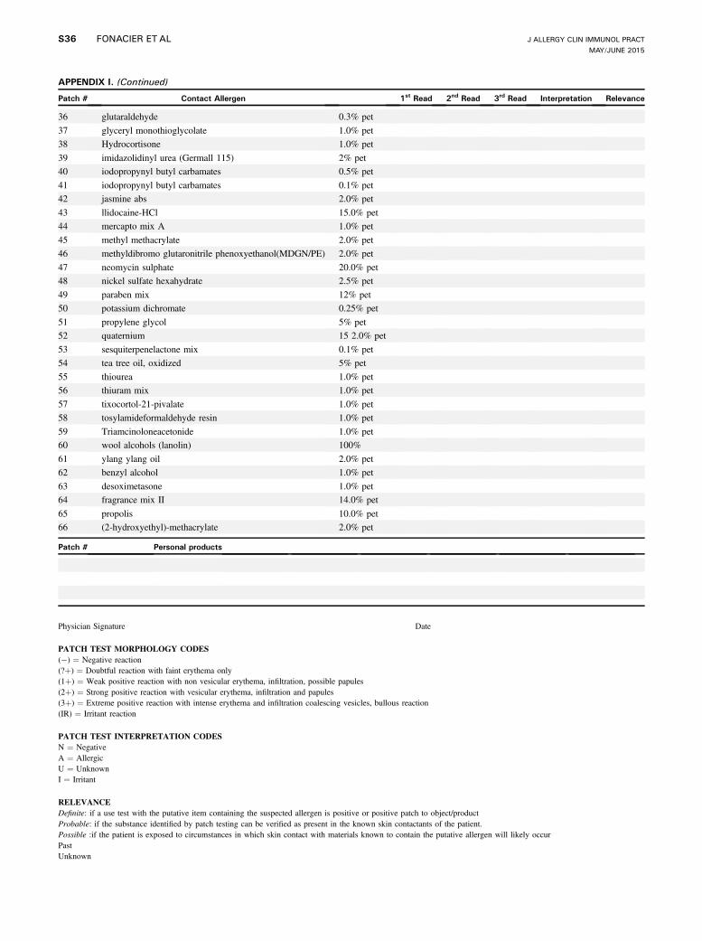
APPENDIX I. (Continued)
Patch # Contact Allergen 1stRead 2
ndRead 3
rdRead Interpretation Relevance
36 glutaraldehyde 0.3% pet
37 glyceryl monothioglycolate 1.0% pet
38 Hydrocortisone 1.0% pet
39 imidazolidinyl urea (Germall 115) 2% pet
40 iodopropynyl butyl carbamates 0.5% pet
41 iodopropynyl butyl carbamates 0.1% pet
42 jasmine abs 2.0% pet
43 llidocaine-HCl 15.0% pet
44 mercapto mix A 1.0% pet
45 methyl methacrylate 2.0% pet
46 methyldibromo glutaronitrile phenoxyethanol(MDGN/PE) 2.0% pet
47 neomycin sulphate 20.0% pet
48 nickel sulfate hexahydrate 2.5% pet
49 paraben mix 12% pet
50 potassium dichromate 0.25% pet
51 propylene glycol 5% pet
52 quaternium 15 2.0% pet
53 sesquiterpenelactone mix 0.1% pet
54 tea tree oil, oxidized 5% pet
55 thiourea 1.0% pet
56 thiuram mix 1.0% pet
57 tixocortol-21-pivalate 1.0% pet
58 tosylamideformaldehyde resin 1.0% pet
59 Triamcinoloneacetonide 1.0% pet
60 wool alcohols (lanolin) 100%
61 ylang ylang oil 2.0% pet
62 benzyl alcohol 1.0% pet
63 desoximetasone 1.0% pet
64 fragrance mix II 14.0% pet
65 propolis 10.0% pet
66 (2-hydroxyethyl)-methacrylate 2.0% pet
Patch # Personal products
Physician Signature Date
PATCH TEST MORPHOLOGY CODES(�) ¼ Negative reaction(?þ) ¼ Doubtful reaction with faint erythema only(1þ) ¼ Weak positive reaction with non vesicular erythema, infiltration, possible papules(2þ) ¼ Strong positive reaction with vesicular erythema, infiltration and papules(3þ) ¼ Extreme positive reaction with intense erythema and infiltration coalescing vesicles, bullous reaction(IR) ¼ Irritant reaction
PATCH TEST INTERPRETATION CODESN ¼ NegativeA ¼ AllergicU ¼ UnknownI ¼ Irritant
RELEVANCEDefinite: if a use test with the putative item containing the suspected allergen is positive or positive patch to object/productProbable: if the substance identified by patch testing can be verified as present in the known skin contactants of the patient.Possible :if the patient is exposed to circumstances in which skin contact with materials known to contain the putative allergen will likely occurPastUnknown
J ALLERGY CLIN IMMUNOL PRACTMAY/JUNE 2015
S36 FONACIER ETAL

APPENDIX J. STRUCTURAL GROUPS OF CORTICOSTEROIDS AND POTENCY CLASSIFICATION WITH
EXAMPLES OF COMMERCIALLY AVAILABLE PREPARATIONS
Steroid group
A: Hydrocortisone
Hydrocortisone &
tixocortol pivalate:
has C17 or C21 short
chain ester
B: TCL acetonide
Acetonides: has C16 C17
cis-ketal or -diol additions
C: BTM
nonesterified
Betamethasone:
has C16 methyl group
D1: BTM-dipropionate
has C16 methyl group &
halogenated B ring
D2: MPL aceponate
labile esters w/o C16
methyl nor B ring
halogen substitution
Prevalence 2.7% 1.5% <0.2% 0.8% 0.8%
Class 7: LeastPotent
HC[Hytone C/L (1%/
2.5%)][Cortaid, C/O/Sp][Egocort C 1%]HC Acetate[Cortisone, Lanacort,
Wellcortin, Gynecort,Lanacort]
Class 6: Low Desonide[DesOwen (0.05%) C/L]FLU acetonide[Capex Sh, Dermasmooth
F/S/ oil (0.01%)]TCL acetonide[Aristocort A C, Kenalog L
(0.025%)]
Aclometasonedipropionate
[Aclovate C/O (0.05%)]
Class 5: LowerMid
Desonide[Tridesilon, DesOwen O
(0.05%)]FLU acetonide[Synalar, Synemol C
(0.025%)]Flurandrenolide[Cordran C/L/Tape
(0.05%)]
Fluticasonepropionate
[Cutivate C (0.05%)]
HC-17-valerate[Westcort C (0.2%)]Prednicarbate[DermAtop C (0.1%)]
Class 4: Mid FLU acetonide[Synalar, Synemol
(0.01%-0.2%)]Flurandrenolide[CordranO (0.05%)]TCL acetonide[Kenalog, Aristocort A O
(0.1%)]
Desoximetasone[Topicort LP C/O(0.05%)]
Mometasone Furorate[Elocon C/L (0.1%)]
HC 17-butyrate[Locoid C/L/O (0.1%)]HC-17-valerate[Westcort O (0.2%)]
Class 3: UpperMid
TCL acetonide[Kenalog, Aristocort C
(0.5%)]Triamcinolone Diacetate[Amcort, Aristocort C/O
(0.025%-0.1%)]
Halometasone[Halometasone (0.05%)]
BTM 17 valerate[Luxiq F (0.12%)][Valisone O (0.1%)]BTM dipropionate[Diprosone C (0.05%)]Clobetasone 17butyrate
[Eumovate C (0.05%)]Fluticasonepropionate
[Cutivate O (0.005%)]Mometasone Furorate[Elocon O (0.1%)]
(Continued)
J ALLERGY CLIN IMMUNOL PRACTVOLUME 3, NUMBER 3S
FONACIER ETAL S37

APPENDIX J. (Continued)
Steroid group
A: Hydrocortisone
Hydrocortisone &
tixocortol pivalate:
has C17 or C21 short
chain ester
B: TCL acetonide
Acetonides: has C16 C17
cis-ketal or -diol additions
C: BTM
nonesterified
Betamethasone:
has C16 methyl group
D1: BTM-dipropionate
has C16 methyl group &
halogenated B ring
D2: MPL aceponate
labile esters w/o C16
methyl nor B ring
halogen substitution
Class 2: High Amcinonide[Cyclocort O/L/C (0.05%-
0.1%)]Fluocinonide[Lidex C/G/O/S (0.05%)]Halcinonide[Halog C/O/S (0.05%-
0.1%)]
Desoximetasone[Topicort C/O (0.25%)][Topicort G (0.05%)]Clocortolone[Cloderm C ( 0.1%)]
BTM 17 valerate[Betnovate C/O (0.1%)]BTM dipropionate[Diprolene AF C(0.05%)]
[Diprosone O (0.05%)]
Class 1: Super Fluocinonide[Vanos C (0.1%)]
BTM dipropionate[Diprolene O/G/L(0.05%)]
Clobetasol propionate[Clobex L/spray/sh,Dermovate C/O,Olux F, Temovate C/O/S/G (0.05%)]
[Olux F (0.05%)]Diflorasone Diacetate[ApexiCon, Psorcon C/O, Florone O(0.05%)]
Halobetasol[Ultravate C/O(0.05%)]
Oral/SystemicPreparation
Cortisone acetateHC-21-acetateMPL acetate[Medrol]intra-articular,
intralesional,intrasynovial
Prednisone[Cortan, Deltasone,
Meticorten, Orasone]Prednisoloneacetate[Prediapred, Prelone
syrup]Cloprednol
FludrocortisoneAcetate
[Florinef]HC sodium[Solucortef]
BudesonideTCM[Atolone Tablets (I)]TCM benetonideTCM diacetateTCM hexacetonide
BTM sodium phosphateInjectable Suspension[Celestone]Dexamethasone acetateDexamethasone-sodiumphosphate Injection
[Decadron]Paramethasone acetate
BTM Oral/IM BTMdipropionate
HC Oral/IV
Cross Reactions Cross reacts with D2 Budesonide specificallycross-reacts with D2
Cross reacts with ClassA & Budesonide
Patch TestSubstance
Tixocortol 21-pivalate BudesonideTCL acetonide
Clobetasole-17-propionate
HC-17-butyrate
(Continued)
J ALLERGY CLIN IMMUNOL PRACTMAY/JUNE 2015
S38 FONACIER ETAL
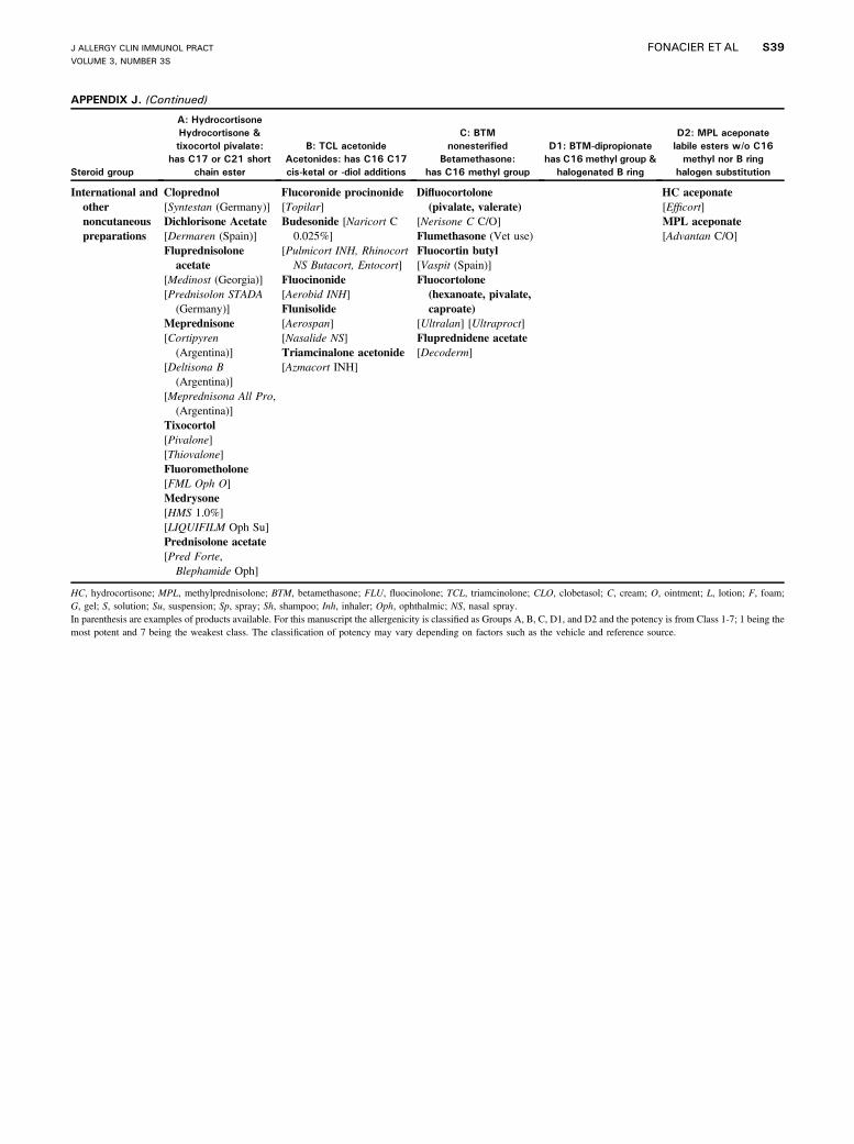
APPENDIX J. (Continued)
Steroid group
A: Hydrocortisone
Hydrocortisone &
tixocortol pivalate:
has C17 or C21 short
chain ester
B: TCL acetonide
Acetonides: has C16 C17
cis-ketal or -diol additions
C: BTM
nonesterified
Betamethasone:
has C16 methyl group
D1: BTM-dipropionate
has C16 methyl group &
halogenated B ring
D2: MPL aceponate
labile esters w/o C16
methyl nor B ring
halogen substitution
International andothernoncutaneouspreparations
Cloprednol[Syntestan (Germany)]Dichlorisone Acetate[Dermaren (Spain)]Fluprednisolone
acetate[Medinost (Georgia)][Prednisolon STADA
(Germany)]Meprednisone[Cortipyren
(Argentina)][Deltisona B
(Argentina)][Meprednisona All Pro,
(Argentina)]Tixocortol[Pivalone][Thiovalone]Fluorometholone[FML Oph O]Medrysone[HMS 1.0%][LIQUIFILM Oph Su]Prednisolone acetate[Pred Forte,
Blephamide Oph]
Flucoronide procinonide[Topilar]Budesonide [Naricort C
0.025%][Pulmicort INH, Rhinocort
NS Butacort, Entocort]Fluocinonide[Aerobid INH]Flunisolide[Aerospan][Nasalide NS]Triamcinalone acetonide[Azmacort INH]
Difluocortolone(pivalate, valerate)
[Nerisone C C/O]Flumethasone (Vet use)Fluocortin butyl[Vaspit (Spain)]Fluocortolone(hexanoate, pivalate,caproate)
[Ultralan] [Ultraproct]Fluprednidene acetate[Decoderm]
HC aceponate[Efficort]MPL aceponate[Advantan C/O]
HC, hydrocortisone; MPL, methylprednisolone; BTM, betamethasone; FLU, fluocinolone; TCL, triamcinolone; CLO, clobetasol; C, cream; O, ointment; L, lotion; F, foam;G, gel; S, solution; Su, suspension; Sp, spray; Sh, shampoo; Inh, inhaler; Oph, ophthalmic; NS, nasal spray.In parenthesis are examples of products available. For this manuscript the allergenicity is classified as Groups A, B, C, D1, and D2 and the potency is from Class 1-7; 1 being themost potent and 7 being the weakest class. The classification of potency may vary depending on factors such as the vehicle and reference source.
J ALLERGY CLIN IMMUNOL PRACTVOLUME 3, NUMBER 3S
FONACIER ETAL S39