Embed Size (px)
Citation preview

Polymyxins: Antibacterial Activity,Susceptibility Testing, and ResistanceMechanisms Encoded by Plasmids orChromosomesLaurent Poirel,a,b,c Aurélie Jayol,a,b,c Patrice Nordmanna,b,c,d
Emerging Antibiotic Resistance Unit, Medical and Molecular Microbiology, Department of Medicine, Universityof Fribourg, Fribourg, Switzerlanda; French INSERM European Unit, University of Fribourg (LEA-IAME), Fribourg,Switzerlandb; National Reference Center for Emerging Antibiotic Resistance, Fribourg, Switzerlandc; Universityof Lausanne and University Hospital Center, Lausanne, Switzerlandd
SUMMARY . . . . . . . . . . . . . . . . . . . . . . . . . . . . . . . . . . . . . . . . . . . . . . . . . . . . . . . . . . . . . . . . . . . . . . . . . . . . . . . . . . . . . . 558INTRODUCTION . . . . . . . . . . . . . . . . . . . . . . . . . . . . . . . . . . . . . . . . . . . . . . . . . . . . . . . . . . . . . . . . . . . . . . . . . . . . . . . . 558
Structure. . . . . . . . . . . . . . . . . . . . . . . . . . . . . . . . . . . . . . . . . . . . . . . . . . . . . . . . . . . . . . . . . . . . . . . . . . . . . . . . . . . . . . 559Mechanism of Action. . . . . . . . . . . . . . . . . . . . . . . . . . . . . . . . . . . . . . . . . . . . . . . . . . . . . . . . . . . . . . . . . . . . . . . . 559Spectrum of Activity. . . . . . . . . . . . . . . . . . . . . . . . . . . . . . . . . . . . . . . . . . . . . . . . . . . . . . . . . . . . . . . . . . . . . . . . . 560Pharmacodynamics . . . . . . . . . . . . . . . . . . . . . . . . . . . . . . . . . . . . . . . . . . . . . . . . . . . . . . . . . . . . . . . . . . . . . . . . . . 560
USE OF COLISTIN IN HUMAN AND VETERINARY MEDICINE. . . . . . . . . . . . . . . . . . . . . . . . . . . . . 560Use in Human Medicine. . . . . . . . . . . . . . . . . . . . . . . . . . . . . . . . . . . . . . . . . . . . . . . . . . . . . . . . . . . . . . . . . . . . . 560
Commercial formulations. . . . . . . . . . . . . . . . . . . . . . . . . . . . . . . . . . . . . . . . . . . . . . . . . . . . . . . . . . . . . . . . . 561Routes of administration . . . . . . . . . . . . . . . . . . . . . . . . . . . . . . . . . . . . . . . . . . . . . . . . . . . . . . . . . . . . . . . . . 561Pharmacokinetics. . . . . . . . . . . . . . . . . . . . . . . . . . . . . . . . . . . . . . . . . . . . . . . . . . . . . . . . . . . . . . . . . . . . . . . . . . 561Dosing regimen . . . . . . . . . . . . . . . . . . . . . . . . . . . . . . . . . . . . . . . . . . . . . . . . . . . . . . . . . . . . . . . . . . . . . . . . . . . 562
(i) Patients with normal renal function . . . . . . . . . . . . . . . . . . . . . . . . . . . . . . . . . . . . . . . . . . . . . . . 562(ii) Patients with renal insufficiency . . . . . . . . . . . . . . . . . . . . . . . . . . . . . . . . . . . . . . . . . . . . . . . . . . . 563
Toxicity. . . . . . . . . . . . . . . . . . . . . . . . . . . . . . . . . . . . . . . . . . . . . . . . . . . . . . . . . . . . . . . . . . . . . . . . . . . . . . . . . . . . . 564Use in Veterinary Medicine . . . . . . . . . . . . . . . . . . . . . . . . . . . . . . . . . . . . . . . . . . . . . . . . . . . . . . . . . . . . . . . . . 564
METHODS FOR SUSCEPTIBILITY TESTING. . . . . . . . . . . . . . . . . . . . . . . . . . . . . . . . . . . . . . . . . . . . . . . . . . 565Dilution Methods . . . . . . . . . . . . . . . . . . . . . . . . . . . . . . . . . . . . . . . . . . . . . . . . . . . . . . . . . . . . . . . . . . . . . . . . . . . . 565
Broth dilution methods. . . . . . . . . . . . . . . . . . . . . . . . . . . . . . . . . . . . . . . . . . . . . . . . . . . . . . . . . . . . . . . . . . . 565(i) Broth microdilution method. . . . . . . . . . . . . . . . . . . . . . . . . . . . . . . . . . . . . . . . . . . . . . . . . . . . . . . . 565(ii) Broth macrodilution method (or tube dilution method) . . . . . . . . . . . . . . . . . . . . . . . . 565
Agar dilution method. . . . . . . . . . . . . . . . . . . . . . . . . . . . . . . . . . . . . . . . . . . . . . . . . . . . . . . . . . . . . . . . . . . . . 566Routine Susceptibility Testing Methods. . . . . . . . . . . . . . . . . . . . . . . . . . . . . . . . . . . . . . . . . . . . . . . . . . . . 566
Nonautomatic systems . . . . . . . . . . . . . . . . . . . . . . . . . . . . . . . . . . . . . . . . . . . . . . . . . . . . . . . . . . . . . . . . . . . 566(i) DD test (Kirby-Bauer procedure) . . . . . . . . . . . . . . . . . . . . . . . . . . . . . . . . . . . . . . . . . . . . . . . . . . . 566(ii) Etest strips. . . . . . . . . . . . . . . . . . . . . . . . . . . . . . . . . . . . . . . . . . . . . . . . . . . . . . . . . . . . . . . . . . . . . . . . . . . 567(iii) UMIC system . . . . . . . . . . . . . . . . . . . . . . . . . . . . . . . . . . . . . . . . . . . . . . . . . . . . . . . . . . . . . . . . . . . . . . . 567
Automatic systems. . . . . . . . . . . . . . . . . . . . . . . . . . . . . . . . . . . . . . . . . . . . . . . . . . . . . . . . . . . . . . . . . . . . . . . . 567(i) Vitek 2 system . . . . . . . . . . . . . . . . . . . . . . . . . . . . . . . . . . . . . . . . . . . . . . . . . . . . . . . . . . . . . . . . . . . . . . . 567(ii) Phoenix automated microbiology system . . . . . . . . . . . . . . . . . . . . . . . . . . . . . . . . . . . . . . . . 567(iii) MicroScan system . . . . . . . . . . . . . . . . . . . . . . . . . . . . . . . . . . . . . . . . . . . . . . . . . . . . . . . . . . . . . . . . . . 568(iv) Sensititre system . . . . . . . . . . . . . . . . . . . . . . . . . . . . . . . . . . . . . . . . . . . . . . . . . . . . . . . . . . . . . . . . . . . 568
Impact of Materials on MIC Determination . . . . . . . . . . . . . . . . . . . . . . . . . . . . . . . . . . . . . . . . . . . . . . . . 568Impact of medium . . . . . . . . . . . . . . . . . . . . . . . . . . . . . . . . . . . . . . . . . . . . . . . . . . . . . . . . . . . . . . . . . . . . . . . . 568Impact of powder composition . . . . . . . . . . . . . . . . . . . . . . . . . . . . . . . . . . . . . . . . . . . . . . . . . . . . . . . . . . 568Impact of the composition and treatment of plates . . . . . . . . . . . . . . . . . . . . . . . . . . . . . . . . . . . 568Presence or absence of P-80 . . . . . . . . . . . . . . . . . . . . . . . . . . . . . . . . . . . . . . . . . . . . . . . . . . . . . . . . . . . . . 569
Impacts of Subcultures and Storage on MICs . . . . . . . . . . . . . . . . . . . . . . . . . . . . . . . . . . . . . . . . . . . . . 569Impact of subcultures . . . . . . . . . . . . . . . . . . . . . . . . . . . . . . . . . . . . . . . . . . . . . . . . . . . . . . . . . . . . . . . . . . . . 569Impact of storage . . . . . . . . . . . . . . . . . . . . . . . . . . . . . . . . . . . . . . . . . . . . . . . . . . . . . . . . . . . . . . . . . . . . . . . . . 569
Interpretive Criteria . . . . . . . . . . . . . . . . . . . . . . . . . . . . . . . . . . . . . . . . . . . . . . . . . . . . . . . . . . . . . . . . . . . . . . . . . . 569Quality Controls. . . . . . . . . . . . . . . . . . . . . . . . . . . . . . . . . . . . . . . . . . . . . . . . . . . . . . . . . . . . . . . . . . . . . . . . . . . . . . 569Correlation between MICs of Colistin and Polymyxin B . . . . . . . . . . . . . . . . . . . . . . . . . . . . . . . . . . 570Qualitative Detection Techniques . . . . . . . . . . . . . . . . . . . . . . . . . . . . . . . . . . . . . . . . . . . . . . . . . . . . . . . . . . 570
(continued)
Published 8 March 2017
Citation Poirel L, Jayol A, Nordmann P. 2017.Polymyxins: antibacterial activity, susceptibilitytesting, and resistance mechanisms encodedby plasmids or chromosomes. Clin MicrobiolRev 30:557–596. https://doi.org/10.1128/CMR.00064-16.
Copyright © 2017 American Society forMicrobiology. All Rights Reserved.
Address correspondence to Laurent Poirel,[email protected].
REVIEW
crossm
April 2017 Volume 30 Issue 2 cmr.asm.org 557Clinical Microbiology Reviews
on May 11, 2020 by guest
http://cmr.asm
.org/D
ownloaded from

Rapid detection of heterogeneous populations among colistin-resistant Gram-negative bacteria by use of capillary electrophoresis . . . . . . . . . . . . . . . . . . . . . . . . . . . . . . . 570
Rapid detection of colistin-resistant A. baumannii isolates by use of the Micromaxassay. . . . . . . . . . . . . . . . . . . . . . . . . . . . . . . . . . . . . . . . . . . . . . . . . . . . . . . . . . . . . . . . . . . . . . . . . . . . . . . . . . . . . 570
Rapid detection of colistin-resistant Enterobacteriaceae isolates by use of the RapidPolymyxin NP test . . . . . . . . . . . . . . . . . . . . . . . . . . . . . . . . . . . . . . . . . . . . . . . . . . . . . . . . . . . . . . . . . . . . . . 571
Selective medium . . . . . . . . . . . . . . . . . . . . . . . . . . . . . . . . . . . . . . . . . . . . . . . . . . . . . . . . . . . . . . . . . . . . . . . . . 571Genotypic Methods. . . . . . . . . . . . . . . . . . . . . . . . . . . . . . . . . . . . . . . . . . . . . . . . . . . . . . . . . . . . . . . . . . . . . . . . . . 571
RESISTANCE MECHANISMS IN ENTEROBACTERIACEAE . . . . . . . . . . . . . . . . . . . . . . . . . . . . . . . . . . . 572Intrinsic Resistance Mechanisms in Proteus mirabilis and Serratia marcescens . . . . . . . . . . 572Mechanisms Responsible for Acquired Resistance in Enterobacteriaceae . . . . . . . . . . . . . . . 572
Genes encoding LPS-modifying enzymes . . . . . . . . . . . . . . . . . . . . . . . . . . . . . . . . . . . . . . . . . . . . . . . 573(i) The pmrC gene . . . . . . . . . . . . . . . . . . . . . . . . . . . . . . . . . . . . . . . . . . . . . . . . . . . . . . . . . . . . . . . . . . . . . . 573(ii) The pmrHFIJKLM operon and the pmrE gene . . . . . . . . . . . . . . . . . . . . . . . . . . . . . . . . . . . . 573(iii) The pmrA and pmrB genes, which encode the PmrAB two-component system. . 573(iv) The phoP and phoQ genes, which encode the PhoPQ two-component system. . 574
Regulators of the PmrAB and PhoPQ Two-Component Systems . . . . . . . . . . . . . . . . . . . . . . . . 574The mgrB gene . . . . . . . . . . . . . . . . . . . . . . . . . . . . . . . . . . . . . . . . . . . . . . . . . . . . . . . . . . . . . . . . . . . . . . . . . . . . 574The crrAB operon . . . . . . . . . . . . . . . . . . . . . . . . . . . . . . . . . . . . . . . . . . . . . . . . . . . . . . . . . . . . . . . . . . . . . . . . . 574
The Intrinsic Regulator RamA. . . . . . . . . . . . . . . . . . . . . . . . . . . . . . . . . . . . . . . . . . . . . . . . . . . . . . . . . . . . . . . 578Plasmid-Mediated Resistance to Polymyxins . . . . . . . . . . . . . . . . . . . . . . . . . . . . . . . . . . . . . . . . . . . . . . 578Other Mechanisms Contributing to Polymyxin Resistance in Enterobacteriaceae. . . . . . . 580
Hyperproduction of CPS. . . . . . . . . . . . . . . . . . . . . . . . . . . . . . . . . . . . . . . . . . . . . . . . . . . . . . . . . . . . . . . . . . 580Role of porins . . . . . . . . . . . . . . . . . . . . . . . . . . . . . . . . . . . . . . . . . . . . . . . . . . . . . . . . . . . . . . . . . . . . . . . . . . . . . 583Role of efflux pumps . . . . . . . . . . . . . . . . . . . . . . . . . . . . . . . . . . . . . . . . . . . . . . . . . . . . . . . . . . . . . . . . . . . . . 583
Mechanisms of Polymyxin Resistance in Pseudomonas aeruginosa and Acinetobacterbaumannii . . . . . . . . . . . . . . . . . . . . . . . . . . . . . . . . . . . . . . . . . . . . . . . . . . . . . . . . . . . . . . . . . . . . . . . . . . . . . . . . . 583Pseudomonas aeruginosa . . . . . . . . . . . . . . . . . . . . . . . . . . . . . . . . . . . . . . . . . . . . . . . . . . . . . . . . . . . . . . . . . 583Acinetobacter baumannii. . . . . . . . . . . . . . . . . . . . . . . . . . . . . . . . . . . . . . . . . . . . . . . . . . . . . . . . . . . . . . . . . . 583
EPIDEMIOLOGY OF RESISTANCE TO POLYMYXINS . . . . . . . . . . . . . . . . . . . . . . . . . . . . . . . . . . . . . . . 584General Epidemiology of Resistance to Polymyxins . . . . . . . . . . . . . . . . . . . . . . . . . . . . . . . . . . . . . . 584
North America. . . . . . . . . . . . . . . . . . . . . . . . . . . . . . . . . . . . . . . . . . . . . . . . . . . . . . . . . . . . . . . . . . . . . . . . . . . . . 584South America. . . . . . . . . . . . . . . . . . . . . . . . . . . . . . . . . . . . . . . . . . . . . . . . . . . . . . . . . . . . . . . . . . . . . . . . . . . . . 584Europe . . . . . . . . . . . . . . . . . . . . . . . . . . . . . . . . . . . . . . . . . . . . . . . . . . . . . . . . . . . . . . . . . . . . . . . . . . . . . . . . . . . . . 584Middle East . . . . . . . . . . . . . . . . . . . . . . . . . . . . . . . . . . . . . . . . . . . . . . . . . . . . . . . . . . . . . . . . . . . . . . . . . . . . . . . . 587Africa . . . . . . . . . . . . . . . . . . . . . . . . . . . . . . . . . . . . . . . . . . . . . . . . . . . . . . . . . . . . . . . . . . . . . . . . . . . . . . . . . . . . . . . 587Asia. . . . . . . . . . . . . . . . . . . . . . . . . . . . . . . . . . . . . . . . . . . . . . . . . . . . . . . . . . . . . . . . . . . . . . . . . . . . . . . . . . . . . . . . . 587
Risk Factors. . . . . . . . . . . . . . . . . . . . . . . . . . . . . . . . . . . . . . . . . . . . . . . . . . . . . . . . . . . . . . . . . . . . . . . . . . . . . . . . . . . 587Specific Epidemiology of the Plasmid-Mediated mcr-1 Resistance Gene. . . . . . . . . . . . . . . . 588
CONCLUSIONS . . . . . . . . . . . . . . . . . . . . . . . . . . . . . . . . . . . . . . . . . . . . . . . . . . . . . . . . . . . . . . . . . . . . . . . . . . . . . . . . . 588ACKNOWLEDGMENTS. . . . . . . . . . . . . . . . . . . . . . . . . . . . . . . . . . . . . . . . . . . . . . . . . . . . . . . . . . . . . . . . . . . . . . . . . 589REFERENCES . . . . . . . . . . . . . . . . . . . . . . . . . . . . . . . . . . . . . . . . . . . . . . . . . . . . . . . . . . . . . . . . . . . . . . . . . . . . . . . . . . . 589AUTHOR BIOS. . . . . . . . . . . . . . . . . . . . . . . . . . . . . . . . . . . . . . . . . . . . . . . . . . . . . . . . . . . . . . . . . . . . . . . . . . . . . . . . . . 596
SUMMARY Polymyxins are well-established antibiotics that have recently regainedsignificant interest as a consequence of the increasing incidence of infections due tomultidrug-resistant Gram-negative bacteria. Colistin and polymyxin B are being seri-ously reconsidered as last-resort antibiotics in many areas where multidrug resis-tance is observed in clinical medicine. In parallel, the heavy use of polymyxins inveterinary medicine is currently being reconsidered due to increased reports ofpolymyxin-resistant bacteria. Susceptibility testing is challenging with polymyxins,and currently available techniques are presented here. Genotypic and phenotypic meth-ods that provide relevant information for diagnostic laboratories are presented. This re-view also presents recent works in relation to recently identified mechanisms of poly-myxin resistance, including chromosomally encoded resistance traits as well as therecently identified plasmid-encoded polymyxin resistance determinant MCR-1. Epidemio-logical features summarizing the current knowledge in that field are presented.
KEYWORDS Gram-negative bacteria, MCR-1, lipopolysaccharide, polymyxins, toxicity
INTRODUCTION
Colistin (also known as polymyxin E) is a polypeptide antibiotic that was originallyisolated in 1947 from the soil bacterium Paenibacillus polymyxa subsp. colistinus (1).
Colistin and polymyxin B belong to the class of polymyxins, which is one of the primaryclasses of antibiotics with activity against most Gram-negative bacteria.
Poirel et al. Clinical Microbiology Reviews
April 2017 Volume 30 Issue 2 cmr.asm.org 558
on May 11, 2020 by guest
http://cmr.asm
.org/D
ownloaded from

Structure
The chemical structure of polymyxins is similar to that of cationic antimicrobialpeptides (CAMPs) (defensins and gramicidins), which represent the first line of defenseagainst bacterial colonization in eukaryotic cells (2). Polymyxins are cationic polypep-tides that consist of a cyclic heptapeptide possessing a tripeptide side chain acylatedat the N terminus by a fatty acid tail (3, 4) (Fig. 1). The inherent toxicity of colistin maybe explained by the hydrophobic properties of the N-terminal fatty acyl segment,which also accounts significantly for its antimicrobial activity, and also by positions 6and 7, which are very important (5, 6).
Colistin and polymyxin B differ by only a single amino acid in the peptide ring, witha phenylalanine in polymyxin B and a leucine in colistin (Fig. 1) (7). Polymyxin B isadministered directly as an active antibiotic, whereas colistin is administered as aninactive prodrug, colistin methanesulfonate (also known as colistimethate [CMS]) (Fig.1) (7).
The terms “colistin” and “colistimethate” are not interchangeable, since they corre-spond to different forms of colistin available for clinical use (4). Indeed, colistimethatesodium is a polyanionic inactive prodrug that is less toxic than colistin sulfate (Fig. 1)(4, 8). Colistimethate is formed by the reaction of colistin with formaldehyde andsodium bisulfite (9). This prodrug is transformed in aqueous media, and also in vivo inbiological fluids, and is converted into colistin and several inactive methanesulfonatedcompounds (10, 11).
Mechanism of Action
The target of polymyxins is the outer membrane of Gram-negative bacteria. Becauseof an electrostatic interaction occurring between the �,�-diaminobutyric acid (Dab)residue of the positively charged polymyxin on one side and the phosphate groups ofthe negatively charged lipid A membrane on the other side, divalent cations (Ca2� andMg2�) are displaced from the negatively charged phosphate groups of membranelipids (12). The lipopolysaccharide (LPS) is therefore destabilized, consequently increas-ing the permeability of the bacterial membrane, leading to leakage of the cytoplasmic
FIG 1 Structures of colistin A and B, colistimethate A and B, and polymyxin B1 and B2.
Polymyxin Activity, Susceptibility, and Resistance Clinical Microbiology Reviews
April 2017 Volume 30 Issue 2 cmr.asm.org 559
on May 11, 2020 by guest
http://cmr.asm
.org/D
ownloaded from

content and ultimately causing cell death (4, 13). Note that even though the LPS is theinitial target, the exact mode of action of polymyxins still remains unclear.
Another antibacterial mechanism is the endotoxin effect. The endotoxin of Gram-negative pathogens corresponds to the lipid A portion of the LPS; polymyxins have theability to bind to and neutralize this LPS molecule released during cell lysis (14).
Finally, another mode of action of the polymyxins is the inhibition of vital respiratoryenzymes (inhibition of type II NADH-quinone oxidoreductases [NDH-2]) in the bacterialinner membrane (15).
Spectrum of Activity
Polymyxins have a narrow antibacterial spectrum, mainly against common Gram-negative bacteria. They are active against most members of the Enterobacteriaceaefamily, including Escherichia coli, Enterobacter spp., Klebsiella spp., Citrobacter spp.,Salmonella spp., and Shigella spp. Polymyxins also have significant activity againstcommon nonfermentative Gram-negative bacteria, including Acinetobacter baumannii,Pseudomonas aeruginosa, and Stenotrophomonas maltophilia (13).
Conversely, some species are naturally resistant to polymyxins, including Proteusspp., Morganella morganii, Providencia spp., Serratia marcescens, Pseudomonas mallei,Burkholderia cepacia, Chromobacterium spp., Edwardsiella spp., Brucella, Legionella, Cam-pylobacter, and Vibrio cholerae. Polymyxins are not active against Gram-negative cocci(Neisseria spp.), Gram-positive bacteria, and anaerobic bacteria (13).
Pharmacodynamics
The antibacterial effect of colistin is concentration dependent (4, 16–18). Thepharmacokinetic-pharmacodynamic (PK-PD) index that best predicts the antibacterialactivity against A. baumannii and P. aeruginosa is the ratio of the area under theconcentration-time curve for free drug from 0 to 24 h to the MIC (fAUC0 –24/MIC), withthis index being superior to the maximum concentration of drug in serum (Cmax)/MICrelationship, suggesting that time-averaged exposure to colistin is more important thanthe achievement of high peak concentrations (19–21). An average steady-state plasmacolistin concentration of 2 �g/ml has been suggested as a reasonable target value forisolates with MICs of �1 �g/ml, maximizing the antimicrobial activity while minimizingthe risk of nephrotoxicity (22). An inadequate AUC/MIC ratio likely leads to treatmentfailure. The colistin antibacterial effect is extremely rapid, occurring as early as 5 minafter exposure (17, 18, 23, 24).
A postantibiotic effect was observed against Klebsiella pneumoniae, P. aeruginosa,and A. baumannii (25). However, it is important to highlight that polymyxins haveminimal postantibiotic effects at clinically relevant concentrations. Despite the majorinitial killing rate observed against colistin-susceptible strains exposed to colistin alone,regrowth has been reported for A. baumannii (17) and K. pneumoniae (18) in statictime-kill studies. Colistin heteroresistance, a phenomenon corresponding to the emer-gence of a colistin-resistant subpopulation (that can grow in the presence of �4 �g/mlof colistin) within a susceptible population (i.e., with a MIC of �2 �g/ml), has beenobserved for A. baumannii (26, 27), K. pneumoniae (18, 28), and P. aeruginosa (23).
USE OF COLISTIN IN HUMAN AND VETERINARY MEDICINEUse in Human Medicine
After its discovery in 1947, colistin was used in Japan and Europe during the 1950s(29). Then, after its approval by the U.S. FDA in 1959, colistimethate (CMS), the inactiveprodrug of colistin, replaced colistin for parenteral administration (29). Colistin and CMShave been used widely for decades for treatment of infections caused by Gram-negative bacteria. However, in the 1970s, because of their toxicity, especially nephro-toxicity (30), their use was reconsidered. They were then replaced by novel, more activeand less toxic antibiotics, such as aminoglycosides, quinolones, and �-lactams. For 20years, the use of colistin was restricted to ophthalmic and topical uses. Systemic ornebulized colistin was used only for cystic fibrosis patients.
Poirel et al. Clinical Microbiology Reviews
April 2017 Volume 30 Issue 2 cmr.asm.org 560
on May 11, 2020 by guest
http://cmr.asm
.org/D
ownloaded from

However, the increasing prevalence of multidrug-resistant (MDR) Gram-negativebacteria (31), particularly K. pneumoniae, A. baumannii, and P. aeruginosa, has forcedphysicians to reintroduce systemic polymyxin as a valuable therapeutic option (4,13, 32).
Considering the paucity of novel antibiotics, colistin is currently often the onlyeffective antibiotic agent against MDR organisms, particularly carbapenemase-pro-ducing bacteria.
Commercial formulations. There are more than 30 polymyxin molecules, amongwhich there are five main chemical compounds (polymyxins A to E), each containingmultiple components. Although colistin (polymyxin E) and polymyxin B are both usedin clinical practice (33), colistin is the most widely used polymyxin (23). The two mostcommon commercially available parenteral formulations of the colistin prodrug, CMS,are Colomycin (Forest Laboratories UK Limited, Dartford, United Kingdom), primarilyemployed in Europe, and Coly-Mycin M Parenteral (Monarch Pharmaceuticals, Inc.,Bristol, TN), primilarily employed in the United States (34). Unfortunately, the vials ofboth formulations contain different dry powder quantities, and the two products aredifferently labeled, with Colomycin being labeled in international units (IU) of CMS andColy-Mycin M Parenteral being labeled in milligrams of CMS or colistin base activity(CBA) (34). The conversion is as follows: 1 million units (MU) CMS � 80 mg CMS � 30mg CBA (35). To add to the confusion, some other brands corresponding to genericproducts are now available (36). The multiplicity of terms used to express contents ofvials and dose regimens unfortunately creates confusion and does not allow anymeaningful comparison of data collected from studies performed in different parts ofthe world.
Routes of administration. Colistin sulfate can be administered orally as tablets andsyrup for selective digestive tract decontamination (no absorption) and topically for thetreatment of bacterial skin infections (13). CMS, the less toxic prodrug, has differentadministration routes, i.e., parenteral (including intravenous) and intramuscular, butintrathecal or intraventricular administration is also possible (13). The intramuscularinjection is rarely used in clinical practice because it may be very painful locally, andalso because its absorption is variable (33). Both colistin sulfate and CMS can bedelivered through inhalation by aerosol therapy, but there is a higher frequency ofbronchoconstriction with colistin sulfate (33). Delivery of CMS by inhalation and by theintrathecal and intraventricular routes allows much higher concentrations in lung fluidand cerebrospinal fluid, respectively, than those seen with systemic administration.Moreover, those routes of administration lead to negligible plasma exposure and areless toxic (in particularly less nephrotoxic) (22).
In aqueous solutions, colistimethate sodium is transformed into colistin; therefore, itshould be administered shortly after reconstitution to avoid the toxicity associated withcolistin (37).
Pharmacokinetics. Because of their discovery and their introduction into clinical usemore than 50 years ago, polymyxins were never subjected to the drug developmentapproval process currently required by international drug regulatory authorities. Con-sequently, the PK and PD data on the rational use of polymyxins (maximizing antibac-terial activity and minimizing toxicity and development of resistance) were not avail-able until recently. The fact that, until recently, plasma concentrations of CMS andformed colistin could not be differentiated because of a lack of suitable techniques wasanother obstacle limiting progress in this area. The recent development of chromato-graphic methods allowing quantitative assessment of each compound separatelysignificantly contributed to the renewed interest in prescribing colistin and colistime-thate (38, 39). It was clearly demonstrated that the observed antimicrobial activityresults from the action of colistin itself, which is generated in vivo when CMS is given.For accurate PK information, a prerequisite is to quantify separately the inactiveprodrug (CMS) and the active entity (colistin) (34).
After its parenteral administration, a large proportion of CMS is eliminated mainlythrough the kidneys by glomerular filtration and tubular secretion (Fig. 2A) (11).
Polymyxin Activity, Susceptibility, and Resistance Clinical Microbiology Reviews
April 2017 Volume 30 Issue 2 cmr.asm.org 561
on May 11, 2020 by guest
http://cmr.asm
.org/D
ownloaded from

Because in a healthy individual the clearance of CMS by the kidneys is much higherthan its conversion clearance to colistin, no more than 20 to 25% of a CMS dose ishydrolyzed in vivo into an active colistin entity (7). Consequently, the colistin concen-trations resulting from the original CMS administration are low. In contrast to CMS,colistin is eliminated predominantly by a nonrenal way because of its extensive renaltubular reabsorption (Fig. 2A) (11, 40). Although colistin is poorly excreted in urine, theurinary concentration of colistin may be relatively high after administration of CMS dueto the conversion of CMS (highly excreted by the kidneys) into colistin within theurinary tract (7).
In contrast to colistin, polymyxin B is administered directly in its active antibacterialform. As for colistin formed from CMS, polymyxin B is subject to very extensive renaltubular reabsorption and is thus eliminated mainly by a nonrenal clearance mecha-nism(s) (Fig. 2B) (7).
Dosing regimen. Due to renewed interest in its use in the context of infectionscaused by multidrug-resistant bacteria, and considering the increasing rates of resis-tance to colistin currently observed, CMS has to be administered carefully. In particular,the regimens allowing maximal antibacterial activity and minimal development ofresistance have to be defined accurately, since the regimens need to minimize adverseeffects (23). A study analyzing product data characteristics of intravenous CMS revealeda lack of uniformity between manufacturers, with quite broad variations in term ofindications, dose regimens (3 to 12 MU/day), and PK (36). Moreover, dosing regimensgiven by manufacturers are often discordant with the dosing regimens recommendedby the recent literature (21, 34, 41).
(i) Patients with normal renal function. The currently used dosage regimens of CMSgenerate suboptimal exposure to colistin in many critically ill patients, in particular inrenally competent patients. Two studies reported low plasma colistin Cmax values
FIG 2 Overview of the pharmacokinetic pathways for colistimethate (CMS) and colistin (A) and forpolymyxin B (B). The thicknesses of the arrows indicate the relative magnitudes of the respectiveclearance pathways when kidney function is normal. CMS includes fully and all partially methanesulfon-ated derivatives of colistin. After administration of CMS, extensive renal excretion of the prodrug occurs,with some of the excreted CMS being converted to colistin within the urinary tract. (The figure is basedin part on data from reference 7.)
Poirel et al. Clinical Microbiology Reviews
April 2017 Volume 30 Issue 2 cmr.asm.org 562
on May 11, 2020 by guest
http://cmr.asm
.org/D
ownloaded from

following administration of 174 mg to 250 mg (2 to 3 MU) of CMS every 8 or 12 h, withsteady-state levels of 1.15 to 5.14 �g/ml or 0.68 to 4.65 �g/ml, respectively (42).Moreover, a significant delay in obtaining steady-state plasma concentrations offormed colistin was reported for CMS treatment started without administration of aloading dose (43). In the latter study, concentrations of colistin in the plasma werereported to be below the MIC breakpoint (2 �g/ml), which is a main drawbackconsidering that a delayed initiation of appropriate antibiotic therapy has been shownto be associated with increased mortality rates, in particular for critically ill patients (44).In addition, low colistin concentrations may induce the amplification of colistin-resistant subpopulations (18, 45). Interestingly, on consideration of the current doserange product recommendations for CMS, it was confirmed that its administration atthe upper limit to patients with normal renal function resulted in low and potentiallysuboptimal plasma colistin concentrations, especially when the MIC for the infectingbacterial strain was in the upper range (2 �g/ml) or if the infection was associated witha high bacterial inoculum (21). That study also revealed that steady-state plasmacolistin concentrations are highly variable, with up to a 10-fold range achieved acrosspatients at a given creatinine clearance (21).
In contrast, there is a relatively low interpatient variability (3.3-fold) across a widerange of creatinine clearance values following administration of polymyxin B (46).Considering that polymyxin B is not given as a prodrug, it is easier to rapidly achievea desired plasma concentration of polymyxin B (46).
There is no consensus about dosing regimens, even though recently publisheddosing suggestions seem to be widely accepted (19). Compared to those suggested bythe manufacturers, the regimens in recent studies support the administration of aloading dose and of higher doses of CMS in order to achieve adequate colistinconcentrations leading to a better therapeutic effect (21, 41, 47). The dosing regimencurrently recommended by the recent literature (for patients with good renal function)is a loading dose of 4.5 MU of CMS followed by maintenance doses of 4.5 MU twicedaily (48–50). A colistin-containing combination therapy has to be considered if theinfecting pathogen shows an MIC of colistin above 1 �g/ml, if there is a high inoculum,or in dealing with deep-seated infections (e.g., in lungs). One therefore has to consideradding antibiotics to colistin regimens, especially for patients with relatively normalrenal function (21, 22).
Data about the pharmacokinetics, effectiveness, and safety of polymyxins wererecently reviewed by the European Medicines Agency (EMA). There have been recom-mended changes in terms of product information in order to ensure the safer use ofpolymyxins (51). According to the EMA, polymyxins should be reserved for the treat-ment of serious infections due to aerobic Gram-negative pathogens with limitedtreatment options (51). Also, they should be given with another suitable antibioticwhen possible. The recommended dose for CMS in adults is 9 MU daily in 2 or 3 divideddoses as a slow intravenous infusion. For dealing with critically ill patients, a loadingdose of 9 MU should be given. For patients with renal impairment, doses shouldobviously be reduced, with consideration of the creatinine clearance.
Because the efficacy and toxicity of colistin are dose dependent, it is crucial thatoptimal dose regimens be used to maximize the antimicrobial activity and to minimizeadverse effects and the development of resistance. This is especially important forcritically ill patients, as they are most at risk for high morbidity and mortality (52).
(ii) Patients with renal insufficiency. A study showed that colistin levels wereelevated in patients with renal insufficiency, presumably due to decreased eliminationof the antibiotic generating a higher rate of conversion of CMS to colistin (43).Development of nephrotoxicity is consequently higher in patients with renal insuffi-ciency than in patients with normal renal function (53).
Dalfino et al. (54) suggested a new dose adjustment for high-dose colistin therapyfor patients with renal insufficiency. For patients with creatinine clearance of 20 to 50ml/min, they recommend a loading dose of 9 MU and maintenance doses of 4.5 MU
Polymyxin Activity, Susceptibility, and Resistance Clinical Microbiology Reviews
April 2017 Volume 30 Issue 2 cmr.asm.org 563
on May 11, 2020 by guest
http://cmr.asm
.org/D
ownloaded from

every 24 h. For patients with creatinine clearance of �20 ml/min, they recommend aloading dose of 9 MU and maintenance doses of 4.5 MU every 48 h (21, 55).
Toxicity. Rates of toxicity following intravenous administration of CMS are consid-ered lower today than those observed in previous studies, and it has to be mentionedthat the criteria for defining toxicity have also been updated (56). The lower toxicitymay be related to the fact that there are fewer chemical impurities in CMS but also tothe fact that monitoring in intensive care units (ICUs) is better nowadays and thecoadministration of other nephrotoxic drugs is significantly avoided (33).
Colistin has a narrow therapeutic window, and major adverse effects related to itsparenteral use are neurotoxicity and nephrotoxicity. Neurotoxicity is dose dependentand reversible (55) and may cause peripheral and facial paresthesia, weakness, dizzi-ness/vertigo, visual disturbances, confusion, ataxia, and neuromuscular blockade, evenleading to respiratory failure or apnea (56). The most common neurotoxicological effectis paresthesia (occurring in 27% of patients), and there is no report of neuromuscularblockade or apnea in the recent literature (56). Nephrotoxicity is the most common andconcerning adverse effect, especially with the newly recommended high-dose regimen.Similarly to neurotoxicity, nephrotoxicity is dose dependent. The risk of colistin-associated nephrotoxicity increases with plasma colistin concentrations above 2.5 to 3�g/ml, as revealed by recent PK-PD analyses (57). Other risk factors for nephrotoxicityinclude coadministration of other drugs that are also nephrotoxic (anti-inflamatorydrugs, vancomycin, or aminoglycoside antibiotics) and patient-related factors (ad-vanced age, male sex, hypoalbuminemia, hyperbilirubinemia, preexisting chronic kid-ney disease, and severity of illness) (33, 56). Nephrotoxicity is reported to be arapid-onset effect, with most cases occurring within the first week of treatment, and ismostly reversible (33, 55). Rates of nephrotoxicity in recent studies ranged from 6% to55% (33). The large range of nephrotoxicity rates may be explained partially by differentdefinitions of renal failure, the dosing regimens used, the concomitant administrationof nephrotoxic drugs, and the use of colistin monitoring to adapt dosing regimens. TheRIFLE (risk–injury–failure–loss– end-stage renal disease) classification is used to deter-mine colistin-associated nephrotoxicity (58).
Two recent comparative studies involving large numbers of patients showed thatthe nephrotoxicity rates were lower for polymyxin B than for CMS/colistin (59, 60).
Use in Veterinary Medicine
As opposed to the case in human medicine, in veterinary medicine colistin has beenused extensively for decades for the treatment and prevention of infectious diseases. Themajority of polymyxin consumption corresponds to orally administered forms, with differ-ent formulations (premix, powder, or oral solutions). The main usage is related to entero-bacterial infections, and in particular to gastrointestinal infections caused by E. coli inpoultry and pigs within intensive husbandry systems (61). Apart from this common usagefor treating infections caused by Enterobacteriaceae, another usage corresponds to growthpromotion, which is a common practice worldwide. Furthermore, the fact that only a thinline exists between oral metaphylactic therapy, preventive starter rations, and growthpromotion adds to the problem. In 2011, polymyxins were the fifth most sold class ofantimicrobials (7%) for treating food-producing animals in Europe (61).
Despite this extensive use in veterinary medicine, the resistance rate to colistin in E.coli strains recovered from healthy animals remains �1% in many European countries(62). However, resistance to colistin has increasingly been reported (10%) amongporcine-pathogenic E. coli strains in Belgium (63), and the emergence of resistance hasbeen described for cattle (64). Moreover, some recent data revealed the possibility ofhorizontal transmission from farm animals to humans in Asia (65). Given the increasingneed to retain the efficacy of colistin to treat MDR infections in humans, the potentialfor spreading colistin-resistant isolates from animals to humans, and the recent iden-tification of colistin-resistant Enterobacteriaceae organisms harboring a plasmid-bornecolistin resistance determinant in animals and food products (see below), the use ofcolistin in veterinary medicine is being reevaluated. As a very recent example, the
Poirel et al. Clinical Microbiology Reviews
April 2017 Volume 30 Issue 2 cmr.asm.org 564
on May 11, 2020 by guest
http://cmr.asm
.org/D
ownloaded from

formal Ministry of Agriculture of China decided to ban colistin as a feed additive foranimals (66). Also, the European Medicines Agency provided a position paper in June2016, in which updated advice on the use of colistin products in animals within theEuropean Union is provided (67).
METHODS FOR SUSCEPTIBILITY TESTING
Despite such a long term of clinical use (decades), the optimal method for polymyxinsusceptibility testing still remains undefined. However, the recent emergence of MDRGram-negative bacteria and the subsequent increased use of colistin prompted the scien-tific community to develop rapid and reliable methods to determine the susceptibility ofisolates to polymyxins, as this is now an urgent need in clinical laboratories. Polymyxinsusceptibility testing is now a major challenge, as human infections with colistin-resistantGram-negative bacteria are associated with higher patient mortality (68). The difficulties intesting susceptibility to polymyxins are diverse, including poor diffusion of polymyxins intoagar, the inherent cationic properties of polymyxins, the occurrence of heteroresistance topolymyxins in many species, and the lack of a reliable reference method that may allowreliable comparisons of commercial tests (69, 70).
Dilution Methods
The aim of dilution methods is to determine the MIC, corresponding to the lowestconcentration of polymyxin that inhibits visible bacterial growth after an incubation of16 to 24 h at 35 � 2°C.
Broth dilution methods. Broth dilution is a technique in which a bacterial suspen-sion at a predetermined concentration is tested against various concentrations ofantimicrobial agent in a liquid medium with a predetermined formulation. Two typesof broth dilution methods are available: (i) the broth macrodilution method, performedwith a minimum volume of 2 ml in standard test tubes; and (ii) the broth microdilution(BMD) method, performed with a volume of 0.05 to 0.1 ml in microtitration trays.
(i) Broth microdilution method. BMD is the reference susceptibility test method. Itis currently the only method recommended by the Clinical and Laboratory StandardsInstitute (CLSI) and the European Committee on Antimicrobial Susceptibility Testing(EUCAST) (71, 72) for polymyxin antimicrobial susceptibility testing.
According to CLSI recommendations, BMD is performed with cation-adjustedMueller-Hinton broth (CA-MHB), a range of 2-fold dilutions of polymyxins (ranging from0.12 to 512 �g/ml), and a final bacterial inoculum of 5 � 105 CFU/ml in each well (73).BMD is considered to be the optimal method and is currently recommended forsusceptibility testing in the recent document proposed by the joint CLSI-EUCASTPolymyxin Breakpoints Working Group (http://www.eucast.org/fileadmin/src/media/PDFs/EUCAST_files/General_documents/Recommendations_for_MIC_determination_of_colistin_March_2016.pdf).
However, BMD is quite laborious, and manual preparation (if the technique used isnot an automated one) of antibiotic solutions may lead to significant errors. It istherefore not adaptable for most clinical microbiology laboratories. Furthermore, non-reproducible and noninterpretable MIC results have been reported due to the presenceof skip wells (i.e., wells that exhibit no growth, whereas growth is observed in wells withhigher antibiotic concentrations) for Enterobacter species (69), P. aeruginosa (72), and A.baumannii (73). This phenomenon might be caused by heteroresistant subpopulationsfor Enterobacter spp. (69). In parallel, “skip well” isolates of P. aeruginosa have beenfound to have increased expression of the pmrAB, phoQ, and arn genes related tochanges in the LPS structure, reducing the potential binding sites of polymyxins (74).
Nevertheless, BMD currently remains the reference method for determination ofMICs because of its reproducibility, reliability, and possibility of automation.
(ii) Broth macrodilution method (or tube dilution method). The growth medium(CA-MHB), the inoculum bacterial suspension, the preparation of 2-fold dilutions ofpolymyxins, the incubation conditions, and the reading of the plate are identical tothose for the broth microdilution method. The only difference is the volume of growth
Polymyxin Activity, Susceptibility, and Resistance Clinical Microbiology Reviews
April 2017 Volume 30 Issue 2 cmr.asm.org 565
on May 11, 2020 by guest
http://cmr.asm
.org/D
ownloaded from

medium and the use of test tubes instead of trays. When evaluated against BMD results,the results of the broth macrodilution method showed the highest agreement (83%)compared to other available methods, and no false susceptibility was observed (70).
Agar dilution method. Agar dilution is another reference method that relies onvarious concentrations of polymyxin molecules in Mueller-Hinton agar (usually 2-foldserial dilutions), followed by the seeding of a defined bacterial inoculum onto the agarplate. In accordance with the CLSI recommendations, the polymyxin powder is dis-solved in sterile water and added to molten MH agar to provide 2-fold dilutions (usuallyranging from 0.12 �g/ml to 512 �g/ml) (70, 71). A bacterial inoculum corresponding toa 0.5 McFarland standard (approximately 108 CFU/ml) is prepared, and then 10-folddilutions are performed. One microliter of this dilution is spotted manually or with anautomated system, and each spot consequently inoculates 104 CFU of bacteria.
Agar dilution may theoretically avoid the adsorption of colistin to the plates, but nostudy has measured the colistin concentration in agar dilution plates to confirm thishypothesis. Numerous studies have demonstrated a strong correlation between agardilution and BMD (70, 75, 76), with the exception of results obtained with P. aeruginosaand S. maltophilia isolates from cystic fibrosis patients (77, 78). One advantage of theagar dilution method is the ability to test multiple strains on the same plate and thepossibility to semiautomate the method. However, the agar dilution method alsopresents some disadvantages, as it is very laborious if not automated and the plates(not available from commercial sources) must be used within a week of preparation.
Many studies have employed the agar dilution method as a standard; however, BMDremains the primary reference method for polymyxin MIC testing. In a recent documentproposed by the joint CLSI-EUCAST Polymyxin Breakpoints Working Group, it is statedthat agar dilution is not recommended for susceptibility testing (http://www.eucast.org/fileadmin/src/media/PDFs/EUCAST_files/General_documents/Recommendations_for_MIC_determination_of_colistin_March_2016.pdf).
Routine Susceptibility Testing MethodsNonautomatic systems. (i) DD test (Kirby-Bauer procedure). The disk diffusion (DD)
test refers to the diffusion of a given concentration of polymyxin from disks into MHagar that has been seeded with a defined bacterial inoculum. According to the CLSI andEUCAST guidelines, the disk diffusion test is performed by applying a bacterial inocu-lum corresponding to a 0.5 McFarland standard (approximately 108 CFU/ml) suspendedin 0.85% NaCl onto the entire surface of an MH agar plate by use of a sterile cottonswab. Paper disks impregnated with polymyxin are placed on the inoculated agarsurface. Following the CLSI guidelines, the contents of colistin and polymyxin B on thepaper disks are 10 �g and 300 U, respectively (72), while following the EUCASTrecommendations, the colistin content is 50 �g (71). The growth inhibition zonediameter around the disk is measured after incubation for 16 to 24 h at 35 � 2°C. Thediameter of the inhibition zone is proportional to the bacterial susceptibility to poly-myxins and inversely correlates with the MIC of the bacterial strain.
The DD test is easy and cheap and does not require specific equipment. Theseadvantages explain why this method is commonly used as a primary test method toscreen large numbers of isolates. However, the poor and slow diffusion of polymyxinsthrough agar gives small zones of inhibition and limits the predictive accuracy of theDD test. In fact, many studies showed that the DD test is a nonreliable method formeasuring susceptibility to colistin for Gram-negative rods, giving an unacceptable andvery high rate of false susceptibility (up to 35%) compared to that with dilutionmethods (76, 78–80). A higher concentration of colistin in the disk (50 �g as recom-mended by EUCAST versus 10 �g as recommended by CLSI) does not improve thereliability of the test (80). Susceptible results should therefore be confirmed by dilutiontests. On the other hand, no false resistance results are found with this method (80).
This method is not reliable and should be abandoned. For human medicine, EUCASTrecommends that precise MIC determination be mandatory before clinical use and nolonger provides disk breakpoints (71). For veterinary medicine, EUCAST recommends
Poirel et al. Clinical Microbiology Reviews
April 2017 Volume 30 Issue 2 cmr.asm.org 566
on May 11, 2020 by guest
http://cmr.asm
.org/D
ownloaded from

precise determination of the MIC each time that the diameter of the inhibition zone isbetween 15 and 18 mm for a given strain.
(ii) Etest strips. Etests are thin plastic test strips impregnated with increasingantibiotic concentrations. MICs are read with the concentration scale marked on theupper surface. According to the manufacturer’s recommendations, this method isperformed by applying a bacterial inoculum of approximately 108 CFU/ml (turbidimetryof a 0.5 McFarland standard) suspended in 0.85% NaCl onto the entire surface of an MHagar plate by use of a sterile cotton swab. Etest strips containing a colistin concentra-tion gradient (ranging from 0.016 to 256 �g/ml) are placed on the inoculated agarsurface, and the MIC is determined after incubation for 16 to 24 h at 35 � 2°C. The MICvalue is defined by the intersection of the lower part of the ellipse-shaped growthinhibition area with the test strip. When the intersection occurs around the MICendpoint, the highest MIC intersection is recorded (75). When small colonies growwithin the zone of inhibition, the strain must be considered heteroresistant to colistin,and the highest MIC intersection is recorded (75, 81).
Several studies, notably including few resistant isolates, found an excellent correla-tion between the Etest and reference techniques (75, 76, 80, 82). However, studiesincluding larger numbers of resistant isolates reported high rates of false susceptibility(up to 32%) for Gram-negative rods compared to those with dilution methods (69, 70,78, 83). The Etest method may fail to detect resistance to colistin even when isolatesexhibit high MICs by dilution methods (70, 83). In addition, there are discrepanciesbetween MICs measured by Etest and MICs measured by dilution methods (70, 82). Ithas been reported that the Etest strip method underestimates the level of resistance ofpolymyxin-resistant strains (MIC � 4 �g/ml) and overestimates the MIC values forsusceptible strains (MIC � 4 �g/ml) (70).
This method is easy to perform but is relatively expensive and does not reliablydetect colistin-resistant isolates. As for the disk diffusion test, susceptibility resultsobtained by Etest require a 24-h delay.
(iii) UMIC system. The UMIC system (Biocentric) consists of broth microdilutionunitary panels in which the wells contain prediluted lyophilized colistin at concentra-tions ranging from 0.06 to 64 �g/ml. The inoculation is performed manually, and therequired incubation time ranges from 18 to 24 h. The performance of this system hasnot yet been evaluated.
Automatic systems. Use of instruments may allow susceptibility testing to beperformed in a shorter period than that required for manual methods, as the sensitiveoptical detection systems of current instruments measure subtle changes in bacterialgrowth. To date, four automated instruments capable of measuring susceptibility topolymyxins are available. Two of them generate overall rapid (4 to 16 h) susceptibilitytest results (Vitek 2 and Phoenix), while the others (MicroScan and Sensititre) areovernight systems. These systems are associated with computer software to interpretsusceptibility results.
(i) Vitek 2 system. The Vitek 2 system (bioMérieux) uses plastic reagent cards thatcontain microliter quantities of antibiotics and test media in wells (84). It tests colistinconcentrations ranging from 0.5 to 16 �g/ml and monitors turbidimetry to determinebacterial growth during a period of 4 to 10 h. Compared to dilution methods, the Vitek2 system displays a low sensitivity for detecting colistin-resistant Gram-negative isolates(83) and is not reliable for detecting heteroresistant subpopulations (76).
(ii) Phoenix automated microbiology system. The BD Phoenix automated micro-biology system (BD Diagnostics) has a large incubator reader. Panels test colistinconcentrations ranging from 0.5 to 4 �g/ml, and the inoculation is manual or automatic(84). MIC results are generated in 6 to 16 h. No study has evaluated the performanceof Phoenix for detection of colistin resistance among Gram-negative bacteria. The onlypublished study evaluating polymyxin susceptibility by using the Phoenix systemunfortunately did not include colistin-resistant strains (85). Nevertheless, we recentlyevaluated the accuracy of this system by testing 100 enterobacterial isolates (60colistin-resistant and 40 colistin-susceptible isolates) and found a high rate (15%) of
Polymyxin Activity, Susceptibility, and Resistance Clinical Microbiology Reviews
April 2017 Volume 30 Issue 2 cmr.asm.org 567
on May 11, 2020 by guest
http://cmr.asm
.org/D
ownloaded from

false-susceptible results. We observed a low sensitivity for detecting colistin heterore-sistance in K. pneumoniae and Enterobacter cloacae isolates (our unpublished data) buta good sensitivity for detecting plasmid-mediated colistin resistance.
(iii) MicroScan system. The MicroScan system (Beckman Coulter Diagnostics) usesmicrodilution trays with colistin concentrations of 2 and 4 �g/ml. The trays areinoculated manually and incubated in the instrument for 16 to 20 h (84). Compared todilution methods, the categorical agreement of the MicroScan system is 87% forAcinetobacter isolates (86), and the sensitivity is 88% for detection of polymyxin Bresistance in K. pneumoniae isolates (87).
(iv) Sensititre system. The Sensititre system (Thermo Fisher Scientific) is an auto-mated incubation and reading system (84). The tests are standard broth microdilutionpanels containing prediluted ranges of lyophilized colistin within the wells (0.12 to 128�g/ml). Inoculation may be performed by using a Sensititre autoinoculator. Growth ismeasured after an incubation of 18 to 24 h. A single study has evaluated the Sensititremethod, and a 96% categorical agreement with BMD was found, with no false suscep-tibility results reported (70).
Impact of Materials on MIC DeterminationImpact of medium. Polymyxin resistance is regulated by the two-component
systems PhoP/PhoQ and PmrA/PmrB (88), which respond to cation (calcium, iron, andmagnesium) concentrations and pH variations. These systems are involved in the LPSmodifications leading to polymyxin resistance.
There is a high variability of cation concentrations in MH medium depending on thecommercial brand, and calcium and magnesium concentrations measured for eachbrand tested are far below the recommendations of the CLSI (89). This is why the CLSIrecommends cation-adjusted MH or supplementation of the culture medium withcations (72, 73).
Iso-Sensitest agar is a well-defined medium with stabilized mineral content that wasdeveloped to overcome problems associated with traditional media used for antimi-crobial susceptibility tests. Comparison of the agar dilution and Etest methods on MHand Iso-Sentitest agar (76) revealed a lack of detection of the resistant subpopulationof heteroresistant E. cloacae isolates for the Etest performed on MH agar, whileIso-Sensitest agar was more sensitive for detecting the resistant subpopulation withboth methods (76).
However, a cation-dependent inhibition of antimicrobial activity has been reportedfor polymyxin antibiotics (90, 91). In fact, it is suspected that the colistin antimicrobialactivity might be overestimated if tested using conventional cation-adjusted MH asrecommended by the CLSI. Note that the calcium concentration recommended by theCLSI for determining colistin susceptibility in vitro is 2-fold higher than the concentra-tion found in human interstitial space fluid in vivo (92). A recent study revealed that theMIC of colistin might be misestimated if tested with conventional cation-adjustedgrowth media (overestimation for P. aeruginosa and A. baumannii and underestimationfor E. coli) (92). The use of cation-adjusted or non-cation-adjusted medium thereforeremains questionable, and a consensus is still needed.
Impact of powder composition. MIC testing is performed using commerciallyavailable polymyxin B and colistin sulfate powders. The variability in the relativeproportions of the mixture components between powder batches and manufacturersis a potential source of variability of the results (93, 94). In parallel, MICs obtained usingBMD with purified forms of the major compounds of polymyxin B were within a log2
dilution of the MICs obtained using the U.S. Pharmacopoeia polymyxin B sulfatepowder mixture (95). These data suggest that the powder composition may not havean impact on polymyxin susceptibility testing. Note that CMS, as a prodrug, cannot beused for susceptibility testing, as it yields erroneously high MIC values (96).
Impact of the composition and treatment of plates. Due to their cationic proper-ties, polymyxins adhere to the negative charges of the microtiter trays commonly usedfor BMD. Karvanen et al. (97) measured the colistin concentrations following incubation
Poirel et al. Clinical Microbiology Reviews
April 2017 Volume 30 Issue 2 cmr.asm.org 568
on May 11, 2020 by guest
http://cmr.asm
.org/D
ownloaded from

in polypropylene, polystyrene, and glass tubes. The adsorption was proportionallyhigher at lower concentrations of the drug. Consequently, the results of colistin BMDmeasurements significantly differ if tests are conducted in microtiter plates withdifferent coated wells (98). The amount of colistin adsorbed to the plate surfacetherefore depends on various factors, such as the coating applied to the plate, and isnot consistent from well to well (K. Sei, presented at the January 2012 Meeting of theCLSI Subcommittee on Antimicrobial Susceptibility Testing, Tempe, AZ, 22 to 24January 2012). Since the nature and treatment of plastics are not addressed in the CLSIrecommendations, significant variability is observed between laboratories performingthe reference BMD method.
Presence or absence of P-80. Polysorbate 80 (P-80 or Tween 80) is a surfactant usedfor the preparation of BMD panels used for susceptibility testing (99). This surfactanthas been recommended by the CLSI to prevent or at least mitigate binding oflipoglycopeptides to plastics (72, 73). The presence of 0.002% P-80 mitigates colistinadsorption to polystyrene microplates (Sei, presented at the January 2012 Meeting ofthe CLSI Subcommittee on Antimicrobial Susceptibility Testing). When P-80 is addedto a final concentration of 0.002% in the well, the polymyxin MICs are 4- to 8-foldlower than those obtained without P-80 among isolates with low MICs by BMDtesting (70, 99).
It is noteworthy that the effect of P-80 on bacterial viability has not been wellevaluated. Also, another concern is that P-80 may act synergistically with polymyxins,consequently giving artificially lower MICs (100). Also, in P. aeruginosa, P-80 increasescell permeability and lyses spheroblasts (101). On the other hand, polymyxins desta-bilize the outer membrane, allowing P-80 to access the inner membrane and induce celllysis. Therefore, isolates resistant to polymyxins would not be affected significantly byP-80. Therefore, only isolates with polymyxin MICs of �1 �g/ml might be affected (94).
In January 2014, the CLSI subcommittee decided to pursue a recommendation ofpolymyxin BMD testing without P-80. However, if a susceptibility breakpoint of �1�g/ml is chosen, the ability to detect susceptible isolates without using P-80 may becompromised (94). Since the use of P-80 is still questionable, a solution might be todetermine MICs in glass plates, as colistin fixation on glass is less extensive (94).However, glass plates are fragile and expensive.
Impacts of Subcultures and Storage on MICsImpact of subcultures. A study by Li et al. (26) revealed a loss of colistin resistance
when resistant isolates were subcultured without selective pressure. For instance, about98% of a colistin-resistant A. baumannii population lost the resistance phenotype aftera single passage in a colistin-free medium.
Impact of storage. A loss of colistin resistance was also observed after 6 to 8 monthsof storage at �70°C (70). Among 25 isolates that initially tested resistant by a dilutionmethod, five (20%) tested susceptible by the same method after freezing. The avail-ability of easy, rapid, and inexpensive techniques allowing screening of colistin resis-tance on fresh cultures in routine laboratories is consequently a real clinical need.
Interpretive Criteria
There is a lack of consensus between the two organizations setting up breakpointsfor polymyxins, namely, the CLSI in the United States (72) and EUCAST in Europe (71).The zone diameter and MIC interpretive criteria given by those two organizations forcolistin and polymyxin B are shown in Table 1. However, recent data related to PKsuggest that the current breakpoints might be too high (21).
Quality Controls
Quality control organisms are required during susceptibility testing in order toensure accuracy and standardization of the procedures. Quality control can be assessedusing the E. coli ATCC 25922 (NCTC 12241; CIP 76.24) and P. aeruginosa ATCC 27853
Polymyxin Activity, Susceptibility, and Resistance Clinical Microbiology Reviews
April 2017 Volume 30 Issue 2 cmr.asm.org 569
on May 11, 2020 by guest
http://cmr.asm
.org/D
ownloaded from

(NCTC 12903; CIP 76110) reference strains. The disk diffusion and MIC quality controlranges for these strains determined by the CLSI are shown in Table 2 (72).
Correlation between MICs of Colistin and Polymyxin B
Despite the high similarity of the molecular structures of colistin and polymyxin B,a recent study including 15,377 Gram-negative bacteria revealed differences betweenthe MICs of colistin and polymyxin B (102). MIC values determined by the Sensititresystem were 2-fold higher for polymyxin B than for colistin for 55% and 53% ofKlebsiella species isolates (n � 4,177) and E. coli isolates (n � 6,311), respectively.However, a categorical agreement of �99% was obtained for enterobacterial strainswhen breakpoints of �2/�4 for both colistin and polymyxin B were applied. That studyshowed a high level of agreement between MICs of colistin and polymyxin B for P.aeruginosa and Acinetobacter spp.
Qualitative Detection TechniquesRapid detection of heterogeneous populations among colistin-resistant Gram-
negative bacteria by use of capillary electrophoresis. Sautrey et al. (103) proposed acapillary electrophoresis method for rapid detection of heterogeneous populations ofcolistin-resistant strains. However, further development is required for such applica-tions to be used in clinical laboratories on a daily basis.
Rapid detection of colistin-resistant A. baumannii isolates by use of the Micromaxassay. The Micromax assay is based on the detection of released nucleotides, indicatingcell wall damage, in the presence of colistin (104). After incubation with 0.5 �g/ml ofcolistin, strains are considered resistant to colistin if �11% of bacteria present cell walldamage. Bacteria are incubated for 90 min in Mueller-Hinton broth to achieve expo-nential growth and then incubated for 60 min with colistin at concentrations of 0 and0.5 �g/ml, respectively. Bacteria embedded in agarose are incubated with a lysissolution removing only weakened cell walls. The released fragmented DNA may bestained with the fluorochrome SYBR gold (Molecular Probes, Eugene, OR) and visualizedby fluorescence microscopy (45 min to 60 min of technical processing and scoringunder the microscope). This method is faster than the routine automatic microdilution
TABLE 1 Colistin and polymyxin B breakpoints according to CLSI and EUCAST in 2014
Criteria andbacterial group
Colistin Polymyxin B
Diskcontent(�g)
Zone diam interpretativecriteria (mm)a
MIC interpretative criteria(�g/ml)a Disk
content(IU)
Zone diaminterpretativecriteria (mm)a
MICinterpretativecriteria (�g/ml)a
Sensitive (S) Resistant (R) S Intermediate (I) R S R S I R
CLSI criteriaEnterobacteriaceae — — — — — 300 —b —b — — —Acinetobacter spp. — — �2 — �4 — —b —b �2 — �4Pseudomonas spp. 10 �11 �10 �2 4 �8 300 �12 �11 �2 4 �8
EUCAST criteriaEnterobacteriaceae 50 �18b,c �15b,c �2 — �2 — —b —b �2 — �2Acinetobacter spp. —c —c �2 — �2 — —b —b �2 — �2Pseudomonas spp. —c —c �4 — �4 — —b —b — — —
a—, not determined or absent.bZone diameter interpretative criteria for Enterobacteriaceae given by EUCAST are only for veterinary medicine; MIC determination is required for diameters rangingfrom 15 to 18 mm.
cNo zone diameter interpretative criteria for human medicine; the MIC must be determined before use.
TABLE 2 Zone diameter and MIC quality control ranges for polymyxins according to CLSIguidelines
Strain
Zone diam (mm) range MIC (�g/ml) range
Colistin Polymyxin B Colistin Polymyxin B
E. coli ATCC 25922 11–17 13–19 0.25–2 0.25–2P. aeruginosa ATCC 27853 11–17 14–18 0.5–4 0.5–2
Poirel et al. Clinical Microbiology Reviews
April 2017 Volume 30 Issue 2 cmr.asm.org 570
on May 11, 2020 by guest
http://cmr.asm
.org/D
ownloaded from

procedure (3 h 30 min versus 6 to 8 h) and is accurate for detecting colistin resistancein A. baumannii (100% sensitivity and 96% specificity). Another advantage is that it canbe automated. However, the manual task and the cost of the materials (fluorochromeand epifluorescence microscope) are disadvantages for its routine use.
Rapid detection of colistin-resistant Enterobacteriaceae isolates by use of theRapid Polymyxin NP test. We developed the Rapid Polymyxin NP test, which is basedon the detection of bacterial growth in the presence of a defined polymyxin concen-tration (105). Detection is based on detection of glucose metabolism upon bacterialgrowth. Glucose metabolism induces the formation of acid, leading to a color changeof the red phenol used as a pH indicator. The test is performed with a final concen-tration of bacteria of ca. 108 CFU/ml in each well (or tube), and the final concentrationof polymyxin is 3.75 �g/ml. Visual inspection of the tray is made after 10 min and thenevery hour for 2 h. The test is considered positive (indicating polymyxin resistance) ifthe isolate grows in the presence of colistin (color change from orange to yellow), whileit is considered negative (indicating polymyxin susceptibility) if the isolate does notgrow in the presence of polymyxin (no color change). This test is rapid (less than 2 h)and easy to perform.
By testing a total of 200 enterobacterial isolates exhibiting either resistance (intrinsicor acquired, or various) or susceptibility to polymyxins, the specificity and sensitivity ofthis test were evaluated at 99.3% and 95.4%, respectively, compared to BMD as thereference method (105). Note that the Rapid Polymyxin NP test identified the isolatesexhibiting a heteroresistance phenotype as well as those producing the plasmid-mediated MCR-1 determinant (see below).
For the Rapid Polymyxin NP test, the adequate culture media for culturing thebacteria prior to the test were Mueller-Hinton agar, Luria-Bertani agar, Columbia agarplus 5% sheep blood, chocolate agar, UriSelect 4 agar, and eosin methylene blue agar.
The Rapid Polymyxin NP test may also detect colistin-resistant Enterobacteriaceaedirectly from blood cultures (106). Results are obtained within 4 h.
Selective medium. So far, no selective medium allowing screening for any type ofpolymyxin-resistant Gram-negative isolates (with intrinsic, chromosomally encoded, orplasmid-mediated polymyxin resistance) has been available. Neither commercial norin-house screening culture media had been designed that might permit screening ofpatients possibly colonized by polymyxin-resistant isolates. Therefore, we developedSuperPolymyxin, a selective culture medium for detection of any type of polymyxin-resistant Gram-negative organism (107). The SuperPolymyxin medium prevents swarm-ing of Proteus spp. (intrinsically resistant to polymyxins) and also the growth ofGram-positive bacteria and fungi, by addition of daptomycin and amphotericin B,respectively. Its base corresponds to the eosin methylene blue medium (Levine’smedium) (108) selective for Gram-negative bacteria, which differentiates lactose fer-menters (black colonies) from nonfermenters (colorless or light lavender colonies). Inaddition, differentiation of lactose fermenters is also possible to some extent. TheSuperPolymyxin medium contains a colistin concentration (3.5 �g/ml) that allows clearcategorization between polymyxin-resistant and -susceptible isolates. The sensitivityand specificity of this medium have been found to be 100% (107).
Genotypic Methods
Although the mechanisms underlying resistance to polymyxins have not all beenelucidated, acquisition of colistin resistance in Gram-negative bacteria has been attrib-uted to lipopolysaccharide (LPS) modifications via diverse routes, including (i) theaddition of cationic groups to the LPS reducing the overall negative charge of the LPSand consequently preventing the fixation of polymyxins; (ii) loss of the LPS and,consequently, loss of the polymyxin target; (iii) the overproduction of capsule polysac-charide (CPS) hiding polymyxin binding sites; and (iv) the release of CPS trappingpolymyxins. Specific modifications of outer membrane porins and overexpression ofefflux pump systems have also been described (88).
Polymyxin Activity, Susceptibility, and Resistance Clinical Microbiology Reviews
April 2017 Volume 30 Issue 2 cmr.asm.org 571
on May 11, 2020 by guest
http://cmr.asm
.org/D
ownloaded from
Several molecular mechanisms have been associated with colistin resistance inGram-negative bacteria, such as alterations in the PmrA/PmrB, PhoP/PhoQ, ParR/ParS,ColR/ColS, and CprR/CprS two-component systems and alterations in the mgrB gene,which encodes a negative regulator of PhoPQ. Mutations leading to the addition ofcationic groups on lipid A result in a less anionic lipid A and, consequently, to lessfixation of polymyxins (88).
Similarly, alterations in the lpxA, lpxC, and lpxD genes of A. baumannii result ininactivation of lipid A biosynthesis, leading to a complete loss of LPS and, consequently,to a loss of the polymyxin target (109).
The mechanisms of polymyxin resistance can be identified by sequencing thosespecific genes. However, molecular techniques cannot be envisioned in the near futureconsidering that (i) many chromosomally encoded mechanisms of resistance remain tobe identified, (ii) it is difficult to extrapolate whether some substitutions identified inproteins known to be involved in LPS biosynthesis lead to resistance, and (iii) the levelsof expression of the corresponding genes may vary and consequently influence thelevel of resistance to polymyxins.
There is an exception that corresponds to the recent identification of the plasmid-borne mcr-1/mcr-2 genes, whose products confer resistance to polymyxins (see below).According to the current knowledge on the topic, identification of these genes may beconsidered a signature of resistance or reduced susceptibility to polymyxins. This is whyidentifying the gene makes sense in this case, since qualitative genetic results may betranslated directly into a nonsusceptibility phenotype. Screening of both mcr-1 andmcr-2 can be performed by using a standard PCR protocol using the primersMCR-1/2-Fw (5=-TAT CGC TAT GTG CTA AAG CC-3=) and MCR-1/2-Rv (5=-TCT TGG TATTTG GCG GTA TC-3=), giving rise to a 715-bp amplicon. Also, a SYBR green-basedreal-time PCR assay that provides a simple, specific, sensitive, and rapid moleculartool for detection of mcr-1-positive isolates was recently published (110). Thattechnique was validated on human and animal isolates and may be applied toextensive surveillance studies.
Porin mutations and overexpression of efflux pump systems may also be involved incolistin resistance (88), and it is very likely that phenotypic resistance to polymyxins inclinical isolates often results from combined resistance mechanisms (e.g., defects in outermembrane proteins combined with structural modification of the LPS). Phenotypic meth-ods, such as the Rapid Polymyxin NP test, are consequently very relevant for determiningthe subsequent therapeutic decision, since they actually concretely determine the suscep-tibility or lack thereof of isolates, in contrast to genotypic methods, which detect onlypotential resistance and require sequencing of multiple genes.
RESISTANCE MECHANISMS IN ENTEROBACTERIACEAEIntrinsic Resistance Mechanisms in Proteus mirabilis and Serratia marcescens
In P. mirabilis and S. marcescens, naturally occurring resistance to polymyxins islinked to the constitutive expression of the arnBCADTEF operon and/or the eptB gene,causing addition of phosphoethanolamine (pEtN) and/or 4-amino-4-deoxy-L-arabinose(L-Ara4N) cationic groups to the LPS. This modification increases the charge of the LPS,which is the initial target of the polymyxins, and therefore decreases polymyxinbinding, giving rise to intrinsic resistance of these species (111–113).
Mechanisms Responsible for Acquired Resistance in Enterobacteriaceae
Acquired resistance to polymyxins has been identified in several genera of theEnterobacteriaceae, such as Klebsiella, Escherichia, Enterobacter, and Salmonella. Colistinresistance mechanisms remain unknown for some bacterial species, but severalmolecular mechanisms have been identified. The most common is modification ofthe LPS via cationic substitution, similar to that observed in bacteria with intrinsicresistance to polymyxins. A single transferable mechanism of resistance has beenidentified so far (see below), with most of the resistance mechanisms beingencoded chromosomally.
Poirel et al. Clinical Microbiology Reviews
April 2017 Volume 30 Issue 2 cmr.asm.org 572
on May 11, 2020 by guest
http://cmr.asm
.org/D
ownloaded from

Similar to what is observed in strains that are naturally resistant to colistin, additionof cationic groups (L-Ara4N and pEtN) to the LPS is responsible for acquisition of colistinresistance in Enterobacteriaceae. A large panel of genes and operons are involved inqualitative modification of the LPS (Fig. 3), including genes and operons coding forenzymes that are directly involved in LPS modifications (genes responsible for synthesisof cationic groups and/or their addition to the LPS), i.e., the pmrC gene, the pmrE gene,and the pmrHFIJKLM operon; regulatory genes, such as those encoding proteinsinvolved in the PmrAB and PhoPQ two-component systems; and the regulators of thesetwo-component systems, i.e., the mgrB gene, which negatively regulates the PhoPQsystem, and the newly described crrAB two-component regulatory system, whichregulates the PmrAB system.
Genes encoding LPS-modifying enzymes. (i) The pmrC gene. The pmrCAB operoncodes for three proteins, namely, the phosphoethanolamine (pEtN) phosphotransferasePmrC, the response regulator PmrA (also called BasR), and the sensor kinase proteinPmrB (also called BasS) (114). The phosphoethanolamine phosphotransferase PmrCadds a pEtN group to the LPS (Fig. 3) (114).
(ii) The pmrHFIJKLM operon and the pmrE gene. The pmrHFIJKLM operon (alsocalled the arnBCADTEF or pbgPE operon) codes for a total of seven proteins (115). ThepmrE gene and the pmrHFIJKLM operon are responsible for the synthesis of theL-aminoarabinose group (L-Ara4N) and its fixation to lipid A (Fig. 3) (115).
(iii) The pmrA and pmrB genes, which encode the PmrAB two-component system.Environmental stimuli, such as macrophage phagosomes, ferric (Fe3�) iron, aluminum(Al3�), and low pH (e.g., pH 5.5), mediate activation of PmrB through its periplasmicdomain (114). The PmrAB and PhoPQ two-component systems are normally activatedwhen bacteria are phagocytized into macrophages, allowing bacterial survival (114).
PmrB is a protein with tyrosine kinase activity that activates PmrA by phosphoryla-tion. PmrA in turn activates the transcription of the pmrCAB operon, the pmrHFIJKLMoperon, and the pmrE gene involved in LPS modification (pEtN and L-Ara4N addition)(Fig. 3) (114).
Specific mutations within the pmrA and pmrB genes have been described as beingresponsible for acquired colistin resistance in K. pneumoniae (105, 116–120), Enterobac-
FIG 3 Regulation pathways of LPS modifications in Klebsiella pneumoniae.
Polymyxin Activity, Susceptibility, and Resistance Clinical Microbiology Reviews
April 2017 Volume 30 Issue 2 cmr.asm.org 573
on May 11, 2020 by guest
http://cmr.asm
.org/D
ownloaded from

ter aerogenes (121), and Salmonella enterica (122, 123) (Table 3). These mutations areresponsible for constitutive activation of the PmrAB two-component system, leading toupregulation of the pmrCAB operon, the pmrHFIJKLM operon, and the pmrE gene, andthus to the synthesis of pEtN and L-Ara4N and their transfer to lipid A (Fig. 3).
Some polymorphism in the pmrAB genes of colistin-resistant E. coli has beenreported (124, 125), but the involvement of these mutations in the colistin resistancephenotype has not formally been demonstrated, since no complementation or site-directed mutagenesis has been performed.
(iv) The phoP and phoQ genes, which encode the PhoPQ two-component system.The phoPQ operon codes for two proteins, namely, the regulator protein PhoP and thesensor protein kinase PhoQ. Environmental stimuli, such as macrophage phagosomes,low magnesium (Mg2�), and low pH (e.g., pH 5.5), mediate activation of PhoQ throughits periplasmic domain (114). The PhoPQ two-component system allows the expressionof genes that code for magnesium transport, enzymes that modify the LPS to allowresistance to cationic antimicrobial peptides, and enzymes that decrease the cell stresscaused by acidic pH or some virulence factors (126, 127). The PhoPQ two-componentsystem therefore allows bacterial survival under conditions of low magnesium or acidicpH or in the presence of cationic antimicrobial peptides.
PhoQ is a protein with tyrosine kinase activity that activates PhoP by phosphoryla-tion. PhoP in turn activates the transcription of the pmrHFIJKLM operon, involved in theaddition of L-Ara4N to the LPS (Fig. 3) (126, 127). PhoP can also activate the PmrAprotein, either directly or indirectly via the PmrD connector protein, causing theaddition of pEtN to the LPS.
Several mutations in the phoP and phoQ genes are responsible for acquired resis-tance to polymyxins in K. pneumoniae (81, 105, 117, 118, 120, 128) (Table 3). Onemutation potentially involved in colistin resistance in E. coli has also been described(65). These mutations are responsible for constitutive activation of the PhoPQ two-component system, leading to upregulation of the pmrHFIJKLM operon and thus to thesynthesis of L-Ara4N and its transfer to lipid A (Fig. 3).
Regulators of the PmrAB and PhoPQ Two-Component SystemsThe mgrB gene. MgrB (also called YobG) is a small transmembrane protein of 47
amino acids (129). Upon activation of PhoP, the mgrB gene is upregulated. The MgrBprotein in turn represses the expression of the PhoQ-encoding gene, leading to negativeregulation of the PhoPQ two-component system (Fig. 3) (129). Inactivation of the mgrBgene (the negative regulator of the PhoPQ two-component system) leads to overexpres-sion of the phoPQ operon, thus causing pmrHFIJKLM operon activation, leading to theproduction of L-Ara4N responsible for the acquisition of colistin resistance.
Several missense mutations resulting in amino acid substitutions and nonsensemutations and therefore leading to a truncated MgrB protein may be responsible foracquired resistance to colistin in K. pneumoniae (Table 3). Other alterations, such asinsertions or deletions of small nucleotide sequences in the mgrB gene, or even somecomplete deletions of the mgrB locus, have been reported (120, 130, 131). Insertionalinactivation caused by diverse insertion sequences (IS), belonging to several familiesand inserted at different locations within the mgrB gene, is often responsible foracquired resistance to colistin in K. pneumoniae (105, 117, 120, 130–132) and Klebsiellaoxytoca (133, 134). Recently, the transposition of genes encoding extended-spectrum�-lactamases (ESBLs) or carbapenemases, leading to disruption of the chromosomalmgrB gene, was reported as a source of resistance to colistin (135, 136). Notably,selective pressure with �-lactams leading to the acquisition of �-lactamase genes maytherefore be responsible for coselection of colistin resistance. Despite the high homol-ogy observed among mgrB gene sequences of Enterobacteriaceae organisms (129),disruption of this gene has so far not been found to be responsible for acquiredresistance to colistin in genera other than Klebsiella.
The crrAB operon. The crrAB (colistin resistance regulation) operon codes for twoproteins, namely, the regulatory protein CrrA and the sensor protein kinase CrrB. The
Poirel et al. Clinical Microbiology Reviews
April 2017 Volume 30 Issue 2 cmr.asm.org 574
on May 11, 2020 by guest
http://cmr.asm
.org/D
ownloaded from

TABLE 3 Chromosomal mutations and amino acid deletions responsible for acquiredcolistin resistance in Klebsiella pneumoniae, Enterobacter aerogenes, Escherichia coli,Salmonella enterica, P. aeruginosa, and A. baumannii isolates
Bacterial group andspecies
Protein (normallength [aa])
Domain involved(residues)a,b
Amino acidchanged Reference(s)
EnterobacteriaceaeK. pneumoniae PmrA (223) REC (1–112) S42N 120
G53C 105, 120G53S 105
Trans_reg_C (145–216)PmrB (365) TM (13–35) ΔR14 118
L17Q 105HAMP (90–142) L82R 116
S85R 120T140P 120
HisKA (143–203) T157P 117–119S208N 118ΔY209 118
HATPase_c (250–358) R256G 117PhoP (223) REC (1–112) V3F 117
L26Q 120S86L 117
Trans_reg_C (145–220) D191Y 81PhoQ (488) PhoQ sensor (10–189) R16C 105
L26P 117L96P 120D150G 117S174N 118
HAMP (195–263) V258F 117HisKA (267–330)
L348Q 120HATPase_c (375–482) G385S 120
D434N 128MgrB (47) K3* 105
L9* 120I13* 120A14S 120W20R 105L24H 130V26* 120M27K 105C28F 120C28Y 117, 120, 128, 130C28* 105, 120Q30* 105, 120D31N 120Q33* 105F35I 120G37S 130C39Y 105N42Y/K43I 105I45T 105W47R 105W47* 105*48Y 117
CrrB (353) Q10L 128, 137TM (12–34) Y31H 137HAMP (81–135) L94 M 128HisKA (136–200) W140R 137
N141I 137P151S 137S195N 137
E. aerogenes PmrA G53S 121E. coli PmrA (222) REC (1–112) R81Sc 125
Trans_reg_C (145–216)PmrB (363) Δ7–12c 124
TM1 (15–37)TM2 (69–91)
(Continued on next page)
Polymyxin Activity, Susceptibility, and Resistance Clinical Microbiology Reviews
April 2017 Volume 30 Issue 2 cmr.asm.org 575
on May 11, 2020 by guest
http://cmr.asm
.org/D
ownloaded from

TABLE 3 (Continued)
Bacterial group andspecies
Protein (normallength [aa])
Domain involved(residues)a,b
Amino acidchanged Reference(s)
HAMP (92–144)HisKA (145–205) T156Kc 124
A159Vc 124V161Gc 125
HATPase_c (252–360)PhoQ_sensor (10–189)HAMP (195–263)HisKA (267–330)
PhoP (223) REC (1–112)Trans_reg_C (145–220)
PhoQ (486) PhoQ_sensor (10–189)HAMP (195–263)HisKA (267–330)HATPase_c (374–480) E375Kc 65
S. enterica PmrA (222) REC (1–112) G15Rc 123G53Ec 123G53Rc 123R81Cc 123R81Hc 123
Trans_reg_C (145–216)PmrB (356) TM (13–35) Δ11–14c 122
L14Fc 123L14Sc 123M15Lc 122L22Pc 123S29Rc 123
HAMP (89–141) T92Ac 123P94Qc 123E121Ac 123S124Pc 123N130Yc 123
HisKA (142–202) T147Pc 123R155Pc 123T156Mc 123T156Pc 123V161Gc 123V161Lc 123V161Mc 123E166Kc 123M186Ic 123
HATPase_c (249–356) G206Rc 123G206Wc 123S305Rc 123
Nonfermentativebacilli
P. aeruginosa PmrA (221) REC (1–112)Trans_reg_C (145–216) L157Q 166
PmrB (477) L14P 167TM1 (15–37)PD (38–160) ΔD45 74, 167
A54V 167TM2 (161–183) L167P 166HAMP (186–238) G188D 167
F237L 118HisKA (239–304) L243Q 167
A247T 168A248V 167S257N 167M292I 167M292T 169
HATPase_c (348–459)PhoQ (448) R6C 170
TM1 (7–29)
(Continued on next page)
Poirel et al. Clinical Microbiology Reviews
April 2017 Volume 30 Issue 2 cmr.asm.org 576
on May 11, 2020 by guest
http://cmr.asm
.org/D
ownloaded from

TABLE 3 (Continued)
Bacterial group andspecies
Protein (normallength [aa])
Domain involved(residues)a,b
Amino acidchanged Reference(s)
ΔV57–Q332 170PD (30–166) N104I 118
K123Q 166K123E 118Q133E 118A143V 166V152* 168
TM2 (167–189) V184G 118A207R 118R214H 118H223R 168
HisKA (238–300) V260G 163, 247HATPase_c (343–448) ΔL364–G365 170
I421* 170Fr at I421 170D433* 170R444C 170
ParR (235) REC (7–117) L18I 118N24S 118S24N 118M59I 171
Trans_reg_C (152–228) E156K 171ParS (428) TM1 (5–27) L14Q 171
PD (28–131) V101 M 171TM2 (132–154) L137P 171HAMP (155–207)HisKA (208–273) Q232E 118HATPase_c (318–428) G361R 118
H398R 247ColS A106V 172CprS R241C 172
A. baumannii PmrA (224) REC (2–112) E8D 177, 180M12I 174P102H 173S119T 174
Trans_reg_C (150–221)PmrB (444) TM1 (10–29) T13N 173
S14L 175S17R 177Fr at F26 176
PD (30–141) ΔA32–E35 174D64V 174A80V 174L87F 175Y116H 177I121F 178
TM2 (142–164) M145K 175ΔL160 174P170L 174, 179P170Q 174A183T 178A184V 178P190S 178T192I 178L208F 174
HisKA (218–280) A226V 174A227V 173, 175, 176Q228P 178R231L 174T232I 177P233S 118, 173–176, 179P233T 173T235I 174N256I 174
(Continued on next page)
Polymyxin Activity, Susceptibility, and Resistance Clinical Microbiology Reviews
April 2017 Volume 30 Issue 2 cmr.asm.org 577
on May 11, 2020 by guest
http://cmr.asm
.org/D
ownloaded from

physiological role of the crrAB operon is still unknown. However, inactivation of the crrBgene leads to overexpression of the pmrAB operon, thus causing activation of thepmrHFIJKLM operon and of the pmrC and pmrE genes, consequently leading to theproduction of L-Ara4N and pEtN, both of which are responsible for the acquisition ofcolistin resistance (Fig. 3) (128). CrrB inactivation may also modify lipid A throughactivation of a glycosyltransferase-like protein (128).
Six amino acid substitutions in the CrrB protein have been identified as beingresponsible for acquired resistance to polymyxins in K. pneumoniae (Table 3) (128, 137).
The Intrinsic Regulator RamA
The intrinsic regulator RamA of K. pneumoniae is known to play a significant role inthe overall response to antimicrobials. It regulates genes that are linked to permeabilitybarriers and therefore may be involved in reduced susceptibility to antibiotics. It wasrecently shown that increased levels of this regulator caused LPS alterations andconsequently reduced susceptibility to polymyxins (138).
Plasmid-Mediated Resistance to Polymyxins
The plasmid-mediated mcr-1 gene, responsible for horizontal transfer of colistinresistance, was described first for E. coli and K. pneumoniae isolates recovered in Chinabetween 2011 and 2014 (139). The encoded MCR-1 protein is a member of thephosphoethanolamine transferase enzyme family, as its acquisition results in theaddition of phosphoethanolamine to lipid A, and consequently in a more cationic LPS,similarly to the chromosomal mutations mentioned above.
Overall, production of MCR-1 in E. coli leads to 4- to 8-fold increases of the MICs ofpolymyxins. Therefore, without additional resistance mechanisms, production of MCR-1 isenough to confer resistance to colistin in E. coli and other enterobacterial species, such as
TABLE 3 (Continued)
Bacterial group andspecies
Protein (normallength [aa])
Domain involved(residues)a,b
Amino acidchanged Reference(s)
A262P 173R263C 174R263L 177R263P 174Q277H 174G315D 174
HATPase_c (326–437) N353Y 175P377L 174F387Y 175S403F 175
LpxA (262) Fr at I25 109G68D 109Q72K 109Fr at H121 109Fr at D130 109H159D 109Q234* 109
LpxC (276) P30L 109Fr at D45 109Fr at T285 109
LpxD (356) Fr at K317 109aDomains predicted in SMART by using protein sequences of Escherichia coli K-12 substrain MG1655,
Klebsiella pneumoniae subsp. pneumoniae MGH 78578, Salmonella enterica serovar Typhimurium LT2, P.aeruginosa PAO1, and A. baumannii AB0057. REC, CheY-homologous receiver domain; Trans_reg_C,transcriptional regulatory protein, C-terminal domain; TM, transmembrane domain; TM1, firsttransmembrane domain; TM2, second transmembrane domain; PD, periplasmic domain; HAMP, histidinekinases, adenylyl cyclases, methyl-binding proteins, and phosphatases; HisKA, histidine kinase A(phosphoacceptor) domain; HATPase_c, histidine kinase-like ATPases; PhoQ sensor, phosphorelay signaltransduction system.
bA periplasmic domain (PD) was not predicted in SMART but was assumed to be between TM1 and TM2.cThe involvement of the mutation in the colistin resistance profile was determined by in silico analysis.dΔ, deletion; Fr, frameshift; *, stop codon.
Poirel et al. Clinical Microbiology Reviews
April 2017 Volume 30 Issue 2 cmr.asm.org 578
on May 11, 2020 by guest
http://cmr.asm
.org/D
ownloaded from
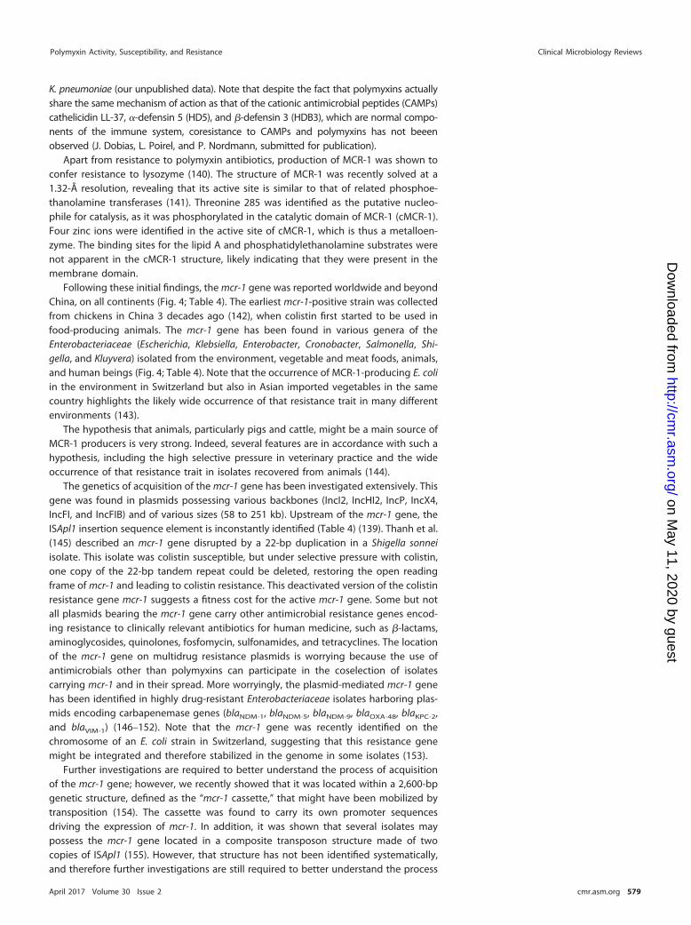
K. pneumoniae (our unpublished data). Note that despite the fact that polymyxins actuallyshare the same mechanism of action as that of the cationic antimicrobial peptides (CAMPs)cathelicidin LL-37, �-defensin 5 (HD5), and �-defensin 3 (HDB3), which are normal compo-nents of the immune system, coresistance to CAMPs and polymyxins has not beeenobserved (J. Dobias, L. Poirel, and P. Nordmann, submitted for publication).
Apart from resistance to polymyxin antibiotics, production of MCR-1 was shown toconfer resistance to lysozyme (140). The structure of MCR-1 was recently solved at a1.32-Å resolution, revealing that its active site is similar to that of related phosphoe-thanolamine transferases (141). Threonine 285 was identified as the putative nucleo-phile for catalysis, as it was phosphorylated in the catalytic domain of MCR-1 (cMCR-1).Four zinc ions were identified in the active site of cMCR-1, which is thus a metalloen-zyme. The binding sites for the lipid A and phosphatidylethanolamine substrates werenot apparent in the cMCR-1 structure, likely indicating that they were present in themembrane domain.
Following these initial findings, the mcr-1 gene was reported worldwide and beyondChina, on all continents (Fig. 4; Table 4). The earliest mcr-1-positive strain was collectedfrom chickens in China 3 decades ago (142), when colistin first started to be used infood-producing animals. The mcr-1 gene has been found in various genera of theEnterobacteriaceae (Escherichia, Klebsiella, Enterobacter, Cronobacter, Salmonella, Shi-gella, and Kluyvera) isolated from the environment, vegetable and meat foods, animals,and human beings (Fig. 4; Table 4). Note that the occurrence of MCR-1-producing E. coliin the environment in Switzerland but also in Asian imported vegetables in the samecountry highlights the likely wide occurrence of that resistance trait in many differentenvironments (143).
The hypothesis that animals, particularly pigs and cattle, might be a main source ofMCR-1 producers is very strong. Indeed, several features are in accordance with such ahypothesis, including the high selective pressure in veterinary practice and the wideoccurrence of that resistance trait in isolates recovered from animals (144).
The genetics of acquisition of the mcr-1 gene has been investigated extensively. Thisgene was found in plasmids possessing various backbones (IncI2, IncHI2, IncP, IncX4,IncFI, and IncFIB) and of various sizes (58 to 251 kb). Upstream of the mcr-1 gene, theISApl1 insertion sequence element is inconstantly identified (Table 4) (139). Thanh et al.(145) described an mcr-1 gene disrupted by a 22-bp duplication in a Shigella sonneiisolate. This isolate was colistin susceptible, but under selective pressure with colistin,one copy of the 22-bp tandem repeat could be deleted, restoring the open readingframe of mcr-1 and leading to colistin resistance. This deactivated version of the colistinresistance gene mcr-1 suggests a fitness cost for the active mcr-1 gene. Some but notall plasmids bearing the mcr-1 gene carry other antimicrobial resistance genes encod-ing resistance to clinically relevant antibiotics for human medicine, such as �-lactams,aminoglycosides, quinolones, fosfomycin, sulfonamides, and tetracyclines. The locationof the mcr-1 gene on multidrug resistance plasmids is worrying because the use ofantimicrobials other than polymyxins can participate in the coselection of isolatescarrying mcr-1 and in their spread. More worryingly, the plasmid-mediated mcr-1 genehas been identified in highly drug-resistant Enterobacteriaceae isolates harboring plas-mids encoding carbapenemase genes (blaNDM-1, blaNDM-5, blaNDM-9, blaOXA-48, blaKPC-2,and blaVIM-1) (146–152). Note that the mcr-1 gene was recently identified on thechromosome of an E. coli strain in Switzerland, suggesting that this resistance genemight be integrated and therefore stabilized in the genome in some isolates (153).
Further investigations are required to better understand the process of acquisitionof the mcr-1 gene; however, we recently showed that it was located within a 2,600-bpgenetic structure, defined as the “mcr-1 cassette,” that might have been mobilized bytransposition (154). The cassette was found to carry its own promoter sequencesdriving the expression of mcr-1. In addition, it was shown that several isolates maypossess the mcr-1 gene located in a composite transposon structure made of twocopies of ISApl1 (155). However, that structure has not been identified systematically,and therefore further investigations are still required to better understand the process
Polymyxin Activity, Susceptibility, and Resistance Clinical Microbiology Reviews
April 2017 Volume 30 Issue 2 cmr.asm.org 579
on May 11, 2020 by guest
http://cmr.asm
.org/D
ownloaded from
of acquisition of that gene from an unknown progenitor in plasmids replicating inEnterobacteriaceae.
MCR-1-producing enterobacterial isolates have often been identified as colonizers ineither humans or animals. Nevertheless, there are some reported cases of infections,including two patients with bacteremia in Switzerland (156).
A functional variant of mcr-1 (Q3L) encoding MCR-1.2 was detected in KPC-3-producing K. pneumoniae in Italy (157), likely sharing the same activity as MCR-1.
In addition, the plasmid-mediated colistin resistance gene mcr-2 was identified in E.coli strains recovered from piglets in Belgium (158). It shared 77% nucleotide identitywith mcr-1 and was carried on an IncX4 plasmid.
Other Mechanisms Contributing to Polymyxin Resistance in EnterobacteriaceaeHyperproduction of CPS. A study showed that the capsule polysaccharide (CPS)
acts as a protective barrier against polymyxins in K. pneumoniae (159). The upregulation
FIG 4 Reports of MCR-1-producing isolates in humans, animals, and both humans and animals.
Poirel et al. Clinical Microbiology Reviews
April 2017 Volume 30 Issue 2 cmr.asm.org 580
on May 11, 2020 by guest
http://cmr.asm
.org/D
ownloaded from

TAB
LE4
Wor
ldw
ide
rep
orts
ofEn
tero
bact
eria
ceae
isol
ates
harb
orin
ga
pla
smid
-med
iate
dm
cr-1
gene
Spec
ies
Cou
ntr
yof
isol
atio
nC
oun
try
ofor
igin
ortr
avel
edre
gio
nSa
mp
leor
igin
Peri
od(y
r)
Plas
mid
feat
ures
a
Refe
ren
ce(s
)In
cty
pe
Size
(kb
)M
obile
elem
ent
Oth
eran
tim
icro
bia
lre
sist
ance
(s)
E.co
liC
hina
Chi
naC
hick
ens
1980
–201
4—
——
—14
2C
hina
Chi
naH
uman
(fec
alca
rria
ge)
Befo
re20
11—
——
—21
8,21
9C
hina
Chi
naC
hick
ens,
pig
s,an
dhu
man
s(in
fect
ions
)20
11–2
014
IncI
264
ISA
pl1
No
139
Chi
naC
hina
2011
–201
4In
cHI2
251
ISA
pl1
Resi
stan
ceto
cefo
taxi
me
(bla
CT
X-M
-14),
amin
ogly
cosi
des,
florf
enic
ol,
olaq
uind
ox,c
otrim
oxaz
ole,
fosf
omyc
in(f
osA
3),a
ndci
pro
floxa
cin
(oqx
AB)
220
Chi
naC
hina
Chi
cken
mea
t20
14In
cI2
65—
146
Chi
naC
hina
Hum
an(b
lood
)20
14–2
015
——
——
147
Chi
naC
hina
Hum
an(f
ecal
carr
iage
)20
15—
——
—22
1C
hina
USA
Hum
an(f
ecal
carr
iage
)20
16In
cFI
33—
Resi
stan
ceto
�-la
ctam
s(b
laC
TX
-M-1
5an
dbl
a TE
M-1
)14
8
Laos
Laos
Hum
an(f
ecal
carr
iage
)an
dp
igs
2012
——
——
222
Thai
land
Thai
land
Hum
an(f
ecal
carr
iage
)20
12—
——
—22
3Vi
etna
mVi
etna
mPi
gs(f
ecal
carr
iage
)20
14–2
015
——
ISA
pl1
Resi
stan
ceto
cefo
taxi
me
(bla
CT
X-M
-55),
trim
etho
prim
(dfr
A12
),te
trac
yclin
e(t
etA
),am
inog
lyco
side
s[a
adA
3,ap
h(3=
)-IA
],p
heni
col
(cm
lA1)
,qui
nolo
nes
(qnr
S1,o
qxA
),lin
cosa
mid
es[in
u(F)
],an
dsu
lfona
mid
es(s
ul2,
sul3
)
224
Cam
bod
iaC
amb
odia
Hum
an(f
eces
)20
12—
—IS
Apl
1—
224
Mal
aysi
aM
alay
sia
Wat
er,c
hick
ens,
pig
s20
13—
—IS
Apl
1or
not
—22
5Ja
pan
Jap
anC
attl
e(m
astit
is)
2008
–201
3In
cl2
60–6
1—
—22
6Fr
ance
Fran
ceVe
alca
lves
(fec
es)
2005
–201
4In
cHI2
——
Resi
stan
ceto
cefo
taxi
me
(bla
CT
X-M
-1),
sulfo
nam
ides
,and
tetr
acyc
lines
227
Fran
ceFr
ance
Broi
ler,
turk
eys,
pig
s20
07–2
014
——
——
228
Ital
yIt
aly
Hum
an(u
rine,
surg
ical
wou
nd)
2013
–201
5—
——
—22
9
Ital
yIt
aly
Hum
an(r
ecta
lsw
abs)
2015
NT
35—
—23
0U
nite
dKi
ngdo
mU
nite
dKi
ngdo
mor
Egyp
tH
uman
(blo
od,s
tool
s)20
12–2
015
IncH
I2,I
ncI2
—IS
Apl
1or
not
—23
1
Switz
erla
nd—
Rive
rw
ater
2012
——
——
143
Switz
erla
ndTh
aila
ndan
dVi
etna
mVe
geta
ble
s20
14—
——
—14
3
Switz
erla
ndSw
itzer
land
Hum
an(u
rinar
ytr
act
infe
ctio
n)20
15—
60—
Resi
stan
ceto
chlo
ram
phe
nico
l,flo
rfen
icol
,an
dco
trim
oxaz
ole
149
Switz
erla
ndSw
itzer
land
Hum
an(b
lood
)20
16In
cFIB
30an
d80
——
156
Belg
ium
Belg
ium
Cal
ves
and
pig
lets
(fec
es)
2011
–201
2In
cP80
Trun
cate
dIS
Apl
1Re
sist
ance
totr
imet
hop
rim(d
frA
1),
tetr
acyc
line
(tet
A),
amin
ogly
cosi
des
[aad
A1,
aph6
-Id/s
trA
,and
aph(
3=)-
Ib/
strB
],an
dsu
lfona
mid
es(s
ul1)
223
Net
herla
nds
Euro
pe
Chi
cken
mea
t20
09–2
014
——
——
232
Net
herla
nds
Net
herla
nds
Chi
cken
s,ve
alca
lves
,tu
rkey
s20
10–2
015
——
——
233
Net
herla
nds
Dut
chfr
esh
mea
tan
dim
por
ted
froz
enm
eat
Reta
ilm
eat
(mos
tly
chic
ken
and
turk
ey)
2009
–201
6—
——
—23
3
Net
herla
nds
Net
herla
nds
Hum
an(b
lood
)20
11—
——
—23
3
(Con
tinue
don
follo
win
gp
age)
Polymyxin Activity, Susceptibility, and Resistance Clinical Microbiology Reviews
April 2017 Volume 30 Issue 2 cmr.asm.org 581
on May 11, 2020 by guest
http://cmr.asm
.org/D
ownloaded from

TAB
LE4
(Con
tinue
d)
Spec
ies
Cou
ntr
yof
isol
atio
nC
oun
try
ofor
igin
ortr
avel
edre
gio
nSa
mp
leor
igin
Peri
od(y
r)
Plas
mid
feat
ures
a
Refe
ren
ce(s
)In
cty
pe
Size
(kb
)M
obile
elem
ent
Oth
eran
tim
icro
bia
lre
sist
ance
(s)
Net
herla
nds
Trav
elin
Tuni
sia,
Sout
hA
mer
ica,
Chi
na,S
outh
east
Asi
a
Hum
an(f
ecal
carr
iage
)20
12–2
013
——
——
234
Net
herla
nds
Ger
man
yVe
alva
lves
,bro
ilers
,and
turk
ey20
10–2
015
IncH
I2or
IncX
4—
ISA
pl1
—23
5
Ger
man
yPi
gsan
dhu
man
(wou
ndin
fect
ion)
2010
–201
5In
cHI2
orIn
cX4
—IS
Apl
1or
not
—15
0
Den
mar
kEu
rop
eC
hick
enm
eat
2012
–201
4In
cI2
orIn
cX4
——
—23
6
Den
mar
kD
enm
ark
Hum
an(b
lood
stre
amin
fect
ion)
2015
IncI
2—
——
236
Can
ada
Unk
now
nBe
efm
eat
2010
IncH
I2A
——
—15
1C
anad
aLi
ved
inEg
ypt
for
5ye
ars
Hum
an(g
astr
osto
my
site
and
rect
um)
2011
IncI
2—
——
151
USA
USA
Hum
an(u
rine)
2016
IncF
225
ISA
pl1
Resi
stan
ceto
�-la
ctam
s(b
laC
TX
-M-5
5)
237
Alg
eria
Alg
eria
Chi
cken
s20
15—
——
—22
2Tu
nisi
aTu
nisi
a/Fr
ance
Chi
cken
s20
15In
cHI2
——
Resi
stan
ceto
�-la
ctam
s(b
laC
TX
-M-1
)23
8Eg
ypt
Egyp
tH
uman
(sp
utum
)20
15—
�90
——
239
Sout
hA
fric
aSo
uth
Afr
ica
Hum
an20
14–2
015
IncI
265
,70
ISA
pl1
orno
t—
154
IncH
I215
015
4In
cX4
3015
4So
uth
Afr
ica
Sout
hA
fric
aBr
oile
rch
icke
n20
08–2
014
IncI
262
ISA
pl1
—24
0Br
azil
Braz
ilC
hick
en,s
win
e20
03–2
015
——
——
241
K.pn
eum
onia
eC
hina
Chi
naH
uman
(sur
gica
lw
ound
,p
erito
neal
fluid
)20
15—
——
—14
7
Chi
naC
hina
Food
anim
als
and
hum
an(in
fect
ions
)20
11–2
014
Incl
264
ISA
pl1
No
139
Den
mar
kD
enm
ark
Hum
an(u
nkno
wn)
2014
——
——
224
Ente
roba
cter
cloa
cae
Chi
naC
hina
Hum
an(u
rine)
2014
IncF
I70
——
242
Ente
roba
cter
aero
gene
sC
hina
Chi
naH
uman
(vag
inal
secr
etio
n)20
14In
cFI
65—
Resi
stan
ceto
�-la
ctam
s(b
laT
EM
-1an
dbl
a CT
X-M
-15)
242
Cron
obac
ter
saka
zaki
iC
hina
Chi
naC
hick
en(d
iarr
hea)
2015
Incl
265
ISA
pl1
—15
2Sa
lmon
ella
ente
rica
sero
typ
eTy
phi
mur
ium
Port
ugal
Port
ugal
Food
anim
als
2011
IncH
I2—
ISA
pl1
—21
8,22
5,24
3
S.en
teric
aJa
pan
Jap
anSw
ine
(sep
ticem
ia)
2013
IncI
258
——
226
Salm
onel
laPa
raty
phi
BFr
ance
Fran
ceC
hick
enm
eat,
guin
eafo
wl
pie
2012
IncX
4—
——
244
Salm
onel
laD
erb
yFr
ance
Fran
cePo
rksa
usag
e20
13In
cP—
——
244
Salm
onel
la1,
4[5]
,12:
i:�Fr
ance
Fran
ceBr
oile
rs20
13In
cP—
——
244
Salm
onel
laTy
phi
mur
ium
and
Salm
onel
laVi
rcho
wU
nite
dKi
ngdo
mU
nite
dKi
ngdo
mH
uman
(fec
es)
2012
–201
5In
cHI2
,In
cX4
—N
o—
231
Salm
onel
laTy
phi
mur
ium
and
Salm
onel
laPa
raty
phi
Bva
r.Ja
va
Uni
ted
King
dom
Trav
elin
Asi
aan
dU
nite
dA
rab
Emira
tes
Hum
an(f
eces
)20
12–2
015
IncH
I2,I
ncI2
—IS
Apl
1or
not
—23
1
Salm
onel
laJa
vaN
ethe
rland
s—
Chi
cken
mea
t20
10–2
015
IncX
430
——
235
Salm
onel
laA
natu
mN
ethe
rland
s—
Turk
eym
eat
2013
IncX
430
——
235
Salm
onel
laSc
hwar
tzen
grun
dN
ethe
rland
s—
Turk
eym
eat
2015
IncX
430
——
235
Salm
onel
laPa
raty
phi
Bva
r.Ja
vaU
nite
d-Ki
ngdo
mEu
rop
ePo
ultr
ym
eat
2012
–201
5In
cHI2
—N
o—
231
Shig
ella
sonn
eiVi
etna
mVi
etna
mH
uman
(fec
es)
2008
IncI
2—
——
145
Kluy
vera
asco
rbat
aC
hina
Chi
naH
osp
ital
sew
age
2015
IncI
257
——
216
aN
T,no
ntyp
eab
le;—
,unk
now
n.
Poirel et al. Clinical Microbiology Reviews
April 2017 Volume 30 Issue 2 cmr.asm.org 582
on May 11, 2020 by guest
http://cmr.asm
.org/D
ownloaded from

of capsular biosynthesis genes indeed reduces the interactions of polymyxins with thebacterial surface, leading to polymyxin resistance.
K. pneumoniae is able to release anionic capsular polysaccharides from its surface(160). This release leads to the trapping of cationic antimicrobial peptides, such aspolymyxins, thus decreasing the amount of antibiotic reaching the bacterial surface.The CPS is connected to the bacterial surface through an ionic interaction with the LPS,and this interaction is stabilized by divalent cations (161). As a consequence, the releaseof CPS in the presence of polymyxins is likely due to perturbation of the cation-dependent bridges between the molecules of LPS.
Role of porins. It has been shown that a periplasmic protein (YdeI) regulated by thePhoPQ and PmrAB two-component systems can interact with the OmpD porin toincrease bacterial resistance to polymyxins in Salmonella enterica (162).
Role of efflux pumps. The role of efflux in colistin resistance is not well understood,but several studies suggested the involvement of efflux pumps in colistin resistance.Mutations in kpnEF and acrAB, encoding components of efflux pumps, may actuallylead to a 2-fold decrease of the MIC of colistin and increase bacterial survival in thepresence of a low concentration of polymyxins (163, 164). Addition to the test mediumof low doses of the efflux pump inhibitor carbonyl cyanide m-chlorophenylhydrazone(CCCP) decreased the MICs for resistant strains (128- to 512-fold reductions) andpartially or completely inhibited the regrowth of resistant subpopulations (165). How-ever, this observation should be considered with caution owing to the nonspecificeffect of CCCP on efflux systems, with a likely wider impact on bacterial metabolism.
Mechanisms of Polymyxin Resistance in Pseudomonas aeruginosa and Acinetobac-ter baumannii
Pseudomonas aeruginosa. The colistin resistance in P. aeruginosa is mediated by fivetwo-component systems that regulate LPS modifications. As for the Enterobacteriaceae,alterations in the PmrAB (74, 118, 166–169) and PhoPQ (118, 166, 168, 170) two-component systems have been shown to be responsible for acquired resistance tocolistin. Mutations in these two-component systems cause constitutive alterations andconsequently activate transcription of the pmrHFIJKLM operon and the subsequentaddition of L-Ara4N to the LPS, finally leading to colistin resistance. Notably, unlike whatis observed in K. pneumoniae, the colistin resistance mediated by PhoPQ modificationsdoes not depend on the PmrAB system.
Three other two-component systems have been proved to contribute to colistinresistance in P. aeruginosa, namely, ParRS, ColRS, and CprRS. The ParRS (polymyxinadaptive resistance) two-component system is involved in adaptative resistance topolymyxins (118, 166, 171). Mutations in this system cause constitutive expression ofthe pmrHFIJKLM operon and thus lead to the addition of L-Ara4N to the LPS, leading tocolistin resistance. Additionally, mutations in the ColRS and CprRS two-componentregulatory systems may also contribute to polymyxin resistance, since the associationof mutations in the phoQ gene and mutations in the colS or cprS gene confers a highlevel of colistin resistance (172). The action of the ColRS and CprRS systems may occurthrough the activation of the phoQ gene and/or through other genes that have not yetbeen identified.
Acinetobacter baumannii. The main mechanism of colistin resistance in A. bauman-nii corresponds to the addition of cationic groups to the LPS (qualitative modificationof the LPS); nevertheless, acquired resistance to colistin may also be the consequenceof a complete loss of LPS production (quantitative modification of the LPS).
The addition of cationic groups in A. baumannii is mediated by mutations in PmrAB(118, 173–180). Mutations in the pmrA and pmrB genes have been shown to causecolistin resistance through upregulation of the pmrCAB operon, leading to pEtN syn-thesis but not to L-Ara4N synthesis (unlike in Enterobacteriaceae).
The second mechanism of colistin resistance in A. baumannii corresponds to thecomplete loss of LPS caused by alterations of the lipid A biosynthesis genes, namely,the lpxA, lpxC, and lpxD genes. Mutations identified in those genes were either
Polymyxin Activity, Susceptibility, and Resistance Clinical Microbiology Reviews
April 2017 Volume 30 Issue 2 cmr.asm.org 583
on May 11, 2020 by guest
http://cmr.asm
.org/D
ownloaded from

substitutions, truncations, frameshifts (109), or insertional inactivation by the insertionsequence ISAba11 (181).
EPIDEMIOLOGY OF RESISTANCE TO POLYMYXINSGeneral Epidemiology of Resistance to Polymyxins
Although polymyxins currently retain significant in vitro activity against most Gram-negative organisms, resistance to these antibiotics is increasingly being reportedamong clinical isolates (29, 182).
The SENTRY antimicrobial surveillance program carried out a worldwide survey in2009 and reported low rates of resistance to polymyxins among Gram-negative patho-gens (Acinetobacter spp., P. aeruginosa, E. coli, and Klebsiella spp.) (�0.1% to 1.5%) (183).However, a rising trend was observed in a 2006-2009 study period focusing on K.pneumoniae isolates (resistance rates of 1.2% in 2006 and 1.8% in 2009), probablybecause of the extensive and/or inadequate usage of polymyxins worldwide fortreating infections with MDR Gram-negative bacteria.
Colistin resistance in K. pneumoniae represents a growing public health concern,since this bacterial species is one of the main pathogens of nosocomial infection andhas gathered a wide range of resistance mechanisms to broad-spectrum antibioticsover the years. Table 5 shows the populations studied (mainly carbapenem-resistant K.pneumoniae [CR-KP] clinical isolates), along with the methods that have been used todetermine the rate of colistin resistance, since some methods are now known tounderestimate the level of colistin resistance and therefore may significantly bias theproposed rates. The occurrence of colistin resistance in K. pneumoniae has beenreported in surveillance studies and clinical case reports worldwide (Table 5) (183).Many studies report an increase of the resistance rate among multidrug-resistant K.pneumoniae isolates, particularly among CR-KP isolates, with high colistin resistancerates reported (Table 5). More worryingly, multiple outbreaks with carbapenem- andcolistin-resistant isolates have been reported in North America and Europe (Table 6;Fig. 5).
North America. Multicenter surveys showed low rates of resistance among K. pneu-moniae isolates in Canada (2.9%) (184) and the United States (4%) (185) (Table 5). However,the colistin resistance rate was higher (6.7 to 18%) among carbapenemase-producingisolates (185–187). In addition, outbreaks with colistin-resistant, KPC-producing K. pneu-moniae, mostly attributed to the international epidemic clone type ST258, have beenreported in the United States (188, 189) and Mexico (190) (Table 6; Fig. 5).
South America. The results from the SENTRY antimicrobial surveillance programshowed a moderate resistance rate in Latin America in 2009 (3%) (183); however, theemergence of colistin-resistant K. pneumoniae isolates has been reported in Argentina(191), Colombia (119), and Brazil (87).
Europe. Multiple outbreaks of both carbapenem- and colistin-resistant K. pneu-moniae isolates have been reported in Europe (Table 6; Fig. 5). Outbreaks with KPC-producing K. pneumoniae isolates attributed to the international epidemic clone typeST258 have been reported in the Netherlands (192), Hungary (193), Greece (194, 195),and Italy (196, 197).
In addition to the two outbreaks attributed to the ST258 clone (196, 197), a largenosocomial outbreak of colistin-resistant and KPC-producing ST512 K. pneumoniaeisolates was reported in Italy (198) (Table 6; Fig. 5). More worryingly, colistin resistancewas recently reported at a high level (�20%) among carbapenemase-producing iso-lates in ICUs of two Italian hospitals (199, 200), with an even higher rate (36.1%) inhospitals in Rome (68) (Table 6). Moreover, a national study reported a countrywidelevel of colistin resistance among KPC-producing K. pneumoniae isolates, with 43% ofisolates being resistant to colistin (201) (Table 5).
In Greece, several outbreaks caused by KPC-producing, colistin-resistant K. pneu-moniae isolates have been reported (194, 195) (Table 6; Fig. 5). Studies performed intwo Greek hospitals reported a huge increase in colistin resistance within a few years(�3.5% incidence before 2010 and �20% incidence after 2010) (202, 203).
Poirel et al. Clinical Microbiology Reviews
April 2017 Volume 30 Issue 2 cmr.asm.org 584
on May 11, 2020 by guest
http://cmr.asm
.org/D
ownloaded from

TAB
LE5
Stud
ies
rep
ortin
gp
reva
lenc
esof
colis
tinre
sist
ance
amon
gK.
pneu
mon
iae
clin
ical
isol
ates
d
Stud
yty
pe
and
per
iod
Cou
ntr
ySe
ttin
gTe
stm
eth
odTo
tal
no.
ofis
olat
esC
arb
apen
emas
ep
rod
ucer
s(n
o.[%
])Po
lym
yxin
-res
ista
nt
isol
ates
(no.
[%])
Refe
ren
ce(s
)
Stud
ies
rep
ortin
gco
listin
resi
stan
cera
tes
amon
gov
eral
lcl
inic
alis
olat
es20
06–2
009
Wor
ldw
ide
Wor
ldw
ide
surv
eilla
nce
BMD
9,77
4N
A(1
.5)
183
2007
–200
8C
anad
aN
atio
nal
BMD
515
NA
15(2
.9)
184
2013
–201
4U
nite
dSt
ates
Mul
ticen
ter
BMD
1,20
5N
A(4
)18
520
06–2
007
Sout
hKo
rea
Mul
ticen
ter
(9ho
spita
ls)
BMD
221a
15(1
3.3)
24(1
0.9)
b20
620
04–2
005
Sing
apor
eSi
ngle
cent
erA
gar
dilu
tion
16N
A1
(6)
207
Stud
ies
rep
ortin
gco
listin
resi
stan
cera
tes
amon
gC
R-KP
isol
ates
2008
–201
1C
anad
aM
ultic
ente
rEt
est
3030
(100
)2
(6.7
)18
4,18
620
13–2
014
Uni
ted
Stat
esM
ultic
ente
rBM
D69
69(1
00)
(18)
c18
520
03–2
004
Uni
ted
Stat
esM
ultic
ente
rBM
Dor
agar
dilu
tion
9696
(100
)(9
)18
720
10–2
013
Gre
ece
Sing
lece
nter
(onl
yIC
U)
Vite
k2,E
test
9292
(100
)20
(21.
7)20
220
10G
reec
eSi
ngle
cent
erVi
tek2
,Ete
st12
012
0(1
00)
25(2
0.8)
203
2014
Ital
ySi
ngle
cent
er(o
nly
ICU
)Vi
tek2
,Ete
st21
421
4(1
00)
47(2
1.9)
199
2013
Ital
ySi
ngle
cent
er(o
nly
ICU
)BM
D25
24(1
00)
6(2
4.0)
200
2013
–201
4It
aly
Nat
iona
lBM
D17
817
8(1
00)
76(4
3)20
120
10–2
011
Ital
yM
ultic
ente
r(9
hosp
itals
)Vi
tek2
,BM
D97
97(1
00)
35(3
6.1)
6820
10–2
012
Spai
nH
osp
ital
Aga
rdi
lutio
n79
79(1
00)
18(2
2.8)
204
2014
Fran
ceN
atio
nal
BMD
561
561
(100
)35
(6.2
)20
520
12–2
013
Turk
eySi
ngle
cent
erVi
tek2
,Ete
st37
36(9
8)(2
.7)
245
2006
–200
7Is
rael
Sing
lece
nter
?88
88(1
00)
(4.5
)24
620
09–2
010
Chi
naSi
ngle
cent
erA
gar
dilu
tion
6868
(100
)3
(4.4
)20
820
12Ta
iwan
Nat
iona
lSe
nsiti
tre
247
55(2
2.3)
(12.
1)20
9aIs
olat
edfr
omb
lood
sam
ple
s.bN
umb
erof
colis
tin-r
esis
tant
isol
ates
acco
rdin
gto
EUC
AST
bre
akp
oint
s.c E
ight
een
per
cent
colis
tinre
sist
ance
amon
g4
E.co
lian
d69
K.pn
eum
onia
eis
olat
esha
rbor
ing
carb
apen
emas
es.
dN
A,n
otap
plic
able
.
Polymyxin Activity, Susceptibility, and Resistance Clinical Microbiology Reviews
April 2017 Volume 30 Issue 2 cmr.asm.org 585
on May 11, 2020 by guest
http://cmr.asm
.org/D
ownloaded from
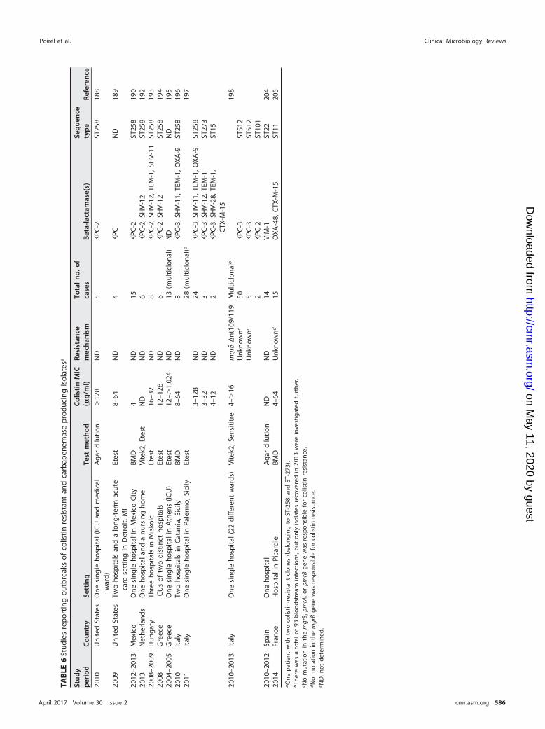
TAB
LE6
Stud
ies
rep
ortin
gou
tbre
aks
ofco
listin
-res
ista
ntan
dca
rbap
enem
ase-
pro
duci
ngis
olat
ese
Stud
yp
erio
dC
oun
try
Sett
ing
Test
met
hod
Col
isti
nM
IC(�
g/m
l)Re
sist
ance
mec
han
ism
Tota
ln
o.of
case
sB
eta-
lact
amas
e(s)
Seq
uen
cety
pe
Refe
ren
ce
2010
Uni
ted
Stat
esO
nesi
ngle
hosp
ital
(ICU
and
med
ical
war
d)A
gar
dilu
tion
�12
8N
D5
KPC
-2ST
258
188
2009
Uni
ted
Stat
esTw
oho
spita
lsan
da
long
-ter
mac
ute
care
sett
ing
inD
etro
it,M
IEt
est
8–64
ND
4KP
CN
D18
9
2012
–201
3M
exic
oO
nesi
ngle
hosp
ital
inM
exic
oC
ityBM
D4
ND
15KP
C-2
ST25
819
020
13N
ethe
rland
sO
neho
spita
lan
da
nurs
ing
hom
eVi
tek2
,Ete
stN
DN
D6
KPC
-2,S
HV-
12ST
258
192
2008
–200
9H
unga
ryTh
ree
hosp
itals
inM
isko
lcEt
est
16–3
2N
D8
KPC
-2,S
HV-
12,T
EM-1
,SH
V-11
ST25
819
320
08G
reec
eIC
Us
oftw
odi
stin
ctho
spita
lsEt
est
12–1
28N
D6
KPC
-2,S
HV-
12ST
258
194
2004
–200
5G
reec
eO
nesi
ngle
hosp
ital
inA
then
s(IC
U)
Etes
t12
–�1,
024
ND
13(m
ultic
lona
l)N
DN
D19
520
10It
aly
Two
hosp
itals
inC
atan
ia,S
icily
BMD
8–64
ND
8KP
C-3
,SH
V-11
,TEM
-1,O
XA
-9ST
258
196
2011
Ital
yO
nesi
ngle
hosp
ital
inPa
lerm
o,Si
cily
Etes
t28
(mul
ticlo
nal)a
197
3–12
8N
D24
KPC
-3,S
HV-
11,T
EM-1
,OX
A-9
ST25
83–
32N
D3
KPC
-3,S
HV-
12,T
EM-1
ST27
34–
12N
D2
KPC
-3,S
HV-
28,T
EM-1
,C
TX-M
-15
ST15
2010
–201
3It
aly
One
sing
leho
spita
l(2
2di
ffer
ent
war
ds)
Vite
k2,S
ensi
titre
4–�
16m
grB
Δnt
109/
119
Mul
ticlo
nalb
198
Unk
now
nc50
KPC
-3ST
512
Unk
now
nc5
KPC
-3ST
512
2KP
C-2
ST10
120
10–2
012
Spai
nO
neho
spita
lA
gar
dilu
tion
ND
ND
14VI
M-1
ST22
204
2014
Fran
ceH
osp
ital
inPi
card
ieBM
D4–
64U
nkno
wnd
15O
XA
-48,
CTX
-M-1
5ST
1120
5aO
nep
atie
ntw
ithtw
oco
listin
-res
ista
ntcl
ones
(bel
ongi
ngto
ST-2
58an
dST
-273
).bTh
ere
was
ato
tal
of93
blo
odst
ream
infe
ctio
ns,b
uton
lyis
olat
esre
cove
red
in20
13w
ere
inve
stig
ated
furt
her.
c No
mut
atio
nin
the
mgr
B,pm
rA,o
rpm
rBge
new
asre
spon
sib
lefo
rco
listin
resi
stan
ce.
dN
om
utat
ion
inth
em
grB
gene
was
resp
onsi
ble
for
colis
tinre
sist
ance
.e N
D,n
otde
term
ined
.
Poirel et al. Clinical Microbiology Reviews
April 2017 Volume 30 Issue 2 cmr.asm.org 586
on May 11, 2020 by guest
http://cmr.asm
.org/D
ownloaded from

During a 2-year period (2010 to 2012) in Spain, a study showed an increase of theprevalence of colistin resistance among carbapenemase-producing K. pneumoniaeisolates, from 13.5 to 31.7%, and an outbreak of colistin-resistant, VIM-1-producing K.pneumoniae was reported (204).
In France, a national survey revealed a low rate of colistin resistance (6.2%) amongcarbapenemase-producing K. pneumoniae isolates; neverthess, an outbreak of OXA-48carbapenemase-producing and colistin-resistant K. pneumoniae isolates was reported(205).
Middle East. In Turkey and Israel, low rates of resistance to colistin among CR-KPisolates have been reported (2.7% and 4.5%, respectively). However, multiclonal out-breaks with OXA-48-, NDM-, and both OXA-48- and NDM-producing K. pneumoniae arecurrently ongoing in Turkey (our unpublished data).
Africa. A very low colistin resistance rate was reported among K. pneumoniaeisolates in Tunisia (1.2%), but this rate was probably underestimated because suscep-tibility to colistin was primarily screened using a DD method generating high falsesusceptibility rates (205). The emergence of colistin resistance was reported for K.pneumoniae isolates recovered in South Africa (81, 119) and Nigeria (120).
Asia. Moderate rates of colistin resistance (about 6 to 11%) have been reported forK. pneumoniae isolates from South Korea (206) and Singapore (207), and similarresistance rates (4.4 to 12.1%) were found among CR-KP isolates in China (208) andTaiwan (209). Colistin-resistant K. pneumoniae isolates have also been reported in Laosand Thailand (120).
Risk Factors
The use of colistin was found to be an independent risk factor for the occurrence ofresistance in Gram-negative bacteria (210, 211). Important increases of colistin resis-tance rates among ESBL-producing K. pneumoniae isolates (from 0 to 71% and from11.1 to 75%) were reported after the introduction of selective digestive tract decon-tamination in two intensive care units (212, 213). Moreover, this decontamination failedto prevent colonization by ESBL-producing Enterobacteriaceae, and such a strategyshould be abandoned. Note that the inappropriate use of colistin (such as suboptimaldosing or prolonged monotherapy) has been shown to be a source of colistin resistanceselection (214, 215). The occurrence of colistin resistance in P. aeruginosa was most
FIG 5 Outbreaks caused by colistin-resistant, carbapenemase-producing K. pneumoniae isolates. Each star indicates a single report.
Polymyxin Activity, Susceptibility, and Resistance Clinical Microbiology Reviews
April 2017 Volume 30 Issue 2 cmr.asm.org 587
on May 11, 2020 by guest
http://cmr.asm
.org/D
ownloaded from
effectively prevented by 8-h dosing intervals compared to 12- or 24-h dosing intervals(45).
Specific Epidemiology of the Plasmid-Mediated mcr-1 Resistance Gene
Notably, plasmid-borne resistance to polymyxins has been reported for few differententerobacterial species so far, mainly among E. coli isolates and rarely for Salmonellaenterica, Enterobacter spp., and K. pneumoniae. There are also some scattered reports ofMCR-1-producing isolates in other species, such as Cronobacter sakazakii (152) andKluyvera ascorbata (216). According to the current literature on the subject, thedistribution of MCR-1 appears to be worldwide, covering all continents (217). It remainsto be determined if the identification of the mcr-1 gene worldwide corresponds tosubsequent spread from an original source (China?) or to simultaneous gene mobili-zation events in different parts of the world. Ongoing epidemiological surveys shouldprovide some important clues.
It is actually speculated that the original source of the gene, or at least of itsmobilization and emergence, might be the animal world. This speculation is based onthe fact that MCR-1-producing E. coli isolates have been identified in several animalsand animal food products, including chickens and chicken meat, pigs and piglets,cattle, calves, and turkeys, but also in humans (Fig. 4). The corresponding samples werecollected from many Asian countries (Cambodia, China, Japan, Laos, Malaysia, Taiwan,Singapore, and Vietnam) but also from Europe (Belgium, Denmark, France, Germany,Portugal, Italy, the Netherlands, Spain, Sweden, Switzerland, and the UK), the Americas(Argentina, Brazil, and Canada), and Africa (Algeria, Egypt, South Africa, and Tunisia)(Table 4). Note that a study conducted in Switzerland identified MCR-1-producing E. coliisolates in vegetables imported from Asia (143), and positive isolates were also iden-tified in environmental water samples in Switzerland and Malaysia (Table 4).
The speculation of an animal origin of the mcr-1 gene is also based on geneticfeatures, since this gene is often associated with the insertion sequence ISApl1, iden-tified in Pasteurella multocida, which is a common animal pathogen, and also with theblaCMY-2 and florR genes, which are often identified in animal enterobacterial isolates(144). Finally, another feature suggesting an animal source of the problem is the heavyusage of polymyxins in veterinary medicine, with usage on many different animalspecies.
Dating the emergence of MCR-1-positive strains remains quite difficult; however, aChinese study retrospectively identified positive isolates recovered from chickensduring the 1980s (142), and they were discovered as early as 2005 in veal calves inFrance (144). It therefore seems that the emergence of MCR-positive isolates, at least inanimals, is not a recent event. Very likely, there has been some silent dissemination ofthat resistance mechanism throughout the last few decades, and the current situationshows an ongoing further dissemination rather than an emerging phenomenon.
CONCLUSIONS
Polymyxins are gaining increasing interest because of the current epidemiologicalsituation, with MDR Gram-negative bacteria spreading worldwide and with a paucity ofnovel marketed antibiotics. In some areas where infections caused by carbapenem-resistant Enterobacteriaceae are now common (such as Greece or Italy), the use ofpolymyxins (alone or often in combination with other antibiotics) is becoming crucialand may even be considered first-line therapy. The reevaluation of some critical issuesin relation to polymyxins (accurate susceptibility testing, defining correct breakpoints,and better appreciating the toxicity issues) now opens new perspectives for its use.Studies that may permit a better evaluation of the PK-PD data, the toxicity level, andappropriate drug combinations are therefore crucial.
The recent identification of plasmid-mediated mechanisms of resistance to poly-myxins also modifies the perspective. Indeed, epidemiological studies have to beinitiated in order to better evaluate the extent of dissemination of this resistance inhuman and veterinary medicine and the impact of its occurrence. The perspective of
Poirel et al. Clinical Microbiology Reviews
April 2017 Volume 30 Issue 2 cmr.asm.org 588
on May 11, 2020 by guest
http://cmr.asm
.org/D
ownloaded from

nosocomial dissemination of MDR Gram-negative organisms possessing resistancedeterminants to all main antibiotics is frightening, in particular for K. pneumoniae,which is one of the main nosocomial pathogens. Whether veterinary medicine isaffecting the epidemiological situation by providing selective pressure with polymyxinshas to be precisely determined. Whether discontinuing some specific usages of thesedrugs (prophylaxis or metaphylaxis in animals and decontamination of MDR bacteria inhumans) should be considered is therefore an open debate.
Ultimately, reinforcing the detection of polymyxin-resistant isolates must be encour-aged. Prospective epidemiological surveys are needed, since the current knowledge onthis issue is very scarce. Actually, the recent development of a rapid diagnostic test fordetection of polymyxin resistance, along with the development of a screening agarmedium, will contribute to facilitating those surveys.
ACKNOWLEDGMENTSThis work was funded by the University of Fribourg, Switzerland, and by a grant from
the ANIWHA ERA-NET project, Switzerland (ANIWHA).
REFERENCES1. Benedict RG, Langlykke AF. 1947. Antibiotic activity of Bacillus poly-
myxa. J Bacteriol 54:24.2. Hancock RE. 1997. Peptide antibiotics. Lancet 349:418 – 422. https://
doi.org/10.1016/S0140-6736(97)80051-7.3. Falagas ME, Rafailidis PI, Matthaiou DK. 2010. Resistance to polymyxins:
mechanisms, frequency and treatment options. Drug Resist Updat13:132–138. https://doi.org/10.1016/j.drup.2010.05.002.
4. Li J, Nation RL, Turnidge JD, Milne RW, Coulthard K, Rayner CR, PatersonDL. 2006. Colistin: the re-emerging antibiotic for multidrug-resistantGram-negative bacterial infections. Lancet Infect Dis 6:589 – 601.https://doi.org/10.1016/S1473-3099(06)70580-1.
5. Brink AJ, Richards GA, Colombo G, Bortolotti F, Colombo P, Jehl F. 2014.Multicomponent antibiotic substances produced by fermentation: im-plications for regulatory authorities, critically ill patients and generics.Int J Antimicrob Agents 43:1– 6. https://doi.org/10.1016/j.ijantimicag.2013.06.013.
6. Gallardo-Godoy A, Muldoon C, Becker B, Elliott AG, Lash LH, Huang JX,Butler MS, Pelingon R, Kavanagh AM, Ramu S, Phetsang W, BlaskovichMA, Cooper MA. 2016. Activity and predicted nephrotoxicity of syn-thetic antibiotics based on polymyxin B. J Med Chem 59:1068 –1077.https://doi.org/10.1021/acs.jmedchem.5b01593.
7. Nation RL, Velkov T, Li J. 2014. Colistin and polymyxin B: peas in a pod,or chalk and cheese? Clin Infect Dis 59:88 –94. https://doi.org/10.1093/cid/ciu213.
8. Bergen PJ, Li J, Rayner CR, Nation RL. 2006. Colistin methanesulfonateis an inactive prodrug of colistin against Pseudomonas aeruginosa.Antimicrob Agents Chemother 50:1953–1958. https://doi.org/10.1128/AAC.00035-06.
9. Barnett M, Bushby SR, Wilkinson S. 1964. Sodium sulphomethyl deriv-atives of polymyxins. Br J Pharmacol Chemother 23:552–574. https://doi.org/10.1111/j.1476-5381.1964.tb01610.x.
10. Li J, Milne RW, Nation RL, Turnidge JD, Coulthard K. 2003. Stability ofcolistin and colistin methanesulfonate in aqueous media and plasma asdetermined by high-performance liquid chromatography. AntimicrobAgents Chemother 47:1364 –1370. https://doi.org/10.1128/AAC.47.4.1364-1370.2003.
11. Li J, Milne RW, Nation RL, Turnidge JD, Smeaton TC, Coulthard K. 2004.Pharmacokinetics of colistin methanesulphonate and colistin in ratsfollowing an intravenous dose of colistin methanesulphonate. J Anti-microb Chemother 53:837– 840. https://doi.org/10.1093/jac/dkh167.
12. Dixon RA, Chopra I. 1986. Leakage of periplasmic proteins from Esch-erichia coli mediated by polymyxin B nonapeptide. Antimicrob AgentsChemother 29:781–788. https://doi.org/10.1128/AAC.29.5.781.
13. Falagas ME, Kasiakou SK. 2005. Colistin: the revival of polymyxins forthe management of multidrug-resistant Gram-negative bacterial infec-tions. Clin Infect Dis 40:1333–1341. https://doi.org/10.1086/429323.
14. Li J, Nation RL, Milne RW, Turnidge JD, Coulthard K. 2005. Evaluation ofcolistin as an agent against multi-resistant Gram-negative bacteria. Int
J Antimicrob Agents 25:11–25. https://doi.org/10.1016/j.ijantimicag.2004.10.001.
15. Deris ZZ, Akter J, Sivanesan S, Roberts KD, Thompson PE, Nation RL, LiJ, Velkov T. 2013. A secondary mode of action of polymyxins againstGram-negative bacteria involves the inhibition of NADH-quinone oxi-doreductase activity. J Antibiot (Tokyo) 67:147–151. https://doi.org/10.1038/ja.2013.111.
16. Bulitta JB, Yang JC, Yohonn L, Ly NS, Brown SV, D’Hondt RE, Jusko WJ,Forrest A, Tsuji BT. 2010. Attenuation of colistin bactericidal activity by highinoculum of Pseudomonas aeruginosa characterized by a new mechanism-based population pharmacodynamic model. Antimicrob Agents Che-mother 54:2051–2062. https://doi.org/10.1128/AAC.00881-09.
17. Owen RJ, Li J, Nation RL, Spelman D. 2007. In vitro pharmacodynamicsof colistin against Acinetobacter baumannii clinical isolates. J Antimi-crob Chemother 59:473– 477. https://doi.org/10.1093/jac/dkl512.
18. Poudyal A, Howden BP, Bell JM, Gao W, Owen RJ, Turnidge JD, NationRL, Li J. 2008. In vitro pharmacodynamics of colistin against multidrug-resistant Klebsiella pneumoniae. J Antimicrob Chemother 62:1311–1318. https://doi.org/10.1093/jac/dkn425.
19. Bergen PJ, Landersdorfer CB, Zhang J, Zhao M, Lee HJ, Nation RL, Li J.2012. Pharmacokinetics and pharmacodynamics of ‘old’ polymyxins:what is new? Diagn Microbiol Infect Dis 74:213–223. https://doi.org/10.1016/j.diagmicrobio.2012.07.010.
20. Cheah SE, Wang J, Nguyen VTT, Turnidge JD, Li J, Nation RL. 2015. Newpharmacokinetic/pharmacodynamic studies of systemically adminis-tered colistin against Pseudomonas aeruginosa and Acinetobacter bau-mannii in mouse thigh and lung infection models: smaller response inlung infection. J Antimicrob Chemother 70:3291–3297. https://doi.org/10.1093/jac/dkv267.
21. Garonzik SM, Li J, Thamlikitkul V, Paterson DL, Shoham S, Jacob J,Silveira FP, Forrest A, Nation RL. 2011. Population pharmacokinetics ofcolistin methanesulfonate and formed colistin in critically ill patientsfrom a multicenter study provide dosing suggestions for various cate-gories of patients. Antimicrob Agents Chemother 55:3284 –3294.https://doi.org/10.1128/AAC.01733-10.
22. Landersdorfer CB, Nation RL. 2015. Colistin: how should it be dosed forthe critically ill? Semin Respir Crit Care Med 36:126 –135. https://doi.org/10.1055/s-0034-1398390.
23. Bergen PJ, Li J, Nation RL. 2011. Dosing of colistin— back to basicPK/PD. Curr Opin Pharmacol 11:464 – 469. https://doi.org/10.1016/j.coph.2011.07.004.
24. Gunderson BW, Ibrahim KH, Hovde LB, Fromm TL, Reed MD, RotschaferJC. 2003. Synergistic activity of colistin and ceftazidime againstmultiantibiotic-resistant Pseudomonas aeruginosa in an in vitro phar-macodynamic model. Antimicrob Agents Chemother 47:905–909.https://doi.org/10.1128/AAC.47.3.905-909.2003.
25. John EB, Bennett RD, Blaser MJ. 2015. Polymyxins (polymyxin B andcolistin), p 549 –555. In Bennett JE, Dolin R, Blaser MJ (ed), Mandell,
Polymyxin Activity, Susceptibility, and Resistance Clinical Microbiology Reviews
April 2017 Volume 30 Issue 2 cmr.asm.org 589
on May 11, 2020 by guest
http://cmr.asm
.org/D
ownloaded from

Douglas, and Bennett’s principles and practice of infectious diseases,8th ed. Elsevier/Saunders, Philadelphia, PA.
26. Li J, Rayner CR, Nation RL, Owen RJ, Spelman D, Tan KE, Liolios L. 2006.Heteroresistance to colistin in multidrug-resistant Acinetobacter bau-mannii. Antimicrob Agents Chemother 50:2946 –2950. https://doi.org/10.1128/AAC.00103-06.
27. Yau W, Owen RJ, Poudyal A, Bell JM, Turnidge JD, Yu HH, Nation RL, LiJ. 2009. Colistin hetero-resistance in multidrug-resistant Acinetobacterbaumannii clinical isolates from the Western Pacific region in theSENTRY antimicrobial surveillance programme. J Infect 58:138 –144.https://doi.org/10.1016/j.jinf.2008.11.002.
28. Meletis G, Tzampaz E, Sianou E, Tzavaras I, Sofianou D. 2011. Colistinheteroresistance in carbapenemase-producing Klebsiella pneumoniae. JAntimicrob Chemother 66:946–947. https://doi.org/10.1093/jac/dkr007.
29. Bialvaei AZ, Samadi Kafil H. 2015. Colistin, mechanisms and prevalenceof resistance. Curr Med Res Opin 31:707–721. https://doi.org/10.1185/03007995.2015.1018989.
30. Koch-Weser J, Sidel VW, Federman EB, Kanarek P, Finer DC, Eaton AE.1970. Adverse effects of sodium colistimethate. Manifestations andspecific reaction rates during 317 courses of therapy. Ann Intern Med72:857– 868.
31. Magiorakos AP, Srinivasan A, Carey RB, Carmeli Y, Falagas ME, Giske CG,Harbarth S, Hindler JF, Kahlmeter G, Olsson-Liljequist B, Paterson DL,Rice LB, Stelling J, Struelens MJ, Vatopoulos A, Weber JT, Monnet DL.2012. Multidrug-resistant, extensively drug-resistant and pandrug-resistant bacteria: an international expert proposal for interim standarddefinitions for acquired resistance. Clin Microbiol Infect 18:268 –281.https://doi.org/10.1111/j.1469-0691.2011.03570.x.
32. Biswas S, Brunel JM, Dubus JC, Reynaud-Gaubert M, Rolain JM. 2012.Colistin: an update on the antibiotic of the 21st century. Expert Rev AntiInfect Ther 10:917–934. https://doi.org/10.1586/eri.12.78.
33. Yahav D, Farbman L, Leibovici L, Paul M. 2012. Colistin: new lessons onan old antibiotic. Clin Microbiol Infect 18:18 –29. https://doi.org/10.1111/j.1469-0691.2011.03734.x.
34. Ortwine JK, Kaye KS, Li J, Pogue JM. 2015. Colistin: understanding andapplying recent pharmacokinetic advances. Pharmacotherapy 35:11–16. https://doi.org/10.1002/phar.1484.
35. Nation RL, Li J, Cars O, Couet W, Dudley MN, Kaye KS, Mouton JW,Paterson DL, Tam VH, Theuretzbacher U, Tsuji BT, Turnidge JD. 2014.Consistent global approach on reporting of colistin doses to promotesafe and effective use. Clin Infect Dis 58:139 –141. https://doi.org/10.1093/cid/cit680.
36. Theuretzbacher U. 2014. Product information for parenteral colistinvaries substantially across Europe. J Antimicrob Chemother 69:1987–1992. https://doi.org/10.1093/jac/dku064.
37. Wallace SJ, Li J, Rayner CR, Coulthard K, Nation RL. 2008. Stability ofcolistin methanesulfonate in pharmaceutical products and solutions foradministration to patients. Antimicrob Agents Chemother 52:3047–3051. https://doi.org/10.1128/AAC.00103-08.
38. Li J, Milne RW, Nation RL, Turnidge JD, Coulthard K, Johnson DW. 2001.A simple method for the assay of colistin in human plasma, usingpre-column derivatization with 9-fluorenylmethyl chloroformate insolid-phase extraction cartridges and reversed-phase high-performance liquid chromatography. J Chromatogr B Biomed Sci Appl761:167–175. https://doi.org/10.1016/S0378-4347(01)00326-7.
39. Li J, Milne RW, Nation RL, Turnidge JD, Coulthard K, Valentine J. 2002.Simple method for assaying colistin methanesulfonate in plasma andurine using high-performance liquid chromatography. AntimicrobAgents Chemother 46:3304 –3307. https://doi.org/10.1128/AAC.46.10.3304-3307.2002.
40. Li J, Milne RW, Nation RL, Turnidge JD, Smeaton TC, Coulthard K. 2003.Use of high-performance liquid chromatography to study the pharma-cokinetics of colistin sulfate in rats following intravenous administra-tion. Antimicrob Agents Chemother 47:1766 –1770. https://doi.org/10.1128/AAC.47.5.1766-1770.2003.
41. Mohamed AF, Karaiskos I, Plachouras D, Karvanen M, Pontikis K, Jans-son B, Papadomichelakis E, Antoniadou A, Giamarellou H, ArmaganidisA, Cars O, Friberg LE. 2012. Application of a loading dose of colistinmethanesulfonate in critically ill patients: population pharmacokinetics,protein binding, and prediction of bacterial kill. Antimicrob AgentsChemother 56:4241– 4249. https://doi.org/10.1128/AAC.06426-11.
42. Markou N, Markantonis SL, Dimitrakis E, Panidis D, Boutzouka E, Karat-zas S, Rafailidis P, Apostolakos H, Baltopoulos G. 2008. Colistin serumconcentrations after intravenous administration in critically ill patients
with serious multidrug-resistant, Gram-negative bacilli infections: aprospective, open-label, uncontrolled study. Clin Ther 30:143–151.https://doi.org/10.1016/j.clinthera.2008.01.015.
43. Plachouras D, Karvanen M, Friberg LE, Papadomichelakis E, AntoniadouA, Tsangaris I, Karaiskos I, Poulakou G, Kontopidou F, Armaganidis A,Cars O, Giamarellou H. 2009. Population pharmacokinetic analysis ofcolistin methanesulfonate and colistin after intravenous administrationin critically ill patients with infections caused by Gram-negative bacte-ria. Antimicrob Agents Chemother 53:3430 –3436. https://doi.org/10.1128/AAC.01361-08.
44. Kumar A, Ellis P, Arabi Y, Roberts D, Light B, Parrillo JE, Dodek P, WoodG, Simon D, Peters C, Ahsan M, Chateau D. 2009. Initiation of inappro-priate antimicrobial therapy results in a fivefold reduction of survival inhuman septic shock. Chest 136:1237–1248. https://doi.org/10.1378/chest.09-0087.
45. Bergen PJ, Li J, Nation RL, Turnidge JD, Coulthard K, Milne RW. 2008.Comparison of once-, twice- and thrice-daily dosing of colistin onantibacterial effect and emergence of resistance: studies with Pseu-domonas aeruginosa in an in vitro pharmacodynamic model. J Antimi-crob Chemother 61:636 – 642. https://doi.org/10.1093/jac/dkm511.
46. Sandri AM, Landersdorfer CB, Jacob J, Boniatti MM, Dalarosa MG, FalciDR, Behle TF, Bordinhao RC, Wang J, Forrest A, Nation RL, Li J, ZavasckiAP. 2013. Population pharmacokinetics of intravenous polymyxin B incritically ill patients: implications for selection of dosage regimens. ClinInfect Dis 57:524 –531. https://doi.org/10.1093/cid/cit334.
47. Vicari G, Bauer SR, Neuner EA, Lam SW. 2013. Association betweencolistin dose and microbiologic outcomes in patients with multidrug-resistant Gram-negative bacteremia. Clin Infect Dis 56:398 – 404.https://doi.org/10.1093/cid/cis909.
48. Cheng CY, Sheng WH, Wang JT, Chen YC, Chang SC. 2010. Safety andefficacy of intravenous colistin (colistin methanesulphonate) for severemultidrug-resistant Gram-negative bacterial infections. Int J AntimicrobAgents 35:297–300. https://doi.org/10.1016/j.ijantimicag.2009.11.016.
49. Michalopoulos AS, Falagas ME. 2011. Colistin: recent data on pharma-codynamics properties and clinical efficacy in critically ill patients. AnnIntensive Care 1:30. https://doi.org/10.1186/2110-5820-1-30.
50. Roberts JA, Lipman J. 2012. Editorial commentary: closing theloop—a colistin clinical study to confirm dosing recommendationsfrom PK/PD modeling. Clin Infect Dis 54:1727–1729. https://doi.org/10.1093/cid/cis311.
51. European Medicines Agency. 2014. European Medicines Agency com-pletes review of polymyxin-based medicines: recommendations issuedfor safe use in patients with serious infections resistant to standardantibiotics. European Medicines Agency, London, United Kingdom.
52. Roberts JA, Abdul-Aziz MH, Lipman J, Mouton JW, Vinks AA, Felton TW,Hope WW, Farkas A, Neely MN, Schentag JJ, Drusano G, Frey OR,Theuretzbacher U, Kuti JL. 2014. Individualised antibiotic dosing forpatients who are critically ill: challenges and potential solutions. LancetInfect Dis 14:498 –509. https://doi.org/10.1016/S1473-3099(14)70036-2.
53. Lee YJ, Wi YM, Kwon YJ, Kim SR, Chang SH, Cho S. 2015. Associationbetween colistin dose and development of nephrotoxicity. Crit CareMed 43:1187–1193. https://doi.org/10.1097/CCM.0000000000000931.
54. Dalfino L, Puntillo F, Mosca A, Monno R, Spada ML, Coppolecchia S,Miragliotta G, Bruno F, Brienza N. 2012. High-dose, extended-intervalcolistin administration in critically ill patients: is this the right dosingstrategy? A preliminary study. Clin Infect Dis 54:1720 –1726. https://doi.org/10.1093/cid/cis286.
55. Ordooei Javan A, Shokouhi S, Sahraei Z. 2015. A review on colistinnephrotoxicity. Eur J Clin Pharmacol 71:801– 810. https://doi.org/10.1007/s00228-015-1865-4.
56. Falagas ME, Kasiakou SK. 2006. Toxicity of polymyxins: a systematicreview of the evidence from old and recent studies. Crit Care 10:R27.https://doi.org/10.1186/cc3995.
57. Sorli L, Luque S, Grau S, Berenguer N, Segura C, Montero MM, Alvarez-Lerma F, Knobel H, Benito N, Horcajada JP. 2013. Trough colistin plasmalevel is an independent risk factor for nephrotoxicity: a prospectiveobservational cohort study. BMC Infect Dis 13:380. https://doi.org/10.1186/1471-2334-13-380.
58. Bellomo R, Kellum JA, Ronco C. 2007. Defining and classifying acuterenal failure: from advocacy to consensus and validation of the RIFLEcriteria. Intensive Care Med 33:409 – 413. https://doi.org/10.1007/s00134-006-0478-x.
59. Akajagbor DS, Wilson SL, Shere-Wolfe KD, Dakum P, Charurat ME,Gilliam BL. 2013. Higher incidence of acute kidney injury with intrave-
Poirel et al. Clinical Microbiology Reviews
April 2017 Volume 30 Issue 2 cmr.asm.org 590
on May 11, 2020 by guest
http://cmr.asm
.org/D
ownloaded from

nous colistimethate sodium compared with polymyxin B in critically illpatients at a tertiary care medical center. Clin Infect Dis 57:1300 –1303.https://doi.org/10.1093/cid/cit453.
60. Phe K, Lee Y, McDaneld PM, Prasad N, Yin T, Figueroa DA, Musick WL,Cottreau JM, Hu M, Tam VH. 2014. In vitro assessment and multicentercohort study of comparative nephrotoxicity rates associated with colis-timethate versus polymyxin B therapy. Antimicrob Agents Chemother58:2740 –2746. https://doi.org/10.1128/AAC.02476-13.
61. Catry B, Cavaleri M, Baptiste K, Grave K, Grein K, Holm A, Jukes H,Liebana E, Navas AL, Mackay D, Magiorakos AP, Romo MA, Moulin G,Madero CM, Pomba MC, Powell M, Pyorala S, Rantala M, Ruzauskas M,Sanders P, Teale C, Threlfall EJ, Torneke K, van Duijkeren E, Edo JT. 2015.Use of colistin-containing products within the European Union andEuropean Economic Area (EU/EEA): development of resistance in ani-mals and possible impact on human and animal health. Int J Antimi-crob Agents 46:297–306. https://doi.org/10.1016/j.ijantimicag.2015.06.005.
62. Kempf I, Fleury MA, Drider D, Bruneau M, Sanders P, Chauvin C, MadecJY, Jouy E. 2013. What do we know about resistance to colistin inEnterobacteriaceae in avian and pig production in Europe? Int J Anti-microb Agents 42:379 –383. https://doi.org/10.1016/j.ijantimicag.2013.06.012.
63. Boyen F, Vangroenweghe F, Butaye P, De Graef E, Castryck F, Heylen P,Vanrobaeys M, Haesebrouck F. 2010. Disk prediffusion is a reliablemethod for testing colistin susceptibility in porcine E. coli strains. VetMicrobiol 144:359 –362. https://doi.org/10.1016/j.vetmic.2010.01.010.
64. Kieffer N, Poirel L, Nordmann P, Madec JY, Haenni M. 2015. Emergenceof colistin resistance in Klebsiella pneumoniae from veterinary medicine.J Antimicrob Chemother 70:1265–1267. https://doi.org/10.1093/jac/dku485.
65. Olaitan AO, Thongmalayvong B, Akkhavong K, Somphavong S, Pabori-boune P, Khounsy S, Morand S, Rolain JM. 2015. Clonal transmission ofa colistin-resistant Escherichia coli from a domesticated pig to a humanin Laos. J Antimicrob Chemother 70:3402–3404. https://doi.org/10.1093/jac/dkv252.
66. Walsh TR, Wu Y. 2016. China bans colistin as a feed additive for animals.Lancet Infect Dis 16:1102–1103. https://doi.org/10.1016/S1473-3099(16)30329-2.
67. European Medicines Agency. 26 May 2016. Updated advice on the useof colistin products in animals within the European Union: develop-ment of resistance and possible impact on human and animal health.EMA/231573/2016. http://www.ema.europa.eu/docs/en_GB/document_library/Scientific_guideline/2016/05/WC500207233.pdf. Accessed 5September 2016.
68. Capone A, Giannella M, Fortini D, Giordano A, Meledandri M, BallardiniM, Venditti M, Bordi E, Capozzi D, Balice MP, Tarasi A, Parisi G, Lappa A,Carattoli A, Petrosillo N. 2013. High rate of colistin resistance amongpatients with carbapenem-resistant Klebsiella pneumoniae infection ac-counts for an excess of mortality. Clin Microbiol Infect 19:E23–E30.https://doi.org/10.1111/1469-0691.12070.
69. Landman D, Salamera J, Quale J. 2013. Irreproducible and uninterpre-table polymyxin B MICs for Enterobacter cloacae and Enterobacteraerogenes. J Clin Microbiol 51:4106 – 4111. https://doi.org/10.1128/JCM.02129-13.
70. Hindler JA, Humphries RM. 2013. Colistin MIC variability by method forcontemporary clinical isolates of multidrug-resistant Gram-negativebacilli. J Clin Microbiol 51:1678 –1684. https://doi.org/10.1128/JCM.03385-12.
71. European Committee on Antimicrobial Susceptibility Testing (EUCAST).2014. Breakpoint tables for interpretation of MICs and zone diameters,version 2.0. http://www.eucast.org/fileadmin/src/media/PDFs/EUCAST_files/Breakpoint_tables/Breakpoint_table_v_2.0_120221.pdf.
72. Clinical and Laboratory Standards Institute (CLSI). 2014. Performancestandards for antimicrobial susceptibility testing; 24th informationalsupplement. CLSI document M100-S24. Clinical and Laboratory Stan-dards Institute, Wayne, PA.
73. Clinical and Laboratory Standards Institute (CLSI). 2012. Methods fordilution of antimicrobial susceptibility tests for bacteria that growaerobically; approved standard, 9th ed. Document M07-A9. Clinical andLaboratory Standards Institute, Wayne, PA.
74. Schurek KN, Sampaio JL, Kiffer CR, Sinto S, Mendes CM, Hancock RE.2009. Involvement of pmrAB and phoPQ in polymyxin B adaptation andinducible resistance in non-cystic fibrosis clinical isolates of Pseudomo-
nas aeruginosa. Antimicrob Agents Chemother 53:4345– 4351. https://doi.org/10.1128/AAC.01267-08.
75. Behera B, Mathur P, Das A, Kapil A, Gupta B, Bhoi S, Farooque K, SharmaV, Misra MC. 2010. Evaluation of susceptibility testing methods forpolymyxin. Int J Infect Dis 14:e596 – e601. https://doi.org/10.1016/j.ijid.2009.09.001.
76. Lo-Ten-Foe JR, de Smet AM, Diederen BM, Kluytmans JA, van KeulenPH. 2007. Comparative evaluation of the VITEK 2, disk diffusion, Etest,broth microdilution, and agar dilution susceptibility testing methodsfor colistin in clinical isolates, including heteroresistant Enterobactercloacae and Acinetobacter baumannii strains. Antimicrob Agents Che-mother 51:3726 –3730. https://doi.org/10.1128/AAC.01406-06.
77. Hogardt M, Schmoldt S, Gotzfried M, Adler K, Heesemann J. 2004.Pitfalls of polymyxin antimicrobial susceptibility testing of Pseudomo-nas aeruginosa isolated from cystic fibrosis patients. J Antimicrob Che-mother 54:1057–1061. https://doi.org/10.1093/jac/dkh470.
78. Moskowitz SM, Garber E, Chen Y, Clock SA, Tabibi S, Miller AK, DoctorM, Saiman L. 2010. Colistin susceptibility testing: evaluation of reliabil-ity for cystic fibrosis isolates of Pseudomonas aeruginosa andStenotrophomonas maltophilia. J Antimicrob Chemother 65:1416 –1423.https://doi.org/10.1093/jac/dkq131.
79. Tan TY, Ng LS. 2006. Comparison of three standardized disc suscepti-bility testing methods for colistin. J Antimicrob Chemother 58:864 – 867. https://doi.org/10.1093/jac/dkl330.
80. Maalej SM, Meziou MR, Rhimi FM, Hammami A. 2011. Comparison ofdisc diffusion, Etest and agar dilution for susceptibility testing ofcolistin against Enterobacteriaceae. Lett Appl Microbiol 53:546 –551.https://doi.org/10.1111/j.1472-765X.2011.03145.x.
81. Jayol A, Nordmann P, Brink A, Poirel L. 2015. Heteroresistance to colistinin Klebsiella pneumoniae associated with alterations in the PhoPQregulatory system. Antimicrob Agents Chemother 59:2780 –2784.https://doi.org/10.1128/AAC.05055-14.
82. Arroyo LA, Garcia-Curiel A, Pachon-Ibanez ME, Llanos AC, Ruiz M,Pachon J, Aznar J. 2005. Reliability of the E-test method for detection ofcolistin resistance in clinical isolates of Acinetobacter baumannii. J ClinMicrobiol 43:903–905. https://doi.org/10.1128/JCM.43.2.903-905.2005.
83. Tan TY, Ng SY. 2007. Comparison of Etest, Vitek and agar dilution forsusceptibility testing of colistin. Clin Microbiol Infect 13:541–544.https://doi.org/10.1111/j.1469-0691.2007.01708.x.
84. Jorgensen JH, Ferraro MJ. 2009. Antimicrobial susceptibility testing: areview of general principles and contemporary practices. Clin Infect Dis49:1749 –1755. https://doi.org/10.1086/647952.
85. Sinirtas M, Akalin H, Gedikoglu S. 2009. Investigation of colistin sensi-tivity via three different methods in Acinetobacter baumannii isolateswith multiple antibiotic resistance. Int J Infect Dis 13:e217– e220.https://doi.org/10.1016/j.ijid.2008.12.012.
86. Lee SY, Shin JH, Lee K, Joo MY, Park KH, Shin MG, Suh SP, Ryang DW,Kim SH. 2013. Comparison of the Vitek 2, MicroScan, and Etest methodswith the agar dilution method in assessing colistin susceptibility ofbloodstream isolates of Acinetobacter species from a Korean universityhospital. J Clin Microbiol 51:1924 –1926. https://doi.org/10.1128/JCM.00427-13.
87. Perez LR. 2015. Evaluation of polymyxin susceptibility profile amongKPC-producing Klebsiella pneumoniae using Etest and MicroScan Walk-Away automated system. APMIS 123:951–954. https://doi.org/10.1111/apm.12438.
88. Olaitan AO, Morand S, Rolain JM. 2014. Mechanisms of polymyxinresistance: acquired and intrinsic resistance in bacteria. Front Microbiol5:643. https://doi.org/10.3389/fmicb.2014.00643.
89. Girardello R, Bispo PJ, Yamanaka TM, Gales AC. 2012. Cation concen-tration variability of four distinct Mueller-Hinton agar brands influencespolymyxin B susceptibility results. J Clin Microbiol 50:2414 –2418.https://doi.org/10.1128/JCM.06686-11.
90. Chen CC, Feingold DS. 1972. Locus of divalent cation inhibition of thebactericidal action of polymyxin B. Antimicrob Agents Chemother2:331–335. https://doi.org/10.1128/AAC.2.5.331.
91. Newton BA. 1953. Reversal of the antibacterial activity of polymyxinby divalent cations. Nature 172:160 –161. https://doi.org/10.1038/172160a0.
92. Matzneller P, Strommer S, Osterreicher Z, Mitteregger D, Zeitlinger M.2015. Target site antimicrobial activity of colistin might be misesti-mated if tested in conventional growth media. Eur J Clin MicrobiolInfect Dis 34:1989 –1994. https://doi.org/10.1007/s10096-015-2441-7.
93. He J, Ledesma KR, Lam WY, Figueroa DA, Lim TP, Chow DS, Tam VH.
Polymyxin Activity, Susceptibility, and Resistance Clinical Microbiology Reviews
April 2017 Volume 30 Issue 2 cmr.asm.org 591
on May 11, 2020 by guest
http://cmr.asm
.org/D
ownloaded from

2010. Variability of polymyxin B major components in commercialformulations. Int J Antimicrob Agents 35:308 –310. https://doi.org/10.1016/j.ijantimicag.2009.11.005.
94. Humphries RM. 2015. Susceptibility testing of the polymyxins: whereare we now? Pharmacotherapy 35:22–27. https://doi.org/10.1002/phar.1505.
95. Tam VH, Cao H, Ledesma KR, Hu M. 2011. In vitro potency of variouspolymyxin B components. Antimicrob Agents Chemother 55:4490 – 4491. https://doi.org/10.1128/AAC.00119-11.
96. Landman D, Georgescu C, Martin DA, Quale J. 2008. Polymyxins revis-ited. Clin Microbiol Rev 21:449 – 465. https://doi.org/10.1128/CMR.00006-08.
97. Karvanen MC, Mohamad A, Lagerback P. 2011. Colistin is extensivelylost during normal experimental conditions, abstr D-690, p 160. Abstr51st Intersci Conf Antimicrob Agents Chemother. American Society forMicrobiology, Washington, DC.
98. Albur M, Noel A, Bowker K, Macgowan A. 2014. Colistin susceptibilitytesting: time for a review. J Antimicrob Chemother 69:1432–1434.https://doi.org/10.1093/jac/dkt503.
99. Sader HS, Rhomberg PR, Flamm RK, Jones RN. 2012. Use of a surfactant(polysorbate 80) to improve MIC susceptibility testing results for poly-myxin B and colistin. Diagn Microbiol Infect Dis 74:412– 414. https://doi.org/10.1016/j.diagmicrobio.2012.08.025.
100. Brown MR, Winsley BE. 1968. Synergistic action of polysorbate 80 andpolymyxin B sulphate on Pseudomonas aeruginosa. J Gen Microbiol50(Suppl):ix.
101. Brown MR, Geaton EM, Gilbert P. 1979. Additivity of action betweenpolysorbate 80 and polymyxin B towards spheroplasts of Pseudomonasaeruginosa NCTC 6750. J Pharm Pharmacol 31:168 –170. https://doi.org/10.1111/j.2042-7158.1979.tb13463.x.
102. Sader HS, Rhomberg PR, Farrell DJ, Jones RN. 2015. Differences inpotency and categorical agreement between colistin and polymyxinB when testing 15,377 clinical strains collected worldwide. DiagnMicrobiol Infect Dis 83:379 –381. https://doi.org/10.1016/j.diagmicrobio.2015.08.013.
103. Sautrey G, Duval RE, Chevalley A, Fontanay S, Clarot I. 2015. Capillaryelectrophoresis for fast detection of heterogeneous population incolistin-resistant Gram-negative bacteria. Electrophoresis 36:2630 –2633. https://doi.org/10.1002/elps.201500064.
104. Tamayo M, Santiso R, Otero F, Bou G, Lepe JA, McConnell MJ, CisnerosJM, Gosalvez J, Fernandez JL. 2013. Rapid determination of colistinresistance in clinical strains of Acinetobacter baumannii by use of theMicromax assay. J Clin Microbiol 51:3675–3682. https://doi.org/10.1128/JCM.01787-13.
105. Nordmann P, Jayol A, Poirel L. 2016. Rapid detection of polymyxinresistance in Enterobacteriaceae. Emerg Infect Dis 22:1038 –1043.https://doi.org/10.3201/eid2206.151840.
106. Jayol A, Dubois V, Poirel L, Nordmann P. 2016. Rapid detection ofpolymyxin-resistant Enterobacteriaceae from blood cultures. J Clin Mi-crobiol 54:2273–2277. https://doi.org/10.1128/JCM.00918-16.
107. Nordmann P, Jayol A, Poirel L. 2016. A universal culture medium forscreening polymyxin-resistant Gram-negative isolates. J Clin Microbiol54:1395–1399. https://doi.org/10.1128/JCM.00446-16.
108. Levine M. 1943. The effect of concentration of dyes on differentiationof enteric bacteria on eosin-methylene-blue agar. J Bacteriol 45:471– 475.
109. Moffatt JH, Harper M, Harrison P, Hale JD, Vinogradov E, Seemann T,Henry R, Crane B, St Michael F, Cox AD, Adler B, Nation RL, Li J, BoyceJD. 2010. Colistin resistance in Acinetobacter baumannii is mediated bycomplete loss of lipopolysaccharide production. Antimicrob AgentsChemother 54:4971– 4977. https://doi.org/10.1128/AAC.00834-10.
110. Bontron S, Poirel L, Nordmann P. 2016. Real-time PCR for detection ofplasmid-mediated polymyxin resistance (mcr-1) from cultured bacteriaand stools. J Antimicrob Chemother 71:2318 –2320. https://doi.org/10.1093/jac/dkw139.
111. Aquilini E, Merino S, Knirel YA, Regue M, Tomas JM. 2014. Functionalidentification of Proteus mirabilis eptC gene encoding a core lipopoly-saccharide phosphoethanolamine transferase. Int J Mol Sci 15:6689 – 6702. https://doi.org/10.3390/ijms15046689.
112. Jiang SS, Liu MC, Teng LJ, Wang WB, Hsueh PR, Liaw SJ. 2010. Proteusmirabilis pmrI, an RppA-regulated gene necessary for polymyxin B resis-tance, biofilm formation, and urothelial cell invasion. Antimicrob AgentsChemother 54:1564–1571. https://doi.org/10.1128/AAC.01219-09.
113. Lin QY, Tsai YL, Liu MC, Lin WC, Hsueh PR, Liaw SJ. 2014. Serratia
marcescens arn, a PhoP-regulated locus necessary for polymyxin Bresistance. Antimicrob Agents Chemother 58:5181–5190. https://doi.org/10.1128/AAC.00013-14.
114. Gunn JS. 2008. The Salmonella PmrAB regulon: lipopolysaccharidemodifications, antimicrobial peptide resistance and more. Trends Mi-crobiol 16:284 –290. https://doi.org/10.1016/j.tim.2008.03.007.
115. Yan A, Guan Z, Raetz CR. 2007. An undecaprenyl phosphate-aminoarabinose flippase required for polymyxin resistance in Esche-richia coli. J Biol Chem 282:36077–36089. https://doi.org/10.1074/jbc.M706172200.
116. Cannatelli A, Di Pilato V, Giani T, Arena F, Ambretti S, Gaibani P,D’Andrea MM, Rossolini GM. 2014. In vivo evolution to colistin resis-tance by PmrB sensor kinase mutation in KPC-producing Klebsiellapneumoniae is associated with low-dosage colistin treatment. Antimi-crob Agents Chemother 58:4399 – 4403. https://doi.org/10.1128/AAC.02555-14.
117. Cheng YH, Lin TL, Pan YJ, Wang YP, Lin YT, Wang JT. 2015. Colistinresistance mechanisms in Klebsiella pneumoniae strains from Taiwan.Antimicrob Agents Chemother 59:2909 –2913. https://doi.org/10.1128/AAC.04763-14.
118. Choi MJ, Ko KS. 2014. Mutant prevention concentrations of colistin forAcinetobacter baumannii, Pseudomonas aeruginosa and Klebsiella pneu-moniae clinical isolates. J Antimicrob Chemother 69:275–277. https://doi.org/10.1093/jac/dkt315.
119. Jayol A, Poirel L, Brink A, Villegas MV, Yilmaz M, Nordmann P. 2014.Resistance to colistin associated with a single amino acid change inprotein PmrB among Klebsiella pneumoniae isolates of worldwide ori-gin. Antimicrob Agents Chemother 58:4762– 4766. https://doi.org/10.1128/AAC.00084-14.
120. Olaitan AO, Diene SM, Kempf M, Berrazeg M, Bakour S, Gupta SK,Thongmalayvong B, Akkhavong K, Somphavong S, Paboriboune P,Chaisiri K, Komalamisra C, Adelowo OO, Fagade OE, Banjo OA, Oke AJ,Adler A, Assous MV, Morand S, Raoult D, Rolain JM. 2014. Worldwideemergence of colistin resistance in Klebsiella pneumoniae from healthyhumans and patients in Lao PDR, Thailand, Israel, Nigeria and Franceowing to inactivation of the PhoP/PhoQ regulator mgrB: an epidemi-ological and molecular study. Int J Antimicrob Agents 44:500 –507.https://doi.org/10.1016/j.ijantimicag.2014.07.020.
121. Diene SM, Merhej V, Henry M, El Filali A, Roux V, Robert C, Azza S,Gavory F, Barbe V, La Scola B, Raoult D, Rolain JM. 2013. The rhizomeof the multidrug-resistant Enterobacter aerogenes genome reveals hownew “killer bugs” are created because of a sympatric lifestyle. Mol BiolEvol 30:369 –383. https://doi.org/10.1093/molbev/mss236.
122. Olaitan AO, Dia NM, Gautret P, Benkouiten S, Belhouchat K, Drali T,Parola P, Brouqui P, Memish Z, Raoult D, Rolain JM. 2015. Acquisitionof extended-spectrum cephalosporin- and colistin-resistant Salmonellaenterica subsp. enterica serotype Newport by pilgrims during Hajj. IntJ Antimicrob Agents 45:600 – 604. https://doi.org/10.1016/j.ijantimicag.2015.01.010.
123. Sun S, Negrea A, Rhen M, Andersson DI. 2009. Genetic analysis ofcolistin resistance in Salmonella enterica serovar Typhimurium. Antimi-crob Agents Chemother 53:2298 –2305. https://doi.org/10.1128/AAC.01016-08.
124. Olaitan AO, Morand S, Rolain JM. 2016. Emergence of colistin-resistantbacteria in humans without colistin usage: a new worry and cause forvigilance. Int J Antimicrob Agents 47:1–3. https://doi.org/10.1016/j.ijantimicag.2015.11.009.
125. Quesada A, Porrero MC, Tellez S, Palomo G, Garcia M, Dominguez L.2015. Polymorphism of genes encoding PmrAB in colistin-resistantstrains of Escherichia coli and Salmonella enterica isolated from poultryand swine. J Antimicrob Chemother 70:71–74. https://doi.org/10.1093/jac/dku320.
126. Groisman EA. 2001. The pleiotropic two-component regulatory systemPhoP-PhoQ. J Bacteriol 183:1835–1842. https://doi.org/10.1128/JB.183.6.1835-1842.2001.
127. Park SY, Groisman EA. 2014. Signal-specific temporal response by theSalmonella PhoP/PhoQ regulatory system. Mol Microbiol 91:135–144.https://doi.org/10.1111/mmi.12449.
128. Wright MS, Suzuki Y, Jones MB, Marshall SH, Rudin SD, van Duin D, KayeK, Jacobs MR, Bonomo RA, Adams MD. 2015. Genomic and transcrip-tomic analyses of colistin-resistant clinical isolates of Klebsiella pneu-moniae reveal multiple pathways of resistance. Antimicrob AgentsChemother 59:536 –543. https://doi.org/10.1128/AAC.04037-14.
129. Lippa AM, Goulian M. 2009. Feedback inhibition in the PhoQ/PhoP
Poirel et al. Clinical Microbiology Reviews
April 2017 Volume 30 Issue 2 cmr.asm.org 592
on May 11, 2020 by guest
http://cmr.asm
.org/D
ownloaded from

signaling system by a membrane peptide. PLoS Genet 5:e1000788.https://doi.org/10.1371/journal.pgen.1000788.
130. Cannatelli A, Giani T, D’Andrea MM, Di Pilato V, Arena F, Conte V,Tryfinopoulou K, Vatopoulos A, Rossolini G. 2014. MgrB inactivation isa common mechanism of colistin resistance in KPC-producing Klebsiellapneumoniae of clinical origin. Antimicrob Agents Chemother 58:5696 –5703. https://doi.org/10.1128/AAC.03110-14.
131. Poirel L, Jayol A, Bontron S, Villegas MV, Ozdamar M, Turkoglu S,Nordmann P. 2015. The mgrB gene as a key target for acquired resis-tance to colistin in Klebsiella pneumoniae. J Antimicrob Chemother70:75– 80. https://doi.org/10.1093/jac/dku323.
132. Lopez-Camacho E, Gomez-Gil R, Tobes R, Manrique M, Lorenzo M,Galvan B, Salvarelli E, Moatassim Y, Salanueva IJ, Pareja E, Codoner FM,Alvarez-Tejado M, Garcillan-Barcia MP, De la Cruz F, Mingorance J. 2014.Genomic analysis of the emergence and evolution of multidrug resis-tance during a Klebsiella pneumoniae outbreak including carbapenemand colistin resistance. J Antimicrob Chemother 69:632– 636. https://doi.org/10.1093/jac/dkt419.
133. Jayol A, Poirel L, Villegas MV, Nordmann P. 2015. Modulation of mgrBgene expression as a source of colistin resistance in Klebsiella oxytoca.Int J Antimicrob Agents 46:108 –110. https://doi.org/10.1016/j.ijantimicag.2015.02.015.
134. Olaitan AO, Rolain JM. 2015. Interruption of mgrB in the mediation ofcolistin resistance in Klebsiella oxytoca. Int J Antimicrob Agents 46:354 –355. https://doi.org/10.1016/j.ijantimicag.2015.06.003.
135. Jayol A, Nordmann P, Desroches M, Descousser JW, Poirel L. 2016.Acquisition of broad-spectrum cephalosporin resistance leading tocolistin resistance in Klebsiella pneumoniae. Antimicrob Agents Che-mother 60:3199 –3201. https://doi.org/10.1128/AAC.00237-16.
136. Zowawi HM, Forde BM, Alfaresi M, Alzarouni A, Farahat Y, Chong TM,Yin WF, Chan KG, Li J, Schembri MA, Beatson SA, Paterson DL. 2015.Stepwise evolution of pandrug-resistance in Klebsiella pneumoniae. SciRep 5:15082. https://doi.org/10.1038/srep15082.
137. Cheng YH, Lin TL, Lin YT, Wang JT. 2016. Amino acid substitutions ofCrrB responsible for resistance to colistin through CrrC in Klebsiellapneumoniae. Antimicrob Agents Chemother 60:3709 –3716. https://doi.org/10.1128/AAC.00009-16.
138. De Majumdar S, Yu J, Fookes M, McAteer SP, Llobet E, Finn S, SpenceS, Monahan A, Kissenpfennig A, Ingram RJ, Bengoechea J, Gally DL,Fanning S, Elborn JS, Schneiders T. 2015. Elucidation of the RamAregulon in Klebsiella pneumoniae reveals a role in LPS regulation. PLoSPathog 11:e1004627. https://doi.org/10.1371/journal.ppat.1004627.
139. Liu YY, Wang Y, Walsh TR, Yi LX, Zhang R, Spencer J, Doi Y, Tian G, DongB, Huang X, Yu LF, Gu D, Ren H, Chen X, Lv L, He D, Zhou H, Liang Z,Liu JH, Shen J. 2016. Emergence of plasmid-mediated colistin resistancemechanism MCR-1 in animals and human beings in China: a microbi-ological and molecular biological study. Lancet Infect Dis 16:161–168.https://doi.org/10.1016/S1473-3099(15)00424-7.
140. Sherman EX, Hufnagel DA, Weiss DS. 2016. MCR-1 confers cross-resistance to lysozyme. Lancet Infect Dis 16:1226 –1227. https://doi.org/10.1016/S1473-3099(16)30395-4.
141. Stojanoski V, Sankaran B, Prasad BV, Poirel L, Nordmann P, Palzkill T. 2016.Structure of the catalytic domain of the colistin resistance enzyme MCR-1.BMC Biol 14:81. https://doi.org/10.1186/s12915-016-0303-0.
142. Shen Z, Wang Y, Shen Y, Shen J, Wu C. 2016. Early emergence of mcr-1in Escherichia coli from food-producing animals. Lancet Infect Dis 16:293. https://doi.org/10.1016/S1473-3099(16)00061-X.
143. Zurfuh K, Poirel L, Nordmann P, Nuesch-Inderbinen M, Hachler H,Stephan R. 2016. Occurrence of the plasmid-borne mcr-1 colistin resis-tance gene in ESBL-producing Enterobacteriacae in river water andimported vegetable samples in Switzerland. Antimicrob Agents Che-mother 60:2594 –2595. https://doi.org/10.1128/AAC.00066-16.
144. Poirel L, Nordmann P. 2016. Plasmid-encoded colistin resistance: theanimal world as the culprit? J Antimicrob Chemother 71:2326 –2327.https://doi.org/10.1093/jac/dkw074.
145. Thanh DP, Tuyen HT, Nguy T, Nguyen ET, The HC, Wick RR, Thwaites G,Baker S, Holt KE. 2016. Inducible colistin resistance via a disrupted plasmid-borne mcr-1 gene in a 2008 Vietnamese Shigella sonnei isolate. J Antimi-crob Chemother 71:2314–2317. https://doi.org/10.1093/jac/dkw173.
146. Yao X, Doi Y, Zeng L, Lv L, Liu JH. 2016. Carbapenem-resistant andcolistin-resistant Escherichia coli co-producing NDM-9 and MCR-1. LancetInfect Dis 16:288 –289. https://doi.org/10.1016/S1473-3099(16)00057-8.
147. Du H, Chen L, Tang YW, Kreiswirth BN. 2016. Emergence of the mcr-1
colistin resistance gene in carbapenem-resistant Enterobacteriaceae.Lancet Infect Dis 16:287–288. https://doi.org/10.1016/S1473-3099(16)00056-6.
148. Zhong LL, Zhang YF, Doi Y, Huang X, Zhang XF, Zeng KJ, Shen C, PatilS, Xing Y, Zou Y, Tian GB. 2017. Coproduction of MCR-1 and NDM-1 bycolistin-resistant Escherichia coli isolated from a healthy individual.Antimicrob Agents Chemother 61:e01962-16. https://doi.org/10.1128/AAC.01962-16.
149. Poirel L, Kieffer N, Liassine N, Thanh D, Nordmann P. 2016. Plasmid-mediated carbapenem and colistin resistance in a clinical isolate ofEscherichia coli. Lancet Infect Dis 16:281. https://doi.org/10.1016/S1473-3099(16)00006-2.
150. Falgenhauer L, Waezsada SE, Yao Y, Imirzalioglu C, Kasbohrer A, RoeslerU, Michael GB, Schwarz S, Werner G, Kreienbrock L, Chakraborty T.2016. Colistin resistance gene mcr-1 in extended-spectrum beta-lactamase-producing and carbapenemase-producing Gram-negativebacteria in Germany. Lancet Infect Dis 16:282–283. https://doi.org/10.1016/S1473-3099(16)00009-8.
151. Mulvey MR, Mataseje LF, Robertson J, Nash JH, Boerlin P, Toye B, IrwinR, Melano RG. 2016. Dissemination of the mcr-1 colistin resistance gene.Lancet Infect Dis 16:289 –290. https://doi.org/10.1016/S1473-3099(16)00067-0.
152. Liu BT, Song FJ, Zou M, Hao ZH, Shan H. 2017. Emergence of colistinresistance gene mcr-1 in Cronobacter sakazakii producing NDM-9 andEscherichia coli from the same animal. Antimicrob Agents Chemother61:e01444-16. https://doi.org/10.1128/AAC.01444-16.
153. Zurfluh K, Tasara T, Poirel L, Nordmann P, Stephan R. 2016. Draftgenome sequence of Escherichia coli S51, a chicken isolate harboring achromosomally encoded mcr-1 gene. Genome Announc 4:e00796-16.https://doi.org/10.1128/genomeA.00796-16.
154. Poirel L, Kieffer N, Brink A, Coetze J, Jayol A, Nordmann P. 2016. Geneticfeatures of MCR-1-producing colistin-resistant Escherichia coli isolates,South Africa. Antimicrob Agents Chemother 60:4394 – 4397. https://doi.org/10.1128/AAC.00444-16.
155. Snesrud E, He S, Chandler M, Dekker JP, Hickman AB, McGann P, DydaF. 2016. A model for transposition of the colistin resistance gene mcr-1by ISApl1. Antimicrob Agents Chemother 60:6973– 6976. https://doi.org/10.1128/AAC.01457-16.
156. Nordmann P, Lienhard R, Kieffer N, Clerc O, Poirel L. 2016. Plasmid-mediated colistin-resistant Escherichia coli in bacteremia in Switzerland.Clin Infect Dis 62:1322–1323. https://doi.org/10.1093/cid/ciw124.
157. Di Pilato V, Arena F, Tascini C, Cannatelli A, Henrici De Angelis L,Fortunato S, Giani T, Menichetti F, Rossolini GM. 2016. mcr-1.2, a newmcr variant carried on a transferable plasmid from a colistin-resistantKPC carbapenemase-producing Klebsiella pneumoniae strain of se-quence type 512. Antimicrob Agents Chemother 60:5612–5615.https://doi.org/10.1128/AAC.01075-16.
158. Xavier BB, Lammens C, Ruhal R, Kumar-Singh S, Butaye P, Goossens H,Malhotra-Kumar S. 2016. Identification of a novel plasmid-mediatedcolistin-resistance gene, mcr-2, in Escherichia coli, Belgium, June 2016. EuroSurvei l l 21 :30280. https : / /doi .org/10 .2807/1560-7917.ES ..21.27.30280.
159. Campos MA, Vargas MA, Regueiro V, Llompart CM, Alberti S, Ben-goechea JA. 2004. Capsule polysaccharide mediates bacterial resis-tance to antimicrobial peptides. Infect Immun 72:7107–7114. https://doi.org/10.1128/IAI.72.12.7107-7114.2004.
160. Llobet E, Tomas JM, Bengoechea JA. 2008. Capsule polysaccharide is abacterial decoy for antimicrobial peptides. Microbiology 154:3877–3886. https://doi.org/10.1099/mic.0.2008/022301-0.
161. Fresno S, Jimenez N, Izquierdo L, Merino S, Corsaro MM, De Castro C,Parrilli M, Naldi T, Regue M, Tomas JM. 2006. The ionic interaction ofKlebsiella pneumoniae K2 capsule and core lipopolysaccharide. Micro-biology 152:1807–1818. https://doi.org/10.1099/mic.0.28611-0.
162. Pilonieta MC, Erickson KD, Ernst RK, Detweiler CS. 2009. A proteinimportant for antimicrobial peptide resistance, YdeI/OmdA, is in theperiplasm and interacts with OmpD/NmpC. J Bacteriol 191:7243–7252.https://doi.org/10.1128/JB.00688-09.
163. Padilla E, Llobet E, Domenech-Sanchez A, Martinez-Martinez L, Ben-goechea JA, Alberti S. 2010. Klebsiella pneumoniae AcrAB efflux pumpcontributes to antimicrobial resistance and virulence. AntimicrobAgents Chemother 54:177–183. https://doi.org/10.1128/AAC.00715-09.
164. Srinivasan VB, Rajamohan G. 2013. KpnEF, a new member of theKlebsiella pneumoniae cell envelope stress response regulon, is anSMR-type efflux pump involved in broad-spectrum antimicrobial resis-
Polymyxin Activity, Susceptibility, and Resistance Clinical Microbiology Reviews
April 2017 Volume 30 Issue 2 cmr.asm.org 593
on May 11, 2020 by guest
http://cmr.asm
.org/D
ownloaded from

tance. Antimicrob Agents Chemother 57:4449 – 4462. https://doi.org/10.1128/AAC.02284-12.
165. Ni W, Li Y, Guan J, Zhao J, Cui J, Wang R, Liu Y. 2016. Effects of effluxpump inhibitors on colistin resistance in multidrug resistant Gram-negative bacteria. Antimicrob Agents Chemother 60:3215–3218.https://doi.org/10.1128/AAC.00248-16.
166. Lee JY, Ko KS. 2014. Mutations and expression of PmrAB and PhoPQrelated with colistin resistance in Pseudomonas aeruginosa clinical iso-lates. Diagn Microbiol Infect Dis 78:271–276. https://doi.org/10.1016/j.diagmicrobio.2013.11.027.
167. Moskowitz SM, Brannon MK, Dasgupta N, Pier M, Sgambati N, Miller AK,Selgrade SE, Miller SI, Denton M, Conway SP, Johansen HK, Høiby N.2012. PmrB mutations promote polymyxin resistance of Pseudomonasaeruginosa isolated from colistin-treated cystic fibrosis patients. Anti-microb Agents Chemother 56:1019 –1030. https://doi.org/10.1128/AAC.05829-11.
168. Barrow K, Kwon DH. 2009. Alterations in two-component regulatorysystems of phoPQ and pmrAB are associated with polymyxin B resis-tance in clinical isolates of Pseudomonas aeruginosa. Antimicrob AgentsChemother 53:5150 –5154. https://doi.org/10.1128/AAC.00893-09.
169. Abraham N, Kwon DH. 2009. A single amino acid substitution in PmrBis associated with polymyxin B resistance in clinical isolate of Pseu-domonas aeruginosa. FEMS Microbiol Lett 298:249 –254. https://doi.org/10.1111/j.1574-6968.2009.01720.x.
170. Miller AK, Brannon MK, Stevens L, Johansen HK, Selgrade SE, Miller SI,Høiby N, Moskowitz SM. 2011. PhoQ mutations promote lipid A mod-ification and polymyxin resistance of Pseudomonas aeruginosa found incolistin-treated cystic fibrosis patients. Antimicrob Agents Chemother55:5761–5769. https://doi.org/10.1128/AAC.05391-11.
171. Muller C, Plesiat P, Jeannot K. 2011. A two-component regulatorysystem interconnects resistance to polymyxins, aminoglycosides, fluo-roquinolones, and beta-lactams in Pseudomonas aeruginosa. Antimi-crob Agents Chemother 55:1211–1221. https://doi.org/10.1128/AAC.01252-10.
172. Gutu AD, Sgambati N, Strasbourger P, Brannon MK, Jacobs MA, HaugenE, Kaul RK, Johansen HK, Høiby N, Moskowitz SM. 2013. Polymyxinresistance of Pseudomonas aeruginosa phoQ mutants is dependent onadditional two-component regulatory systems. Antimicrob AgentsChemother 57:2204 –2215. https://doi.org/10.1128/AAC.02353-12.
173. Adams MD, Nickel GC, Bajaksouzian S, Lavender H, Murthy AR, JacobsMR, Bonomo RA. 2009. Resistance to colistin in Acinetobacter bauman-nii associated with mutations in the PmrAB two-component system.Antimicrob Agents Chemother 53:3628 –3634. https://doi.org/10.1128/AAC.00284-09.
174. Arroyo LA, Herrera CM, Fernandez L, Hankins JV, Trent MS, Hancock RE.2011. The pmrCAB operon mediates polymyxin resistance in Acineto-bacter baumannii ATCC 17978 and clinical isolates through phosphoe-thanolamine modification of lipid A. Antimicrob Agents Chemother55:3743–3751. https://doi.org/10.1128/AAC.00256-11.
175. Beceiro A, Llobet E, Aranda J, Bengoechea JA, Doumith M, Hornsey M,Dhanji H, Chart H, Bou G, Livermore DM, Woodford N. 2011. Phosphoe-thanolamine modification of lipid A in colistin-resistant variants ofAcinetobacter baumannii mediated by the pmrAB two-component reg-ulatory system. Antimicrob Agents Chemother 55:3370 –3379. https://doi.org/10.1128/AAC.00079-11.
176. Kim Y, Bae IK, Lee H, Jeong SH, Yong D, Lee K. 2014. In vivo emergenceof colistin resistance in Acinetobacter baumannii clinical isolates ofsequence type 357 during colistin treatment. Diagn Microbiol Infect Dis79:362–366. https://doi.org/10.1016/j.diagmicrobio.2014.03.027.
177. Lesho E, Yoon EJ, McGann P, Snesrud E, Kwak Y, Milillo M, Onmus-Leone F, Preston L, St Clair K, Nikolich M, Viscount H, Wortmann G,Zapor M, Grillot-Courvalin C, Courvalin P, Clifford R, Waterman PE. 2013.Emergence of colistin-resistance in extremely drug-resistant Acineto-bacter baumannii containing a novel pmrCAB operon during colistintherapy of wound infections. J Infect Dis 208:1142–1151. https://doi.org/10.1093/infdis/jit293.
178. Park YK, Choi JY, Shin D, Ko KS. 2011. Correlation between overexpres-sion and amino acid substitution of the PmrAB locus and colistinresistance in Acinetobacter baumannii. Int J Antimicrob Agents 37:525–530. https://doi.org/10.1016/j.ijantimicag.2011.02.008.
179. Pournaras S, Poulou A, Dafopoulou K, Chabane YN, Kristo I, Makris D,Hardouin J, Cosette P, Tsakris A, Dé E. 2014. Growth retardation,reduced invasiveness, and impaired colistin-mediated cell death asso-ciated with colistin resistance development in Acinetobacter baumannii.
Antimicrob Agents Chemother 58:828 – 832. https://doi.org/10.1128/AAC.01439-13.
180. Rolain JM, Diene SM, Kempf M, Gimenez G, Robert C, Raoult D. 2013.Real-time sequencing to decipher the molecular mechanism of resis-tance of a clinical pan-drug-resistant Acinetobacter baumannii isolatefrom Marseille, France. Antimicrob Agents Chemother 57:592–596.https://doi.org/10.1128/AAC.01314-12.
181. Moffatt JH, Harper M, Adler B, Nation RL, Li J, Boyce JD. 2011. Insertionsequence ISAba11 is involved in colistin resistance and loss of lipopoly-saccharide in Acinetobacter baumannii. Antimicrob Agents Chemother55:3022–3024. https://doi.org/10.1128/AAC.01732-10.
182. Ah YM, Kim AJ, Lee JY. 2014. Colistin resistance in Klebsiella pneu-moniae. Int J Antimicrob Agents 44:8 –15. https://doi.org/10.1016/j.ijantimicag.2014.02.016.
183. Gales AC, Jones RN, Sader HS. 2011. Contemporary activity of colistinand polymyxin B against a worldwide collection of Gram-negativepathogens: results from the SENTRY Antimicrobial Surveillance Pro-gram (2006 – 09). J Antimicrob Chemother 66:2070 –2074. https://doi.org/10.1093/jac/dkr239.
184. Walkty A, DeCorby M, Nichol K, Karlowsky JA, Hoban DJ, Zhanel GG.2009. In vitro activity of colistin (polymyxin E) against 3,480 isolates ofGram-negative bacilli obtained from patients in Canadian hospitals inthe CANWARD study, 2007–2008. Antimicrob Agents Chemother 53:4924 – 4926. https://doi.org/10.1128/AAC.00786-09.
185. Sutherland CA, Nicolau DP. 2015. Susceptibility profile of ceftolozane/tazobactam and other parenteral antimicrobials against Escherichia coli,Klebsiella pneumoniae, and Pseudomonas aeruginosa from U.S. hospi-tals. Clin Ther 37:1564 –1571. https://doi.org/10.1016/j.clinthera.2015.05.501.
186. Tijet N, Sheth PM, Lastovetska O, Chung C, Patel SN, Melano RG. 2014.Molecular characterization of Klebsiella pneumoniae carbapenemase(KPC)-producing Enterobacteriaceae in Ontario, Canada, 2008 –2011.PLoS One 9:e116421. https://doi.org/10.1371/journal.pone.0116421.
187. Bratu S, Tolaney P, Karumudi U, Quale J, Mooty M, Nichani S, LandmanD. 2005. Carbapenemase-producing Klebsiella pneumoniae in Brooklyn,NY: molecular epidemiology and in vitro activity of polymyxin B andother agents. J Antimicrob Chemother 56:128 –132. https://doi.org/10.1093/jac/dki175.
188. Bogdanovich T, Adams-Haduch JM, Tian GB, Nguyen MH, Kwak EJ,Muto CA, Doi Y. 2011. Colistin-resistant, Klebsiella pneumoniae carbap-enemase (KPC)-producing Klebsiella pneumoniae belonging to theinternational epidemic clone ST258. Clin Infect Dis 53:373–376. https://doi.org/10.1093/cid/cir401.
189. Marchaim D, Chopra T, Pogue JM, Perez F, Hujer AM, Rudin S, EndimianiA, Navon-Venezia S, Hothi J, Slim J, Blunden C, Shango M, Lephart PR,Salimnia H, Reid D, Moshos J, Hafeez W, Bheemreddy S, Chen TY, DharS, Bonomo RA, Kaye KS. 2011. Outbreak of colistin-resistant,carbapenem-resistant Klebsiella pneumoniae in metropolitan Detroit,Michigan. Antimicrob Agents Chemother 55:593–599. https://doi.org/10.1128/AAC.01020-10.
190. Garza-Ramos U, Barrios H, Reyna-Flores F, Sanchez-Perez A, Tamayo-Legorreta E, Ibarra-Pacheco A, Salazar-Salinas J, Nunez-Ceballos R,Silva-Sanchez J. 2014. Characteristics of KPC-2-producing Klebsiellapneumoniae (ST258) clinical isolates from outbreaks in 2 Mexican med-ical centers. Diagn Microbiol Infect Dis 79:483– 485. https://doi.org/10.1016/j.diagmicrobio.2014.05.010.
191. Arduino SM, Quiroga MP, Ramirez MS, Merkier AK, Errecalde L, DiMartino A, Smayevsky J, Kaufman S, Centron D. 2012. Transposons andintegrons in colistin-resistant clones of Klebsiella pneumoniae and Acin-etobacter baumannii with epidemic or sporadic behaviour. J Med Mi-crobiol 61:1417–1420. https://doi.org/10.1099/jmm.0.038968-0.
192. Weterings V, Zhou K, Rossen JW, van Stenis D, Thewessen E, KluytmansJ, Veenemans J. 2015. An outbreak of colistin-resistant Klebsiella pneu-moniae carbapenemase-producing Klebsiella pneumoniae in the Neth-erlands (July to December 2013), with inter-institutional spread. Eur JClin Microbiol Infect Dis 34:1647–1655. https://doi.org/10.1007/s10096-015-2401-2.
193. Toth A, Damjanova I, Puskas E, Janvari L, Farkas M, Dobak A, Borocz K,Paszti J. 2010. Emergence of a colistin-resistant KPC-2-producing Kleb-siella pneumoniae ST258 clone in Hungary. Eur J Clin Microbiol InfectDis 29:765–769. https://doi.org/10.1007/s10096-010-0921-3.
194. Kontopoulou K, Protonotariou E, Vasilakos K, Kriti M, Koteli A, Antonia-dou E, Sofianou D. 2010. Hospital outbreak caused by Klebsiella pneu-
Poirel et al. Clinical Microbiology Reviews
April 2017 Volume 30 Issue 2 cmr.asm.org 594
on May 11, 2020 by guest
http://cmr.asm
.org/D
ownloaded from

moniae producing KPC-2 beta-lactamase resistant to colistin. J HospInfect 76:70 –73. https://doi.org/10.1016/j.jhin.2010.03.021.
195. Antoniadou A, Kontopidou F, Poulakou G, Koratzanis E, Galani I, Papado-michelakis E, Kopterides P, Souli M, Armaganidis A, Giamarellou H. 2007.Colistin-resistant isolates of Klebsiella pneumoniae emerging in intensivecare unit patients: first report of a multiclonal cluster. J Antimicrob Che-mother 59:786–790. https://doi.org/10.1093/jac/dkl562.
196. Mezzatesta ML, Gona F, Caio C, Petrolito V, Sciortino D, Sciacca A,Santangelo C, Stefani S. 2011. Outbreak of KPC-3-producing, andcolistin-resistant, Klebsiella pneumoniae infections in two Sicilian hos-pitals. Clin Microbiol Infect 17:1444 –1447. https://doi.org/10.1111/j.1469-0691.2011.03572.x.
197. Mammina C, Bonura C, Di Bernardo F, Aleo A, Fasciana T, Sodano C,Saporito MA, Verde MS, Tetamo R, Palma DM. 2012. Ongoing spread ofcolistin-resistant Klebsiella pneumoniae in different wards of an acutegeneral hospital, Italy, June to December 2011. Euro Surveill 17:20248.
198. Giani T, Arena F, Vaggelli G, Conte V, Chiarelli A, Henrici De Angelis L,Fornaini R, Grazzini M, Niccolini F, Pecile P, Rossolini GM. 2015. Largenosocomial outbreak of colistin-resistant, carbapenemase-producingKlebsiella pneumoniae traced to clonal expansion of an mgrB deletionmutant. J Clin Microbiol 53:3341–3344. https://doi.org/10.1128/JCM.01017-15.
199. Parisi SG, Bartolini A, Santacatterina E, Castellani E, Ghirardo R, Berto A,Franchin E, Menegotto N, De Canale E, Tommasini T, Rinaldi R, Basso M,Stefani S, Palu G. 2015. Prevalence of Klebsiella pneumoniae strainsproducing carbapenemases and increase of resistance to colistin in anItalian teaching hospital from January 2012 to December 2014. BMCInfect Dis 15:244. https://doi.org/10.1186/s12879-015-0996-7.
200. Mezzatesta ML, Caio C, Gona F, Cormaci R, Salerno I, Zingali T, DenaroC, Gennaro M, Quattrone C, Stefani S. 2014. Carbapenem and multidrugresistance in Gram-negative bacteria in a single centre in Italy: consid-erations on in vitro assay of active drugs. Int J Antimicrob Agents44:112–116. https://doi.org/10.1016/j.ijantimicag.2014.04.014.
201. Monaco M, Giani T, Raffone M, Arena F, Garcia-Fernandez A, Pollini S,Grundmann H, Pantosti A, Rossolini GM. 2014. Colistin resistance su-perimposed to endemic carbapenem-resistant Klebsiella pneumoniae: arapidly evolving problem in Italy, November 2013 to April 2014. EuroSurveill 19:20939. https://doi.org/10.2807/1560-7917.ES2014.19.42.20939.
202. Meletis G, Oustas E, Botziori C, Kakasi E, Koteli A. 2015. Containment ofcarbapenem resistance rates of Klebsiella pneumoniae and Acinetobac-ter baumannii in a Greek hospital with a concomitant increase incolistin, gentamicin and tigecycline resistance. New Microbiol 38:417– 421.
203. Zagorianou A, Sianou E, Iosifidis E, Dimou V, Protonotariou E, Miyakis S,Roilides E, Sofianou D. 2012. Microbiological and molecular character-istics of carbapenemase-producing Klebsiella pneumoniae endemic in atertiary Greek hospital during 2004 –2010. Euro Surveill 17:20088.
204. Pena I, Picazo JJ, Rodriguez-Avial C, Rodriguez-Avial I. 2014.Carbapenemase-producing Enterobacteriaceae in a tertiary hospital inMadrid, Spain: high percentage of colistin resistance among VIM-1-producing Klebsiella pneumoniae ST11 isolates. Int J Antimicrob Agents43:460 – 464. https://doi.org/10.1016/j.ijantimicag.2014.01.021.
205. Jayol A, Poirel L, Dortet L, Nordmann P. 2016. National survey of colistinresistance among carbapenemase-producing Enterobacteriaceae andoutbreak caused by colistin-resistant OXA-48-producing Klebsiellapneumoniae, France, 2014. Euro Surveill 21:30339. https://doi.org/10.2807/1560-7917.ES.2016.21.37.30339.
206. Suh JY, Son JS, Chung DR, Peck KR, Ko KS, Song JH. 2010. Nonclonalemergence of colistin-resistant Klebsiella pneumoniae isolates fromblood samples in South Korea. Antimicrob Agents Chemother 54:560 –562. https://doi.org/10.1128/AAC.00762-09.
207. Tan TY, Ng SY. 2006. The in-vitro activity of colistin in Gram-negativebacteria. Singapore Med J 47:621– 624.
208. Chen S, Hu F, Zhang X, Xu X, Liu Y, Zhu D, Wang H. 2011. Independentemergence of colistin-resistant Enterobacteriaceae clinical isolates with-out colistin treatment. J Clin Microbiol 49:4022– 4023. https://doi.org/10.1128/JCM.01233-11.
209. Chiu SK, Wu TL, Chuang YC, Lin JC, Fung CP, Lu PL, Wang JT, Wang LS,Siu LK, Yeh KM. 2013. National surveillance study on carbapenemnon-susceptible Klebsiella pneumoniae in Taiwan: the emergence andrapid dissemination of KPC-2 carbapenemase. PLoS One 8:e69428.https://doi.org/10.1371/journal.pone.0069428.
210. Matthaiou DK, Michalopoulos A, Rafailidis PI, Karageorgopoulos DE,
Papaioannou V, Ntani G, Samonis G, Falagas ME. 2008. Risk factorsassociated with the isolation of colistin-resistant Gram-negativebacteria: a matched case-control study. Crit Care Med 36:807– 811.https://doi.org/10.1097/CCM.0B013E3181652FAE.
211. Kontopidou F, Plachouras D, Papadomichelakis E, Koukos G, Galani I,Poulakou G, Dimopoulos G, Antoniadou A, Armaganidis A, GiamarellouH. 2011. Colonization and infection by colistin-resistant Gram-negativebacteria in a cohort of critically ill patients. Clin Microbiol Infect 17:E9 –E11. https://doi.org/10.1111/j.1469-0691.2011.03649.x.
212. Halaby T, Al Naiemi N, Kluytmans J, van der Palen J, Vandenbroucke-Grauls CM. 2013. Emergence of colistin resistance in Enterobacteriaceaeafter the introduction of selective digestive tract decontamination in anintensive care unit. Antimicrob Agents Chemother 57:3224 –3229.https://doi.org/10.1128/AAC.02634-12.
213. Strenger V, Gschliesser T, Grisold A, Zarfel G, Feierl G, Masoud L,Hoenigl M, Resch B, Muller W, Urlesberger B. 2011. Orally administeredcolistin leads to colistin-resistant intestinal flora and fails to preventfaecal colonisation with extended-spectrum beta-lactamase-producingenterobacteria in hospitalised newborns. Int J Antimicrob Agents 37:67– 69. https://doi.org/10.1016/j.ijantimicag.2010.09.010.
214. Durante-Mangoni E, Del Franco M, Andini R, Bernardo M, Giannouli M,Zarrilli R. 2015. Emergence of colistin resistance without loss of fitnessand virulence after prolonged colistin administration in a patient withextensively drug-resistant Acinetobacter baumannii. Diagn MicrobiolInfect Dis 82:222–226. https://doi.org/10.1016/j.diagmicrobio.2015.03.013.
215. Silvestri L, Taylor N, van Saene HK, Bakker J. 2014. Colistin, SDD andresistance: nihil novi sub sole. Intensive Care Med 40:1065. https://doi.org/10.1007/s00134-014-3321-9.
216. Zhao F, Zong Z. 2016. Kluyvera ascorbata strain from hospital sewagecarrying the mcr-1 colistin resistance gene. Antimicrob Agents Che-mother 60:7498 –7501.
217. Skov RL, Monnet DL. 2016. Plasmid-mediated colistin resistance (mcr-1gene): three months later, the story unfolds. Euro Surveill 21:30155.https://doi.org/10.2807/1560-7917.ES.2016.21.9.30155.
218. Hu Y, Liu F, Lin IY, Gao GF, Zhu B. 2016. Dissemination of the mcr-1colistin resistance gene. Lancet Infect Dis 16:146 –147. https://doi.org/10.1016/S1473-3099(15)00533-2.
219. Ruppe E, Chatelier EL, Pons N, Andremont A, Ehrlich SD. 2016. Dissem-ination of the mcr-1 colistin resistance gene. Lancet Infect Dis 16:290 –291. https://doi.org/10.1016/S1473-3099(16)00066-9.
220. Zhi C, Lv L, Yu LF, Doi Y, Liu JH. 2016. Dissemination of the mcr-1colistin resistance gene. Lancet Infect Dis 16:292–293. https://doi.org/10.1016/S1473-3099(16)00063-3.
221. Zhang R, Huang Y, Chan EW, Zhou H, Chen S. 2016. Dissemination ofthe mcr-1 colistin resistance gene. Lancet Infect Dis 16:291–292. https://doi.org/10.1016/S1473-3099(16)00062-1.
222. Olaitan AO, Chabou S, Okdah L, Morand S, Rolain JM. 2016. Dissemi-nation of the mcr-1 colistin resistance gene. Lancet Infect Dis 16:147.https://doi.org/10.1016/S1473-3099(15)00540-X.
223. Malhotra-Kumar S, Xavier BB, Das AJ, Lammens C, Hoang HT, Pham NT,Goossens H. 2016. Colistin-resistant Escherichia coli harbouring mcr-1isolated from food animals in Hanoi, Vietnam. Lancet Infect Dis 16:286 –287. https://doi.org/10.1016/S1473-3099(16)00014-1.
224. Stoesser N, Mathers AJ, Moore CE, Day NP, Crook DW. 2016. Colistinresistance gene mcr-1 and pHNSHP45 plasmid in human isolates ofEscherichia coli and Klebsiella pneumoniae. Lancet Infect Dis 16:285–286. https://doi.org/10.1016/S1473-3099(16)00010-4.
225. Petrillo M, Angers-Loustau A, Kreysa J. 2016. Possible genetic eventsproducing colistin resistance gene mcr-1. Lancet Infect Dis 16:280.https://doi.org/10.1016/S1473-3099(16)00005-0.
226. Suzuki S, Ohnishi M, Kawanishi M, Akiba M, Kuroda M. 2016. Investi-gation of a plasmid genome database for colistin-resistance genemcr-1. Lancet Infect Dis 16:284 –285. https://doi.org/10.1016/S1473-3099(16)00008-6.
227. Haenni M, Poirel L, Kieffer N, Chatre P, Saras E, Metayer V, Dumoulin R,Nordmann P, Madec JY. 2016. Co-occurrence of extended spectrum�-lactamase and MCR-1 encoding genes on plasmids. Lancet Infect Dis16:281–282. https://doi.org/10.1016/S1473-3099(16)00007-4.
228. Perrin-Guyomard A, Bruneau M, Houee P, Deleurme K, Legrandois P,Poirier C, Soumet C, Sanders P. 2016. Prevalence of mcr-1 in commensalEscherichia coli from French livestock, 2007 to 2014. Euro Surveill21:30135. https://doi.org/10.2807/1560-7917.ES.2016.21.6.30135.
229. Cannatelli A, Giani T, Antonelli A, Principe L, Luzzaro F, Rossolini GM.
Polymyxin Activity, Susceptibility, and Resistance Clinical Microbiology Reviews
April 2017 Volume 30 Issue 2 cmr.asm.org 595
on May 11, 2020 by guest
http://cmr.asm
.org/D
ownloaded from

2016. First detection of the mcr-1 colistin resistance gene in Escherichiacoli, Italy. Antimicrob Agents Chemother 60:3257–3258. https://doi.org/10.1128/AAC.00246-16.
230. Giufrè M, Monaco M, Accogli M, Pantosti A, Cerquetti M, PAMURSAStudy Group. 2016. Emergence of the colistin resistance mcr-1 deter-minant in commensal Escherichia coli from residents of long-term-carefacilities in Italy. J Antimicrob Chemother 71:2329 –2331. https://doi.org/10.1093/jac/dkw195.
231. Doumith M, Godbole G, Ashton P, Larkin L, Dallman T, Day M, Day M,Muller-Pebody B, Ellington MJ, de Pinna E, Johnson AP, Hopkins KL,Woodford N. 2016. Detection of the plasmid-mediated mcr-1 geneconferring colistin resistance in human and food isolates of Salmonellaenterica and Escherichia coli in England and Wales. J Antimicrob Che-mother 71:2300 –2305. https://doi.org/10.1093/jac/dkw093.
232. Kluytmans-van den Bergh MF, Huizinga P, Bonten MJ, Bos M, De BruyneK, Friedrich AW, Rossen JW, Savelkoul PH, Kluytmans JA. 2016. Presenceof mcr-1-positive Enterobacteriaceae in retail chicken meat but not inhumans in the Netherlands since 2009. Euro Surveill 21:30149. https://doi.org/10.2807/1560-7917.ES.2016.21.9.30149.
233. Bonten MJ. 2016. Antimicrobial resistance: is it really all going wrongnow? Ned Tijdschr Geneeskd 160:D81.
234. Arcilla MS, van Hattem JM, Matamoros S, Melles DC, Penders J, de JongMD, Schultsz C. 2016. Dissemination of the mcr-1 colistin resistancegene. Lancet Infect Dis 16:147–149. https://doi.org/10.1016/S1473-3099(15)00541-1.
235. Veldman K, van Essen-Zandbergen A, Rapallini M, Wit B, Heymans R,van Pelt W, Mevius D. 2016. Location of colistin resistance gene mcr-1in Enterobacteriaceae from livestock and meat. J Antimicrob Chemother71:2340 –2342. https://doi.org/10.1093/jac/dkw181.
236. Hasman H, Hammerum AM, Hansen F, Hendriksen RS, Olesen B, AgersoY, Zankari E, Leekitcharoenphon P, Stegger M, Kaas RS, Cavaco LM,Hansen DS, Aarestrup FM, Skov RL. 2015. Detection of mcr-1 encodingplasmid-mediated colistin-resistant Escherichia coli isolates from hu-man bloodstream infection and imported chicken meat, Denmark2015. Euro Surveill 20:30085. https://doi.org/10.2807/1560-7917.ES.2015.20.49.30085.
237. McGann P, Snesrud E, Maybank R, Corey B, Ong AC, Clifford R, Hinkle M,Whitman T, Lesho E, Schaecher KE. 2016. Escherichia coli harboringmcr-1 and blaCTX-M on a novel IncF plasmid: first report of mcr-1 in theUnited States. Antimicrob Agents Chemother 60:4420 – 4421. https://doi.org/10.1128/AAC.01103-16.
238. Grami R, Mansour W, Mehri W, Bouallegue O, Boujaafar N, Madec JY,
Haenni M. 2016. Impact of food animal trade on the spread of mcr-1-mediated colistin resistance, Tunisia, July 2015. Euro Surveill 21:30144.https://doi.org/10.2807/1560-7917.ES.2016.21.8.30144.
239. Elnahriry SS, Khalifa HO, Soliman AM, Ahmed AM, Moustafa AH, Shi-mamoto T. 2016. Emergence of plasmid-mediated colistin resistance gene,mcr-1, in a clinical Escherichia coli isolate from Egypt. Antimicrob AgentsChemother 60:3249–3250. https://doi.org/10.1128/AAC.00269-16.
240. Perreten V, Strauss C, Collaud A, Gerber D. 2016. Colistin resistancegene mcr-1 in avian pathogenic Escherichia coli in South Africa. Anti-microb Agents Chemother 60:4414 – 4415. https://doi.org/10.1128/AAC.00548-16.
241. Fernandes MR, Moura Q, Sartori L, Silva KC, Cunha MP, Esposito F,Lopes R, Otutumi LK, Gonçalves DD, Dropa M, Matté MH, Monte DF,Landgraf M, Francisco GR, Bueno MF, de Oliveira Garcia D, Knöbl T,Moreno AM, Lincopan N. 2016. Silent dissemination of colistin-resistantEscherichia coli in South America could contribute to the global spreadof the mcr-1 gene. Euro Surveill 21:30214. https://doi.org/10.2807/1560-7917.ES.2016.21.17.30214.
242. Zeng KJ, Doi Y, Patil S, Huang X, Tian GB. 2016. Emergence of plasmid-mediated mcr-1 gene in colistin-resistant Enterobacter aerogenes andEnterobacter cloacae. Antimicrob Agents Chemother 60:3862–3863.https://doi.org/10.1128/AAC.00345-16.
243. Tse H, Yuen KY. 2016. Dissemination of the mcr-1 colistin resistancegene. Lancet Infect Dis 16:145–146. https://doi.org/10.1016/S1473-3099(15)00532-0.
244. Webb HE, Granier SA, Marault M, Millemann Y, den Bakker HC, Night-ingale KK, Bugarel M, Ison SA, Scott HM, Loneragan GH. 2016. Dissem-ination of the mcr-1 colistin resistance gene. Lancet Infect Dis 16:144 –145. https://doi.org/10.1016/S1473-3099(15)00538-1.
245. Iraz M, Ozad Duzgun A, Sandalli C, Doymaz MZ, Akkoyunlu Y, Saral A,Peleg AY, Ozgumus OB, Beris FS, Karaoglu H, Copur Cicek A. 2015.Distribution of beta-lactamase genes among carbapenem-resistantKlebsiella pneumoniae strains isolated from patients in Turkey. Ann LabMed 35:595– 601. https://doi.org/10.3343/alm.2015.35.6.595.
246. Hussein K, Sprecher H, Mashiach T, Oren I, Kassis I, Finkelstein R. 2009.Carbapenem resistance among Klebsiella pneumoniae isolates: risk fac-tors, molecular characteristics, and susceptibility patterns. Infect Con-trol Hosp Epidemiol 30:666 – 671. https://doi.org/10.1086/598244.
247. Lee JY, Chung ES, Na IY, Kim H, Shin D, Ko KS. 2014. Development ofcolistin resistance in pmrA-, phoP-, parR- and cprR-inactivated mutantsof Pseudomonas aeruginosa. J Antimicrob Chemother 69:2966 –2971.https://doi.org/10.1093/jac/dku238.
Laurent Poirel, Ph.D., is an Associate Profes-sor in the Medical and Molecular Microbiol-ogy Unit, Department of Medicine, Univer-sity of Fribourg, Switzerland. His researchinterests focus on antibiotic resistance inGram-negative bacteria, with a special focuson the identification of molecular mecha-nisms responsible for acquisition of carbap-enem and polymyxin resistance and the ge-netics of acquisition of resistance genes.
Aurélie Jayol, Pharm.D., is a Microbiologistcurrently preparing a Ph.D. thesis at the Uni-versity of Fribourg, Switzerland. Her researchinterests focus on antibiotic resistance, espe-cially the molecular mechanisms responsiblefor acquisition of colistin resistance in Enter-obacteriaceae and the use of diagnostic testsfor detection of colistin resistance.
Patrice Nordmann, M.D., Ph.D., is Chair Pro-fessor and Head of the Medical and Molec-ular Microbiology Unit of the French INSERMEuropean Unit (University of Fribourg) of theNational Reference Center for Emerging An-tibiotic Resistance (Switzerland), Departmentof Medicine, University of Fribourg, Switzer-land. His research interests focus on antibi-otic resistance in Gram-negative bacteria,encompassing identification of molecularmechanisms, performance of epidemiologi-cal surveys, development of rapid diagnostic tests for antibiotic resis-tance, and development of novel antibiotic molecules.
Poirel et al. Clinical Microbiology Reviews
April 2017 Volume 30 Issue 2 cmr.asm.org 596
on May 11, 2020 by guest
http://cmr.asm
.org/D
ownloaded from