Embed Size (px)
DESCRIPTION
penyakit paru
Citation preview

Management of spontaneous pneumothorax: BritishThoracic Society pleural disease guideline 2010
Andrew MacDuff,1 Anthony Arnold,2 John Harvey,3 on behalf of the BTS PleuralDisease Guideline Group
INTRODUCTIONThe term ‘pneumothorax’ was first coined by Itardand then Laennec in 1803 and 1819 respectively,1
and refers to air in the pleural cavity (ie, inter-spersed between the lung and the chest wall). Atthat time, most cases of pneumothorax weresecondary to tuberculosis, although some wererecognised as occurring in otherwise healthypatients (‘pneumothorax simple’). This classifica-tion has endured subsequently, with the firstmodern description of pneumothorax occurring inhealthy people (primary spontaneous pneumo-thorax, PSP) being that of Kjærgaard2 in 1932. It isa significant global health problem, with a reportedincidence of 18e28/100 000 cases per annum formen and 1.2e6/100 000 for women.3
Secondary pneumothorax (SSP) is associatedwith underlying lung disease, in distinction to PSP,although tuberculosis is no longer the commonestunderlying lung disease in the developed world. Theconsequences of a pneumothorax in patients withpre-existing lung disease are significantly greater,and the management is potentially more difficult.Combined hospital admission rates for PSP and SSPin the UK have been reported as 16.7/100 000 formen and 5.8/100 000 for women, with corre-sponding mortality rates of 1.26/million and 0.62/million per annum between 1991 and 1995.4
With regard to the aetiology of pneumothorax,anatomical abnormalities have been demonstrated,even in the absence of overt underlying lungdisease. Subpleural blebs and bullae are found at thelung apices at thoracoscopy and on CT scanning inup to 90% of cases of PSP,5 6 and are thought toplay a role. More recent autofluorescence studies7
have revealed pleural porosities in adjacent areasthat were invisible with white light. Small airwaysobstruction, mediated by an influx of inflammatorycells, often characterises pneumothorax and maybecome manifest in the smaller airways at an earlierstage with ‘emphysema-like changes’ (ELCs).8
Smoking has been implicated in this aetiologicalpathway, the smoking habit being associated witha 12% risk of developing pneumothorax in healthysmoking men compared with 0.1% in non-smokers.9 Patients with PSP tend to be taller thancontrol patients.10 11 The gradient of negativepleural pressure increases from the lung base to theapex, so that alveoli at the lung apex in tall indi-viduals are subject to significantly greaterdistending pressure than those at the base of thelung, and the vectors in theory predispose to thedevelopment of apical subpleural blebs.12
Although it is to some extent counterintuitive,there is no evidence that a relationship exists
between the onset of pneumothorax and physicalactivity, the onset being as likely to occur duringsedentary activity.13
Despite the apparent relationship betweensmoking and pneumothorax, 80e86% of youngpatients continue to smoke after their first episode ofPSP.14 The risk of recurrence of PSP is as high as 54%within the first 4 years, with isolated risk factorsincluding smoking, height and age >60 years.12 15
Risk factors for recurrence of SSP include age,pulmonary fibrosis and emphysema.15 16 Thus,efforts should be directed at smoking cessation afterthe development of a pneumothorax.The initial British Thoracic Society (BTS)
guidelines for the treatment of pneumothoraceswere published in 1993.17 Later studies suggestedthat compliance with these guidelines wasimproving but remained suboptimal at only20e40% among non-respiratory and A&E staff.Clinical guidelines have been shown to improveclinical practice,18 19 compliance being related tothe complexity of practical procedures20 andstrengthened by the presence of an evidencebase.21 The second version of the BTS guidelineswas published in 200322 and reinforced the trendtowards safer and less invasive managementstrategies, together with detailed advice on a rangeof associated issues and conditions. It includedalgorithms for the management of PSP and SSPbut excluded the management of trauma. Thisguideline seeks to consolidate and update thepneumothorax guidelines in the light of subse-quent research and using the SIGN methodology.Traumatic pneumothorax is not covered by thisguideline.< SSP is associated with a higher morbidity
and mortality than PSP. (D)< Strong emphasis should be placed on
smoking cessation to minimise the risk ofrecurrence. (D)
< Pneumothorax is not usually associatedwith physical exertion. (D)
CLINICAL EVALUATION< Symptoms in PSP may be minimal or
absent. In contrast, symptoms are greaterin SSP, even if the pneumothorax is rela-tively small in size. (D)
< The presence of breathlessness influencesthe management strategy. (D)
< Severe symptoms and signs of respiratorydistress suggest the presence of tensionpneumothorax. (D)
The typical symptoms of chest pain and dyspnoeamay be relatively minor or even absent,23 so that
1Respiratory Medicine, RoyalInfirmary of Edinburgh, UK2Department of RespiratoryMedicine, Castle Hill Hospital,Cottingham, East Yorkshire, UK3North Bristol Lung Centre,Southmead Hospital, Bristol, UK
Correspondence toDr John Harvey, North BristolLung Centre, SouthmeadHospital, Bristol BS10 5NB, UK;[email protected]
Received 12 February 2010Accepted 4 March 2010
ii18 Thorax 2010;65(Suppl 2):ii18eii31. doi:10.1136/thx.2010.136986
BTS guidelines

a high index of initial diagnostic suspicion is required. Manypatients (especially those with PSP) therefore present severaldays after the onset of symptoms.24 The longer this period oftime, the greater is the risk of re-expansion pulmonary oedema(RPO).25 26 In general, the clinical symptoms associated withSSP are more severe than those associated with PSP, and mostpatients with SSP experience breathlessness that is out ofproportion to the size of the pneumothorax.27 28 These clinicalmanifestations are therefore unreliable indicators of the size ofthe pneumothorax.29 30 When severe symptoms are accompa-nied by signs of cardiorespiratory distress, tension pneumo-thorax must be considered.
The physical signs of a pneumothorax can be subtle but, char-acteristically, include reduced lung expansion, hyper-resonanceand diminished breath sounds on the side of the pneumothorax.Added sounds such as ‘clicking’ can occasionally be audible at thecardiac apex.23 The presence of observable breathlessness hasinfluenced subsequent management in previous guidelines.17 23
In association with these signs, cyanosis, sweating, severetachypnoea, tachycardia and hypotension may indicate thepresence of tension pneumothorax (see later section).
Arterial blood gas measurements are frequently abnormal inpatients with pneumothorax, with the arterial oxygen tension(PaO2) being <10.9 kPa in 75% of patients,31 but are not requiredif the oxygen saturations are adequate (>92%) on breathingroom air. The hypoxaemia is greater in cases of SSP,31 the PaO2
being <7.5 kPa, together with a degree of carbon dioxide reten-tion in 16% of cases in a large series.32 Pulmonary function testsare poor predictors of the presence or size of a pneumothorax7
and, in any case, tests of forced expiration are generally bestavoided in this situation.
The diagnosis of pneumothorax is usually confirmed byimaging techniques (see below) which may also yield informa-tion about the size of the pneumothorax, but clinical evaluationshould probably be the main determinant of the managementstrategy as well as assisting the initial diagnosis.
IMAGINGInitial diagnosis< Standard erect chest x-rays in inspiration are recom-
mended for the initial diagnosis of pneumothorax,rather than expiratory films. (A)
< The widespread adoption of digital imaging (PACS)requires diagnostic caution and further studies since thepresence of a small pneumothorax may not be imme-diately apparent. (D)
< CT scanning is recommended for uncertain or complexcases. (D)
The following numerous imaging modalities have beenemployed for the diagnosis and management of pneumothorax:1. Standard erect PA chest x-ray.2. Lateral x-rays.3. Expiratory films.4. Supine and lateral decubitus x-rays.5. Ultrasound scanning.6. Digital imaging.7. CT scanning.
Standard erect PA chest x-rayThis has been the mainstay of clinical management of primaryand secondary pneumothorax for many years, although it isacknowledged to have limitations such as the difficulty inaccurately quantifying pneumothorax size. Major technologicaladvances in the last decade have resulted in the advent of digital
chest imaging, so that conventional chest films are no longereasily available in clinical practice in the UK or in many othermodern healthcare systems. The diagnostic characteristic isdisplacement of the pleural line. In up to 50% of cases an air-fluid level is visible in the costophrenic angle, and this is occa-sionally the only apparent abnormality.33 The presence ofbullous lung disease can lead to the erroneous diagnosis ofpneumothorax, with unfortunate consequences for the patient.If uncertainty exists, then CT scanning is highly desirable (seebelow).
Lateral x-raysThese may provide additional information when a suspectedpneumothorax is not confirmed by a PA chest film33 but, again,are no longer routinely used in everyday clinical practice.
Expiratory filmsThese are not thought to confer additional benefit in the routineassessment of pneumothorax.34e36
Supine and lateral decubitus x-raysThese imaging techniques have mostly been employed fortrauma patients who cannot be safely moved. They are generallyless sensitive than erect PA x-rays for the diagnosis of pneu-mothorax37 38 and have been superseded by ultrasound or CTimaging for patients who cannot assume the erect posture.
Ultrasound scanningSpecific features on ultrasound scanning are diagnostic ofpneumothorax39 but, to date, the main value of this techniquehas been in the management of supine trauma patients.40
Digital imagingDigital radiography (Picture-Archiving Communication Systems,PACS) has replaced conventional film-based chest radiographyacross most UK hospitals within the last 5 years, conferringconsiderable advantages such as magnification, measurement andcontrast manipulation, ease of transmission, storage and repro-duction. Relatively few studies have addressed the specific issueof pneumothorax and its diagnosis, and these have tended tofocus on expert diagnosis (by consultant radiologists) and themore discriminating departmental (rather than ward-based)workstations. Even so, some difficulties were found in the diag-nosis of pneumothorax in early studies.41 42 Since then there havebeen technological advances, such that digital imaging may nowbe as reliable as more conventional chest x-rays in pneumothoraxdiagnosis, but there have been no more recent studies to confirmthis. Differences exist between the characteristics (screen size,pixel count, contrast and luminescence) and therefore the sensi-tivity of the more expensive departmental devices and thedesktop and mobile consoles available in the ward environment.It is currently recommended that, where primary diagnosticdecisions are made based on the chest x-ray, a diagnostic PACSworkstation is available for image review.In addition, digital images do not directly lend themselves to
measurement and size calculations; an auxiliary function anduse of a cursor is required, but this is almost certainly moreaccurate than using a ruler and is easy to learn to do. Non-specialist clinicians and trainees may not always be familiarwith these functions.
CT scanningThis can be regarded as the ‘gold standard’ in the detection ofsmall pneumothoraces and in size estimation.43 It is also useful
Thorax 2010;65(Suppl 2):ii18eii31. doi:10.1136/thx.2010.136986 ii19
BTS guidelines

in the presence of surgical emphysema and bullous lungdisease44 and for identifying aberrant chest drain placement45 oradditional lung pathology. However, practical constraintspreclude its general use as the initial diagnostic modality.
Size of pneumothorax< In defining a management strategy, the size of a pneu-
mothorax is less important than the degree of clinicalcompromise. (D)
< The differentiation of a ‘large’ from a ‘small’ pneumo-thorax continues to be the presence of a visible rim of>2 cm between the lung margin and the chest wall (atthe level of the hilum) and is easily measured with thePACS system. (D)
< Accurate pneumothorax size calculations are bestachieved by CT scanning. (C)
The size of pneumothoraces does not correlate well with theclinical manifestations.29 30 The clinical symptoms associatedwith secondary pneumothoraces are more severe in general thanthose associated with primary pneumothoraces, and may seemout of proportion to the size of the pneumothorax.27 28 Theclinical evaluation is therefore probably more important thanthe size of the pneumothorax in determining the managementstrategy.
Commonly, the plain PA chest x-ray has been used toquantify the size of the pneumothorax. However, it tends tounderestimate the size because it is a two-dimensional imagewhile the pleural cavity is a three-dimensional structure. The2003 BTS guidelines22 advocated a more accurate means of sizecalculation than its predecessor in 1993,15 using the cubefunction of two simple measurements, and the fact that a 2 cmradiographic pneumothorax approximates to a 50% pneumo-thorax by volume. There are difficulties with this approach,including the fact that some pneumothoraces are localised(rather than uniform), so that measurement ratios cannot beapplied. The shape of the lung cannot be assumed to remainconstant during collapse.46 The measurement of the ratio ofthe lung to the hemithorax diameter is accurate and relativelyeasy with the new PACS systems by means of a cursor, oncefamiliar with the PACS auxiliary functions.
The choice of a 2 cm depth is a compromise between thetheoretical risk of needle trauma with a more shallow pneu-mothorax and the significant volume and length of time tospontaneous resolution of a greater depth of pneumothorax.47 48
Assuming a symmetrical pattern of lung collapse, then thismeasure is normally taken from the chest wall to the outer edgeof the lung at the level of the hilum (figure 1). Guidelines fromthe USA49 estimated the volume of a pneumothorax bymeasuring the distance from the lung apex to the cupola, butthis method would tend to overestimate the volume in a local-ised apical pneumothorax. Belgian guidelines have used yetanother technique for measuring pneumothorax size, andcomparisons between the different techniques have shown pooragreement.50
CT scanning is regarded as the best means of establishing thesize of a pneumothorax51 and has been calibrated in a lungmodel experiment.52
TREATMENT OPTIONS FOR PNEUMOTHORAX< Patients with pre-existing lung disease tolerate a pneu-
mothorax less well, and the distinction between PSPand SSP should be made at the time of diagnosis toguide appropriate management. (D)
< Breathlessness indicates the need for active interventionas well as supportive treatment (including oxygen). (D)
< The size of the pneumothorax determines the rate ofresolution and is a relative indication for activeintervention. (D)
Primary pneumothorax occurs in patients with no evidence ofother underlying lung disease. Although histological abnormali-ties are usually present, associated in particular with cigarettesmoking, they have not been manifested by symptoms or loss offunction. In contrast, secondary pneumothorax usually occurs inpatients with overt underlying lung disease, most commonlychronic obstructive pulmonary disease (COPD). It is importantto make this fundamental distinction as pneumothorax in COPDis much less well tolerated by the patient and tends to respondless favourably to management interventions and because theunderlying lung disease requires appropriate treatment in addi-tion. Several series have shown a reduced success rate for aspi-ration in patients aged >50 years as well as for chronic lungdisease. It seems likely that these older patients had unrecognisedunderlying lung disease. This age criterion was included in theflowchart for SSP in the 2003 guidelines and is incorporated intothe new flowchart (figure 2), serving as a prompt to consider thelikelihood of SSP. Further criteria that are important in the deci-sion-making process are the presence of significant breathlessnessand the size of the pneumothorax. The rate of resolution/reab-sorption of spontaneous pneumothoraces has been gauged asbeing between 1.25% and 2.2% of the volume of the hemithoraxevery 24 h,47 48 52 the higher and more recent estimate52 beingderived from CT volumetry. Thus, a complete pneumothoraxmight be expected to take up to 6 weeks to resolve spontaneouslyand, conceivably, in the presence of a persistent air leak, evenlonger.
Management of PSP< Patients with PSP or SSP and significant breathlessness
associated with any size of pneumothorax shouldundergo active intervention. (A)
< Chest drains are usually required for patients withtension or bilateral pneumothorax who should beadmitted to hospital. (D)
Figure 1 Depth of pneumothorax.
ii20 Thorax 2010;65(Suppl 2):ii18eii31. doi:10.1136/thx.2010.136986
BTS guidelines
< Observation is the treatment of choice for small PSPwithout significant breathlessness. (B)
< Selected asymptomatic patients with a large PSP may bemanaged by observation alone. (A)
< Patients with a small PSP without breathlessness shouldbe considered for discharge with early outpatient review.These patients should also receive clear written advice toreturn in the event of worsening breathlessness. (D)
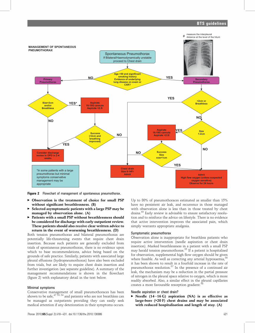
Both tension pneumothorax and bilateral pneumothorax arepotentially life-threatening events that require chest draininsertion. Because such patients are generally excluded fromtrials of spontaneous pneumothorax, there is no evidence uponwhich to base recommendations, advice being based on thegrounds of safe practice. Similarly, patients with associated largepleural effusions (hydropneumothorax) have also been excludedfrom trials, but are likely to require chest drain insertion andfurther investigation (see separate guideline). A summary of themanagement recommendations is shown in the flowchart(figure 2) with explanatory detail in the text below.
Minimal symptomsConservative management of small pneumothoraces has beenshown to be safe,47 53 54 and patients who are not breathless canbe managed as outpatients providing they can easily seekmedical attention if any deterioration in their symptoms occurs.
Up to 80% of pneumothoraces estimated as smaller than 15%have no persistent air leak, and recurrence in those managedwith observation alone is less than in those treated by chestdrains.55 Early review is advisable to ensure satisfactory resolu-tion and to reinforce the advice on lifestyle. There is no evidencethat active intervention improves the associated pain, whichsimply warrants appropriate analgesia.
Symptomatic pneumothoraxObservation alone is inappropriate for breathless patients whorequire active intervention (needle aspiration or chest draininsertion). Marked breathlessness in a patient with a small PSPmay herald tension pneumothorax.55 If a patient is hospitalisedfor observation, supplemental high flow oxygen should be givenwhere feasible. As well as correcting any arterial hypoxaemia,56
it has been shown to result in a fourfold increase in the rate ofpneumothorax resolution.57 In the presence of a continued airleak, the mechanism may be a reduction in the partial pressureof nitrogen in the pleural space relative to oxygen, which is morereadily absorbed. Also, a similar effect in the pleural capillariescreates a more favourable resorption gradient.58
Needle aspiration or chest drain?< Needle (14e16 G) aspiration (NA) is as effective as
large-bore (>20 F) chest drains and may be associatedwith reduced hospitalisation and length of stay. (A)
tnacifingis dna 05> egA
yrotsih gnikoms
gniylrednu fo ecnedivE
ro maxe no esaesid gnul
?RXC
yramirP
xarohtomuenP
yradnoceS
xarohtomuenP
mc2>eziS
ro/dna
sselhtaerB
etaripsA
alunnac G81-61
l5.2< etaripsA
sseccuS
dna mc2<(
gnihtaerb
)devorpmi
niard tsehC
rF41-8 eziS
timdA
egrahcsid redisnoC
4-2 ni DPO ni weiver
skeew
ro mc2>
sselhtaerB
etaripsA
alunnac G81-61
l5.2< etaripsA
sseccuS
eziS
mc1<won
timdA
detcepsus sselnu( negyxo wolf hgiH
)evitisnes negyxo
sruoh 42 rof evresbO
xarohtomuenP suoenatnopS elbatsnuyllacimanydomeaH/laretaliB fI
niard tsehC ot deecorp
ON
ON
ON
ON
ON
SEY
SEY
*SEY
SEY
SEY
egral a htiw stneitap emos nI* laminim tub xarohtomuenp
evitavresnoc smotpmys eb yam tnemeganam
etairporppa
eziS
mc2-1
ON
SEY
SUOENATNOPS FO TNEMEGANAM
XAROHTOMUENP
##
measure the interpleuraldistance at the level of the hilum#
Figure 2 Flowchart of management of spontaneous pneumothorax.
Thorax 2010;65(Suppl 2):ii18eii31. doi:10.1136/thx.2010.136986 ii21
BTS guidelines

< NA should not be repeated unless there were technicaldifficulties. (B)
< Following failed NA, small-bore (<14 F) chest draininsertion is recommended. (A)
< Large-bore chest drains are not needed for pneumo-thorax. (D)
Needle aspiration (NA) was recommended in the previousguidelines17 22 as the initial intervention for PSP on the basis ofstudies59 60 showing equivalent success to the insertion of large-bore chest drains, although this was not shown in anotherstudy.61 Seldinger (catheter over guide wire) chest drains haveentered widespread usage since then and further studies havebeen published. A randomised controlled trial in a Kuwaitipopulation has confirmed equivalence between NA and chestdrains (16 Fr), plus a reduction in hospital admission and lengthof stay for NA.62 A smaller study in India has also confirmedequivalence.63 Two recent case series have reported NA successrates of 69%64 and 50.5%.65 Several meta-analyses66e68 werelimited by the small numbers of patients and studies69e77 butconfirm equivalence, with NA success rates ranging from 30% to80% (see evidence table available on the BTS website www.brit-thoracic.org.uk). If undertaken, NA should cease after 2.5 l of airhas been aspirated, further re-expansion being unlikely59 becauseof the likely presence of a persistent air leak.
Guidelines that encourage NA are not always followed78e82
and the ease of insertion of small-bore (<14 F) Seldinger chestdrains may be regarded as a simpler option to NA. Their successhas been documented in several studies,83e89 the attachment ofHeimlich valves facilitating mobilisation and outpatient care.Small-bore chest drains have been shown to have a similarsuccess rate to larger drains90 while being less painful,91 92 butthere have been no randomised controlled trials comparing themwith NA. More detail on chest drain insertion and managementand complications of chest drain insertion are found in theguideline on pleural procedures. Catheter aspiration wasdescribed in the last guideline,22 with success in up to 59%, andfurther improvement with the addition of Heimlich valves andsuction.93e95 Seldinger chest drains have also permitted a ‘step-wise’ approach to PSP management, following a predefinedpathway that culminates in surgical referral where there isa persistent air leak.96
The choice of initial intervention for PSP should take intoaccount operator experience and patient choice; NA is lesspainful than chest drain insertion60 but failure in approximatelyone-third of patients will require a second procedure. Othernational and consensus guidelines recommend either NA orsmall-bore chest drain insertion,97 or chest drain insertionalone.49 We believe that NA remains the procedure of first choicein most cases. Repeat NA is unlikely to be successful unless therewere technical difficulties such as a blocked or kinked catheter.There is some limited evidence that VATS is the preferred‘salvage’ strategy after failed NA,98 but this is not the usualpractice currently in the UK where small-bore chest draininsertion is usually employed. Following successful NA, thepatient can be considered for hospital discharge.
Suction< Suction should not be routinely employed. (B)< Caution is required because of the risk of RPO. (B)< High-volume low-pressure suction systems are recom-
mended. (C)A persistent air leak with or without incomplete re-expansion ofthe lung is the usual reason for consideration of the use ofsuction, although there is no evidence for its routine use.99e101 It
is arbitrarily defined as the continued bubbling of air througha chest drain after 48 h in situ. A retrospective review of 142cases of pneumothorax102 found a median time to resolution of8 days which was not related to the initial size of pneumo-thorax, but longer for SSP. A persistent air leak was observed in43 cases, 30 of which were treated with suction. The theory thatunderpins the role of suction is that air might be removed fromthe pleural cavity at a rate that exceeds the egress of air throughthe breach in the visceral pleura and to subsequently promotehealing by apposition of the visceral and parietal pleural layers.It has been suggested that optimal suction should entail pres-sures of �10 to �20 cm H2O (compared with normal intra-pleural pressures of between �3.4 and �8 cm H2O, according tothe respiratory cycle), with the capacity to increase the air flowvolume to 15e20 l/min.103 Other forms of suction are notrecommended. High-pressure high-volume suction may lead toair stealing, hypoxaemia or the perpetuation of air leaks.104
Likewise, high-pressure low-volume systems should beavoided.105 High-volume low-pressure systems such as Vernon-Thompson pumps or wall suction with low pressure adaptorsare therefore recommended.The addition of suction too early after chest drain insertion
may precipitate RPO, especially in the case of a PSP that mayhave been present for more than a few days,106 and is thought tobe due to the additional mechanical stress applied to capillariesthat are already ‘leaky’.107 The clinical manifestations are cough,breathlessness and chest tightness after chest drain insertion.The incidence may be up to 14% (higher in younger patientswith a large PSP), although no more than a radiologicalphenomenon in the majority of cases.106 Sometimes pulmonaryoedema is evident in the contralateral lung.108 Fatalities havebeen reported in as many as 20% of 53 cases in one series,108 socaution should be exercised in this particular group of patients.
Specialist referral< Referral to a respiratory physician should be made
within 24 h of admission. (C)< Complex drain management is best effected in areas
where specialist medical and nursing expertise is avail-able. (D)
Failure of a pneumothorax to re-expand or a persistent air leakshould prompt early referral to a respiratory physician, prefer-ably within the first 24 h. Such patients may require prolongedchest drainage with complex drain management (suction, chestdrain repositioning) and liaison with thoracic surgeons. Drainmanagement is also best delivered by nurses with specialistexpertise. Surgical referral is discussed in a later section.
Surgical emphysemaThis is a well-recognised complication of chest drainage.109
Generally it is of cosmetic importance only, although alarmingfor patients and their relatives, and subsides spontaneously aftera few days. It is usually seen in the context of a malpositioned,kinked, blocked or clamped chest drain. It can also occur with animbalance between a large air leak and a relatively small-borechest drain. Occasionally, acute airway obstruction or thoraciccompression may lead to respiratory compromise109 110 in whichcase tracheostomy, skin incision decompression and insertion oflarge-bore chest drains have all been used.109 For most, thetreatment is conservative.
Management of SSP< All patients with SSP should be admitted to hospital for
at least 24 h and receive supplemental oxygen in
ii22 Thorax 2010;65(Suppl 2):ii18eii31. doi:10.1136/thx.2010.136986
BTS guidelines

compliance with the BTS guidelines on the use ofoxygen. (D)
< Most patients will require the insertion of a small-borechest drain. (B)
< All patients will require early referral to a chestphysician. (D)
< Those with a persistent air leak should be discussedwith a thoracic surgeon at 48 h. (B)
As stated previously, SSP is less likely to be tolerated bypatients than PSP because of co-existing lung disease.Furthermore, the air leak is less likely to settle spontane-ously,111 112 so that most patients will require active inter-vention. Oxygen is indicated,56 57 but caution is required forpatients with carbon dioxide retention.113 Aspiration is lesslikely to be successful in SSP (see evidence table available on theBTS website at www.brit-thoracic.org.uk) but can be consid-ered in symptomatic patients with small pneumothoraces in anattempt to avoid chest drain insertion. Otherwise, the inser-tion of a small-bore chest drain is recommended, a study inSSP114 having found equivalent success to the use of largedrains. Early referral to a chest physician is encouraged for allpatients with SSP, both for management of the pneumothoraxand also of the underlying lung disease. Similarly, those witha persistent air leak should be discussed with a thoracic surgeonafter 48 h,112 115 even though many will resolve spontaneously ifmanaged conservatively for as long as 14 days.111
Patients with SSP but unfit for surgery< Medical pleurodesis may be appropriate for inoperable
patients. (D)< Patients with SSP can be considered for ambulatory
management with a Heimlich valve. (D)These patients are at heightened risk of a persistent air leak butmay not be fit for surgical intervention by virtue of the severityof their underlying lung disease, or they may be unwilling toproceed. Their optimal management is challenging and requiresclose medical and surgical liaison. Medical pleurodesis is anoption for such patients, as is ambulatory management with theuse of a Heimlich valve.86
DISCHARGE AND FOLLOW-UP< Patients should be advised to return to hospital if
increasing breathlessness develops. (D)< All patients should be followed up by respiratory
physicians until full resolution. (D)< Air travel should be avoided until full resolution. (C)< Diving should be permanently avoided unless the
patient has undergone bilateral surgical pleurectomyand has normal lung function and chest CT scanpostoperatively. (C)
All patients discharged after active treatment or otherwiseshould be given verbal and written advice to return to theAccident and Emergency department immediately should theydevelop further breathlessness. It is recommended that allpatients should be followed up by a respiratory physician toensure resolution of the pneumothorax, to institute optimal careof any underlying lung disease, to explain the risk of recurrenceand the possible later need for surgical intervention and toreinforce lifestyle advice on issues such as smoking and air travel.Those managed by observation alone or by NA should beadvised to return for a follow-up chest x-ray after 2e4 weeks tomonitor resolution. Those with successful lung re-expansionbefore hospital discharge will also require early review becauserecurrence may occur relatively early.
Since there is no evidence to link recurrence with physicalexertion, the patient can be advised to return to work and toresume normal physical activities once all symptoms haveresolved, although it is reasonable to advise that sports thatinvolve extreme exertion and physical contact should be deferreduntil full resolution. Patients should be made aware of thedanger of air travel in the presence of a current closed pneu-mothorax, and should be cautioned against commercial flights athigh altitude until full resolution of the pneumothorax has beenconfirmed by a chest x-ray. While there is no evidence that airtravel per se precipitates pneumothorax recurrence, the conse-quences of a recurrence during air travel may be serious. Manycommercial airlines previously advised arbitrarily a 6-weekinterval between the pneumothorax event and air travel, butthis has since been amended to a period of 1 week after fullresolution. The BTS guidelines on air travel116 emphasise thatthe recurrence risk only significantly falls after a period of 1 yearfrom the index pneumothorax so that, in the absence ofa definitive surgical procedure, patients with SSP in particularmay decide to minimise the risk by deferring air travel accord-ingly. After a pneumothorax, diving should be discouragedpermanently117 unless a very secure definitive preventionstrategy has been performed such as surgical pleurectomy. TheBTS guidelines on respiratory aspects of fitness for diving118 dealwith this in greater detail. Smoking influences the risk ofrecurrence,12 15 so cessation should be advised. Pregnancy is anissue to be discussed with younger female patients.
MEDICAL CHEMICAL PLEURODESIS< Chemical pleurodesis can control difficult or recurrent
pneumothoraces (A) but, since surgical options aremore effective, it should only be used if a patient iseither unwilling or unable to undergo surgery. (B)
< Chemical pleurodesis for pneumothorax should only beperformed by a respiratory specialist. (C)
Chemical pleurodesis has generally been advocated by respira-tory physicians experienced in thoracoscopy. The instillation ofsubstances into the pleural space should lead to an asepticinflammation, with dense adhesions leading ultimately topleural symphysis. There is a significant rate of recurrence ofboth primary and secondary pneumothoraces,12 and efforts toreduce recurrence by instilling various sclerosantsdeither viaa chest drain, video-assisted thoracoscopic surgery (VATS) oropen surgerydare often undertaken without clear guidelines todirect physicians in their use. In the vast majority of cases theprevention of recurrent pneumothoraces should be undertakensurgically using either an open or VATS approach, as the rateof recurrence following surgical pleurodesis via thoracotomyor VATS is far less than following simple medical pleurodesiswith chemical agents,32 119e121 although direct comparativetrials are lacking. A small number of patients are either toofrail or are unwilling to undergo any surgical treatment and,in these situations, medical chemical pleurodesis may beappropriate.Many sclerosing agents suitable for instillation into the
pleural space have been studied.32 119 122e125 Tetracycline used tobe recommended as the first-line sclerosant therapy for bothprimary and secondary pneumothoraces as it proved to be themost effective sclerosant in animal models.123 126 127 Recently,however, parenteral tetracycline for pleurodesis has becomemore difficult to obtain owing to problems with themanufacturing process. Minocycline and doxycycline have alsobeen shown to be reasonable alternative sclerosing agents inanimal models.126 127
Thorax 2010;65(Suppl 2):ii18eii31. doi:10.1136/thx.2010.136986 ii23
BTS guidelines

The rate of recurrence of pneumothorax is the primary indi-cator for success for any recurrence prevention techniques.Although tetracycline has been shown to reduce the incidence ofearly recurrence, the incidence of late recurrence remains at10e20% which is unacceptably high when compared withsurgical methods of pleurodesis.119 121 125 128 129 Tetracycline canbe recommended for recurrent primary and secondary pneumo-thorax when surgery is not an option, and graded talc may also beused on the grounds that it is the most effective agent in treatingmalignant pleural effusion and is also commonly used for surgicalchemical pleurodesis.130e133 There is conflicting evidence as towhether tetracycline is effective for the treatment of a fullyexpanded pneumothorax with a persistent air leak.32 134 135 Thelargest of these studies, the Veterans Administration Study, didnot support the use of intrapleural tetracycline to facilitate theclosure of a persistent air leak.31 Macoviak and colleagues135
suggest that intrapleural tetracycline can facilitate the closure ofa persistent air leak provided the lung can be kept expanded sothat symphysis can occur. Likewise, there is conflicting evidenceas to whether intrapleural tetracycline shortens the length of stayin hospital with pneumothorax.32 119 125
The dosage of intrapleural tetracycline requires clarification.Almind119 found a reduction in the recurrence rate in a groupreceiving 500 mg tetracycline via chest drains compared withthose treated by tube drainage alone. This reduction was notsignificant. The Veterans Administration Study,32 which used1500 mg tetracycline, showed a significant reduction in therecurrence rate of pneumothorax without significant extramorbidity. This dose of intrapleural tetracycline is thereforerecommended as the standard dose for medical pleurodesis.While pain was reported more frequently in the group treatedwith tetracycline at a dose of 1500 mg,32 others have reported noincrease in pain with doses of 500 mg provided adequate anal-gesia is given.119 Adequate analgesia may be achieved with theadministration of intrapleural local anaesthesia. Standard doses(200 mg (20 ml) of 1% lidocaine) are significantly less effectivethan larger doses (250 mg (25 ml) of 1% lidocaine), the higherdoses having been shown to increase the number of pain-freeepisodes from 10% to 70% with no appreciable toxicity.136
Chemical pleurodesis using graded talc is an effective alter-native to tetracycline pleurodesis, but there are no controlledtrials comparing the two in the treatment of pneumothorax.The issue of talc pleurodesis is discussed in the later section onsurgical chemical pleurodesis as most trials using talc relate to itsuse in either thoracoscopic or open surgical techniques. Since werecognise chemical pleurodesis as an inferior option to surgicalpleurodesis, we recommend that chemical pleurodesis should beundertaken by respiratory physicians or thoracic surgeons only.
REFERRAL TO THORACIC SURGEONS< In cases of persistent air leak or failure of the lung to re-
expand, an early (3e5 days) thoracic surgical opinionshould be sought. (C)
There is no evidence on which to base the ideal timing forthoracic surgical intervention in cases of persistent air leak. A cut-off point of 5 days has been widely advocated in the past55 but isarbitrary. Chee et al111 showed that 100% of primary pneumo-thoraces with a persistent air leak for>7 days and treated by tubedrainage had resolved by 14 days. Also, 79% of those withsecondary pneumothoraces and a persistent air leak had resolvedby 14 days, with no mortality in either group. However, surgicalintervention carries a low morbidity128 129 137e140 and post-surgical recurrence rates are low.128 129 Surgical intervention asearly as 3 days has advocates,141 142 but there is no evidence that
intervention before 5 days is necessary for PSP. Each case shouldbe assessed individually on its own merit. Patients with pneu-mothoraces should be managed by a respiratory physician, anda thoracic surgical opinion will often form an early part of themanagement plan.Accepted indications for surgical advice should be as follows:
< Second ipsilateral pneumothorax.< First contralateral pneumothorax.< Synchronous bilateral spontaneous pneumothorax.< Persistent air leak (despite 5e7 days of chest tube drainage) or
failure of lung re-expansion.< Spontaneous haemothorax.143 144
< Professions at risk (eg, pilots, divers).111 138 145e147
< Pregnancy.Increasingly, patient choice will play a part in decision-
making, and even those without an increased risk in the event ofa pneumothorax because of their profession may elect toundergo surgical repair after their first pneumothorax,148 149
weighing the benefits of a reduced recurrence risk against that ofchronic pain,150 paraesthesia151 or the possibility of increasedcosts.152
Surgical strategies: open thoracotomy or VATS?< Open thoracotomy and pleurectomy remain the proce-
dure with the lowest recurrence rate (approximately1%) for difficult or recurrent pneumothoraces. (A)
< Video-assisted thoracoscopic surgery (VATS) withpleurectomy and pleural abrasion is better tolerated buthas a higher recurrence rate of approximately 5%. (A)
There are two main objectives in the surgical repair of persistentair leak from a pneumothorax and in the prevention of recur-rence. The first objective is to resect any visible bullae or blebs onthe visceral pleura and also to obliterate emphysema-likechanges9 or pleural porosities under the surface of the visceralpleura.8 The second objective is to create a symphysis betweenthe two opposing pleural surfaces as an additional means ofpreventing recurrence. In the past, surgeons have tended tofavour a surgical pleurodesis with pleural abrasion while othershave stressed the importance of various degrees of pleurectomyin recurrence prevention.137 153 154 Although there may be slightadvantages of pleurectomy over pleural abrasion,137 a combina-tion of the two is often used.155e158 Unfortunately there isa paucity of good comparative case-controlled studies in thisarea.128 129 In recent years, less invasive procedures using VATShave become more popular with lower morbidity although withslightly higher recurrence rates.Open thoracotomy with pleural abrasion was the original
surgical treatment for pneumothorax, described by Tyson andCrandall in 1941.159 In 1956 Gaensler introduced parietalpleurectomy for recurrent pneumothoraces, encouragingpleural symphysis through adhesions between the visceralpleura and the chest wall.153 Closure of the leaking visceralpleura with direct cautery and ligation or suture of associatedblebs147 is also thought to be important. Although openthoracotomy has the lowest pneumothorax recurrence rates,there are also lesser surgical procedures with comparablerecurrence rates but less morbidity.160 These include trans-axillary minithoracotomy, using a 5e6 cm incision in theaxillary margin with apical pleurectomy and pleural abrasion,introduced in the 1970s.161 Open thoracotomy is generallyperformed with a limited posterolateral approach and singlelung ventilation. This allows for a parietal pleurectomy withexcision, stapling or ligation of visible bullae and pleural abra-sion.162 Isolated lung ventilation during open thoracotomy
ii24 Thorax 2010;65(Suppl 2):ii18eii31. doi:10.1136/thx.2010.136986
BTS guidelines
renders easier visualisation of the visceral pleura than duringa VATS procedure.163e165 Meta-analyses of studies comparingopen with limited or VATS procedures128 129 have shown lowerrecurrence rates (approximately 1%) with open procedures butgreater blood loss, more postoperative pain166 and longerhospital stays.167 Some non-randomised studies have found nosignificant differences.168 169 A complicated meta-analysis ofthree retrospective studies and one prospective studycomparing the cost of open thoracotomy versus VATS (notexclusively for pneumothoraces) concluded that the totaleconomic cost of VATS was lower,170 and it can be undertakenwithout general anaesthesia.149 There is a need for betterquality prospective randomised studies in this area. Severalauthors suggest that VATS offers a significant advantage overopen thoracotomy, including a shorter postoperative hospitalstay,145 162 167 171e173 less postoperative pain160 162 166 174 175
and improved pulmonary gas exchange postoperatively,176
although not all trials have confirmed shorter hospital stayswith VATS.169 177
Much of the literature contains heterogeneous comparisonsbetween PSP and SSP, but the most recent ‘clinical bottomline’129 concludes that VATS pleurectomy is comparable to openpleurectomy, with several randomised controlled trials showingreductions in length of hospital stay, analgesic requirement andpostoperative pulmonary dysfunction. Clearly this needs to beweighed against the slight increase in recurrence rate when usinga less invasive approach.128
Surgical chemical pleurodesis< Surgical chemical pleurodesis is best achieved by using
5 g sterile graded talc, with which the complications ofadult respiratory distress syndrome and empyema arerare. (A)
With the advent of VATS for pneumothorax repair and recur-rence prevention, the use of surgical chemical pleurodesis hasdeclined significantly. Previous reports have shown that talc canachieve pleurodesis successfully in 85e90% of cases, similar toother thoracoscopic techniques for complicated pneumo-thorax.121 145 171 178 179 A meta-analysis of the success rates oftalc pleurodesis in the treatment of pneumothorax shows anoverall success rate of 91%.178 Graded talc is preferable to tetra-cycline, which is less available now, and is associated with muchhigher recurrence rates.120 Much of the literature concerning theuse of talc in achieving pleurodesis relates to its use in the controlof malignant pleural effusions, although talc poudrage has beenused successfully in secondary pneumothoraces.180 On the basisof a systematic review of uncontrolled trials, 5 g of intrapleuraltalc via VATS achieves a success rate of 87%.178
The adult respiratory distress syndrome has been describedfollowing the use of talc. This probably relates to the size of thetalc particles181 and is unlikely to occur with the use of gradedtalc.182 183 If talc is correctly sterilised, the incidence ofempyema is very low.178 184 185 There does not appear to bea difference between talc poudrage and talc slurry pleurodesis.The advent of successful and well-tolerated VATS surgery willlead to less use of surgical chemical pleurodesis with talc. Inthose patients who are either unwilling or too unwell toundergo a VATS procedure, then medical pleurodesis with talcvia a chest drain would be the preferred option.
TENSION PNEUMOTHORAX< Tension pneumothorax is a medical emergency that
requires heightened awareness in a specific range ofclinical situations. (D)
< Treatment is with oxygen and emergency needledecompression. (D)
< A standard cannula may be insufficiently long if used inthe second intercostal space. (D)
This is a medical emergency that can arise in a variety ofclinical situations, so a high index of suspicion is required inorder to make the correct diagnosis and to manage it effec-tively. The most frequent situations are shown in box 1,although the list does not include all eventualities. It arises asa result of the development of a one-way valve system at thesite of the breach in the pleural membrane, permitting air toenter the pleural cavity during inspiration but preventingegress of air during expiration, with consequent increase in theintrapleural pressure such that it exceeds atmospheric pressurefor much of the respiratory cycle. As a result, impaired venousreturn and reduced cardiac output results in the typical featuresof hypoxaemia and haemodynamic compromise.186 187
A recent review188 has emphasised the important differencesbetween the presentation in ventilated and non-ventilatedpatients, where it is typically seen after trauma or resuscitation.The former group is associated with a uniformly rapid presen-tation with hypotension, tachycardia, falling oxygen saturationand cardiac output, increased inflation pressures and cardiacarrest. This is frequently missed in the ICU setting37 and canalso occur after nasal non-invasive ventilation (NIV). The lattergroup of awake patients show a greater variability of presenta-tions which are generally progressive with slower decompensa-tion. Tachypnoea, tachycardia and hypoxaemia lead eventuallyto respiratory arrest. Apart from these general physical signs, themost frequent lateralising sign found in a review of 18 casereports188 was that of decreased air entry (50e75%), with signsof tracheal deviation, hyperexpansion, hypomobility andhyperresonance present only in the minority.In neither group is imaging especially helpful; there is usually
insufficient time to obtain a chest x-ray and, even if available,the size of the pneumothorax or the presence of mediastinaldisplacement correlate poorly with the presence of tensionwithin a pneumothorax. However, a chest x-ray can, whentime is available, confirm the presence of a pneumothorax (ifuncertain) and the correct side.Treatment is with high concentration oxygen and emergency
needle decompression, a cannula usually being introduced in thesecond anterior intercostal space in the mid-clavicular line. Theinstantaneous egress of air through the majority of the respira-tory cycle is an important confirmation of the diagnosis and thecorrect lateralisation. A standard 14 gauge (4.5 cm) cannula maynot be long enough to penetrate the parietal pleura, however,with up to one-third of patients having a chest wall thickness
Box 1 Typical clinical situations where tensionpneumothorax arises
1. Ventilated patients on ICU.2. Trauma patients.3. Resuscitation patients (CPR).4. Lung disease, especially acute presentations of asthma and
chronic obstructive pulmonary disease.5. Blocked, clamped or displaced chest drains.6. Patients receiving non-invasive ventilation (NIV).7. Miscellaneous group, for example patients undergoing
hyperbaric oxygen treatment.
Thorax 2010;65(Suppl 2):ii18eii31. doi:10.1136/thx.2010.136986 ii25
BTS guidelines

>5 cm in the second interspace.189 The chest wall may be lessdeep in the fourth or fifth interspace, and this could provide analternative site for decompression or a chest drain may need tobe inserted if there is an initial treatment failure. In any case,a chest drain should be inserted immediately after needledecompression and the cannula left in place until bubbling isconfirmed in the underwater seal system to confirm properfunction of the chest drain.186
PNEUMOTHORAX AND PREGNANCY< Pneumothorax recurrence is more common in preg-
nancy, poses risks to the mother and fetus, and requiresclose cooperation between chest physicians, obstetri-cians and thoracic surgeons. (C)
< The modern and less invasive strategies of simpleobservation and aspiration are usually effective duringpregnancy, with elective assisted delivery and regionalanaesthesia at or near term. (C)
< A corrective surgical procedure (VATS) should beconsidered after delivery. (D)
Although less common in women than in men, the occurrenceof PSP in women of childbearing age is not unusual. Thereappears to be an increased risk of recurrence during pregnancyand during parturition,190 with potential risks to the mother andfetus. The earlier literature consists largely of case reports anddescribed varied and relatively invasive management strategiessuch as prolonged intrapartum chest tube drainage, intrapartumthoracotomy, premature induction of labour or caesareansection. A more recent case series and literature review191 hasrecommended the use of more modern conservative manage-ment methods for which favourable outcomes have now beenexperienced. Pneumothorax that occurs during pregnancy can bemanaged by simple observation if the mother is not dyspnoeic,there is no fetal distress and the pneumothorax is small (<2 cm).Otherwise aspiration can be performed, chest drain insertionbeing reserved for those with a persistent air leak.
Close cooperation between the respiratory physician, obste-trician and thoracic surgeon is essential. To avoid spontaneousdelivery or caesarean section, both of which have been associatedwith an increased risk of recurrence, the safest approach willusually be that of elective assisted delivery (forceps or ventouseextraction) at or near term, with regional (epidural) anaesthesia.Less maternal effort is required with forceps delivery, which istherefore preferable. If caesarean section is unavoidable becauseof obstetric considerations, then a spinal anaesthetic is preferableto a general anaesthetic.
Because of the risk of recurrence in subsequent pregnancies,a minimally invasive VATS surgical procedure should beconsidered after convalescence. Successful pregnancies andspontaneous deliveries without pneumothorax recurrence havebeen reported after a VATS procedure.191
CATAMENIAL PNEUMOTHORAX< Catamenial pneumothorax is underdiagnosed in women
with pneumothorax. (C)< A combination of surgical intervention and hormonal
manipulation requires cooperation with thoracicsurgeons and gynaecologists. (D)
Catamenial is a term that derives from the Greek meaning‘monthly ’. The typical combination of chest pain, dyspnoea andhaemoptysis occurring within 72 h before or after menstruationin young women has been thought to be relatively rare. There areapproximately 250 cases described in the medical literature,192
but it is likely that the majority of cases are not reported. Most ofthese references are of solitary case reports or small series. Theassociated pneumothorax is usually right-sided and there isa heightened tendency to recurrence coinciding with themenstrual cycle. Many cases have evidence of pelvic endometri-osis. Although the aetiology is not fully understood, inspection ofthe pleural diaphragmatic surface at thoracoscopy often revealsdefects (termed fenestrations) as well as small endometrialdeposits. These deposits have also been seen on the visceralpleural surface. Among women undergoing routine surgicaltreatment for recurrent pneumothorax, however, catamenialpneumothorax has been diagnosed in as many as 25%.193 Thus, itmay be relatively underdiagnosed.Extragenital or ‘ectopic’ endometriosis is an uncommon
condition that can affect almost any organ system and tissuewithin the body, the thorax being the most frequent extrapelviclocation. What has been termed the thoracic endometriosissyndrome (TES) includes catamenial pneumothorax, catamenialhaemothorax, catamenial haemoptysis and lung nodules (purpleor brown coloured). The most accepted theory to explain thephenomenon of catamenial pneumothorax is that of aspirationof air from the abdomen and genital tract via the diaphragmaticfenestrations, but the appearance of endometriosis deposits onthe visceral pleural surface raises the possibility that erosion ofthe visceral pleura might be an alternative mechanism.Haemoptysis is thought to result from intrapulmonary endo-metriosis deposits, the mechanism by which endometrial tissuereaches the lung being poorly understood.The management strategies can be divided into thoracic
surgical techniques and hormonal manipulation although, in thepast, total abdominal hysterectomy and bilateral salpingohys-terectomy have been employed. Thoracic surgical techniqueshave been varied and include diaphragmatic resection or plica-tion of the fenestrations seen at thoracoscopy, the insertion ofa mesh or patch over these fenestrations, electrocoagulation ofthe endometriosis deposits and pleurodesis. This variabilityreflects the general lack of success with surgical interventionalone, recurrence rates of up to 30% being documented.194 Whencombined with gonadotrophin-releasing hormone analoguesamenorrhoea results, but recurrence has been avoided withfollow-up approaching periods of 4 years.195 Successful patientmanagement requires close cooperation between respiratoryphysicians, thoracic surgeons and gynaecologists.
PNEUMOTHORAX AND AIDS< The combination of pneumothorax and HIV infection
requires early intercostal tube drainage and surgicalreferral, in addition to appropriate treatment for HIVand PJP infection. (C)
Over the course of the last 20 years a strong association hasbeen observed between HIV infection and pneumothorax.Historically, up to 5% of AIDS patients developed pneumo-thorax196e198 and up to 25% of spontaneous pneumothoracesoccurred in HIV-infected patients in large urban settings wherea high prevalence occurred.27 28 199 Pneumocystis jiroveci (PJP)dpreviously known as Pneumocystis carinii (PCP)dinfection hasbeen considered to be the main aetiological factor for thisassociation, because of a severe form of necrotising alveolitisthat occurs in which the subpleural pulmonary parenchyma isreplaced by necrotic thin-walled cysts and pneumatoceles.200 201
The administration of nebulised pentamidine has also beensuggested as a possible independent risk factor.196 The use ofsystemic corticosteroids may also contribute to the morbidity insuch patients.202
ii26 Thorax 2010;65(Suppl 2):ii18eii31. doi:10.1136/thx.2010.136986
BTS guidelines

Due to the histopathology outlined above, pneumothoracescaused by PJP have a tendency to more prolonged air leaks,treatment failure, recurrence and higher hospital mortality.203
Up to 40% of these patients can develop bilateral pneumo-thorax. Treatment failures have been observed to correlate withthe degree of immunosuppression, as reflected by CD4counts.203 In view of these features, management strategies havebeen evolved that incorporate early and aggressive interventionincluding tube drainage, pleurodesis and surgical techniques suchas pleurectomy.197 199 202e205 Observation and simple aspirationare not likely to suffice, even in the first instance.
Over the last 5 years, and since the last BTS guidelines, theglobal spectrum of HIV infection has changed significantly asa result of the more widespread use of both antiretroviraltherapy and PJP prophylaxis. While the disease burden remainsvery high in the underdeveloped world, the prognosis for suchpatients in Western societies has greatly improved,206 where thiscombination is now much less frequently encountered. As HIVis now becoming a more chronic disease associated with a highincidence of smoking and therefore of COPD, pneumothoracesmight become more significant when they occur.
However, the mortality of patients who require intensive carefor PJP in HIV infection remains high, especially when pneu-mothorax occurs during ventilation. Although antiretroviraltherapy that is commenced before or during hospitalisation canimprove the outcome,207 the potential risk of the ‘immunereconstitution syndrome’ has to be taken into consideration.
PNEUMOTHORAX AND CYSTIC FIBROSIS< The development of a pneumothorax in a patient with
cystic fibrosis requires early and aggressive treatmentwith early surgical referral. (C)
< Pleural procedures, including pleurodesis, do not havea significant adverse effect on the outcome of subse-quent lung transplantation. (D)
Even though long-term survival has improved significantly, spon-taneous pneumothorax remains a common complication of cysticfibrosis, occurring in 0.64% of patients per annum and 3.4% ofpatients overall.208 It occurs more commonly in older patients andthose with more advanced lung disease, and is associated with apoor prognosis, the median survival being 30 months.209 Contra-lateral pneumothoraces occur in up to 40% of patients.209 210 Anincreased morbidity also results, with increased hospitalisationand a measurable decline in lung function.208 While a smallpneumothorax without symptoms can be observed or aspirated,larger pneumothoraces require a chest drain. The collapsed lungcan be stiff and associated with sputum retention, thus requiringa longer time to re-expand. During this time other generalmeasures, such as appropriate antibiotic treatment, are needed.
Chest tube drainage alone has a recurrence rate of 50%, butinterventions such as pleurectomy, pleural abrasion and pleu-rodesis have lower rates.211e213 With a success rate of 95% andwith little associated reduction in pulmonary function, partialpleurectomy is generally regarded as the treatment of choice inpatients with cystic fibrosis and recurrent pneumothoraceswho are fit to undergo surgery.209 In those who are not fit forsurgery and in whom re-expansion may take several weekswith a chest drain and suction, pleurodesis offers an alternativestrategy.209 This had been thought to be a relative contraindi-cation to later transplantation because of the need fora lengthier transplant procedure and excessive bleeding.214 Amore recent study215 has concluded that previous pleuralprocedures should not be considered as a contraindication fortransplantation, there being no significant effect on surgical
outcome although more dense pleural adhesions were observedthan in a control population.
IATROGENIC PNEUMOTHORAXIatrogenic pneumothorax has been shown to be even morecommon than spontaneous pneumothorax in several largereviews,216 217 the most common causes being transthoracicneedle aspiration (24%), subclavian vessel puncture (22%), thor-acocentesis (22%), pleural biopsy (8%) andmechanical ventilation(7%).218 It is also a complication of transbronchial biopsy. Duringtransthoracic needle aspiration the two primary risk factors arethe depth of the lesion and the presence of COPD.219 A largeretrospective survey in the USA found an incidence of 2.68%among patients undergoing thoracocentesis.220 No means ofreducing this risk has yet been identified. Positioning of the patientso that the procedure is performed in a dependent area has had nobeneficial effect.221 Excluding iatrogenic penumothorax thatoccurs in intensive care units, the treatment seems to be relativelysimple with less likelihood of recurrence (the underlying riskfactors for SP not usually being present). The majority resolvespontaneously by observation alone. If intervention is required,simple aspiration has been shown to be effective in 89% ofpatients.94 For the remainder a chest drain is required, this beingmore likely in patients with COPD.222
In the intensive care unit iatrogenic pneumothorax is a life-threatening complication that may be seen in up to 3% ofpatients.223 Those on positive pressure ventilation require chestdrain insertion as positive pressure maintains the air leak.224
CONCLUDING REMARKSThese pneumothorax guidelines differ from the last (2003) BTSguidelines in that they have been produced in accordancewith theSIGN methodology and therefore have necessitated a carefulanalysis of the current underlying evidence. Unfortunately thereare relatively few adequate studies that address the main areas ofuncertainty, and few additions to the knowledge base in the last 7years.Nevertheless, some subtle changes inpractice have occurred.These are incorporated, together with coverage of some additionaltopics of relevance suchas catamenial pneumothorax and the issueof pneumothorax in pregnancy. The treatment algorithm is nowillustrated on a single flowchart for both PSP and SSP and placesslightly less emphasis on the size of the pneumothorax and moreon the clinical features. However, the trend towards moreconservative management is maintained, with observation formany patients with PSP, aspiration for the remainder, and small-bore chest drains for persistent air leaks. The imaging of pneu-mothorax has undergone a major change due to the advent ofPACS technology, and the implications of this are now described.Surgical practice has also developedwith thewidespread adoptionof less invasive (VATS) procedures rather than open thoracoto-mies. While the challenge of pneumothorax management inpatients with cystic fibrosis remains, there has been a significantreduction in pneumothorax in patients with HIV since theintroduction of antiretroviral therapy and PJP prophylactictherapy, in the countries with advanced healthcare systems atleast. It is hoped that these guidelines build upon their predeces-sors and lead to improved care for patients with pneumothorax,and that they informand support the clinicianswho care for them.
Competing interests No member of the Guideline Group is aware of any competinginterests.
Provenance and peer review The draft guideline was available for online publicconsultation (July/August 2009) and presented to the BTS Winter Meeting (December2009). Feedback was invited from a range of stakeholder institutions (see
Thorax 2010;65(Suppl 2):ii18eii31. doi:10.1136/thx.2010.136986 ii27
BTS guidelines

Introduction). The draft guideline was reviewed by the BTS Standards of CareCommittee (September 2009).
REFERENCES1. Laennec RTH. Traite du diagnostic des maladies des poumons et du coeur.
Tome Second, Paris: Brosson and Chaude, 1819. (4).2. Kjærgaard H. Spontaneous pneumothorax in the apparently healthy. Acta Med
Scand (Suppl) 1932;43:1e159. (3).3. Melton LJ, Hepper NCG, Offord KP. Incidence of spontaneous pneumothorax in
Olmsted County, Minnesota: 1950e1974. Am Rev Respir Dis1987;29:1379e82. (3).
4. Gupta D, Hansell A, Nichols T, et al. Epidemiology of pneumothorax in England.Thorax 2000;55:666e71. (3).
5. Donahue DM, Wright CD, Viale G, et al. Resection of pulmonary blebs andpleurodesis for spontaneous pneumothorax. Chest 1993;104:1767e9. (2L).
6. Lesur O, Delorme N, Frogamet JM, et al. Computed tomography in theaetiological assessment of idiopathic spontaneous pneumothorax.Chest 1990;98:341e7. (2+).
7. Noppen M, Dekeukeleire T, Hanon S, et al. Fluorescein-enhanced autofluorescencethoracoscopy in patients with primary spontaneous pneumothorax. Am J Respir CritCare Med 2006;174:26e30. (3).
8. Schramel F, Meyer CJ, Postmus P. Inflammation as a cause of spontaneouspneumothorax (SP) and emphysematous like changes (ELC)? Results ofbronchoalveolar lavage (BAL). Eur Respir J 1995;8(Suppl 19):397. (2L).
9. Bense L, Eklund G, Odont D, et al. Smoking and the increased risk of contractingpneumothorax. Chest 1987;92:1009e12. (2++).
10. Withers JN, Fishback ME, Kiehl PV, et al. Spontaneous pneumothorax. Am J Surg1964;108:772e6. (3).
11. Sadikot RT, Greene T, Meadows K, et al. Recurrence of primary pneumothorax.Thorax 1997;52:805e9. (3).
12. West JB. Distribution of mechanical stress in the lung, a possible factor inlocalisation of pulmonary disease. Lancet 1971;1:839e41. (4).
13. Bense L, Wiman L-G, Hedenstierna G. Onset of symptoms in spontaneouspneumothorax: correlations to physical activity. Eur J Respir Dis 1987;71:181e6. (3).
14. Smit HJM, Chatrou M, Postmus PE. The impact of spontaneous pneumothorax,and its treatment, on the smoking behaviour of young adult smokers. Respir Med1998;92:1132e6. (2L).
15. Lippert HL, Lund O, Blegrad S, et al. Independent risk factors for cumulativerecurrence rate after first spontaneous pneumothorax. Eur Respir J1991;4:324e31. (2+).
16. Videm V, Pillgram-Larsen J, Ellingsen O, et al. Spontaneous pneumothorax inchronic obstructive pulmonary disease: complications, treatment and recurrences.Eur J Respir Dis 1987;71:365e71. (2+).
17. Miller AC, Harvey JE. Guidelines for the management of spontaneouspneumothorax. BMJ 1993;307:114e16. (4).
18. Grimshaw JM, Russell IT. Effect of clinical guidelines on medical practice:a systematic review of rigorous evaluation. Lancet 1993;342:1317e21. (3).
19. Bero BA, Grilli R, Grimshaw JM, et al. Closing the gap between research andpractice: an overview of systematic reviews of interventions to promote theimplementation of research findings. BMJ 1998;317:465e8. (4).
20. Grilli R, Lomas J. Evaluating the message: the relationship between compliancerate and the subject of a practice guideline. Med Care 1994;32:202e13. (2+).
21. Woolf SH, Grol R, Hutchinson A, et al. Potential benefits, limitations and harms ofclinical guidelines. BMJ 1999;318:527e30. (4).
22. Henry M, Arnold T, Harvey J. BTS guidelines for the management of spontaneouspneumothorax. Thorax 2003;58(Suppl II):39e52. (4).
23. Miller A. Spontaneous pneumothorax. In: Light RW, Lee YCG, eds. Textbook ofpleural diseases. 2nd edn. London: Arnold Press, 2008:445e63. (4).
24. O’Hara VS. Spontaneous pneumothorax. Milit Med 1978;143:32e5. (3).25. Pavlin J, Cheney FW Jr. Unilateral pulmonary edema in rabbits after re-expansion
of collapsed lung. J Appl Physiol 1979;46:31e5. (3).26. Miller WC, Toon R, Palat H, et al. Experimental pulmonary edema following re-
expansion of pneumothorax. Am Rev Respir Dis 1973;108:664e6. (3).27. Wait MA, Estrera A. Changing clinical spectrum of spontaneous pneumothorax.
Am J Surg 1992;164:528e31. (2+).28. Tanaka F, Itoh M, Esaki H, et al. Secondary spontaneous pneumothorax.
Ann Thorac Surg 1993;55:372e6. (2L).29. Vail WJ, Alway AE, England NJ. Spontaneous pneumothorax. Dis Chest
1960;38:512e15. (3).30. Serementis MG. The management of spontaneous pneumothorax. Chest
1970;57:65e8. (3).31. Norris RM, Jones JG, Bishop JM. Respiratory gas exchange in patients with
spontaneous pneumothorax. Thorax 1968;23:427e33. (2+).32. Light RW, O’Hara VS, Moritz TE, et al. Intrapleural tetracycline for the prevention of
recurrent spontaneous pneumothorax: results of a Department of Veterans AffairsCo-operative Study. JAMA 1990;264:2224e30. (1+).
33. Glazer H, Anderson J, Wilson BS, et al. Pneumothorax:appearances on lateralchest radiographs. Radiology 1989;173:707e11. (3).
34. Schramel FMNH, Wagenaar M, Sutedja TG, et al. Diagnosis of pneumothorax notimproved by additional roentgen pictures of the thorax in the expiratory phase.Ned Tijdschr Geneeskd 1995;139:131e3. (2+).
35. Schramel FM, Golding RP, Haakman CD, et al. Expiratory chest radiographs do notimprove visibility of small apical pneumothoraces by enhanced contrast.Eur Respir J 1996;9:406e9. (2+).
36. Seow A, Kazerooni EA, Cascade PN, et al. Comparison of upright inspiratory andexpiratory chest radiographs for detecting pneumothoraces. AJR1996;166:313e16. (1+).
37. Tocino IM, Miller MH, Fairfax WR. Distribution of pneumothorax in the supine andsemirecumbent critically ill adult. AJR 1985;144:901e5. (3).
38. Beres RA, Goodman LR. Pneumothorax:detection with upright versus decubitusradiography. Radiology 1993;186:19e26. (2+).
39. Warakaulle DR, Traill ZC. Imaging of pleural disease. Imaging 2004;16:10e21. (4).40. Zhang H, Liu Z-H, Yang J-X, et al. Rapid detection of pneumothorax by
ultrasonography in patients with multiple trauma. Crit Care 2006;10:R112. (2+).41. Fajardo LL, Hillman BJ, Pond GD, et al. Detection of pneumothorax: comparison of
digital and conventional chest imaging. AJR 1989;152:475e80. (2+).42. Elam EA, Rehm K, Maloney K, et al. Efficacy of digital radiography for the detection
of pneumothorax: comparison with conventional chest radiography. AJR1992;158:509e14. (2+).
43. Kelly A-M, Weldon D, Tsang AYL, et al. Comparison between two methods forestimating pneumothorax size from chest x-rays. Respir Med2006;100:1356e9. (2+).
44. Phillips GD, Trotman-Dickenson B, Hodson ME, et al. Role of CT in themanagement of pneumothorax in patients with complex cystic lung disease. Chest1997;112:275e8. (3).
45. Gayer G, Rozenman J, Hoffmann C, et al. CT diagnosis of malpositioned chesttubes. Br J Radiol 2000;73:786e90. (3).
46. Engdahl O, Toft T, Boe J. Chest radiographda poor method for determining thesize of a pneumothorax. Chest 1993;103:26e9. (3).
47. Flint K, Al-Hillawi AH, Johnson NM. Conservative management of spontaneouspneumothorax. Lancet 1984;ii:687e8. (3).
48. Kircher LT Jr, Swartzel RL. Spontaneous pneumothorax and its treatment. JAMA1954;155:24e9. (3).
49. Baumann MH, Strange C, Heffner JE, et al. Management of spontaneouspneumothorax. An American College of Chest Physicians Delphi Consensusstatement. Chest 2001;119:590e602. (1L).
50. Kelly AM, Druda D. Comparison of size classification of primary spontaneouspneumothorax by three international guidelines: a case for international consensus?Respir Med 2008;102:1830e2. (2+).
51. Hoi K, Turchin B, Kelly AM. How accurate is the light index for estimatingpneumothorax size? Austral Radiol 2007;51:196e8. (2+).
52. Kelly AM, Loy J, Tsang AYL, et al. Estimating the rate of re-expansion ofspontaneous pneumothorax by a formula derived from computed tomographyvolumetry studies. Emerg Med J 2006;23:780e2. (2+).
53. Stradling P, Poole G. Conservative management of spontaneous pneumothorax.Thorax 1966;21:145e9. (3).
54. Hart GJ, Stokes TC, Couch AHC. Spontaneous pneumothorax in Norfolk. Br J DisChest 1983;77:164e70. (3).
55. O’Rourke JP, Yee ES. Civilian spontaneous pneumothorax: treatment options andlong term results. Chest 1989;96:1302e6. (3).
56. Chadha TS, Cohn MA. Noninvasive treatment of pneumothorax with oxygeninhalation. Respiration 1983;44:147e52. (3).
57. Northfield TC. Oxygen therapy for spontaneous pneumothorax.BMJ 1971;4:86e8. (2L).
58. Light RW, Lee YCG. Pneumothorax, chylothorax, hemothorax and fibrothorax. In:Murray J, Nadel J, Mason R, et al, eds. Textbook of respiratory diseases. 5th edn,Philadelphia: Saunders Elsevier, 2010:1764e91. (4).
59. Noppen M, Alexander P, Driesen P, et al. Manual aspiration versus chest tubedrainage in first episodes of primary spontaneous pneumothorax. Am J Respir CritCare Med 2002;165:1240e4. (1+).
60. Harvey J, Prescott RJ. Simple aspiration versus intercostal tube drainage forspontaneous pneumothorax in patients with normal lungs.BMJ 1994;309:1338e9. (1L).
61. Andrivert P, Djedaim K, Teboul JL, et al. Spontaneous pneumothorax: comparisonof thoracic drainage vs immediate or delayed needle aspiration. Chest1995;108:335e40. (1L).
62. Ayed AK, Chandrasekaran C, Sukumar M, et al. Aspiration versus tube drainage inprimary spontaneous pneumothorax: a randomised study. Eur Respir J2006;27:477e82. (1+).
63. Masood I, Ahmad Z, Pandey DK, et al. Role of simple needle aspiration in themanagement of spontaneous pneumothorax. J Assoc Phys Ind2007;55:628e9. (1L).
64. Camuset J, Laganier J, Brugiere O, et al. Needle aspiration as first-line managementof primary spontaneous pneumothorax. Presse Med 2006;35:765e8. (3).
65. Chan SSW, Lam PKW. Simple aspiration as initial treatment for primaryspontaneous pneumothorax: results of 91 consecutive cases. J Emerg Med2005;28:133e8. (3).
66. Devanand A, Koh MS, Ong TH, et al. Simple aspiration versus chest-tube insertionin the management of primary spontaneous pneumothorax: a systematic review(provisional record). Respir Med 2004;98:579e90. (1+).
67. Zehtabchi S, Rios CL. Management of emergency department patients withprimary spontaneous pneumothorax: needle aspiration or tube thoracostomy?Ann Emerg Med 2008;51:91e100. (1+).
ii28 Thorax 2010;65(Suppl 2):ii18eii31. doi:10.1136/thx.2010.136986
BTS guidelines

68. Wakai A, O’Sullivan RG, McCabe G. Simple aspiration versus intercostal tubedrainage for primary spontaneous pneumothorax in adults. Cochrane Database SystRev 2007;(1):CD 004479. (1+).
69. Archer GJ, Hamilton AA, Upadhyay R, et al. Results of simple aspiration ofpneumothoraces. Br J Dis Chest 1985;79:177e82. (3).
70. Markos J, McGonigle P, Phillips MJ. Pneumothorax: treatment by small-lumencatheter aspiration. Aust NZ Med 1990;20:775e81. (3).
71. Bevelaqua FA, Aranda C. Management of spontaneous pneumothorax with smalllumen catheter manual aspiration. Chest 1982;81:693e4. (3).
72. Seaton D, Yoganathan K, Coady T, et al. Spontaneous pneumothorax: marker gastechnique for predicting outcome of manual aspiration. BMJ 1991;302:697. (3).
73. Ng AW, Chan KW, Lee SK. Simple aspiration of pneumothorax. Singapore Med J1994;35:50e2. (3).
74. Raja OG, Lalor AJ. Simple aspiration of spontaneous pneumothorax. Br J Dis Chest1981;75:207e8. (3).
75. Jones JS. A place for aspiration in the treatment of spontaneous pneumothorax.Thorax 1985;40:66e7. (3).
76. Talbot-Stern J, Richardson H, Tomlanovich MC, et al. Catheter aspiration forsimple pneumothorax. J Emerg Med 1986;4:437e42. (3).
77. Chan SS. The role of simple aspiration in the management of primary spontaneouspneumothorax. J Emerg Med 2008;34:131e8. (3).
78. Soulsby T. British Thoracic Society guidelines for the management of spontaneouspneumothorax: do we comply with them and do they work? J Accid Emerg Med1998;15:317e21. (3).
79. Yeoh JH, Ansari S, Campbell IA. Management of spontaneous pneumothoraxdaWelsh survey. Postgrad Med J 2000;76:496e500. (3).
80. Mendis D, El-Shanawany T, Mathur A, et al. Management of spontaneouspneumothorax: are British Thoracic Society guidelines being followed? PostgradMed J 2002;87:80e4. (3).
81. Packham S, Jaiswal P. Spontaneous pneumothorax: use of aspiration andoutcomes of management by respiratory and general physicians. Postgrad Med J2003;79:345e7. (3).
82. Medford ARL, Pepperell CT. Management of spontaneous pneumothoraxcompared to British Thoracic Society (BTS) 2003 guidelines: a district generalhospital audit. Prim Care Respir J 2007;16:291e8. (3).
83. Reinhold C, Illescas FF, Atri M, et al. Treatment of pleural effusions andpneumothorax with catheters placed percutaneously under imaging guidance.AJR 1989;152:1189e91. (3).
84. Conces DJ Jr, Tarver RD, Gray WC, et al. Treatment of pneumothoraces utilizingsmall calibre chest tubes. Chest 1988;94:55e7. (3).
85. Minami H, Saka H, Senda K, et al. Small calibre catheter drainage for spontaneouspneumothorax. Am J Med Sci 1992;304:345e7. (3).
86. Campisi P, Voitk AJ. Outpatient treatment of spontaneous pneumothorax ina community hospital using a Heimlich flutter valve: a case series. J Emerg Med1997;15:115e19. (3).
87. Dernevik L, Roberts D, Hamraz B, et al. Management of pneumothorax witha mini-drain in ambulatory and hospitalized patients. Scand Cardiovasc J2003;37:172e6. (3).
88. Horsley A, Jones L, White J, et al. Efficacy and complications of small-bore, wire-guided chest drains. Chest 2006;130:1857e63. (3).
89. Funaki B. Pneumothorax treated by small-bore chest tube. Semin Intervent Radiol2007;24:272e6. (3).
90. Vedam H, Barnes DJ. Comparison of large- and small-bore intercostal catheters inthe management of spontaneous pneumothorax. Int Med J 2003;33:495e9. (3).
91. Clementsen P, Evald T, Grode G, et al. Treatment of malignant pleural effusion:pleurodesis using a small percutaneous catheter. A prospective randomized study.Respir Med 1998;92:593e6. (2+).
92. Akowuah E, Ho EC, George R, et al. Less pain with flexible fluted silicone chestdrains than with conventional rigid chest tubes after cardiac surgery. J ThoracCardiovasc Surg 2002;124:1027e8. (2+).
93. Vallee P, Sullivan M, Richardson H, et al. Sequential treatment of a simplepneumothorax. Ann Emerg Med 1988;17:936e47. (3).
94. Delius RE, Obeid FN, Horst HM, et al. Catheter aspiration for simple pneumothorax.Arch Surg 1989;124:833e6. (3).
95. Laub M, Milman N, Muller D. Role of small calibre chest tube drainage foriatrogenic pneumothorax. Thorax 1990;45:748e9. (3).
96. Marquette CH, Marx A, Leroy S, et al. Simplified stepwise management of primaryspontaneous pneumothorax: a pilot study. Eur Respir J 2006;27:470e6. (3).
97. De Leyn P, Lismonde M, Ninane V, et al. Belgian Society of Pneumology.Guidelines on the management of spontaneous pneumothorax. Acta Chir Belg2005;105:265e7. (1+).
98. Chen JS, Hsu HH, Tsai KT, et al. Salvage for unsuccessful aspiration of primarypneumothorax: thoracoscopic surgery or chest tube drainage? Ann Thorac Surg2008;85:1908e13. (3).
99. So SY, Yu DY. Catheter drainage of spontaneous pneumothorax: suction or nosuction, early or late removal? Thorax 1982;37:46e8. (1L).
100. Sharma TN, Agrihotri SP, Jain NK, et al. Intercostal tube thoracostomy inpneumothorax: factors influencing re-expansion of lung. Ind J Chest Dis All Sci1988;30:32e5. (1L).
101. Reed MF, Lyons JM, Luchette FA, et al. Preliminary report of a prospective,randomized trial of underwater seal for spontaneous and iatrogenic pneumothorax.J Am Coll Surg 2007;204:84e90. (3).
102. Mathur R, Cullen J, Kinnear WJM, et al. Time course of resolution of persistent airleak in spontaneous pneumothorax. Respir Med 1995;89:129e32. (3).
103. Munnell ER. Thoracic drainage. Ann Thorac Surg 1997;63:1497e502. (4).104. Pierson DJ. Persistent bronchopleural air-leak during mechanical ventilation:
a review. Respir Care 1982;27:408e15. (4).105. Harriss DR, Graham TR. Management of intercostal drains. Br J Hosp Med
1991;45:383e6. (4).106. Matsuura Y, Nomimura T, Nurikami H, et al. Clinical evidence of re-expansion
pulmonary edema. Chest 1991;100:1562e6. (3).107. Pavlin DJ, Nessly MC, Cheney FW. Increased pulmonary vascular permeability as
a cause of re-expansion edema in rabbits. Am Rev Respir Dis 1981;124:422e7. (3).108. Mahfood S, Hix WR, Aaron BL, et al. Re-expansion pulmonary edema. Ann Thorac
Surg 1988;45:340e5. (3).109. Maunder RJ, Pierson DJ, Hudson LD. Subcutaneous and mediastinal emphysema.
Pathophysiology, diagnosis and management. Arch Intern Med1984;144:1447e53. (3).
110. Conetta R, Barman AA, Lakovou C, et al. Acute ventilatory failure from massivesubcutaneous emphysema. Chest 1993;104:978e80. (3).
111. Chee CBE, Abisheganaden J, Yeo JKS, et al. Persistent air-leak in spontaneouspneumothoraxdclinical course and outcome. Respir Med 1998;92:757e61. (3).
112. Schoenenberger RA, Haefeli WE, Weiss P, et al. Timing of invasive procedures intherapy for primary and secondary spontaneous pneumothorax. Arch Surg1991;126:764e6. (3).
113. British Thoracic Society Emergency Oxygen Guideline Group. Guideline foremergency oxygen use in adult patients. Thorax 2008;63(Suppl V1):1e68. (4).
114. Tsai WK, Chen W, Lee JC, et al. Pigtail catheters vs large-bore chest tubes for themanagement of secondary spontaneous pneumothoraces in adults. Am J EmergMed 2006;24:795e800. (2L).
115. Waller DA, McConnell SA, Rajesh PB. Delayed referral reduces the success ofvideo-assisted thoracoscopic surgery for spontaneous pneumothorax. Respir Med1998;92:246e9. (3).
116. British Thoracic Society Standards of Care Committee. Managing passengerswith respiratory disease planning air travel: British Thoracic Societyrecommendations. Thorax 2002;57:289e304. (4).
117. Ziser A, Vaananen A, Melamed Y. Diving and chronic spontaneous pneumothorax.Chest 1985;87:264e5. (3).
118. British Thoracic Society Fitness to Dive Group. BTS guidelines on respiratoryaspects of fitness for diving. Thorax 2003;58:3e11. (4).
119. Almind M, Lange P, Viskum K. Spontaneous pneumothorax: comparison ofsimple drainage, talc pleurodesis and tetracycline pleurodesis.Thorax 1989;44:627e30. (1+).
120. Olsen PS, Anderson HO. Long term results after tetracycline pleurodesis inspontaneous pneumothorax. Ann Thorac Surg 1992;53:1015e17. (3).
121. Massard G, Thomas P, Wihlm J-M. Minimally invasive management for first andrecurrent pneumothorax. Ann Thorac Surg 1998;66:592e9. (4).
122. Torre M, Grassi M, Nerli FP, et al. Nd-Yag laser pleurodesis via thoracoscopy.Chest 1994;106:338e41. (3).
123. Vargas FS, Wang N-S, Lee HM, et al. Effectiveness of bleomycin in comparisonto tetracycline as pleural sclerosing agents in rabbits. Chest 1993;104:1582e4.(2+).
124. Van den Brande P, Staelans I. Chemical pleurodesis in primary spontaneouspneumothorax. J Thorac Cardiovasc Surg 1989;37:180e2. (1L).
125. Alfegeme I, Moreno L, Heurtas C, et al. Spontaneous pneumothorax: long termresults with tetracycline pleurodesis. Chest 1994;106:347e50. (2+).
126. Light RW, Wang N-S, Sassoon CS, et al. Comparison of the effectiveness oftetracycline and minocycline as pleural sclerosing agents in rabbits.Chest 1994;106:577e82. (2+).
127. Hurewitz AN, Lidonicci K, Wu CL, et al. Histologic changes of doxycyclinepleurodesis in rabbits. Chest 1994;106:1241e5. (2+).
128. Barker A, Maratos EC, Edmonds L, et al. Recurrence rates of video-assistedthoracoscopic versus open surgery in the prevention of recurrent pneumothorax:a systematic review of randomised and non-randomised trials. Lancet2007;370:329e35. (1++).
129. Vohra HA, Adamson L, Weeden DF. Does video-assisted thoracoscopicpleurectomy result in better outcomes than open pleurectomy for primaryspontaneous pneumothorax? Int Cardiovasc Thorac Surg 2008;7:673e7. (1++).
130. Schramel FM, Postmus PE, Vanderschueren RG. Current aspects of spontaneouspneumothorax. Eur Respir J 1997;10:1372e9. (4).
131. Hatz RA, Kaps MF, Meimarakis G, et al. Long-term results after video-assistedthoracoscopic surgery for first-time and recurrent spontaneous pneumothorax.Ann Thorac Surg 2000;70:253e7. (3).
132. Noppen M, Meysman M, d’Haese J, et al. Comparison of video-assistedthoracoscopic talcage for primary versus persistent secondary spontaneouspneumothorax. Eur Respir J 1997;10:412e16. (3).
133. Cardillo G, Carleo F, Giunti R, et al. Videothorascopic talc poudrage in primaryspontaneous pneumothorax: a single-institution experience in 861 cases. J ThoracCardiovasc Surg 2006;131:322e8. (3).
134. Wang YT, Ng KY, Poh SC. Intrapleural tetracycline for spontaneous pneumothoraxwith persistent air leak. Singapore Med J 1988;29:72e3. (3).
135. Macoviak JA, Stephenson LW, Ochs R, et al. Tetracycline pleurodesis duringactive pulmonary-pleural air leak for prevention of recurrent pneumothorax. Chest1982;81:78e81. (1+).
Thorax 2010;65(Suppl 2):ii18eii31. doi:10.1136/thx.2010.136986 ii29
BTS guidelines

136. Sherman S, Ravikrishnan KP, Patel A, et al. Optimum anaesthesia with intrapleurallidocaine during chemical pleurodesis with tetracycline. Chest 1993;3:533e6.(2L).
137. Thevenet F, Gamondes JP, Bodzongo D, et al. Spontaneous and recurrentpneumothorax. Surgical review of 278 cases. Ann Chir 1992;46:165e9. (3).
138. Weedon D, Smith GH. Surgical experience in the management of spontaneouspneumothorax, 1972e1982. Thorax 1983;38:737e43. (3).
139. Korner H, Anderson KS, Strangeland L, et al. Surgical treatment of spontaneouspneumothorax by wedge resection without pleurodesis or pleurectomy. Eur JCardiothorac Surg 1996;10:656e9. (3).
140. Thomas P, Le Mee F, Le Hars H, et al. Results of surgical treatment of persistent orrecurrent pneumothorax. Ann Chir 1993;47:136e40. (3).
141. Granke K, Fischer CR, Gago O, et al. The efficacy and timing of operative interventionfor spontaneous pneumothorax. Ann Thorac Surg 1986;42:540e2. (2L).
142. Shah SS, Cohen AS, Magee PG, et al. Surgery remains a late and under-utilisedoption in the management of spontaneous pneumothorax: should the BritishThoracic Society guidelines be revisited? Thorax 1998;53(Suppl 4):A52. (3).
143. Luh SP, Tsao TCY. Video-assisted thoracic surgery for spontaneoushaemopneumothorax. Respirology 2007;12:443e7. (3).
144. Hwong TMT, Ng CSH, Lee TW, et al. Video-assisted thoracic surgery for primaryspontaneous haemopneumothorax. Eur J Cardio-Thorac Surg 2004;26:893e6. (3).
145. Inderbitzi RG, Leiser A, Furrer M, et al. Three years experience in video-assistedthoracic surgery (VATS) for spontaneous pneumothorax. J Thorac Cardiovasc Surg1994;107:1410e15. (3).
146. Cran IR, Rumball CA. Survey of spontaneous pneumothorax in the Royal Air Force.Thorax 1967;22:462e5. (3).
147. Parry GN, Juniper ME, Dussek JE. Surgical intervention in spontaneouspneumothorax. Respir Med 1992;86:1e2. (4).
148. Chou SH, Cheng YJ, Kao EL. Is video-assisted thoracic surgery indicated in the firstepisode primary spontaneous pneumothorax? Int Cardiovasc Thorac Surg2003;2:552e4. (3).
149. Pompeo E, Tacconi F, Mineo D, et al. The role of awake video-assistedthoracoscopic surgery in spontaneous pneumothorax. J Thorac Cardiovasc Surg2007;133:786e90. (1+).
150. Passlick B, Born C, Sienel W, et al. Incidence of chronic pain after minimal-invasivesurgery for spontaneous pneumothorax. Eur J Cardio-Thorac Surg2001;19:355e8. (3).
151. Sihoe ADL, Au SSW, Cheung ML, et al. Incidence of chest wall paresthesia aftervideo-assisted thoracic surgery for primary spontaneous pneumothorax. Eur JCardio-Thorac Surg 2004;25:1054e8. (3).
152. Morimoto T, Shimbo T, Noguchi Y, et al. Effects of timing of thoracoscopic surgeryfor primary spontaneous pneumothorax on prognosis and costs. Am J Surg2004;187:767e74. (3).
153. Gaensler EA. Parietal pleurectomy for recurrent spontaneous pneumothorax.Surg Gynecol Obstet 1956;102:293e308. (3, 4).
154. Claggett OT. The management of spontaneous pneumothorax. J ThoracCardiovasc Surg 1968;55:761e2. (4).
155. Cheynel N, Bernard A, Belichard C, et al. Comparative study of thoracoscopy andthoracotomy in spontaneous pneumothorax. Lyon Chir 1994;90:193e6. (2L).
156. Elfeldt RJ, Schroder DW, Thies J. Long term follow-up of different therapy proceduresin spontaneous pneumothorax. J Cardiovasc Surg Torino 1994;35:229e33. (2L).
157. Gilliland R, McGuigan JA, McManus KG. Thoracoscopic management of primaryspontaneous pneumothoraxda comparative study. Min Invas Ther Allied Technol1996;5:69e74. (2L).
158. Ingolfssen I, Gyllstedt E, Lillo-Gil R, et al. Reoperations are common followingVATS for spontaneous pneumothorax: study of risk factors. Interact CardiovascThorac Surg 2006;5:602e7. (3).
159. Tyson MD, Crandall WB. The surgical treatment of recurrent idiopathicspontaneous pneumothorax. J Thorac Surg 1941;10:566e70. (3).
160. Deslauriers J, Beaulieu M, Despres JP, et al. Transaxillary pleurectomy fortreatment of spontaneous pneumothorax. Ann Thorac Surg 1980;30:567e74. (3).
161. Becker RM, Munro DD. Transaxillary minithoracotomy: the optimal approach forcertain pulmonary and mediastinal lesions. Ann Thorac Surg 1976;22:254e9. (3).
162. Waller DA, Forty J, Morritt GN. Video-assisted thoracoscopic surgery versusthoracotomy for spontaneous pneumothorax. Ann Thorac Surg1994;58:373e7. (1+).
163. Waller DA. Video-assisted thoracoscopic surgery (VATS) in the management ofspontaneous pneumothorax. Thorax 1997;52:307e8. (4).
164. Naunheim KS, Mack MJ, Hazelrigg SR, et al. Safety and efficacy of video-assistedthoracic surgical techniques for the treatment of spontaneous pneumothorax.J Thorac Cardiovasc Surg 1995;109:1198e204. (3).
165. Horio H, Nomori H, Fuyuno G, et al. Limited axillary thoracotomy vs video-assistedthoracoscopic surgery for spontaneous pneumothorax. Surg Endosc1998;12:1155e8. (3).
166. Al-Tarshihi M. Comparison of the efficacy and safety of video-assistedthoracoscopic surgery with the open method for the treatment of primaryspontaneous pneumothorax. Ann Thorac Med 2008;3:9e12. (2L).
167. Sedrakyan A, van der Meulen J, Lewsey J, et al. Video assisted thoracic surgeryfor treatment of pneumothorax and lung resections: systematic review ofrandomised clinical trials. BMJ 2004;329:1008e10. (1+).
168. Sawada S, Watanabe Y, Moriyama S. Video-assisted thoracoscopic surgery forprimary spontaneous pneumothorax: evaluation of indications and long-term
outcome compared with conservative treatment and open thoracotomy.Chest 2005;127:2226e30. (3).
169. Freixinet JL, Canalis E, Julia G, et al. Axillary thoracotomy versusvideothoracoscopy for the treatment of primary spontaneous pneumothorax.Ann Thorac Surg 2004;78:417e20. (1+).
170. Van Schil P. Cost analysis of video-assisted thoracic surgery versus thoracotomy:critical review. Eur Respir J 2003;22:735e8. (1++).
171. Dumont P, Diemont F, Massard G, et al. Does a thoracoscopic approach forsurgical treatment of spontaneous pneumothorax represent progress? Eur JCardioThorac Surg 1997;11:22e31. (3).
172. Mouroux J, Elkaim D, Padavoni B, et al. Video-assisted thoracoscopic treatment ofspontaneous pneumothorax: technique and results of one hundred cases. J ThoracCardiovasc Surg 1996;112:385e91. (3).
173. Bertrand PC, Regnard JP, Spaggiari L, et al. Immediate and long term results aftersurgical treatment of primary spontaneous pneumothorax by VATS. Ann ThoracSurg 1996;61:1641e5. (3).
174. Gebhard FT, Becker HP, Gerngross H, et al. Reduced inflammatory response inminimally invasive surgery of pneumothorax. Arch Surg 1996;131:1079e82. (1L).
175. Cole FH Jr, Cole FH, Khandekar A, et al. Video-assisted thoracic surgery: primarytherapy for spontaneous pneumothorax? Ann Thorac Surg 1995;60:931e5. (2L).
176. Sekine Y, Miyata Y, Yamada H, et al. Video-assisted thoracoscopic surgery doesnot deteriorate postoperative pulmonary gas exchange in spontaneouspneumothorax patients. Eur J Cardiothorac Surg 1999;16:48e53. (1+).
177. Kim KH, Kim HK, Han JY, et al. Transaxillary minithoracotomy versus video-assistedthoracic surgery for spontaneous pneumothorax. Ann Thorac Surg1996;61:1510e12. (1+).
178. Kennedy L, Sahn SA. Talc pleurodesis for the treatment of pneumothorax andpleural effusion. Chest 1994;106:1215e22. (2).
179. Tschopp JM, Brutsche M, Frey JG. Treatment of complicated pneumothorax bysimple talc pleurodesis under thoracoscopy and local anaesthesia.Thorax 1997;52:329e32. (3).
180. Lee P, Yap W, Pek W, et al. An audit of medical thoracoscopy and talc poudragefor pneumothorax prevention in advanced COPD. Chest 2004;125:1315e20. (3).
181. Rinaldo JE, Owens GR, Roger RM. Adult respiratory distress syndrome followingintrapleural instillation of talc. J Thorac Cardiovasc Surg 1983;85:523e6. (4).
182. Maskell NA, Lee YC, Gleeson FV, et al. Randomized trials describing lunginflammation after pleurodesis with talc of varying particle size. Am J Respir CritCare Med 2004;170:377e82. (1+).
183. Janssen JP, Collier G, Astoul P, et al. Safety of pleurodesis with talc poudrage inmalignant pleural effusion: a prospective cohort study. Lancet 2007;369:1535e9.(2++).
184. Kennedy L, Rusch VW, Strange C, et al. Pleurodesis using talc slurry.Chest 1994;106:342e6. (3).
185. Hamed H, Fentiman IS, Chaudary MA, et al. Comparison of bleomycin and talc forthe control of pleural effusions secondary to carcinoma of the breast. Br J Surg1989;76:1266e7. (1L).
186. Light RW. Tension pneumothorax. Intensive Care Med 1994;20:468e9. (4).187. Baumann MH, Sahn SA. Tension pneumothorax: diagnostic and therapeutic
pitfalls. Crit Care Med 1993;21:177e9. (4).188. Leigh-Smith S, Harris T. Tension pneumothoraxdtime for a re-think?
Emerg Med J 2005;22:8e16. (4).189. Marinaro JL, Kenny CV, Smith SR, et al. Needle thoracostomy in trauma patients:
what catheter length is adequate? Acad Emerg Med 2003;10:495. (3).190. Terndrup TE, Bosco SF, McLean ER. Spontaneous pneumothorax complicating
pregnancy: case report and review of the literature. J Emerg Med 1989;7:245e8. (3).191. Lal A, Anderson G, Cowen M, et al. Pneumothorax and pregnancy.
Chest 2007;132:1044e8. (3).192. Korom S, Canyurt H, Missbach A, et al. Catamenial pneumothorax revisited: clinical
approach and systematic review of the literature. J Thorac Cardiovasc Surg2004;128:502e8. (3).
193. Alifano M, Roth T, Broet SC, et al. Catamenial pneumothorax: a prospective study.Chest 2003;124:1004e8. (3).
194. Alifano M, Jablonski C, Kadiri H, et al. Catamenial and noncatamenial,endometriosis-related or nonendometriosis-related pneumothorax referred forsurgery. Am J Respir Crit Care Med 2007;176:1048e53. (3).
195. Leong AC, Coonar AS, Lang-Lazdunski L. Catamenial pneumothorax: surgical repairof the diaphragm and hormone treatment. Ann R Coll Surg 2006;88:547e9. (3).
196. Sepkowitz KA, Telzac EE, Gold JWM, et al. Pneumothorax in AIDS. Ann InternMed 1991;114:455e9. (2+).
197. Gerein AN, Brumwell ML, Lawson LM, et al. Surgical management ofpneumothorax in patients with acquired immune deficiency syndrome. Arch Surg1991;126:1272e7. (3).
198. Delorenzo LT, Huang CT, Maguire GP, et al. Roentgenographic patterns ofPneumocystis carinii in 104 patients with AIDS. Chest 1987;91:323e7. (3).
199. Light RW, Hamm H. Pleural disease and acquired immune deficiency syndrome.Eur Respir J 1997;10:2638e43. (4).
200. Suster B, Akerman M, Orenstein M, et al. Pulmonary manifestations of AIDS:review of 106 episodes. Radiology 1986;161:87e93. (3).
201. Eng RHK, Bishburg E, Smith SM. Evidence for destruction of lung tissue duringPneumocystis carinii infection. Arch Intern Med 1987;147:746e9. (3).
202. Metersky ML, Colt HG, Olson LK, et al. AIDS-related spontaneous pneumothorax.Risk factors and treatment. Chest 1995;108:946e51. (2+).
ii30 Thorax 2010;65(Suppl 2):ii18eii31. doi:10.1136/thx.2010.136986
BTS guidelines

203. Wait MA, Dal Nogare AR. Treatment of AIDS-related spontaneous pneumothorax:a decade of experience. Chest 1994;106:693e6. (3).
204. Coker RJ, Moss F, Peters B, et al. Pneumothorax in patients with AIDS.Respir Med 1993;87:43e7. (3).
205. Asboe D, Fisher M, Nelson MR, et al. Pneumothorax in AIDS: case reviews andproposed clinical management. Genitourin Med 1996;72:258e60. (3).
206. Mahungu TW, Rodger AJ, Johnson MA. HIV as a chronic disease. Clin Med2009;9:125e8. (4).
207. Morris A, Wachter RM, Luce J, et al. Improved survival with highly activeantiretroviral therapy in HIV-infected patients with severe Pneumocystis cariniipneumonia. AIDS 2003;17:73e80. (2+).
208. Flume PA, Strange C, Ye X, et al. Pneumothorax in cystic fibrosis.Chest 2005;128:720e8. (2++).
209. Spector ML, Stern SC. Pneumothorax in cystic fibrosis: a 26-year experience.Ann Thorac Surg 1989;47:204e7. (3).
210. Tribble CG, Selden RF, Rodgers BM. Talc poudrage in the treatment ofspontaneous pneumothoraces in patients with cystic fibrosis. Ann Surg1986;204:677e80. (3).
211. Davis PB, di Sant’Agnese PA. Diagnosis and treatment of cystic fibrosis: anupdate. Chest 1984;85:802e9. (4).
212. Schuster SR, McLaughlin FJ, Matthews WJ, et al. Management of pneumothoraxin cystic fibrosis. J Pediatr Surg 1983;18:492e7. (3).
213. Penketh ARL, Knight RK, Hodson ME, et al. Management of pneumothorax inadults with cystic fibrosis. Thorax 1982;37:850e3. (3).
214. Noyes BE, Orenstein DM. Treatment of pneumothorax in cystic fibrosis in the eraof lung transplantation. Chest 1992;101:1187e8. (4).
215. Curtis HJ, Bourke SJ, Dark JH, et al. Lung transplantation outcome in cysticfibrosis patients with previous pneumothorax. J Heart Lung Transplant2005;24:865e9. (3).
216. Sassoon CSH, Light RW, O’Hara VS, et al. Iatrogenic pneumothorax: etiology andmorbidity. Respiration 1992;59:215e20. (3).
217. Despars JA, Sassoon CSH, Light RW. Significance of iatrogenic pneumothoraces.Chest 1994;105:1147e50. (3).
218. Britten S, Palmer SH, Snow TM. Needle thoracocentesis in tension pneumothorax:insufficient cannula length and potential failure. Injury 1996;27:321e2. (3).
219. Poe RH, Kallay MC, Wicks CM, et al. Predicting risk of pneumothorax in needlebiopsy of the lung. Chest 1984;85:232e5. (3).
220. Zhan C, Smith M, Stryer D. Accidental iatrogenic pneumothorax in hospitalizedpatients. Med Care 2006;44:182e6. (3).
221. Berger R, Smith D. Efficacy of the lateral decubitus position in preventingpneumothorax after needle biopsy of the lung. South Med J 1988;81:1140e3. (2+).
222. Anderson CLV, Crespo JCA, Lie TH. Risk of pneumothorax not increased byobstructive lung disease in percutaneous needle biopsy. Chest1994;105:1705e8. (3).
223. de Lassence A, Timsit J-F, Tafflet M, et al. Pneumothorax in the intensive careunit: incidence, risk factors, and outcome. Anesthesiology 2006;104:5e13. (2++).
224. Pollack MM, Fields AI, Holbrook PR. Pneumothorax and pneumomediastinumduring pediatric mechanical ventilation. Crit Care Med 1979;7:536e9. (3).
Thorax 2010;65(Suppl 2):ii18eii31. doi:10.1136/thx.2010.136986 ii31
BTS guidelines