Embed Size (px)
Citation preview
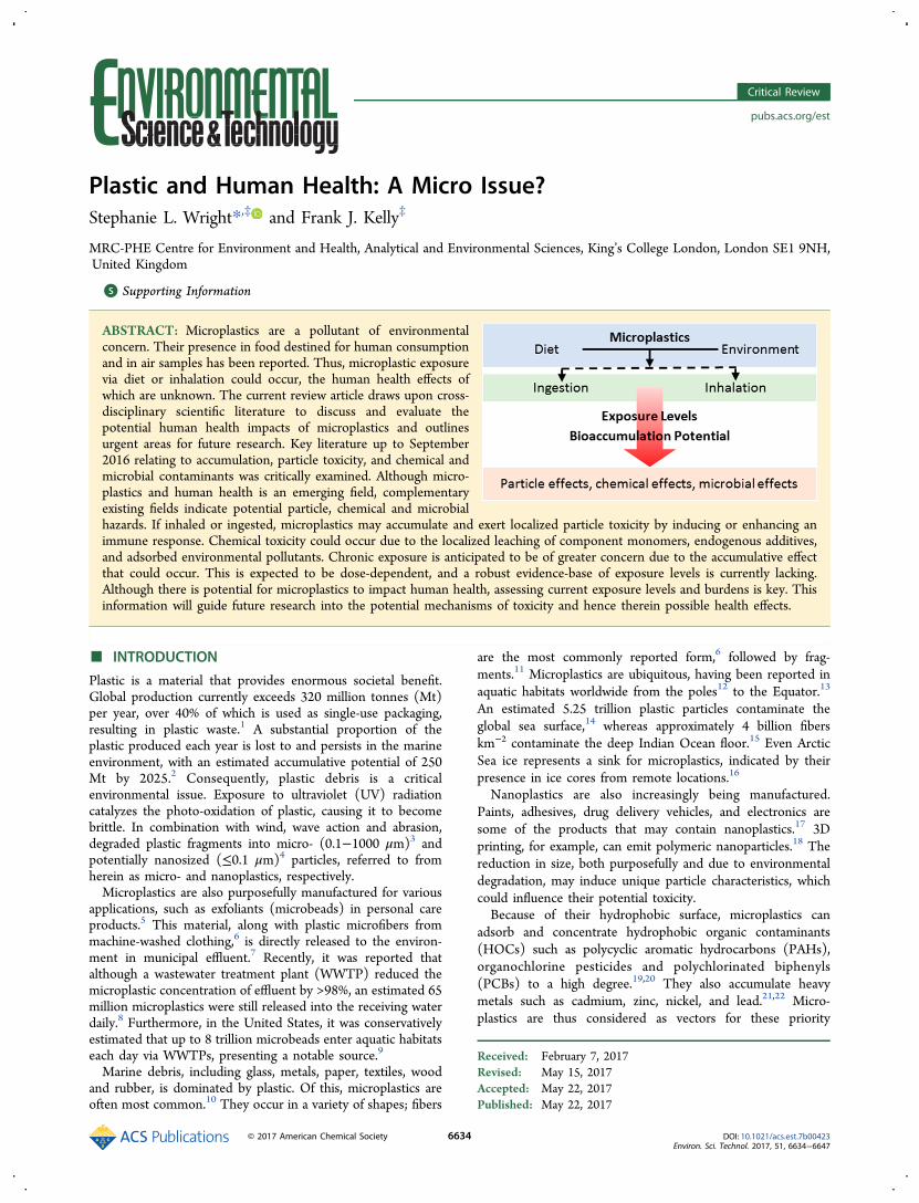
Plastic and Human Health: A Micro Issue?Stephanie L. Wright*,‡ and Frank J. Kelly‡
MRC-PHE Centre for Environment and Health, Analytical and Environmental Sciences, King’s College London, London SE1 9NH,United Kingdom
*S Supporting Information
ABSTRACT: Microplastics are a pollutant of environmentalconcern. Their presence in food destined for human consumptionand in air samples has been reported. Thus, microplastic exposurevia diet or inhalation could occur, the human health effects ofwhich are unknown. The current review article draws upon cross-disciplinary scientific literature to discuss and evaluate thepotential human health impacts of microplastics and outlinesurgent areas for future research. Key literature up to September2016 relating to accumulation, particle toxicity, and chemical andmicrobial contaminants was critically examined. Although micro-plastics and human health is an emerging field, complementaryexisting fields indicate potential particle, chemical and microbialhazards. If inhaled or ingested, microplastics may accumulate and exert localized particle toxicity by inducing or enhancing animmune response. Chemical toxicity could occur due to the localized leaching of component monomers, endogenous additives,and adsorbed environmental pollutants. Chronic exposure is anticipated to be of greater concern due to the accumulative effectthat could occur. This is expected to be dose-dependent, and a robust evidence-base of exposure levels is currently lacking.Although there is potential for microplastics to impact human health, assessing current exposure levels and burdens is key. Thisinformation will guide future research into the potential mechanisms of toxicity and hence therein possible health effects.
■ INTRODUCTION
Plastic is a material that provides enormous societal benefit.Global production currently exceeds 320 million tonnes (Mt)per year, over 40% of which is used as single-use packaging,resulting in plastic waste.1 A substantial proportion of theplastic produced each year is lost to and persists in the marineenvironment, with an estimated accumulative potential of 250Mt by 2025.2 Consequently, plastic debris is a criticalenvironmental issue. Exposure to ultraviolet (UV) radiationcatalyzes the photo-oxidation of plastic, causing it to becomebrittle. In combination with wind, wave action and abrasion,degraded plastic fragments into micro- (0.1−1000 μm)3 andpotentially nanosized (≤0.1 μm)4 particles, referred to fromherein as micro- and nanoplastics, respectively.Microplastics are also purposefully manufactured for various
applications, such as exfoliants (microbeads) in personal careproducts.5 This material, along with plastic microfibers frommachine-washed clothing,6 is directly released to the environ-ment in municipal effluent.7 Recently, it was reported thatalthough a wastewater treatment plant (WWTP) reduced themicroplastic concentration of effluent by >98%, an estimated 65million microplastics were still released into the receiving waterdaily.8 Furthermore, in the United States, it was conservativelyestimated that up to 8 trillion microbeads enter aquatic habitatseach day via WWTPs, presenting a notable source.9
Marine debris, including glass, metals, paper, textiles, woodand rubber, is dominated by plastic. Of this, microplastics areoften most common.10 They occur in a variety of shapes; fibers
are the most commonly reported form,6 followed by frag-ments.11 Microplastics are ubiquitous, having been reported inaquatic habitats worldwide from the poles12 to the Equator.13
An estimated 5.25 trillion plastic particles contaminate theglobal sea surface,14 whereas approximately 4 billion fiberskm−2 contaminate the deep Indian Ocean floor.15 Even ArcticSea ice represents a sink for microplastics, indicated by theirpresence in ice cores from remote locations.16
Nanoplastics are also increasingly being manufactured.Paints, adhesives, drug delivery vehicles, and electronics aresome of the products that may contain nanoplastics.17 3Dprinting, for example, can emit polymeric nanoparticles.18 Thereduction in size, both purposefully and due to environmentaldegradation, may induce unique particle characteristics, whichcould influence their potential toxicity.Because of their hydrophobic surface, microplastics can
adsorb and concentrate hydrophobic organic contaminants(HOCs) such as polycyclic aromatic hydrocarbons (PAHs),organochlorine pesticides and polychlorinated biphenyls(PCBs) to a high degree.19,20 They also accumulate heavymetals such as cadmium, zinc, nickel, and lead.21,22 Micro-plastics are thus considered as vectors for these priority
Received: February 7, 2017Revised: May 15, 2017Accepted: May 22, 2017Published: May 22, 2017
Critical Review
pubs.acs.org/est
© 2017 American Chemical Society 6634 DOI: 10.1021/acs.est.7b00423Environ. Sci. Technol. 2017, 51, 6634−6647
pollutants,23 which are listed in the Stockholm Convention fortheir potential adverse health effects.24
Microplastics may harbor endogenous chemical additives,due to their incorporation during the manufacture of plasticproducts. Because these additives are not chemically bound tothe plastic polymer matrix, they are susceptible to leaching tothe external medium.25 There is potential for the constantmigration of intrinsic chemicals along a concentration gradientto the surface of microplastics as they continue to fragment.Such pollutants can be released upon ingestion and transfer tosurrounding tissue.26,27 If microplastics have the capacity toaccumulate, they potentially present a source of chemicals totissues and fluids, if there is any additive remaining to leach.Emerging evidence suggests that human exposure to
microplastics is plausible. Microplastics have been reported inseafood,28−30 and in processed food and beverages such assugar,31 beer,32 and salt.33 In addition, the sludge byproducts ofWWTPs that are applied to agricultural land have been foundto contain synthetic (plastic) clothing fibers, which persist up to5 years postapplication.34 The wind-driven transport ofmicroplastics from sludge-based fertilizer, in addition to othersources such as the degradation of agricultural polyethylene(PE) sheets or the release of fibers from drying clothes outside,could also result in airborne microplastics.35 The atmosphericfallout of microplastics has recently been reported,36,37
representing a possible inhalation exposure pathway. Whethermicroplastics and their associated chemicals are transferred tohumans via diet and/or inhalation is unknown.The quantity of microplastics in the environment is likely to
increase due to the legacy of plastic items that contaminate theplanet. Given the evidence suggesting human exposure tomicroplastics and their associated pollutants is possible, it isimportant to assess the risk they pose to human health. To ourknowledge, there are two peer-reviewed articles that review thissubject;35,38 however, neither article considers inhalation as apotential exposure pathway, and the subsequent toxicity thiscould exert on the respiratory tract. We build on these twopublications to incorporate the marine environment, diet, andinhalation as pathways to microplastic exposure. This reviewtherefore aims to assess the evidence for this new potentialenvironmental challenge by addressing the following issues: (1)dietary exposure pathways; (2) inhalation exposure pathway;(3) microplastic uptake and translocation; and (4) potentialhuman health risks of microplastics.
■ EVIDENCE FOR DIETARY EXPOSURE PATHWAYSSeafood. Given the prevalence of microplastics in the
marine environment, an anticipated route of human exposure isvia seafood, which forms an essential dietary component.Seafood provides almost 3 billion people worldwide withapproximately 20% of their animal protein intake.39 It istherefore one of the most important food commoditiesconsumed globally; however, it can also be a source ofenvironmental contaminants such as PCBs and dioxins. Ifseafood were to exceed regulatory levels of contaminants, therecould be negative health impacts following consumption;however, these regulations are only in place for specificcontaminants, e.g., mercury, not for contaminants of emergingconcern such as microplastics.Fish. Globally, fish provides approximately 4.3 billion people
with 15% of their animal protein intake.40 The capacity for fishto ingest microplastics has been demonstrated in laboratorystudies,41,42 although these employed substantially higher
concentrations of microplastics than those found in nature.43,44
Importantly, the ingestion of microplastics by fish in situ hasbeen widely reported, including by commercial species,although the quantity of ingested microplastics is low (TableS1).The occurrence of microplastics in the gastrointestinal tract
(GIT) of fish does not provide direct evidence for humanexposure, as this organ is usually not consumed. There ispotential for the leaching and accumulation of associatedchemical contaminants in edible tissue, post-microplasticingestion. Dietary microplastic exposure via fish could bepossible if microplastics were able to translocate across the GITor gill via transcellular uptake or paracellular diffusion and enterthe circulatory fluid. The respiratory epithelium of the gill ismuch tighter than that of mammalian lungs, decreasing thelikelihood of this route of exposure; uptake across the fish gut ismore likely.45
There is evidence for the uptake of 1 μm latex spheres fromthe surrounding water in rainbow trout, with particles localizingand persisting in the surface and subsurface epidermal cells ofthe skin and in phagocytes underlying the gill surface.46 Thishighlights the importance of fish epithelial cells in theattachment and entry of microplastics. Additionally, con-sumption of the skin or gill tissue could present a directroute of human exposure to microplastics (≥1 μm).
Shellfish. Perhaps the most important source of dietaryexposure to microplastics at present is via bivalve molluscs(shellfish). Shellfish represents an important food source,comprising approximately 22 Mt of world fish production fromcapture and aquaculture in 2012 (almost 15 million USD).40
Bivalves feed by pumping large volumes of water through thepallial cavity within their shells, retaining particles fromsuspension on their gills for subsequent ingestion.47 Thus,they are directly exposed to microplastics via the water column.There is ample evidence for the capture and ingestion ofmicroplastics by bivalves in laboratory studies,48−50 andmicroplastics in wild and aquaculture shellfish for humanconsumption have been detected.Bivalves are a popular seafood in China28 where >60% of the
global aquaculture volume is produced. This coincides withwhere the greatest volume of plastic enters the marineenvironment from land-based sources.2 Consequently, concen-trations reaching 8720 microplastics/kg of sediment, includingpolyethylene terephthalate (PET), polystyrene (PS) and PEparticles, have been found on beaches.51 Nine of the mostcommercially popular species of bivalves purchased from afishing market in Shanghai, were found to be contaminatedwith microplastics (Table S2).28 Based on the abundancesobserved, it was estimated that Chinese shellfish consumerscould be exposed to 100 000s of microplastics each year.The contamination of shellfish by microplastics is not limited
to China. In Canada and Belgium, both wild and purchasedfarmed mussels were contaminated by microplastic fibers.29,52
Farmed mussels are often cultured on deployed polypropylene(PP) lines, which may present a source of microplastics as theline degrades.29,52 In Belgium, microplastics were recoveredfrom farmed mussels and shop-bought Pacific oysters, whichwere subjected to a 3 day depuration period. Based on theaverage recovered concentrations, it was estimated that theaverage European shellfish consumer may ingest up to 11 000microplastics per year.30 It is of concern that following 3 days ofdepuration, microplastics remained in the bivalves, suggestingstandard depuration periods may not be sufficient to ensure
Environmental Science & Technology Critical Review
DOI: 10.1021/acs.est.7b00423Environ. Sci. Technol. 2017, 51, 6634−6647
6635
microplastic clearance. Shellfish food safety is an increasinglyimportant issue with respect to microplastics.Other Foods. In addition to seafood, potential microplastics
have been reported in other foods. The presence of syntheticmicrofibers (minimum 40 μm in length) and fragments (mostly10−20 μm in size) was reported in honey and sugar.31 Anaverage of 174 (maximum of 660) fibers and 8 (maximum of38) fragments/kg honey, and an average of 217 (maximum of388) fibers and 32 (maximum of 270) fragments/kg of sugarwere found. Negative Rose Bengal staining determined whichfibers and fragments were of synthetic origin; however, nofurther methods were used to identify definitively whether theparticles were plastic.31
The contamination of honey suggests synthetic micro-particles and microplastics are airborne. In support of thishypothesis, the authors reported finding 18 fibers and 4fragments/L of rain during precipitation events. If airborne,microplastics may be deposited on flowers and foliage, wherethey could become incorporated with pollen and transportedby bees to the hive. In support of this, the authors reportedfinding fibers in flowers.31
Perhaps even more remarkable is that the contamination ofGerman beer by potential microplastics has been reported.32 Inall 24 samples tested, contamination by potential microplasticswas found. Fragments were the most abundant, reaching up to109 fragments/L. One suggested source was the atmosphericdeposition of microplastics while a second related to thematerials used in the production process.32 Given theprevalence of microplastics in freshwater systems,53 the watersource may also be a source of contamination.Microplastics have recently been identified in 15 brands of
shop-bought sea salt. Up to 681 microplastics/kg sea salt werereported down to 45 μm. PET was the most common type ofplastic found, followed by PE. It is likely that the coastal watersused to produce sea salt were the source of contamination,33
although microplastics could also be present due toatmospheric deposition at these sites.Clearly, microplastics currently contaminate food destined
for human consumption, the impacts of which are unknown.The presence of microplastics in other foods also suggests theycontaminate the atmospheric environment.
■ EVIDENCE FOR AN INHALATION EXPOSUREPATHWAY
In the ocean, sea salt aerosol (SSA) formation occurs due tobubbles bursting during white cap formation and wind stress, ordue to waves breaking in the coastal surf zone. SSAs can rangein size from <0.2 to >2000 μm diameter; the ambient mass istypically dominated by particles in the 1−10 μm range. Duringperiods of onshore winds, they can be transported to urbanenvironments close to the coast. Particles <50 μm are likely tohave an extended atmospheric lifetime.54 Because many plasticshave a specific gravity less than seawater, it is plausible thatwind action and sea spray may aerosolise sea-surface micro-plastics of appropriate size; however, this theory remains to betested.WWTP sludge byproducts applied to agricultural land have
been found to contain synthetic clothing fibers, which persist inboth the sludge and soil columns up to 5 years postapplication.Synthetic fibers have even been detected in field site soils 15years after application.34 This suggests that microplasticsreleased via municipal effluent are retained in sludge, which isthen applied as fertilizer, representing a persistent terrestrial
contaminant. The wind-driven transport of microplastics fromdried sludge-based fertilizer in addition to other sources such asthe degradation of agricultural PE sheets55 or the release offibers from drying clothes32 could all represent sources ofairborne microplastics.Recently, evidence for the presence of microplastics in
atmospheric fallout has been reported.37 The total atmosphericfallout of microplastics was assessed in a densely populatedurban area and a less-dense suburban area in Paris. Themajority of particles observed were fibers, approximately 30% ofwhich were confirmed plastic. Diameters varied mainly between7 and 15 μm and almost 25% of fibers were 100−500 μm inlength; 50 μm was the limit of detection. Up to 355 particles/m2/d were reported, with an average of 110 ± 96 particles/m2/d. Abundances were substantially greater in urban thansuburban areas.37 Periods of heavy rainfall corresponded withsome of the highest concentrations observed.37 This prelimi-nary study highlights the potential for human exposure tomicroplastics via inhalation, especially in densely populatedareas.To quantify the level of fiber exposure, a small scale study
assessing 24 h personal exposure to respirable inorganic andorganic fibers was undertaken at 3 European sites. Meanpersonal exposure levels to organic fibers (diameter <3 μm)were 0.003−0.011 fibers/mL with lengths <5 μm, 0.009−0.019fibers/mL with lengths >5 μm, and 0.0008−0.002 fibers/mLwith lengths >20 μm.56 Although “organic” fibers (natural andmanmade) included PE, PP, poly(vinyl alcohol), polyester,polyamide (PA), polytetrafluoroethylene, carbon, and naturalcellulose,56 no distinction was made regarding the compositionof the fibers sampled.Tires have recently been acknowledged as a source of
microplastics. Synthetic rubber is a variation on plastic,produced by the plastics industry. It is also a hydrocarbon-based polymer, although it has different properties to plastic,such as elasticity. Tire abrasion products are a reportedcomponent of ambient particulate matter (PM). In Japan, an airsample was reported to contain 0.16 μg/m3 of tire wearparticles in the PM10 fraction.
57 Furthermore, the concentrationof tire and road wear particles (TRWP), tire particles with roadmineral incrustations, was low, with global (United States,Europe, and Japan) averages ranging from 0.05 to 0.70 mg/m3.This comprised an average PM10 contribution of 0.84%.58
On an occupational level, indoor exposure levels can reach0.5 and 0.8 particles/mL for polyvinyl chloride (PVC) andnylon (PA), respectively; critical particle concentrations (aspectratio ≥3 μm) were 0.06 and 0.02/mL, respectively.59 In theflocking area (where many small fibers are deposited onto asurface) of a flock (microfibre) manufacturing plant, the highestconcentration of airborne particles reached 7 mg/m3.60
Polyester concentrations of 700 000 (up to 1 000 000) totalfibers/m3 and 10 000 critical fibers/m3 were reported duringprocessing operations.59
Size and exposure concentrations influence the potential riskthat microplastics pose to human health. Thus, to understandthe risk, it is first important to consolidate current exposureconcentrations to inhalable, thoracic, and respirable particles.Airborne fibers are ubiquitous and some of these fibers arelikely to be inhaled. Once they gain entry to the respiratorytract, most fibers are likely to be trapped by the lung liningfluid. However, some fibers may avoid the mucociliaryclearance mechanisms of the lung, especially in individualswhose clearance mechanisms have been impaired. Occupational
Environmental Science & Technology Critical Review
DOI: 10.1021/acs.est.7b00423Environ. Sci. Technol. 2017, 51, 6634−6647
6636
health literature specific to the synthetic textile industryprovides a good indication of the anticipated hazards thatmicroplastics, particularly fibers, may incur to human health.
■ MICROPLASTIC UPTAKE AND TRANSLOCATION
Fibers and Occupational Health. Studies among nylonflock workers suggest there is no evidence of increased cancerrisk, although workers had a higher prevalence of respiratoryirritation.61 Interstitial lung disease, a work-related conditionthat induces coughing, dyspnea (breathlessness), and reducedlung capacity, has been identified in 4% of workers from nylonflock plants in the US and Canada.62,63 Workers processingpara-aramid, polyester, and PA fibers in the Netherlandspresented similar symptoms, including coughing, dyspnoea,wheezing, and increased phlegm production.64 Prick tests andnasal and inhalation provocation tests in nylon workers alsofound synthetic fibers, such as nylon, may act as haptens,causing an allergic reaction leading to occupational asthma.65
Histopathological analysis of lung biopsies from workers inthe textile (nylon, polyester, polyolefin, and acrylic) industryshowed interstitial fibrosis and foreign-body-containing gran-ulomatous lesions, postulated to be acrylic, polyester, and/ornylon dust. The clinical symptoms presented were similar toallergic alveolitis (a form of inflammation in the lung).66
Although occupational exposure likely occurs at levels higherthan those in the environment, the health outcomes evidencethe potential for microplastics to trigger localized biologicalresponses, given their uptake and persistence.Both cellulosic and plastic microfibers have been observed in
non-neoplastic and malignant lung tissue taken from patientswith different types of lung cancer.67 The fibers exhibited littledeterioration, supporting the notion that they are biopersistent.Additionally, these observations suggest that the human airwayis of a sufficient size for plastic fibers to penetrate the deeplung; one fiber found was 135 μm in length, approximately one-quarter of the diameter of a respiratory bronchiole ofgeneration 17 (540 μm diameter, 1410 μm length).67 Theseobservations confirm that some fibers avoid clearancemechanisms and, as they persist, these foreign bodies mayinduce acute or chronic inflammation. Importantly, rigoroussterile methods were employed throughout the sampleprocessing in this study to prevent contamination byenvironmental fibers.In addition to biopersistence, fiber dimensions play a role in
toxicity. Thinner fibers are respirable, whereas longer fibers aremore persistent and toxic to pulmonary cells; fibers 15−20 μmcannot be efficiently cleared from the lung by alveolarmacrophages and the mucociliary escalator,61 and fibers <0.3μm thick and >10 μm long are most carcinogenic.68 The use offine-diameter (1−5 μm) plastic fibers has increased, such as inthe sports clothing industry.61 Nylon fibers of a respirable size(2 μm diameter, 14 μm length on average) interacted with thealveolar macrophages of exposed rats and were retained up to atleast 29 days postexposure, causing an acute inflammatoryresponse.69 Shorter (9.8 μm) but wider (1.6 μm diameter)“finish-free” nylon respirable fibers, however, showed nosignificant impact on lung weights, pulmonary inflammationor macrophage function in male rats up to the highestconcentration tested (57 fibers/cm3) compared to controlanimals.70 The burden of fibers, site of deposition and thepotential for chemicals to desorb from the fiber surface alsocontribute to toxicity,67 e.g., the affinity of PAHs for the
hydrophobic surface of plastic23,71 may present a route ofcarcinogenicity.
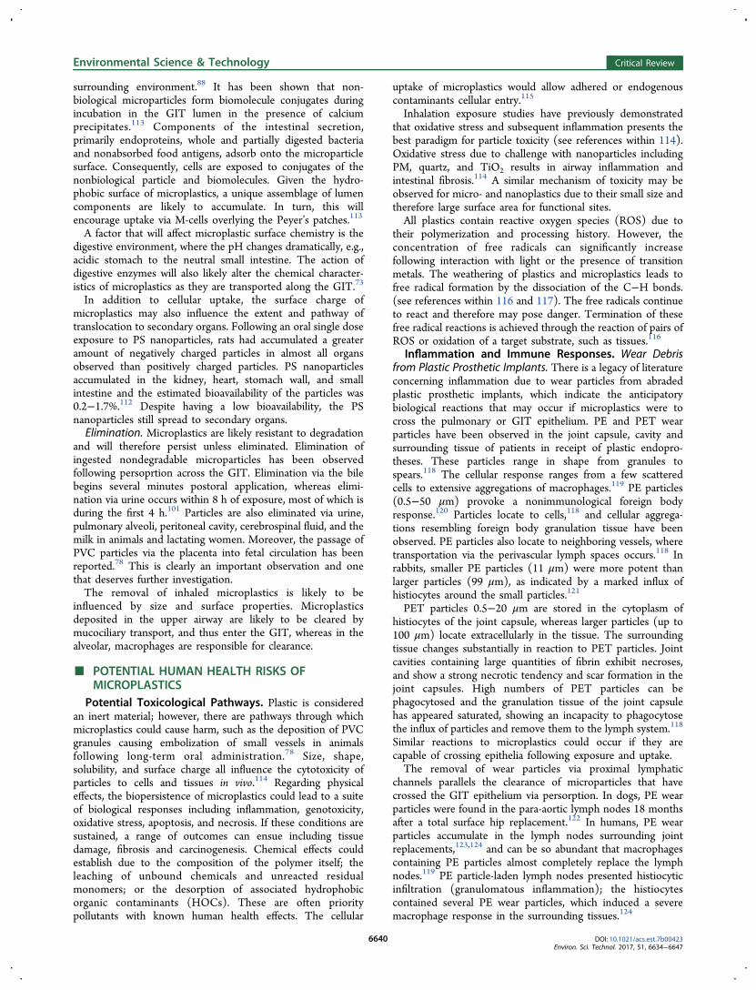
Potential for and Factors That May Affect Bioaccu-mulation. An essential factor determining whether micro-plastics present a physical threat or act as a vector for chemicaltransfer is the ability for these particles to accumulate.Throughout evolution, it is likely that both the lungs andGIT have been exposed to nondegradable, exogenous nano-and microparticles, and endogenous nanoparticles.72,73 Con-sequently, the body has evolved mechanisms to respond toparticle exposure (Figures 1 and 2). Recently, there has been an
increased dietary influx of nondegradable microparticles,approximately 40 mg/person/day, primarily due to theirinclusion as additives in processed foods.73,74 The contributionof microplastics to exogenous microparticle exposure isunknown, however the biological response to microplastics incomparison to other nondegradable microparticles could differdue to their unique chemical composition and properties.Microplastics are resistant to chemical degradation in vivo. If
inhaled or ingested, they may also resist mechanical clearance,becoming lodged or embedded. Their biopersistence is anessential factor contributing to their risk, along with dose. Theuptake and toxicity of several types of polymeric nano- and
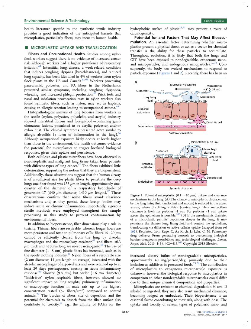
Figure 1. Potential microplastic (0.1 > 10 μm) uptake and clearancemechanisms in the lung. (A) The chance of microplastic displacementby the lung lining fluid (surfactant and mucus) is reduced in the upperairway, where the lining is thick (central lung). Here mucociliaryclearance is likely for particles >1 μm. For particles <1 μm, uptakeacross the epithelium is possible.107 (B) If the aerodynamic diameterof a microplastic permits deposition deeper in the lung, it maypenetrate the thinner lung lining fluid and contact the epithelium,translocating via diffusion or active cellular uptake (adapted from ref162). Reprinted from Ruge, C. A.; Kirch, J.; Lehr, C. M. Pulmonarydrug delivery: From generating aerosols to overcoming biologicalbarriers-therapeutic possibilities and technological challenges. Lancet.Respir. Med. 2013, 1(5), 402−413.162 Copyright 2013 Elsevier.
Environmental Science & Technology Critical Review
DOI: 10.1021/acs.est.7b00423Environ. Sci. Technol. 2017, 51, 6634−6647
6637
microparticles have been studied in model mammalian systems.The findings suggest they can translocate across living cells tothe lymphatic and/or circulatory system,72,75 potentiallyaccumulating in secondary organs,76−78 or impacting theimmune system and health of cells.79,80
Retention time, and therefore the likelihood of uptake andclearance, is influenced by particle characteristics such as size,shape, solubility, and surface chemistry; by biological factorssuch as the anatomical site of deposition and structure; and bythe nature of particle interaction with different biologicalstructures, including the air−liquid interface, aqueous phase
and free cells (e.g., macrophages, dendritic cells, epithelialcells).81 Uptake of inhaled microplastics will depend on theirwettability; it is possible that inhaled microplastics deposited onthe airway will not be immersed in the lung lining fluid due totheir hydrophobicity, and may therefore be subjected tomucociliary clearance leading to exposure via the gut (Figure1). Shape also affects displacement at the air−liquid interface;shapes with sharper edges are less likely to be displaced inliquid.82 However, the histological prevalence of plasticmicrofibers in flock worker66 and lung cancer67 tissue biopsiesimplies that uptake and embedment of at least plasticmicrofibers is possible.As with lining fluid in the lung, mucus is the first layer in the
GIT that foreign particles interact with. Here, mucus can causeparticles to aggregate; surfactants reduce mucus viscosity,increasing the uptake of particles.83 Size and surface charge alsoinfluence the ability for microplastics to cross the GIT mucusgel layer and contact the underlying epithelial cells;84,85 smallersizes and negative surface charge are most likely to lead toincreased uptake.If a microplastic contacts the airway or gastrointestinal
epithelium, there are several routes of uptake and translocationthat may occur. This is primarily via endocytic pathways in thelung and GIT, and also via persorption in the GIT (Figures 1and 2). Paracellular transfer of nanoparticles through the tightjunctions of the epithelium has been postulated for the GIT.Although tight junctions are extremely efficient at preventingsuch permeation, their integrity can be affected, potentiallyallowing for particles to pass through.73
Uptake Pathways. Endocytosis: Airway Surface. If aninhaled microplastic encounters the respiratory epithelium, itmay translocate via diffusion, direct cellular penetration oractive cellular uptake, as has been reported for othernonbiological micro- and nanoparticles.86 The active uptakeof nano- and microparticles by epithelial and endothelial cellsoccurs via energy-dependent endocytic and phagocyticprocesses.87 Phagocytosis is the primary clearance pathwayfor particles 1−3 μm from the alveoli.88 PS microparticles (1μm) were phagocytosed by porcine pulmonary macrophages,whereas smaller PS microparticles and nanoparticles (0.2 and0.078 μm) seemed to be passively transported via diffusionacross membrane pores, as endocytic particles were notmembrane-bound (Figure 1).88
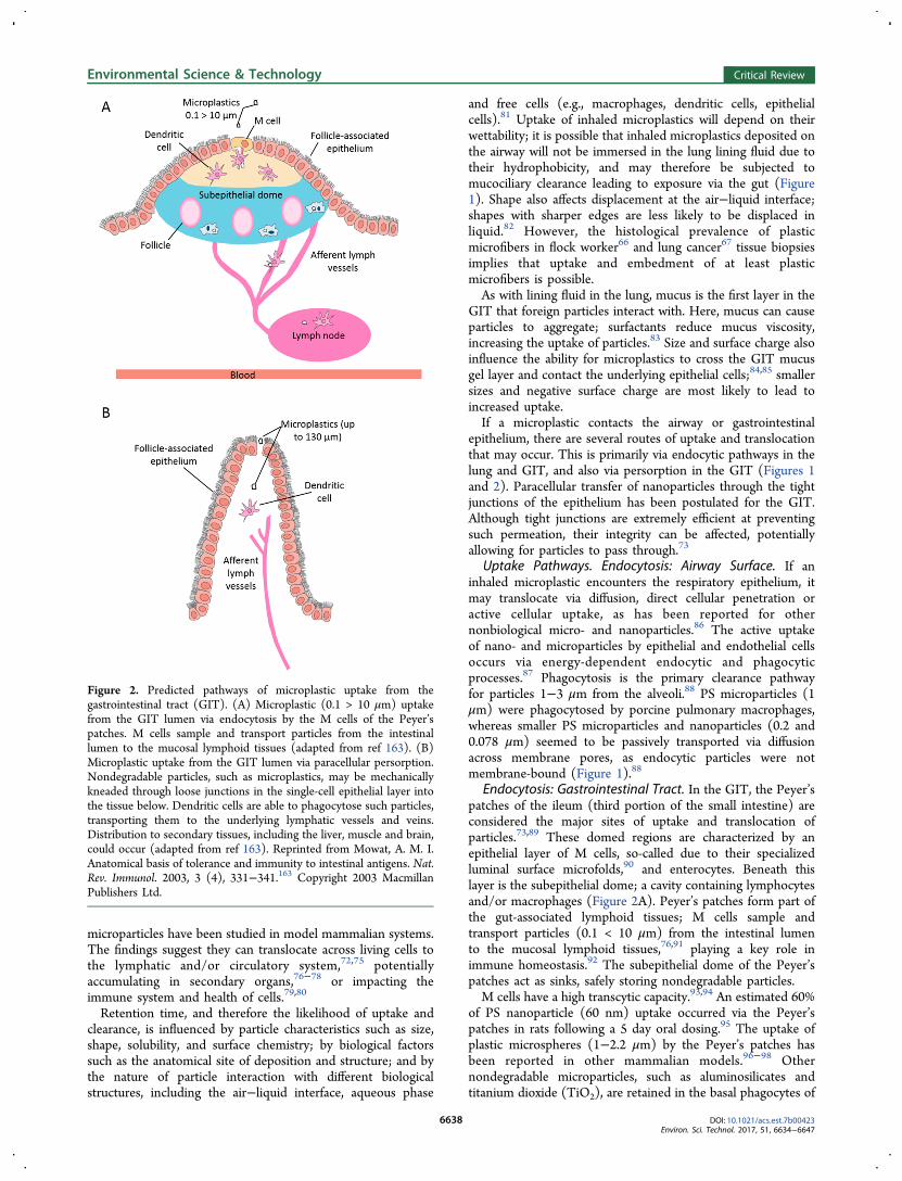
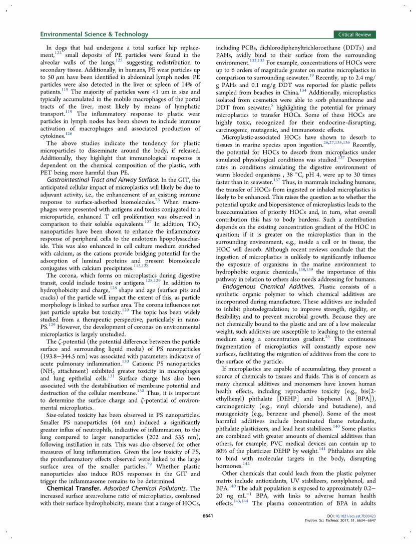
Endocytosis: Gastrointestinal Tract. In the GIT, the Peyer’spatches of the ileum (third portion of the small intestine) areconsidered the major sites of uptake and translocation ofparticles.73,89 These domed regions are characterized by anepithelial layer of M cells, so-called due to their specializedluminal surface microfolds,90 and enterocytes. Beneath thislayer is the subepithelial dome; a cavity containing lymphocytesand/or macrophages (Figure 2A). Peyer’s patches form part ofthe gut-associated lymphoid tissues; M cells sample andtransport particles (0.1 < 10 μm) from the intestinal lumento the mucosal lymphoid tissues,76,91 playing a key role inimmune homeostasis.92 The subepithelial dome of the Peyer’spatches act as sinks, safely storing nondegradable particles.M cells have a high transcytic capacity.93,94 An estimated 60%
of PS nanoparticle (60 nm) uptake occurred via the Peyer’spatches in rats following a 5 day oral dosing.95 The uptake ofplastic microspheres (1−2.2 μm) by the Peyer’s patches hasbeen reported in other mammalian models.96−98 Othernondegradable microparticles, such as aluminosilicates andtitanium dioxide (TiO2), are retained in the basal phagocytes of
Figure 2. Predicted pathways of microplastic uptake from thegastrointestinal tract (GIT). (A) Microplastic (0.1 > 10 μm) uptakefrom the GIT lumen via endocytosis by the M cells of the Peyer’spatches. M cells sample and transport particles from the intestinallumen to the mucosal lymphoid tissues (adapted from ref 163). (B)Microplastic uptake from the GIT lumen via paracellular persorption.Nondegradable particles, such as microplastics, may be mechanicallykneaded through loose junctions in the single-cell epithelial layer intothe tissue below. Dendritic cells are able to phagocytose such particles,transporting them to the underlying lymphatic vessels and veins.Distribution to secondary tissues, including the liver, muscle and brain,could occur (adapted from ref 163). Reprinted from Mowat, A. M. I.Anatomical basis of tolerance and immunity to intestinal antigens. Nat.Rev. Immunol. 2003, 3 (4), 331−341.163 Copyright 2003 MacmillanPublishers Ltd.
Environmental Science & Technology Critical Review
DOI: 10.1021/acs.est.7b00423Environ. Sci. Technol. 2017, 51, 6634−6647
6638
the Peyer’s patch, where they can occur in large numbers.73 Ifmicroplastics also accumulate in this compartment, they couldhijack the route for endogenous microparticle uptake andconsequently interfere with immunosensing and surveillance,compromising local immunity.Persorption. Another route of uptake in the GIT, and
perhaps the most applicable to microplastics due to the sizerange it covers, is via a phenomenon known as persorption.Persorption describes the mechanical kneading of solid particles(up to 130 μm diameter) through gaps in the single-layerepithelium at the villus tips of the GIT (desquamation zones),99
and into the circulatory system (Figure 2B).100,101
PVC particles (5−110 μm) have been used as modelnondegradable microparticles, along with starch, to study thisphenomenon.78 Following exposure via feeding or rectaladministration, the microplastics were observed to passbetween enterocytes in a paracellular manner, especially indesquamation zones and between the villi. The transportationof PVC particles occurred via two routes. First by the chyle(lumen) of the underlying lymph vessels, seen in rats, guineapigs, rabbits, chickens, dogs and pigs. Second by portalcirculation, suggested by the increased occurrence of particlesin blood taken from the mesenteric veins of intestinal segmentsof dogs fed PVC particles.78 The appearance of PVC particlesin the blood of dogs occurred rapidly postingestion; however,exposure concentrations were high −200 g of PVC powder,resulting in 10−15 PVC particles/mL of venous blood 1−2 hpostingestion. PVC particles were subsequently found in bile,urine, and cerebrospinal fluid.78 Larger particles are found intissues and organs; PVC microparticles appeared in the liver ofexposed rats, peaking 2−3 and 10 min postesophagealadministration.78 The reason for this multipeak curve has notbeen clarified but the study suggests that if ingested,microplastics may persorb across the intestinal wall and betransported to secondary tissues by the lymphatic and portalsystems. Cerebral softening, micronecroses and scarring wereobserved in the brains of dogs postexposure via femoral arterycatheterization into the left ventricular cavity.100
Persorption has been reported in human subjects. Theingestion of starch particles (200 g) led to granules beingobserved in urine, bile, cerebrospinal fluid, peritoneal fluid, andbreast milk.102 Particles peaked in the blood at 10 min (70particles/10 mL) and at 110 min (90 particles/10 mL)postingestion.102 However, the same authors found the rate(particles recovered in the blood over 24 h postexposure) ofpersorption to be low (0.002%).99 Persorption is influenced byboth rigidity of the particle and the level of motor activity in theGIT.99 The rigidity of microplastics combined with a likelyexposure pathway via diet suggests persorption of microplasticsduring the consumption of contaminated food could occur.Factors Affecting Uptake. Following uptake, translocation
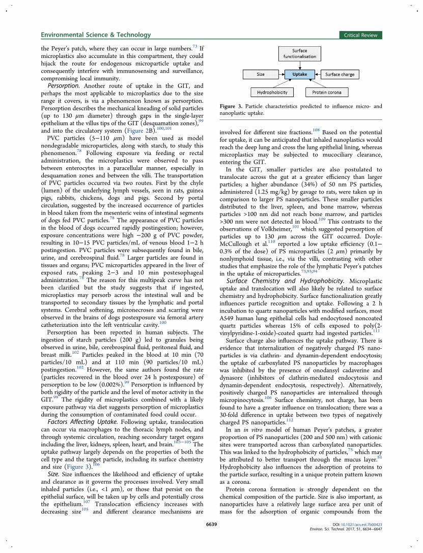
can occur via macrophages to the thoracic lymph nodes, andthrough systemic circulation, reaching secondary target organsincluding the liver, kidneys, spleen, heart, and brain.103−105 Theuptake pathway largely depends on the properties of both thecell type and the target particle, including its surface chemistryand size (Figure 3).106
Size. Size influences the likelihood and efficiency of uptakeand clearance as it governs the processes involved. Very smallinhaled particles (i.e., <1 μm), or those that persist on theepithelial surface, will be taken up by cells and potentially crossthe epithelium.107 Translocation efficiency increases withdecreasing size105 and different clearance mechanisms are
involved for different size fractions.108 Based on the potentialfor uptake, it can be anticipated that inhaled nanoplastics wouldreach the deep lung and cross the lung epithelial lining, whereasmicroplastics may be subjected to mucociliary clearance,entering the GIT.In the GIT, smaller particles are also postulated to
translocate across the gut at a greater efficiency than largerparticles; a higher abundance (34%) of 50 nm PS particles,administered (1.25 mg/kg) by gavage to rats, were taken up incomparison to larger PS nanoparticles. These smaller particlesdistributed to the liver, spleen, and bone marrow, whereasparticles >100 nm did not reach bone marrow, and particles>300 nm were not detected in blood.109 This contrasts to theobservations of Volkheimer,101 which suggested persorption ofparticles up to 130 μm across the GIT occurred. Doyle-McCullough et al.110 reported a low uptake efficiency (0.1−0.3% of the dose) of PS microparticles (2 μm) primarily bynonlymphoid tissue, i.e., via the villi, contrasting with otherstudies that emphasize the role of the lymphatic Peyer’s patchesin the uptake of microparticles.73,93,94
Surface Chemistry and Hydrophobicity. Microplasticuptake and translocation will also likely be related to surfacechemistry and hydrophobicity. Surface functionalization greatlyinfluences particle recognition and uptake. Following a 2 hincubation to quartz nanoparticles with modified surfaces, mostA549 human lung epithelial cells had endocytosed noncoatedquartz particles whereas 15% of cells exposed to poly(2-vinylpyridine-1-oxide)-coated quartz had ingested particles.111
Surface charge also influences the uptake pathway. There isevidence that internalization of negatively charged PS nano-particles is via clathrin- and dynamin-dependent endocytosis;the uptake of carboxylated PS nanoparticles by macrophageswas inhibited by the presence of onodansyl cadaverine anddynasore (inhibitors of clathrin-mediated endocytosis anddynamin-dependent endocytosis, respectively). Alternatively,positively charged PS nanoparticles are internalized throughmicropinocytosis.106 Surface chemistry, not charge, has beenfound to have a greater influence on translocation; there was a30-fold difference in uptake between two types of negativelycharged PS nanoparticles.112
In an in vitro model of human Peyer’s patches, a greaterproportion of PS nanoparticles (200 and 500 nm) with cationicsites were transported across than carboxylated nanoparticles.This was linked to the hydrophobicity of particles,75 which maybe attributed to better transport through the mucus layer.81
Hydrophobicity also influences the adsorption of proteins tothe particle surface, resulting in a unique protein pattern knownas a corona.Protein corona formation is strongly dependent on the
chemical composition of the particle. Size is also important, asnanoparticles have a relatively large surface area per unit ofmass for the adsorption of organic compounds from the
Figure 3. Particle characteristics predicted to influence micro- andnanoplastic uptake.
Environmental Science & Technology Critical Review
DOI: 10.1021/acs.est.7b00423Environ. Sci. Technol. 2017, 51, 6634−6647
6639
surrounding environment.88 It has been shown that non-biological microparticles form biomolecule conjugates duringincubation in the GIT lumen in the presence of calciumprecipitates.113 Components of the intestinal secretion,primarily endoproteins, whole and partially digested bacteriaand nonabsorbed food antigens, adsorb onto the microparticlesurface. Consequently, cells are exposed to conjugates of thenonbiological particle and biomolecules. Given the hydro-phobic surface of microplastics, a unique assemblage of lumencomponents are likely to accumulate. In turn, this willencourage uptake via M-cells overlying the Peyer’s patches.113
A factor that will affect microplastic surface chemistry is thedigestive environment, where the pH changes dramatically, e.g.,acidic stomach to the neutral small intestine. The action ofdigestive enzymes will also likely alter the chemical character-istics of microplastics as they are transported along the GIT.73
In addition to cellular uptake, the surface charge ofmicroplastics may also influence the extent and pathway oftranslocation to secondary organs. Following an oral single doseexposure to PS nanoparticles, rats had accumulated a greateramount of negatively charged particles in almost all organsobserved than positively charged particles. PS nanoparticlesaccumulated in the kidney, heart, stomach wall, and smallintestine and the estimated bioavailability of the particles was0.2−1.7%.112 Despite having a low bioavailability, the PSnanoparticles still spread to secondary organs.Elimination. Microplastics are likely resistant to degradation
and will therefore persist unless eliminated. Elimination ofingested nondegradable microparticles has been observedfollowing persoprtion across the GIT. Elimination via the bilebegins several minutes postoral application, whereas elimi-nation via urine occurs within 8 h of exposure, most of which isduring the first 4 h.101 Particles are also eliminated via urine,pulmonary alveoli, peritoneal cavity, cerebrospinal fluid, and themilk in animals and lactating women. Moreover, the passage ofPVC particles via the placenta into fetal circulation has beenreported.78 This is clearly an important observation and onethat deserves further investigation.The removal of inhaled microplastics is likely to be
influenced by size and surface properties. Microplasticsdeposited in the upper airway are likely to be cleared bymucociliary transport, and thus enter the GIT, whereas in thealveolar, macrophages are responsible for clearance.
■ POTENTIAL HUMAN HEALTH RISKS OFMICROPLASTICS
Potential Toxicological Pathways. Plastic is consideredan inert material; however, there are pathways through whichmicroplastics could cause harm, such as the deposition of PVCgranules causing embolization of small vessels in animalsfollowing long-term oral administration.78 Size, shape,solubility, and surface charge all influence the cytotoxicity ofparticles to cells and tissues in vivo.114 Regarding physicaleffects, the biopersistence of microplastics could lead to a suiteof biological responses including inflammation, genotoxicity,oxidative stress, apoptosis, and necrosis. If these conditions aresustained, a range of outcomes can ensue including tissuedamage, fibrosis and carcinogenesis. Chemical effects couldestablish due to the composition of the polymer itself; theleaching of unbound chemicals and unreacted residualmonomers; or the desorption of associated hydrophobicorganic contaminants (HOCs). These are often prioritypollutants with known human health effects. The cellular
uptake of microplastics would allow adhered or endogenouscontaminants cellular entry.115
Inhalation exposure studies have previously demonstratedthat oxidative stress and subsequent inflammation presents thebest paradigm for particle toxicity (see references within 114).Oxidative stress due to challenge with nanoparticles includingPM, quartz, and TiO2 results in airway inflammation andintestinal fibrosis.114 A similar mechanism of toxicity may beobserved for micro- and nanoplastics due to their small size andtherefore large surface area for functional sites.All plastics contain reactive oxygen species (ROS) due to
their polymerization and processing history. However, theconcentration of free radicals can significantly increasefollowing interaction with light or the presence of transitionmetals. The weathering of plastics and microplastics leads tofree radical formation by the dissociation of the C−H bonds.(see references within 116 and 117). The free radicals continueto react and therefore may pose danger. Termination of thesefree radical reactions is achieved through the reaction of pairs ofROS or oxidation of a target substrate, such as tissues.116
Inflammation and Immune Responses. Wear Debrisfrom Plastic Prosthetic Implants. There is a legacy of literatureconcerning inflammation due to wear particles from abradedplastic prosthetic implants, which indicate the anticipatorybiological reactions that may occur if microplastics were tocross the pulmonary or GIT epithelium. PE and PET wearparticles have been observed in the joint capsule, cavity andsurrounding tissue of patients in receipt of plastic endopro-theses. These particles range in shape from granules tospears.118 The cellular response ranges from a few scatteredcells to extensive aggregations of macrophages.119 PE particles(0.5−50 μm) provoke a nonimmunological foreign bodyresponse.120 Particles locate to cells,118 and cellular aggrega-tions resembling foreign body granulation tissue have beenobserved. PE particles also locate to neighboring vessels, wheretransportation via the perivascular lymph spaces occurs.118 Inrabbits, smaller PE particles (11 μm) were more potent thanlarger particles (99 μm), as indicated by a marked influx ofhistiocytes around the small particles.121
PET particles 0.5−20 μm are stored in the cytoplasm ofhistiocytes of the joint capsule, whereas larger particles (up to100 μm) locate extracellularly in the tissue. The surroundingtissue changes substantially in reaction to PET particles. Jointcavities containing large quantities of fibrin exhibit necroses,and show a strong necrotic tendency and scar formation in thejoint capsules. High numbers of PET particles can bephagocytosed and the granulation tissue of the joint capsulehas appeared saturated, showing an incapacity to phagocytosethe influx of particles and remove them to the lymph system.118
Similar reactions to microplastics could occur if they arecapable of crossing epithelia following exposure and uptake.The removal of wear particles via proximal lymphatic
channels parallels the clearance of microparticles that havecrossed the GIT epithelium via persorption. In dogs, PE wearparticles were found in the para-aortic lymph nodes 18 monthsafter a total surface hip replacement.122 In humans, PE wearparticles accumulate in the lymph nodes surrounding jointreplacements,123,124 and can be so abundant that macrophagescontaining PE particles almost completely replace the lymphnodes.119 PE particle-laden lymph nodes presented histiocyticinfiltration (granulomatous inflammation); the histiocytescontained several PE wear particles, which induced a severemacrophage response in the surrounding tissues.124
Environmental Science & Technology Critical Review
DOI: 10.1021/acs.est.7b00423Environ. Sci. Technol. 2017, 51, 6634−6647
6640
In dogs that had undergone a total surface hip replace-ment,122 small deposits of PE particles were found in thealveolar walls of the lungs,125 suggesting redistribution tosecondary tissue. Additionally, in humans, PE wear particles upto 50 μm have been identified in abdominal lymph nodes. PEparticles were also detected in the liver or spleen of 14% ofpatients.119 The majority of particles were <1 um in size andtypically accumulated in the mobile macrophages of the portaltracts of the liver, most likely by means of lymphatictransport.119 The inflammatory response to plastic wearparticles in lymph nodes has been shown to include immuneactivation of macrophages and associated production ofcytokines.126
The above studies indicate the tendency for plasticmicroparticles to disseminate around the body, if released.Additionally, they highlight that immunological response isdependent on the chemical composition of the plastic, withPET being more harmful than PE.Gastrointestinal Tract and Airway Surface. In the GIT, the
anticipated cellular impact of microplastics will likely be due toadjuvant activity, i.e., the enhancement of an existing immuneresponse to surface-adsorbed biomolecules.73 When macro-phages were presented with antigens and toxins conjugated to amicroparticle, enhanced T cell proliferation was observed incomparison to their soluble equivalents.127 In addition, TiO2nanoparticles have been shown to enhance the inflammatoryresponse of peripheral cells to the endotoxin lipopolysacchar-ide. This was also enhanced in cell culture medium enrichedwith calcium, as the cations provide bridging potential for theadsorption of luminal proteins and present biomoleculeconjugates with calcium precipitates.113,128
The corona, which forms on microplastics during digestivetransit, could include toxins or antigens.128,129 In addition tohydrophobicity and charge,128 shape and age (surface pits andcracks) of the particle will impact the extent of this, as particlemorphology is linked to surface area. The corona influences notjust particle uptake but toxicity.129 The topic has been widelystudied from a therapeutic perspective, particularly in nano-PS.129 However, the development of coronas on environmentalmicroplastics is largely unstudied.The ζ-potential (the potential difference between the particle
surface and surrounding liquid media) of PS nanoparticles(193.8−344.5 nm) was associated with parameters indicative ofacute pulmonary inflammation.130 Cationic PS nanoparticles(NH2 attachment) exhibited greater toxicity in macrophagesand lung epithelial cells.131 Surface charge has also beenassociated with the destabilization of membrane potential anddestruction of the cellular membrane.130 Thus, it is importantto determine the surface charge and ζ-potential of environ-mental microplastics.Size-related toxicity has been observed in PS nanoparticles.
Smaller PS nanoparticles (64 nm) induced a significantlygreater influx of neutrophils, indicative of inflammation, to thelung compared to larger nanoparticles (202 and 535 nm),following instillation in rats. This was also observed for othermeasures of lung inflammation. Given the low toxicity of PS,the proinflammatory effects observed were linked to the largesurface area of the smaller particles.79 Whether plasticnanoparticles also induce ROS responses in the GIT andtrigger the inflammasome remains to be determined.Chemical Transfer. Adsorbed Chemical Pollutants. The
increased surface area:volume ratio of microplastics, combinedwith their surface hydrophobicity, means that a range of HOCs,
including PCBs, dichlorodiphenyltrichloroethane (DDTs) andPAHs, avidly bind to their surface from the surroundingenvironment.132,133 For example, concentrations of HOCs wereup to 6 orders of magnitude greater on marine microplastics incomparison to surrounding seawater.19 Recently, up to 2.4 mg/g PAHs and 0.1 mg/g DDT was reported for plastic pelletssampled from beaches in China.134 Additionally, microplasticsisolated from cosmetics were able to sorb phenanthrene andDDT from seawater,5 highlighting the potential for primarymicroplastics to transfer HOCs. Some of these HOCs arehighly toxic, recognized for their endocrine-disrupting,carcinogenic, mutagenic, and immunotoxic effects.Microplastic-associated HOCs have shown to desorb to
tissues in marine species upon ingestion.26,27,135,136 Recently,the potential for HOCs to desorb from microplastics undersimulated physiological conditions was studied.137 Desorptionrates in conditions simulating the digestive environment ofwarm blooded organisms , 38 °C, pH 4, were up to 30 timesfaster than in seawater.137 Thus, in mammals including humans,the transfer of HOCs from ingested or inhaled microplastics islikely to be enhanced. This raises the question as to whether thepotential uptake and biopersistence of microplastics leads to thebioaccumulation of priority HOCs and, in turn, what overallcontribution this has to body burdens. Such a contributiondepends on the existing concentration gradient of the HOC inquestion; if it is greater on the microplastics than in thesurrounding environment, e.g., inside a cell or in tissue, theHOC will desorb. Although recent reviews conclude that theingestion of microplastics is unlikely to significantly influencethe exposure of organisms in the marine environment tohydrophobic organic chemicals,138,139 the importance of thispathway in relation to others also needs addressing for humans.
Endogenous Chemical Additives. Plastic consists of asynthetic organic polymer to which chemical additives areincorporated during manufacture. These additives are includedto inhibit photodegradation; to improve strength, rigidity, orflexibility; and to prevent microbial growth. Because they arenot chemically bound to the plastic and are of a low molecularweight, such additives are susceptible to leaching to the externalmedium along a concentration gradient.25 The continuousfragmentation of microplastics will constantly expose newsurfaces, facilitating the migration of additives from the core tothe surface of the particle.If microplastics are capable of accumulating, they present a
source of chemicals to tissues and fluids. This is of concern asmany chemical additives and monomers have known humanhealth effects, including reproductive toxicity (e.g., bis(2-ethylhexyl) phthalate [DEHP] and bisphenol A [BPA]),carcinogenicity (e.g., vinyl chloride and butadiene), andmutagenicity (e.g., benzene and phenol). Some of the mostharmful additives include brominated flame retardants,phthalate plasticizers, and lead heat stabilizers.140 Some plasticsare combined with greater amounts of chemical additives thanothers, for example, PVC medical devices can contain up to80% of the plasticizer DEHP by weight.141 Phthalates are ableto bind with molecular targets in the body, disruptinghormones.142
Other chemicals that could leach from the plastic polymermatrix include antioxidants, UV stabilizers, nonylphenol, andBPA.140 The adult population is exposed to approximately 0.2−20 ng mL−1 BPA, with links to adverse human healtheffects.143,144 The plasma concentration of BPA in adults
Environmental Science & Technology Critical Review
DOI: 10.1021/acs.est.7b00423Environ. Sci. Technol. 2017, 51, 6634−6647
6641
exceed levels predicted from exposure via food and drinkalone,145 suggesting alternate pathways of exposure.The ingestion and inhalation of household dust is a widely
recognized human exposure pathway to flame-retardingpolybrominated diphenyl ethers (PBDEs), which can reach>90 ng/g dust.146,147 PBDEs released from plastic components,such as upholstery, carpets and electronics, lead to inhalation ofultrafine particulate PBDEs associated with dust.148 However,the migration pathways from treated products to dust isunderstudied. One postulated mechanism is the transfer ofPBDEs and other brominated flame retardants via the abrasionof particles and/or fibers from plastic products, i.e., micro-plastics. Recently, fibers and particles generated by the abrasionof brominated-flame-retardant- (BFR) treated curtain uphols-tery accounted for BFR concentrations in spiked ambient dustsamples.149 Thus, the accumulation of PBDEs via house dustmay be due to leaching of PBDEs following the ingestion orinhalation of microplastics resulting from the wear of plastichousehold products and textiles.In addition to chemical additives, plastic can also leach
hazardous unreacted residual monomers. Polyurethanes, PVC,epoxy resins, and styrenic polymers have been identified asplastics of the greatest concern in terms of environmental andhealth effects, as their monomers are classified as carcinogenic,mutagenic, or both.140 Currently, there is no informationconcerning the direct transfer of additives from plastic tohuman tissues, although this has been suggested for seabirds.150
Recently, in a study investigating whether peritoneal dialysissolution (PDS) contains leached contaminants, toxic effectswere observed in mice and linked to the leaching additives ofthe plastic PDS solution storage bags.151
Microbiome. In the environment, the surface of micro-plastics becomes rapidly colonised by microbes; well-developedbiofilms establish on the surface of plastic after 7 days in wateror sediment.152,153 Such biofilms significantly differ from theambient environment154 and can include harmful humanpathogens such as strains of Vibrio spp.154,155
The microbiome refers to the collection of microbialcommunities living on or in the body, the physiological activityof which influences host well-being.156 It is known that thecomposition of the GIT microbiome can significantly differbetween liquid and solid phases.157 Thus, it can be anticipatedthat, in the instance that microplastics are colonised during GITtransit, the composition will differ to the surroundingenvironment. This is emphasized by the unique microbialassemblage that plastic attracts.154,155 The unique coating mayinfluence the body’s response to microplastics, by enhancingbioavailability or triggering an immune response.Environmental pollutants have shown to affect the micro-
biome, as microbes have the capacity to metabolize a range ofenvironmental toxicants.158 This can have knock on effects forthe host, compromising immunity and stimulating inflamma-tion.156 Mice exhibited changes to the composition andfunction of the colonic microbiome following long-termexposure to PM10 administered via lavage.159 This potentiallycontributed to the induction of proinflammatory cytokines inthe host. However, it was unknown whether this was a directcause of PM10, PM10-induced immune changes, or both.159
The lungs also host a microbial community that ismaintained by alveolar macrophages, antibacterial surfactant,and other environmental conditions.160 Colonisation is low incomparison to the GIT, although growth and community shiftscoincide with disease.161 Oxidative stress and inflammation
have a key role in the pathogenesis of inhaled pollutants, andalso modify local conditions, which potentially influence themicrobiome.Thus, the response to inhaled or ingested PM, including
microplastics, may cause a shift in the microbial compositioncolonising the lung or GIT. Microplastics may causeinflammation or leach HOCs, the microbial metabolism ofwhich could lead to oxidative stress. Microplastics could carrypathogenic species, or the additional substrate in the lung orGIT may facilitate growth of specific groups, shifting theassembly. Through this, alterations in the community structureand functions of the lung or GIT microbiome could occur, withknock on effects for host well-being and therefore humanhealth.
■ CONCLUSIONS AND RECOMMENDATIONS FORFUTURE RESEARCH
Although microplastics are widely studied in the context of themarine environment where they are a prolific pollutant, we are
only just recognizing the potential human exposure pathways.Following exposure, via diet and/or inhalation, uptake isplausible, as evidenced by the observations of plastic micro-fibers in lung tissue biopsy samples, and by the capacity forbiopersistent particles up to >100 μm to cross the GITepithelium. Following uptake, particles <2.5 μm and fibers areanticipated to be of greatest concern in the lung, whereas largerparticles are of concern in the GIT due the presence of M cells
Table 1. Key Knowledge Gaps and Recommendations forFuture Research into Microplastics and Human Health
Key Knowledge Gaps
What are the overall exposure concentrations from dietary and airbornesources?
What proportion of microparticle exposure do microplastic comprise?Do different biological responses to microplastics manifest due to their uniquechemical compositions/properties?
What effect does the interchangeable gastric environment/lung lining fluidhave on the surface charge and chemistry, and therefore handling ofmicroplastics?
What is the composition of the protein corona on microplastics?Is there evidence of microplastic uptake in humans?Are microplastics able to accumulate in the body? Do they become lodged orare they engulfed by cells?
If taken up by cells, what is the cellular mechanism of uptake? Does subcellularlocalization or translocation occur?
If subcellular location occurs, does this “hijack” the route for endogenousmicroparticle uptake or compromise immune homeostasis?
Does dissemination and/or elimination occur? Are there target secondaryorgans?
Are accumulative effects the same as those observed in occupational exposures?Are larger particles a greater issue for the GIT due to the process ofpersorption?
What is the toxicological response to biopersistent microplastics? Doinflammatory responses mimic those observed in response to plasticprosthetics wear debris?
Do size and shape influence toxicity? Does this depend on the point of entry,e.g., are plastic microfibres of greater concern for the lung than the GIT?
Do polymer type and hydrophobicity influence toxicity?Does surface charge of microplastics affect toxicity and does this vary with timein the environment (and therefore exposure to UV)?
Once uptaken, can microplastics deliver their chemical burden and does thiscause localized toxicity?
What will the addition of the novel hard surface of microplastics, for whichspecific microbes and biomolecules have an affinity for, have on themicrobiome?
Environmental Science & Technology Critical Review
DOI: 10.1021/acs.est.7b00423Environ. Sci. Technol. 2017, 51, 6634−6647
6642
in the Peyer’s Patches, capable of engulfing micrometer-sizedparticles, and the phenomenon of persorption. Toxicity is viainflammation due to the biopersistent nature of microplastics,and their unique hydrophobicity and surface chemistry.Toxicity is likely to have an accumulative effect, dependenton dose. Key knowledge gaps are outlined in Table 1.Exposure concentrations are predicted to be low, although
this is partly due to the present technical limitations in samplingand identifying microplastics. Measuring and assessing trueexposure concentrations is a current scientific challenge, largelylimited by particle size. Thus, current predicted exposure levelsare also probably an underestimation. Once we have a betterunderstanding of human exposure levels, and whether micro-plastics are uptaken/able to translocate, we can begin to unravelthe potential toxicological mechanisms of microplastics andhence therein possible health effects.
■ ASSOCIATED CONTENT
*S Supporting InformationThe Supporting Information is available free of charge on theACS Publications website at DOI: 10.1021/acs.est.7b00423.
Occurrence of microplastics in the gastrointestinal tractsof fish in situ; Tables S1 and S2 (PDF)
■ AUTHOR INFORMATION
Corresponding Author*S. L. Wright. Tel.: +4420 7848 4007. E-mail: [email protected].
ORCIDStephanie L. Wright: 0000-0003-1894-2365Author Contributions‡These authors contributed equally.
FundingWe thank the Medical Research Council for funding thisresearch (MR/M501669/1).
NotesThe authors declare no competing financial interest.
■ REFERENCES(1) PlasticsEurope. Plastics - the Facts 2016 - an Analysis of EuropeanPlastics Production, Demand and Waste Data; PlasticsEurope, 2016.(2) Jambeck, J. R.; Geyer, R.; Wilcox, C.; Siegler, T. R.; Perryman,M.; Andrady, A.; Narayan, R.; Law, K. L. Plastic waste inputs from landinto the ocean. Science 2015, 347 (6223), 768−771.(3) Cozar, A.; Echevarría, F.; Gonzalez-Gordillo, J. I.; Irigoien, X.;Ubeda, B.; Hernandez-Leon, S.; Palma, A. T.; Navarro, S.; Garcia-de-Lomas, J.; Ruiz, A.; Fernandez-de-Pelles, M. L.; Duarte, C. M. Plasticdebris in the open ocean. Proc. Natl. Acad. Sci. U. S. A. 2014, 111 (28),10239−10244.(4) Lambert, S.; Wagner, M. Characterisation of nanoplastics duringthe degradation of polystyrene. Chemosphere 2016, 145, 265−268.(5) Napper, I. E.; Bakir, A.; Rowland, S. J.; Thompson, R. C.Characterisation, quantity and sorptive properties of microplasticsextracted from cosmetics. Mar. Pollut. Bull. 2015, 99 (1−2), 178−185.(6) Browne, M. A.; Crump, P.; Niven, S. J.; Teuten, E.; Tonkin, A.;Galloway, T.; Thompson, R. Accumulation of microplastic onshorelines worldwide: sources and sinks. Environ. Sci. Technol. 2011,45 (21), 9175−9179.(7) Fendall, L. S.; Sewell, M. A. Contributing to marine pollution bywashing your face: Microplastics in facial cleansers. Mar. Pollut. Bull.2009, 58 (8), 1225−1228.
(8) Murphy, F.; Ewins, C.; Carbonnier, F.; Quinn, B. WastewaterTreatment Works (WwTW) as a source of microplastics in the aquaticenvironment. Environ. Sci. Technol. 2016, 50 (11), 5800−5808.(9) Rochman, C. M.; Kross, S. M.; Armstrong, J. B.; Bogan, M. T.;Darling, E. S.; Green, S. J.; Smyth, A. R.; Verissimo, D. Scientificevidence supports a ban on microbeads. Environ. Sci. Technol. 2015, 49(18), 10759−10761.(10) Andrady, A. L. The plastic in microplastics: a review.Mar. Pollut.Bull. 2017, DOI: 10.1016/j.marpolbul.2017.01.082.(11) Desforges, J. P.; Galbraith, M.; Dangerfield, N.; Ross, P. S.Widespread distribution of microplastics in subsurface seawater in theNE Pacific Ocean. Mar. Pollut. Bull. 2014, 79 (1−2), 94−99.(12) Lusher, A. L.; Tirelli, V.; O’Connor, I.; Officer, R. Microplasticsin Arctic polar waters: the first reported values of particles in surfaceand sub-surface samples. Sci. Rep. 2015, 5, 14947.(13) Ivar do Sul, J. A.; Costa, M. F.; Barletta, M.; Cysneiros, F. J.Pelagic microplastics around an archipelago of the Equatorial Atlantic.Mar. Pollut. Bull. 2013, 75 (1−2), 305−309.(14) Eriksen, M.; Lebreton, L. C. M.; Carson, H. S.; Thiel, M.;Moore, C. J.; Borerro, J. C.; Galgani, F.; Ryan, P. J.; Reisser, J. Plasticpollution in the World’s oceans: more than 5 trillion plastic piecesweighing over 250,000 tons afloat at sea. PLoS One 2014, 9 (12),e111913.(15) Woodall, L. C.; Sanchez-Vidal, A.; Canals, M.; Paterson, G. L. J.;Coppock, R.; Sleight, V.; Calafat, A.; Rogers, A. D.; Narayanaswamy, B.E.; Thompson, R. C. The deep sea is a major sink for microplasticdebris. R. Soc. Open Sci. 2014, 1 (4), 140317.(16) Obbard, R. W.; Sadri, S.; Wong, Y. Q.; Khitun, A. A.; Baker, I.;Thompson, R. C. Global warming releases microplastic legacy frozenin Arctic Sea ice. Earth's Future. 2014, 2 (6), 31510.1002/2014EF000240.(17) Koelmans, A. A.; et al. Nanoplastics in the aquatic environment.Critical Review. In Marine Anthropogenic Litter; Bergmann, M., Gutow,L., Klages, M., Eds.; Springer International Publishing, 2015; pp 325−340.(18) Stephens, B.; Azimi, P.; El Orch, Z.; Ramos, T. Ultrafine particleemissions from desktop 3D printers. Atmos. Environ. 2013, 79, 334−339.(19) Mato, Y.; Isobe, T.; Takada, H.; Kanehiro, H.; Ohtake, C.;Kaminuma, T. Plastic resin pellets as a transport medium for toxicchemicals in the marine environment. Environ. Sci. Technol. 2001, 35(2), 318−324.(20) Ogata, Y.; Takada, H.; Mizukawa, K.; Hirai, H.; Iwasa, S.; Endo,S.; Mato, Y.; Saha, M.; Okuda, K.; Nakashima, A.; Murakami, M.;Zurcher, N.; Booyatumanondo, R.; Zakaria, M. P.; Dung, L. Q.;Gordon, M.; Miguez, C.; Suzuki, S.; Moore, C.; Karapanagioti, H. K.;Weerts, S.; McClurg, T.; Burres, E.; Smith, W.; Van Velkenburg, M.;Selby Lang, J.; Lang, R. C.; Laursen, D.; Danner, B.; Stewardson, N.;Thompson, R. C. International Pellet Watch: Global monitoring ofpersistent organic pollutants (POPs) in coastal waters. 1. Initial phasedata on PCBs, DDTs, and HCHs. Mar. Pollut. Bull. 2009, 58 (10),1437−1446.(21) Holmes, L. A.; Turner, A.; Thompson, R. C. Adsorption of tracemetals to plastic resin pellets in the marine environment. Environ.Pollut. 2012, 160 (1), 42−48.(22) Rochman, C. M.; Hentschel, B. T.; Teh, S. J. Long-termsorption of metals is similar among plastic types: implications forplastic debris in aquatic environments. PLoS One 2014, 9 (1), e85433.(23) Hirai, H.; Takada, H.; Ogata, Y.; Yamashita, R.; Mizukawa, K.;Saha, M.; Kwan, C.; Moore, C.; Gray, H.; Laursen, D.; Zettler, E. R.;Farrington, J. W.; Reddy, C. M.; Peacock, E. E.; Ward, M. W. Organicmicropollutants in marine plastics debris from the open ocean andremote and urban beaches.Mar. Pollut. Bull. 2011, 62 (8), 1683−1692.(24) Vanden Bilcke, C. The Stockholm Convention on PersistentOrganic Pollutants. Review of European Community & InternationalEnvironmental Law. 2002, 11 (3), 328−342.(25) Tickner, J. The use of Di-2-Ethylhexyl Phthalate in PVC medicaldevices: exposure, toxicity, and alternatives; Lowell Centre forSustainable Production, 1999.
Environmental Science & Technology Critical Review
DOI: 10.1021/acs.est.7b00423Environ. Sci. Technol. 2017, 51, 6634−6647
6643
(26) Browne, M. A.; Niven, S. J.; Galloway, T. S.; Rowland, S. J.;Thompson, R. C. Microplastic moves pollutants and additives toworms, reducing functions linked to health and biodiversity. Curr. Biol.2013, 23 (23), 2388−2392.(27) Rochman, C. M.; Hoh, E.; Kurobe, T.; Teh, S. J. Ingested plastictransfers hazardous chemicals to fish and induces hepatic stress. Sci.Rep. 2013, 3, 3263.(28) Li, J.; Yang, D.; Li, L.; Jabeen, K.; Shi, H. Microplastics incommercial bivalves from China. Environ. Pollut. 2015, 207, 190−195.(29) Mathalon, A.; Hill, P. Microplastic fibers in the intertidalecosystem surrounding Halifax Harbor, Nova Scotia. Mar. Pollut. Bull.2014, 81 (1), 69−79.(30) Van Cauwenberghe, L.; Janssen, C. R. Microplastics in bivalvescultured for human consumption. Environ. Pollut. 2014, 193 (0), 65−70.(31) Liebezeit, G.; Liebezeit, E. Non-pollen particulates in honey andsugar. Food Addit. Contam., Part A 2013, 30 (12), 2136−2140.(32) Liebezeit, G.; Liebezeit, E. Synthetic particles as contaminants inGerman beers. Food Addit. Contam., Part A 2014, 31 (9), 1574−1578.(33) Yang, D.; Shi, H.; Li, L.; Li, J.; Jabeen, K.; Kolandhasamy, P.Microplastic pollution in table salts from China. Environ. Sci. Technol.2015, 49 (22), 13622−13627.(34) Zubris, K. A. V.; Richards, B. K. Synthetic fibers as an indicatorof land application of sludge. Environ. Pollut. 2005, 138 (2), 201−211.(35) Bouwmeester, H.; Hollman, P. C. H.; Peters, R. J. B. Potentialhealth impact of environmentally released micro- and nanoplastics inthe human food production chain: experiences from nanotoxicology.Environ. Sci. Technol. 2015, 49 (15), 8932−8947.(36) Dris, R.; Gasperi, J.; Rocher, V.; Saad, M.; Renault, N.; Tassin,B. Microplastic contamination in an urban area: a case study in GreaterParis. Envir. Chem. 2015, 12 (5), 592−599.(37) Dris, R.; Gasperi, J.; Saad, M.; Mirande, C.; Tassin, B. Syntheticfibers in atmospheric fallout: a source of microplastics in theenvironment? Mar. Pollut. Bull. 2016, 104 (1−2), 290−293.(38) Galloway, T. G. Micro- and Nano-plastics and Human Health.In Marine Anthropogenic Litter; Bergmann, M., Gutow, L., Klages, M.,Eds.; Springer International Publishing 2015; pp 343−366.(39) FAO. World review of fisheries and aquaculture; The State ofWorld Fisheries and Aquaculture, 2012.(40) FAO. FAO Yearbook; Fishery and Aquaculture Statistics 2012,2014.(41) Mazurais, D.; Ernande, B.; Quazuguel, P.; Severe, A.; Huelvan,C.; Madec, L.; Mouchel, O.; Soudant, P.; Robbens, J.; Huvet, A.;Zambonino-Infante, J. Evaluation of the impact of polyethylenemicrobeads ingestion in European sea bass (Dicentrarchus labrax)larvae. Mar. Environ. Res. 2015, 112 (A), 78−85.(42) Oliveira, M.; Ribeiro, A.; Hylland, K.; Guilhermino, L. Singleand combined effects of microplastics and pyrene on juveniles (0+group) of the common goby Pomatoschistus microps (Teleostei,Gobiidae). Ecol. Indic. 2013, 34, 641−647.(43) da Costa, J. P.; Santos, P. S. M.; Duarte, A. C.; Rocha-Santos, T.(Nano)plastics in the environment − sources, fates and effects. Sci.Total Environ. 2016, 566−567, 15−26.(44) Phuong, N. N.; Zalouk-Vergnoux, A.; Poirier, L.; Kamari, A.;Chatel, A.; Mouneyrac, C.; Lagarde, F. Is there any consistencybetween the microplastics found in the field and those used inlaboratory experiments? Environ. Pollut. 2016, 211, 111−123.(45) Handy, R. D.; Henry, T. B.; Scown, T. M.; Johnston, B. D.;Tyler, C. R. Manufactured nanoparticles: their uptake and effects onfish–a mechanistic analysis. Ecotoxicology 2008, 17 (5), 396−409.(46) Moore, J. D.; Ototake, M.; Nakanishi, T. Particulate antigenuptake during immersion immunisation of fish: The effectiveness ofprolonged exposure and the roles of skin and gill. Fish ShellfishImmunol. 1998, 8 (6), 393−408.(47) Ward, E. J.; Shumway, S. E. Separating the grain from the chaff:particle selection in suspension- and deposit-feeding bivalves. J. Exp.Mar. Biol. Ecol. 2004, 300 (1−2), 83−130.
(48) Brillant, M. G. S.; MacDonald, B. A. Postingestive selection inthe sea scallop, Placopecten magellanicus (Gmelin): the role of particlesize and density. J. Exp. Mar. Biol. Ecol. 2000, 253 (2), 211−227.(49) Browne, M. A.; Dissanayake, A.; Galloway, T. S.; Lowe, D. M.;Thompson, R. C. Ingested microscopic plastic translocates to thecirculatory system of the mussel, Mytilus edulis (L). Environ. Sci.Technol. 2008, 42 (13), 5026−5031.(50) von Moos, N.; Burkhardt-Holm, P.; Kohler, A. Uptake andeffects of microplastics on cells and tissue of the blue mussel Mytilusedulis L. after an experimental exposure. Environ. Sci. Technol. 2012, 46(20), 11327−11335.(51) Qiu, Q.; Peng, J.; Yu, X.; Chen, F.; Wang, J.; Dong, F.Occurrence of microplastics in the coastal marine environment: Firstobservation on sediment of China. Mar. Pollut. Bull. 2015, 98 (1−2),274−280.(52) De Witte, B.; Devriese, L.; Bekaert, K.; Hoffman, S.;Vandermeersch, G.; Cooreman, K.; Robbens, J. Quality assessmentof the blue mussel (Mytilus edulis): comparison between commercialand wild types. Mar. Pollut. Bull. 2014, 85 (1), 146−155.(53) Eerkes-Medrano, D.; Thompson, R. C.; Aldridge, D. C.Microplastics in freshwater systems: a review of the emerging threats,identification of knowledge gaps and prioritisation of research needs.Water Res. 2015, 75, 63−82.(54) Athanasopoulou, E.; Tombrou, M.; Pandis, S. N.; Russell, A. G.The role of sea-salt emissions and heterogeneous chemistry in the airquality of polluted coastal areas. Atmos. Chem. Phys. 2008, 8, 5755−5769.(55) Kasirajan, S.; Ngouajio, M. Polyethylene and biodegradablemulches for agricultural applications: a review. Agron. Sustainable Dev.2012, 32 (2), 501−529.(56) Schneider, T.; Burdett, G.; Martinon, L.; Brochard, P.;Guillemin, M.; Teichert, U.; Draeger, U. Ubiquitous fiber exposurein selected sampling sites in Europe. Scand. J. Work, Environ. Health1996, 22 (4), 274−284.(57) Unice, K. M.; Kreider, M. L.; Panko, J. M. Use of a deuteratedinternal standard with pyrolysis-GC/MS dimeric marker analysis toquantify tire tread particles in the environment. Int. J. Environ. Res.Public Health 2012, 9 (11), 4033−4055.(58) Panko, J. M.; Chu, J.; Kreider, M. L.; Unice, K. M. Measurementof airborne concentrations of tire and road wear particles in urban andrural areas of France, Japan, and the United States. Atmos. Environ.2013, 72, 192−199.(59) Bahners, T.; Ehler, P.; Hengstberger, M. Erste Untersuchungenzur Erfassung und Charakterisierung textiler Feinstaube. MelliandTextbilber. 1994, 75, 24−30.(60) Burkhart, J.; Piacitelli, C.; Schwegler-Berry, D.; Jones, W.Environmental study of nylon flocking process. J. Toxicol. Environ.Health, Part A 1999, 57, 1−23.(61) Warheit, D. B.; Hart, G. A.; Hesterberg, T. W.; Collins, J. J.;Dyer, W. M.; Swaen, G. M. H.; Castranova, V.; Soiefer, A. I.; Kennedy,G. L., Jr. Potential pulmonary effects of man-made organic fiber(MMOF) dusts. Crit. Rev. Toxicol. 2001, 31 (6), 697−736.(62) Boag, A. H.; Colby, T. V.; Fraire, A. E.; Kuhn, C., 3rd.; Roggli,V. L.; Travis, W. D.; Vallyathan, V. The pathology of interstitial lungdisease in nylon flock workers. Am. J. Surg. Pathol. 1999, 23 (12),1539−1545.(63) Eschenbacher, W. L.; Kreiss, K.; Lougheed, M. D.; Pransky, G.S.; Day, B.; Castellan, R. M. Nylon flock associated interstitial lungdisease. Am. J. Respir. Crit. Care Med. 1999, 159 (6), 2003−2008.(64) Kremer, A. M.; Pal, T. M.; Boleij, J. S.; Schouten, J. P.; Rijcken,B. Airway hyper-responsiveness and the prevalence of work-relatedsymptoms in workers exposed to irritants. Am. J. Ind. Med. 1994, 26(5), 655−669.(65) Muittari, A.; Veneskoski, T. Natural and synthetic fibers ascauses of asthma and rhinitis. Ann. Allergy 1978, 41 (1), 48−50.(66) Pimentel, J. C.; Avila, R.; Lourenco, A. G. Respiratory diseasecaused by synthetic fibres: a new occupational disease. Thorax 1975,30 (2), 204−219.
Environmental Science & Technology Critical Review
DOI: 10.1021/acs.est.7b00423Environ. Sci. Technol. 2017, 51, 6634−6647
6644

(67) Pauly, J. L.; Stegmeier, S. J.; Allaart, H. A.; Cheney, R. T.;Zhang, P. J.; Mayer, A. G.; Streck, R. J. Inhaled cellulosic and plasticfibers found in human lung tissue. Cancer Epidemiol. Biomarkers Prev.1998, 7 (5), 419−428.(68) Omenn, G. S.; Merchant, J.; Boatman, E.; Dement, J. M.;Kuschner, M.; Nicholson, W.; Peto, J.; Rosenstock, L. Contribution ofenvironmental fibers to respiratory cancer. Environ. Health Perspect.1986, 70, 51−56.(69) Porter, D. W.; Castranova, V.; Robinson, V. A.; Hubbs, A. F.;Mercer, R. R.; Scabilloni, J.; Goldsmith, T.; Schwegler-Berry, D.;Battelli, L.; Washko, R.; Burkhart, J.; Piacitelli, C.; Whitmer, M.; Jones,W. Acute inflammatory reaction in rats after intratracheal instillation ofmaterial collected from a nylon flocking plant. J. Toxicol. Environ.Health, Part A 1999, 57 (1), 25−45.(70) Warheit, D. B.; Webb, T. R.; Reed, K. L.; Hansen, J. F.;Kennedy, G. L., Jr. Four-week inhalation toxicity study in rats withnylon respirable fibers: rapid lung clearance. Toxicology 2003, 192 (2−3), 189−210.(71) Mizukawa, K.; Takada, H.; Ito, M.; Geok, Y. B.; Hosoda, J.;Yamashita, R.; Saha, M.; Suzuki, S.; Miguez, C.; Frias, J.; Antunes, J.C.; Sobral, P.; Santos, I.; Micaelo, C.; Ferreira, A. M. Monitoring of awide range of organic micropollutants on the Portuguese coast usingplastic resin pellets. Mar. Pollut. Bull. 2013, 70 (1−2), 296−302.(72) Hodges, G. M.; Carr, E. A.; Hazzard, R. A.; Carr, K. E. Uptakeand translocation of microparticles in small intestine. Morphology andquantification of particle distribution. Dig. Dis. Sci. 1995, 40 (5), 967−975.(73) Powell, J. J.; Faria, N.; Thomas-McKay, E.; Pele, L. C. Originand fate of dietary nanoparticles and microparticles in the gastro-intestinal tract. J. Autoimmun. 2010, 34 (3), J226−233.(74) Lomer, M. C. E.; Thompson, R. P. H.; Powell, J. J. Fine andultrafine particles of the diet: influence on the mucosal immuneresponse and association with Crohn’s disease. Proc. Nutr. Soc. 2002,61 (01), 123−130.(75) Rieux, A. D.; Ragnarsson, E. G. E.; Gullberg, E.; Preat, V.;Schneider, Y. J.; Artursson, P. Transport of nanoparticles across an invitro model of the human intestinal follicle associated epithelium. Eur.J. Pharm. Sci. 2005, 25 (4−5), 455−465.(76) Eldridge, J. H.; et al. Vaccine-containing biodegradablemicrospheres specifically enter the gut-associated lymphoid tissuefollowing oral administration and induce a disseminated mucosalimmune response. In Immunobiology of Proteins and Peptides V:Vaccines Mechanisms, Design, and Applications; Atassi, M. Z., Ed.;Springer, 1989; pp 191−202.(77) Jani, P. U.; McCarthy, D. E.; Florence, A. T. Nanosphere andmicrosphere uptake via Peyer’s patches: observation of the rate ofuptake in the rat after a single oral dose. Int. J. Pharm. 1992, 86 (2),239−246.(78) Volkheimer, G. Hematogenous dissemination of ingestedpolyvinyl chloride particles. Ann. N. Y. Acad. Sci. 1975, 246 (1),164−171.(79) Brown, D. M.; Wilson, M. R.; MacNee, W.; Stone, V.;Donaldson, K. Size-dependent proinflammatory effects of ultrafinepolystyrene particles: a role for surface area and oxidative stress in theenhanced activity of ultrafines. Toxicol. Appl. Pharmacol. 2001, 175 (3),191−199.(80) Frohlich, E.; Samberger, C.; Kueznik, T.; Absenger, M.;Roblegg, E.; Zimmer, A.; Pieber, T. R. Cytotoxicity of nanoparticlesindependent from oxidative stress. J. Toxicol. Sci. 2009, 34 (4), 363−375.(81) Schurch, S.; Geiser, M.; Lee, M. M.; Gehr, P. Particles at theairway interfaces of the lung. Colloids Surf., B 1999, 15 (3−4), 339−353.(82) Oliver, J. F.; Mason, S. G. Microspreading studies on roughsurfaces by scanning electron microscopy. J. Colloid Interface Sci. 1977,60 (3), 480−487.(83) Rabanel, J. M.; Aoun, V.; Elkin, I.; Mokhtar, M.; Hildgen, P.Drug-Loaded Nanocarriers: Passive targeting and crossing of biologicalbarriers. Curr. Med. Chem. 2012, 19 (19), 3070−3102.
(84) Behrens, I.; Pena, A. I.; Alonso, M. J.; Kissel, T. Comparativeuptake studies of bioadhesive and non-bioadhesive nanoparticles inhuman intestinal cell lines and rats: the effect of mucus on particleadsorption and transport. Pharm. Res. 2002, 19 (8), 1185−1193.(85) Szentkuti, L. Light microscopical observations on luminallyadministered dyes, dextrans, nanospheres and microspheres in the pre-epithelial mucus gel layer of the rat distal colon. J. Controlled Release1997, 46 (3), 233−242.(86) Geiser, M.; Kreyling, W. G. Deposition and biokinetics ofinhaled nanoparticles. Part. Fibre Toxicol. 2010, 7 (2), 2.(87) Deville, S.; Penjweini, R.; Smisdom, N.; Notelaers, K.; Nelissen,I.; Hooyberghs, J.; Ameloot, M. Intracellular dynamics and fate ofpolystyrene nanoparticles in A549 Lung epithelial cells monitored byimage (cross-) correlation spectroscopy and single particle tracking.Biochim. Biophys. Acta, Mol. Cell Res. 2015, 1853 (10), 2411−2419.(88) Geiser, M.; Rothen-Rutishauser, B.; Kapp, N.; Schurch, S.;Kreyling, W.; Schulz, H.; Semmler, M.; Hof, V. I.; Heyder, J.; Gehr, P.Ultrafine particles cross cellular membranes by nonphagocyticmechanisms in lungs and in cultured cells. Environ. Health. Perspect.2005, 113 (11), 1555−1560.(89) Sass, W.; Dreyer, H. P.; Seifert, J. Rapid insorption of smallparticles in the gut. Am. J. Gastroenterol. 1990, 85 (3), 255−260.(90) Owen, R. L.; Jones, A. L. Epithelial cell specialization withinhuman Peyer’s patches: an ultrastructural study of intestinal lymphoidfollicles. Gastroenterology 1974, 66 (2), 189−203.(91) Kucharzik, T.; Lugering, N.; Rautenberg, K.; Lugering, A.;Schmidt, M. A.; Stoll, R.; Domschke, W. Role of M cells in intestinalbarrier function. Ann. N. Y. Acad. Sci. 2000, 915 (1), 171−183.(92) Powell, J. J.; Thomas-McKay, E.; Thoree, V.; Robertson, J.;Hewitt, R. E.; Skepper, J. N.; Brown, B.; Hernandez-Garrido, J. C.;Midgley, P. A.; Gomez-Morilla, I.; Grime, G. W.; Kirkby, K. J.;Mabbott, N. A.; Donaldson, D. S.; Williams, I. R.; Rios, D.; Girardin, S.E.; Haas, C. T.; Bruggraber, S. F. A.; Laman, J. D.; Tanriver, Y.;Lombardi, G.; Lechler, R.; Thompson, R. P. H.; Pele, L. C. Anendogenous nanomineral chaperones luminal antigen and peptidogly-can to intestinal immune cells. Nat. Nanotechnol. 2015, 10 (4), 361−369.(93) Seifert, J.; Sass, W. Intestinal absorption of macromolecules andsmall particles. Dig. Dis. 1990, 8 (3), 169−178.(94) Beier, R.; Gebert, A. Kinetics of particle uptake in the domes ofPeyer’s patches. Am. J. Physiol. Gastrointest. Liver Physiol. 1998, 275(1), G130−G137.(95) Hillery, A.; Jani, P.; Florence, A. Comparative, quantitative studyof lymphoid and non-lymphoid uptake of 60 nm polystyrene particles.J. Drug. Target. 1994, 2 (2), 151−156.(96) Jani, P.; Halbert, G. W.; Langridge, J.; Florence, A. T. Theuptake and translocation of latext nanospheres and microspheres afteroral administration to rats. J. Pharm. Pharmacol. 1989, 41 (12), 809−12.(97) LeFevre, M. E.; Boccio, A. M.; Joel, D. D. Intestinal uptake offluorescent microspheres in young and aged mice. Exp. Biol. Med.(London, U. K.) 1989, 190, 23−27.(98) Sanders, E.; Ashworth, C. T. A study of particulate intestinalabsorption and hepatocellular uptake: use of polystyrene latexparticles. Exp. Cell Res. 1961, 22, 137−145.(99) Steffens, K. J. PersorptionCriticism and Agreement as Basedupon In Vitro and In Vivo Studies on Mammals. In Absorption of OrallyAdministered Enzymes; Gardner, M. L. G, Steffens, K. J., Eds; Springer,1995; pp 9−21.(100) Freedman, B. J. Persorption of raw starch: A cause of seniledementia? Med. Hypotheses 1991, 35 (2), 85−87.(101) Volkheimer, G. The phenomenom of persorption: persorption,dissemination, and elimination of microparticles. In Old HerbornUniversity Seminar Monograph; Heidt, P. J., Nieuwenhuis, P., Rusch, V.D., Waaij, D. V. D., Eds.; Old Herborn University, 2001; Vol. 14, pp7−13.(102) Volkheimer, G.; Schulz, F. H. The phenomenon ofpersorption. Digestion 2004, 1 (4), 213−218.
Environmental Science & Technology Critical Review
DOI: 10.1021/acs.est.7b00423Environ. Sci. Technol. 2017, 51, 6634−6647
6645

(103) Geiser, M.; Stoeger, T.; Casaulta, M.; Chen, S.; Semmler-Behnke, M.; Bolle, I.; Takenaka, S.; Kreyling, W. G.; Schulz, H.Biokinetics of nanoparticles and susceptibility to particulate exposurein a murine model of cystic fibrosis. Part. Fibre Toxicol. 2014, 11 (19),19.(104) Kreyling, W. G.; Semmler, M.; Erbe, F.; Mayer, P.; Takenaka,S.; Schulz, H.; Oberdorster, G.; Ziesenis, A. Translocation of ultrafineinsoluble iridium particles from lung epithelium to extrapulmonaryorgans is size dependent but very low. J. Toxicol. Environ. Health, PartA 2002, 65 (20), 1513−1530.(105) Kreyling, W. G.; Semmler-Behnke, M.; Seitz, J.; Scymczak, W.;Wenk, A.; Mayer, P.; Takenaka, S.; Oberdorster, G. Size dependenceof the translocation of inhaled iridium and carbon nanoparticleaggregates from the lung of rats to the blood and secondary targetorgans. Inhalation Toxicol. 2009, 21 (Suppl 1), 55−60.(106) Lunov, O.; Syrovets, T.; Loos, C.; Beil, J.; Delacher, M.; Tron,K.; Nienhaus, G. U.; Musyanovych, A.; Mailander, V.; Landfester, K.;Simmet, T. Differential uptake of functionalized polystyrene nano-particles by human macrophages and a monocytic cell line. ACS Nano2011, 5 (3), 1657−1669.(107) Geiser, M.; Schurch, S.; Gehr, P. Influence of surface chemistryand topography of particles on their immersion into the lung’s surface-lining layer. J. Appl. Physiol. 2003, 94 (5), 1793−1801.(108) Oberdorster, G.; Ferin, J.; Lehnert, B. E. Correlation betweenparticle size, in vivo particle persistence, and lung injury. Environ.Health Perspect. 1994, 102 (Suppl 5), 173−179.(109) Jani, P.; Halbert, G. W.; Langridge, J.; Florence, A. T.Nanoparticle uptake by the rat gastrointestinal mucosa: quantitationand particle size dependency. J. Pharm. Pharmacol. 1990, 42 (12),821−826.(110) Doyle-McCullough, M.; Smyth, S. H.; Moyes, S. M.; Carr, K.E. Factors influencing intestinal microparticle uptake in vivo. Int. J.Pharm. 2007, 335 (1−2), 79−89.(111) Schins, R. P.; Duffin, R.; Hohr, D.; Knaapen, A. M.; Shi, T.;Weishaupt, C.; Borm, P. J.; Stone, V.; Donaldson, K. Surfacemodification of quartz inhibits toxicity, particle uptake, and oxidativeDNA damage in human lung epithelial cells. Chem. Res. Toxicol. 2002,15 (9), 1166−1173.(112) Walczak, A.; Hendriksen, P. M.; Woutersen, R.; van der Zande,M.; Undas, A.; Helsdingen, R.; van den Berg, H. H. J.; Rietjens, I. M.C. M.; Bouwmeester, H. Bioavailability and biodistribution ofdifferently charged polystyrene nanoparticles upon oral exposure inrats. J. Nanopart. Res. 2015, 17 (5), 1−13.(113) Ashwood, P.; Thompson, R. P.; Powell, J. J. Fine particles thatadsorb lipopolysaccharide via bridging calcium cations may mimicbacterial pathogenicity towards cells. Exp. Biol. Med. (Maywood). 2007,232 (1), 107−117.(114) Nel, A.; Xia, T.; Madler, L.; Li, N. Toxic Potential of Materialsat the Nanolevel. Science 2006, 311 (5761), 622−627.(115) Khan, F. R.; Syberg, K.; Shashoua, Y.; Bury, N. R. Influence ofpolyethylene microplastic beads on the uptake and localization ofsilver in zebrafish (Danio rerio). Environ. Pollut. 2015, 206, 73−79.(116) White, J.; Turnbull, A. Weathering of polymers: mechanisms ofdegradation and stabilization, testing strategies and modelling. J. Mater.Sci. 1994, 29 (3), 584−613.(117) Gewert, B.; Plassmann, M. M.; MacLeod, M. Pathways fordegradation of plastic polymers floating in the marine environment.Environ. Sci.: Processes Impacts. 2015, 17, 1513.(118) Willert, H. G.; Semlitsch, M.; Peltier, L. F. Tissue reactions toplastic and metallic wear products of joint endoprostheses. Clin.Orthop. Relat. Res. 1996, 333, 4−14.(119) Urban, R. M.; Jacobs, J. J.; Tomlinson, M. J.; Gavrilovic, J.;Black, J.; Peoc’h, M. Dissemination of wear particles to the liver,spleen, and abdominal lymph nodes of patients with hip or kneereplacement. J. Bone. Joint. Surg. 2000, 82 (4), 457−457.(120) Doorn, P. F.; Campbell, P. A.; Amstutz, H. C. Metal versuspolyethylene wear particles in total hip replacements. a review. Clin.Orthop. Relat. Res. 1996, 329 (329 Suppl), S206−S216.
(121) Kubo, T.; Sawada, K.; Hirakawa, K.; Shimizu, C.; Takamatsu,T.; Hirasawa, Y. Histiocyte reaction in rabbit femurs to UHMWPE,metal, and ceramic particles in different sizes. J. Biomed. Mater. Res.1999, 45 (4), 363−369.(122) Mendes, D. G.; Walker, P. S.; Figarola, F.; Bullough, P. G.Total surface hip replacement in the dog. A preliminary study of localtissue reaction. Clin. Orthop. Relat. Res. 1974, 100, 256−264.(123) Leugering, H. J.; Puschner, H. Identification of wear particlesin tissue after implantation of different plastic materials. J. Biomed.Mater. Res. 1978, 12 (4), 571−578.(124) Morawski, D. R.; Coutts, R. D.; Handal, E. G.; Luibel, F. J.;Santore, R. F.; Ricci, J. L. Polyethylene debris in lymph nodes after atotal hip arthroplasty. A report of two cases. J. Bone Joint Surg. 1995, 77(5), 772−776.(125) Walker, P. S.; Bullough, P. G. The effects of friction and wearin artificial joints. Orthop. Clin. North Am. 1973, 4 (2), 275−293.(126) Hicks, D. G.; Judkins, A. R.; Sickel, J. Z.; Rosier, R. N.; Puzas, J.E.; O’Keefe, R. J. Granular histiocytosis of pelvic lymph nodesfollowing total hip arthroplasty. The presence of wear debris, cytokineproduction, and immunologically activated macrophages. J. Bone Jt.Surg. 1996, 78 (4), 482−496.(127) Kovacsovics-Bankowski, M.; Clark, K.; Benacerraf, B.; Rock, K.L. Efficient major histocompatibility complex class I presentation ofexogenous antigen upon phagocytosis by macrophages. Proc. Natl.Acad. Sci. U. S. A. 1993, 90 (11), 4942−4946.(128) Evans, S. M.; Ashwood, P.; Warley, A.; Berisha, F.; Thompson,R. P. H.; Powell, J. J. The role of dietary microparticles and calcium inapoptosis and interleukin-1β release of intestinal macrophages.Gastroenterology 2002, 123 (5), 1543−1553.(129) Lundqvist, M.; Stigler, J.; Elia, G.; Lynch, I.; Cedervall, T.;Dawson, K. A. Nanoparticle size and surface properties determine theprotein corona with possible implications for biological impacts. Proc.Natl. Acad. Sci. U. S. A. 2008, 105 (38), 14265−14270.(130) Kim, J.; Chankeshwara, S. V.; Thielbeer, F.; Jeong, J.;Donaldson, K.; Bradley, M.; Cho, W. S. Surface charge determinesthe lung inflammogenicity: A study with polystyrene nanoparticles.Nanotoxicology 2016, 10 (1), 94−101.(131) Xia, T.; Kovochich, M.; Liong, M.; Zink, J. I.; Nel, A. E.Cationic polystyrene nanosphere toxicity depends on cell-specificendocytic and mitochondrial injury pathways. ACS Nano 2008, 2 (1),85−96.(132) Browne, M. A.; Galloway, T.; Thompson, R. Microplastic −am emerging contaminant of potential concern? Intergr. Integr.Environ. Assess. Manage. 2007, 3 (4), 559−561.(133) Endo, S.; Yuyama, M.; Takada, H. Desorption Kinetics ofhydrophobic organic contaminants from marine plastic pellets. Mar.Pollut. Bull. 2013, 74 (1), 125−131.(134) Zhang, W.; Ma, X.; Zhang, Z.; Wang, Y.; Wang, J.; Wang, J.;Ma, D. Persistent organic pollutants carried on plastic resin pelletsfrom two beaches in China. Mar. Pollut. Bull. 2015, 99 (1−2), 28−34.(135) Avio, C. G.; Gorbi, S.; Regoli, F. Experimental development ofa new protocol for extraction and characterization of microplastics infish tissues: First observations in commercial species from Adriatic Sea.Mar. Environ. Res. 2015, 111, 18−26.(136) Besseling, E.; Wegner, A.; Foekema, E. M.; van den Heuvel-Greve, M. J.; Koelmans, A. A. Effects of microplastic on fitness andPCB bioaccumulation by the lugworm Arenicola marina (L.). Environ.Sci. Technol. 2013, 47 (1), 593−600.(137) Bakir, A.; Rowland, S. J.; Thompson, R. C. Enhanceddesorption of persistent organic pollutants from microplastics undersimulated physiological conditions. Environ. Pollut. 2014, 185, 16−23.(138) Ziccardi, L. M.; Edgington, A.; Hentz, K.; Kulacki, K. J.; KaneDriscoll, S. Microplastics as vectors for bioaccumulation of hydro-phobic organic chemicals in the marine environment: A state-of-the-science review: Role of microplastics in marine contaminant transfer.Environ. Toxicol. Chem. 2016, 35 (7), 1667−1676.(139) Koelmans, A. A.; Bakir, A.; Burton, G. A.; Janssen, C. R.Microplastic as a vector for chemicals in the aquatic environment:
Environmental Science & Technology Critical Review
DOI: 10.1021/acs.est.7b00423Environ. Sci. Technol. 2017, 51, 6634−6647
6646
Critical review and model-supported reinterpretation of empiricalstudies. Environ. Sci. Technol. 2016, 50 (7), 3315−3326.(140) Lithner, D.; Larsson, A.; Dave, G. Environmental and healthhazard ranking and assessment of plastic polymers based on chemicalcomposition. Sci. Total Environ. 2011, 409 (18), 3309−3324.(141) Tickner, J. A.; Schettler, T.; Guidotti, T.; McCally, M.; Rossi,M. Health risks posed by use of Di-2 ethylhexyl phthalate (DEHP) inPVC medical devices: A critical review. Am. J. Ind. Med. 2001, 39 (1),100−111.(142) Mariana, M.; Feiteiro, J.; Verde, I.; Cairrao, E. The effects ofphthalates in the cardiovascular and reproductive systems: A review.Environ. Int. 2016, 94, 758−776.(143) Lang, I. A.; Galloway, T. S.; Scarlett, A.; Henley, W. E.;Depledge, M.; Wallace, R. B.; Melzer, D. Association of urinarybisphenol A concentration with medical disorders and laboratoryabnormalities in adults. JAMA 2008, 300 (11), 1303−1310.(144) Melzer, D.; Osborne, N. J.; Henley, W. E.; Cipelli, R.; Young,A.; Money, C.; McCormack, P.; Luben, R.; Khaw, K. T.; Wareham, N.J.; Galloway, T. S. Urinary bisphenol A concentration and risk of futurecoronary artery disease in apparently healthy men and women.Circulation 2012, 125 (12), 1482−1490.(145) Mielke, H.; Gundert-Remy, U. Bisphenol A levels in blooddepend on age and exposure. Toxicol. Lett. 2009, 190 (1), 32−40.(146) Fromme, H.; Hilger, B.; Kopp, E.; Miserok, M.; Volkel, W.Polybrominated diphenyl ethers (PBDEs), hexabromocyclododecane(HBCD) and “novel” brominated flame retardants in house dust inGermany. Environ. Int. 2014, 64, 61−68.(147) Linares, V.; Belles, M.; Domingo, J. L. Human exposure toPBDE and critical evaluation of health hazards. Arch. Toxicol. 2015, 89(3), 335−356.(148) Johnson-Restrepo, B.; Kannan, K. An assessment of sourcesand pathways of human exposure to polybrominated diphenyl ethersin the United States. Chemosphere 2009, 76 (4), 542−548.(149) Rauert, C.; Harrad, S.; Suzuki, G.; Takigami, H.; Uchida, N.;Takata, K. Test chamber and forensic microscopy investigation of thetransfer of brominated flame retardants into indoor dust via abrasionof source materials. Sci. Total Environ. 2014, 493, 639−648.(150) Tanaka, K.; Takada, H.; Yamashita, R.; Mizukawa, K.;Fukuwaka, M.; Watanuki, Y. Accumulation of plastic-derived chemicalsin tissues of seabirds ingesting marine plastics. Mar. Pollut. Bull. 2013,69 (1−2), 219−22.(151) Gader Al-Khatim, A. S. A.; Galil, K. A. A. Postnatal toxicity inmice attributable to plastic leachables in peritoneal dialysis solution(PDS). Arch. Environ. Occup. Health 2015, 70 (2), 91−97.(152) Harrison, J. P.; Schratzberger, M.; Sapp, M.; Osborn, A. M.Rapid bacterial colonization of low-density polyethylene microplasticsin coastal sediment microcosms. BMC Microbiol. 2014, 14 (1), 1−15.(153) Lobelle, D.; Cunliffe, M. Early microbial biofilm formation onmarine plastic debris. Mar. Pollut. Bull. 2011, 62 (1), 197−200.(154) Zettler, E. R.; Mincer, T. J.; Amaral-Zettler, L. A. Life in the‘plastisphere’: microbial communities on plastic marine debris. Environ.Sci. Technol. 2013, 47 (13), 7137−7146.(155) Kirstein, I. V.; Kirmizi, S.; Wichels, A.; Garin-Fernandez, A.;Erler, R.; Loder, M.; Gerdts, G. Dangerous hitchhikers? Evidence forpotentially pathogenic Vibrio spp. on microplastic particles. Mar.Environ. Res. 2016, 120, 1−8.(156) Cho, I.; Blaser, M. J. The human microbiome: at the interfaceof health and disease. Nat. Rev. Genet. 2012, 13 (4), 260−270.(157) Walker, A. W.; Duncan, S. H.; Harmsen, H. J. M.; Holtrop, G.;Welling, G. W.; Flint, H. J. The species composition of the humanintestinal microbiota differs between particle-associated and liquidphase communities. Environ. Microbiol. 2008, 10 (12), 3275−3283.(158) Vanhaecke, L.; Van Hoof, N.; Van Brabandt, W.; Soenen, B.;Heyerick, A.; De Kimpe, N.; De Keukeleire, D.; Verstraete, W.; Van deWiele, T. Metabolism of the Food-Associated Carcinogen 2-Amino-1-methyl-6-phenylimidazo[4,5 b]pyridine by Human Intestinal Micro-biota. J. Agric. Food Chem. 2006, 54 (9), 3454−3461.(159) Kish, L.; Hotte, N.; Kaplan, G. G.; Vincent, R.; Tso, R.; Ganzle,M.; Rioux, A. T.; Barkema, H. W.; Wine, E.; Madsen, K. L.; Thiesen,
A. Environmental particulate matter induces murine intestinalinflammatory responses and alters the gut microbiome. PLoS One2013, 8 (4), e62220.(160) Adar, S. D.; Huffnagle, G. B.; Curtis, J. L. The respiratorymicrobiome: an underappreciated player in the human response toinhaled pollutants? Ann. Epidemiol. 2016, 26 (5), 355−359.(161) Marri, P. R.; Stern, D. A.; Wright, A. L.; Billheimer, D.;Martinez, F. D. Asthma-associated differences in microbial composi-tion of induced sputum. J. Allergy Clin. Immunol. 2013, 131 (2), 346−352.(162) Ruge, C. A.; Kirch, J.; Lehr, C. M. Pulmonary drug delivery:from generating aerosols to overcoming biological barriers-therapeuticpossibilities and technological challenges. Lancet Respir. Med. 2013, 1,402−413.(163) Mowat, A. M. I. Anatomical basis of tolerance and immunity tointestinal antigens. Nat. Rev. Immunol. 2003, 3 (4), 331−341.
Environmental Science & Technology Critical Review
DOI: 10.1021/acs.est.7b00423Environ. Sci. Technol. 2017, 51, 6634−6647
6647