Embed Size (px)
Citation preview

Abstract—A transmission-based surface plasmon resonance
(SPR) sensor for label-free detection of protein-carbohydrateand protein-protein binding proximate to a perforated goldsurface is demonstrated. An SPR instrument makes real-timemeasurements of the resonant wavelength and/or the resonantangle of incidence of transmitted light; both are influenced bythe presence of proteins at the gold surface-liquid interface.Ethylene glycol solutions with known refractive indices wereused to calibrate the instrument. A paired polarization-sensitive detector achieved an overall detection resolution of~6.6 X 10⁻⁵ refractive index units (RIU). Proof of principleexperiments were performed with concanavalin A (Con A)binding to gold-adsorbed ovomucoid and anti-bovine serumalbumin (BSA) binding to gold-adsorbed BSA.
Key Words—Surface plasmon resonance (SPR), surfaceplasmon polariton (SPP), gold nanohole array, bioplasmonics,biosensors
I. INTRODUCTION
HE deployment of biosensing systems that can rapidlydetect and identify airborne and waterborne pathogens in
situ with acceptable false alarm rates presents enormouschallenges. For example, pathogens have been shown tospread exponentially via commercial airline traffic [1].Numerous rapid biosensor technologies (reviewed in [2, 3]) arebeing developed, but currently available systems fail to meetsome or all of the performance criteria needed for large- scaledeployment. These criteria include low false alarmprobabilities (PFA <~ 10-4), highly sensitive detection for a widerange of bioagents (probability of detection PD > 0.9), rapidresponse time (on the order of minutes or less), limited
Manuscript received February 22, 2008; accepted August 12, 2008. Thiswork was supported in part by The MITRE Corporation and DARPA Centerfor Opto-Fluidic Integration.
G. M. Hwang, Ph.D., Lead Biosensors Scientist, The MITRECorporation, E090, 202 Burlington Road, Bedford, MA 01730 USA, tel 781-271-2165, fax 781-271-3086, email [email protected]
L. Pang, Project Scientist, Department of Electrical and ComputerEngineering, University of California, San Diego, 9500 Gilman Drive, LaJolla, CA 92093-0407 USA, tel 858-534-2495, fax 858-534-1225, [email protected] E. H. Mullen, Sr. Multi-Discipline Systems Engineer, The MITRECorporation, H419, 7515 Colshire Drive, McLean, VA 22102-7539 USA,tel 703-983-7110, fax 703-983-5963, email [email protected]
Y. Fainman, Professor, Department of Electrical and Computer,Engineering, University of California, San Diego, 9500 Gilman Drive, LaJolla, CA 92093-0407 USA, tel 858-534-8909, fax 858-534-1225e-mail [email protected]
use of liquid consumable reagents, energy efficiency andcompact packaging. Here, we present a sensitive biosensor thatis suited for highly parallel, multi-analyte sensing in anenergy efficient and compact footprint.
Surface plasmons (SPs) can be described as electron densitywaves formed at the interface of a metal and a dielectric. Asurface plasmon polariton (SPP) is an oscillation in which theelectron density wave from the metal is coupled with thephoton from the excitation source [4]. Due to the large surfaceenergy confinement of SPPs, they are extremely sensitive toperturbations in the index of refraction at the metal-dielectricinterface. The most common commercially available surfaceplasmon resonance (SPR) method uses the prism-basedKretschmann-Raether geometry [5]. Recently, SPR techniquesare experiencing a reemergence, largely due to advances innanofabrication that have made it possible to excite surfaceplasmons using metallic subwavelength structures instead ofprism-coupling (reviewed in [6-11]). Enhanced transmissionthrough metallic subwavelength structures already show greatpromise for high throughput applications [12-17]. Moreimportantly, many features of an SPR biosensor meet therequirements for compact, portable packaging and low powernecessary for field deployment [18-20]. SPR systems do notrequire extrinsic fluorophore tags; require only smallquantities of consumables in typical assays; demonstrate nearreal-time detector responses; and can regenerate the sensingsurface with a low pH wash. To date, prism-based SPRsystems have been shown to detect proteins, bacteria, toxins,allergens, HIV, the West Nile Virus, and the SARS-associatedcorona virus [21]. Despite these benefits, conventional SPRdesigns are not as sensitive as fluorescence sensors thatemploy extrinsic labels.
In this paper, we discuss the use of an integratedmicrofluidic chip SPR biosensor configured in a transmissionsetup. The system consists of a nanohole array etched into athin gold film [17, 22, 23] that is coated with carbohydratereceptor molecules to capture specific pathogens. The systemwas calibrated with a series of ethylene glycol solutions atcontrolled refractive indices. A paired polarization-sensitivedetector achieved an overall detection resolution of ~6.6 x 10⁻⁵
refractive index units (RIU). Motivated by recent findingswhich implicated the role of carbohydrate receptors indifferentiating human from non-human influenza viruses [24,25], we measured real-time interaction between an infectiousagent simulant, conconavalin A (Con A) carbohydrate-bindinglectin, and mannose carbohydrate receptors on the glycoproteinovomucoid. In addition, real-time, label-free, protein-protein
Plasmonic Sensing of Biological AnalytesThrough Nanoholes
Grace M. Hwang, Lin Pang, Elaine H. Mullen, and Yeshaiahu Fainman
T

binding measurements were obtained using BSA and anti-BSA. Rough estimates of detection limits for Con A andBSA from the nanohole SPR sensor are discussed. Weconclude with recommendations for carbohydrate receptorintegration and our path forward on a design for highthroughput applications and methods to enhance systemresolution and sensitivity.
II. MATERIALS AND METHODOLOGY
A. MaterialsThe SPP sample holder, consisting of a gold nanohole
array on a glass slide and microfluidic channel (10 x 2 x 0.1mm3) molded in polydimethylsiloxane (PDMS), was preparedfollowing standard holographic lithography patterning,described in our previous work [17, 22].
Mouse monoclonal antibody (MAb, B2901, ~140kiloDalton [kD]), Albumin bovine fraction V power (BSA,A9647, ~66 kD), and the Protein A Antibody Purification Kit(PURE1A-1Kt) were purchased from Sigma–Aldrich (SaintLouis, MO). MAb was purified with PURE1A-1Kt; MAbrecovery concentration was determined via UV absorption at280 nm.
Ovomucoid from chicken egg white (Trypsin InhibitorT9253, MW ~28 kD) was purchased from Sigma–Aldrich.Concanavalin A (Con A) (FL-1001, MW~26.8 kD) waspurchased from Vector Laboratories (Burlingame, CA).
All solutions were sterilized through a 0.2 micron-poresyringe filter (Fisherbrand, nylon).
B. Experimental Setup and ProcedureWe previously explored resonant SPP transmission through
a 2D nanohole array in an angular setup [17]. In this priorwork, using index-calibrated solutions to create a controlledrefractive index in the overlayer at the gold-fluid interface, wereported that the resolving power and interrogation range weremore sensitive in an angular than in a wavelengthconfiguration. However, in terms of future systemdeployment, we prefer to build a setup that uses as fewmoving parts as possible. Thus, our experiments wereconducted in a wavelength-interrogation configuration wherethe angle of incidence remained fixed after initial calibration.The period of the nanohole array was designed to match theillumination wavelength range. Nanoholes are approximately300 nm in diameter and spaced 1.5 microns apart. Gold filmthickness ranges between 150 and 200 nm. The propagation ofcoupled SPP modes on the grating coupler is described as
,sin yxsSPP jGiGnc
k ±±= qw(1)
where w is the angular frequency, c is the speed of light in avacuum, ns is the refractive index of the dielectric overlayer, qis the angle of incidence, and i and j are integer values. Gx andGy are the reciprocal vectors for a square lattice with |Gx| =|Gy|= 2p/ao, with ao being the period [7, 26].
To measure the intensity of the SPP modes at the
overlayer, we used a collimated tunable laser beam (<0.5 mmdiameter) with 1 picometer wavelength resolution (1520-1570nm, 6.9 dBm, scan rate 100 nm/s, 5-trace averaging) toilluminate a sample area of 200 x 200 mm2 on the goldnanohole array. As shown in Fig. 1, the sample holder wasplaced between an orthogonally crossed polarizer-analyzer pairin such a way that the surface wave was excited by aprojection of the incident electric field polarization. The re-radiated resonant field was then projected onto the analyzerand the greater part of the non-resonant photon transmissionwas rejected. The transmitted light was simultaneously usedto image the sample holder onto an InGaAs camera foralignment and to measure light transmission using aphotodiode. Any change of the in-plane wave vector wasachieved by rotating the sample in the x-z plane (angle q inFig. 1) via a mechanized rotation stage with a 0.001o angleresolution.
The SPR transmission mechanism involves coupling to anSPP mode, evanescent transmission through the below-cutoffwaveguide hole, and scattering of radiation again from thenanohole array to produce propagating free-space modes.Normalized transmittance spectra from wavelengthinterrogation are shown in Fig. 2a in the vicinity of the (1,0)type SPP mode from the overlayer solution. Transmissionspectra were obtained at an incidence angle 18o from thenormal.
Prior to each experiment, the sample holder was flushedwith 5 ml of sterile, de-ionized (DI) water in situ. Then ~3 mlof receptor molecules concentrated at 1 mg/ml were flowedinto the SPP sample holder, enabling the receptor moleculesto adhere to the surface of the gold film via thiol-bondformation. Adsorption strength of thiol groups in Cysteineresidues on gold surface depends on the deposition time asdescribed below in Results. Excess receptor molecules werewashed away with sterile DI water for 5 min. A microfluidicchannel was used to transport analytes to the overlayer at aflow rate of ~100 ml/min.
C. Receptor Molecule SelectionIn order to perform biological experiments using
carbohydrate receptors we had to identify a suitable analyte-receptor pair. We utilized SugarBindDBTM, a web-enabled,searchable database that catalogs all known published data oncarbohydrates expressed by host cells to which pathogenicbacteria, viruses, and biotoxins bind [27]. We targetedEscherichia coli K12 as a surrogate. We started by employingSugarBindDB to identify which complex carbohydrate wouldinteract with E. Coli’s Type 1 fimbriael adhesins. From theSugarBindDB search results, we recognized that the non-reducing terminal mannose should be sufficient for bindingthe fimbriae of E. coli K12. We then employed GlycoSuiteDB

(a)
(b)
Glycoprotein molecule
Carbohydrate receptor
Perforated gold layer
S S
Proteins (lectins) on the surface of a pathogen bind to specific sugars
(c)
Fig. 1. (a) Schematic of the 2D nanohole-array surface plasmon polariton(SPP) transmission setup. Inset: scanning electron micrograph of a section ofthe gold nanohole array. (b) Illustration of the SPP sample holder. (c)Illustration of pathogen capture via carbohydrate receptor and proteinbinding.
[28], a database that catalogs all known sugar sequences onglycoproteins by tissue type, to identify which glycoproteinsnaturally bear the carbohydrates we required, i.e., terminalmannose. Based on our findings in GlycoSuiteDB, the eggwhite protein ovomucoid was shown to have glycans withterminal mannose receptors. We recognized that thecommercially available plant lectin Con A binds to bothterminal mannose and to the trimanylsyl core common to allN-linked glycans; so it would suffice as a model surrogate forE. Coli. Therefore, we chose to use ovomucoid and Con A asour model receptor molecule and analyte pair to simulatepathogen capture.
III. RESULTS
A series of ethylene glycol (EG) solutions ranging from 0to 9% by volume was introduced into the microfluidicchannel. Fig. 2a depicts normalized transmission spectra ofthe EG solution series. Fig. 2b shows the resonant wavelengthas a function of time. At t = 0, the DI water was devoid ofEG. The EG solutions were added to the channel in increasingconcentrations of about 2% until a maximum of 9% EG wasreached, as depicted by each jump along the time axis.Following the 9% EG test, DI water was introduced into thechannel returning the SPP resonant wavelength to 1533 nm.(Accurate concentrations of the solution were 1.96, 3.85, 5.60,7.40, and 9.10 %). The wavelength-time trace exhibitedstability at each EG concentration and the increase in SPPresonant wavelength was proportional to the increase in EGconcentration. Fig. 2c shows the resonant wavelength as afunction of the refractive index of the fluid at the overlayer.The resonant wavelength and RIUs are linearly related (R2 =0.99, Pearson’s). Open circles represent data based on five-shot averaging. The line represents a least squares linearregression on the experimental data, the slope of whichapproximates the sensitivity (Sl) for the (1,0) type SPP mode,Sl = 1520 nm/RIU. If we assume a system repeatability of 0.1nm, then Sl corresponds to a resolution of 6.6 x 10⁻⁵ RIU [0.1nm/(1520 nm/RIU)].
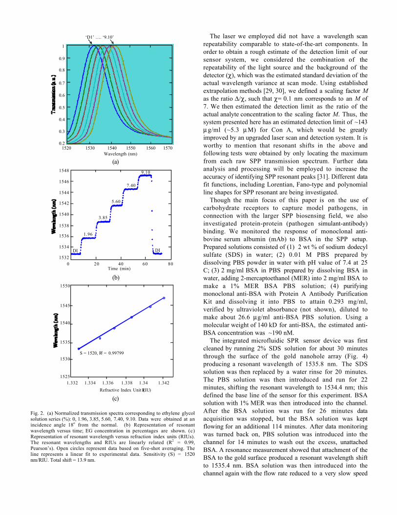
We monitored the response of Con A to ovomucoid in theSPP setup. Prepared solutions consisted of (1) 0.01 Mphosphate-buffered saline (PBS) prepared by dissolving PBSpowder in water with pH value of 7.4 at 25 C; (2) 1 mg/mlCon A in PBS, (3) an aqueous solution of ovomucoid (1mg/ml) heated for 10 minutes at 95 degrees C, stirred for 30minutes, passed through a 0.2 micron-pore syringe filter(Fisherbrand, nylon) and allowed to cool on ice.
The integrated microfluidic SPR sensor device waspretreated with DI water for 8 minutes. The PBS solution wasthen introduced and run for 10 minutes, shifting the resonantwavelength to 1543.2 nm (Fig. 3); this defined the base lineof the sensor for this experiment. Solution with 1 mg/ml ofovomucoid was then introduced into the channel. After theovomucoid solution was run for 11 minutes, PBS was addedto the channel for flushing away excess unbound ovomucoid,leading to a resonant wavelength shift to 1544.2 nm. Afterflowing PBS for 20 minutes, the purified Con A solution wasintroduced into the channel to allow for specific binding to theovomucoid attached to the gold surface. After 25 minutesPBS was introduced to wash out the unbound Con A,although disassociation of weakly bound ovomucoid fromgold surface could also have occurred. The resulting resonantwavelength was 1544.9 nm, which corresponds to a bindingshift D of 0.7 nm.
0.5
0.6
0.7
0.8
0.9
1
1520 1530 1540 1550 1560
0.4
0.3
0.2
Wavelength (nm)1570
‘D1’ …. ‘9.10’
(a)
1540
1542
1544
1546
1548
0 20 40 60 80Time (min)
1532
1534
1536
1538
DI
1.96
3.85
5.60
7.40
DI
9.10
(b)
1525
1530
1535
1540
1545
1550
1.332 1.334 1.336 1.338 1.34 1.342
S = 1520, R2 = 0.99799
Refractive Index Unit (RIU)
(c)
Fig. 2. (a) Normalized transmission spectra corresponding to ethylene glycolsolution series (%): 0, 1.96, 3.85, 5.60, 7.40, 9.10. Data were obtained at anincidence angle 18o from the normal. (b) Representation of resonantwavelength versus time; EG concentration in percentages are shown. (c)Representation of resonant wavelength versus refraction index units (RIUs).The resonant wavelengths and RIUs are linearly related (R2 = 0.99,Pearson’s). Open circles represent data based on five-shot averaging. Theline represents a linear fit to experimental data. Sensitivity (S) = 1520nm/RIU. Total shift = 13.9 nm.
The laser we employed did not have a wavelength scanrepeatability comparable to state-of-the-art components. Inorder to obtain a rough estimate of the detection limit of oursensor system, we considered the combination of therepeatability of the light source and the background of thedetector (c), which was the estimated standard deviation of theactual wavelength variance at scan mode. Using establishedextrapolation methods [29, 30], we defined a scaling factor Mas the ratio D/c, such that c= 0.1 nm corresponds to an M of7. We then estimated the detection limit as the ratio of theactual analyte concentration to the scaling factor M. Thus, thesystem presented here has an estimated detection limit of ~143m g/ml (~5.3 m M) for Con A, which would be greatlyimproved by an upgraded laser scan and detection system. It isworthy to mention that resonant shifts in the above andfollowing tests were obtained by only locating the maximumfrom each raw SPP transmission spectrum. Further dataanalysis and processing will be employed to increase theaccuracy of identifying SPP resonant peaks [31]. Different datafit functions, including Lorentian, Fano-type and polynomialline shapes for SPP resonant are being investigated.
Though the main focus of this paper is on the use ofcarbohydrate receptors to capture model pathogens, inconnection with the larger SPP biosensing field, we alsoinvestigated protein-protein (pathogen simulant-antibody)binding. We monitored the response of monoclonal anti-bovine serum albumin (mAb) to BSA in the SPP setup.Prepared solutions consisted of (1) 2 wt % of sodium dodecylsulfate (SDS) in water; (2) 0.01 M PBS prepared bydissolving PBS powder in water with pH value of 7.4 at 25C; (3) 2 mg/ml BSA in PBS prepared by dissolving BSA inwater, adding 2-mercaptoethanol (MER) into 2 mg/ml BSA tomake a 1% MER BSA PBS solution; (4) purifyingmonoclonal anti-BSA with Protein A Antibody PurificationKit and dissolving it into PBS to attain 0.293 mg/ml,verified by ultraviolet absorbance (not shown), diluted tomake about 26.6 mg/ml anti-BSA PBS solution. Using amolecular weight of 140 kD for anti-BSA, the estimated anti-BSA concentration was ~190 nM.
The integrated microfluidic SPR sensor device was firstcleaned by running 2% SDS solution for about 30 minutesthrough the surface of the gold nanohole array (Fig. 4)producing a resonant wavelength of 1535.8 nm. The SDSsolution was then replaced by a water rinse for 20 minutes.The PBS solution was then introduced and run for 22minutes, shifting the resonant wavelength to 1534.4 nm; thisdefined the base line of the sensor for this experiment. BSAsolution with 1% MER was then introduced into the channel.After the BSA solution was run for 26 minutes dataacquisition was stopped, but the BSA solution was keptflowing for an additional 114 minutes. After data monitoringwas turned back on, PBS solution was introduced into thechannel for 14 minutes to wash out the excess, unattachedBSA. A resonance measurement showed that attachment of theBSA to the gold surface produced a resonant wavelength shiftto 1535.4 nm. BSA solution was then introduced into thechannel again with the flow rate reduced to a very slow speed
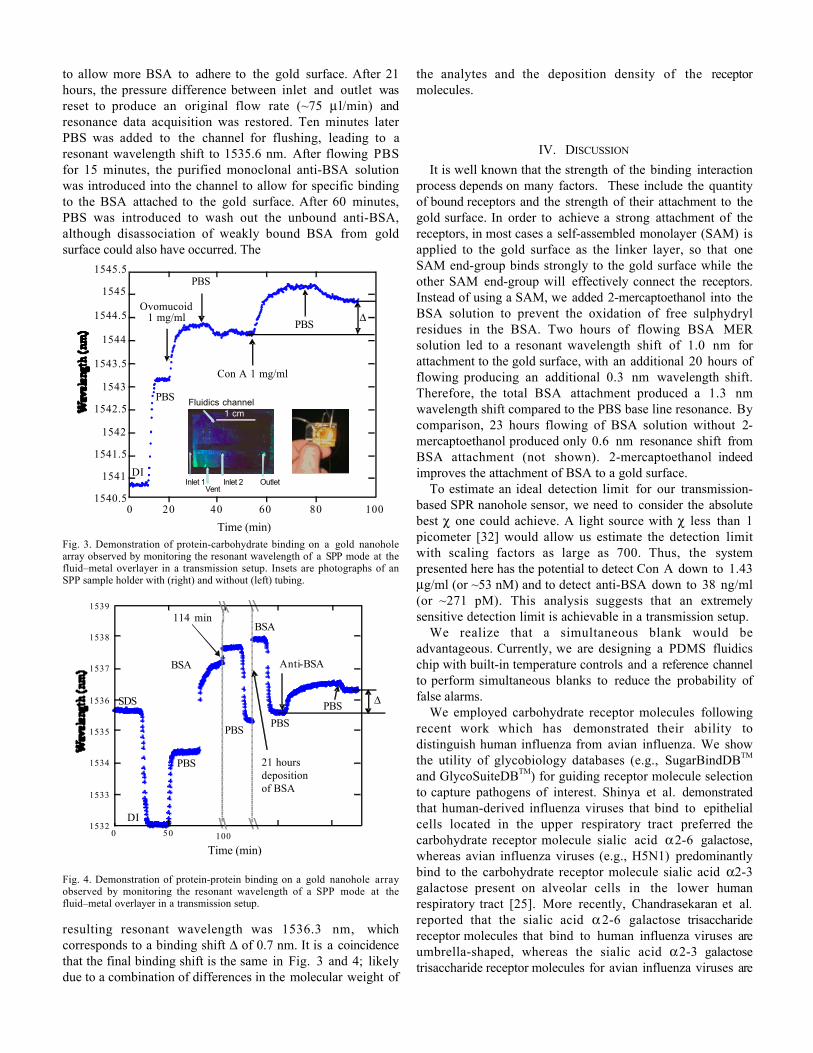
to allow more BSA to adhere to the gold surface. After 21hours, the pressure difference between inlet and outlet wasreset to produce an original flow rate (~75 ml/min) andresonance data acquisition was restored. Ten minutes laterPBS was added to the channel for flushing, leading to aresonant wavelength shift to 1535.6 nm. After flowing PBSfor 15 minutes, the purified monoclonal anti-BSA solutionwas introduced into the channel to allow for specific bindingto the BSA attached to the gold surface. After 60 minutes,PBS was introduced to wash out the unbound anti-BSA,although disassociation of weakly bound BSA from goldsurface could also have occurred. The
1545.5
1545
1544.5
1544
1543.5
1543
1542.5
1542
1541.5
1541
1540.50 20 40 60 80 100
DI
Ovomucoid1 mg/ml
PBS
PBS
Con A 1 mg/ml
PBS
Time (min)
D
Inlet 1 Inlet 2 OutletVent
Fluidics channel
1 cm1 cm
Fig. 3. Demonstration of protein-carbohydrate binding on a gold nanoholearray observed by monitoring the resonant wavelength of a SPP mode at thefluid–metal overlayer in a transmission setup. Insets are photographs of anSPP sample holder with (right) and without (left) tubing.
0 50 1001532
1533
1534
1535
1536
1537
1538
1539
Time (min)
SDS
DI
PBS
PBSPBS
BSA
BSA
Anti-BSA
PBS
114 min
21 hours deposition of BSA
D
Fig. 4. Demonstration of protein-protein binding on a gold nanohole arrayobserved by monitoring the resonant wavelength of a SPP mode at thefluid–metal overlayer in a transmission setup.
resulting resonant wavelength was 1536.3 nm, whichcorresponds to a binding shift D of 0.7 nm. It is a coincidencethat the final binding shift is the same in Fig. 3 and 4; likelydue to a combination of differences in the molecular weight of
the analytes and the deposition density of the receptormolecules.
IV. DISCUSSION
It is well known that the strength of the binding interactionprocess depends on many factors. These include the quantityof bound receptors and the strength of their attachment to thegold surface. In order to achieve a strong attachment of thereceptors, in most cases a self-assembled monolayer (SAM) isapplied to the gold surface as the linker layer, so that oneSAM end-group binds strongly to the gold surface while theother SAM end-group will effectively connect the receptors.Instead of using a SAM, we added 2-mercaptoethanol into theBSA solution to prevent the oxidation of free sulphydrylresidues in the BSA. Two hours of flowing BSA MERsolution led to a resonant wavelength shift of 1.0 nm forattachment to the gold surface, with an additional 20 hours offlowing producing an additional 0.3 nm wavelength shift.Therefore, the total BSA attachment produced a 1.3 nmwavelength shift compared to the PBS base line resonance. Bycomparison, 23 hours flowing of BSA solution without 2-mercaptoethanol produced only 0.6 nm resonance shift fromBSA attachment (not shown). 2-mercaptoethanol indeedimproves the attachment of BSA to a gold surface.
To estimate an ideal detection limit for our transmission-based SPR nanohole sensor, we need to consider the absolutebest c one could achieve. A light source with c less than 1picometer [32] would allow us estimate the detection limitwith scaling factors as large as 700. Thus, the systempresented here has the potential to detect Con A down to 1.43mg/ml (or ~53 nM) and to detect anti-BSA down to 38 ng/ml(or ~271 pM). This analysis suggests that an extremelysensitive detection limit is achievable in a transmission setup.
We realize that a simultaneous blank would beadvantageous. Currently, we are designing a PDMS fluidicschip with built-in temperature controls and a reference channelto perform simultaneous blanks to reduce the probability offalse alarms.
We employed carbohydrate receptor molecules followingrecent work which has demonstrated their ability todistinguish human influenza from avian influenza. We showthe utility of glycobiology databases (e.g., SugarBindDBTM
and GlycoSuiteDBTM) for guiding receptor molecule selectionto capture pathogens of interest. Shinya et al. demonstratedthat human-derived influenza viruses that bind to epithelialcells located in the upper respiratory tract preferred thecarbohydrate receptor molecule sialic acid a 2-6 galactose,whereas avian influenza viruses (e.g., H5N1) predominantlybind to the carbohydrate receptor molecule sialic acid a2-3galactose present on alveolar cells in the lower humanrespiratory tract [25]. More recently, Chandrasekaran et al.reported that the sialic acid a 2-6 galactose trisaccharidereceptor molecules that bind to human influenza viruses areumbrella-shaped, whereas the sialic acid a 2-3 galactosetrisaccharide receptor molecules for avian influenza viruses are

cone-shaped. This provides a topological explanation for whythe complementary protein lectin of H5N1 prefers sialic acida2-3 galactose [24]. Collectively, these findings suggest theimportance of carbohydrate receptor binding specificities fordifferentiating human from non-human influenza viruses. Inthe future, we plan to use E. coli K12 and a non-virulentstrain of influenza for testing this approach.
V. CONCLUSION
We have implemented a label-free method to utilizeglycoproteins as carbohydrate receptor molecules on a goldnanohole array to capture pathogen simulants in atransmission-based nanohole array SPR setup. We devised amethod that utilizes glycomics databases to guide the designof receptor molecules for use inside a biosensor. We showed,through estimates, that our SPR sensor can be improved toexhibit picomolar detection limits. In the future, we willexplore methods to improve wavelength illuminationrepeatability; experiment with different illuminationwavelength bands; model nanohole performance as a functionof diameter, lattice spacing; and fabricate different types ofnanostructures to enhance overall system resolution andsensitivity. We will also increase the total number of sensingelements in the PDMS mold in order to perform highly-parallel, multi-element SPR detection.
REFERENCES
[1] A. Mangili and M. A. Gendreau, "Transmission of infectious diseasesduring commercial air travel," The Lancet, vol. 365, pp. 989-996, 2005.
[2] J. J. Gooding, "Biosensor technology for detecting biological warfareagents: Recent progress and future trends," Anal. chim. acta, vol. 559,pp. 137-151, 2006.
[3] N. O. Fischer, T. M. Tarasow, and J. B. H. Tok, "Heightened sense forsensing: recent advances in pathogen immunoassay sensing platforms,"Analyst, vol. 132, pp. 187-191, 2007.
[4] H. Raether, Surface Plasmons on Smooth and Rough Surfaces and onGratings, Springer Tracts in Modern Physics. vol. 111. Berlin: Springer,1988.
[5] Biacore, "Flexchip Product Information," 2006, Product description.[6] R.L. Rich and D.G. Myszka "Survey of the year 2005 commercial
optical biosensor literature," Journal of Molecular Recognition, vol. 19,pp. 478-534, 2006.
[7] C. Genet and T. W. Ebbesen, "Light in tiny holes," Nature, vol. 445, pp.39-46, 2007.
[ 8 ] J. Homola, "Present and future of surface plasmon resonancebiosensors," Anal. Bioanal. Chem., vol. 377, pp. 528-539, 2003.
[9] R. Ince and R. Narayanaswamy, "Analysis of the performance ofinterferometry, surface plasmon resonance and luminescence asbiosensors and chemosensors," Anal. Chim. acta., vol. 569, pp. 1-20,2006.
[10] K. Phillips and Q. Cheng, "Recent advances in surface plasmonresonance based techniques for bioanalysis," Anal. Bioanal. Chem., vol.387, pp. 1831-1840, 2007.
[11] N. Ramachandran, D. N. Larson, P. R. H. Stark, E. Hainsworth, and J.LaBaer, "Emerging tools for real-time label-free detection ofinteractions on functional protein microarrays," Febs J., vol. 272, pp.5412-5425, 2005.
[12] A. D. Leebeeck, L. K. Swaroop Kumar, V. de Lange, D. Sinton, R.Gordon, and A. G. Brolo, "On-Chip Surface-Based Detection withNanohole Arrays," Anal. Chem., vol. 79, pp. 4094-4100, 2007.
[13] G. Steiner, "Surface plasmon resonance imaging," Anal. Bioanal.Chem., vol. 379, pp. 328-331, 2004.
[14] A. G. Brolo, R. Gordon, B. Leathem, and K. L. Kavanagh, "Surfaceplasmon sensor based on the enhanced light transmission througharrays of nanoholes in gold films," Langmuir, vol. 20, pp. 4813-4815,2004.
[15] T. W. Ebbesen, H. J. Lezec, H. F. Ghaemi, T. Thio, and P. A. Wolff,"Extraordinary optical transmission through sub-wavelength holearrays," Nature, vol. 391, pp. 667-669, 1998.
[16] M. E. Stewart, N. H. Mack, V. Malyarchuk, J. A. N. T. Soares, T.-W.Lee, S. K. Gray, R. G. Nuzzo, and J. A. Rogers, "Quantitativemultispectral biosensing and 1D imaging using quasi-3D plasmoniccrystals," Proceedings of the National Academy of Sciences, vol. 103,pp. 17143-17148, 2006.
[17] K. A. Tetz, L. Pang, and Y. Fainman, "High-resolution surface plasmonresonance sensor based on linewidth-optimized nanohole arraytransmittance," Opt. Lett., vol. 31, pp. 1528-1530, 2006.
[18] T. M. Chinowsky, M. S. Grow, K. S. Johnston, K. Nelson, T. Edwards,E. Fu, and P. Yager, "Compact, high performance surface plasmonresonance imaging system," Biosens. Bioelectron., vol. 22, pp. 2208-2215, 2007.
[19] T. M. Chinowsky, S. D. Soelberg, P. Baker, N. R. Swanson, P.Kauffman, A. Mactutis, M. S. Grow, R. Atmar, S. S. Yee, and C. E.Furlong, "Portable 24-analyte surface plasmon resonance instrumentsfor rapid, versatile biodetection," Biosens. Bioelectron., vol. 22, pp.2268-2275, 2007.
[20] A. N. Naimushin, C. B. Spinelli, S. D. Soelberg, T. Mann, R. C. Stevens,T. Chinowsky, P. Kauffman, S. Yee, and C. E. Furlong, "Airborneanalyte detection with an aircraft-adapted surface plasmon resonancesensor system," Sensor. Actuat B-Chem., vol. 104, pp. 237-248, 2005.
[21] G. P. Anderson, E. C. Merrick, S. A. Trammell, T. M. Chinowsky, andD. K. Shenoy, "Simplified avidin-biotin mediated antibody attachmentfor a surface plasmon resonance biosensor," Sens. Lett., vol. 3, pp. 151-156, 2005.
[22] L. Pang, K. A. Tetz, and Y. Fainman, "Observation of the splitting ofdegenerate surface plasmon polariton modes in a two-dimensionalmetallic nanohole array," App. Phys. Lett., vol. 90, p. 111103, 2007.
[23] K. A. Tetz, R. Rokitski, M. Nezhad, and Y. Fainman, "Excitation anddirect imaging of surface plasmon polariton modes in a two-dimensional grating," Appl. Phys. Lett., vol. 86, p. 111110, 2005.
[24] A. Chandrasekaran, A. Srinivasan, R. Raman, K. Viswanathan, S.Raguram, T. M. Tumpey, V. Sasisekharan, and R. Sasisekharan,"Glycan topology determines human adaptation of avian H5N1 virushemagglutinin," Nat Biotech, vol. 26, pp. 107-113, 2008.
[25] K. Shinya, M. Ebina, S. Yamada, M. Ono, N. Kasai, and Y. Kawaoka,"Avian flu: Influenza virus receptors in the human airway," Nature,vol. 440, pp. 435-436, 2006.
[26] H. F. Ghaemi, T. Thio, D. E. Grupp, T. W. Ebbesen, and H. J. Lezec,"Surface plasmons enhance optical transmission throughsubwavelength holes," Physical Review B, vol. 58, p. 6779, 1998.
[27] "Glycan Database ( http://sugarbinddb.mitre.org), " in Glycobiology. vol.15, pp. 9G-14G, 2005.
[28] C. A. Cooper, H. J. Joshi, M. J. Harrison, M. R. Wilkins, and N. H.Packer, "GlycoSuiteDB: a curated relational database of glycoproteinglycan structures and their biological sources. 2003 update," Nucl.Acids Res., vol. 31, pp. 511-513, 2003.
[29] L. Pang, G. M. Hwang, B. Slutsky, and Y. Fainman, "Spectral sensitivityof two-dimensional nanohole array surface plasmon polaritonresonance sensor," App. Phys. Lett., vol. 91, p. 123112, 2007.
[30] W. Yuan, H. P. Ho, C. L. Wong, S. K. Kong, and C. L. Lin, "Surfaceplasmon resonance biosensor incorporated in a Michelsoninterferometer with enhanced sensitivity," IEEE Sensors Journal, vol.7, pp. 70-73, 2007.
[31] A. B. Dahlin, J. O. Tegenfeldt, and F. Hook, "Improving theinstrumental resolution of sensors based on localized surface plasmonresonance," Analytical Chemistry, vol. 78, pp. 4416-4423, 2006.
[32] A. M. Armani, R. P. Kulkarni, S. E. Fraser, R. C. Flagan, and K. J.Vahala, "Label-free, single-molecule detection with opticalmicrocavities," Science, vol. 317, pp. 783-787, 2007.
Grace Hwang received a B.S. in civil and environmental engineering fromNortheastern University in 1996. She earned a S.M. in civil engineering fromthe Massachusetts Institute of Technology in 1998. She received M.S. andPh.D. degrees in biophysics and structural biology from Brandeis Universityin 2004 and 2005, respectively. She worked at Raytheon Company between

1990 and 2000 where she developed a variety of optical systems forchemical analysis and biological detection. At Brandeis University shedeveloped a confocal microscopy system with single-molecule resolution forfluorescent correlation spectroscopy of proteins. In 2000, she joined theMITRE Corporation, a not-for-profit, federally funded research anddevelopment center in the U.S. She is presently a Lead Biosensors Scientist.Her current research interests include biosensor development based onsurface plasmon resonance and bioaerosol sensor design and integration.
Elaine Mullen received her education in biology and biochemistry fromVirginia Commonwealth and Johns Hopkins Universities. As a researchscientist at The MITRE Corporation in McLean, VA, she studies bioactiveproperties of patented glycoprotein films. To support the development ofglycoprotein biocapture films, she initiated an ongoing effort to listpublications on carbohydrate receptors of human pathogens, now publishedon-line in a searchable database at http://SugarBindDB.mitre.org . Hercurrent research includes a collaborative effort involving the Consortium forFunctional Glycomics and the US Department of Agriculture to preciselydefine carbohydrate affinities of pathogens responsible for food- andwaterborne-illness.
Lin Pang received his BS and MS degrees in physics from LanzhouUniversity, China, in 1987 and 1990, respectively, and his Ph.D. degree inoptics from Sichuan University in 1998 with a thesis on the fabrication ofmicrostructures. Since 2001 he has been a project scientist with the Ultrafastand Nanoscale Optics Group of the University of California at San Diego.His current interests include the holographic lithography, photonic crystal,nanostructure fabrication and biosensors based on surface plasmonresonance.
Yeshaiahu Fainman received his PhD degree from Technion-Israel Instituteof Technology in 1983. He is a Cymer Professor of Advanced OpticalTechnologies and Professor of Electrical and Computer Engineering at theUniversity of California, San Diego. His current research interests are inoptical signal and information processing of ultrafast signals using both linearand nonlinear optical processes; nonlinear space-time processes usingfemtosecond laser pulses for optical communications, quantum cryptographyand communication; near field phenomena in optical nanostructures andnanophotonic devices; diffractive and nonlinear optics; and multidimensionalquantitative imaging. He has contributed 200 manuscripts in refereedjournals and over 350 conference presentations and conferenceproceedings. He is a Fellow of the Optical Society of America, Fellow of theInstitute of Electrical and Electronics Engineers, and Fellow of the Societyof Photo-Optical Instrumentation Engineers. His honors and awards includeMiriam and Aharon Gutvirt Prize, Technion, Haifa, Israel (1982), LadyDavis Fellowship (2006), and Brown award (2006). He Chaired and co-Chaired various conferences including IEEE/LEOS sub-Committee onOptical Interconnects and Signal Processing, 2004-2008 and served as aGeneral Chair for Inaugural OSA Topical Meeting on Nanophotonics forInformation Systems, 2005. He also served on numerous conferenceprogram committees, organized symposia and workshops, and between 1993and 2001 served as a topical editor of the Journal of the Optical Society ofAmerica: A on Optical Signal Processing and Imaging Science; he served asAssociate Editor for “The European Physical Journals – Applied Physics” in2004-2006, and is currently serving as Editor for an International Journal onOptical Memory and Neural Networks from 1998 to the present, EditorialBoard Member of Nanotechnology Journal, and serving on Advisory Boardfor Journal Metamaterials.
Grace M. HwangLead Biosensors ScientistThe MITRE CorporationE090202 Burlington RoadBedford, MA 01730tel 781-271-2165fax 781-271-3086email [email protected]
Lin PangProject ScientistDepartment of Electrical and Computer EngineeringUniversity of California, San Diego9500 Gilman DriveLa Jolla, CA 92093-0407tel 858-534-2495fax 858-534-1225e-mail [email protected]
Elaine H. MullenSr. Multi-Discipline Systems EngineerThe MITRE CorporationH4197515 Colshire DriveMcLean, VA 22102-7539tel 703-983-7110fax 703-983-5963email [email protected]
Yeshaiahu FainmanProfessorDepartment of Electrical and Computer EngineeringUniversity of California, San Diego9500 Gilman DriveLa Jolla, CA 92093-0407tel 858-534-8909fax 858-534-1225e-mail [email protected]