Embed Size (px)
Citation preview
Case ReportPlasmablastic Lymphoma, a Rare Entity in Bone Marrow withUnusual Immunophenotype and ChallengingDifferential Diagnosis
Majd Al Shaarani,1 Rodney E. Shackelford,1 Samip R. Master,2 Glenn M. Mills,2
Yasir AlZubaidi,1 Ahmed Mamilly,3 and Eric X. Wei 1
1Department of Pathology and Translational Pathobiology, Louisiana State University Health Sciences Center,Shreveport, LA, USA2Department of Hematology and Oncology, Louisiana State University Health Sciences Center, Shreveport, LA, USA3Department of Radiology, Louisiana State University Health Sciences Center, Shreveport, LA, USA
Correspondence should be addressed to Eric X. Wei; [email protected]
Received 28 December 2018; Revised 8 July 2019; Accepted 14 August 2019; Published 2 September 2019
Academic Editor: Marie-Christine Kyrtsonis
Copyright © 2019 Majd Al Shaarani et al. .is is an open access article distributed under the Creative Commons AttributionLicense, which permits unrestricted use, distribution, and reproduction in any medium, provided the original work isproperly cited.
Plasmablastic lymphoma (PBL) is an aggressive malignancy that usually occurs in the setting of immunosuppression. .eimmunohistochemical profile of PBL is that of terminally differentiated B lymphocytes. CD138, CD38, and MUM1 are usuallyimmunopositive. However, pan B-cell markers such as CD20 and PAX-5 are usually negative. MYC rearrangement is the mostcommonly encountered genetic alteration, with immunoglobulin (IG), especially immunoglobulin heavy (IGH) chain, being themost frequent partner. We report a case of PBL in a 48-year-old human immunodeficiency virus- (HIV-) positive male who wasadmitted to the hospital with signs and symptoms suspicious for tumor lysis syndrome. Bone marrow examination revealedhypercellular marrow with trilineage hypoplasia and sheets of intermediate to large neoplastic cells with basophilic vacuolatedcytoplasm comprising the majority of cellular elements of the bone marrow. .e neoplastic cells were negative for conventionalB-cell, T-cell, plasma cell, and myeloid markers, while flow cytometric analysis revealed an abnormal CD45-dim population thatwas partially weakly positive for CD71 and CD79b..e diagnosis was initially thought to be a high-grade primitive hematopoieticneoplasm, possibly an acute undifferentiated leukemia. BOB-1, however, was immunopositive in the neoplastic cells, confirmingits B-cell origin. MYC was positive by immunohistochemistry and break-apart FISH, as were CD45, MUM-1, and EMAimmunostains. .ere was immunoglobulin kappa (IGK) light chain gene rearrangement by polymerase chain reaction (PCR).Additionally, Epstein–Barr virus- (EBV–) encoded small RNAs (EBER) were positive by in situ hybridization (ISH). .e tumorproliferation index by Ki-67 immunostaining approached 95%. Although the tumor cells were negative for CD38 and CD138, thediagnosis of PBL was still rendered. We recommend using a broad spectrum of B-cell markers, including BOB-1 and OCT-2, insuch challenging cases of B-cell lymphomas with no expression of conventional B-cell markers. We also emphasize that thenegative CD38 and CD138 should not exclude PBL from the differential diagnosis.
1. Introduction
Plasmablastic lymphoma (PBL) is an aggressive postgerminalcenter, terminally differentiated B-cell malignancy thatusually occurs in the setting of immunosuppression. Initially,PBL was considered a subtype of diffuse large B-cell lym-phoma arising in the oral cavity of human immunodeficiency
virus- (HIV–) positive patients, as was first described byDelecluse et al. [1–3]. It was later categorized by the WHO asa separate entity in 2008, and accumulative data havedemonstrated cases at other anatomical sites and in im-munocompetent patients [4]. Although the majority of casesare associated with HIV infection, other causes of immu-nosuppression such as posttransplantation can also be
HindawiCase Reports in HematologyVolume 2019, Article ID 1586328, 9 pageshttps://doi.org/10.1155/2019/1586328
associated with this lymphoma. About 35% of the patients,however, are actually immunocompetent, indicating thatPBL is not confined to the immunosuppressed status. In themajority of cases, the disease arises in the oral cavity of HIV-infected patients. However, other sites are also reportedincluding the gastrointestinal (GI) system, soft tissue, skin,and much less frequently the lungs, bone, bone marrow, andcentral nervous system (CNS) [5, 6]. .e immunohisto-chemical profile of PBL is that of terminally differentiatedB lymphocytes or plasma cells, where CD138, CD38, andMUM1 are usually positive and pan B-cell markers includingCD20 and PAX-5 are frequently negative. MYC rearrange-ments are common, often translocated with the immuno-globulin (IG) heavy chains (IGH), as the most commonlyencountered genetic alteration [3, 7]..e rarity of such cases,the difficulty in making the diagnosis, and the very poorprognosis all make PBL a clinicopathological challenge [4].
2. Clinical History
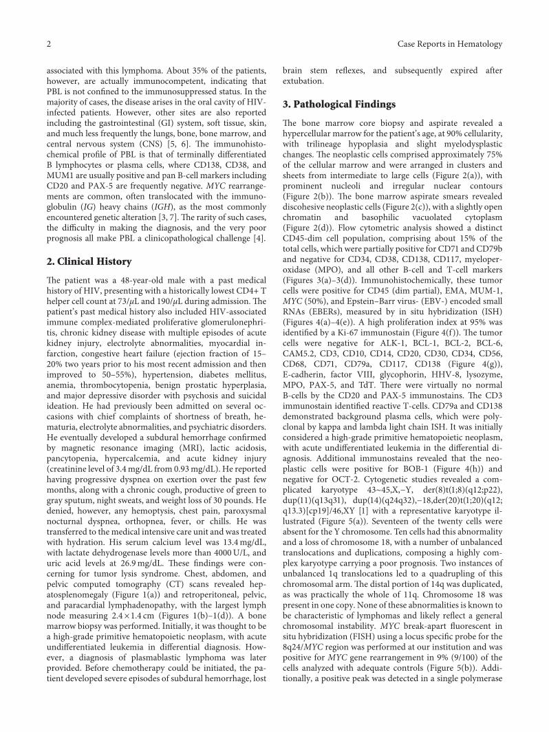
.e patient was a 48-year-old male with a past medicalhistory of HIV, presenting with a historically lowest CD4+ Thelper cell count at 73/µL and 190/µL during admission..epatient’s past medical history also included HIV-associatedimmune complex-mediated proliferative glomerulonephri-tis, chronic kidney disease with multiple episodes of acutekidney injury, electrolyte abnormalities, myocardial in-farction, congestive heart failure (ejection fraction of 15–20% two years prior to his most recent admission and thenimproved to 50–55%), hypertension, diabetes mellitus,anemia, thrombocytopenia, benign prostatic hyperplasia,and major depressive disorder with psychosis and suicidalideation. He had previously been admitted on several oc-casions with chief complaints of shortness of breath, he-maturia, electrolyte abnormalities, and psychiatric disorders.He eventually developed a subdural hemorrhage confirmedby magnetic resonance imaging (MRI), lactic acidosis,pancytopenia, hypercalcemia, and acute kidney injury(creatinine level of 3.4mg/dL from 0.93mg/dL). He reportedhaving progressive dyspnea on exertion over the past fewmonths, along with a chronic cough, productive of green togray sputum, night sweats, and weight loss of 30 pounds. Hedenied, however, any hemoptysis, chest pain, paroxysmalnocturnal dyspnea, orthopnea, fever, or chills. He wastransferred to the medical intensive care unit and was treatedwith hydration. His serum calcium level was 13.4mg/dL,with lactate dehydrogenase levels more than 4000U/L, anduric acid levels at 26.9mg/dL. .ese findings were con-cerning for tumor lysis syndrome. Chest, abdomen, andpelvic computed tomography (CT) scans revealed hep-atosplenomegaly (Figure 1(a)) and retroperitoneal, pelvic,and paracardial lymphadenopathy, with the largest lymphnode measuring 2.4×1.4 cm (Figures 1(b)–1(d)). A bonemarrow biopsy was performed. Initially, it was thought to bea high-grade primitive hematopoietic neoplasm, with acuteundifferentiated leukemia in differential diagnosis. How-ever, a diagnosis of plasmablastic lymphoma was laterprovided. Before chemotherapy could be initiated, the pa-tient developed severe episodes of subdural hemorrhage, lost
brain stem reflexes, and subsequently expired afterextubation.
3. Pathological Findings
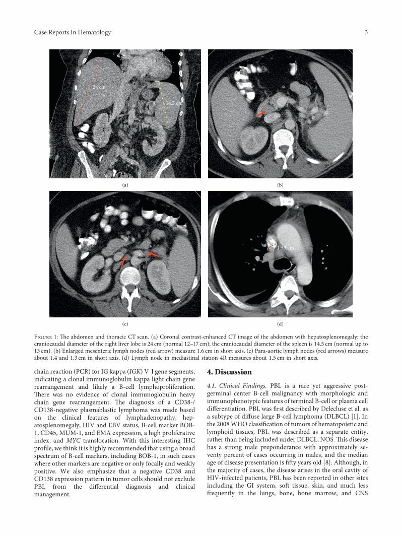
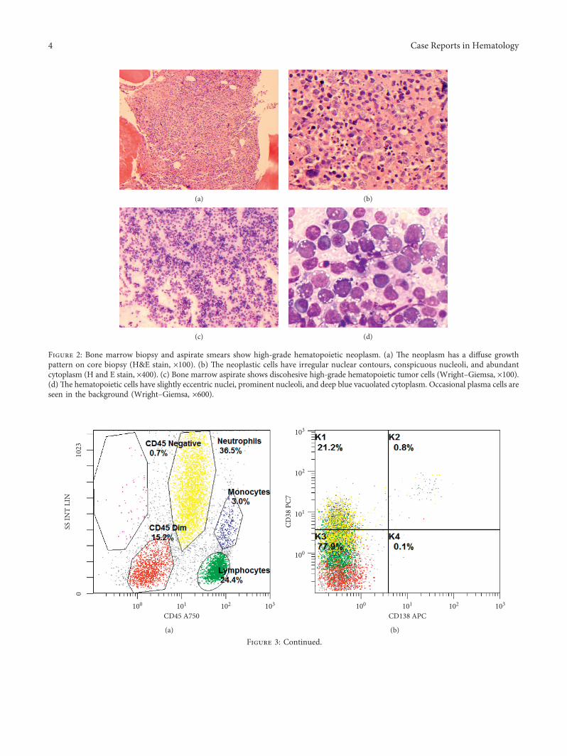
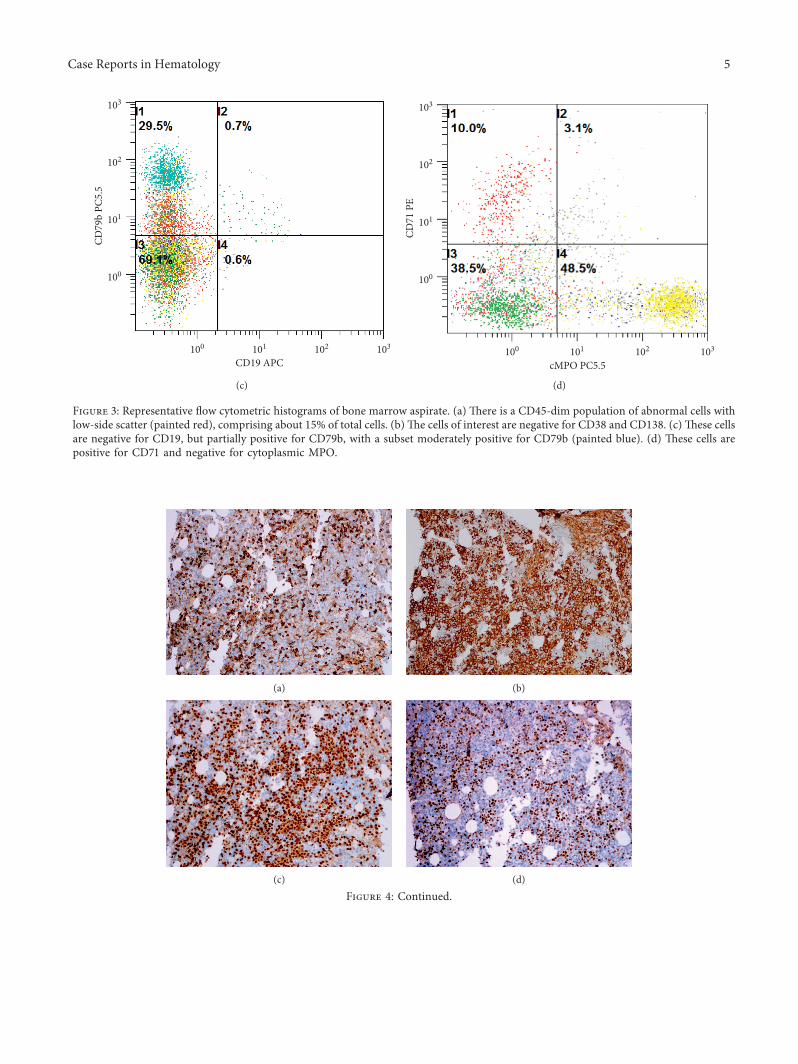
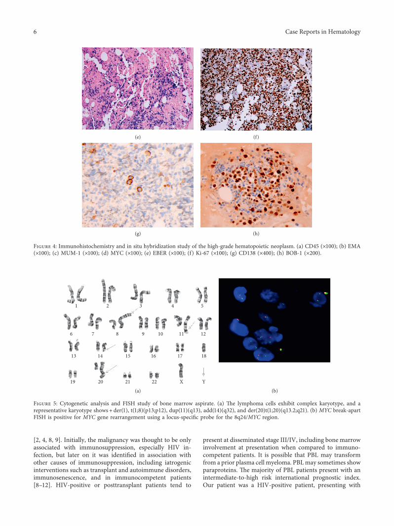
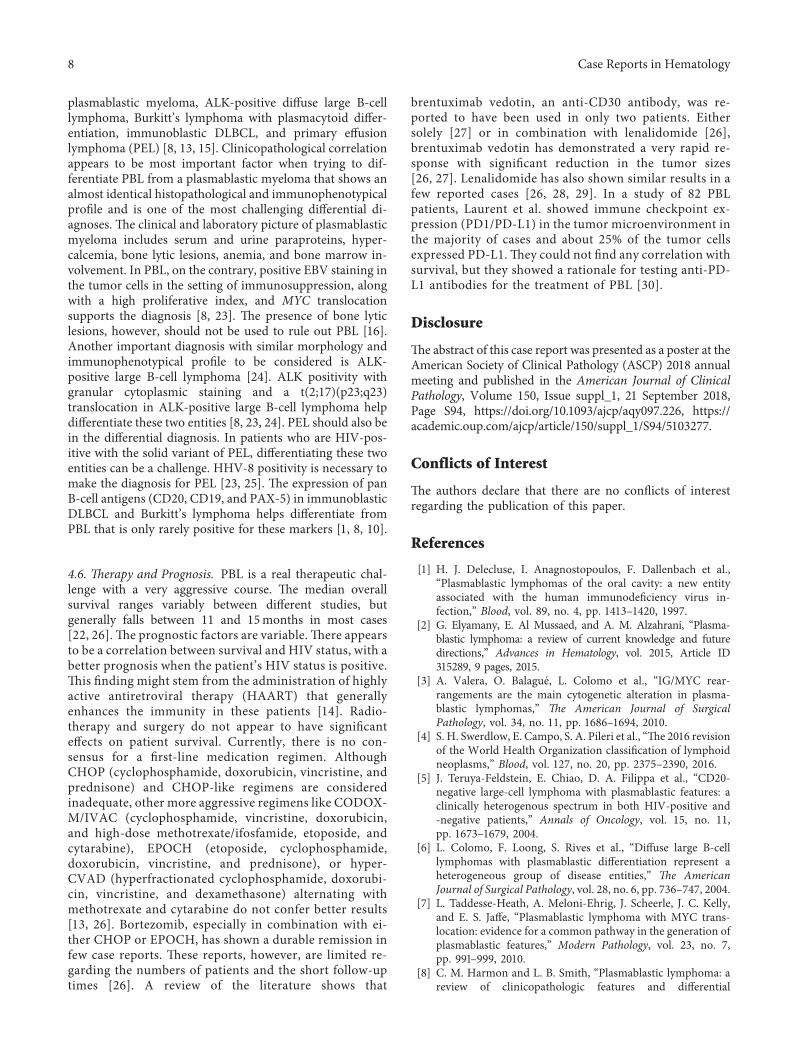
.e bone marrow core biopsy and aspirate revealed ahypercellular marrow for the patient’s age, at 90% cellularity,with trilineage hypoplasia and slight myelodysplasticchanges. .e neoplastic cells comprised approximately 75%of the cellular marrow and were arranged in clusters andsheets from intermediate to large cells (Figure 2(a)), withprominent nucleoli and irregular nuclear contours(Figure 2(b)). .e bone marrow aspirate smears revealeddiscohesive neoplastic cells (Figure 2(c)), with a slightly openchromatin and basophilic vacuolated cytoplasm(Figure 2(d)). Flow cytometric analysis showed a distinctCD45-dim cell population, comprising about 15% of thetotal cells, which were partially positive for CD71 and CD79band negative for CD34, CD38, CD138, CD117, myeloper-oxidase (MPO), and all other B-cell and T-cell markers(Figures 3(a)–3(d)). Immunohistochemically, these tumorcells were positive for CD45 (dim partial), EMA, MUM-1,MYC (50%), and Epstein–Barr virus- (EBV-) encoded smallRNAs (EBERs), measured by in situ hybridization (ISH)(Figures 4(a)–4(e)). A high proliferation index at 95% wasidentified by a Ki-67 immunostain (Figure 4(f)). .e tumorcells were negative for ALK-1, BCL-1, BCL-2, BCL-6,CAM5.2, CD3, CD10, CD14, CD20, CD30, CD34, CD56,CD68, CD71, CD79a, CD117, CD138 (Figure 4(g)),E-cadherin, factor VIII, glycophorin, HHV-8, lysozyme,MPO, PAX-5, and TdT. .ere were virtually no normalB-cells by the CD20 and PAX-5 immunostains. .e CD3immunostain identified reactive T-cells. CD79a and CD138demonstrated background plasma cells, which were poly-clonal by kappa and lambda light chain ISH. It was initiallyconsidered a high-grade primitive hematopoietic neoplasm,with acute undifferentiated leukemia in the differential di-agnosis. Additional immunostains revealed that the neo-plastic cells were positive for BOB-1 (Figure 4(h)) andnegative for OCT-2. Cytogenetic studies revealed a com-plicated karyotype 43∼45,X,− Y, der(8)t(1;8)(q12;p22),dup(11)(q13q31), dup(14)(q24q32),− 18,der(20)t(1;20)(q12;q13.3)[cp19]/46,XY [1] with a representative karyotype il-lustrated (Figure 5(a)). Seventeen of the twenty cells wereabsent for the Y chromosome. Ten cells had this abnormalityand a loss of chromosome 18, with a number of unbalancedtranslocations and duplications, composing a highly com-plex karyotype carrying a poor prognosis. Two instances ofunbalanced 1q translocations led to a quadrupling of thischromosomal arm. .e distal portion of 14q was duplicated,as was practically the whole of 11q. Chromosome 18 waspresent in one copy. None of these abnormalities is known tobe characteristic of lymphomas and likely reflect a generalchromosomal instability. MYC break-apart fluorescent insitu hybridization (FISH) using a locus specific probe for the8q24/MYC region was performed at our institution and waspositive for MYC gene rearrangement in 9% (9/100) of thecells analyzed with adequate controls (Figure 5(b)). Addi-tionally, a positive peak was detected in a single polymerase
2 Case Reports in Hematology
chain reaction (PCR) for IG kappa (IGK) V-J gene segments,indicating a clonal immunoglobulin kappa light chain generearrangement and likely a B-cell lymphoproliferation..ere was no evidence of clonal immunoglobulin heavychain gene rearrangement. .e diagnosis of a CD38-/CD138-negative plasmablastic lymphoma was made basedon the clinical features of lymphadenopathy, hep-atosplenomegaly, HIV and EBV status, B-cell marker BOB-1, CD45, MUM-1, and EMA expression, a high proliferativeindex, and MYC translocation. With this interesting IHCprofile, we think it is highly recommended that using a broadspectrum of B-cell markers, including BOB-1, in such caseswhere other markers are negative or only focally and weaklypositive. We also emphasize that a negative CD38 andCD138 expression pattern in tumor cells should not excludePBL from the differential diagnosis and clinicalmanagement.
4. Discussion
4.1. Clinical Findings. PBL is a rare yet aggressive post-germinal center B-cell malignancy with morphologic andimmunophenotypic features of terminal B-cell or plasma celldifferentiation. PBL was first described by Delecluse et al. asa subtype of diffuse large B-cell lymphoma (DLBCL) [1]. Inthe 2008WHO classification of tumors of hematopoietic andlymphoid tissues, PBL was described as a separate entity,rather than being included under DLBCL, NOS..is diseasehas a strong male preponderance with approximately se-venty percent of cases occurring in males, and the medianage of disease presentation is fifty years old [8]. Although, inthe majority of cases, the disease arises in the oral cavity ofHIV-infected patients, PBL has been reported in other sitesincluding the GI system, soft tissue, skin, and much lessfrequently in the lungs, bone, bone marrow, and CNS
14.5 cm
24 cm
(a) (b)
(c)
1.53 cm
(d)
Figure 1: .e abdomen and thoracic CT scan. (a) Coronal contrast-enhanced CT image of the abdomen with hepatosplenomegaly: thecraniocaudal diameter of the right liver lobe is 24 cm (normal 12–17 cm); the craniocaudal diameter of the spleen is 14.5 cm (normal up to13 cm). (b) Enlarged mesenteric lymph nodes (red arrow) measure 1.6 cm in short axis. (c) Para-aortic lymph nodes (red arrows) measureabout 1.4 and 1.3 cm in short axis. (d) Lymph node in mediastinal station 4R measures about 1.5 cm in short axis.
Case Reports in Hematology 3
(a) (b)
(c) (d)
Figure 2: Bone marrow biopsy and aspirate smears show high-grade hematopoietic neoplasm. (a) .e neoplasm has a diffuse growthpattern on core biopsy (H&E stain, ×100). (b) .e neoplastic cells have irregular nuclear contours, conspicuous nucleoli, and abundantcytoplasm (H and E stain, ×400). (c) Bone marrow aspirate shows discohesive high-grade hematopoietic tumor cells (Wright–Giemsa, ×100).(d).e hematopoietic cells have slightly eccentric nuclei, prominent nucleoli, and deep blue vacuolated cytoplasm. Occasional plasma cells areseen in the background (Wright–Giemsa, ×600).
SS IN
T LI
N10
230
CD45 A750100 101 103102
(a)
100
101
102
103
CD38
PC7
100 101 103102
CD138 APC
(b)
Figure 3: Continued.
4 Case Reports in Hematology
CD79
b PC
5.5
101 102 103100
CD19 APC
100
101
102
103
(c)
100
101
102
103
CD71
PE
100
cMPO PC5.5101 102 103
(d)
Figure 3: Representative flow cytometric histograms of bone marrow aspirate. (a) .ere is a CD45-dim population of abnormal cells withlow-side scatter (painted red), comprising about 15% of total cells. (b).e cells of interest are negative for CD38 and CD138. (c) .ese cellsare negative for CD19, but partially positive for CD79b, with a subset moderately positive for CD79b (painted blue). (d) .ese cells arepositive for CD71 and negative for cytoplasmic MPO.
(a) (b)
(c) (d)
Figure 4: Continued.
Case Reports in Hematology 5
[2, 4, 8, 9]. Initially, the malignancy was thought to be onlyassociated with immunosuppression, especially HIV in-fection, but later on it was identified in association withother causes of immunosuppression, including iatrogenicinterventions such as transplant and autoimmune disorders,immunosenescence, and in immunocompetent patients[8–12]. HIV-positive or posttransplant patients tend to
present at disseminated stage III/IV, including bone marrowinvolvement at presentation when compared to immuno-competent patients. It is possible that PBL may transformfrom a prior plasma cell myeloma. PBLmay sometimes showparaproteins. .e majority of PBL patients present with anintermediate-to-high risk international prognostic index.Our patient was a HIV-positive patient, presenting with
(e) (f )
(g) (h)
Figure 4: Immunohistochemistry and in situ hybridization study of the high-grade hematopoietic neoplasm. (a) CD45 (×100); (b) EMA(×100); (c) MUM-1 (×100); (d) MYC (×100); (e) EBER (×100); (f ) Ki-67 (×100); (g) CD138 (×400); (h) BOB-1 (×200).
1 2 3 4 5
6 7 8 9 10 11 12
13 14 15 16 17 18
19 20 21 22 YX
(a) (b)
Figure 5: Cytogenetic analysis and FISH study of bone marrow aspirate. (a) .e lymphoma cells exhibit complex karyotype, and arepresentative karyotype shows + der(1), t(1;8)(p13;p12), dup(11)(q13), add(14)(q32), and der(20)t(1;20)(q13.2;q21). (b) MYC break-apartFISH is positive for MYC gene rearrangement using a locus-specific probe for the 8q24/MYC region.
6 Case Reports in Hematology

pancytopenia, hepatosplenomegaly, general lymphadenop-athy, possible tumor lysis syndrome, and a high clinical stageinvolving bone marrow. He did not have oral cavity in-volvement. Even though hepatosplenomegaly and generallymphadenopathy may suggest high-grade lymphoma, itmay also be due to his HIV infection status. His pancyto-penia and tumor lysis syndrome may implicate acute leu-kemia. .us, accurate pathological diagnosis is essential forrecognition of this disease entity and clinical management.
4.2. Histopathology. Morphologically, PBL may show vari-ation depending on the sites and possibly the HIV status[6, 8]. Some of the common features include plasmablasticor immunoblastic morphology and plasmacytic differenti-ation. Mitoses, apoptosis, tingible body macrophages, andoccasionally confluent necrosis are characteristic of differentdisease variations [4, 8, 13]. In HIV-positive patients and inoral and nasal mucosal areas, PBL usually reveals sheets ofmonomorphic plasmablastic or immunoblastic cell pro-liferations, with vesicular chromatin, large round- to oval-shaped nuclei, prominent centrally located nucleoli, andabundant cytoplasm. In HIV-negative patients, and inlymph nodes and extraoral cavities, it exhibits a more ap-parent plasmacytic differentiation with eccentric nuclei,inconspicuous nucleoli, and paranuclear hofs [6, 8, 14].
4.3. Immunophenotype. .e immunophenotypic profile ofPBL is that of terminally differentiated B lymphocytes. CD138,CD38, VS38c, MUM1, PRDMA, and XBP1 positivity isdemonstrated in the majority of the cases. However, pan B-cellmarkers including CD20 and PAX-5 and leukocyte commonantigen CD45 are usually negative or can be weakly positive in aminority of cases. CD79a, EMA, and CD30 are often expressed,as well as cytoplasmic immunoglobulin, commonly IgG, fre-quently with either kappa or lambda light chain restriction.CD4, CD10, CD43, CD45RO, and CD56 have been reported tobe expressed in a subset of cases. BCL-1, BCL-2, and BCL-6expression is usually absent. Ki-67 is usually diffusely positive,typically above 90% [2, 4, 10, 13, 15]. Although the majority ofthe cases are CD138- andCD38-positive, the diagnosismust notbe overlooked if these stains are negative, such as in our case. Ina clinical study of 60 Chinese PBL cases, it was found about 12%and 16% of cases were negative for CD38 and CD138, re-spectively [10]. Dittus and Sarosiek have recently reported aninteresting case of PBL in the bonemarrow that was positive forCD45, CD79a, CD117, and lambda immunoglobulin lightchain but negative for CD38, CD138, PAX-5, and MUM1 [16].Among the 12 PBL cases described by Teruya-Felstein et al.,only one case of a 28-year-old HIV +ve male patient wasnegative for CD20, CD45, CD79a, CD138, and p63, with thebone and rectum involvement [5]. No other confirmatory B-cellmarkers were performed. Due to the similar immunohisto-chemical profile and anatomical locations between our case andreported cases in the literature, it is possible this lymphomamayexhibit different tumor markers when in different anatomicalstructures..e rarity of such cases and the lack of sufficient dataprevent such generalizations. Rare types of B-cell lymphomasmay not express conventional B-cell markers, and accurate
diagnosis of such cases can be challenging. In an analysis among34 cases of B-cell lymphomas with no expression of typicalB-cell lymphomas, it was found that, in 13 such PBL cases, 9cases expressed OCT-2, 10 cases had BOB-1 expression, andwhen combined together, 11 cases were positive for eitherOCT-2 or BOB-1 [17]. Compared to CD20, CD79a, and PAX-5,which on rare occasions can be found expressed in T-celllymphomas, acute myeloid leukemia, or nonhematopoietictumors, OCT-2 and BOB-1 are very sensitive and specific forconfirmation of B-cell lymphoma or plasma cell neoplasm thatare negative or equivocal for conventional B-cell antigen ex-pression [17]. CD71, a transferrin receptor, is present on activelyproliferative cells. It was partially positive in PBL tumor cells inour case by flow cytometry but was negative in bone marrowcore biopsy by immunohistochemistry. CD71 expression hasbeen detected by flow cytometry in B-cell lymphomas and acuteleukemia [18, 19]. Although weak CD71 expression has beenfound in DLBCL and subset of acute leukemias, it is selectivelyexpressed at high levels in early and late erythroid precursorsand can be used as a lineage-specific marker for erythroid cellsin bone marrow by immunohistochemistry [20]. .us, CD71partial expression in our PBL case by flow cytometry is non-specific, and negative CD71 immunohistochemical stain con-firms that tumor cells are not in erythroid lineage.
4.4.Cytogenetic andMolecularStudies. Our patient showed acomplex karyotype with positive MYC break-apart FISHresults. .e dissociation between the karyotype and FISHdata may be possibly due to a cryptic MYC translocationwith an unknown partner gene, undetected by conventionalcytogenetics. MYC gene rearrangements are the mostcommonly encountered PBL genetic alteration, with IG,particularly IGH, being the most frequent partner. .esealterations are usually associated with several other complexheterogeneous cytogenetic alterations [3, 7, 8, 10, 21, 22].Although the causality between EBV infection and MYCrearrangements has not been established, MYC alterationswere more frequently detected when EBV infection wasconfirmed. Furthermore, Morcsio et al. have shown thatEBVwas detected in about 75% of AIDS patients’ tumor cellsbut dropped down to 67% in transplant patients and furtherdown to 50% in immunocompetent patients [11]. Morestudies are needed to clearly demonstrate the causativemechanism, if one actually exists. Al-Malki et al. havedemonstrated that the presence ofMYC gene rearrangementand HIV negativity were adverse prognostic factors [21]. Intheir review of 60 non-HIV-related PBL cases from Chinesepatients, Han et al. found that IGH rearrangement was themost frequent alteration accounting for 33.3% of all 14different types of genetic alterations they found. .ey alsoreported, for the first time, the presence of PML/RARAfusion in PBL [10].
4.5. Differential Diagnosis. .e diagnosis of this rare entitydepends not only on the histopathological features, but alsoon the integration of the clinical history, radiologic studies,and laboratory tests. .e differential diagnosis encompasseslymphoid tumors with plasmacytic features, including
Case Reports in Hematology 7
plasmablastic myeloma, ALK-positive diffuse large B-celllymphoma, Burkitt’s lymphoma with plasmacytoid differ-entiation, immunoblastic DLBCL, and primary effusionlymphoma (PEL) [8, 13, 15]. Clinicopathological correlationappears to be most important factor when trying to dif-ferentiate PBL from a plasmablastic myeloma that shows analmost identical histopathological and immunophenotypicalprofile and is one of the most challenging differential di-agnoses. .e clinical and laboratory picture of plasmablasticmyeloma includes serum and urine paraproteins, hyper-calcemia, bone lytic lesions, anemia, and bone marrow in-volvement. In PBL, on the contrary, positive EBV staining inthe tumor cells in the setting of immunosuppression, alongwith a high proliferative index, and MYC translocationsupports the diagnosis [8, 23]. .e presence of bone lyticlesions, however, should not be used to rule out PBL [16].Another important diagnosis with similar morphology andimmunophenotypical profile to be considered is ALK-positive large B-cell lymphoma [24]. ALK positivity withgranular cytoplasmic staining and a t(2;17)(p23;q23)translocation in ALK-positive large B-cell lymphoma helpdifferentiate these two entities [8, 23, 24]. PEL should also bein the differential diagnosis. In patients who are HIV-pos-itive with the solid variant of PEL, differentiating these twoentities can be a challenge. HHV-8 positivity is necessary tomake the diagnosis for PEL [23, 25]. .e expression of panB-cell antigens (CD20, CD19, and PAX-5) in immunoblasticDLBCL and Burkitt’s lymphoma helps differentiate fromPBL that is only rarely positive for these markers [1, 8, 10].
4.6. 1erapy and Prognosis. PBL is a real therapeutic chal-lenge with a very aggressive course. .e median overallsurvival ranges variably between different studies, butgenerally falls between 11 and 15months in most cases[22, 26]. .e prognostic factors are variable. .ere appearsto be a correlation between survival and HIV status, with abetter prognosis when the patient’s HIV status is positive..is finding might stem from the administration of highlyactive antiretroviral therapy (HAART) that generallyenhances the immunity in these patients [14]. Radio-therapy and surgery do not appear to have significanteffects on patient survival. Currently, there is no con-sensus for a first-line medication regimen. AlthoughCHOP (cyclophosphamide, doxorubicin, vincristine, andprednisone) and CHOP-like regimens are consideredinadequate, other more aggressive regimens like CODOX-M/IVAC (cyclophosphamide, vincristine, doxorubicin,and high-dose methotrexate/ifosfamide, etoposide, andcytarabine), EPOCH (etoposide, cyclophosphamide,doxorubicin, vincristine, and prednisone), or hyper-CVAD (hyperfractionated cyclophosphamide, doxorubi-cin, vincristine, and dexamethasone) alternating withmethotrexate and cytarabine do not confer better results[13, 26]. Bortezomib, especially in combination with ei-ther CHOP or EPOCH, has shown a durable remission infew case reports. .ese reports, however, are limited re-garding the numbers of patients and the short follow-uptimes [26]. A review of the literature shows that
brentuximab vedotin, an anti-CD30 antibody, was re-ported to have been used in only two patients. Eithersolely [27] or in combination with lenalidomide [26],brentuximab vedotin has demonstrated a very rapid re-sponse with significant reduction in the tumor sizes[26, 27]. Lenalidomide has also shown similar results in afew reported cases [26, 28, 29]. In a study of 82 PBLpatients, Laurent et al. showed immune checkpoint ex-pression (PD1/PD-L1) in the tumor microenvironment inthe majority of cases and about 25% of the tumor cellsexpressed PD-L1. .ey could not find any correlation withsurvival, but they showed a rationale for testing anti-PD-L1 antibodies for the treatment of PBL [30].
Disclosure
.e abstract of this case report was presented as a poster at theAmerican Society of Clinical Pathology (ASCP) 2018 annualmeeting and published in the American Journal of ClinicalPathology, Volume 150, Issue suppl_1, 21 September 2018,Page S94, https://doi.org/10.1093/ajcp/aqy097.226, https://academic.oup.com/ajcp/article/150/suppl_1/S94/5103277.
Conflicts of Interest
.e authors declare that there are no conflicts of interestregarding the publication of this paper.
References
[1] H. J. Delecluse, I. Anagnostopoulos, F. Dallenbach et al.,“Plasmablastic lymphomas of the oral cavity: a new entityassociated with the human immunodeficiency virus in-fection,” Blood, vol. 89, no. 4, pp. 1413–1420, 1997.
[2] G. Elyamany, E. Al Mussaed, and A. M. Alzahrani, “Plasma-blastic lymphoma: a review of current knowledge and futuredirections,” Advances in Hematology, vol. 2015, Article ID315289, 9 pages, 2015.
[3] A. Valera, O. Balague, L. Colomo et al., “IG/MYC rear-rangements are the main cytogenetic alteration in plasma-blastic lymphomas,” 1e American Journal of SurgicalPathology, vol. 34, no. 11, pp. 1686–1694, 2010.
[4] S. H. Swerdlow, E. Campo, S. A. Pileri et al., “.e 2016 revisionof the World Health Organization classification of lymphoidneoplasms,” Blood, vol. 127, no. 20, pp. 2375–2390, 2016.
[5] J. Teruya-Feldstein, E. Chiao, D. A. Filippa et al., “CD20-negative large-cell lymphoma with plasmablastic features: aclinically heterogenous spectrum in both HIV-positive and-negative patients,” Annals of Oncology, vol. 15, no. 11,pp. 1673–1679, 2004.
[6] L. Colomo, F. Loong, S. Rives et al., “Diffuse large B-celllymphomas with plasmablastic differentiation represent aheterogeneous group of disease entities,” 1e AmericanJournal of Surgical Pathology, vol. 28, no. 6, pp. 736–747, 2004.
[7] L. Taddesse-Heath, A. Meloni-Ehrig, J. Scheerle, J. C. Kelly,and E. S. Jaffe, “Plasmablastic lymphoma with MYC trans-location: evidence for a common pathway in the generation ofplasmablastic features,” Modern Pathology, vol. 23, no. 7,pp. 991–999, 2010.
[8] C. M. Harmon and L. B. Smith, “Plasmablastic lymphoma: areview of clinicopathologic features and differential
8 Case Reports in Hematology
diagnosis,” Archives of Pathology & Laboratory Medicine,vol. 140, no. 10, pp. 1074–1078, 2016.
[9] J. J. Castillo, E. S. Winer, D. Stachurski et al., “HIV-negativeplasmablastic lymphoma: not in the mouth,” Clinical Lym-phoma Myeloma and Leukemia, vol. 11, no. 2, pp. 185–189,2011.
[10] X. Han, M. Duan, L. Hu, D. Zhou, and W. Zhang, “Plas-mablastic lymphoma: review of 60 Chinese cases and prog-nosis analysis,” Medicine, vol. 96, no. 9, p. e5981, 2017.
[11] J. Morscio, D. Dierickx, J. Nijs et al., “Clinicopathologiccomparison of plasmablastic lymphoma in HIV-positive,immunocompetent, and posttransplant patients,” 1eAmerican Journal of Surgical Pathology, vol. 38, no. 7,pp. 875–886, 2014.
[12] C. Cattaneo, F. Facchetti, A. Re et al., “Oral cavity lymphomasin immunocompetent and human immunodeficiency virusinfected patients,” Leukemia & Lymphoma, vol. 46, no. 1,pp. 77–81, 2005.
[13] J. J. Castillo and J. L. Reagan, “Plasmablastic lymphoma: asystematic review,” 1e Scientific World Journal, vol. 11,pp. 687–696, 2011.
[14] J. J. Castillo, E. S. Winer, D. Stachurski et al., “Clinical andpathological differences between human immunodeficiencyvirus-positive and human immunodeficiency virus-negativepatients with plasmablastic lymphoma,” Leukemia & Lym-phoma, vol. 51, no. 11, pp. 2047–2053, 2010.
[15] G. Elyamany, A. M. Alzahrani, M. Aljuboury et al., “Clini-copathologic features of plasmablastic lymphoma: single-center series of 8 cases from Saudi Arabia,” Diagnostic Pa-thology, vol. 10, no. 1, 2015.
[16] C. Dittus and S. Sarosiek, “A case of HIV-negative plasma-blastic lymphoma of the bone marrow with a uniqueimmunophenotype,” Clinical Case Reports, vol. 5, no. 6,pp. 902–904, 2017.
[17] L. Yin, J. Xu, M. Li et al., “Oct2 and Bob1 are sensitive andspecific markers in lineage determination of B cell lymphomaswith no expression of conventional B cell markers,” Histo-pathology, vol. 69, no. 5, pp. 775–783, 2016.
[18] J. M. Wu, M. J. Borowitz, and E. G. Weir, “.e usefulness ofCD71 expression by flow cytometry for differentiating in-dolent from aggressive CD10+ B-cell lymphomas,” AmericanJournal of Clinical Pathology, vol. 126, no. 1, pp. 39–46, 2006.
[19] Q. Liu, MWang, Y Hu et al., “Significance of CD71 expressionby flow cytometry in diagnosis of acute leukemia,” Leukemia& Lymphoma, vol. 55, no. 4, pp. 892–898, 2014.
[20] H. Y. Dong, S. Wilkes, and H. Yang, “CD71 is selectively andubiquitously expressed at high levels in erythroid precursorsof all maturation stages: a comparative immunochemicalstudy with glycophorin A and hemoglobin A,” 1e AmericanJournal of Surgical Pathology, vol. 35, no. 5, pp. 723–732, 2011.
[21] M. M. Al-Malki, J. J. Castillo, J. M. Sloan, and A. Re, “He-matopoietic cell transplantation for plasmablastic lymphoma:a review,” Biology of Blood and Marrow Transplantation,vol. 20, no. 12, pp. 1877–1884, 2014.
[22] J. J. Castillo, M. Bibas, and R. N. Miranda, “.e biology andtreatment of plasmablastic lymphoma,” Blood, vol. 125, no. 15,pp. 2323–2330, 2015.
[23] J. S. Ahn, R. Okal, J. A. Vos, M. Smolkin, A. S. Kanate, andF. G. Rosado, “Plasmablastic lymphoma versus plasmablasticmyeloma: an ongoing diagnostic dilemma,” Journal of ClinicalPathology, vol. 70, no. 9, pp. 775–780, 2017.
[24] Z. Pan, S. Hu, M. Li et al., “ALK-positive large B-cell lym-phoma: a clinicopathologic study of 26 cases with review of
additional 108 cases in the literature,”1eAmerican Journal ofSurgical Pathology, vol. 41, no. 1, pp. 25–38, 2017.
[25] H. Hashmi, D. Murray, S. Al-Quran, and W. Tse, “Primaryeffusion lymphoma without an effusion: a rare case of solidextracavitary variant of primary effusion lymphoma in anHIV-positive patient,” Case Reports in Hematology, vol. 2018,Article ID 9368451, 5 pages, 2018.
[26] D. Pretscher, A. Kalisch, M. Wilhelm, and J. Birkmann,“Refractory plasmablastic lymphoma—a review of treatmentoptions beyond standard therapy,” Annals of Hematology,vol. 96, no. 6, pp. 967–970, 2017.
[27] B. M. Holderness, S. Malhotra, N. B. Levy, and A. V. Danilov,“Brentuximab vedotin demonstrates activity in a patient withplasmablastic lymphoma arising from a background ofchronic lymphocytic leukemia,” Journal of Clinical Oncology,vol. 31, no. 12, pp. e197–e199, 2013.
[28] J. M. Schmit, J. DeLaune, M. Norkin, and A. Grosbach, “Acase of plasmablastic lymphoma achieving complete responseand durable remission after lenalidomide-based therapy,”Oncology Research and Treatment, vol. 40, no. 1-2, pp. 46–48,2017.
[29] W. D. Marrero, A. Cruz-Chacon, C. Castillo, andF. Cabanillas, “Successful use of bortezomib-lenalidomidecombination as treatment for a patient with plasmablasticlymphoma,” Clinical Lymphoma Myeloma and Leukemia,vol. 18, no. 7, pp. e275–e277, 2018.
[30] C. Laurent, B. Fabiani, C. Do et al., “Immune-checkpointexpression in epstein-barr virus positive and negative plas-mablastic lymphoma: a clinical and pathological study in 82patients,” Haematologica, vol. 101, no. 8, pp. 976–984, 2016.
Case Reports in Hematology 9

Stem Cells International
Hindawiwww.hindawi.com Volume 2018
Hindawiwww.hindawi.com Volume 2018
MEDIATORSINFLAMMATION
of
EndocrinologyInternational Journal of
Hindawiwww.hindawi.com Volume 2018
Hindawiwww.hindawi.com Volume 2018
Disease Markers
Hindawiwww.hindawi.com Volume 2018
BioMed Research International
OncologyJournal of
Hindawiwww.hindawi.com Volume 2013
Hindawiwww.hindawi.com Volume 2018
Oxidative Medicine and Cellular Longevity
Hindawiwww.hindawi.com Volume 2018
PPAR Research
Hindawi Publishing Corporation http://www.hindawi.com Volume 2013Hindawiwww.hindawi.com
The Scientific World Journal
Volume 2018
Immunology ResearchHindawiwww.hindawi.com Volume 2018
Journal of
ObesityJournal of
Hindawiwww.hindawi.com Volume 2018
Hindawiwww.hindawi.com Volume 2018
Computational and Mathematical Methods in Medicine
Hindawiwww.hindawi.com Volume 2018
Behavioural Neurology
OphthalmologyJournal of
Hindawiwww.hindawi.com Volume 2018
Diabetes ResearchJournal of
Hindawiwww.hindawi.com Volume 2018
Hindawiwww.hindawi.com Volume 2018
Research and TreatmentAIDS
Hindawiwww.hindawi.com Volume 2018
Gastroenterology Research and Practice
Hindawiwww.hindawi.com Volume 2018
Parkinson’s Disease
Evidence-Based Complementary andAlternative Medicine
Volume 2018Hindawiwww.hindawi.com
Submit your manuscripts atwww.hindawi.com


![Plasmablastic lymphoma presenting as a large cervical-thoracic … · 2020. 7. 3. · associated with human immunodeficiency virus infection (HIV) [1]. In the AIDS population, PBL](https://img.pdfslide.us/doc/110x75/6069f36ff8cff70a315119ce/plasmablastic-lymphoma-presenting-as-a-large-cervical-thoracic-2020-7-3-associated.jpg)