Embed Size (px)
Citation preview
Lymphoma Imaging: Diagnosis to Follow-up
Kevin Lian MD Line Srour, MD
Charlotte Yong-Hing, MD, FRCPC
Outline
1. Clinical Background 2. Staging System 3. Imaging
• Modalities • Staging • Treatment response • Surveillance
4. Cases
Classification: Hodgkin
• 10% of all lymphomas • Characterized by the presence of (few) Reed-Sternberg
cells in an inflammatory background • Subtypes:
• Classical (95%) • Nodular sclerosing • Mixed cellularity • Lymphocyte-rich • Lymphocyte-deplete
• Nodular lymphocyte predominant (5%)
WHO classification 2008

Classification: Non-Hodgkin
WHO classification 2008
Type B-cell T-cell Indolent Follicular (22%)
Small lymphocytic/CLL Lymphoplasmacytic Marginal zone extranodal (MALT) nodal splenic
Mycosis fungoides Primary cutaneous anaplastic large cell Lymphoproliferative disease of large granular lymphocytes
Aggressive Diffuse large B-cell lymphoma (31%)
Mantle cell DLBCL with features intermediate between
DLBCL and Burkitt lymphoma
Peripheral T-cell, NOS Peripheral T-cell, specified Angioimmunoblastic (AILD++-type) Nasal T/NK cell-type Subcutaneous panniculitic Intestinal enteropathy associated Hepatosplenic Anaplastic large cell including null cell
Very Aggressive
B-cell Lymphoblastic Burkitt (Double-hit lymphoma)
T-cell Lymphoblastic

Clinical Features
HL NHL (variable depending on histology)
Age Young adults 40-70 yo
B –Symptoms 40% 20%
Spread Contiguous nodal groups Multiple remote nodal groups
Stage at Presentation
>80% early (I & II) >85% late (III and IV)
Nodal Groups Thoracic Para-aortic Mesenteric
65-85% 25-35%
5%
25-40% 45-55% 50-60%
Extra-nodal disease
CNS GI GU BM
Lung Bone
<1% <1% <1% 5%
8-12% <1%
2% 5-15% 1-5%
20-40% 3-6% 1-2

Workup
• H & P • Blood work
• CBC, diff, platelets, electrolytes, LFTs, uric acid, LDH, beta 2 microglobulin, SPEP, viral serologies for hepatitis B, C and HIV
• Initial imaging • Tissue biopsy • Staging scan • +/- BM biopsy

Tissue Diagnosis
• Morphology, architecture, immunohistochemistry, flow cytometry +/- molecular studies
• Excisional biopsy preferred
• Core biopsy can be considered when excision is not possible. However, non-diagnostic sample must be followed by excisional biopsy
• FNA is inadequate
Staging
• Define location and extent • Prognostic information • Allow comparison among studies • Provides baseline for assessing response or
progression.

Staging
• Ann Arbor Classification • Cotswold Classification • Lugano Classification (2014)

Staging
* Tonsils, Waldeyer’s Ring, and spleen are considered nodal tissue

Staging - Qualifiers
• “Bulky / X” • Bulk is a negative prognostic factor for some lymphoma, but little
agreement on its definition • Largest measurement of single nodal mass • NHL
• Various threshold suggested (6-10 cm as cut-off) • HL
• Single nodal mass > 10 cm or >1/3 of transthoracic diameter • None of the proposed sizes have been validated in current therapeutic
era. • Lugano Classification: Give longest measurement rather than
designation of “X” for bulky disease • BCCA: “X” still used. Stage II bulky disease in HL treated as advanced
disease

Staging - Qualifiers
• “A / B” • A: absence of constitutional symptoms • B: presence of constitutional symptoms
• Unexplained weight loss > 10% baseline during 6 months prior to staging • Reccurrent unexplained fever > 38 degrees • Recurrent night sweats

Staging - Qualifiers
• “E” – Extra-nodal disease • Only apply in the setting of limited disease • Stage 1E = Single extranodal lesion without nodal involvement • Stage 2E = Stage 1 or 2 by nodal extent with limited contiguous
extranodal involvement • Designation “E” is not applicable in stage 3 and 4

Staging
Limited • Stages I and II (non-bulky)
Advanced • Stages III and IV
Stage II bulky • Limited or advanced as determined by histology and number of
prognostic marker • Usually treated as advanced

Staging – Bone Marrow Biopsy
• Previously standard in lymphoma staging • Often performed even if likelihood of involvement is low
• Hodgkin • If PET-CT is performed, BMB is not required.
• Non-Hodgkin (DLBCL) • PET-CT more sensitive than BMB, but may miss low-volume diffuse involvement
(<20%) • If PET-CT is positive and indicate bone marrow involvement, BMB is not
required. However, it is still routinely performed in BC currently
• Non-Hodgkin (Other) • Data insufficient to change standard of practice • 2.5 cm unilateral BMB is recommended
Cheson BD, et al. Recommendations for Initial Evaluation, Staging, and Response Assessment of Hodgkin and Non-Hodgkin Lymphoma: The Lugano Classification. J Clinical Oncology. 2014.Sep 20;32(27):3059-68.

Imaging
Goal: 1. Define local extent of clinically overt disease 2. To seek occult disease elsewhere
Primary modalities: CT and PET-CT Other: X-ray, ultrasound, MRI

Computed Tomography
• High spatial resolution • Detection of lymphadenopathy
by size/morphology • Staging scan:
– Neck – Chest – Abdomen – Pelvis

PET-CT
• CT • Detailed anatomic information
• PET • Functional information
• Overall • More accurate than CT for staging in HL and
NHL, with increased sensitivity, particularly extranodal disease
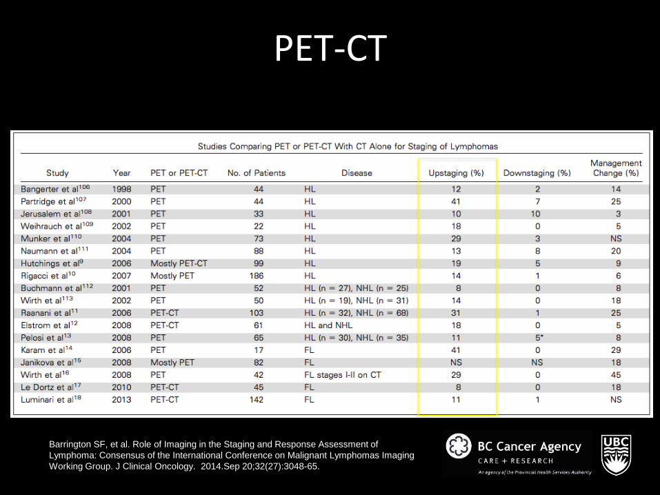
PET-CT
Barrington SF, et al. Role of Imaging in the Staging and Response Assessment of Lymphoma: Consensus of the International Conference on Malignant Lymphomas Imaging Working Group. J Clinical Oncology. 2014.Sep 20;32(27):3048-65.

Normal PET CT

Abnormal PET CT

Pitfalls
• False negative Lesions smaller than 8mm Diabetes/Non-fasting patient Tumor histology
• False positive Granulomas and infections Adenomas Normal physiology

PET - CT
• CT component can be done in 2 ways:
* Rare alters management* Recommendation: ceCT ideally should occur during single visit in
combination with PET-CT as baseline, if not already performed. Baseline findings will determine whether cePET-CT or lower-dose unenhanced PET-CT will suffice for additional exams.
Full Dose with Contrast Low Dose without Contrast -Improve detection of abdominal and pelvic disease
-Helps with planning for RT
-Accurate nodal measurement for trial purposes
-Lower radiation -Less error in measurement of FDG uptake in tumor (10-15% overestimation with contrast)
Barrington SF, et al. Role of Imaging in the Staging and Response Assessment of Lymphoma: Consensus of the International Conference on Malignant Lymphomas Imaging Working Group. J Clinical Oncology. 2014.Sep 20;32(27):3048-65.

Chest X-ray
• Cheap and accessible • No added information
compared to CT • Initial imaging that raises
suspicion for lymphoma • BCCA
• Often performed on follow-up visits after treatment

Ultrasound
• Limited role • Provide guidance for biopsy • Primary modality for evaluation of
testicular lymphoma • Non-specific appearance on US

MRI
• Limited availability. Not routinely used for staging.
• Accuracy overall comparable to CT in staging • Detection of lymphadenopathy by size • Compared to CT:
• Superior to CT for CNS and bone marrow Inferior to CT for thorax
• Whole-body MRI and diffusion-weighted imaging are radiation-free alternatives (i.e for children)

STAGING
PET-CT for FDG-avid lymphomas
CT for non-FDG-avid lymphomas •Small lymphocytic lymphoma
•Lymphoplasmacytic lymphoma •Waldenstrom macroglobulinemia
•Mycosis fungoides •Marginal zone NHL
(unless suspicion for aggressive transformation)
Cheson BD, et al. Recommendations for Initial Evaluation, Staging, and Response Assessment of Hodgkin and Non-Hodgkin Lymphoma: The Lugano Classification. J Clinical Oncology. 2014.Sep 20;32(27):3059-68.

TREATMENT RESPONSE
Interim PET-CT (early treatment response) • Ensure effectiveness of treatment and exclude possibility of
progression • iPET-CT is a strong prognostic indicator in HL and aggressive NHL • Frequently performed in clinical practice and trials and is
recommended by some international guidelines. • No conclusive evidence that changing treatment based on iPET-CT
improves outcome. • Lugano classification: Do not change treatment solely on the basis of
iPET-CT unless clear evidence of disease progression • In BC, the established guidelines are that interim PET scan will guide
treatment in limited and advanced-stage Hodgkin lymphoma, and limited-stage aggressive NHL.
Cheson BD, et al. Recommendations for Initial Evaluation, Staging, and Response Assessment of Hodgkin and Non-Hodgkin Lymphoma: The Lugano Classification. J Clinical Oncology. 2014.Sep 20;32(27):3059-68.

Limited-stage Hodgkin : BC approach
Stages 1 to 2A* Non-bulky ( <10 cm)
*Stage 2B Rx following the advance-stage algorithm ABVD x 2
PET/CT
Negative PET (Score 1,2)
Positive PET (Score 3, 4, 5)
ABVD x 2 Radiation therapy

Advanced-stage Hodgkin: BC approach
Stages 2B, 3 or 4 Stages 1 or 2 with bulky dz (≥ 10 cm)
ABVD x 2
PET/CT
Negative PET (Score 1, 2, 3)
Positive PET (Score 4, 5)
AVD x 4 ABVD x 4
Radiation Therapy

Limited-stage aggressive NHL: BC approach
Stages 1, 2 Non-bulky
R-CHOP x 3
PET/CT
Negative PET (Score 1, 2)
Positive PET (Score 3, 4, 5)
R-CHOP x 1 Radiation therapy

TREATMENT RESPONSE
End of Treatment • Lugano classification
• PET-CT for FDG-avid lymphoma • CT for non-FDG-avid lymphoma
• BCCA • Start with CT(neck, chest, abdo, pelvis) for all histologies (limited
resources) • If CT shows complete response, stop. (Usually PET would be negative) • If CT shows lesion > 2 cm, then get PET-CT
• PET-CT done at least 4-6 weeks after end of treatment to avoid false positive.
Cheson BD, et al. Recommendations for Initial Evaluation, Staging, and Response Assessment of Hodgkin and Non-Hodgkin Lymphoma: The Lugano Classification. J Clinical Oncology. 2014.Sep 20;32(27):3059-68.

Criteria for Response
PET •Five point scale (Deauville Criteria) validated for use at interim and end of treatment •Scores the most intense uptake in a site of initial disease:
1. No uptake 2. Uptake < mediastinum 3. Uptake > mediastinum but < liver 4. Uptake moderately higher than liver 5. Uptake markedly higher than liver and/or new lesions X. Area of uptake unlikely to be related to lymphoma

Criteria for Response
PET • 1 or 2 = complete metabolic response • 3 = complete metabolic response at interim and good prognosis
at completion • 4 or 5 (with reduced uptake) = partial metabolic response • Increase in FDG uptake, score of 5 with no decrease in uptake,
and new FDG avid foci = treatment failure / progression

Criteria for Response
CT •Complete response:
•Target nodes/nodal masses regress <1.5 cm (in longest transverse diameter) and no extra-lymphatic sites of disease •Spleen regress to normal

Criteria for Response
CT •Complete response:
•Target nodes/nodal masses regress <1.5 cm (in longest transverse diameter) and no extra-lymphatic sites of disease •Spleen regress to normal
•Partial response: •>50% decrease in sum of product diameter of up to 6 target measurable nodes and extranodal sites •Spleen regressed > 50% in length beyond normal
•Stable disease: •< 50% decrease from baseline in SPD of up to 6 dominant, measurable nodes and extranodal sites •No criteria for progressive disease are met

Criteria for Response
CT •Disease progression
•Require at least 1 of the following 1. New or increased adenopathy 2. Splenic volume increase 3. New or larger non-measured lesions 4. Recurrent previously resolved lesion 5. New extranodal lesion > 1cm in any axis

Follow-up / Surveillance
• Clinical surveillance (history, PE, labs) • Routine surveillance scan is discouraged
• PET-CT has false-positive rate of 20% leading to unnecessary investigations, radiation, biopsies, expense, and patient anxiety
• Follow-up scan should be prompted by clinical indication • Exception: Indolent lymphomas, asymptomatic intra-abdominal
or retroperitoneal disease progression may be clinically occult. Judicious use of scans can be considered.
Cheson BD, et al. Recommendations for Initial Evaluation, Staging, and Response Assessment of Hodgkin and Non-Hodgkin Lymphoma: The Lugano Classification. J Clinical Oncology. 2014.Sep 20;32(27):3059-68.

Cases

Case 1 – Limited HL

Case 1 – CMR

Case 2 – 14 yo

Case 2
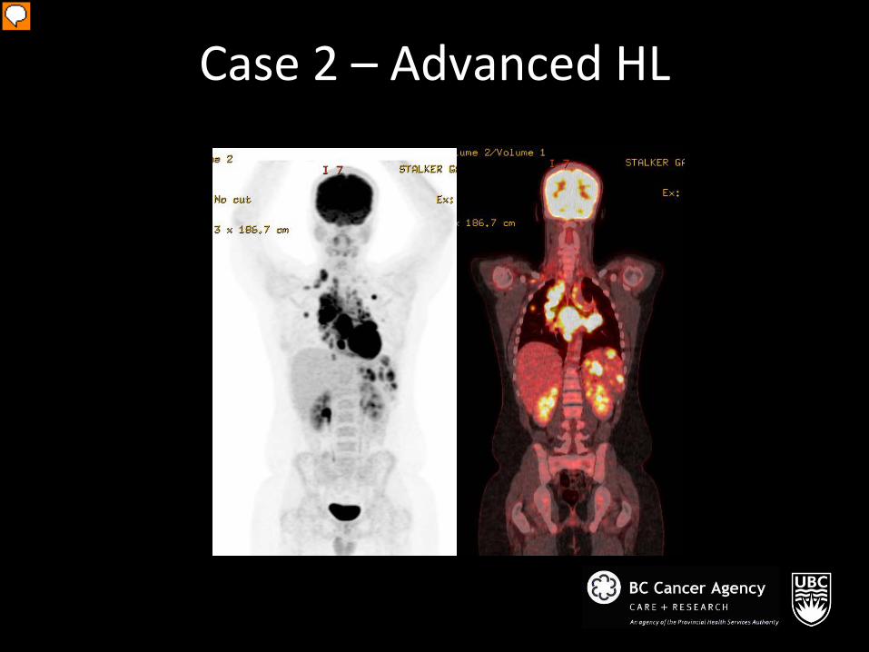
Case 2 – Advanced HL

Case 2 – CMR
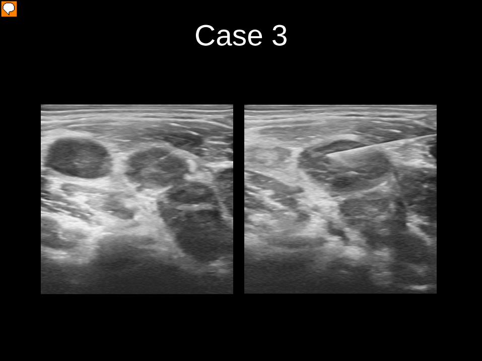
Case 3

Case 3 – Limited HL

Case 3 – Partial Response

Case 4

Case 4 – Limited HL

Case 4 – CMR

Case - DLBCL

Case - DLBCL

Case - DLBCL

Case - Progression

Extra-nodal Disease

CNS

Orbit

Nasopharyngeal NHL

Parotid

Thyroid

Spine
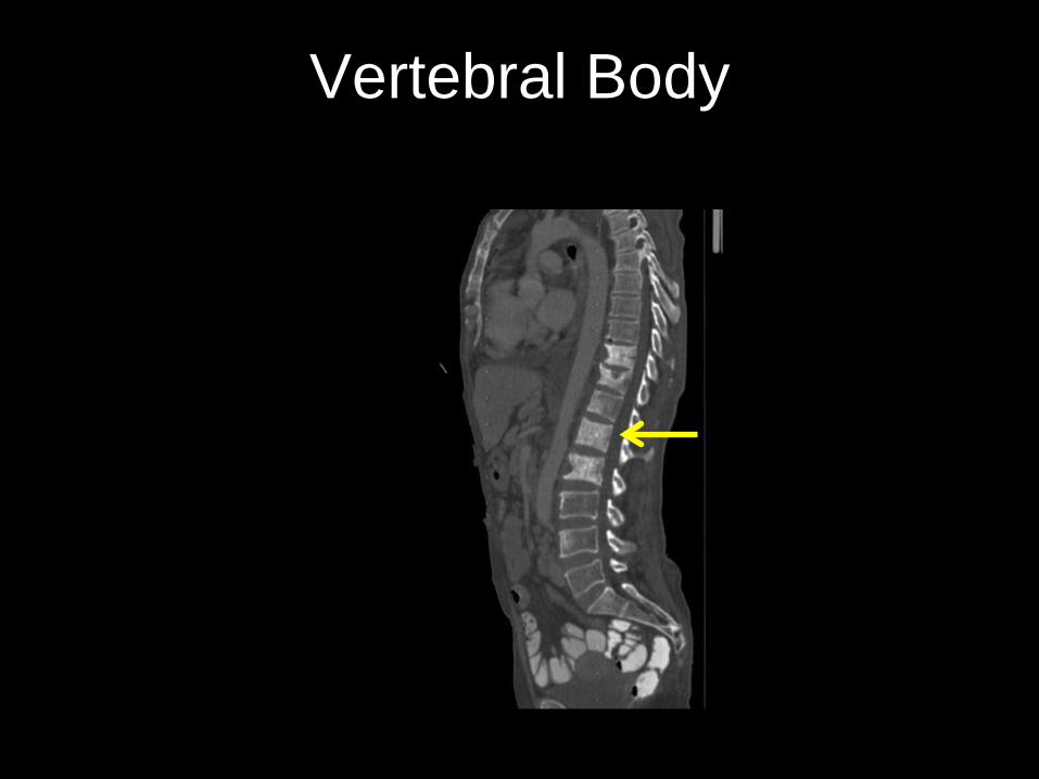
Vertebral Body
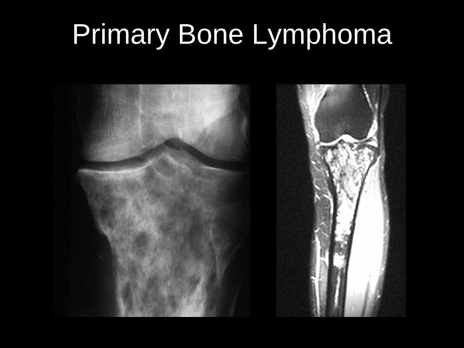
Primary Bone Lymphoma

Breast

Gastric

Small Bowel
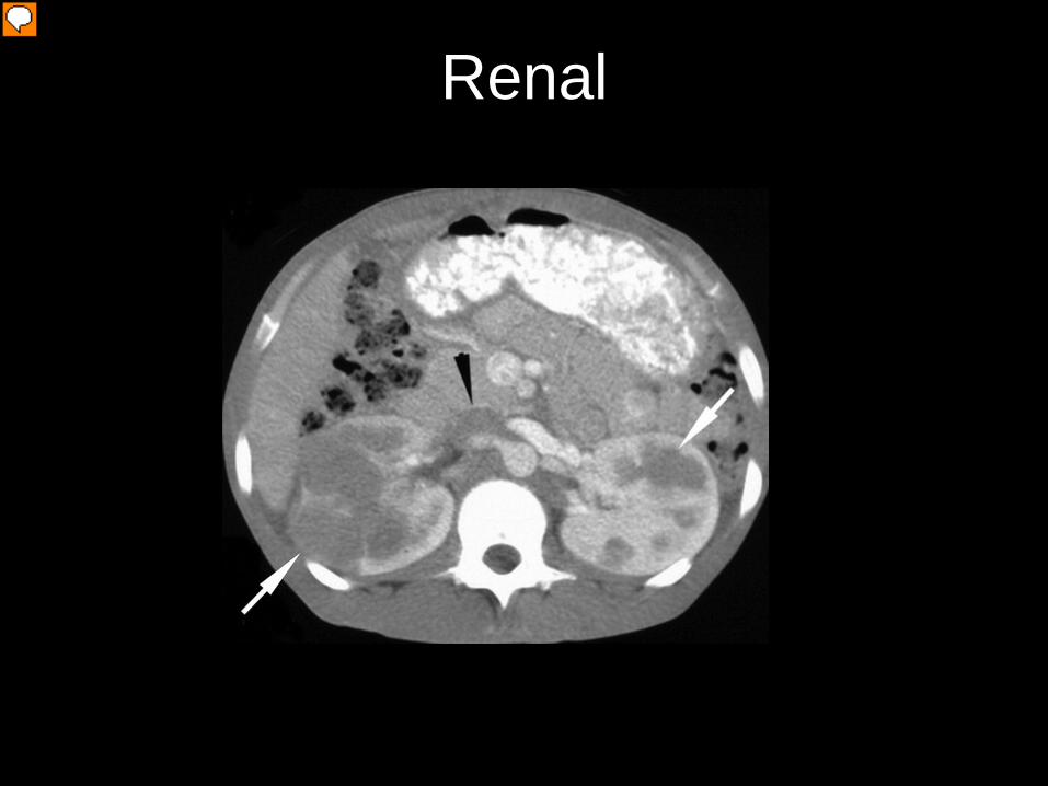
Renal

Testicular

Take Home Points
• Excisional biopsy is preferred for diagnosis, although core-needle biopsy may suffice when not feasible. FNA is considered inadequate.
• For staging, PET-CT is the standard for FDG-avid lymphomas, whereas CT is indicated for non-FDG-avid histology.
• Staging (limited vs advanced) is only one of many factors guide
treatment. • The designation X for bulky disease is no longer necessary per
Lugano Classification, but still used in BC.
Take Home Points
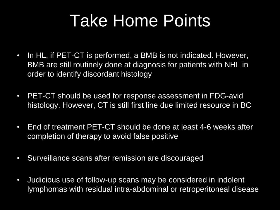
• In HL, if PET-CT is performed, a BMB is not indicated. However, BMB are still routinely done at diagnosis for patients with NHL in order to identify discordant histology
• PET-CT should be used for response assessment in FDG-avid
histology. However, CT is still first line due limited resource in BC • End of treatment PET-CT should be done at least 4-6 weeks after
completion of therapy to avoid false positive
• Surveillance scans after remission are discouraged
• Judicious use of follow-up scans may be considered in indolent lymphomas with residual intra-abdominal or retroperitoneal disease

Lymphoma Imaging - PET
Sharon Gershony MD UBC Radiology and Nuclear medicine Resident
UBC Radiology and Nuclear medicine Resident

BCCA Indications for FDG-PET in the Clinical Management of Adult Cancer Patients:
Lymphoma 1.Post-chemotherapy for patients with advanced stage aggressive non-Hodgkin lymphoma (including primary mediastinal large B cell lymphoma) and Hodgkin lymphoma with residual CT abnormalities or initial bulky (bulky = 10 cm or larger in any single diameter) disease to assess need for radiation therapy 2. Staging of Hodgkin lymphoma 3. Staging of aggressive non-Hodgkin lymphoma 4.PET to plan duration of chemotherapy for patients with limited stage (IA or IIA, non- bulky) Hodgkin lymphoma. 5.PET to plan duration and type of treatment for limited stage (IA or IIA, non-bulky) aggressive histology (diffuse large B cell, mantle cell, peripheral T cell) lymphoma. NOTE: No defined indication in the routine evaluation of low grade lymphomas.


DEAUVILLE CRITERIA

Other cancers given specific clinical indications, as approved by the BC Cancer Agency, on an individual basis. It is well recognized in clinical practice that there may be clinical scenarios that do not meet specific guidelines but where expert medical opinion indicates the procedure could have a major impact on patient management. PET scan referrals in these cases will be reviewed on an individual basis by physician representatives from the appropriate Provincial Tumor Group and the Functional Imaging department. If approved by consensus, the patient will be offered participation in the study.
http://www.bccancer.bc.ca/PPI/PET/indications.htm


![Lymphoma - ISD Scotland · [DLBCL/Burkitts Lymphoma] MYCDATE Date (DD/MM/CCYY) 10 15 MYC Testing Result [DLBCL/Burkitts Lymphoma] MYCRESULT Integer 2 16 Location of Diagnosis {Cancer}](https://img.pdfslide.us/doc/110x75/5fe202d4c67e945f1a036fa7/lymphoma-isd-scotland-dlbclburkitts-lymphoma-mycdate-date-ddmmccyy-10-15.jpg)