Embed Size (px)
Citation preview
PlanmecaProMax® 2D S3ProMax® 2D S2
ENG
LISH
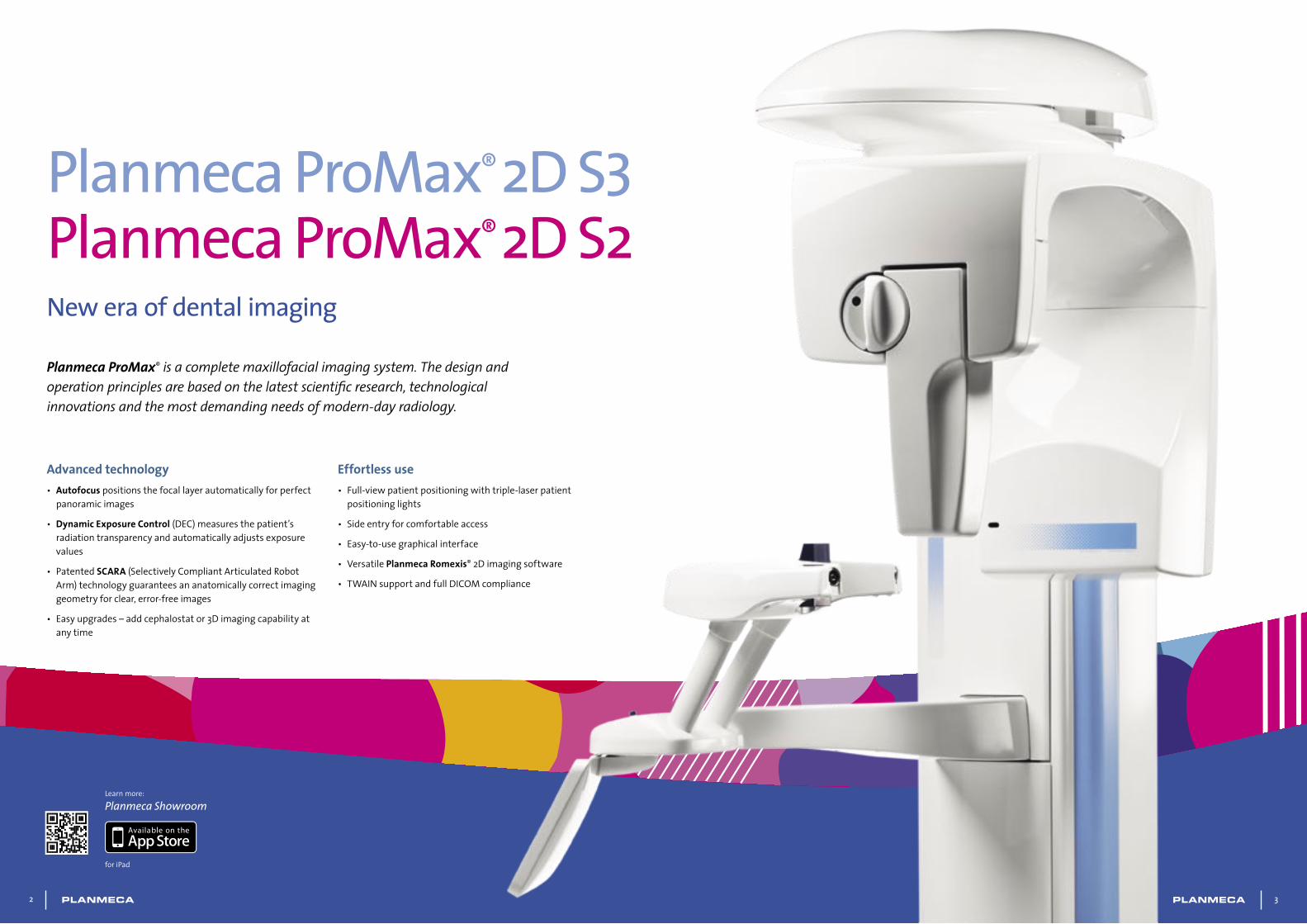
Planmeca ProMax® is a complete maxillofacial imaging system. The design and operation principles are based on the latest scientific research, technological innovations and the most demanding needs of modern-day radiology.
Planmeca ProMax® 2D S3 Planmeca ProMax® 2D S2New era of dental imaging
Advanced technology• Autofocus positionsthefocallayerautomaticallyforperfect
panoramicimages
• Dynamic Exposure Control(DEC)measuresthepatient’sradiationtransparencyandautomaticallyadjustsexposurevalues
• Patented SCARA (SelectivelyCompliantArticulatedRobotArm)technologyguaranteesananatomicallycorrectimaginggeometryforclear,error-freeimages
• Easyupgrades–addcephalostator3Dimagingcapabilityatanytime
Learnmore:
Planmeca Showroom
foriPad
Effortless use• Full-viewpatientpositioningwithtriple-laserpatient
positioninglights
• Sideentryforcomfortableaccess
• Easy-to-usegraphicalinterface
• VersatilePlanmeca Romexis®2Dimagingsoftware
• TWAINsupportandfullDICOMcompliance
2 3
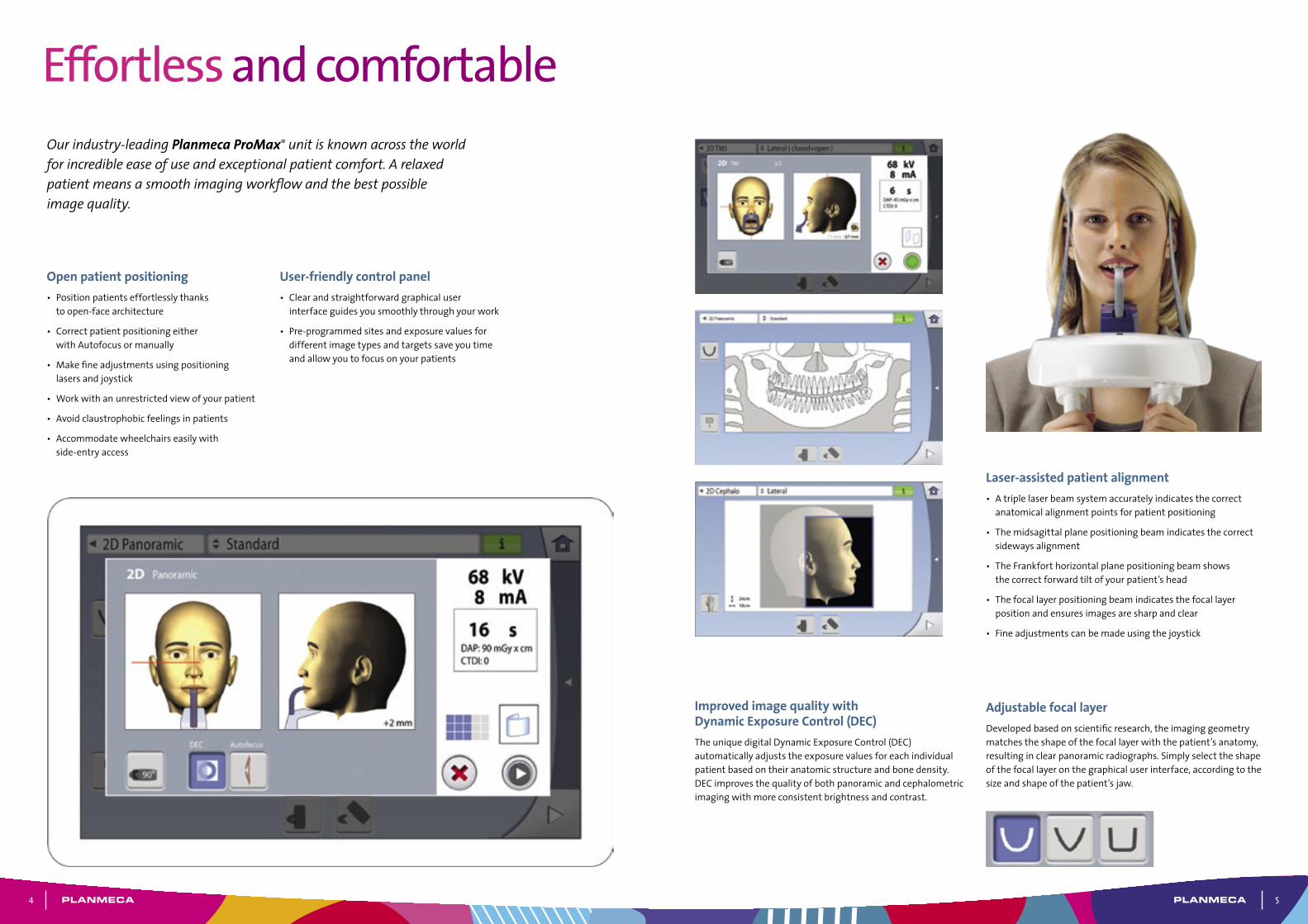
Laser-assisted patient alignment• Atriplelaserbeamsystemaccuratelyindicatesthecorrect
anatomicalalignmentpointsforpatientpositioning
• Themidsagittalplanepositioningbeamindicatesthecorrectsidewaysalignment
• TheFrankforthorizontalplanepositioningbeamshowsthecorrectforwardtiltofyourpatient’shead
• Thefocallayerpositioningbeamindicatesthefocallayerpositionandensuresimagesaresharpandclear
• Fineadjustmentscanbemadeusingthejoystick
Improved image quality with Dynamic Exposure Control (DEC)TheuniquedigitalDynamicExposureControl(DEC)automaticallyadjuststheexposurevaluesforeachindividualpatientbasedontheiranatomicstructureandbonedensity.DECimprovesthequalityofbothpanoramicandcephalometricimagingwithmoreconsistentbrightnessandcontrast.
Adjustable focal layerDevelopedbasedonscientificresearch,theimaginggeometrymatchestheshapeofthefocallayerwiththepatient’sanatomy,resultinginclearpanoramicradiographs.Simplyselecttheshapeofthefocallayeronthegraphicaluserinterface,accordingtothesizeandshapeofthepatient’sjaw.
Effortless and comfortableOur industry-leading Planmeca ProMax® unit is known across the world for incredible ease of use and exceptional patient comfort. A relaxed patient means a smooth imaging workflow and the best possible image quality.
Open patient positioning• Positionpatientseffortlesslythanks
toopen-facearchitecture
• CorrectpatientpositioningeitherwithAutofocusormanually
• Makefineadjustmentsusingpositioninglasersandjoystick
• Workwithanunrestrictedviewofyourpatient
• Avoidclaustrophobicfeelingsinpatients
• Accommodatewheelchairseasilywithside-entryaccess
User-friendly control panel• Clearandstraightforwardgraphicaluser
interfaceguidesyousmoothlythroughyourwork
• Pre-programmedsitesandexposurevaluesfordifferentimagetypesandtargetssaveyoutimeandallowyoutofocusonyourpatients
4 5
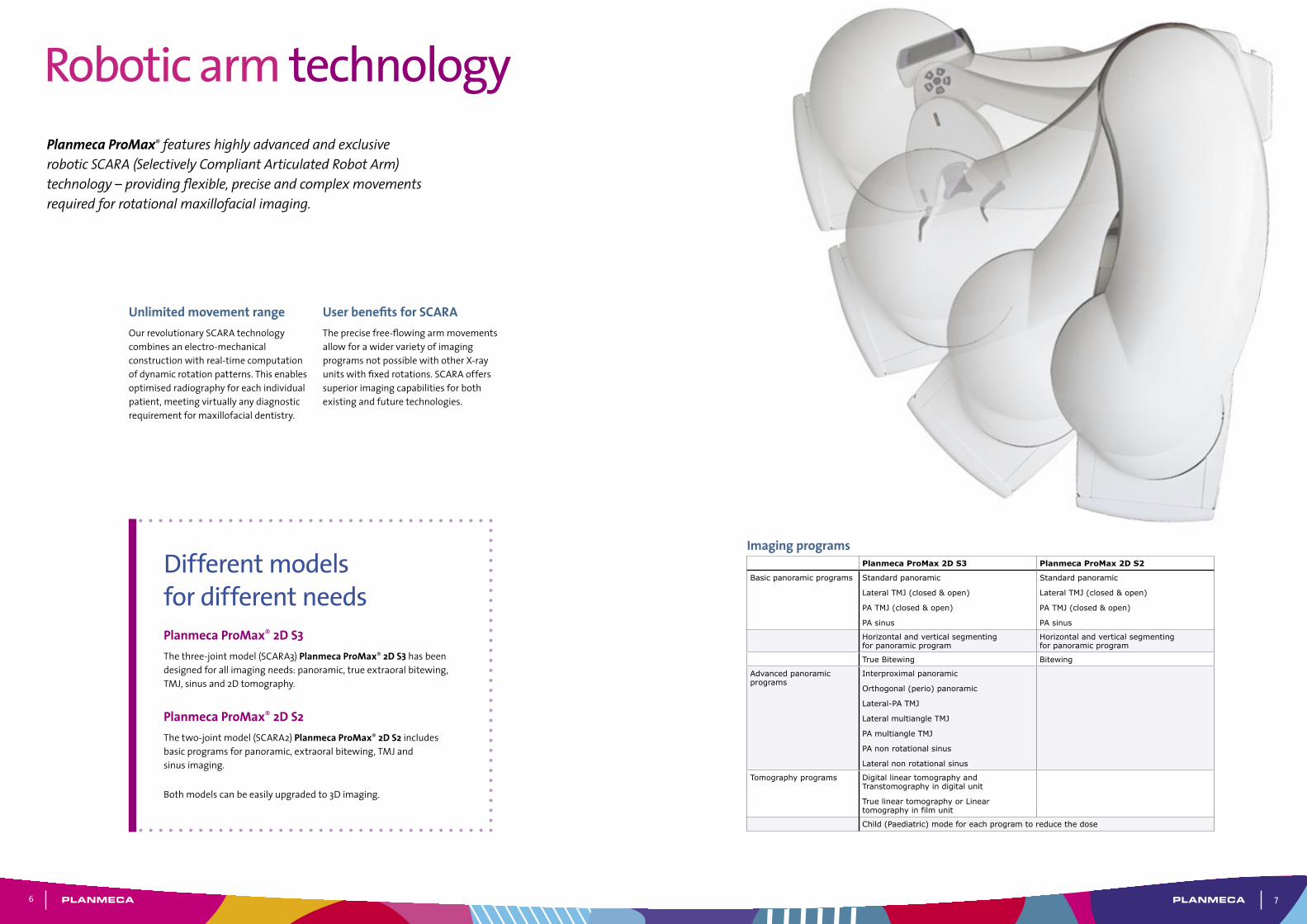
Planmeca ProMax® features highly advanced and exclusive robotic SCARA (Selectively Compliant Articulated Robot Arm) technology – providing flexible, precise and complex movements required for rotational maxillofacial imaging.
Imaging programsPlanmeca ProMax 2D S3 Planmeca ProMax 2D S2
Basic panoramic programs Standard panoramic
Lateral TMJ (closed & open)
PA TMJ (closed & open)
PA sinus
Standard panoramic
Lateral TMJ (closed & open)
PA TMJ (closed & open)
PA sinus
Horizontal and vertical segmenting for panoramic program
Horizontal and vertical segmenting for panoramic program
True Bitewing Bitewing
Advanced panoramic programs
Interproximal panoramic
Orthogonal (perio) panoramic
Lateral-PA TMJ
Lateral multiangle TMJ
PA multiangle TMJ
PA non rotational sinus
Lateral non rotational sinus
Tomography programs Digital linear tomography and Transtomography in digital unit
True linear tomography or Linear tomography in film unit
Child (Paediatric) mode for each program to reduce the dose
Unlimited movement rangeOurrevolutionarySCARAtechnologycombinesanelectro-mechanicalconstructionwithreal-timecomputationofdynamicrotationpatterns.Thisenablesoptimisedradiographyforeachindividualpatient,meetingvirtuallyanydiagnosticrequirementformaxillofacialdentistry.
User benefits for SCARATheprecisefree-flowingarmmovementsallowforawidervarietyofimagingprogramsnotpossiblewithotherX-rayunitswithfixedrotations.SCARAofferssuperiorimagingcapabilitiesforbothexistingandfuturetechnologies.
Robotic arm technology
Different models for different needsPlanmeca ProMax® 2D S3Thethree-jointmodel(SCARA3)PlanmecaProMax® 2D S3 hasbeendesignedforallimagingneeds:panoramic,trueextraoralbitewing,TMJ,sinusand2Dtomography.
Planmeca ProMax® 2D S2Thetwo-jointmodel(SCARA2)PlanmecaProMax® 2D S2 includesbasicprogramsforpanoramic,extraoralbitewing,TMJandsinusimaging.
Bothmodelscanbeeasilyupgradedto3Dimaging.
6 7
Panoramic imagingInadditiontotheStandardpanoramicprogram,thefollowingprogramsareoffered:
• Interproximalpanoramicprogram:generatesanimage,whereinterproximalteethcontactsareopen.Primarilyusedforcariesdetection.
• Orthogonalpanoramicprogram:producesanimagewithclearlyvisiblealveolarcrestforimproveddiagnostics.Idealforperiodontalimagingandimplantplanning.
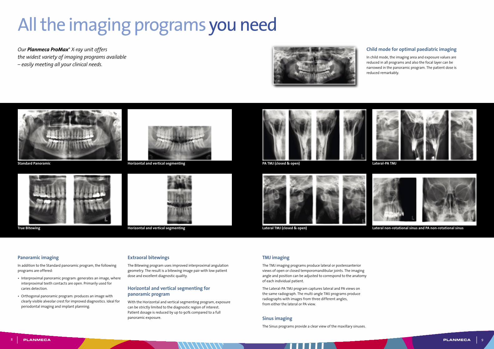
All the imaging programs you needChild mode for optimal paediatric imagingInchildmode,theimagingareaandexposurevaluesarereducedinallprogramsandalsothefocallayercanbenarrowedinthepanoramicprogram.Thepatientdoseisreducedremarkably.
Our Planmeca ProMax® X-ray unit offers the widest variety of imaging programs available – easily meeting all your clinical needs.
Extraoral bitewingsTheBitewingprogramusesimprovedinterproximalangulationgeometry.Theresultisabitewingimagepairwithlowpatientdoseandexcellentdiagnosticquality.
Horizontal and vertical segmenting for panoramic programWiththeHorizontalandverticalsegmentingprogram,exposurecanbestrictlylimitedtothediagnosticregionofinterest.Patientdosageisreducedbyupto90%comparedtoafullpanoramicexposure.
TMJ imagingTheTMJimagingprogramsproducelateralorposteroanteriorviewsofopenorclosedtemporomandibularjoints.Theimagingangleandpositioncanbeadjustedtocorrespondtotheanatomyofeachindividualpatient.
TheLateral-PATMJprogramcaptureslateralandPAviewsonthesameradiograph.Themulti-angleTMJprogramsproduceradiographswithimagesfromthreedifferentangles,fromeitherthelateralorPAview.
Sinus imaging TheSinusprogramsprovideaclearviewofthemaxillarysinuses.
Standard Panoramic PA TMJ (closed & open)Horizontal and vertical segmenting Lateral-PA TMJ
Horizontal and vertical segmenting Lateral non-rotational sinus and PA non-rotational sinusTrue Bitewing Lateral TMJ (closed & open)
8 9
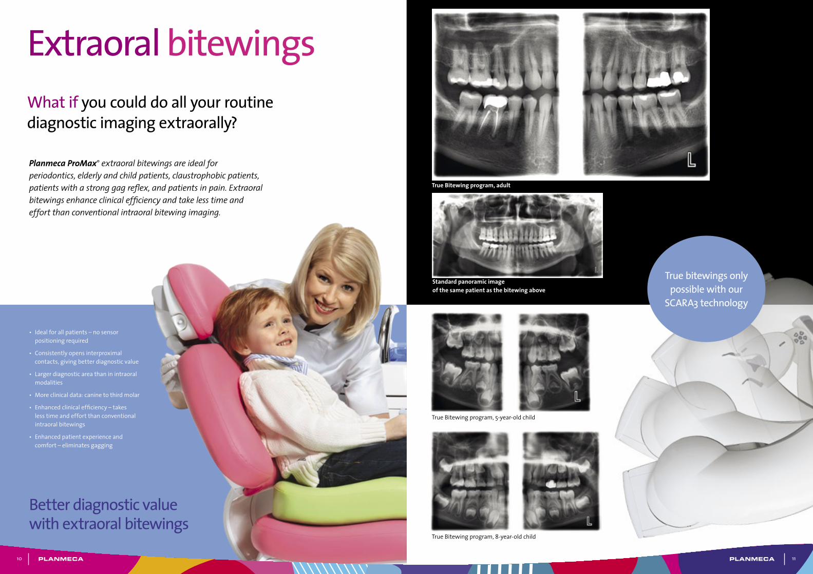
Planmeca ProMax® extraoral bitewings are ideal for periodontics, elderly and child patients, claustrophobic patients, patients with a strong gag reflex, and patients in pain. Extraoral bitewings enhance clinical efficiency and take less time and effort than conventional intraoral bitewing imaging.
What are the advantages of extraoral bitewings?
Extraoral bitewings
• Idealforallpatients–nosensorpositioningrequired
• Consistentlyopensinterproximalcontacts,givingbetterdiagnosticvalue
• Largerdiagnosticareathaninintraoralmodalities
• Moreclinicaldata:caninetothirdmolar
• Enhancedclinicalefficiency–takeslesstimeandeffortthanconventionalintraoralbitewings
• Enhancedpatientexperienceandcomfort–eliminatesgagging
True bitewings only possible with our
SCARA3 technology
Better diagnostic value with extraoral bitewings
What if you could do all your routine diagnostic imaging extraorally?
True Bitewing program, adult
TrueBitewingprogram,5-year-oldchild
TrueBitewingprogram,8-year-oldchild
Standard panoramic image of the same patient as the bitewing above
10 11
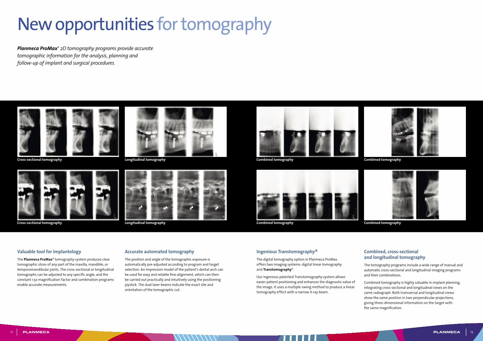
Valuable tool for implantologyThePlanmeca ProMax®tomographysystemproducescleartomographicslicesofanypartofthemaxilla,mandible,ortemporomandibularjoints.Thecross-sectionalorlongitudinaltomographscanbeadjustedtoanyspecificangle,andtheconstant1.5xmagnificationfactorandcombinationprogramsenableaccuratemeasurements.
New opportunities for tomographyPlanmeca ProMax® 2D tomography programs provide accurate tomographic information for the analysis, planning and follow-up of implant and surgical procedures.
Accurate automated tomographyThepositionandangleofthetomographicexposureisautomaticallypre-adjustedaccordingtoprogramandtargetselection.Animpressionmodelofthepatient’sdentalarchcanbeusedforeasyandreliablefine-alignment,whichcanthenbecarriedoutpracticallyandintuitivelyusingthepositioningjoystick.Theduallaserbeamsindicatetheexactsiteandorientationofthetomographiccut.
Ingenious Transtomography®ThedigitaltomographyoptioninPlanmecaProMaxofferstwoimagingsystems:digitallineartomographyandTranstomography®.
OuringeniouspatentedTranstomographysystemallowseasierpatientpositioningandenhancesthediagnosticvalueoftheimage.Itusesamultiple-swingmethodtoproducealineartomographyeffectwithanarrowX-raybeam.
Combined, cross-sectional and longitudinal tomographyThetomographyprogramsincludeawiderangeofmanualandautomaticcross-sectionalandlongitudinalimagingprogramsandtheircombinations.
Combinedtomographyishighlyvaluableinimplantplanning,integratingcross-sectionalandlongitudinalviewsonthesameradiograph.Bothtransversalandlongitudinalviewsshowthesamepositionintwoperpendicularprojections,givingthree-dimensionalinformationonthetargetwiththesamemagnification.
Combined tomographyCross-sectional tomography Combined tomographyLongitudinal tomography
Combined tomographyLongitudinal tomography Combined tomographyCross-sectional tomography
12 13
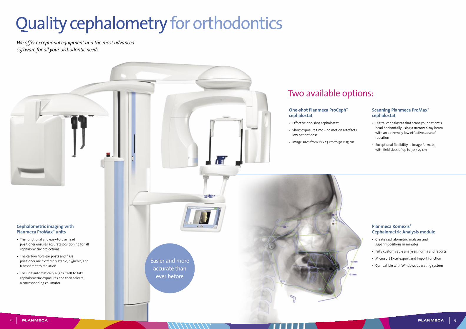
We offer exceptional equipment and the most advanced software for all your orthodontic needs.
Cephalometric imaging with Planmeca ProMax® units• Thefunctionalandeasy-to-usehead
positionerensuresaccuratepositioningforallcephalometricprojections
• Thecarbonfibreearpostsandnasalpositionerareextremelystable,hygienic,andtransparenttoradiation
• Theunitautomaticallyalignsitselftotakecephalometricexposuresandthenselectsacorrespondingcollimator
Two available options:
Planmeca Romexis® Cephalometric Analysis module• Createcephalometricanalysesand
superimpositionsinminutes
• Fullycustomisableanalyses,normsandreports
• MicrosoftExcelexportandimportfunction
• CompatiblewithWindowsoperatingsystem
Quality cephalometry for orthodontics
One-shot Planmeca ProCeph™ cephalostat• Effectiveone-shotcephalostat
• Shortexposuretime–nomotionartefacts,lowpatientdose
• Imagesizesfrom18x25cmto30x25cm
Scanning Planmeca ProMax® cephalostat • Digitalcephalostatthatscansyourpatient’s
headhorizontallyusinganarrowX-raybeamwithanextremelyloweffectivedoseofradiation
• Exceptionalflexibilityinimageformats,withfieldsizesofupto30x27cm
Easier and more accurate than
ever before
14 15
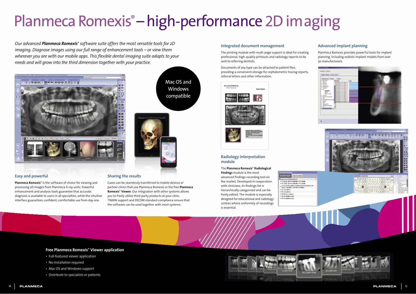
Planmeca Romexis® – high-performance 2D imaging
Mac OS and Windows
compatible
Free Planmeca Romexis® Viewer application• Full-featuredviewerapplication
• Noinstallationrequired
• MacOSandWindowssupport
• Distributetospecialistsorpatients
Our advanced Planmeca Romexis® software suite offers the most versatile tools for 2D imaging. Diagnose images using our full range of enhancement tools – or view them wherever you are with our mobile apps. This flexible dental imaging suite adapts to your needs and will grow into the third dimension together with your practice.
Easy and powerfulPlanmeca Romexis®isthesoftwareofchoiceforviewingandprocessing2DimagesfromPlanmecaX-rayunits.Powerfulenhancementandanalysistoolsguaranteethataccuratediagnosisisavailabletousersinallspecialties,whiletheintuitiveinterfaceguaranteesconfident,comfortableusefromdayone.
Integrated document managementTheprintingmodulewithmulti-pagesupportisidealforcreatingprofessional,high-qualityprintoutsandradiologyreportstobesenttoreferringdentists.
Documentsofanytypecanbeattachedtopatientfiles,providingaconvenientstorageforcephalometrictracingreports,referrallettersandotherinformation.
Radiology interpretation moduleThePlanmeca Romexis® Radiological Findingsmoduleisthemostadvancedfindings-recordingtoolonthemarket.Developedincooperationwithclinicians,itsfindingslistishierarchicallycategorisedandcanbefreelyedited.Themoduleisespeciallydesignedforeducationalandradiologycentreswhereuniformityofrecordingsisessential.
Advanced implant planning PlanmecaRomexisprovidespowerfultoolsforimplantplanning,includingrealisticimplantmodelsfromover30manufacturers.
Sharing the resultsCasescanbeseamlesslytransferredtomobiledevicesorpartnerclinicsthatusePlanmecaRomexisorthefreePlanmeca Romexis®Viewer.Ourintegrationwithothersystemsallowsyoutofreelyutilisethird-partyproductsatyourclinic.TWAINsupportandDICOMstandardcomplianceensurethatthesoftwarecanbeusedtogetherwithmostsystems.
16 17
Pink Sky SteelLime
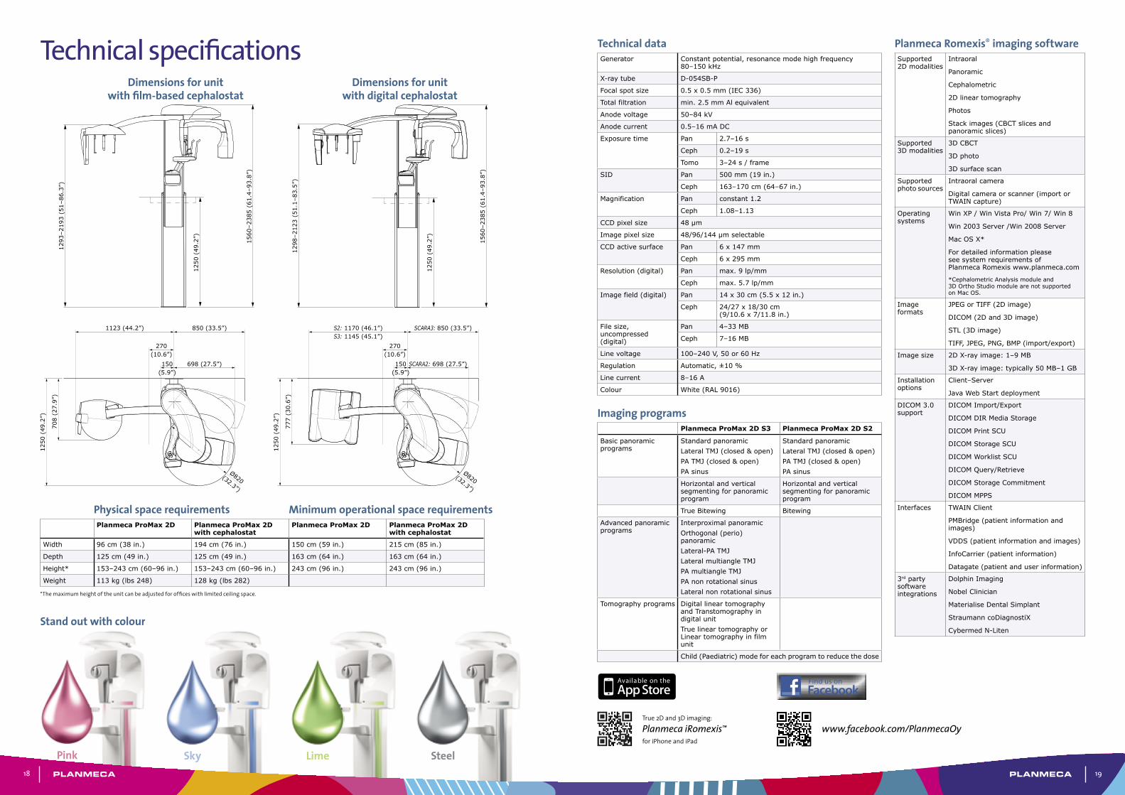
Technical specifications Technical dataGenerator Constant potential, resonance mode high frequency
80–150 kHz
X-ray tube D-054SB-P
Focal spot size 0.5 x 0.5 mm (IEC 336)
Total filtration min. 2.5 mm Al equivalent
Anode voltage 50–84 kV
Anode current 0.5–16 mA DC
Exposure time Pan 2.7–16 s
Ceph 0.2–19 s
Tomo 3–24 s / frame
SID Pan 500 mm (19 in.)
Ceph 163–170 cm (64–67 in.)
Magnification Pan constant 1.2
Ceph 1.08–1.13
CCD pixel size 48 µm
Image pixel size 48/96/144 µm selectable
CCD active surface Pan 6 x 147 mm
Ceph 6 x 295 mm
Resolution (digital) Pan max. 9 lp/mm
Ceph max. 5.7 lp/mm
Image field (digital) Pan 14 x 30 cm (5.5 x 12 in.)
Ceph 24/27 x 18/30 cm (9/10.6 x 7/11.8 in.)
File size, un compressed (digital)
Pan 4–33 MB
Ceph 7–16 MB
Line voltage 100–240 V, 50 or 60 Hz
Regulation Automatic, ±10 %
Line current 8–16 A
Colour White (RAL 9016)
Imaging programsPlanmeca ProMax 2D S3 Planmeca ProMax 2D S2
Basic panoramic programs
Standard panoramicLateral TMJ (closed & open)PA TMJ (closed & open)PA sinus
Standard panoramicLateral TMJ (closed & open)PA TMJ (closed & open)PA sinus
Horizontal and vertical segmenting for panoramic program
Horizontal and vertical segmenting for panoramic program
True Bitewing Bitewing
Advanced panoramic programs
Interproximal panoramicOrthogonal (perio) panoramicLateral-PA TMJLateral multiangle TMJPA multiangle TMJPA non rotational sinusLateral non rotational sinus
Tomography programs Digital linear tomography and Transtomography in digital unitTrue linear tomography or Linear tomography in film unit
Child (Paediatric) mode for each program to reduce the dose
Physical space requirements Minimum operational space requirementsPlanmeca ProMax 2D Planmeca ProMax 2D
with cephalostatPlanmeca ProMax 2D Planmeca ProMax 2D
with cephalostat
Width 96 cm (38 in.) 194 cm (76 in.) 150 cm (59 in.) 215 cm (85 in.)
Depth 125 cm (49 in.) 125 cm (49 in.) 163 cm (64 in.) 163 cm (64 in.)
Height* 153–243 cm (60–96 in.) 153–243 cm (60–96 in.) 243 cm (96 in.) 243 cm (96 in.)
Weight 113 kg (lbs 248) 128 kg (lbs 282)
*Themaximumheightoftheunitcanbeadjustedforofficeswithlimitedceilingspace.
Stand out with colour
�
��
�
��
Dimensions for unit with film-based cephalostat
Dimensions for unit with digital cephalostat
1298
–212
3 (5
1.1–
83.5
”)
1293
–219
3 (5
1–86
.3”)
1560
–238
5 (6
1.4–
93.8
”)
1560
–238
5 (6
1.4–
93.8
”)
1250
(49
.2”)
1250
(49
.2”)
1250
(49
.2”)
1250
(49
.2”)
777
(30.
6”)
708
(27.
9”)
150(5.9”)
150(5.9”)
270(10.6”)
270(10.6”)
S2: 1170 (46.1”)S3: 1145 (45.1”)
1123 (44.2”)
SCARA2: 698 (27.5”)698 (27.5”)
Ø820(32.3”)
Ø820(32.3”)
SCARA3: 850 (33.5”)850 (33.5”)
Planmeca Romexis® imaging softwareSupported 2D modalities
Intraoral
Panoramic
Cephalometric
2D linear tomography
Photos
Stack images (CBCT slices and panoramic slices)
Supported 3D modalities
3D CBCT
3D photo
3D surface scan
Supported photo sources
Intraoral camera
Digital camera or scanner (import or TWAIN capture)
Operating systems
Win XP / Win Vista Pro/ Win 7/ Win 8
Win 2003 Server /Win 2008 Server
Mac OS X*
For detailed information please see system requirements of Planmeca Romexis www.planmeca.com
*Cephalometric Analysis module and 3D Ortho Studio module are not supported on Mac OS.
Image formats
JPEG or TIFF (2D image)
DICOM (2D and 3D image)
STL (3D image)
TIFF, JPEG, PNG, BMP (import/export)
Image size 2D X-ray image: 1–9 MB
3D X-ray image: typically 50 MB–1 GB
Installation options
Client–Server
Java Web Start deployment
DICOM 3.0 support
DICOM Import/Export
DICOM DIR Media Storage
DICOM Print SCU
DICOM Storage SCU
DICOM Worklist SCU
DICOM Query/Retrieve
DICOM Storage Commitment
DICOM MPPS
Interfaces TWAIN Client
PMBridge (patient information and images)
VDDS (patient information and images)
InfoCarrier (patient information)
Datagate (patient and user information)
3rd party software integrations
Dolphin Imaging
Nobel Clinician
Materialise Dental Simplant
Straumann coDiagnostiX
Cybermed N-Liten
www.facebook.com/PlanmecaOy
Find us onFacebook
True2Dand3Dimaging:
Planmeca iRomexis™
foriPhoneandiPad
18 19
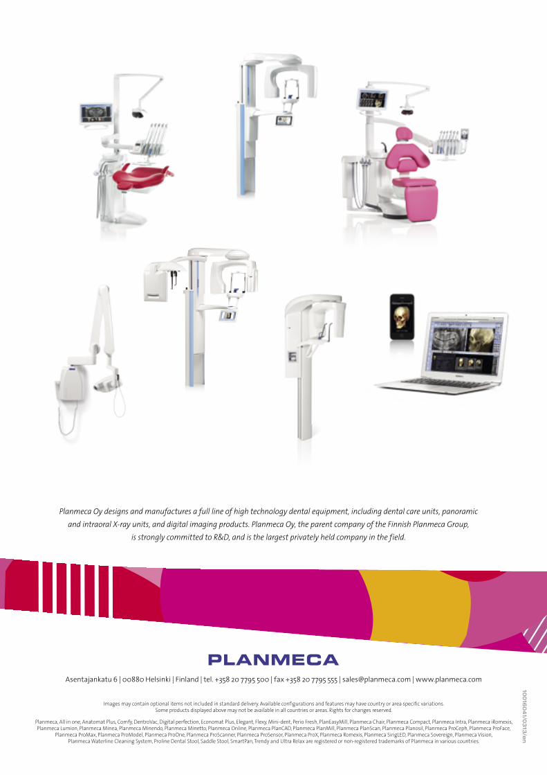
Planmeca Oy designs and manufactures a full line of high technology dental equipment, including dental care units, panoramic and intraoral X-ray units, and digital imaging products. Planmeca Oy, the parent company of the Finnish Planmeca Group,
is strongly committed to R&D, and is the largest privately held company in the field.
10016
041/0
313
/en
Asentajankatu 6 | 00880 Helsinki | Finland | tel. +358 20 7795 500 | fax +358 20 7795 555 | [email protected] | www.planmeca.com
Images may contain optional items not included in standard delivery. Available configurations and features may have country or area specific variations. Some products displayed above may not be available in all countries or areas. Rights for changes reserved.
Planmeca, All in one, Anatomat Plus, Comfy, DentroVac, Digital perfection, Economat Plus, Elegant, Flexy, Mini-dent, Perio Fresh, PlanEasyMill, Planmeca Chair, Planmeca Compact, Planmeca Intra, Planmeca iRomexis, Planmeca Lumion, Planmeca Minea, Planmeca Minendo, Planmeca Minetto, Planmeca Online, Planmeca PlanCAD, Planmeca PlanMill, Planmeca PlanScan, Planmeca Planosil, Planmeca ProCeph, Planmeca ProFace,
Planmeca ProMax, Planmeca ProModel, Planmeca ProOne, Planmeca ProScanner, Planmeca ProSensor, Planmeca ProX, Planmeca Romexis, Planmeca SingLED, Planmeca Sovereign, Planmeca Vision, Planmeca Waterline Cleaning System, Proline Dental Stool, Saddle Stool, SmartPan, Trendy and Ultra Relax are registered or non-registered trademarks of Planmeca in various countries.