Embed Size (px)
Citation preview
Pituitary Gland – Hyperplasia
1
Pituitary Gland – Hyperplasia
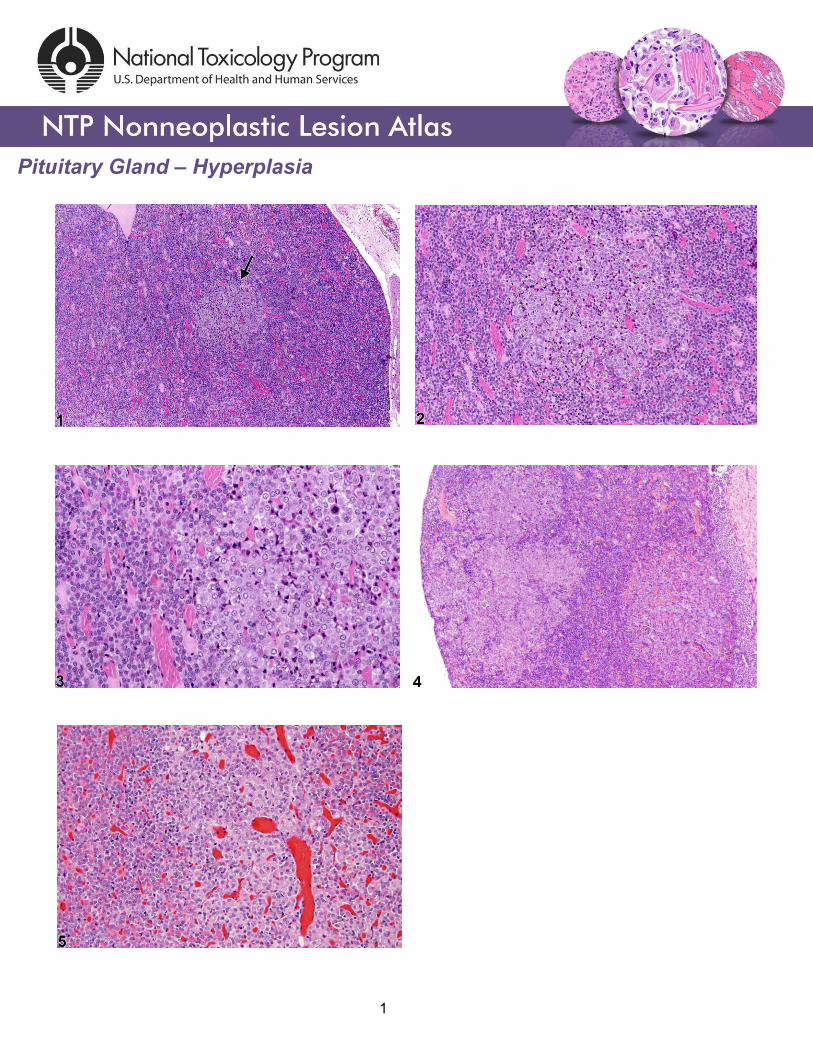
Figure Legend: Figure 1 Pituitary Gland, Pars distalis - Hyperplasia in a female Harlan Sprague-
Dawley rat from a chronic study. A small focus of hyperplasia (arrow) in the pars distalis is recognized
by the paler staining cells. Figure 2 Pituitary Gland, Pars distalis - Hyperplasia in a female Harlan
Sprague-Dawley rat from a chronic study. Higher magnification of Figure 1 shows the focus of
hyperplasia in greater detail. Figure 3 Pituitary Gland, Pars distalis - Hyperplasia in a female Harlan
Sprague-Dawley rat from a chronic study. Higher magnification of Figure 2 highlights the larger, paler
staining cells in this focus of hyperplasia in the pars distalis. Figure 4 Pituitary Gland, Pars distalis -
Hyperplasia in a female Harlan Sprague-Dawley rat from a chronic study. Multiple focal areas of
hyperplasia consisting of paler staining cells compared with the surrounding parenchyma are present in
the pars distalis. Figure 5 Pituitary Gland, Pars distalis - Hyperplasia in a male F344/N rat from a
chronic study. The focus of hyperplasia consists of paler staining cells (compared with the surrounding
normal parenchyma) and dilated blood-filled vascular structures which are consistent with minimal to
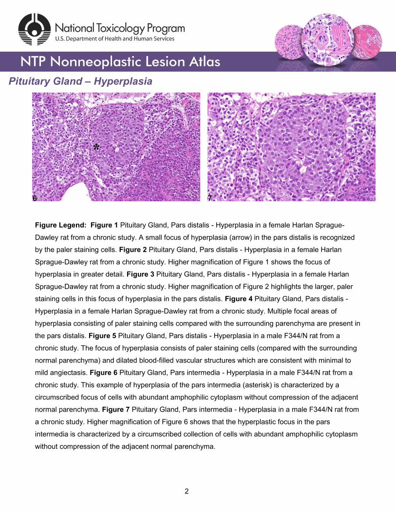
mild angiectasis. Figure 6 Pituitary Gland, Pars intermedia - Hyperplasia in a male F344/N rat from a
chronic study. This example of hyperplasia of the pars intermedia (asterisk) is characterized by a
circumscribed focus of cells with abundant amphophilic cytoplasm without compression of the adjacent
normal parenchyma. Figure 7 Pituitary Gland, Pars intermedia - Hyperplasia in a male F344/N rat from
a chronic study. Higher magnification of Figure 6 shows that the hyperplastic focus in the pars
intermedia is characterized by a circumscribed collection of cells with abundant amphophilic cytoplasm
without compression of the adjacent normal parenchyma.
2
Pituitary Gland – Hyperplasia
Comment: Focal hyperplasia is a frequent spontaneous as well as induced change and is seen more
commonly in rats than in mice. The microscopic appearance is variable but consists primarily of a
single cell type that blends into the adjacent parenchyma without compression (Figure 1, figure 2,
Figure 3, and Figure 4). Hyperplasia is typically a combination of increased cell number and increased
cell size and increases with animal age. On occasion there may be multiple foci of hyperplasia (Figure
4). Angiectasis may occur within a focus of hyperplasia (Figure 5) and may sometimes cause slight
compression of adjacent parenchyma.
Focal hyperplasia of the pars intermedia (Figure 6 and Figure 7) is less common than hyperplasia of
the pars distalis. The hyperplastic cells in the pars intermedia are similar to normal pars intermedia
cells, and identification of a focal proliferative lesion may rely on alteration of growth pattern or
asymmetrical enlargement of the pars intermedia since there may be no compression of the adjacent
parenchyma. Diffuse hyperplasia may also occur and must be distinguished from a tangential section of
the pars intermedia. Diffuse hyperplasia is usually of a specific cell type that can be confirmed by
immunohistochemistry and typically represents a physiologic response.
There is a morphologic continuum between hyperplasia and pituitary neoplasia, with compression of
the adjacent parenchyma being a primary diagnostic feature of neoplasia. Immunohistochemistry for
pituitary hormones can be used to determine cell type.
Recommendation: Focal hyperplasia should be diagnosed and given a severity grade whenever
present, and the part of the pituitary involved should be specified in the diagnosis (e.g., Pituitary Gland,
Pars distalis - Hyperplasia). Any remarkable features of hyperplasia may be described in the pathology
narrative. Angiectasis within the hyperplastic lesion should not be diagnosed separately.
3
Pituitary Gland – Hyperplasia
References: Capen CC. 1983. Functional and pathologic interrelationships of the pituitary gland and hypothalamus. In: Endocrine System (Jones TC, Mohr U, Hunt RD, eds). Springer, New York, 103-120. Abstract: http://www.springer.com/medicine/pathology/book/978-3-642-96722-1 Capen CC, DeLellis RA, Yarrington JT. 1991. Endocrine system. In: Handbook of Toxicologic Pathology (Haschek WM, Rousseaux CG, eds). Academic Press, New York, 697-705. Abstract: http://www.sciencedirect.com/science/book/9780123302151 Carlton WW, Gries CL. 1983. Cysts, pituitary; rat, mouse, and hamster. In: Endocrine System (Jones TC, Mohr U, Hunt RD, eds). Springer, New York, 161-163. Abstract: http://www.springer.com/medicine/pathology/book/978-3-642-96722-1 Greaves P. 2007. Histopathology of Preclinical Toxicity Studies: Interpretation and Relevance in Drug Safety Evaluation, 3rd ed. Academic Press, Amsterdam, 953. Abstract: http://www.sciencedirect.com/science/book/9780444527714 MacKenzie WF, Boorman GA. 1991. Pituitary gland. In: Pathology of the Fischer Rat: Reference and Atlas (Boorman GA, Eustis SL, Elwell MR, Montgomery CA, MacKenzie WF, eds). Academic Press, San Diego, 485-500. Abstract: http://www.ncbi.nlm.nih.gov/nlmcatalog/9002563 Mahler J, Elwell M. 1999. The pituitary gland. In: Pathology of the Mouse: Reference and Atlas (Maronpot RR, Boorman GA, Gaul BW, eds). Cache River Press, Vienna, IL, 491-508. Abstract: http://www.cacheriverpress.com/books/pathmouse.htm Tucker MJ. 1998. The endocrine system. In: Target Organ Pathology (Turton J, Hoodon J, eds). Taylor and Francis, London, 311-334. Abstract: http://www.amazon.com/Target-Organ-Pathology-Basic-Text/dp/0748401571 Authors: Robert R. Maronpot, DVM, MS, MPH, DACVP, DABT, FIATP Senior Pathologist Experimental Pathology Laboratories, Inc. Research Triangle Park, NC Amy Brix, DVM, PhD, DACVP Senior Pathologist Experimental Pathology Laboratories, Inc. Research Triangle Park, NC
4