Embed Size (px)
Citation preview
PRR5 regulates phosphorylation, nuclear importand subnuclear localization of TOC1 in theArabidopsis circadian clock
Lei Wang, Sumire Fujiwara1
and David E Somers*
Department of Plant Cellular and Molecular Biology, Ohio StateUniversity, Columbus, OH, USA
Many core oscillator components of the circadian clock are
nuclear localized but how the phase and rate of their entry
contribute to clock function is unknown. TOC1/PRR1,
a pseudoresponse regulator (PRR) protein, is a central
element in one of the feedback loops of the Arabidopsis
clock, but how it functions is unknown. Both TOC1 and
a closely related protein, PRR5, are nuclear localized,
expressed in the same phase, and shorten period when
deficient, but their molecular relationship is unclear. Here,
we find that both proteins interact in vitro and in vivo
through their conserved N-termini. TOC1–PRR5 oligomer-
ization enhances TOC1 nuclear accumulation two-fold,
most likely through enhanced nuclear import. In addition,
PRR5 recruits TOC1 to large subnuclear foci and promotes
phosphorylation of the TOC1 N-terminus. Our results show
that nuclear TOC1 is essential for normal clock function
and reveal a mechanism to enhance phase-specific TOC1
nuclear accumulation. Interestingly, this process of regu-
lated nuclear import is reminiscent of similar oligomeric
pairings in animal clock systems (e.g. timeless/period and
clock/cycle), suggesting evolutionary convergence of a
conserved mechanism across kingdoms.
The EMBO Journal (2010) 29, 1903–1915. doi:10.1038/
emboj.2010.76; Published online 20 April 2010
Subject Categories: signal transduction; plant biology
Keywords: Arabidopsis thaliana; circadian clock; nuclear
import; oligomerization; phosphorylation
Introduction
The timing of many physiological and developmental pro-
cesses in most eukaryotes is under the control of a circadian
clock. This endogenous, self-sustaining oscillator maintains a
rhythm of ca. 24 h in processes as diverse as human sleep/
wake cycles (Ebisawa, 2007), insect pupal eclosion
(Pittendrigh, 1981), fungal sporulation (Bell-Pedersen et al,
2005) and the movement of plant leaves (Sweeney, 1987).
The apparent adaptive significance of a circadian clock lies in
the ability to anticipate the regular changes in the environ-
ment (light/dark; warm/cold) that arise from the 24-h
rotation of the earth, and to adjust its physiology and deve-
lopment accordingly (Ouyang et al, 1998; Dodd et al, 2005;
Johnson and Kyriacou, 2005).
Classical and molecular genetics have helped to identify
genes in Drosophila, Arabidopsis, Neurospora and mammals
(clock genes) that alter clock function and a common mole-
cular mechanism has emerged. In all systems, both the
mRNA and protein products of key genes cycle in abundance
and/or activity, creating a transcription–translation feedback
loop. The 24-h delay occurs, in part, through the time it takes
for the entry of the protein product(s) to the nucleus, where
they contribute to the repression of their own transcription,
and/or in the time required for these proteins to degrade in
the nucleus. More recently, the importance of post-transla-
tional control has emerged, particularly the function of
protein phosphorylation, and three or more interlocked loops
are now apparent in all known eukaryotic clocks (Hardin,
2005; Ueda, 2007; de Paula et al, 2007; Harmer, 2009).
In Arabidopsis, 420 genes are known to affect circadian
cycling (Gardner et al, 2006; McClung, 2006; Harmer, 2009).
Current models posit three or more interlocked loops that
contain both negative and positive regulatory elements that
control gene transcription, but the majority of loci known to
affect circadian period have not been incorporated into these
schemes, either conceptually or functionally (Gardner et al,
2006; Locke et al, 2006; Zeilinger et al, 2006; Harmer, 2009).
Those components that have been successfully linked largely
belong to two gene families, the closely related MYB trans-
cription factors CIRCADIAN CLOCK-ASSOCIATED 1 (CCA1)
and LATE ELONGATED HYPORCOTYL (LHY), and the five-
member PSEUDO-RESPONSE REGULATOR (PRR) gene fa-
mily (TOC1/PRR1, PRR3, PRR5, PRR7 and PRR9) (Schaffer
et al, 1998; Wang and Tobin, 1998; Matsushika et al, 2000;
Strayer et al, 2000). The first ‘loop’ to be established involved
the upregulation of CCA1 and LHY by TOC1, whose transcrip-
tion is negatively regulated by CCA1 and LHY (Alabadi et al,
2001). Subsequent work tied CCA1 and LHY as positive
regulators of PRR7 and PRR9 transcription, which act to
negatively regulate CCA1/LHY (Farre et al, 2005), and effec-
tively interlink the two different loops. Other components,
some postulated and some demonstrated, have been incor-
porated to comprise a third, transcription-based loop (Locke
et al, 2006; Zeilinger et al, 2006).
In addition, a tremendous explosion of new work in
eukaryotic clock systems has shown the fundamental impor-
tance of post-transcriptional processes in circadian clocks,
particularly proteolysis and phosphorylation (Bell-Pedersen
et al, 2005; Cha et al, 2007; Gallego and Virshup, 2007;
Somers et al, 2007; Vanselow and Kramer, 2007). In
Arabidopsis, the F-box protein ZEITLUPE (ZTL) controlsReceived: 30 November 2009; accepted: 30 March 2010; publishedonline: 20 April 2010
*Corresponding author. Department of Plant Cellular and MolecularBiology, Ohio State University, 244B Rightmire Hall, 1060 CarmackRoad, Columbus, OH 43210, USA. Tel.: þ 1 614 292 2551;Fax: þ 1 614 292 5379; E-mail: [email protected] address: Bioproduction Research Institute, National Institute ofAdvanced Industrial Science and Technology (AIST), Central 4, Higashi1-1-1, Tsukuba, Ibaraki 305-8562, Japan
The EMBO Journal (2010) 29, 1903–1915 | & 2010 European Molecular Biology Organization | All Rights Reserved 0261-4189/10
www.embojournal.org
&2010 European Molecular Biology Organization The EMBO Journal VOL 29 | NO 11 | 2010
EMBO
THE
EMBOJOURNAL
THE
EMBOJOURNAL
1903

circadian period through the targeted proteasome-dependent
degradation of TOC1 and PRR5 (Mas et al, 2003b; Kiba et al,
2007; Fujiwara et al, 2008). In addition, ZTL itself undergoes
clock-controlled phase-specific proteolysis, regulated by the
circadian cycling of GIGANTEA (GI), which acts as a blue-
light enhanced stabilizer of ZTL (Kim et al, 2003, 2007;
Somers and Fujiwara, 2009). The SCFZTL substrate, TOC1,
exhibits circadian phase-specific variation in both abundance
and phosphorylation (Fujiwara et al, 2008). Although clock
control of TOC1 transcription contributes to much of TOC1
protein cycling, oligomerization with PRR3 shields
TOC1 from ZTL-mediated degradation and boosts overall
TOC1 levels (Para et al, 2007; Fujiwara et al, 2008).
Phosphorylation of both TOC1 and PRR3 is necessary for
their optimal binding and this interaction exemplifies one
mechanism by which oligomerization controls protein abun-
dance in the plant clock (Fujiwara et al, 2008).
In this study, we report a second, related, mechanism.
We show that PRR5 and TOC1 oligomerize through their
N-terminal PRR domains. Through this interaction, PRR5
promotes TOC1 nuclear import and phosphorylation, and
directs TOC1 subnuclear localization to PRR5-type nuclear
foci. Consistent with these results, TOC1 total protein accu-
mulation is significantly decreased and nuclear accumulation
is strongly reduced in the prr5 loss-of-function mutant.
Our findings indicate that the PRR5–TOC1 interaction
increases TOC1 steady-state levels, similar to the effect of
the PRR3–TOC1 interaction. However, PRR3 achieves this as
a competitive inhibitor of the cytosolic TOC1–ZTL interac-
tion, whereas PRR5 acts primarily to enhance TOC1 nuclear
accumulation, thereby avoiding ZTL-dependent degradation.
This PRR5-mediated nuclear import contributes to enhanced
TOC1 levels in a manner similar to that observed in other
clock systems, indicating a well-conserved mechanism across
kingdoms.
Results
PRR5 and TOC1 interact through their N-termini
Recent studies have shown that TOC1 protein levels can be
altered by the presence of other PRR proteins, notably PRR3,
through direct protein–protein interactions (Para et al, 2007;
Fujiwara et al, 2008). We expanded on this idea by testing
TOC1 interactions with the remaining PRR family members.
As the TOC1–PRR3 interaction is mediated through the two
N-terminal PRR domains (Fujiwara et al, 2008), we next
tested whether the N-terminal domain (aa 1–242) of TOC1
can interact with full-length TOC1, PRR5, PRR7 and PRR9
in vitro and in vivo.
TAP-TOC11�242 (TAP-TOC1NT) was expressed in Nicotiana
benthamiana and resin-bound TOC1NT was incubated with
extracts from Arabidopsis plants expressing specific
PRRnHPRRn-GFP genes (Fujiwara et al, 2008). Only PRR5
and PRR9 protein were pulled down, with the PRR5–TOC1
interaction markedly stronger than PRR9–TOC1 (Figure 1A).
When specific deletions of TOC1 were further tested with full-
length PRR5, only full-length and N-terminal TOC1 were able
to pull down PRR5, both in vitro and when transiently
co-expressed in N. benthamiana (Figure 1B).
Reciprocal in vivo co-IP experiments using full-length,
N-terminal and C-terminal deletions of TAP-PRR5 co-
expressed with TOC1-YFP in N. benthamiana showed that
the N-terminal domain of PRR5 is sufficient to mediate their
interaction (Figure 1C). Taken together, these data show that
TOC1 and PRR5 oligomerize through their N-termini.
Specific TOC1 short-period alleles diminish interaction
with PRR5 and ZTL
We took advantage of specific toc1 mutant alleles to further
probe the nature of the PRR5–TOC1 interaction. Two short-
period point mutations (toc1-5 and toc1-8) alter two different
amino-acid residues in the TOC1 PRR domain at proline 96
(P96L) and proline 124 (P124L), respectively (Hazen et al,
2005; Kevei et al, 2006). Both mutations severely diminish
the TOC1–PRR5 interaction three-fold or greater based on
co-immunoprecipitation in a transient expression assay
(Figure 2A and B). Mutations in highly conserved residues
in the C-terminal CCT domain that affect either TOC1 activity
(toc1-1) or CONSTANS (CO) activity (co-5 and co-7) were also
tested (Strayer et al, 2000; Robson et al, 2001). These muta-
tions had no effect on the TOC1–PRR5 interaction (Figure 2A
and B). Taken together, these data support the notion that the
PRR domain is essential for the TOC1–PRR5 interaction and
suggest an explanation for the short period of the PRR-
domain toc1 alleles.
However, we further tested the effect of these mutations on
the TOC1–ZTL interaction, knowing that the N-terminus of
TOC1 is also essential for its interaction with ZTL (Fujiwara
et al, 2008). Interestingly, the same PRR domain mutations
strongly reduced the TOC1–ZTL interaction, whereas the
introduced CCT domain mutations had no effect on the
interaction (Figure 2C and D). As reduced accessibility of
TOC1 to ZTL is predicted to mimic a ztl mutant and result in a
long period, it is likely that the short period of the toc1-5 and
toc1-8 alleles results instead from the diminished TOC1–PRR5
interaction.
PRR5 stabilizes TOC1 post-transcriptionally
and independent of phosphorylation state
The significance of the TOC1–PRR5 interaction was first
sought by observing the level of TOC1 in the Arabidopsis
prr5 mutant. The TOC-YFP minigene (TMG) (Mas et al,
2003a) was crossed into prr5-1 and the TMG/TMG prr5/
prr5 genotype was isolated. Total protein extracts from the
WT and prr5 mutant were collected every 4 h from plants
grown in 12/12 h light/dark cycles and TMG levels were
determined in total protein extracts. Consistently lower levels
of TMG were observed during the entire time of detectable
expression (Figure 3A and B). RT–PCR analysis indicated that
TOC1 message levels are not significantly different in the two
backgrounds, indicating post-transcriptional regulation of
TOC1 expression (Figure 3C and D).
We next sought to understand how TOC1 protein levels are
sustained by PRR5. Earlier reports showed that all the PRR
proteins are phosphorylated in vivo, and that the TOC1–PRR3
interaction is highly enhanced by phosphorylation of both
proteins (Fujiwara et al, 2008). We tested this requirement
for TOC1 and PRR5. We bound TAP-TOC1 expressed in
N. benthamiana to IgG resin and tested whether TOC1
phosphorylation affects the in vitro binding of PRR5-GFP
from Arabidopsis seedling extracts. PRR5-GFP is able to bind
both forms of TOC1 equally well (Supplementary Figure 1A,
lower panel, lanes 3 and 4). Similarly, when PRR5-GFP
extracts were mock or phosphatase treated, both forms of
PRR5 regulates TOC1 nuclear importL Wang et al
The EMBO Journal VOL 29 | NO 11 | 2010 &2010 European Molecular Biology Organization1904

PRR5-GFP bound TOC1 similarly (Supplementary Figure 1B,
lower panel, lanes 5 and 6). These data indicate that the
TOC1–PRR5 interaction is not affected by the phosphorylation
status of either of the proteins.
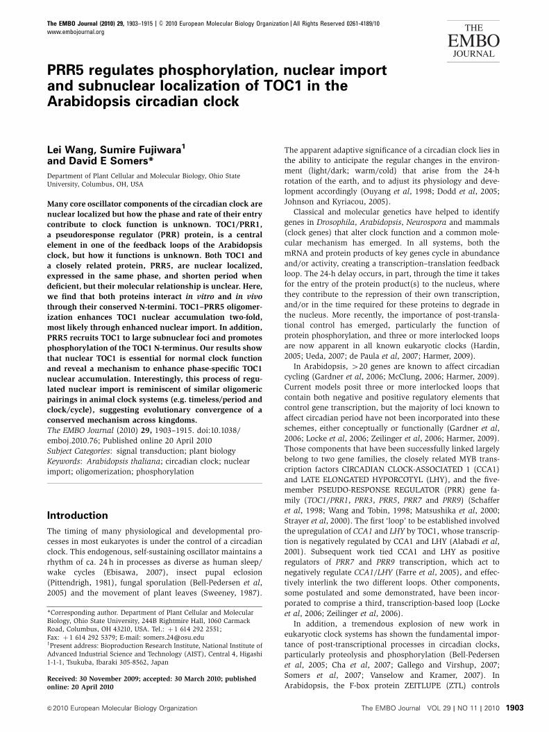
PRR5 enhances the proportion of phosphorylated TOC1
Although the phosphorylation state of PRR5 does not affect
binding to TOC1, we observed that the migration of TOC1
was more rapid when the TOC1–PRR5 interaction was dimin-
ished by the P96L and P124L mutations in the TOC1 PRR
domain (Figure 2A). This suggested that a TOC1–PRR5
interaction might facilitate TOC1 phosphorylation in vivo.
We tested this idea by co-expressing both proteins at different
abundance ratios in N. benthamiana and then observed the
relative abundance of upper (phosphorylated) and lower
(unphosphorylated) forms of TOC1 in total protein extracts.
We adjusted the relative concentrations of two Agro-
bacterium strains expressing either PRR5-GFP or TAP-TOC1
to obtain different amounts of PRR5 expressed relative
to TOC1 in N. benthamiana (Figure 4). As the relative abundance
of PRR5-GFP was increased, we observed a concomitant
increase in the phosphorylated form of TOC1, relative to the
unphosphorylated form using both full-length TOC1 and the
TOC1 N-terminus alone (Figure 4A and B; Supplementary
Figure S2). Similar experiments conducted with PRR9-GFP
had no effect on TOC1 phosphorylation, indicating a specific
function for PRR5 (Figure 4A). When the N-terminal and C-
terminal domains of PRR5 were co-expressed separately with
TAP-TOC1, we did not observe a shift to a greater proportion of
phosphorylated TOC1, indicating the need for full-length PRR5
(Supplementary Figure S3A). We observed a similar enhance-
ment of TOC1 phosphorylation in Arabidopsis using ecto-
pically expressed full-length PRR5 (PRR5 FL OX), but no effect
when N-terminal PRR5 (PRR5 NT OX) was used (Supple-
mentary Figure S3B).
PRR5 promotes TOC1 nuclear accumulation
and phosphorylation
Studies in other circadian systems have indicated that phos-
phorylation often enhances or facilitates nuclear import
Figure 1 PRR5 interacts with TOC1 in vitro and in vivo. (A) Test of TOC1 interactions with Arabidopsis extracts expressing specific PRR clock-related proteins using TAP-tagged amino-terminal TOC1 (TAP-TOC1NT, aa 1–242). (B) The TOC1 N-terminus is necessary and sufficient tointeract with PRR5 in vitro and in vivo. Only full length and amino-terminal TOC1 resin are able to pull down PRR5 in vitro (left panel). TOC1interaction with PRR5 in vivo was detected by co-immunoprecipitation after transient co-expression of PRR5-GFP with full-length TOC1 (FL)or N-terminal (NT) or C-terminal (CT) TOC1 in N. benthamiana (right panel). (C) The PRR5 N-terminus (NT, aa 1–171) is necessary andsufficient to interact with TOC1 in vivo. Assay performed as in (B) except full length and deleted forms of TAP-PRR5 (NT, CT) wereco-expressed with full-length YFP-TOC1 in N. benthamiana. Arrows indicate the migration position of the three forms of PRR5-TAP proteins(FL, CTand NT) with the vertical order of the arrows corresponding, from left to right, to the running position of TAP-tagged PRR5-FL, CT andNT, respectively. The lower bands in some lanes in the upper portion of the panel may be due to partial degradation of PRR5.
PRR5 regulates TOC1 nuclear importL Wang et al
&2010 European Molecular Biology Organization The EMBO Journal VOL 29 | NO 11 | 2010 1905

Figure 2 Interactions between TOC1 and PRR5 and ZTL are disrupted by specific PRR domain point mutations in TOC1. (A) Interactionsbetween TOC1 and PRR5 in vivo were detected by co-immunoprecipitation after transient co-expression in N. benthamiana of PRR5-GFP withwild-type (WT) and various single amino-acid mutants of TOC1 in the PRR domain (P96L, P124S) and C-terminal region (A562V, P566L andR567Q). After resin binding and enzymatic release, TAP-TOC1 was detected by anti-myc and PRR5 detected by anti-GFP antibodies,respectively. (B) Quantification of the densitometric ratio of co-immunoprecipitated (co-IP) PRR5 to immunoprecipitated TOC1 in (A). Errorbars indicate s.d. (n¼ 3). (C) Co-expression and co-IP experiments conducted as in (A) but with ZTL as the test partner for interaction withTOC1. Western blots as in (A) using an anti-ZTL antibody. (D) Quantification of densitometric ratio of co-immunoprecipitated ZTL toimmunoprecipitated TOC1 in (C). Error bars indicate s.d. (n¼ 3).
Figure 3 PRR5 promotes post-transcriptional accumulation of TOC1. (A, B) Immunodetection of TOC1-YFP in TOC1HTOC1-YFP (TMG) wild-type and prr5-1 mutant plants. TOC1 protein levels are significantly decreased in the prr5-1 mutant. Representative data are shown in (A).Quantitation of (A) shown in (B) with values normalized to the maximum expression level (ZT13). Histone H3 and ADK were used as loadingcontrols for nuclear and cytosolic compartments, respectively. (C, D) TOC1 mRNA levels are not significantly affected by the absence of PRR5.Semi-quantitative RT–PCR experiments were repeated three times from the same tissue used in (A). Representative data are shown in (C).Quantitation of (C) are shown in (D) with values normalized to the maximum expression level (ZT13). All error bars are s.d. (n¼ 3). Seedlingswere entrained in 12-h light/12-h dark cycles and harvested at the indicated times (ZT; Zeitgeber time indicating the number of hours sincelights-on.). White and black bars indicate light and dark periods, respectively.
PRR5 regulates TOC1 nuclear importL Wang et al
The EMBO Journal VOL 29 | NO 11 | 2010 &2010 European Molecular Biology Organization1906
(Blau, 2008; Diernfellner et al, 2009). We tested this notion in
plants by first observing how co-expression of different PRR
proteins effects TOC1 nucleocytoplasmic partitioning. When
transiently expressed alone in N. benthamiana, TAP-TOC1
clearly partitions in both the cytoplasm and nucleus
(Figure 5A, lanes 5 and 9). Co-expression of TAP-TOC1
with GFP-PRR3, a demonstrated TOC1 interactor (Para et al,
2007; Fujiwara et al, 2008), has no detectable effect on this
partitioning of TAP-TOC1 (Figure 5A, lanes 6 and 10).
Similarly, co-expression of TAP-TOC1 with GFP-PRR7 has
little effect on TOC1 subcellular localization (Figure 5A,
lanes 8 and 12). It is notable that neither PRR3 nor PRR7
affect TOC1 localization, despite the differences between
these two in their own relative nucleocytoplasmic partition-
ing. PRR3 is present in both compartments, whereas PRR7 is
found largely in the nucleus (Figure 5A; Supplementary
Figure S4). Strikingly, GFP-PRR5 co-expression results in
near exclusive nuclear localization of TOC1-TAP, with little
protein detectable in the cytoplasm (Figure 5A, lanes 7 and
11). In addition, this effect depends on PRR5 levels, with
increasing amounts of co-expressed PRR5 resulting in a
corresponding increase in the proportion of nuclear TOC1
(Figure 5B). This localization requires full-length PRR5
(Supplementary Figure S5) and depends on TOC1–PRR5
oligomerization, as the same PRR mutations that diminish
their interaction strongly reduce the enhanced nuclear pre-
sence of TOC1 (Figure 5C, compare lanes 2, 3, 4 with 9, 10,
11; Figure 5D). These results implicate PRR5 as a strong
facilitator of TOC1 nuclear localization. In contrast, under
these conditions, TOC1 co-expression has no effect on the
localization of PRR3, PRR5 or PRR7 (compare Figure 5A with
Supplementary Figure S4).
As the TOC1–PRR5 interaction only requires the N-termi-
nus of TOC1 (Figure 1B), we further tested the coupling of
PRR5-mediated nuclear import and phosphorylation of TOC1.
We co-expressed the TAP-TOC1 N-terminus and C-terminus
with full-length GFP-PRR5 in N. benthamiana and examined
the partitioning and phosphorylation state of these deletions.
When expressed in the absence of PRR5, TOC1 NT remains in
the cytosol (Figure 5E, lanes 6 and 10), whereas TOC1 CT is
found in both the cytosol and nucleus (Figure 5E, lanes 5 and 9),
as expected given that the putative NLS is in the CCT
domain (Strayer et al, 2000). When co-expressed with GFP-
PRR5 TOC1 NT is both mobilized to the nucleus and phos-
phorylated (Figure 5E, lane 12), and cytosolic TOC1 NT is
also phosphorylated (Figure 5E, lane 8). As there is no
detectable nuclear TOC1 NT in the absence of PRR5
(Figure 5E, lane 10), these results reinforce the notion that
PRR5 facilitates the nuclear import of TOC1, rather than
causes an increase in the stability of nuclear TOC1 or a
diminished nuclear export. Coupled with PRR5-mediated
nuclear import is the phosphorylation of TOC1 NT, although
it is not known whether this occurs in the cytosol before
nuclear import or whether the cytosolic fraction of phos-
phorylated TOC1 NT results from the export of nuclearly
phosphorylated TOC1 NT.
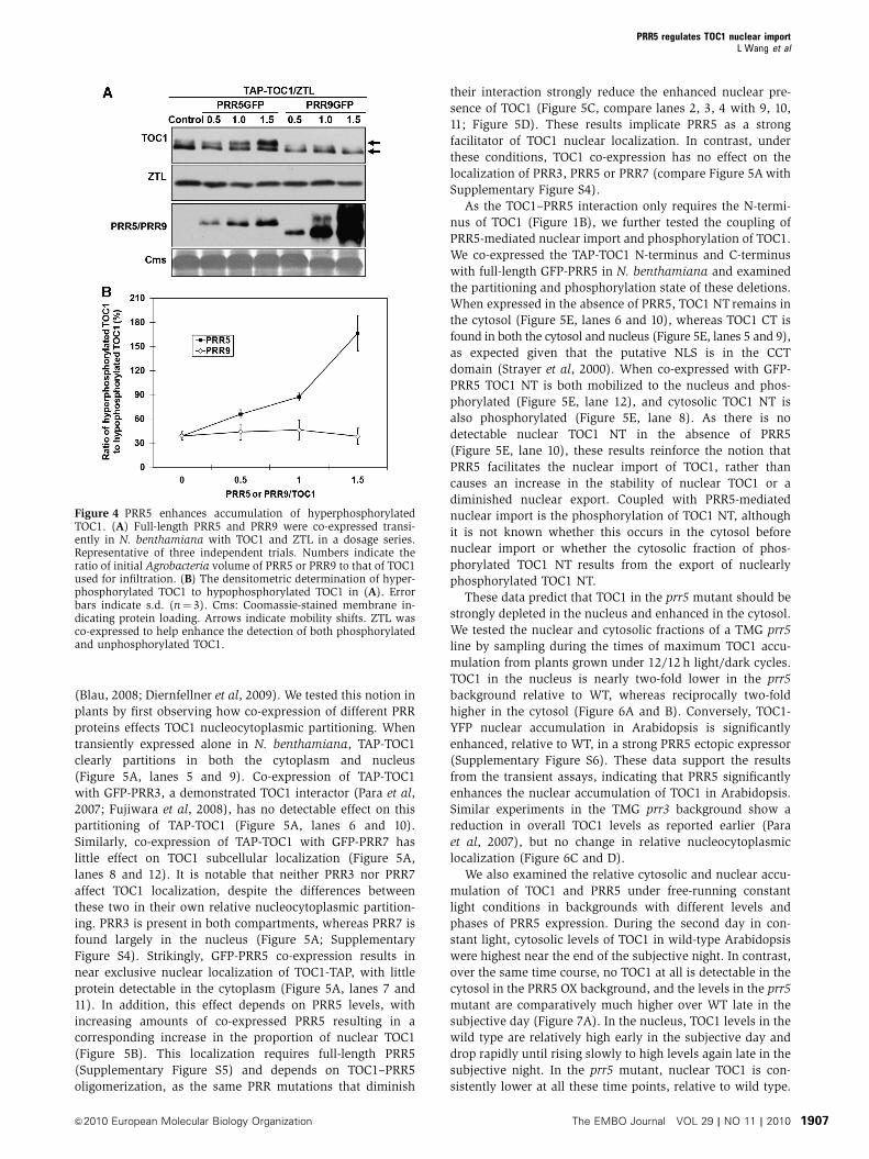
These data predict that TOC1 in the prr5 mutant should be
strongly depleted in the nucleus and enhanced in the cytosol.
We tested the nuclear and cytosolic fractions of a TMG prr5
line by sampling during the times of maximum TOC1 accu-
mulation from plants grown under 12/12 h light/dark cycles.
TOC1 in the nucleus is nearly two-fold lower in the prr5
background relative to WT, whereas reciprocally two-fold
higher in the cytosol (Figure 6A and B). Conversely, TOC1-
YFP nuclear accumulation in Arabidopsis is significantly
enhanced, relative to WT, in a strong PRR5 ectopic expressor
(Supplementary Figure S6). These data support the results
from the transient assays, indicating that PRR5 significantly
enhances the nuclear accumulation of TOC1 in Arabidopsis.
Similar experiments in the TMG prr3 background show a
reduction in overall TOC1 levels as reported earlier (Para
et al, 2007), but no change in relative nucleocytoplasmic
localization (Figure 6C and D).
We also examined the relative cytosolic and nuclear accu-
mulation of TOC1 and PRR5 under free-running constant
light conditions in backgrounds with different levels and
phases of PRR5 expression. During the second day in con-
stant light, cytosolic levels of TOC1 in wild-type Arabidopsis
were highest near the end of the subjective night. In contrast,
over the same time course, no TOC1 at all is detectable in the
cytosol in the PRR5 OX background, and the levels in the prr5
mutant are comparatively much higher over WT late in the
subjective day (Figure 7A). In the nucleus, TOC1 levels in the
wild type are relatively high early in the subjective day and
drop rapidly until rising slowly to high levels again late in the
subjective night. In the prr5 mutant, nuclear TOC1 is con-
sistently lower at all these time points, relative to wild type.
Figure 4 PRR5 enhances accumulation of hyperphosphorylatedTOC1. (A) Full-length PRR5 and PRR9 were co-expressed transi-ently in N. benthamiana with TOC1 and ZTL in a dosage series.Representative of three independent trials. Numbers indicate theratio of initial Agrobacteria volume of PRR5 or PRR9 to that of TOC1used for infiltration. (B) The densitometric determination of hyper-phosphorylated TOC1 to hypophosphorylated TOC1 in (A). Errorbars indicate s.d. (n¼ 3). Cms: Coomassie-stained membrane in-dicating protein loading. Arrows indicate mobility shifts. ZTL wasco-expressed to help enhance the detection of both phosphorylatedand unphosphorylated TOC1.
PRR5 regulates TOC1 nuclear importL Wang et al
&2010 European Molecular Biology Organization The EMBO Journal VOL 29 | NO 11 | 2010 1907

Constitutive high expression of PRR5 causes TOC1 to accu-
mulate in the nucleus at times not normally detectable in the
wild type (e.g. at 33 h), though levels early in the subjective
day (25 h) are unexpectedly low (Figure 7A). Overall, these
findings can be explained by the strong phase-specific ex-
pression of PRR5 in the wild type where peak accumulation
of PRR5 precedes that of TOC1 in both the cytosol and
nucleus (compare Figure 7A and B) (Fujiwara et al, 2008).
This earlier timing and the effect of the PRR5 overexpression
and absence on TOC1 levels are consistent with PRR5 acting
phase specifically to facilitate TOC1 nuclear accumulation
and cytosolic depletion through enhanced nuclear import.
Taken together, our data indicate that PRR5 enhances both
the nuclear accumulation and phosphorylation of TOC1.
Co-localization of TOC1 and PRR5 in the nucleus
Our initial demonstration of a TOC1–PRR5 interaction did not
distinguish between nuclear and cytoplasmic compartments.
We tested for nuclear oligomers by sonic lysis of the nuclear
fraction followed by an IgG pull down of TAP-TOC1. GFP-
PRR5 is detected in this bound fraction, indicating that the
interaction occurs in the nucleus (Supplementary Figure S7).
We then investigated the subcellular in vivo interaction by
observing the nuclear localization of both TOC1-mCHERRY
Figure 5 PRR5 promotes TOC1 cytosolic depletion and nuclear accumulation. (A) PRR5 alone is able to alter the TOC1 nucleocytoplasmicdistribution. GFP-PRR3, GFP-PRR5 and GFP-PRR7 were transiently co-expressed with TAP-TOC1 in N. benthamiana and tissues wereprocessed for cytosolic and nuclear protein fractions at ZT13 2 days after infiltration. Representative of three trials. (B) PRR5 promotion ofTOC1 cytosolic depletion and nuclear accumulation is dose dependent. Numbers indicate the ratio of initial Agrobacteria infiltration volumeof PRR5 to that of TOC1. (C) Point mutations in the TOC1 PRR domain specifically reduce TOC1 nuclear accumulation. Left panel shows thetotal protein expression for each infiltration combination; right panel shows TOC1 and PRR5 accumulation in cytosolic and nuclear fractions.(D) Quantitation of relative cytosolic-to-nuclear TOC1 levels from (C). See Materials and methods for explanation. Error bars indicate s.d.(n¼ 2). (E) TOC1 N-terminus is nuclear localized by PRR5. TOC1 NT and CT as in Figure 1. Histone H3 and ADK antibodies used are as inFigure 3. All immunoblots performed with anti-myc (TAP-TOC1) and anti-GFP (PRR5) antibodies.
PRR5 regulates TOC1 nuclear importL Wang et al
The EMBO Journal VOL 29 | NO 11 | 2010 &2010 European Molecular Biology Organization1908
and PRR5-GFP when transiently expressed separately or
together in N. benthamiana (Figure 8). When expressed
alone, TOC1-mCHERRY exhibits a pattern of small, dispersed
nuclear foci (Figure 8A), very similar to an earlier report
describing YFP-TOC1 expression (Strayer et al, 2000). In
contrast, PRR5-GFP occurs in distinctly larger foci dispersed
within the nucleus (Figure 8B), consistent with an earlier
report (Matsushika et al, 2007). When both proteins are co-
expressed, numerous medium-sized PRR5-like nuclear inclu-
sions are evident with TOC1 now co-localized with PRR5
(Figure 8C). These data indicate a co-residence of both
proteins, and strongly suggest that TOC1 is recruited by
PRR5 to a particular class of nuclear foci.
LKP2 contributes to the degradation of PRR5
Although the post-translational effects of PRR5 on TOC1
nuclear localization and phosphorylation strongly affect
TOC1 function, PRR5 is also linked to TOC1 through a shared
degradation mechanism. Earlier reports have shown that
both TOC1 and PRR5 are targeted for degradation by the
SCFZTL (Kiba et al, 2007; Fujiwara et al, 2008). However, the
ZTL family also includes the closely related LKP2 and FKF1
proteins (Kiyosue and Wada, 2000; Nelson et al, 2000;
Somers et al, 2000). Whereas FKF1 is strongly linked to
photoperiodic timing in Arabidopsis, until now there has
been no demonstrated function for LKP2 in the plant clock
or flowering time, apart from the effects of strong ectopic
expression, which has effects on periodicity, hypocotyl length
and flowering time identical to ZTL overexpression (Kiyosue
and Wada, 2000; Schultz et al, 2001; Somers et al, 2004).
We transformed the lkp2-1, ztl-1 and the ztl-3 lkp2-1
double mutants with PRR5HPRR5-GFP and identified and
characterized plants expressing PRR5-GFP in each of the
mutant genotypes. As reported earlier, PRR5 levels are
strongly stabilized in the ztl mutant under light/dark cycles
(Figure 9A) (Kiba et al, 2007; Fujiwara et al, 2008).
In contrast, we observed the same strongly cycling PRR5
steady-state levels in the lkp2-1 mutant as in the WT Col
background (Figure 9A). Oscillation of PRR5 in the ztl-3 lkp2-1
appeared slightly less robust compared with the ztl single
mutant, so we tested the effects of cyclohexamide on PRR5
stability in all the backgrounds. We reasoned that if activity of
SCFZTL was further compromised by the absence of LKP2, the
half life of PRR5 would be longer in the ztl lkp2 double
mutant than in the ztl single mutant. PRR5 half-life in the
lkp2-1 mutant was indistinguishable from WT, consistent
with no distinguishable differences in free-running period
between these backgrounds (Figure 9B and C; Supplementary
Figure S8). In contrast, there was a significant increase in
the PRR5 half-life in the ztl lkp2 double mutant, relative
to ztl-3 (Figure 9B and C). A similar stabilization of PRR5 has
been observed in the zlt lkp2 fkf1 triple mutant (Takato
Imaizumi, personal communication). These data indicate
that LKP2 contributes to the proteasome-dependent degrad-
ation of PRR5.
Discussion
PRR5 controls the nucleocytoplasmic and subnuclear
distribution of TOC1
The five-member family of PRR proteins is intricately linked
to the control of circadian period, but the specific function of
each has remained unclear. Genetic and molecular evidence
implicates most of them in the transcriptional control of a
number of core clock genes, but how this is effected is
obscure. Our results now show a novel function for PRR5
Figure 6 Nucleocytoplasmic ratio of TOC1 distribution is altered in prr5-1. (A) Subcellular determination of cytosolic and nuclear TOC1-YFP(TOC1) in Arabidopsis wild-type and prr5-1 mutant. Anti-GFP antibody was used for immunodetection of TOC1-YFP. Histone H3 and ADKantibodies used are as in Figure 3. (B) prr5-1/WT ratio of quantitated cytosolic and nuclear TOC1 from (A). A value of 1.0 would indicateidentical TOC1 levels in both backgrounds. (C) Subcellular determination of cytosolic and nuclear TOC1-YFP (TOC1) in Arabidopsis wild-typeand prr3-1 mutant. (D) prr3-1/WT ratio of quantitated cytosolic and nuclear TOC1 from (C). Error bars indicate s.d. (n¼ 3). Seedlings wereentrained in 12-h light/12-h dark cycles and harvested at the indicated ZT times.
PRR5 regulates TOC1 nuclear importL Wang et al
&2010 European Molecular Biology Organization The EMBO Journal VOL 29 | NO 11 | 2010 1909

through a specific oligomerization with TOC1 that appears, in
part, unrelated to a direct function in control of transcription.
The nearly two-fold decrease in steady-state nuclear TOC1
abundance in the prr5 mutant concomitant with a similar
increase in TOC1 cytoplasmic levels demonstrates a critical
function for PRR5 in the control of TOC1 nuclear accumula-
tion and localization. These findings imply that the short
period of the prr5 mutant is at least partially due to the
diminished levels of nuclear TOC1. Similarly, the decreased
ability of the toc1-5 and toc1-8 proteins to interact with PRR5
explains the short period of those two mutant alleles not as a
result of diminished activities but from lower accumulations
in the nucleus.
Consideration of earlier studies of prr5 and toc1 single
mutants and ectopic expression work reveals that manipulat-
ing the levels of both genes affects the clock in similar ways.
In addition to the similarly shortened circadian period in the
single mutants, the effects on the gene expression of a
number of clock genes in toc1 and prr5 backgrounds are
very similar. Of the genes examined, the toc1 mutant invari-
ably causes a more rapid damping of mRNA rhythms than
prr5, and is largely epistatic to prr5 when compared with the
toc1 prr5 double mutant (Ito et al, 2008). There is a slight
degree of additivity apparent, but these results are consistent
with TOC1 as the primary effector and PRR5 as an enhancer
of TOC1 activity. Similarly, in the PRR5 and TOC1 over-
expressors morning genes such as LHY and CCA1 both
damp to low expression, whereas expression of PRR9 and
GI become undetectable very quickly in constant light (LL)
(Makino et al, 2002; Sato et al, 2002). The similarity of these
molecular phenotypes, together with data presented here, is
consistent with the idea that PRR5 promotes TOC1 function,
at least in part, by enhancing nuclear accumulation and
subnuclear localization.
Overexpression of the PRR5 receiver domain (PRR5OX NT)
causes a long period in Arabidopsis, very different from the
strongly damped of rhythms of overexpressed full-length
PRR5 (PRR5OX FL) (Matsushika et al, 2007). These data
now can be re-interpreted in view of the altered nuclear
localization of TOC1 in these two backgrounds
(Supplementary Figure S5). The higher accumulation of
cytosolic TOC1 in the PRR5OX NT line likely arises from a
competitive inhibition of the TOC1–ZTL interaction by PRR5,
resulting in an accumulation of TOC1, and probably also
endogenous PRR5. Together, the increased level of both may
account for the longer period in PRR5OX NT plants
(Matsushika et al, 2007). At the same time, the much higher
levels of nuclear TOC1 in the PRR5OX FL (Supplementary
Figure S4) can explain the damped rhythms reported by
Matsushika et al (2007) and alterations in clock gene expres-
sions (see above), which are similar to those reported when
TOC1 levels are strongly elevated (Makino et al, 2002; Mas
et al, 2003a). These results are consistent with the notion that
the PRR5 overexpression phenotypes arise from effects on
TOC1 nucleocytoplasmic partitioning. In addition, the above
findings, along with recent results that identify TOC1 in
association with the CCA1 and ABAR1 promoters, support
the notion that a nuclear population of TOC1 is necessary and
a limiting factor in the control of circadian period (Legnaioli
et al, 2009; Pruneda-Paz et al, 2009).
Further support for this idea comes from the re-positioning
of TOC1 subnuclear localization to the larger PRR5-type
inclusions when the two are expressed together. The specific
function of these foci remains unclear, but it is plausible
and testable that PRR5 may facilitate TOC1 binding to the
E-box of the CCA1 and ABAR1 promoters. As neither protein
bears a recognizable DNA-binding domain, it is likely that
they are part of a larger multi-protein complex at these
promoters. However, as the period of the prr5 toc1 double
mutant is considerably shorter than the toc1 mutant alone
(Fujiwara et al, 2008; Ito et al, 2008), it is likely that PRR5
has an additional function in the clock separate from its
effects on TOC1.
Complex dynamics: PRR5, TOC1, PRR3, ZTL and LKP2
The PRR5–TOC1 dimerization domain maps to the same
N-terminal receiver domain that facilitates the TOC1–ZTL
interaction. The toc1-5 and toc1-8 mutations that diminish
the TOC1–ZTL interaction similarly reduce TOC1–PRR5 bind-
ing. These results imply a complex dynamic between TOC1,
Figure 7 Phase-specific effect of PRR5 expression on TOC1 nucleo-cytoplasmic distribution. (A) Cytosolic and nuclear TOC1-YFP(TMG) levels in Arabidopsis wild-type, prr5-1 and PRR5 OX back-grounds under free-running constant light conditions. (B) Cytosolicand nuclear PRR5-GFP levels in Arabidopsis wild type under free-running constant light conditions. Seedlings were entrained in 12-hlight/12-h dark cycles then maintained in constant white light forthe number of hours indicated until harvested. Different immuno-blot exposure levels are shown for TMG (anti-GFP) panels in (A) toallow comparisons between earlier and later time points within andbetween genotypes. SDS–PAGE fractionated protein extracts fromall genotypes and time points within each TMG-probed panel wereimmunoblotted and probed together and are directly comparable,relative to the cytosolic and nuclear loading controls. Direct com-parisons of abundance between TMG and PRR5-GFP panels are notappropriate. All data are representative of two independent trials.Histone H3 and ADK antibodies used are as in Figure 3.
PRR5 regulates TOC1 nuclear importL Wang et al
The EMBO Journal VOL 29 | NO 11 | 2010 &2010 European Molecular Biology Organization1910

PRR5 and ZTL in view of the fact that PRR5 is also a
proteolytic target of ZTL (Kiba et al, 2007; Fujiwara et al,
2008). However, although PRR5 might also act as a compe-
titive inhibitor of the ZTL–TOC1 interaction, our data indicate
a more potent function as a facilitator of TOC1 nuclear
import, where it would escape proteolysis by the SCFZTL,
which is undetectable in the nucleus (Kim et al, 2007). In this
way, PRR5 has a function similar to PRR3 in that both
facilitate an increase in TOC1 accumulation. However, PRR3
achieves this as a competitive inhibitor of the cytosolic
TOC1–ZTL interaction, whereas PRR5 acts primarily to
enhance TOC1 nuclear accumulation. Thus, through two
different mechanisms PRR3 and PRR5 contribute to post-
translationally enhancing TOC1 levels.
Figure 8 PRR5 and TOC1 co-localize in the nucleus. 35SHTOC1-mCherry and 35SHGFP-PRR5 were individually or co-expressed inN. bentamiana leaves as indicated. Signals from GFP, mCherry, 40,6-diamidino-2-phenylindole (DAPI), and the merged signals (overlay) areshown. Bars¼ 10mm.
Figure 9 LKP2 contributes to PRR5 turnover. (A) Diurnal oscillation of PRR5 is further diminished in the ztl lkp2 background. Time course ofPRR5GFP protein levels in 10-day-old plants grown under 12/12L/D cycles probed with anti-GFP antibodies. Coomassie-stained regions (Cms)shown as loading controls. Blots are representative of more than three trials. (B) PRR5 stability regulated by both ZTL and LKP2. Plant growthand tissue processing as in (A) with plants transferred to continuous light at ZT0 and treated with 100mM cyclohexamide (CHX) after 19 h.Plants were harvested at 0, 1, 4 and 10 h after adding CHX. PRR5GFP protein was detected by anti-GFP antibodies. (C) Quantitation relative toCoomassie-stained regions and normalized to time 0. Blots representative of three trials. Means of three trials±s.e.m. are shown.
PRR5 regulates TOC1 nuclear importL Wang et al
&2010 European Molecular Biology Organization The EMBO Journal VOL 29 | NO 11 | 2010 1911

In addition, LKP2 can now also be added to the interplay
between these four proteins. Although strong ectopic expres-
sion of LKP2 can disrupt clock function (Schultz et al, 2001),
lkp2 and lkp2 ztl double mutants show no detectable affect on
circadian period (Supplementary Figure S6). These findings
initially suggested that normally LKP2 either acts outside the
clock or that its contribution to period is minor or not easily
detectable. The observed increase in PRR5 stability in the ztl
lkp2 double mutant indicates the latter case; that LKP2 does
contribute to the proteolytic degradation of a target protein
previously ascribed entirely to the SCFZTL (Kiba et al, 2007;
Fujiwara et al, 2008), but effects on period are subtle. This
finding does not exclude the possibility that LKP2 may have
effects on other proteolyses apart from its effect on PRR5.
One possible explanation for the additivity between ZTL and
LKP2 is heterodimerization or multimerization among the
three ZTL family members that might enhance SCF activity
(Yasuhara et al, 2004). For example, the WD 40 class of
F-box proteins contain a short conserved repeat (D domain)
directly adjacent to the F-box that is essential for their function
by mediating homodimerization and/or heterodimerzation be-
tween closely related forms (Zhang and Koepp, 2006; Tang
et al, 2007). The ZTL F-box protein family is of a different class,
and although there is a region directly upstream of the F-box in
that is highly conserved among the three, it is not known
whether this functions as an interaction domain.
TOC1 phosphorylation: multiple functions?
It is likely that the phosphorylation of TOC1 is critical to its
function in a number of ways. As the TOC1–PRR3 interaction
is strongly enhanced by the phosphorylated form of both
proteins (Fujiwara et al, 2008), and the TOC1–PRR3 interac-
tion sequesters some fraction of TOC1 from degradation by
SCFZTL (Para et al, 2007; Fujiwara et al, 2008) at least one
function of phosphorylation is to stabilize TOC1. At the same
time, the TOC1–ZTL interaction is also enhanced by TOC1
phosphorylation (Fujiwara et al, 2008), typical of F-box
protein–substrate interactions (Willems et al, 2004). Thus,
TOC1 phosphorylation facilitates both its degradation and its
stabilization, depending on the relative abundances of ZTL
and phosphorylated PRR3 (Fujiwara et al, 2008).
Our results indicate that in contrast to the PRR3–TOC1
interaction, PRR5 and TOC1 oligomerize independent of their
phosphorylation states. Rather, the PRR5–TOC1 interaction
can facilitate a kinase activity that phosphorylates the
N-terminus of TOC1, although it is not known whether that
kinase activity is responsible for all TOC1 phosphorylations.
Unlike the strong TOC1 mobility shifts seen when PRR5 is
strongly co-expressed, we were unable to detect changes in the
TOC1 mobility in the prr5 mutant (data not shown). This
suggests either that the PRR5-mediated kinase activity contri-
butes to only a portion of TOC1 phosphorylation, and a
reduced phosphorylation level is not detectable by mobility
shift, or that other phosphorylation mechanisms may substitute
in the absence of a PRR5-based kinase recruitment platform. It
is well known that multiple, often sequential, phosphorylations
resulting from the action of multiple kinases are involved in the
post-translational control of clock proteins in other systems
(Huang et al, 2007; Virshup et al, 2007; Gallego and Virshup,
2007; Baker et al, 2009; Tang et al, 2009).
It is possible that the marked increase in the proportion of
hyperphosphorylated TOC1 late in the circadian cycle
(Fujiwara et al, 2008) is tied to its nuclear function, possibly
related to interaction with partners as part of a transcription
complex (Pruneda-Paz et al, 2009). Alternatively, PRR5-
mediated phosphorylation may only be important in facilitat-
ing the cytoplasmic interaction of TOC1 and PRR3. The dual
functions of PRR5 in enhancing both TOC1 phosphorylation
and subnuclear relocalization may be causal or separable:
PRR5-mediated phosphorylation may facilitate the re-posi-
tioning of nuclear TOC1 or the two processes depend on
PRR5 independent of each other. Clearly, further identifica-
tion of interaction partners along with the phosphorylation
sites and the kinases involved will be necessary for a full
understanding of the function of phosphorylation in TOC1
function.
Conserved mechanism of post-translational control
of nuclear import of core clock components
The transcription–translation feedback loop that forms the
backbone of all eukaryotic oscillators also relies heavily on
post-translational mechanisms, and phosphorylation is one
of the best characterized (Gallego and Virshup, 2007; Cha
et al, 2008; Chiu et al, 2008; Diernfellner et al, 2009). Two
well-studied heterodimerzations in the Drosophila clock are
between timeless (TIM) and period (PER), and clock (CLK)
and cycle (CYC) (Benito et al, 2007; Saez et al, 2007; Maurer
et al, 2009). Both pairs act to control transcription, with the
TIM–PER dimer repressing gene activations initiated by the
binding of the CLK-CYC dimer to the E-box of numerous
clock-controlled promoters, including per and tim (Benito
et al, 2007). As well, both pairs of proteins undergo phos-
phorylations that can affect their turnover, nuclear localiza-
tion and regulatory activity (Gallego and Virshup, 2007;
Vanselow and Kramer, 2007). In the former case, the TIM–
PER interaction specifically protects PER from proteosome-
dependent degradation, and facilitates PER phosphorylation.
TIM-dependent enhancement of PER import is also likely,
although maintenance of the interaction during passage
through the nuclear pore is not clear (Meyer et al, 2006).
Similarly, CLK and CYC proteins interact both in the cyto-
plasm and the nucleus, and heterodimerization of CLK with
CYC promotes both the phosphorylation and nuclear import
of CLK (Maurer et al, 2009). Thus, our findings here show
that in Arabidopsis, PRR5 has a similar function to TOC1 as
TIM does to PER and CYC to CLK. Interestingly, none of these
three pairs is structurally related to each other. Taken to-
gether, these findings show that clock-regulated nuclear
import through heterodimer formation is fundamental to
the molecular circuitry of circadian clocks. Indeed, in the
mammalian clock as well, the period2 (PER2)–cryptochrome
(CRY) interaction facilitates nuclear import of CRY in mam-
malian cells (Miyazaki et al, 2001).
One notable common feature of these examples is that the
transcription of one or both of the dimeric partners is clock
regulated. This permits a phase-dependent accentuation of
nuclear accumulation of one or both proteins, and may be the
reason such a mechanism is found across such disparate
circadian systems, which otherwise do not share homologous
components. In the present case, PRR5 nuclear accumulation
is very similar regardless of the level of TOC1 (Supplementary
Figures S2 and S6), suggesting that PRR5 is the key regulatory
element in this partnership. With its very strong nuclear
localization tendency, it is possible that PRR5 also partners
PRR5 regulates TOC1 nuclear importL Wang et al
The EMBO Journal VOL 29 | NO 11 | 2010 &2010 European Molecular Biology Organization1912
with other clock components, including PRR3, 7 or 9, to act
as a phase-specific nuclear shuttle. With the large number of
still uncharacterized proteins in the Arabidopsis clock, it is
likely that this type of nuclear transport mechanism will be
more commonly found.
Materials and methods
Plant material and growth conditionsConstruction of TMG (TOC1HTOC1-YFP minigene) in Arabidopsis(Col-0 accession) has been described earlier (Mas et al, 2003a, b).Generation of TMG prr5-1 and TMG prr3-1, PRR5HPRR5-GFP in wildtype, ztl-1, lkp2 and ztl-1 lkp2 is described in detail in Supplemen-tary data. Arabidopsis seedlings were entrained under 12-h whitefluorescent light (50–60mmol m�2 s�1)/12 h dark cycles for 8–12days on MS plates with 3% sucrose and 1% agar before harvesting.The tissues were harvested at the indicated time points. Fortransient expression experiments, 3–4-week-old N. benthamianaplants were infiltrated with Agrobacterium as described earlier(Fujiwara et al, 2008). Tissues were harvested on the third day atZT12 (16 h light/8 h dark).
PlasmidsConstructs of TAP-tagged TOC1 full-length, N-terminus (1–242) andC-terminus (243–618) have been described earlier (Fujiwara et al,2008). Constructs of TAP-tagged PRR5 and its deletions, 35SHTOC1-mCherry and TAP-tagged TOC1 point mutations are described in theSupplementary data.
Protein extraction and immunoblot analysesTotal protein extraction was carried out as described earlier (Kimet al, 2007; Fujiwara et al, 2008). For the immunoblot analysis,proteins were size fractionated by 8% SDS–PAGE (acrylamide:bis-acrylamide, 149:1) for PRRs proteins or 12% SDS–PAGE (acrylami-de:bisacrylamide, 37.5:1) for histone H3 and ADK, then analysedby immunoblot. Immunoblotting was performed using 1:1000dilution anti-PAP antibodies (Sigma-Aldrich P-1291) or primarypolyclonal anti-GFP antibody (Abcam ab6556) or primaryanti-histone H3 antibody (Abcam 1791) and 1:4000 polyclonalADK primary antibodies (gift from Dr David Bisaro) followed byECL detection using anti-rabbit IgG with horseradish peroxidase-linked whole antibody (GE healthcare NA934V). For immunoblotof primary anti-Myc antibody (Santa Cruz, sc40, 1:300 dilution),1:1000 HRP-linked anti-mouse IgG (Sigma-Aldrich, A0198) wasused as secondary antibody. Chemoluminance reactions wereperformed with Supersignal West Pico Chemiluminescent Sub-strates from Pierce. Quantity One 4.1.1 software was adopted forcalculation and quantification of protein signal intensity. InFigure 5D, the cytosolic and nuclear TOC1 signals for WT TOC1(no PRR5 co-infiltration) were first normalized to the respectivecytosolic (ADK) or nuclear (H3) loading controls. The ratios ofthese values were determined (cytosolic TOC1 to nuclear TOC1) andmultiplied by its reciprocal to obtain a reference value of 1. Thisvalue was then used as a multiplier for remaining ratios ofnormalized cytosolic TOC1 to normalized nuclear TOC1 withPRR5 co-infiltrated with the different TOC1 point mutations.
Fractionation of cytosolic and nuclear proteinsThe cytosolic and nuclear protein extractions were carried out withCELLYTPN1 CelLytic PN isolation/Extraction Kit (Sigma-Aldrich)according to the manufacturer’s instruction. Briefly, the tissueswere ground in liquid nitrogen and stored at �801C before use. Theground tissues were gently resuspended with 1� nuclei isolationbuffer supplemented with 1 mM DTT, 2 mM Na3VO4, 2 mM NaF and25 mM phenylmethylsulfonyl fluoride (PMSF), and were filteredthrough five layers Miracloth (Calbiochem, San Diego, CA) by
centrifugation at 41C. The pellet was resuspended with 1� nucleiisolation buffer supplemented with 0.3% Triton X-100, 1 mM DTT,2 mM Na3VO4, 2 mM NaF and 25 mM PMSF, 2.5mg/ml antipain,2.5 mg/ml chymostatin, 1 mg/ml pepstatin, 5mg/ml leupeptin, 5 mg/ml aprotinin, and 50mM MG132, 50mM MG115, 50 mM ALLN. Afterincubation on ice (5 min), extracts were centrifuged at 6800 r.p.m.for 5 min. The resulting supernatant was transferred to a new tubecontaining SDS buffer and designated as the cytosolic fraction. Thepellet was resuspended in the above buffer and centrifuged andresuspended repeatedly until the pellet appeared totally white orslightly gray. The pellet was washed with 1� nuclei isolation buffersupplemented with 1 mM DTT, 2 mM Na3VO4, 2 mM NaF and25 mM PMSF, dissolved in SDS buffer and designated as the nuclearcompartment. The cytosolic and nuclear compartments were loadedonto SDS–PAGE gels in proportion to the volume used in the initialextractions and the resuspension volumes of each compartment.
In vitro pull-down assayIn vitro pull-down assays were performed as described earlier(Fujiwara et al, 2008).
Co-immunoprecipitationAgrobacteria containing both TOC1 full length and its amino- orcarboxyl-deletions by TAP tagged with GFP-PRR5 or PRR5 fulllength and its deletions tagged by TAP with TOC1-YFP were co-infiltrated. Tissues were harvested at ZT12 after infiltration on thethird day. The co-immunoprecipitation was performed as describedearlier (Fujiwara et al, 2008).
mRNA expression analysisTotal RNA was extracted with TRIzol reagents (Invitrogen, Carlbad,CA) from tissue of 10-day-old seedlings grown on MS media. Afterdigestion with DNase I (Amplification grade, Invitrogen, Carlbad,CA), cDNA synthesis was performed with Oligo-dT primer andSuperscriptIII reverse transcriptase kit (Invitrogen, Carlbad, CA) asper the manufacturer’s instruction. cDNA templates were digestedwith RNaseH before amplification. Gene-specific primers were usedfor the quantification of corresponding mRNA levels through semi-quantitative RT–PCR as described earlier (Fujiwara et al, 2008).
Confocal laser scanning microscopyAgrobacteria containing 35SHGFP-PRR5 or 35SHTOC1-mCherrywas singly or co-infiltrated into N. benthamiana plants as describedearlier. Images were collected using a Zeiss LSM 510 coanfocalmicroscope with a Plan-Apochromat 63� /1.4 oil objective usingthe multi-track method. For GFP and mCherry confocal microscopy,GFP was imaged using 488 nm laser light and a 505–530 nm BPemission filter, and mCherry was imaged using 543 nm laser lightand a 585–615 BP emission filter. For DAPI staining, a high-pressuremercury bulb and Zeiss DAPI filter set were used, and the DAPIemission was detected at 461–525 nm on ChS1.
Supplementary dataSupplementary data are available at The EMBO Journal Online(http://www.embojournal.org).
Acknowledgements
We thank Dr D Bisaro for his kind gift of the ADK antibody and Dr TImaizumi for sharing unpublished results. This work was supportedby grants to DES from NSF grants MCB-0544137 and IBN-0748749and in part by Support for Long-term Visit from the Yamada ScienceFoundation to SF.
Conflict of interest
The authors declare that they have no conflict of interest.
References
Alabadi D, Oyama T, Yanovsky MJ, Harmon FG, Mas P,Kay SA (2001) Reciprocal regulation between TOC1 andLHY/CCA1 within the Arabidopsis circadian clock. Science 293:880–883
Baker CL, Kettenbach AN, Loros JJ, Gerber SA, Dunlap JC (2009)Quantitative proteomics reveals a dynamic interactome andphase-specific phosphorylation in the Neurospora circadianclock. Mol Cell 34: 354–363
PRR5 regulates TOC1 nuclear importL Wang et al
&2010 European Molecular Biology Organization The EMBO Journal VOL 29 | NO 11 | 2010 1913

Bell-Pedersen D, Cassone VM, Earnest DJ, Golden SS, Hardin PE,Thomas TL, Zoran MJ (2005) Circadian rhythms frommultiple oscillators: lessons from diverse organisms. Nat RevGenet 6: 544–556
Benito J, Zheng H, Ng FS, Hardin PE (2007) Transcriptional feed-back loop regulation, function, and ontogeny in Drosophila. ColdSpring Harb Symp Quant Biol 72: 437–444
Blau J (2008) PERspective on PER phosphorylation. Genes Dev 22:1737–1740
Cha J, Chang SS, Huang G, Cheng P, Liu Y (2008) Control of WHITECOLLAR localization by phosphorylation is a critical step in thecircadian negative feedback process. EMBO J 27: 3246–3255
Cha J, Huang G, Guo J, Liu Y (2007) Posttranslational control of theNeurospora circadian clock. Cold Spring Harb Symp Quant Biol72: 185–191
Chiu JC, Vanselow JT, Kramer A, Edery I (2008) The phospho-occupancy of an atypical SLIMB-binding site on PERIOD that isphosphorylated by DOUBLETIME controls the pace of the clock.Genes Dev 22: 1758–1772
de Paula RM, Vitalini MW, Gomer RH, Bell-Pedersen D (2007)Complexity of the Neurospora crassa circadian clock system:multiple loops and oscillators. Cold Spring Harb Symp QuantBiol 72: 345–351
Diernfellner AC, Querfurth C, Salazar C, Hofer T, Brunner M (2009)Phosphorylation modulates rapid nucleocytoplasmic shuttlingand cytoplasmic accumulation of Neurospora clock protein FRQon a circadian time scale. Genes Dev 23: 2192–2200
Dodd AN, Salathia N, Hall A, Kevei E, Toth R, Nagy F, Hibberd JM,Millar AJ, Webb AA (2005) Plant circadian clocks increasephotosynthesis, growth, survival, and competitive advantage.Science 309: 630–633
Ebisawa T (2007) Circadian rhythms in the CNS and peripheralclock disorders: human sleep disorders and clock genes.J Pharmacol Sci 103: 150–154
Farre EM, Harmer SL, Harmon FG, Yanovsky MJ, Kay SA (2005)Overlapping and distinct roles of PRR7 and PRR9 in theArabidopsis circadian clock. Curr Biol 15: 47–54
Fujiwara S, Wang L, Han L, Suh SS, Salome PA, McClung CR,Somers DE (2008) Post-translational regulation of theArabidopsis circadian clock through selective proteolysis andphosphorylation of pseudo-response regulator proteins. J BiolChem 283: 23073–23083
Gallego M, Virshup DM (2007) Post-translational modificationsregulate the ticking of the circadian clock. Nat Rev Mol Cell Biol8: 139–148
Gardner MJ, Hubbard KE, Hotta CT, Dodd AN, Webb AA (2006)How plants tell the time. Biochem J 397: 15–24
Hardin PE (2005) The circadian timekeeping system of Drosophila.Curr Biol 15: R714–R722
Harmer SL (2009) The Circadian system in higher plants. Annu RevPlant Biol 60: 357–377
Hazen SP, Borevitz JO, Harmon FG, Pruneda-Paz JL, Schultz TF,Yanovsky MJ, Liljegren SJ, Ecker JR, Kay SA (2005) Rapid arraymapping of circadian clock and developmental mutations inArabidopsis. Plant Physiol 138: 990–997
Huang G, Chen S, Li S, Cha J, Long C, Li L, He Q, Liu Y (2007)Protein kinase A and casein kinases mediate sequential phos-phorylation events in the circadian negative feedback loop. GenesDev 21: 3283–3295
Ito S, Niwa Y, Nakamichi N, Kawamura H, Yamashino T, Mizuno T(2008) Insight into missing genetic links between two evening-expressed pseudo-response regulator genes TOC1 and PRR5 inthe circadian clock-controlled circuitry in Arabidopsis thaliana.Plant Cell Physiol 49: 201–213
Johnson CH, Kyriacou C (2005) Clock evolution and adaptation:whence and whither? In Endogenous Plant Rhythms, Hall A,McWatters H (eds). Oxford: Blackwell, pp 237–260
Kevei E, Gyula P, Hall A, Kozma-Bognar L, Kim WY, Eriksson ME,Toth R, Hanano S, Feher B, Southern MM, Bastow RM,Viczian A, Hibberd V, Davis SJ, Somers DE, Nagy F, Millar AJ(2006) Forward genetic analysis of the circadian clockseparates the multiple functions of ZEITLUPE. Plant Physiol140: 933–945
Kiba T, Henriques R, Sakakibara H, Chua NH (2007) Targeteddegradation of pseudo-response regulator5 by an SCFZTL com-plex regulates clock function and photomorphogenesis inArabidopsis thaliana. Plant Cell 19: 2516–2530
Kim WY, Fujiwara S, Suh SS, Kim J, Kim Y, Han L, David K,Putterill J, Nam HG, Somers DE (2007) ZEITLUPE is a circadianphotoreceptor stabilized by GIGANTEA in blue light. Nature 449:356–360
Kim WY, Geng R, Somers DE (2003) Circadian phase-specificdegradation of the F-box protein ZTL is mediated by the protea-some. Proc Natl Acad Sci USA 100: 4933–4938
Kiyosue T, Wada M (2000) LKP1 (LOV kelch protein 1): a factorinvolved in the regulation of flowering time in Arabidopsis. PlantJ 23: 807–815
Legnaioli T, Cuevas J, Mas P (2009) TOC1 functions as a molecularswitch connecting the circadian clock with plant responses todrought. EMBO J 28: 3745–3757
Locke JC, Kozma-Bognar L, Gould PD, Feher B, Kevei E, Nagy F,Turner MS, Hall A, Millar AJ (2006) Experimental validation of apredicted feedback loop in the multi-oscillator clock ofArabidopsis thaliana. Mol Syst Biol 2: 59
Makino S, Matsushika A, Kojima M, Yamashino T, Mizuno T (2002)The APRR1/TOC1 quintet implicated in circadian rhythms ofArabidopsis thaliana: I. Characterization with APRR1-overexpres-sing plants. Plant Cell Physiol 43: 58–69
Mas P, Alabadi D, Yanovsky MJ, Oyama T, Kay SA (2003a) Dual roleof TOC1 in the control of circadian and photomorphogenicresponses in Arabidopsis. Plant Cell 15: 223–236
Mas P, Kim WY, Somers DE, Kay SA (2003b) Targeted degradation ofTOC1 by ZTL modulates circadian function in Arabidopsis thali-ana. Nature 426: 567–570
Matsushika A, Kawamura M, Nakamura Y, Kato T, Murakami M,Yamashino T, Mizuno T (2007) Characterization of circadian-associated pseudo-response regulators: II. The function of PRR5and its molecular dissection in Arabidopsis thaliana. BiosciBiotechnol Biochem 71: 535–544
Matsushika A, Makino S, Kojima M, Mizuno T (2000) Circadianwaves of expression of the APRR1/TOC1 family of pseudo-response regulators in Arabidopsis thaliana: insight into theplant circadian clock. Plant Cell Physiol 41: 1002–1012
Maurer C, Hung HC, Weber F (2009) Cytoplasmic interaction withCYCLE promotes the post-translational processing of the circa-dian CLOCK protein. FEBS Lett 583: 1561–1566
McClung CR (2006) Plant circadian rhythms. Plant Cell 18: 792–803Meyer P, Saez L, Young MW (2006) PER-TIM interactions in living
Drosophila cells: an interval timer for the circadian clock. Science311: 226–229
Miyazaki K, Mesaki M, Ishida N (2001) Nuclear entry mechanism ofrat PER2 (rPER2): role of rPER2 in nuclear localization of CRYprotein. Mol Cell Biol 21: 6651–6659
Nelson DC, Lasswell J, Rogg LE, Cohen MA, Bartel B (2000) FKF1, aclock-controlled gene that regulates the transition to flowering inArabidopsis. Cell 101: 331–340
Ouyang Y, Andersson CR, Kondo T, Golden SS, Johnson CH (1998)Resonating circadian clocks enhance fitness in Cyanobacteria.Proc Natl Acad Sci USA 95: 8660–8664
Para A, Farre EM, Imaizumi T, Pruneda-Paz JL, Harmon FG, Kay SA(2007) PRR3 is a vascular regulator of TOC1 stability in theArabidopsis circadian clock. Plant Cell 19: 3462–3473
Pittendrigh CS (1981) Circadian organization and the photoperiodicphenomena. In Biological Clocks in Reproductive Cycles, Follet BK(ed) Bristol: John Wright, pp 1–35
Pruneda-Paz JL, Breton G, Para A, Kay SA (2009) A functionalgenomics approach reveals CHE as a component of theArabidopsis circadian clock. Science 323: 1481–1485
Robson F, Costa MM, Hepworth SR, Vizir I, Pineiro M, Reeves PH,Putterill J, Coupland G (2001) Functional importance of conserveddomains in the flowering-time gene CONSTANS demonstrated byanalysis of mutant alleles and transgenic plants. Plant J 28: 619–631
Saez L, Meyer P, Young MW (2007) A PER/TIM/DBT interval timerfor Drosophila’s circadian clock. Cold Spring Harb Symp QuantBiol 72: 69–74
Sato E, Nakamichi N, Yamashino T, Mizuno T (2002) Aberrantexpression of the Arabidopsis circadian-regulated APRR5 genebelonging to the APRR1/TOC1 quintet results in early floweringand hypersensitiveness to light in early photomorphogenesis.Plant Cell Physiol 43: 1374–1385
Schaffer R, Ramsay N, Samach A, Corden S, Putterill J, Carre IA,Coupland G (1998) The late elongated hypocotyl mutation ofArabidopsis disrupts circadian rhythms and the photoperiodiccontrol of flowering. Cell 93: 1219–1229
PRR5 regulates TOC1 nuclear importL Wang et al
The EMBO Journal VOL 29 | NO 11 | 2010 &2010 European Molecular Biology Organization1914

Schultz TF, Kiyosue T, Yanovsky M, Wada M, Kay SA (2001) A rolefor LKP2 in the circadian clock of Arabidopsis. Plant Cell 13:2659–2670
Somers DE, Fujiwara S (2009) Thinking outside the F-box: novelligands for novel receptors. Trends Plant Sci 14: 206–213
Somers DE, Fujiwara S, Kim WY, Suh SS (2007) Posttranslationalphotomodulation of circadian amplitude. Cold Spring Harb SympQuant Biol 72: 193–200
Somers DE, Kim WY, Geng R (2004) The F-box protein ZEITLUPEconfers dosage-dependent control on the circadian clock, photo-morphogenesis, and flowering time. Plant Cell 16: 769–782
Somers DE, Schultz TF, Milnamow M, Kay SA (2000) ZEITLUPEencodes a novel clock-associated PAS protein from Arabidopsis.Cell 101: 319–329
Strayer C, Oyama T, Schultz TF, Raman R, Somers DE, Mas P, PandaS, Kreps JA, Kay SA (2000) Cloning of the Arabidopsis clock geneTOC1, an autoregulatory response regulator homolog. Science289: 768–771
Sweeney BM (1987) Rhythmic Phenomena in Plants. San Diego:Academic Press
Tang CT, Li S, Long C, Cha J, Huang G, Li L, Chen S, Liu Y (2009)Setting the pace of the Neurospora circadian clock by multipleindependent FRQ phosphorylation events. Proc Natl Acad Sci USA106: 10722–10727
Tang X, Orlicky S, Lin Z, Willems A, Neculai D, Ceccarelli D,Mercurio F, Shilton BH, Sicheri F, Tyers M (2007) Suprafacial
orientation of the SCFCdc4 dimer accommodates multiple geo-metries for substrate ubiquitination. Cell 129: 1165–1176
Ueda HR (2007) Systems biology of mammalian circadian clocks.Cold Spring Harb Symp Quant Biol 72: 365–380
Vanselow K, Kramer A (2007) Role of phosphorylation in themammalian circadian clock. Cold Spring Harb Symp Quant Biol72: 167–176
Virshup DM, Eide EJ, Forger DB, Gallego M, Harnish EV (2007)Reversible protein phosphorylation regulates circadian rhythms.Cold Spring Harb Symp Quant Biol 72: 413–420
Wang ZY, Tobin EM (1998) Constitutive expression of theCIRCADIAN CLOCK ASSOCIATED 1 (CCA1) gene disruptscircadian rhythms and suppresses its own expression. Cell 93:1207–1217
Willems AR, Schwab M, Tyers M (2004) A hitchhiker’s guide to thecullin ubiquitin ligases: SCF and its kin. Biochim Biophys Acta1695: 133–170
Yasuhara M, Mitsui S, Hirano H, Takanabe R, Tokioka Y, Ihara N,Komatsu A, Seki M, Shinozaki K, Kiyosue T (2004) Identification ofASK and clock-associated proteins as molecular partners of LKP2(LOV kelch protein 2) in Arabidopsis. J Exp Bot 55: 2015–2027
Zeilinger MN, Farre EM, Taylor SR, Kay SA, Doyle III FJ (2006) Anovel computational model of the circadian clock in Arabidopsisthat incorporates PRR7 and PRR9. Mol Syst Biol 2: 58
Zhang W, Koepp DM (2006) Fbw7 isoform interaction contributes tocyclin E proteolysis. Mol Cancer Res 4: 935–943
PRR5 regulates TOC1 nuclear importL Wang et al
&2010 European Molecular Biology Organization The EMBO Journal VOL 29 | NO 11 | 2010 1915