Embed Size (px)
Citation preview
PINK1-dependent recruitment of Parkin tomitochondria in mitophagyCristofol Vives-Bauzaa,1, Chun Zhoua,1, Yong Huanga,1, Mei Cuib, Rosa L.A. de Vriesa, Jiho Kimc, Jessica Maya,Maja Aleksandra Tocilescua, Wencheng Liud, Han Seok Koe,f, Jordi Magranéd, Darren J. Mooree,f,2, Valina L. Dawsone,f,g,h,Regis Grailhec, Ted M. Dawsone,f,h, Chenjian Lid, Kim Tieub, and Serge Przedborskia,i,j,3
Departments of aNeurology and iPathology and Cell Biology and the jCenter for Motor Neuron Biology and Disease, Columbia University, New York, NY10032; bDepartment of Neurology, Center for Translational Medicine, University of Rochester, Rochester, NY 14642; cInstitut Pasteur Korea, Gyeonggi-do 463-400, Republic of Korea; dDepartment of Neurology and Neurosciences, Weill Medical College of Cornell University, New York, NY 10065; eNeuroRegenerationand Stem Cell Programs, Institute for Cell Engineering, Departments of fNeurology and gPhysiology, and the hSolomon H. Snyder Department ofNeuroscience, Johns Hopkins University School of Medicine, Baltimore, MD 21205
Edited by Solomon H. Snyder, Johns Hopkins University School of Medicine, Baltimore, MD, and approved November 9, 2009 (received for review September29, 2009)
Phosphatase and tensin homolog (PTEN)-induced putative kinase 1(PINK1) and PARK2/Parkin mutations cause autosomal recessiveforms of Parkinson's disease. Upon a loss of mitochondrial mem-brane potential (ΔΨm) in human cells, cytosolic Parkin has been re-ported to be recruited to mitochondria, which is followed by astimulation of mitochondrial autophagy. Here, we show that therelocation of Parkin to mitochondria induced by a collapse of ΔΨm
relies on PINK1expression and that overexpression ofWTbutnot ofmutated PINK1 causes Parkin translocation tomitochondria, even incellswith normalΔΨm.Wealso showthat once at themitochondria,Parkin is in close proximity to PINK1, but we find no evidence thatParkin catalyzes PINK1 ubiquitination or that PINK1 phosphorylatesParkin. However, co-overexpression of Parkin and PINK1 collapsesthe normal tubular mitochondrial network into mitochondrial ag-gregates and/or large perinuclear clusters, many of which are sur-rounded by autophagic vacuoles. Our results suggest that Parkin,together with PINK1, modulates mitochondrial trafficking, espe-cially to the perinuclear region, a subcellular area associated withautophagy. Thus by impairing this process, mutations in either Par-kin or PINK1 may alter mitochondrial turnover which, in turn, maycause the accumulation of defective mitochondria and, ultimately,neurodegeneration in Parkinson's disease.
autophagy | Parkinson's disease | phosphatase and tensin homolog-inducedputative kinase 1
The common neurodegenerative disorder Parkinson's disease(PD) occasionally can be inherited (1, 2). Parkinson disease 6/
phosphatase and tensin homolog (PTEN)-induced putativekinase-1 (PARK6/PINK1) is among the gene products associatedwith familial PD (2, 3). This 581-amino acid polypeptide is lo-calized to the mitochondria and has only a single recognizedfunctional domain, a serine/threonine kinase with a high degree ofhomology to that of the Ca2+/calmodulin kinase family. Over-expression of WT PINK1 rescues abnormal mitochondrial mor-phology that has been described in Drosophila carrying Pink1mutations (4, 5), a finding that supports the notion that the mu-tated allele gives rise to a loss-of-function phenotype. Loss-of-function mutations in the gene encoding PARK2/Parkin (an E3ubiquitin ligase) also can cause an autosomal recessive form offamilial PD (2, 6). Parkin is thought to operate within the samemolecular pathway as PINK1 to modulate mitochondrial dynam-ics (4, 5, 7). This possibility is intriguing, because Parkin has beenreported to be essentially cytosolic (8, 9). However, we have shownthat PINK1 spans the outer mitochondrial membrane, with itskinase domain facing the cytoplasm (10). These details of PINK1topology are relevant to the reported Parkin/PINK1 genetic in-teraction because they place the only known functional domain ofPINK1 in the same subcellular compartment as Parkin.However, the role played by Parkin, PINK1, or both in mito-
chondrial dynamics is still uncertain. Perhaps, the beginning of an
answer to this unresolved issue can be found in the recent studyby Narendra et al. (9) in which they showed that, following a loss ofmitochondrial membrane potential (ΔΨm), cytosolic Parkinrelocates to the mitochondria (9). After this recruitment, mi-tochondrial depletion occurs through an autophagy-related gene 5(Atg5)-dependent mechanism (9). These findings have led to thehypothesis that Parkin contributes to the removal of damaged mi-tochondria, an action that is essential to the well-being of neurons.Given this mitochondrial Parkin-related effect and the
reported Parkin/PINK1 interaction, we sought to determinewhether PINK1 is involved in the recruitment of Parkin to themitochondria and to define the role played by Parkin, PINK1, orboth in mitochondrial turnover. Our work confirms that cytosolicWT but not mutated Parkin relocates to the mitochondria inresponse to a loss of ΔΨm and also demonstrates that thisphenomenon does not occur in the absence of PINK1. Fur-thermore, we show that overexpression of WT but not of mu-tated PINK1 is sufficient to trigger Parkin relocation to themitochondria, even in cells with normal ΔΨm. We also show thatco-overexpression of PINK1 and Parkin causes a collapse of thenormal tubular mitochondrial network into mitochondrial ag-gregates and/or large perinuclear clusters. Many of these clustersare surrounded by a double-membrane structure that is positivefor the autophagosome marker LC3 and the lysosome markerLamp2. Based on these results, we propose a physiological sce-nario in which, once Parkin is recruited to the mitochondria by aPINK1-dependent mechanism, damaged mitochondria are de-livered to the perinuclear area, where they are then degradedby autophagy. Because we have demonstrated that mutationsin either Parkin or PINK1 impair this trafficking, neuro-degeneration in these familial forms of PD may result from adefect in the turnover of dysfunctional mitochondria.
Protonophores Induce Parkin Relocalization to MitochondriaMounting evidence indicates that Parkinmodulatesmitochondrialdynamics and autophagy (4, 5, 7, 9). A prerequisite for Parkin’sactions on mitochondria may be its translocation from the cytosol
Author contributions: C.V.-B., C.Z., Y.H., and S.P. designed research; C.V.-B., C.Z., Y.H.,M.C., R.L.A.d.V., J.K., J. May, M.A.T., W.L., H.S.K., J. Magrané, and R.G. performed re-search; D.J.M., V.L.D., T.M.D., C.L., and K.T. contributed new reagents/analytic tools;C.V.-B., C.Z., Y.H., and S.P. analyzed data; and C.V.-B., C.Z., Y.H., and S.P. wrote the paper.
The authors declare no conflict of interest.
This article is a PNAS Direct Submission.1C.V.-B., C.Z., and Y.H. contributed equally to this work.2Present address: Laboratory of Molecular Neurodegenerative Research, Brain Mind In-stitute, Ecole Polytechnique Federale de Lausanne, Lausanne, CH 1015, Switzerland.
3To whom correspondence should be addressed at: BB–302, Columbia University, 650West 168th Street, New York, NY 10032. E–mail: [email protected].
This article contains supporting information online at www.pnas.org/cgi/content/full/0911187107/DCSupplemental.
378–383 | PNAS | January 5, 2010 | vol. 107 | no. 1 www.pnas.org/cgi/doi/10.1073/pnas.0911187107
Dow
nloa
ded
by g
uest
on
June
19,
202
0
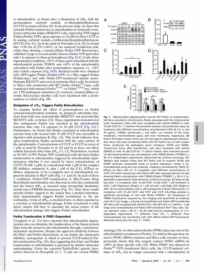
to mitochondria, as shown after a dissipation of ΔΨm with theprotonophore carbonyl cyanide m-chlorophenylhydrazone(CCCP) in clonal cell lines (9). In the present study, we show thatcytosolic Parkin translocates to mitochondria in transiently trans-fected embryonic kidney HEK293T cells, expressing YFP-taggedParkin (Parkin-YFP), upon exposure to 10 μM of either CCCP orits analog, carbonyl cyanide p-trifluoromethoxyphenylhydrazone(FCCP) (Fig. S1). As in the study by Narendra et al. (9), we foundthat >150 out of 250 (>60%) of our analyzed transfected cells,rather than showing a normal diffuse Parkin-YFP fluorescence,exhibited 1 large or several smaller discrete Parkin-YFP spots afteronly 1-h exposure to these protonophores (Fig. S1A). Under theseexperimental conditions,>85% of these spots colocalized with themitochondrial protein TOM20, and >65% of the mitochondriacolocalized with Parkin after protonophore exposure, vs. ∼10%after vehicle exposure (Fig. S1B). Identical results were obtainedwith GFP-tagged Parkin (Parkin-GFP) or c-Myc–tagged Parkin(Parkin-myc) and with Parkin-YFP–transfected human neuro-blastoma SH-SY5Y and cervical carcinomaHeLa cells. In contrastto HeLa cells transfected with WT Parkin (ParkinWT)-myc, cellstransfected with mutated ParkinT415N- or ParkinG430D-myc, whichare 2 PD pathogenic mutations (2), retained a normal diffuse cy-tosolic fluorescence whether cells were incubated with a proto-nophore or vehicle (Fig. 1B).
Dissipation of ΔΨm Triggers Parkin RelocalizationTo examine further the effect of protonophores on Parkincytosolic/mitochondrial partition, we prepared subcellular frac-tions from both non–neuronal-like HEK293T and neuronal-likeSH-SY5Y cells, as before (10). These experiments demonstratedthat endogenous Parkin was enriched in the mitochondrialfractions after only 1-h exposure to 10 μM CCCP (Fig. S1C).Furthermore, we found that Parkin contained in mitochondrialextracts from cells treated with 10 μM CCCP was accessible todigestion with proteinase-K (Fig. 1A). This result indicates that,upon translocation, Parkin associates with the outer surface ofthe mitochondria. Concentrations of CCCP or FCCP in excess of1 μM, as used by Narendra et al. (9) and by us here, can affectcellular functions other than ΔΨm (11, 12). Nonetheless, as muchas 30–50% of WT Parkin-YFP–transfected cells did show Parkintranslocation to mitochondria triggered by mitochondrial depo-larization, whether it was caused by lower concentrations ofCCCP (10 nM–1 μM), by coincubation with 1 μM of the complexIII inhibitor, antimycin A, plus 1 μM of the F1F0 ATPase in-hibitor, oligomycin, or by a complete loss of mitochondrial res-piratory function in Rho0 cells (Fig. 1 C and D). In each of these3 conditions, Parkin-YFP translocation to MitoTracker DeepRed-labeled mitochondria was observed in cells that consistentlyhad the lowest ΔΨm as assessed using tetramethyl rhodaminemethyl ester (TMRM) fluorescence (Fig. 1E). Thus, these resultsprovide further support to the notion that a marked mitochon-drial depolarization triggers Parkin recruitment to the mi-tochondria. A loss of ΔΨm, as modeled here, is often regarded asa correlate to mitochondrial damage. If that correlation is valid,future studies will have to elucidate the actual nature of themitochondrial damage that triggers Parkin relocalization.
Parkin Translocation Is PINK1-DependentCereghetti et al. (13) have reported that mitochondrial depola-rization also can stimulate the translocation of the fission proteinDrp1 from the cytosol to the mitochondria through a calcineurin-dependent mechanism. Despite the apparent similarity betweenthe Drp1 and Parkin observations, in our hands, the calcineurininhibitor, cyclosporine A, failed to prevent CCCP-induced Par-kin translocation (Fig. 1D), thus suggesting that Drp1 and Parkintranslocation to mitochondria is governed by distinct molecularunderpinnings. Given the reported Parkin/Pink1 genetic inter-action observed in Drosophila (4, 5, 7) and our revised PINK1
topology (10), we then asked whether PINK1 plays any role in themitochondrial recruitment of Parkin. To address this question, weused a PINK1 siRNA construct and HeLa cells, because we havepreviously shown that this reagent reduces PINK1 mRNA by>80% in these specific cells (10). When PINK1 was silenced inParkinWT-YFP–transfected HeLa cells, the CCCP-induced col-lapse of ΔΨm was no longer associated with a relocalization of
Fig. 1. Mitochondrial depolarization recruits WT Parkin to mitochondria.(A) Once recruited to mitochondria, Parkin associates with the mitochondrialouter membrane. HeLa cells were incubated with vehicle (DMSO) or with10 μM CCCP for 1 h before mitochondrial isolation and protection assay bytreatment with different concentrations of proteinase K (PK) (0, 0.2, 2, and20 μg/mL). TOM20, cytochrome c, and COX-1 are markers of the outermembrane, intermembrane space, and inner membrane, respectively. (B)Parkin PD-linked mutant forms are not recruited to depolarized mitochon-dria. HeLa cells were transfected either with WT Parkin-myc or with mutantforms containing the pathogenic point mutations T415N and G430D.Twenty-four hours after transfection, cells were incubated with vehicle(DMSO) or with 10 μM CCCP for 1 h before fixation. Bars represent percentof cells showing Parkin-myc colocalization with TOM20 (Parkin+/TOM20+) ±SD of 3 independent experiments, determined by confocal microcopy. (B′)Western blot analysis shows that WT Parkin and its mutants T415D andG430P achieved comparable levels of protein expression. Parkin is im-munodetected using anti-myc antibody. Loading is normalized by TIM23 (C)Effects on HeLa cells of 1-h incubation with different concentrations ofCCCP, 24 h after transfection with Parkin-GFP. Bars represent percent of cellsshowing Parkin colocalization with TOM20 (Parkin+/TOM20+) ± SD of 3 in-dependent experiments, determined by confocal microcopy. (D) Same as (B),but after 1-h incubation with 10 μM FCCP, 10 μM CCCP, 1 μM Antimycin A(AA), 1 μM Oligomycin (Oligo), or 1 μM AA and 1 μM Oligo (AA+Oligo) orafter 30-min preincubation with 2 μM cyclosporine A (CsA), followed by 1-hincubation with 10 μM CCCP. Like FCCP and CCCP, AA+Oligo produce asignificant percentage of cells with Parkin+/TOM20+. (E) Comparison ofTMRM fluorescence acquired by live imaging and quantified as arbitraryunits (A.U.) by Image J, among nontransfected and Parkin-GFP–transfectedWT HeLa cells incubated with vehicle (CTL), 100 nM CCCP, or 1 μMAA + 1 μMOligo and nontransfected and Parkin-GFP–transfected Rho0 HeLa cells. Val-ues represent mean ± SD (n = 35–50 cells) and are representative of 3 in-dependent experiments. **, Different from CTL. *, Different fromnontransfected and transfected cells with diffuse Parkin-GFP fluorescence(Newman–Keuls post hoc test; P < 0.001).
Vives-Bauza et al. PNAS | January 5, 2010 | vol. 107 | no. 1 | 379
NEU
ROSC
IENCE
Dow
nloa
ded
by g
uest
on
June
19,
202
0
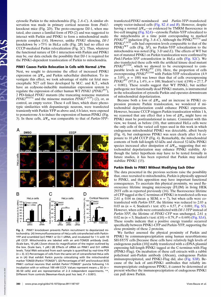
cytosolic Parkin to the mitochondria (Fig. 2 A–C). A similar ob-servation was made in primary cortical neurons from Pink1-knockout mice (Fig. 2D). The proto-oncogene DJ-1, when mu-tated, also causes a familial form of PD (2) and was suggested tointeract with Parkin and PINK1 to form a mitochondrial multi-protein complex (14). However, unlike PINK1 silencing, DJ-1knockdown by >75% in HeLa cells (Fig. 2B) had no effect onCCCP-mediated Parkin relocalization (Fig. 2C). Thus, whateverthe functional nature of DJ-1 interaction with Parkin and PINK1may be, our data exclude the possibility that DJ-1 is required forthe PINK1-dependent translocation of Parkin to mitochondria.
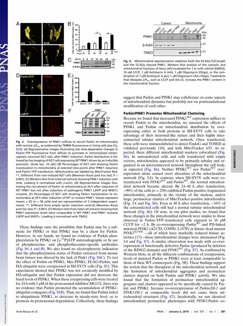
PINK1 Causes Parkin Relocation in Cells with Normal ΔΨmNext, we sought to determine the effect of increased PINK1expression on ΔΨm and Parkin subcellular distribution. To in-vestigate this effect, we took advantage of stable rat fetal mes-encephalic N27 cell lines developed by M.C. and K.T., whichhave an ecdysone-inducible mammalian expression system toregulate the expression of either human WT PINK1 (PINK1WT),2 PD-linked PINK1 mutants (the truncating nonsense mutationPINK1W437× and the missense mutation PINK1L347P) (2), or, ascontrol, an empty vector. These 4 cell lines, which share pheno-typic similarities with dopaminergic neurons, were transfectedtransiently with Parkin-YFP as above and, 6 h later, were exposedto ponasterone A to induce the expression of human PINK1 (Fig.3). In these cells, ΔΨm was comparable to that of Parkin-YFP–
transfected/PINK1-noninduced and Parkin-YFP–transfected/empty vector-induced cells (Fig. S2 A and B). However, despitehaving a normal ΔΨm—as evidenced by TMRM fluorescence inlive-cell imaging (Fig. S2A)—cytosolic Parkin-YFP relocalized tothe mitochondria at a time point corresponding to markedPINK1WT induction (Fig. 3 A–C). Although the PINK1W437× andPINK1L347P cells had expression levels comparable to that of thePINK1WT cells (Fig. 3F), no Parkin-YFP relocalization to themitochondria was noted (Fig. 3 D and E). The effects of WT butnot of mutated PINK1 on Parkin translocation were confirmed byPink1/Parkin-YFP cotransfection in HeLa cells (Fig. S2C). Wealso transfected these cells with the artificial kinase dead mutantPINK1K219M, which we showed to be overexpressed to com-parable levels as PINK1WT (10). Here, the proportion of cellsoverexpressing PINK1K219M with Parkin-YFP relocalization (4.9± 3.0%, n = 100) was lower than that of cells overexpressingPINK1WT (97.0 ± 1.4%, n = 100; Student’s t test: t(198) = 27.7, P< 0.001). These results suggest that WT PINK1, but neitherpathogenic nor functionally dead PINK1 mutants, is instrumentalin the relocalization of cytosolic Parkin and operates downstreamof mitochondrial depolarization.Because both a loss of ΔΨm and an increase in PINK1 ex-
pression promote Parkin translocation, we wondered if mi-tochondrial depolarization could enhance PINK1 expression.However, because CCCP triggers Parkin translocation within 1 h,we reasoned that any effect that a loss of ΔΨm might have onPINK1 must be posttranslational in nature. Consistent with thisview, we found, as before (10), that untreated HeLa cells werethe only cells of the varied cell types used in this work in whichendogenous mitochondrial PINK1 was detectable, albeit barely(Fig. 4), but endogenous PINK1 was seen clearly after 1-h ex-posure to 10 μM CCCP (Fig. 4). Remarkably, the mitochondrialcontents of both full-length 63-kDa and cleaved 52-kDa PINK1species increased after dissipation of ΔΨm, suggesting that mi-tochondrial depolarization may enhance PINK1 stability. Al-though the latter hypothesis may have to be tested formally infuture studies, it has been reported that Parkin may indeedstabilize PINK1 (15).
Parkin Binds to PINK1 Without Modifying Each OtherThe data presented in the previous sections raise the possibilitythat, once recruited to mitochondria, Parkin is physically apposedto PINK1, and this apposition may have important functionalconsequences. To ascertain this physical proximity, we used flu-orescence lifetime imaging microscopy (FLIM) in living HEK293T cells as reported previously (16). The fluorescence lifetimeof CFP tagged at the C-terminus of PINK1 in transfected cells was2.61 ± 0.04 ns (mean ± SEM; n = 7), but when cells were co-transfected with Parkin-YFP, the lifetime was reduced to 2.03 ±0.03 ns (n = 4; Student's t test: t(9) = 8.57, P < 0.001; Fig. S3).However, when cells were cotransfected withDJ-1-YFP instead ofParkin-YFP, the lifetime of PINK1-CFP was unchanged, 2.61 ±0.02 ns (n = 5; Student's t test: t(10) = 0.79, P = 0.449) (Fig. S3A).These results indicate that a positive energy transfer occurredspecifically between PINK1-CFP and Parkin-YFP, supporting theclose proximity of these 2 proteins.We further assessed the physical proximity of Parkin and
PINK1 by coimmunoprecipitation using human neuroblastomaSH-SY5Y cells [because these cells have relatively high levels ofendogenous parkin (10)] stably transfected with a cDNA plasmidexpressing full-length PINK1 tagged at the C-terminus with Flag(PINK1-Flag). On incubation of these cell extracts with a rabbitpolyclonal anti-Parkin antibody (Abcam), endogenous Parkinimmunoprecipitated, and PINK1-Flag did, also (Fig. S3B). Be-cause of the lack of anti-PINK1 antibodies that reliably im-munoprecipitate endogenous PINK1, it cannot be determined atpresent whether the immunoprecipitation of endogenous PINK1can pull down Parkin.
Fig. 2. PINK1 knockdown prevents Parkin recruitment to depolarized mi-tochondria. (A) Immunofluorescence of HeLa cells cotransfected with Parkin-YFP and scrambled (scr) PINK1 or DJ-1 siRNA, and incubated for 1 h with 10μM CCCP. Mitochondria are labeled with an anti-TOM20 antibody (red).(Scale bars, 10 μM.) Zoom shows 6× magnification of the region outlined bythe box. (Scale bars, 1 μM.) (B) Effects of siRNA on PINK1 and DJ1 mRNAlevels. Total RNA extracted from each sample is quantified by real-time PCR(n = 3). (C) Percentages of cells from the same set of cotransfected HeLa cellsas in (A) that exhibit Parkin puncta colocalizing with the mitochondrialmarker TOM20 (Parkin+/TOM20+). (D) Percentages of WT and knockout (KO)PINK1 cortical neurons that exhibit Parkin+/TOM20+ puncta following 1-hincubation with or without 100 nM CCCP. Values represent means ± SD (n =30–50 cells) and are representative of 2–3 independent experiments. **,Different from controls (Newman-Keuls post hoc test; P < 0.001).
380 | www.pnas.org/cgi/doi/10.1073/pnas.0911187107 Vives-Bauza et al.
Dow
nloa
ded
by g
uest
on
June
19,
202
0
These findings raise the possibility that Parkin may be a sub-strate for PINK1 or that PINK1 may be a client for Parkin.However, in our hands, we found no evidence of Parkin phos-phorylation by PINK1 on [γ-32P]ATP autoradiography or by useof phosphoserine- and phosphothreonine-specific antibodies(Fig. S4 A and B). We also found no electrophoretic indicationthat the phosphorylation status of Parkin extracted from mousebrain tissues was altered by the lack of Pink1 (Fig. S4C). To testthe effect of Parkin on PINK1, Myc-PINK1, FLAG-Parkin, andHA-ubiquitin were coexpressed in SH-SY5Y cells (Fig. S5). Thisexperiment showed that PINK1 was not covalently modified byHA-ubiquitin and that Parkin expression did not decrease thebasal levels of PINK1.When these coexpressing cells were treatedfor 24 h with 5 μMof the proteasomal inhibitor MG132, there wasno evidence that Parkin promoted the accumulation of PINK1-ubiquitin conjugates (Fig. S5B). Thus, we found that Parkin failedto ubiquitinate PINK1, to decrease its steady-state level, or topromote its proteasomal degradation. Collectively, these findings
suggest that Parkin and PINK1 may collaborate on some aspectsof mitochondrial dynamics but probably not via posttranslationalmodification of each other.
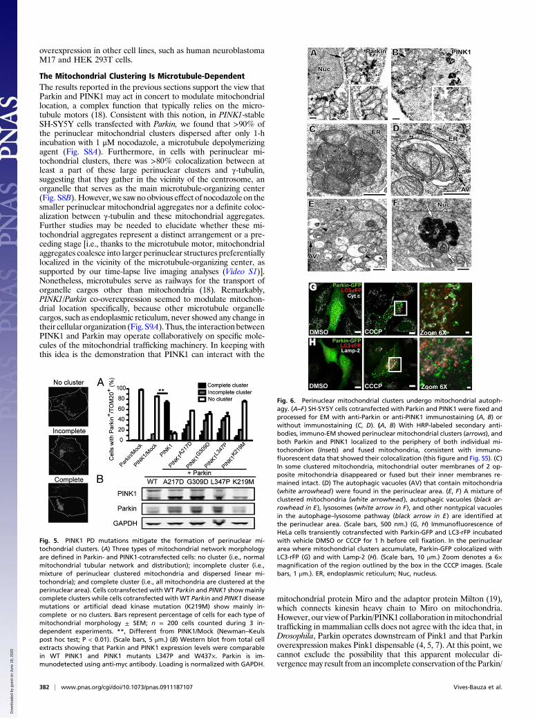
Parkin/PINK1 Promotes Mitochondrial ClusteringBecause we found that increased PINK1WT expression suffices torecruit Parkin to the mitochondria, we assessed the effects ofPINK1 and Parkin on mitochondrial distribution by over-expressing either or both proteins in SH-SY5Y cells to takeadvantage of their neuronal-like nature and their highly inter-connected tubular mitochondrial network. Once transfected,these cells were immunolabeled to detect EndoG and TOM20 asvalidated previously (10), and with MitoTracker 633. As ex-pected, these different mitochondrial markers colocalized (Fig.S6). In untransfected cells and cells transfected with emptyvectors, mitochondria appeared to be primarily tubular and or-ganized in an interconnected network throughout the cell body,as expected (Fig. S6). Neither PINK1WT nor ParkinWT over-expression alone caused overt alteration of the mitochondrialnetwork (Fig. 5A). In contrast, when SH-SY5Y cells were co-transfected with PINK1WT and ParkinWT, the normal mitochon-drial network became altered. By 24–48 h after transfection,∼90% of the cells (n = 250) exhibited Parkin-positive fragmentedmitochondria, primarily in the vicinity of the nucleus, and/orlarge, perinuclear clusters of MitoTracker-positive mitochondria(Fig. 5A and Fig. S6). Even at 48 h after transfection, ∼10% ofthe cotransfected cells still had a normal tubular mitochondrialnetwork (Fig. S6). Of note, in our pilot studies, we found thatthese changes in the mitochondrial network were similar to thoseobserved in Parkin-YFP–transfected cells exposed to 10 μMCCCP for ∼2 h. By co-overexpressing ParkinWT and PD-linkedmutated PINK1 (A217D, G309D, L347P) or kinase dead mutantPINK1K219M —all of which have markedly reduced kinase ac-tivities (17)—these mitochondrial changes were attenuated (Fig.5A and Fig. S7). A similar observation was made with co-over-expression of functionally defective Parkin (produced by deletionof the RING2 domain) and PINK1WT (Fig. S7). As confirmed byWestern blots, in all the different combinations of coexpression,levels of mutated Parkin or PINK1 were at least comparable tothose of their WT counterparts (Fig. 5B). These findings supportthe notion that the disruption of the mitochondrial network andthe formation of mitochondrial aggregates and perinuclearclusters depend on both Parkin and PINK1 activity. We alsofound that the formation of perinuclear mitochondrial ag-gregates and clusters appeared to be specifically caused by Par-kin and PINK1, because co-overexpression of Parkin/DJ-1 andPINK1/DJ-1 at comparable levels did not cause these mi-tochondrial structures (Fig. S7). Incidentally, we saw identicalmitochondrial perinuclear phenotypes with PINK1/Parkin co-
Fig. 3. Overexpression of PINK1 suffices to recruit Parkin to mitochondriawith normal ΔΨm, as evidenced by TMRM fluorescence in living cells (see Fig.S2A). (A) Representative images illustrating the time-dependent changes inParkin-YFP fluorescence from diffuse to punctate in immortalized mesen-cephalic neuronal N27 cells, after PINK1 induction. Parkin distribution is fol-lowed by live imaging of N27 cells expressingWT PINK1 driven by an induciblepromoter. (Scale bar, 10 μM.) (B) Percentages of N27 cells showing Parkintranslocation to mitochondria at selected time points after PINK1 inductionand Parkin-YFP transfection. Mitochondria are labeled by MitoTracker Red.**, Different from non-induced N27 cells (Newman–Keuls post hoc test; P <0.001). (C) Western blot from total cell extracts showing PINK1 induction overtime. Loading is normalized with β-actin. (D) Representative images illus-trating the recruitment of Parkin to mitochondria at 24 h after induction ofWT PINK1 but not after induction of pathogenic PINK1 L347P and W437×mutants. (E) Percentages of N27 cells showing Parkin translocation to mi-tochondria at 24 h after induction of WT or mutant PINK1. Values representmeans ± SD (n = 50 cells) and are representative of 3 independent experi-ments. **, Different from empty vector induction controls (Newman–Keulspost hoc test; P < 0.001). (F) Western blot from total cell extracts showing thatPINK1 expression levels were comparable in WT PINK1 and PINK1 mutantsL347P and W437×. Loading is normalized with TIM23.
Fig. 4. Mitochondrial depolarization stabilizes both the 63-kDa full-lengthand the 52-kDa cleaved PINK1. Western blot analysis of the cytosolic andmitochondrial fractions of HeLa cells incubated for 1 hr with vehicle (DMSO),10 μM CCCP, 1 μM Antimycin A (AA), 1 μM Oligomycin (Oligo), or the com-bination of 1 μM Antimycin A plus 1 μM Oligomycin (AA+Oligo). Treatmentsthat dissipate ΔΨm, such as CCCP and AA+O, increase the PINK1 content inthe mitochondrial fraction.
Vives-Bauza et al. PNAS | January 5, 2010 | vol. 107 | no. 1 | 381
NEU
ROSC
IENCE
Dow
nloa
ded
by g
uest
on
June
19,
202
0
overexpression in other cell lines, such as human neuroblastomaM17 and HEK 293T cells.
The Mitochondrial Clustering Is Microtubule-DependentThe results reported in the previous sections support the view thatParkin and PINK1 may act in concert to modulate mitochondriallocation, a complex function that typically relies on the micro-tubule motors (18). Consistent with this notion, in PINK1-stableSH-SY5Y cells transfected with Parkin, we found that >90% ofthe perinuclear mitochondrial clusters dispersed after only 1-hincubation with 1 μM nocodazole, a microtubule depolymerizingagent (Fig. S8A). Furthermore, in cells with perinuclear mi-tochondrial clusters, there was >80% colocalization between atleast a part of these large perinuclear clusters and γ-tubulin,suggesting that they gather in the vicinity of the centrosome, anorganelle that serves as the main microtubule-organizing center(Fig. S8B). However, we sawnoobvious effect of nocodazoleon thesmaller perinuclear mitochondrial aggregates nor a definite coloc-alization between γ-tubulin and these mitochondrial aggregates.Further studies may be needed to elucidate whether these mi-tochondrial aggregates represent a distinct arrangement or a pre-ceding stage [i.e., thanks to the microtubule motor, mitochondrialaggregates coalesce into larger perinuclear structures preferentiallylocalized in the vicinity of the microtubule-organizing center, assupported by our time-lapse live imaging analyses (Video S1)].Nonetheless, microtubules serve as railways for the transport oforganelle cargos other than mitochondria (18). Remarkably,PINK1/Parkin co-overexpression seemed to modulate mitochon-drial location specifically, because other microtubule organellecargos, such as endoplasmic reticulum, never showed any change intheir cellular organization (Fig. S9A).Thus, the interaction betweenPINK1 and Parkin may operate collaboratively on specific mole-cules of the mitochondrial trafficking machinery. In keeping withthis idea is the demonstration that PINK1 can interact with the
mitochondrial protein Miro and the adaptor protein Milton (19),which connects kinesin heavy chain to Miro on mitochondria.However, our viewof Parkin/PINK1 collaboration inmitochondrialtrafficking in mammalian cells does not agree with the idea that, inDrosophila, Parkin operates downstream of Pink1 and that Parkinoverexpression makes Pink1 dispensable (4, 5, 7). At this point, wecannot exclude the possibility that this apparent molecular di-vergencemay result froman incomplete conservation of the Parkin/
Fig. 5. PINK1 PD mutations mitigate the formation of perinuclear mi-tochondrial clusters. (A) Three types of mitochondrial network morphologyare defined in Parkin- and PINK1-cotransfected cells: no cluster (i.e., normalmitochondrial tubular network and distribution); incomplete cluster (i.e.,mixture of perinuclear clustered mitochondria and dispersed linear mi-tochondria); and complete cluster (i.e., all mitochondria are clustered at theperinuclear area). Cells cotransfected with WT Parkin and PINK1 showmainlycomplete clusters while cells cotransfected with WT Parkin and PINK1 diseasemutations or artificial dead kinase mutation (K219M) show mainly in-complete or no clusters. Bars represent percentage of cells for each type ofmitochondrial morphology ± SEM; n = 200 cells counted during 3 in-dependent experiments. **, Different from PINK1/Mock (Newman–Keulspost hoc test; P < 0.01). (Scale bars, 5 μm.) (B) Western blot from total cellextracts showing that Parkin and PINK1 expression levels were comparablein WT PINK1 and PINK1 mutants L347P and W437×. Parkin is im-munodetected using anti-myc antibody. Loading is normalized with GAPDH.
Fig. 6. Perinuclear mitochondrial clusters undergo mitochondrial autoph-agy. (A–F) SH-SY5Y cells cotransfected with Parkin and PINK1 were fixed andprocessed for EM with anti-Parkin or anti-PINK1 immunostaining (A, B) orwithout immunostaining (C, D). (A, B) With HRP-labeled secondary anti-bodies, immuno-EM showed perinuclear mitochondrial clusters (arrows), andboth Parkin and PINK1 localized to the periphery of both individual mi-tochondrion (Insets) and fused mitochondria, consistent with immuno-fluorescent data that showed their colocalization (this figure and Fig. S5). (C)In some clustered mitochondria, mitochondrial outer membranes of 2 op-posite mitochondria disappeared or fused but their inner membranes re-mained intact. (D) The autophagic vacuoles (AV) that contain mitochondria(white arrowhead) were found in the perinuclear area. (E, F) A mixture ofclustered mitochondria (white arrowhead), autophagic vacuoles (black ar-rowhead in E), lysosomes (white arrow in F), and other nontypical vacuolesin the autophage–lysosome pathway (black arrow in E) are identified atthe perinuclear area. (Scale bars, 500 nm.) (G, H) Immunofluorescence ofHeLa cells transiently cotransfected with Parkin-GFP and LC3-rFP incubatedwith vehicle DMSO or CCCP for 1 h before cell fixation. In the perinucleararea where mitochondrial clusters accumulate, Parkin-GFP colocalized withLC3-rFP (G) and with Lamp-2 (H). (Scale bars, 10 μm.) Zoom denotes a 6×magnification of the region outlined by the box in the CCCP images. (Scalebars, 1 μm.). ER, endoplasmic reticulum; Nuc, nucleus.
382 | www.pnas.org/cgi/doi/10.1073/pnas.0911187107 Vives-Bauza et al.
Dow
nloa
ded
by g
uest
on
June
19,
202
0
PINK1 pathway between invertebrate and vertebrate organisms. Italso shouldbe taken into account that, here,we investigated the roleof Parkin/PINK1 interaction on mitochondrial distribution anddisposition, whereas in all the Drosophila studies the authors as-certained mitochondrial morphology and fission/fusion, very dif-ferent aspects of mitochondrial dynamics that are not necessarilygoverned by an identical molecular machinery.
Parkin/PINK1 May Regulate Mitochondrial TraffickingTo examine the ultrastructure of perinuclear clustered mitochon-dria induced by Parkin/PINK1, we performed EM and observed arange of different types of mitochondrial perinuclear clusters thatwerenot present in empty vector-transfected cells. In all cases, boththe length and the width of perinuclear-clustered mitochondria inParkin/PINK1-cotransfected cells were smaller than in mock-transfected control cells (Fig. S9B), suggesting that Parkin/PINK1coexpression distorts the mitochondrial network, perhaps by pro-motingmitochondrial fragmentation. Furthermore, in some cases,clusters were made of nearly normal-appearing mitochondria, andboth PINK1andParkin localized to the outside boundaries of eachindividual mitochondrion (Fig. 6A andB). In other cases, multiplemitochondria were fused together (Fig. 6 A and C). Among clus-tered mitochondria, the gap between 2 mitochondria was ∼6 nm,similar to the gap of the mitochondria clusters induced by mi-tochondrial phospholipase-D(20).However, unlikemitochondrialphospholipase-D, Parkin/PINK1 overexpression was associatedwith mitochondrial outer-membrane fusion (Fig. 6A). We alsoidentified perinuclear lysosomal vacuoles as well as autophago-somes, and someof these containedmitochondria (Fig. 6D andF),suggesting a mitochondrial autophagic event. The autophagic na-ture of these vacuoles was confirmed by fluorescence for the au-tophagosomemarkerLC3-rFPandby immunofluorescence for thelysosome marker Lamp2 (Fig. 6 G and H). Notably, in untreatedParkin-GFP/LC3-rFP cotransfected cells, the LC3-rFP signal wasdetected throughout the cytoplasm (Fig. 6G). In contrast, inCCCP-treated cells, the LC3-rFP signal was localized mostly in theperinuclear region, where it colocalized with Parkin and the mi-tochondrial marker cytochrome c (Fig. 6G). Consistent with thepreferential subcellular localization of lysosomes, Lamp2 im-munofluorescence was detected primarily in the perinuclear area,which is the only subcellular region where we observed definitecolocalization between Lamp2 and Parkin (Fig. 6H). Together
with our results for the microtubule experiments, these datasuggest that autophagosomes containing Parkin/PINK1-en-riched mitochondria may form at some distance from the ly-sosomes and then are delivered by the microtubule motor tothe perinuclear lysosomes for degradation. This scenario isreminiscent of that proposed for the clearance of aggresomes(21), in which proteinaceous inclusion bodies are thought tobe targeted to the perinuclear area to be disposed of by au-tophagy. Although our study is in agreement with that ofNarendra et al. (9), in that we also found that cytosplasmicParkin can translocate to the mitochondria, we argue that theensuing autophagy of mitochondria requires the trafficking ofdamaged mitochondria to the perinuclear area to be degraded.We thus propose that both PINK1 and Parkin are key ele-ments of the trafficking machinery responsible for deliveringdefective mitochondria to the lysosome-rich perinuclear area,rather than being part of the actual autophagy systems. Theinterplay between PINK1 and Parkin in mitochondrial func-tioning also may modulate the trafficking of mitochondria indendrites, perhaps accounting for the synaptic dysfunction thatis observed in PINK1- or Parkin-knockout mice (22, 23).
Materials and MethodsAllmethods employed in this article are routinely used in our laboratories andare thus referenced (10, 16, 24, 25) and are described in SI Materials andMethods. For immunoblotting, the primary antibodies used were PINK1 (100-494; Novus), Parkin, GAPDH, Hsp60, and HA (Santa Cruz Biotechnology),TIM23, cytochrome c, and COX-I (Invitrogen). For immunostaining, primaryantibodies were PINK1 (Novus), myc (9E10; Abcam), EndoG (ProSci), TOM20(BD Biosciences), α- and γ-tubulin (Sigma-Aldrich), calreticulin (AbCam), andtyrosine hydroxylase (Chemicon-Millipore).
ACKNOWLEDGMENTS. We thank Drs. Liza Pon, Eric Schon, William Dauer,Anna-Maria Cuervo, and Richard Vallee for their insightful comments on themanuscript and Jie Shen for providing the Pink1 knockout mice. The authorsare supported by National Institutes of Health Grants AG021617, ES014899,ES017470, NS042269, NS054773, NS062180, NS064191, NS38370, NS38377,and NS48206; US Department of Defense Grants W81XWH-08-1-0522,W81XWH-08-1-0465, and DAMD 17-03-1; the Parkinson Disease Foundation;the Thomas Hartman Foundation for Parkinson's Research; and the MuscularDystrophy Association's Wings-over-Wall Street. T.M.D. is the Leonard andMadlyn Abramson Professor of Neurodegenerative Diseases, and S.P. is thePage and William Black Professor of Neurology.
1. Dauer W, Przedborski S (2003) Parkinson's disease: Mechanisms and models. Neuron
39:889–909.2. Moore DJ, West AB, Dawson VL, Dawson TM (2005) Molecular pathophysiology of
Parkinson's disease. Annu Rev Neurosci 28:57–87.3. Valente EM, et al. (2004) Hereditary early-onset Parkinson's disease caused by
mutations in PINK1. Science 304:1158–1160.4. Clark IE, et al. (2006) Drosophila pink1 is required for mitochondrial function and
interacts genetically with Parkin. Nature 441:1162–1166.5. Park J, et al. (2006) Mitochondrial dysfunction in Drosophila PINK1 mutants is
complemented by parkin. Nature 441:1157–1161.6. Kitada T, et al. (1998) Mutations in the Parkin gene cause autosomal recessive juvenile
parkinsonism. Nature 392:605–608.7. Poole AC, et al. (2008) The PINK1/Parkin pathway regulates mitochondrial morphology.
Proc Natl Acad Sci USA 105:1638–1643.8. Darios F, et al. (2003) Parkin prevents mitochondrial swelling and cytochrome c
release in mitochondria-dependent cell death. Hum Mol Genet 12:517–526.9. Narendra D, Tanaka A, Suen DF, Youle RJ (2008) Parkin is recruited selectively to
impaired mitochondria and promotes their autophagy. J Cell Biol 183:795–803.10. Zhou C, et al. (2008) The kinase domain of mitochondrial PINK1 faces the cytoplasm.
Proc Natl Acad Sci USA 105:12022–12027.11. Zhang GJ, Liu HW, Yang L, Zhong YG, Zheng YZ (2000) Influence of membrane
physical state on the lysosomal proton permeability. J Membr Biol 175:53–62.12. Maro B, Marty MC, Bornens M (1982) In vivo and in vitro effects of the mitochondrial
uncoupler FCCP on microtubules. EMBO J 1:1347–1352.13. Cereghetti GM, et al. (2008) Dephosphorylation by calcineurin regulates translocation
of Drp1 to mitochondria. Proc Natl Acad Sci USA 105:15803–15808.
14. Xiong H, et al. (2009) Parkin, PINK1, and DJ-1 form a ubiquitin E3 ligase complexpromoting unfolded protein degradation. J Clin Invest 119:650–660.
15. Shiba K, et al. (2009) Parkin stabilizes PINK1 through direct interaction. BiochemBiophys Res Commun 383:331–335.
16. Grailhe R, et al. (2006) Monitoring protein interactions in the living cell through thefluorescence decays of the cyan fluorescent protein. ChemPhysChem 7:1442–1454.
17. Beilina A, et al. (2005) Mutations in PTEN-induced putative kinase 1 associated withrecessive parkinsonism have differential effects on protein stability. Proc Natl Acad SciUSA 102:5703–5708.
18. Hirokawa N (1998) Kinesin and dynein superfamily proteins and the mechanism oforganelle transport. Science 279:519–526.
19. Weihofen A, Thomas KJ, Ostaszewski BL, Cookson MR, Selkoe DJ (2009) Pink1 forms amultiprotein complex with Miro and Milton, linking Pink1 function to mitochondrialtrafficking. Biochemistry 48:2045–2052.
20. Choi SY, et al. (2006) A common lipid links Mfn-mediated mitochondrial fusion andSNARE-regulated exocytosis. Nat Cell Biol 8:1255–1262.
21. Garcia-Mata R, Gao YS, Sztul E (2002) Hassles with taking out the garbage:Aggravating aggresomes. Traffic 3:388–396.
22. Goldberg MS, et al. (2003) Parkin-deficient mice exhibit nigrostriatal deficits but notloss of dopaminergic neurons. J Biol Chem 278:43628–43635.
23. Kitada T, et al. (2007) Impaired dopamine release and synaptic plasticity in thestriatum of PINK1-deficient mice. Proc Natl Acad Sci USA 104:11441–11446.
24. Vives-Bauza C, et al. (2008) The age lipid A2E and mitochondrial dysfunctionsynergistically impair phagocytosis by retinal pigment epithelial cells. J Biol Chem 283:24770–24780.
25. Nagai M, et al. (2007) Astrocytes expressing ALS-linked mutated SOD1 release factorsselectively toxic to motor neurons. Nat Neurosci 10:615–622.
Vives-Bauza et al. PNAS | January 5, 2010 | vol. 107 | no. 1 | 383
NEU
ROSC
IENCE
Dow
nloa
ded
by g
uest
on
June
19,
202
0