Embed Size (px)
Citation preview
1
PilMNOPQ from the Pseudomonas aeruginosa Type IV Pilus System form a Transenvelope 1
Protein Interaction Network that Interacts with PilA* 2
3
Stephanie Tammam1,2, Liliana M. Sampaleanu1, Jason Koo1,2, Kumararaaj Manoharan1, Mark Daubaras1, 4
Lori L. Burrows3, P. Lynne Howell1,2 5
6 1From the Program in Molecular Structure and Function, Research Institute, The Hospital for Sick 7
Children, Toronto, ON, CANADA 8
9 2From the Department of Biochemistry, University of Toronto, ON, CANADA 10
11 3From the Department of Biochemistry and Biomedical Sciences, McMaster University, Hamilton, ON, 12
CANADA 13
14
15
16
17
18
19
To whom correspondence should be addressed: 20
P. L. Howell, Phone: 416-813-5378, Fax: 416-813-5379, E-mail: [email protected] 21
L. L. Burrows, Phone: 905-525-9140, ext. 22029�Fax: 905-522-9033, E-mail: [email protected] 22
23
24
*Running title: PilMNOPQ form a transenvelope complex 25
26
Keywords: Type IV pili, P. aeruginosa, protein-protein interactions 27
28 29
Copyright © 2013, American Society for Microbiology. All Rights Reserved.J. Bacteriol. doi:10.1128/JB.00032-13 JB Accepts, published online ahead of print on 1 March 2013
on April 9, 2018 by guest
http://jb.asm.org/
Dow
nloaded from
2
Abstract 30
Pseudomonas aeruginosa Type IV pili (T4P) are virulence factors that promote infection of cystic fibrosis 31
and immunosuppressed patients. As the absence of T4P impairs colonization, they are attractive targets 32
for the development of novel therapeutics. Genes in the pilMNOPQ operon are important for both T4P 33
assembly and a form of bacterial movement called twitching motility, required for pathogenicity. The 34
type II membrane proteins, PilN and PilO, dimerize via their periplasmic domains and anchor this 35
complex in the inner membrane. Our earlier work showed that PilNO binds PilP, a periplasmic 36
lipoprotein. Here we show that PilP interacts with the N0 segment of the outer membrane secretin PilQ 37
via its C-terminal domain, and that the N-terminal cytoplasmic tail of PilN binds to the actin-like protein 38
PilM, thus connecting all cellular compartments via the PilMNOPQ protein interaction network. We show 39
that PilA, the major pilin subunit, interacts with PilNOPQ. The results allow us to propose a model 40
whereby PilA makes extensive contacts with the transenvelope complex, possibly to increase local 41
concentrations of PilA monomers for polymerization. The PilNOP complex could provide a stable anchor 42
in the inner membrane, while the PilMNOPQ transenvelope complex facilitates transit of the pilus 43
through the periplasm and clamps the pilus in the cell envelope. The PilMN interaction is proposed to be 44
responsible for communicating signals from the cytoplasmic to periplasmic components of this complex 45
macromolecular machine. 46
47
Introduction 48
Type IV pili (T4P) are surface appendages involved in many processes including adhesion to biotic and 49
abiotic surfaces, aggregation, DNA uptake and twitching motility [1–3]. T4P are essential virulence 50
factors that have been extensively studied in the model organism P. aeruginosa and other bacteria. Since 51
adhesion and twitching motility play important roles in pathogenicity, understanding structure/function 52
relationships involved in T4P assembly is vital for the development of novel therapeutic strategies to 53
combat infection. 54
55
The intricacies of the T4P machinery are puzzling and a comprehensive understanding of how these thin 56
filaments are rapidly extended and retracted while resisting mechanical forces upwards of 100 pN [4] 57
remains elusive. On a macromolecular scale, the machine has a modular organization, with four sub-58
complexes: the cytoplasmic motor subcomplex (consisting of PilB, PilT, PilU, PilC and potentially PilD), 59
the inner membrane alignment subcomplex (PilM, PilN, PilO, and PilP), the outer membrane secretin 60
pore subcomplex (PilQ and PilF), and the pilus itself (PilA and the minor pilins) [5]. The dynamics of 61
PilA (pilin) polymerization/de-polymerization rely on the action of the motor ATPases PilB (pilus 62
extension), PilT and PilU (pilus retraction), and the integral membrane protein, PilC, the putative platform 63
on April 9, 2018 by guest
http://jb.asm.org/
Dow
nloaded from
3
protein. T4P exit the cell through the secretin, comprised of multiple PilQ monomers [6]. Assembly of the 64
PilQ secretin requires its cognate pilotin (a protein essential for assembly of the secretin in the outer 65
membrane), PilF [7]. Bridging the cytoplasmic and outer membrane components is the alignment 66
complex comprised of PilMNOP [8]. PilM is a cytoplasmic actin-like protein that has been shown in 67
Thermus thermophilus to bind the N terminus of PilN [9], while the cytoplasmic domain of the PilM 68
homolog in the Type 2 secretion system (T2SS) interacts with the single ATPase and the PilC homolog 69
[10]. PilN is a type II membrane protein that heterodimerizes with PilO, a protein with a similar domain 70
organization [11]. The periplasmic domains of PilNO interact with the inner membrane lipoprotein PilP, 71
forming a 1:1:1 complex [12]. The pilus is composed of hundreds of copies of PilA, plus low abundance 72
pilin-like proteins termed minor pilins [13]. 73
74
While there are significant structural and functional similarities between components of T4P and T2SS 75
assembly machineries [14–16], there are a few notable differences. For example, PilM and PilN appear to 76
be the structural and functional equivalents of the cytoplasmic and periplasmic portions, respectively, of 77
the T2SS protein EpsL [17]. However, while PilM has been demonstrated to bind ATP, the cytoplasmic 78
domain of EpsL does not [18]. Also, EpsL interacts with the hexameric ATPase EpsE [19, 20]; an 79
interaction that has not yet been demonstrated for PilM and the equivalent ATPase PilB. There is further 80
structural similarity between the T4P inner membrane lipoprotein PilP and the HR region of the T2SS 81
inner membrane protein, GspC [12, 21, 22]. Here, we highlight a unique structural difference between 82
PilP and GspC that may reflect a role of the transenvelope complex in retraction of long extracellular pili. 83
84
The current study expands our understanding of the role of PilMNOPQ in T4P assembly. We demonstrate 85
that the cytoplasmic extension of P. aeruginosa PilN interacts directly with PilM, consistent with 86
structural and bacterial two-hybrid data from T. thermophilus and Neisseria meningitis, respectively [9, 87
23]. Further, we show that in P. aeruginosa, the N-terminal disordered domain of PilP interacts with the 88
periplasmic regions of PilNO, while the C-terminal β-domain of PilP interacts with the N0 domain of 89
PilQ, further refining the boundaries of the PilP-PilQ interaction identified in N. meningitidis [24]. We 90
show that PilNO have no affinity for PilQ under our experimental conditions, but that PilP interacts with 91
both PilNO and PilQ, allowing all four proteins to be co-purified using 6xHis tagged PilQ as bait. 92
Together, these data suggest that PilP connects the inner and outer membrane subcomplexes. We also 93
demonstrate that PilQ is able to pull-down PilN, PilO, PilP and PilA from PAO1 (a well characterized P. 94
aeruginosa strain) lysates, indicating that PilA (and possibly the pilus itself) forms multiple interactions 95
with the transenvelope complex formed by PilMNOPQ. 96
97
on April 9, 2018 by guest
http://jb.asm.org/
Dow
nloaded from
4
Materials and Methods 98
Bacterial Strains 99
Supplemental Table 1 summarizes the bacterial strains and vectors used in this study. The pET-Duet and 100
pET vectors were transformed by heat shock into chemically competent E. coli. 101
102
Western Blot analysis 103
All protein samples analyzed by Western blotting were mixed with 2xSDS-PAGE sample buffer at a 1:1 104
ratio, boiled for 10 min, separated on 16 % SDS-PAGE minigels and transferred to polyvinylidene 105
difluoride (PVDF) membranes. Proteins of interest were detected using the purified rabbit polyclonal 106
antibodies to PilMNOPQA [8]. The primary antibodies were diluted in TBST (25 mM TrisHCl pH 7.5, 107
150 mM NaCl and 0.1 % v/v Tween20). The secondary anti-rabbit or anti-mouse antibodies conjugated to 108
alkaline phosphatase were used as per the manufacturer’s instructions (BioRad) and the blots developed 109
with nitroblue tetrazolium/5-bromo-4-chloro-3-indoyl-phosphate (NBT/BCIP) from BioShop Canada Inc. 110
Detection of the biotinylated PilN_N20 peptide was performed using the streptavidin-horse radish 111
peroxidase (HRP) conjugated reagent from GenScript in TBST followed by detection of HRP using the 112
Super Signal West Pico chemiluminescent substrate from Pierce (Thermo Scientific) 113
114
Construct generation and peptide synthesis 115
(i) PilN N-terminal peptides: The peptides encompassing the N terminus 20 amino acids of PilN: 116
MARINLLPWREELREQRKQQ (PilN_N20, 95% pure and reconstituted in water at 1 mg/ml) and the 117
Asp5 to Ala variant (PilN_N20_N5A, 86 % purity) were synthesized by GenScript. For both wild type 118
and mutant PilN_N20 peptides, a lysine residue and a mini-PEG linker were added at the C terminus to 119
facilitate the addition of a biotin molecule. 120
(ii) Generation of heterologous protein expression vectors. The generation of the co-expression vector that 121
produces N-terminally 6xHis tagged PilNΔ44 and untagged PilOΔ51 (PilNΔ44/PilOΔ51) and the expression 122
vectors for the 6xHis tagged proteins PilOΔ43 and PilM or the untagged PilPΔ18 (PilPΔ18T7) were reported 123
previously in [8, 11, 12]. Details about these vectors are shown in Supplemental Table 1. The untagged 124
versions N- and C-terminal constructs of PilP (PilP18-84T7 and PilPΔ71_TAAT respectively) were generated 125
using PilPΔ18T7 as a template to amplify the appropriate regions and subsequently ligated into the BamI 126
and XhoI sites of pET24a. The PilQ expression constructs were amplified with specific primers 127
containing a 5’ NcoI site and a 3’ XhoI site. Purified PCR fragments were digested and ligated into an 128
appropriately digested pET28a vector. Clones were verified by sequencing (TCAG or AGTC, Toronto, 129
Ontario). 130
131
on April 9, 2018 by guest
http://jb.asm.org/
Dow
nloaded from

5
Protein expression 132
PilM, PilNΔ44/PilOΔ51, PilPΔ18, PilPΔ71 and the PilQ constructs used in pull-down studies were expressed in 133
E. coli. The pET vectors containing the genes of interest were transformed into BL21 Codon Plus cells 134
(Stratagene) and plated on LB plates supplemented with the 50 mg/ml kanamycin. One colony was then 135
used to inoculate 5 ml LB containing the appropriate antibiotic. Each overnight culture was used to 136
inoculate 500 ml of LB-antibiotic and the cells were grown at 37 °C until OD600 of 0.6-0.7, when protein 137
expression was induced by adding IPTG to a final concentration of 1.0 mM. The cells were allowed to 138
grow either overnight at 18 °C or for 4 h at 37 °C, prior to being harvested by centrifugation (7700 g, 30 139
min, 4 °C). Cell pellets were stored at -20 °C until required. 140
141
Bioinformatics analyses 142
(i) PilQ domain identification. Sequence alignments were performed by T-Coffee [25]. Initially the 143
sequences for the T2SS and T4P secretins were aligned separately. These individual alignments were 144
subsequently combined using the COMBINE algorithm in the T-Coffee suite, and then manipulated 145
manually to refine the alignment in JalView [26]. Secondary structure predictions were performed by 146
JnetPRED [27] in the JalView alignment editor program [26]. Figures for sequence alignments were 147
produced in JalView. Structural analysis and alignments were performed using PyMOL [28]. 148
(ii) PilP and GspC structural comparison. Sequence alignments were performed as described in [12] and 149
structural superpositions were performed and displayed using PyMOL [28]. 150
151
PilM co-purification with PilN_N20 and PilN_N20_N5A peptides 152
N-terminally 6xHis tagged PilM (PilMNt6xHis) was co-purified with PilN_N20 peptides using NiMagBeads 153
(GenScipt). Initially, 0.2 g of dry PilMNt6xHis pellet was solubilized in 4 ml Easy BacLysis buffer 154
(GenScript) and 2 ml buffer A (20 mM Tris HCl, pH 7.5, 150 mM NaCl) with added 100 mg/ml 155
lysozyme, 10 mg/ml DNase and 10 mg/ml RNase. Cells were lysed by incubation for 1 h at 4 °C on a 156
rotator. Subsequently, 2 ml of cell lysate solution was added to each of the three falcon tubes containing 157
100 �l of Ni MagBeads pre-equilibrated in lysis buffer. After mixing for 1.5 h at room temperature on a 158
rotator, the falcon tubes were inserted in the magnetic base and the supernatant was discarded. The 159
MagBeads with bound PilMNt6xHis were then incubated with either 1 ml of buffer A alone (control) or 1 ml 160
buffer A and 100 �l of a 1 mg/ml wild type PilN_N20 or mutant PilN_N20_N5A peptides (freshly made 161
solutions of lyophilized synthetic peptides in water). The control experiments of both PilN peptides in the 162
absence of PilM were set up in a similar manner. After an overnight incubation at 4 °C on a rotator the 163
Ni-NTA beads were washed in a batch-wise manner using the magnetic base and the 1.1 ml flow through 164
together with 3, 3, 0.5, 0.5 and 0.5 ml elute fractions containing 10, 30, 75, 150 or 400 mM imidazole, 165
on April 9, 2018 by guest
http://jb.asm.org/
Dow
nloaded from
6
respectively, were collected and analyzed by SDS-PAGE and Western blotting using both anti-PilM 166
antibody and a streptavidin conjugated to horseradish peroxidase (Strep-HRP) reagent. 167
168
PilN/O and PilP N-terminal pull-downs 169
The expression, lysis, co-purification and analysis were performed as described in Tammam et al. [12] for 170
the PilNΔ44/PilOΔ51/PilPΔ18_6xHis co-fractionation. In brief, the pET28a-pilPΔ18-6His and pET-Duet-pilNΔ44-171
pilOΔ51 expression vectors were transformed separately into E. coli BL21 DE3 cells (Novagen). Overnight 172
cultures were used to inoculate 1L of LB-kanamycin and cells were grown at 37 °C until OD600 of 0.6-0.7, 173
when protein expression was induced by adding IPTG to a final concentration of 1 mM. The cells were 174
grown for 4 hours after induction at 37 °C, and were harvested by centrifugation and stored at -20°C until 175
required. The frozen cells were thawed, resuspended and then mixed together. The cells were lysed by 176
homogenization and the cellular debris was removed by centrifugation. The cell lysate was mixed with 2 177
mL of Ni-agarose (Qiagen) at 4 °C for 2 h and then packed into a column and washed with 2 x 20 mL of 178
Tris buffer containing successively 10 and 30 mM imidazole. Bound protein was eluted from the column 179
in three 10 mL batches with Tris buffer and 75, 150 and 300 mM imidazole, respectively. The 75 mM 180
imidazole fraction was subsequently concentrated to 2 mL (Amicon unit with 10 kDa cut-off, Millipore), 181
to a final protein concentration of 10-15 mg/ml. The concentrated protein sample was applied to a 182
Superdex S-200 column (GE Health) and eluted using 20 mM Tris HCl pH 7.5, and 150 mM NaCl. 183
184
PilQ pull-down experiments 185
All proteins were over expressed as described above. Each protein construct was expressed individually, 186
and appropriate cell pellets were then mixed prior to lysis. PilPΔ18, PilPΔ71, PilNΔ44/PilOΔ51, PilQ24-445, 187
PilQ24-280, PilQ281-445, PilQ281-410, PilQ281-376 were used in 0.5 L aliquots, while 1 L aliquots of PilQ377-445 188
were used. 189
190
Individual cell pellets were initially resuspended in 10 ml of lysis buffer (50 mM Tris pH 7.5, 150 mM 191
NaCl, 5 mM imidazole, 1 mM PMSF, 0.1 mg/ml DNase, RNase, and Lysozyme, protease inhibitor 192
cocktail (Sigma)). After mixing the appropriate resuspended pellets, cells were lysed by homogenization 193
using an Avestin EmulsiFlex-C3, 3 x 15-20 kpsi and insoluble debris removed by centrifugation (39,000 194
g, 30 min, 4 °C). The cell lysate was then loaded on a pre-equilibrated Ni-NTA column, and the flow 195
through collected and loaded a second time on the column. The column was washed with 30 ml of buffer 196
A containing 5 mM imidazole and a second wash of 10 ml buffer A with 30 mM imidazole. Bound 197
protein was eluted from the column in three 10 ml batches with buffer A with 75, 150 and 300 mM 198
on April 9, 2018 by guest
http://jb.asm.org/
Dow
nloaded from

7
imidazole, respectively. Fractions were analyzed by SDS-PAGE gel electrophoresis and Western blot 199
analysis with PilN, PilO, PilP and 6xHis specific antibodies as appropriate. 200
201
A negative control for PilPΔ18_TAAT, PilPΔ71_TAAT, and PilNΔ44/PilOΔ51 were run, where soluble lysate of 202
cells over expressing these constructs was loaded onto a Ni-NTA column in the absence of PilQ. 203
Purification was carried out as described above, and the eluted fractions analyzed by SDS-PAGE gel 204
electrophoresis. 205
206
PilA pull-down assays 207
Cell pellets containing expressed PilQ24-445, PilQ24-280, and PilQ281-445 were resuspended in 10 ml lysis 208
buffer (see above). Cells were lysed using an Avestin EmulsiFlex-C3 (see above) and cellular debris was 209
then removed by centrifugation (39000 g, 30 min, 4 °C). The soluble supernatant mixed with 1 ml Ni-210
NTA agarose (Qiagen), pre-equilibrated in buffer A, for 2 h at 4 °C. The beads were collected by 211
centrifugation, the supernatant fraction being the flow through. The beads were washed once with 10 ml 212
buffer A for 1 h at 4 °C. The beads were again collected by centrifugation, resulting in Ni-NTA beads 213
with pre-bound proteins were subsequently used to pull-down PilA from PAO1 lysates prepared as 214
described below. 215
216
PAO1 cell lysates were prepared by growing cells streaked in a grid pattern on LB agar plates over night 217
at 37 °C. Cells were collected by scraping them off the surface, and cell pellets were lysed in 6 ml Easy 218
BacLysis and 14 ml buffer A with 100 mg/ml lysozyme, 10 mg/ml DNase and 10 mg/ml RNase. Cell 219
pellets were homogenized by brief vortexing and incubation at room temperature for 30 min on a rotator. 220
Cellular debris was then removed by centrifugation (39000 g, 30 min, 4 °C) and 4 ml of the cell lysate 221
supernatant was applied to the Ni-NTA beads charged with specific His-tagged protein, as described 222
above. 223
224
The PAO1 cell lysates and Ni-NTA beads pre-bound with PilQ24-445, PilQ24-280, and PilQ281-445 were 225
incubated at 4 °C overnight. The beads were collected by centrifugation while the supernatant contained 226
the flow through fraction. The resin was washed twice using a batch method (mixed for 1 h on a rotator at 227
4 °C and centrifuged to solution to pellet the beads and then removed the supernatants) with 10 ml buffer 228
A containing successively 10 and 30 mM imidazole, and the bound proteins were eluted from the column 229
in three 2 ml batches, containing buffer A and 75, 150 or 400 mM imidazole, respectively. The identity of 230
each protein was confirmed by Western blot analysis of the flow through and 400 mM fractions using 231
PilN, PilO, PilP, and PilA specific antibodies. 232
on April 9, 2018 by guest
http://jb.asm.org/
Dow
nloaded from
8
RESULTS 233
The conserved N terminus of PilN is important for T4P function and interacts with PilM in vitro 234
We previously identified the PilN N-terminal sequence “I4N5L6L7P8” as a signature sequence for PilN 235
family members and predicted that this motif would be involved in interactions with PilM [11]. To test 236
this hypothesis, we examined the interaction of P. aeruginosa PilM and PilN using N-terminally tagged 237
PilM (PilMNt6xHis) and a biotinylated peptide of PilN that contains the first 20 N-terminal residues of the 238
protein conjugated via a C-terminal lysine and a mini-PEG linker to biotin (PilN_N20). This 2.5kDa 239
peptide co-purified with PilMNt6xHis using either nickel- (Figure 1) or streptavidin-affinity chromatography 240
(Supplemental Figure 1). When HRP-conjugated streptavidin was used to probe the fractions from the 241
nickel-affinity purification, the biotinylated PilN_N20 peptide was recovered in the 10, 30 and 75 mM 242
imidazole washes at ~40 kDa. Western blot analysis with an anti-6xHis antibody confirmed that these 243
bands correspond to PilM (Figure 1A). In the presence of PilM, the PilN_N20 peptide appeared at the 244
approximate molecular weight of PilM, indicating that this peptide forms a high affinity interaction with 245
PilMNt6xHis that is not disrupted by SDS. In the absence of PilM, some peptide bound non-specifically to 246
the column (Figure 1A) and under these conditions the peptide eluted at ~2.5 kDa. However, when PilM 247
was present, the levels of PilN_N20 in the high imidazole fractions were greatly diminished, presumably 248
because most of the peptide is bound to PilM. Compared with the purification profile of PilM in the 249
absence of the PilN peptide (Figure 1C), it is clear that the addition of the peptide does not affect binding 250
of PilM to the column. Together, these data suggest that PilN_N20 binds to PilM in an SDS stable 251
manner. 252
253
The effects of point mutations at the highly conserved Asn5 (N5A/Q/D) of PilN, and a series of N-254
terminal deletions (Δ2-9, Δ10-18, Δ4-17) were tested in vivo by complementation of a P. aeruginosa pilN 255
mutant (pilN::FRT). Constructs expressing the PilN_N5A variant or deletion constructs lacking residues 256
2-9 or 4-18 did not restore twitching motility, phage sensitivity or surface PilA in a pilN mutant 257
(Supplemental Figure 2). Other deletions and site-specific mutations decreased T4P biogenesis and 258
function to varying degrees (Supplemental Figure 2). Mutations that prevent association of PilM and 259
PilN in vivo were previously shown to lead to degradation of PilNO [8], consistent with the phenotypes 260
seen here. When the pull-down experiments were repeated with a peptide containing the N5A mutation 261
(PilN_N20_N5A) PilM was still pulled down (Figure 1B), despite the in vivo data suggesting that this 262
variant was non-functional (Supplemental Figure 2) [23]. Together, these data suggest that Asn5 in PilN 263
may play an additional role in communicating a signal from the cytoplasm through PilN to the 264
periplasmic components or that the binding of PilM to the PilN peptide in vivo is influenced by other 265
proteins or protein interactions that may be present. 266
on April 9, 2018 by guest
http://jb.asm.org/
Dow
nloaded from
9
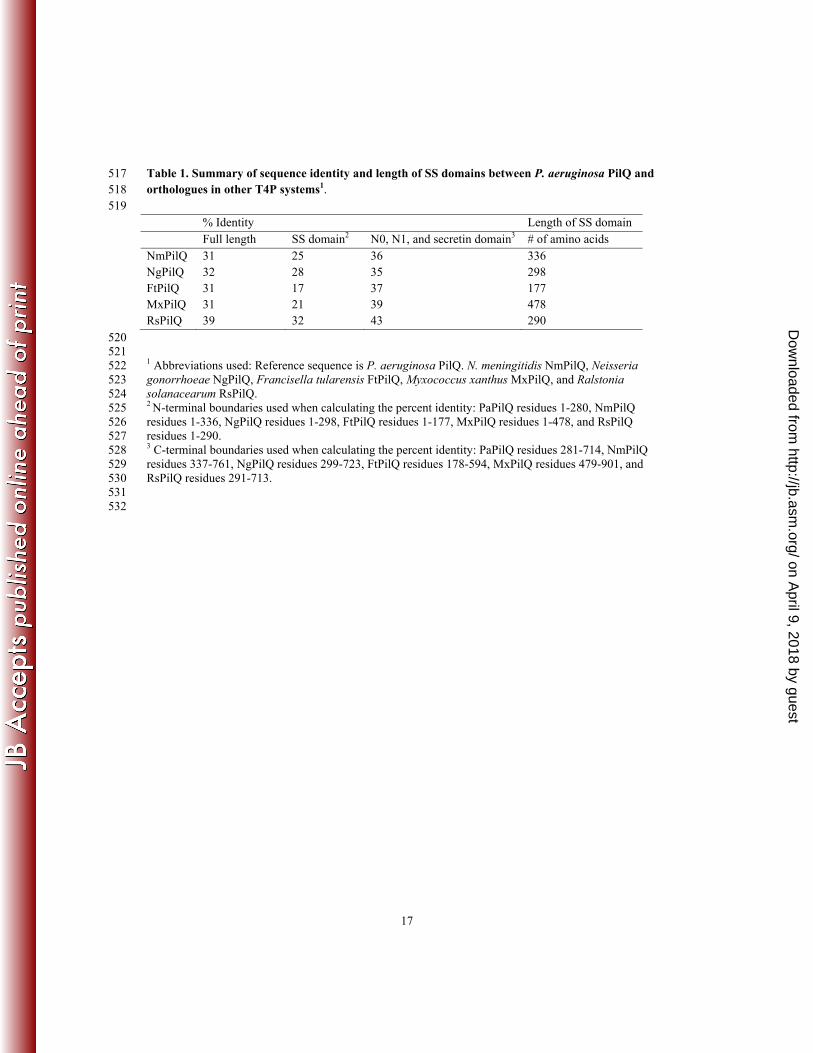
267
The N terminus of PilP is sufficient for the interaction with PilNO 268
Previously we showed that full length PilP (less its signal sequence) interacted with a stable complex of 269
PilNO, and that the C-terminal β-sandwich domain of PilP alone could not bind PilNO [12]. To directly 270
probe the potential interaction of the N terminus of PilP with PilNO, a construct corresponding to the 271
unstructured domain of PilP (PilP19-84T7, 8.9 kDa) was used. Under the conditions used previously to 272
characterize the interaction of mature PilP (PilPΔ18_6xHis) with the periplasmic PilNO (PilNΔ44/PilOΔ51) 273
heterodimer [12], the PilP N-terminal construct co-fractionated with PilNO, and this interaction was 274
stable during size exclusion chromatography (Figure 2). Although the N-terminal PilP fragment was 275
unstable when expressed in the absence of PilNO (Supplemental Figure 3), it was protected from 276
proteolytic degradation when in complex with PilNO. Examination of the PilNOP19-84T7 complex by 277
limited proteolysis suggested that the protected region of PilP spans residues 19-76, a region that our 278
bioinformatics analysis predicted to be entirely disordered [12]. These data support our hypothesis that 279
the N terminus of PilP mediates the interaction with PilNO [12]. 280
281
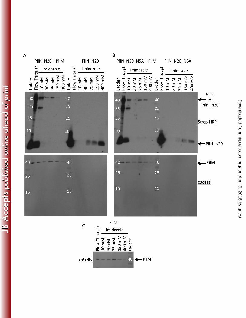
An important structural difference between PilP and T2SS GspC HR region. 282
Our earlier work highlighted a pair of highly conserved residues in PilP, Pro85 (found in the N-terminal 283
disordered region) and Trp161 (found on β7), which interact and tether the N-terminal disordered region 284
to the β-sandwich domain in solution [12]. The recent release of two structures of the HR domain of the 285
T2SS protein GspC from E. coli (PDBID:3OSS; GspCEc) and Dickeya dadantii 3937 (PDBID:2LNV; 286
GspCDd), allowed us to extend our analysis of the PilP structure. The structures of PilPΔ71 and GspCEc or 287
GspCDd can be superimposed with root mean squared deviations of 0.92 Å and 1.5 Å, over 51 and 43 Cα 288
atoms, respectively (Figure 3). The β-sandwich domains are very similar, but β-strands 6 and 7 in the 289
GspC proteins are each two residues shorter than those in PilP (Figure 3). Notably, this β6-β7 extension is 290
the site of the highly conserved Trp residue (Trp161 in PilP) found in all PilP proteins. There is no 291
equivalent conserved aromatic residue in the GspC family (data not shown). Mutation of Pro85 to either 292
Ala or Glu, or the mutation of residues in the β6-β7 extension (Gly157 and Leu162) impaired P. 293
aeruginosa twitching motility (Supplemental Figure 4) indicating that this region of the protein is 294
important for T4P biogenesis. However, in the case of the Pro85Val mutation, twitching was not 295
diminished, reinforcing the notion that a hydrophobic interaction between the residue at this position and 296
Trp161 is important for function. We propose that the interaction formed between Pro85 and Trp161 is 297
important for stabilizing the relative orientations of the disordered N terminus and the β-sandwich 298
on April 9, 2018 by guest
http://jb.asm.org/
Dow
nloaded from
10
domain, and that the β6-β7 extension and the Pro85-Trp161 interaction are T4P-specific adaptations for 299
function. 300
301
The C terminus of PilP interacts with the periplasmic domain of PilQ 302
Genin and Boucher [29] analyzed a number of secretin sequences from the T2SS and T3SS (Type III 303
secretion system) and identified four highly conserved regions towards the C-termini of the proteins that 304
they termed ‘secretin consensus regions’. Alignments of PilQ protein sequences from various T4P-305
producing bacteria show the same conserved regions (Supplemental Figure 5). Using these sequence 306
alignments, we identified four discrete periplasmic domains in T4P secretins. The two N-terminal β-307
strand rich domains share the lowest percent identity across PilQ homologues (Table 1, Supplemental 308
Figure 5) and thus we termed these regions of PilQ the ‘species-specific’ (SS) domains (SS1 and SS2). C-309
terminal to the SS domains we predicted that there are two additional domains – recently confirmed to be 310
structurally homologous to N0 and N1 from T2SS secretins [6] – and we use that terminology here. From 311
this analysis, we designed and expressed six soluble constructs spanning the periplasmic region of PilQ to 312
examine the roles of these individual domains in more detail (Figure 4). Briefly, these soluble constructs 313
are PilQ24-445 (containing the entire periplasmic domain), PilQ24-280 (SS domains), PilQ281-445 (N0 + N1), 314
PilQ281-410 (N0 + ~1/3 N1), PilQ281-376 (N0) and PilQ377-445 (N1). 315
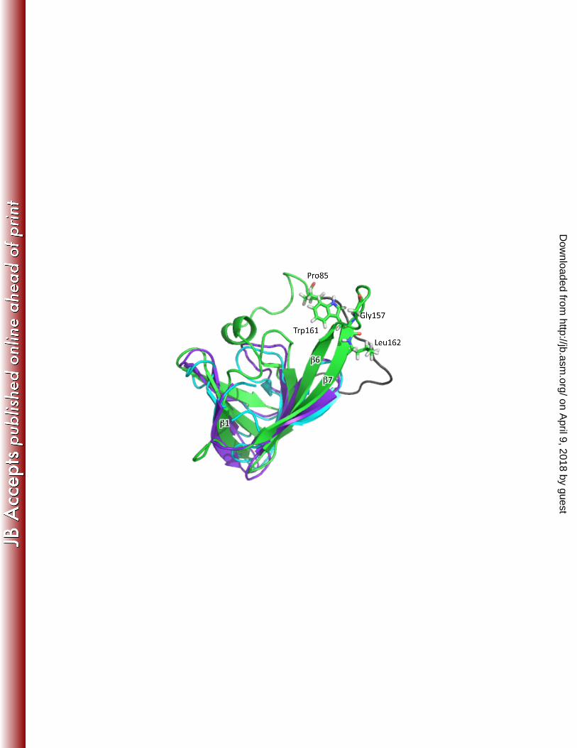
316
Each of the six 6xHis-tagged constructs was tested for its ability to pull-down untagged PilP from E. coli 317
lysates. Three constructs of PilP were used: mature PilP (PilPΔ18); the N-terminal region (PilP19-84T7) and 318
the C-terminal region (PilPΔ71). PilP19-84T7 was rapidly degraded after lysis, suggesting that no PilQ 319
fragment could stabilize this region of PilP (Supplemental Figure 3). In contrast, PilPΔ18 and PilPΔ71 were 320
pulled down by three of the six soluble PilQ constructs (Figure 5 and Table 2), while the negative control 321
showed that the PilP fragments did not bind to the Ni-NTA column in the absence of PilQ (data not 322
shown). PilPΔ18 and PilPΔ71 interacted with PilQ281-445, PilQ281-410, and PilQ281-376, the three constructs 323
containing the N0 domain at their N-termini (Table 2). Together these data suggest that the C-terminal β-324
domain region of PilP is sufficient for the interaction with PilQ, and that the site of PilP interaction with 325
PilQ is the N0 domain. 326
327
Notably, the full-length periplasmic domain of PilQ (PilQ24-445) did not pull-down either PilPΔ18 or PilPΔ71 328
from cell lysates, even though this construct contains the N0 domain (Figure 5B). It is possible that in this 329
context in vitro, the binding site for PilP on the N0 domain is occluded. This PilQ periplasmic fragment 330
may be conformationally flexible when expressed as a monomer in vitro, compared to the in vivo 331
oligomer where its orientation would be constrained. 332
on April 9, 2018 by guest
http://jb.asm.org/
Dow
nloaded from
11
333
The PilNOP heterotrimer interacts with PilQ 334
Previously, we showed that PilP forms a stable heterotrimer with PilNO in vitro [12], and in this study we 335
have shown that the extended N terminus of PilP interacts with PilNO, while its C-terminal β-domain 336
interacts with PilQ. To test whether PilP could interact simultaneously with PilNO and PilQ, the pull-337
down experiments were repeated with all four proteins. PilNOP were pulled down by the same PilQ 338
constructs that pulled down PilPΔ18 and PilPΔ71 (Table 2). Negative controls consisting of PilNO alone or 339
PilNO with tagged PilQ constructs showed that PilNO was not retained on the column unless both PilP 340
and tagged PilQ were present (Table 2). Therefore, PilP is sufficient to coordinate a stable interaction 341
network between the inner membrane components PilNO and the outer membrane component PilQ. 342
343
PilA interacts with a PilNOPQ complex in vivo 344
Bacterial two-hybrid experiments using N. meningitidis T4P components expressed in E. coli indicated 345
potential interactions of the major pilin subunit PilE (equivalent to PilA in P. aeruginosa) with specific 346
inner membrane proteins [23]. To test for potential interactions in P. aeruginosa and to include the 347
periplasmic domains of PilQ, we performed a series of pull-down experiments using 6xHis tagged PilQ 348
bait to capture prey proteins from detergent-solubilized P. aeruginosa lysates. Negative controls showed 349
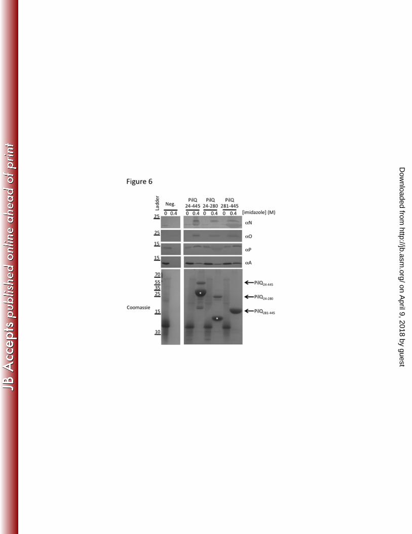
no interaction of proteins from detergent solubilized PAO1 lysates with the Ni-NTA column (Figure 6). 350
In contrast, after extensive washing of the column, the bound PilQ24-445, PilQ24-280, or PilQ281-445 fragments 351
captured PilA from the lysates (Figure 6). Western blots of imidazole eluates with antibodies specific to 352
PilN, PilO and PilP showed that PilQ captured all three proteins. Together, these data support formation 353
of a stable PilNOPQ complex and suggest that PilA interacts with this complex. These data support the 354
idea that PilA and/or the pilus make extensive contacts with the PilMNOPQ transenvelope complex. 355
DISCUSSION 356
This is the first report to provide evidence for a complete T4P transenvelope protein interaction network 357
formed by the products of the pilMNOPQ operon. This complex has components in the cytoplasm (PilM), 358
inner membrane (PilN, PilO, and PilP), and outer membrane (PilQ). This arrangement of the inner and 359
outer membrane components of the pilus assembly complex may provide an unobstructed path through 360
the periplasm, including the peptidoglycan, for the assembled pilus. As well, hypothesized interactions of 361
PilM with the platform protein PilC, as demonstrated for orthologs in the T2SS and the T4bP system [30–362
32], would connect the cytoplasmic motor with the outer membrane secretin, allowing for the efficient 363
transmission of the signals that control its opening and closing. 364
365
on April 9, 2018 by guest
http://jb.asm.org/
Dow
nloaded from

12
Interaction of the major pilin subunit with the periplasmic components PilNOP has been reported for the 366
N. meningitidis T4P system [23] and for corresponding components of the T2SS [23, 33, 34] and the 367
T4bP system [35]. Our pull-down experiment from PAO1 lysates provides indirect evidence that a 368
similar interaction occurs in P. aeruginosa. Based on the efficiency of the pull-down observed in our 369
experiments (Figure 6), the interaction of PilA with PilNOP may be transient. This scenario would be 370
expected for a dynamic interaction where the PilA monomers encounter the PilNOP complex in transit to 371
or from the pilus. One potential reason for PilA’s interaction with the alignment complex (PilMNOP) 372
might be to transiently cluster pilin monomers at the polar site of pilus assembly, providing a high local 373
concentration of PilA to promote rapid pilus assembly, and to prevent diffusion of PilA monomers away 374
from the system following disassembly. Further, PilMNOP and the periplasmic domains of PilQ may 375
participate in selection for the T4P major and minor pilins over the T2SS pseudopilins and minor 376
pseudopilins, maintaining the fidelity of pilus composition and function. Finally, as the pilus experiences 377
forces in excess of 100 pN upon retraction [4], PilA interaction sites in the periplasm could contribute to 378
anchoring of the pilus. Indeed the SS domain in the T4P-specific secretin (PilQ) may be a unique 379
adaptation that could act as a clamp on the assembled fiber. 380
381
The T4P-specific interaction between Pro85 and Trp161 of PilP may also be important for the efficient 382
generation of long retractable extracellular filaments. We suggest that this interaction may be related to 383
PilP’s bridging of PilMNO in the inner membrane and PilQ in the outer membrane, and that maintaining 384
the orientation of the disordered N terminus and β-sandwich domains of PilP may be important for 385
stabilizing the PilMNOPQ complex in vivo. Also, the SS domains in PilQ (absent in the T2SS secretins) 386
may constrain the dynamics of the PilPQ interaction, and the Pro85-Trp161 tether may promote an 387
optimal PilPQ interface. 388
389
The full T4P transenvelope complex is unlikely to have a closed lumen as in the T3SS needle complex 390
[36, 37], since addition and removal of PilA subunits at the periplasmic face of the inner membrane are 391
absolutely required for pilus extension and retraction to occur. Our earlier work indicated that PilNOP 392
interact in a 1:1:1 complex [12], and the PilMN interaction suggests a 1:1 stoichiometry as well [9]. 393
Recent structural studies of T2SS and T4P components further suggest that PilP and PilQ form a 1:1 394
interaction, and comparison of the available data regarding this heterodimeric interface suggests that this 395
organization is conserved between the two systems [6, 22]. Also, when the N-terminal domain of PilP is 396
attached to the inner membrane via its lipid anchor and interacting with PilNO, the nature of the Pro85-397
Trp161 interaction would position the PilQ interaction interface on PilP “up” towards the outer 398
membrane, possibly facilitating the PilPQ interaction. In this orientation, one could imagine a series of 399
on April 9, 2018 by guest
http://jb.asm.org/
Dow
nloaded from

13
rings through the periplasm, where PilNO form a ring closest to the inner membrane, PilP forms a ring 400
above PilNO, and PilQ would form a ring closest to the outer membrane, extending downward into the 401
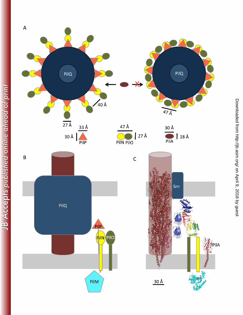
periplasm (Figure 7B&C). Since PilQ is predicted to be a dodecameric complex [6], there could be twelve 402
PilMNOPQ units arranged in a circular manner, similar to the arrangement in T2SS, T3SS and T4SS 403
(Type IV secretion system) assembly complexes [37–39] (Figure 7). Alternatively, data from the T2SS 404
suggests that the secretin may be assembled as a hexamer of dimers [40–42]; in this case, the arrangement 405
may be one PilMNOP complex for every two PilQ monomers (1:1:1:1:2 stoichiometry). Differences in 406
orientation of the N0 domain of PilQ in a dimer versus monomer configuration might explain why we 407
saw differences in PilPQ interactions when fragments versus full periplasmic domains of PilQ were used 408
for pull-downs. 409
410
The requirement for addition and removal of PilA subunits at the periplasmic face of the inner membrane 411
means that the periplasmic constituents are likely arranged in a way that allows room for this process. 412
Using the dimensions of the structures determined by NMR, X-ray crystallography and electron 413
microscopy for PilNOPQ [11, 12, 43], a scale model showing the putative arrangement of periplasmic 414
components can be derived (Figure 7A). The outer diameter of the P. aeruginosa PilQ secretin was 415
measured from negative stained PilQ complexes to be ~183 Å [43], leading to a circumference of 575 Å 416
or ~48 Å per monomer in a dodecamer. When viewed from above the β-sandwich domain of PilP is ~33 417
Å by ~30 Å, and the dimensions of the ferredoxin-like fold of a PilNO heterodimer (based on the 418
fragment of PilO that was determined by X-ray crystallography) are ~47 Å by 27 Å. As the N terminus of 419
PilP facilitates the PilNOP interaction, it is unlikely to substantially affect the width of an PilNOP 420
complex. If twelve PilNO heterdimers are oriented with the broad (47 Å) face facing the conduit, there 421
would be no room for the lateral introduction of PilA monomers (Figure 7A, right). However, if PilNO 422
are oriented with the narrow (27 Å) face towards the conduit, there is ample room for entry and exit of 423
PilA (~30 Å by 18 Å) in the gap between PilNOP complexes (~40 Å) (Figure 7A left). There are five 424
major pilin subtypes in P. aeruginosa [44], with head groups of various sizes [45], supporting the need 425
for a large enough gap between the “spokes” formed by the PilMNOP transenvelope complex to 426
accommodate structurally distinct pilins and likely the minor pilins as well. Alternatively, if there is one 427
PilMNOP complex per PilQ dimer, there would be sufficient space between the PilMNOP spokes, 428
regardless of the orientation of PilNOP, to facilitate the addition and removal of pilin and minor pilin 429
monomers. 430
431
Drawing the components to scale and including available structures (Figure 7B), it is clear that the SS 432
domain of PilQ extends a significant distance across the periplasm towards the inner membrane. As has 433
on April 9, 2018 by guest
http://jb.asm.org/
Dow
nloaded from

14
been suggested elsewhere [46], we propose that the periplasmic region of PilQ forms the inside of the 434
channel, while the PilMNOP subcomplex forms the exterior. In this arrangement, both PilMNOP and 435
PilQ could interact with monomeric PilA and/or the minor pilins. This interaction could serve a number 436
of roles, including concentrating the pilin subunits at the site of assembly/disassembly; communicating 437
assembly/disassembly signals from the cytoplasm to the PilA subunits; or stimulating opening of the 438
secretin. The SS domains of PilQ could be involved in the initial stages of polymerization (through as yet 439
uncharacterized interactions with a priming complex) and/or clamping on the pilus to ensure it remains 440
anchored in the membrane under tension. In our favored model, the transenvelope complex composed of 441
PilMNOPQ is a stable assembly through the cell envelope, while the signal(s) for assembly/disassembly 442
of the pilus come from the communication between the cytoplasmic motor complex (made up of the 443
platform protein PilC [47] and the ATPases PilB/T/U) and PilM. Further studies are needed to better 444
understand the nature of the communication between the motor and alignment subcomplexes and the 445
exact mechanism of PilA polymerization/deploymerization. With this work, we have provided a 446
framework for the further examination of these processes. 447
448
Acknowledgements 449
This work was supported by Operating Grant MOP 93585 from the Canadian Institutes of Health 450
Research (CIHR) to L.L.B. and P.L.H.. J.K. and S.T. have been funded, in part, by graduate scholarships 451
from CIHR and Cystic Fibrosis Canada, respectively. P.L.H. is the recipient of a Canada Research Chair. 452
FIGURE LEGENDS 453
454
Figure 1. PilMNt6xHis pulls down PilN_N20. (A) The wild type PilN_N20 peptide binds to PilM in an 455
SDS resistant manner. Western blots developed by enhanced chemiluminescence (ECL) with Strep-HRP 456
detecting biotinylated PilN_N20 peptide (top panel) and a blot probed with a monoclonal 6xHis antibody 457
conjugated to alkaline phosphatase probing for PilM and detected by NBT-BCIP (bottom panel). (B) 458
PilN_N20_N5A peptide binds to PilM in an SDS resistant manner. The top and bottom panels are as 459
described for (A). (C) Western blot probed with monoclonal 6xHis antibody conjugated to alkaline 460
phosphatase showing that PilM binds to the column in absence of either PilN peptide indicating that the 461
binding of PilM to the Ni-beads is not altered by the presence of the peptide. Ladder markers are labeled 462
in kDa. 463
464
Figure 2. Co-purification of PilNOP. A) Coomassie stained SDS-PAGE analysis of the 465
PilNΔ44_6xHis/PilOΔ51/PilP19-84T7 fractions eluted from a Ni-NTA column in the presence of increasing 466
on April 9, 2018 by guest
http://jb.asm.org/
Dow
nloaded from

15
imidazole concentrations. B) Elution of the 75 mM imidazole Ni-NTA fraction in (A) from a Superdex 467
200 gel filtration column. Inset is the Coomassie stained SDS-PAGE gel of the fractions from the gel 468
filtration column. 469
470
Figure 3. Structural comparison of PilP and GspC proteins. Cartoon representations of PilP (PDBID: 471
2LC4, green), GspCDd (PDBID: 2LNV, cyan), and GspCEc (PDBID: 3OOS chain C, purple). For clarity 472
the following residues have been removed from the cartoon representations: PilPΔ71 the C-terminal 6xHis 473
tag, GspCDd the N-terminal residues 70-94 and the C-terminal residues 158-173, and in GspCEc the N-474
terminal residues 122-127. The location of the highly conserved Trp161 and residues selected for 475
mutagenesis (Pro85, Gly157, and Leu162) in PilP are shown in sticks. The N-terminal β-strand, strand 476
β1, and the two C-terminal strands, strands β6 and β7, have been labeled. The disordered N-terminal 477
region of PilP is colored in black. 478
479
Figure 4. Schematic of soluble PilQ constructs. All constructs were cloned into pET28a with a C-480
terminal 6xHis tag. Amino acid numbers are above the schematic of the full-length protein. The first 24 481
residues are the signal peptidase signal sequence (SSq), residues 24-445 constitute the full periplasmic 482
domain, and 445-714 the putative membrane embedded secretin domain (Secretin). In the periplasm there 483
are two distinct regions, residues 24-280 are the species specific domain (SS) which can be broken down 484
into two sub domains (SS1 spanning residues 24-139, and SS2 spanning residues 140-280), while 485
residues 281-445 share sequence and secondary structural homology to other secretins, and have been 486
found to play a role in multimerization. This multimerization domain can be broken into two sub 487
domains: residues 281-376 and 377-445, corresponding to the highly conserved N0 and N1 domains, 488
respectively. 489
490
Figure 5. Co-fractionation of PilP and PilQ from a Ni-NTA affinity column. A) The Ni-NTA results 491
of PilPΔ18 and PilQ. The absence of any PilP in the high imidazole fractions when PilQ24-445 is used as bait 492
indicates that PilP is unable to interact with this construct, but when PilQ281-445 is used as the bait protein 493
PilPΔ18 is found in the high imidazole fractions. B) The Ni-NTA results of PilPΔ71 and PilQ. A similar 494
interaction pattern for PilPΔ71 is observed as in (A). Ladder markers are labeled in kDa. 495
496
Figure 6. Periplasmic regions of PilQ pull-down PilA. After binding various PilQ6xHis constructs on a 497
Ni-NTA column, the flow through (0 M imidazole) and highest elution (0.4 M imidazole) fractions were 498
examined by Coomassie staining and with PilN, PilO, PilP, or PilA specific antibodies. The locations of 499
the molecular weight markers are shown as labeled bars on the left hand side of the figure (in kDa). The 500
on April 9, 2018 by guest
http://jb.asm.org/
Dow
nloaded from

16
location of the PilQ constructs in the 0.4 M imidazole elution fractions are shown by arrows at the right of 501
the figure, and white asterisks denote PilQ degradation products. 502
503
Figure 7. Model of the PilMNOPQ transenvelope complex. A) Scale schematic of T4P complex, and 504
possible orientations of periplasmic components (top down orientation). Circumference of PilQ pore is 505
approximately 575 Å. PilN, PilO, PilP, and PilQ are drawn to scale, PilM, which is omitted for clarity as 506
it is located in the cytoplasm, is believed to interact with PilN with a 1:1 stoichiometry. The left panel 507
depicts the narrow face of PilNOP adjacent to the pore, while the right panel depicts the wide edge of 508
PilNOP adjacent to the pore. B) Schematic representation of the PilMNOPQ complex approximately to 509
scale. C) Scale model using experimentally determined or modeled structures for PilMNOP and PilA, a 510
homology model for the N0 and N1 domains, and the structure of the B2 domain of N. meningitidis 511
(PDBID: 4AQZ) for SS1 and SS2. The orientation of PilP and the N0 domain of PilQ was achieved by 512
overlaying the PilPΔ71 and the homology model of PilQ N0 and N1 domains on the C and D chains of the 513
GspC/D co-crystal structure (PDBID: 3OSS). Scale and coloring are as shown in (B). For PilQ the size of 514
the secretin (Srn) domain is estimated based on the size of the N0 and N1 domains. 515
516
on April 9, 2018 by guest
http://jb.asm.org/
Dow
nloaded from
17
Table 1. Summary of sequence identity and length of SS domains between P. aeruginosa PilQ and 517 orthologues in other T4P systems1. 518 519 % Identity Length of SS domain Full length SS domain2 N0, N1, and secretin domain3 # of amino acids NmPilQ 31 25 36 336 NgPilQ 32 28 35 298 FtPilQ 31 17 37 177 MxPilQ 31 21 39 478 RsPilQ 39 32 43 290
520 521 1 Abbreviations used: Reference sequence is P. aeruginosa PilQ. N. meningitidis NmPilQ, Neisseria 522 gonorrhoeae NgPilQ, Francisella tularensis FtPilQ, Myxococcus xanthus MxPilQ, and Ralstonia 523 solanacearum RsPilQ. 524 2 N-terminal boundaries used when calculating the percent identity: PaPilQ residues 1-280, NmPilQ 525 residues 1-336, NgPilQ residues 1-298, FtPilQ residues 1-177, MxPilQ residues 1-478, and RsPilQ 526 residues 1-290. 527 3 C-terminal boundaries used when calculating the percent identity: PaPilQ residues 281-714, NmPilQ 528 residues 337-761, NgPilQ residues 299-723, FtPilQ residues 178-594, MxPilQ residues 479-901, and 529 RsPilQ residues 291-713. 530 531 532
on April 9, 2018 by guest
http://jb.asm.org/
Dow
nloaded from
18
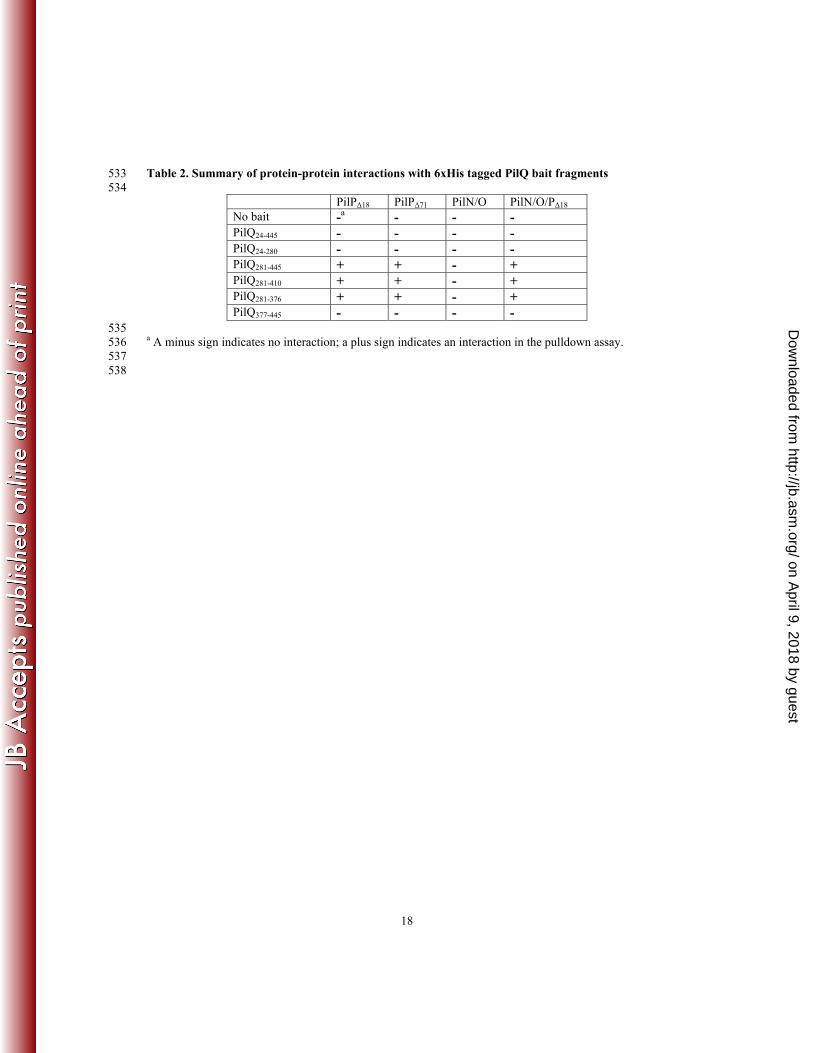
Table 2. Summary of protein-protein interactions with 6xHis tagged PilQ bait fragments 533 534
PilPΔ18 PilPΔ71 PilN/O PilN/O/PΔ18 No bait -a - - - PilQ24-445 - - - - PilQ24-280 - - - - PilQ281-445 + + - + PilQ281-410 + + - + PilQ281-376 + + - + PilQ377-445 - - - -
535 a A minus sign indicates no interaction; a plus sign indicates an interaction in the pulldown assay. 536 537 538
on April 9, 2018 by guest
http://jb.asm.org/
Dow
nloaded from
19
REFERENCES 539 1. Bradley DE 1974. Basic Characterization of a Pseudomonas aeruginosa Pilus-Dependent 540
Bacteriophage with a Long Noncontractile Tail. J Virol 12:1139–1148. 541 2. Bradley DE 1974. The adsorption of pilus-dependent bacteriophages to a host mutant with 542
nonretractile pili. Virology 58:149–63. 543 3. Burrows LL 2005. Weapons of Mass Retraction. Mol Microbiol 57:878–888. 544 4. Maier B, Potter L, So M, Long CD, Seifert HS, and Sheetz MP. 2002. Single Pilus 545
Motor Forces Exceed 100 pN. Proc Natl Acad Sci U S A 99:16012–16017. 546 5. Burrows LL 2012. Pseudomonas aeruginosa twitching motility: type IV pili in action, p. 547
493–520. In Annual Review of Microbiology. 548 6. Berry J-L, Phelan MM, Collins RF, Adomavicus T, Tonjum T, Frye SA, Bird L, 549
Owens RJ, Ford RC, Lian L-Y, and Derrick JP. 2012. Structure and Assembly of a 550 Trans-Periplasmic Channel for Type IV Pili in Neisseria meningitidis. PLoS Pathog 551 8:e1002923. 552
7. Koo J, Tammam SD, Ku S-Y, Sampaleanu L, Burrows LL, and Howell PL. 2008. PilF 553 is an Outer Membrane Lipoprotein Required for Multimerization and Localization of the 554 Pseudomonas aeruginosa Type IV Pilus Secretin. J Bacteriol 190:6961–6969. 555
8. Ayers M, Sampaleanu L, Tammam SD, Koo J, Harvey H, Howell PL, and Burrows 556 LL. 2009. PilM/N/O/P proteins form an inner membrane complex that affects the stability 557 of the Pseudomonas aeruginosa type IV pilus secretin. J Mol Biol 394:128–42. 558
9. Karuppiah V, and Derrick JP. 2011. Structure of the PilM-PilN Inner Membrane Type 559 IV Pilus Biogenesis Complex from Thermus thermophilus. J Biol Chem 286:24434–24442. 560
10. Yamagata A, Milgotina E, ScanlonK, Craig L, Tainer JA, and Donnenberg MS. 2012. 561 Structure of an Essential Type IV Pilus Biogenesis Protein Provides Insights into Pilus and 562 Type II Secretion Systems. J Mol Biol 419:110–124. 563
11. Sampaleanu LM, Bonanno JB, Ayers M, Koo J, Tammam S, Burley SK, Almo SC, 564 Burrows LL, and Howell PL. 2009. Periplasmic domains of Pseudomonas aeruginosa 565 PilN and PilO form a stable heterodimeric complex. J Mol Biol 394:143–59. 566
12. Tammam S, Sampaleanu LM, Koo J, Sundaram P, Ayers M, Chong PA, Forman-Kay 567 JD, Burrows LL, and Howell PL. 2011. Characterization of the PilN, PilO and PilP type 568 IVa pilus subcomplex. Mol Microbiol 82:1496–1514. 569
13. Giltner CL, Habash M, and Burrows LL. 2010. Pseudomonas aeruginosa Minor Pilins 570 Are Incorporated into Type IV Pilus. J Mol Biol 398:444–461. 571
14. Ayers M, Howell PL, and Burrows LL. 2010. Architectures of the type II secretion and 572 type IV pilus machineries. Future Microbiol 5:1203–1218. 573
15. Peabody CR, Chung YJ, Yen M-R, Vidal-Ingigliardi D, Pugsley AP, and Saier MHJ. 574 2003. Type II Protein Secretion and its Relationship to Bacterial Type IV Pili and Archael 575 Flagella. Microbiology 149:3051–3072. 576
16. Russel M. 1998. Macromolecular assembly and secretion across the bacterial cell envelope: 577 type II protein secretion systems. J Mol Biol 279:485–499. 578
17. Abendroth J, Kreger AC, and Hol WG. 2009. The dimer formed by the periplasmic 579 domain of EpsL from the Type 2 Secretion System of Vibrio parahaemolyticus. J Struct 580 Biol 168:313–322. 581
18. Abendroth J, Bagdasarian M, Sandkvist M, and Hol WGJ. 2004. The Structure of the 582 Cytoplasmic Domain of EpsL, An Inner Membrane Component of the Type II Secretion 583 System of Vibrio cholerae: An Unusual Member of the Actin-like ATPase Superfamily. J 584 Mol Biol 344:619–633. 585
on April 9, 2018 by guest
http://jb.asm.org/
Dow
nloaded from

20
19. Abendroth J, Murphy P, Sandkvist M, Bagdasarian M, and Hol WGJ. 2005. The X-586 ray Structure of the Type II Secretion system Complex Formed by the N-terminal Domain 587 of EpsE and the Cytoplasmic Domain of EpsL of Vibrio cholerae. J Mol Biol 348:845–855. 588
20. Sandkvist M, Keith JM, Bagdasarian M, and Howard SP. 2000. Two regions of EpsL 589 involved in species-specific protein-protein interactions with EpsE and EpsM of the general 590 secretion pathway in Vibrio cholerae. J Bacteriol 182:742–748. 591
21. Gu S, Kelly G, Wang X, Frenkiel T, Shevchik VE, and Pickersgill RW. 2012. Solution 592 structure of the HR domain of the type II secretion system. J Biol Chem 287:9072–9080. 593
22. Korotkov KV, Johnson TL, Jobling MG, Pruneda J, Pardon E, Héroux A, Turley S, 594 Steyaert J, Holmes RK, Sandkvist M, and Hol WGJ. 2011. Structural and Functional 595 Studies on the Interaction of GspC and GspD in the Type II Secretion System. PLoS Pathog 596 7:e1002228. 597
23. Georgiadou M, Castagnini M, Karimova G, Ladant D, and Pelicic V. 2012. Large-scale 598 study of the interactions between proteins involved in type IV pilus biology in Neisseria 599 meningitidis: characterization of a subcomplex involved in pilus assembly. Mol Microbiol 600 84:857–873. 601
24. Balasingham SV, Collins RF, Assalkhou R, Homberset H, Frye SA, Derrick JP, and 602 Tonjum T. 2007. Interactions Between the Lipoprotein PilP and the Secretin PilQ in 603 Neisseria meningitidis. J Bacteriol 189:5716–5727. 604
25. Notredame C, Higgins DG, and Heringa J. 2000. T-Coffee: A novel method for fast and 605 accurate multiple sequence alignment. J Mol Biol 302:205–217. 606
26. Waterhouse AM, Procter JB, Martin DMA, Clamp M, and Barton GJ. 2009. Jalview 607 Version 2--a multiple sequence alignment editor and analysis workbench. Bioinformatics 608 25:1189–1191. 609
27. Cole C, Barber JD, and Barton GJ. 2008. The Jpred 3 secondary structure prediction 610 server. Nucleic Acids Res 36:W197–201. 611
28. DeLano WL. 2002. The PyMOL Molecular Graphics System. DeLano Scientific, San 612 Carlos, CA, USA. 613
29. Genin S, and Boucher CA. 1994. A Superfamily of Proteins Involved in Different 614 Scretion Pathways in Gram-Negative Bacteria: Modular Structure and Specificity of the N-615 terminal Domain. Mol Gen Genet 243:112–118. 616
30. Arts J, de Groot A, Ball G, Durand E, Khattabi ME, Filloux A, Tommassen J, and 617 Koster M. 2007. Interaction Domains in the Pseudomonas aeruginosa Type II Secretory 618 Apparatus Component XcpS (GspF). Microbiology 153:1582–1592. 619
31. Py B, Loiseau L, and Barras F. 2001. An Inner Membrane Platform in the Type II 620 Secretion Machinery of Gram-Negative Bacteria. EMBO Rep 2:244–248. 621
32. Milgotina EI, Lieberman JA, and Donnenberg MS. 2011. The inner membrane 622 subassembly of the enteropathogenic Escherichia coli bundle-forming pilus machine. Mol 623 Microbiol 81:1125–1127. 624
33. Lee M-S, ChenL-Y, Leu W-M, Shiau R-J, and Hu N-T. 2005. Associations of the Major 625 Pseudopilin XpsG with XpsN (GspC) and Secretin XpsD of Xanthomonas campsetris pv. 626 campestris Type II Secretion Apparatus Revealed by Cross-linking Analysis. J Biol Chem 627 280:4585–4591. 628
34. Gray MD, Bagdasarian M, Hol WGJ, and Sandkvist M. 2011. In vivo cross-linking of 629 EpsG to EpsL suggests a role for EpsL as an ATPase-pseudopilin coupling protein in the 630 Type II secretion system of Vibrio cholerae. Mol Microbiol 79:786–798. 631
on April 9, 2018 by guest
http://jb.asm.org/
Dow
nloaded from
21
35. Hwang J, Bieber D, Ramer SW, Wu C-Y, and Schoolnik GK. 2003. Structural and 632 Topographical Studies of the Type IV Bundle-Forming Pilus Assembly Complex of 633 Enteropathogenic Escherichia coli. J Bacteriol 185:6695–6701. 634
36. Blocker A, Jouihri N, Larquet E, Gounon P, Ebel F, Parsot C, Sansonetti P, and 635 Allaoui A. 2001. Structure and composition of the Shigella flexneri “needle complex”, a 636 part of its type III secreton. Mol Microbiol 39:652–63. 637
37. Schraidt O, and Marlovits TC. 2011. Three-dimensional model of Salmonella’s needle 638 complex at subnanometer resolution. Science 331:1192–1195. 639
38. McLaughlin LS, Haft RJ, and Forest KT. 2012. Structural insights into the Type II 640 secretion nanomachine. Curr Opin Struct Biol 22:208–216. 641
39. Fronzes R, Scharer E, Wang L, Saibil H, Orlova EV, and Waksman G. 2009. Structure 642 of a Type IV Secretions System Core Complex. Science 323:266–268. 643
40. Van der Meeren R, Wen Y, Van Gelder P, Tommassen J, Devreese B, and Savvides 644 SN. 2012. New insights into the assembly of bacterial secretins: structural studies of the 645 periplasmic domain of XcpQ from Pseudomonas aeruginosa. J Biol Chem 288:1214-1225. 646
41. Wang X, Pineau C, Gu S, Guschinskaya N, Pickersgill RW, and Shevchik VE. 2012. 647 Cysteine scanning mutagenesis and disulfide mapping analysis of the arrangement of GspC 648 and GspD protomers within the T2SS. J Biol Chem 287:19082–19093. 649
42. Lieberman JA, Frost NA, Hoppert M, Fernandes PJ, Vogt SL, Raivio TL, Blanpied 650 TA, and Donnenberg MS. 2012. Outer Membrane Targeting, Ultrastructure and Single 651 Molecule Localization of the Enteropathogenic Escherichia coli Type IV Pilus Secretin 652 BfpB. J Bacteriol 194:1646–1658. 653
43. Bitter W, Koster M, Latijnhouwers M, de Cock H, and Tommassen J. 1998. Formation 654 of Oligomeric Rings by XcpQ and PilQ, Which are Involved in Protein Transport Across 655 the Outer Membrane of Pseudomonas aeruginosa. Mol Microbiol 27:209–219. 656
44. Kus JV, Tullis E, Cvitkovitch DG, and Burrows LL. 2004. Significant Differences in 657 Type IV Pilin Allele Distribution Among Pseudomonas aeruginosa Isolated from Cyctic 658 Fibrosis (CF) Versus Non-CF Patients. Microbiology 150:1315–1326. 659
45. Nguyen Y, Jackson SG, Aidoo F, Junop M, and Burrows LL. 2010. Structural 660 characterization of novel Pseudomonas aeruginosa type IV pilins. J Mol Biol 395:491–503. 661
46. Douzi B, Ball G, Cambillau C, Tegoni M, and Voulhoux R. 2011. Deciphering the Xcp 662 Pseudomonas aeruginosa type II secretion machinery through multiple interactions with 663 substrates. J Biol Chem 286:40792–40801. 664
47. Takhar HK, Kemp K, Kim M, Howell PL, and Burrows LL. 2013. The Platform Protein 665 is essential for Type IV pilus biogenesis. J Biol Chem doi: 10.1074/jbc.M113.453506 666
667 668
on April 9, 2018 by guest
http://jb.asm.org/
Dow
nloaded from