Embed Size (px)
DESCRIPTION
physiology of hearing
Citation preview

PHYSIOLOGY OF
HEARING
J.P. SOUAID, M.D., C.M., FRCS(C)QUEENSWAY-CARLETON HOSPITAL
THE OTTAWA HOSPITALDEPARTMENT OF OTOLARYNGOLOGY
April 10, 2014J. G. Marsan MD FRCSC

Physics of SoundDEFINITIONMechanical radiant energyTransmitted by longitudinal pressure wavesIn a material medium
CHARACTERISTICSFrequency – pitchAmplitude - loudness
Mismatch of Impedance
Air Water
Sound pressure energy
99.9% Energy reflected
0.1% Energy absorbed
Loss of sound pressure energy: 30dB

OBJECTIVES Explain how sound travels from a source to the
temporal lobe Explain how mechanical energy is transformed
into electrical energy in hearing Define and differentiate between conductive
hearing loss and sensorineural hearing loss Describe the neural pathways involved in the
stapedial reflex Describe pure-tone audiometry and
tympanometry. Describe the physiology of otoacoustic emissions


EXTERNAL EAR Concha and External Auditory Canal act
as acoustic resonators 10-15 db gain in 3-5kHz region. Concha:
Resonance of 5kHz, Irregular surface introduces other
resonances and anti-resonances. Ear canal acts roughly like a simple tube
resonator, open at one end Resonant peak at a frequency of 2.5 kHz
in human ears Effects of the ear canal resonance
contribute substantially toward an increase in sound pressure level at the tympanum

EXTERNAL EAR CANAL2.5kHz

MIDDLE EAR
Purpose: Transfer sound energy from the air space of the external auditory meatus to the fluid in the cochlea.
Accomplished through the vibration of the three middle-ear ossicles.
Piston-like vibration of the stapes in the oval window results in a pressure differential between the oval window (and hence scala vestibuli) and the round window (and scala tympani). This pressure difference between the two scalae is critical to the mechanical excitation of the cochlear structures.
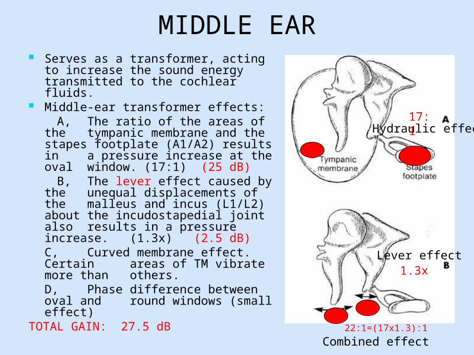
MIDDLE EAR Serves as a transformer, acting to
increase the sound energy transmitted to the cochlear fluids.
Middle-ear transformer effects: A, The ratio of the areas of the
tympanic membrane and the stapes footplate (A1/A2) results in a pressure increase at the oval window. (17:1) (25 dB)
B, The lever effect caused by the unequal displacements of the malleus and incus (L1/L2) about the incudostapedial joint also results in a pressure increase. (1.3x) (2.5 dB)C, Curved membrane effect. Certain areas of TM vibrate more than others.D, Phase difference between oval and round windows (small effect)
TOTAL GAIN: 27.5 dB
17:1
1.3x
22:1=(17x1.3):1
Lever effect
Hydraulic effect
Combined effect

Next Step
Mechanical energyMechanical energy Electrical nerve impulses
External and middle ear Inner ear (cochlea)

COCHLEAR PHYSIOLOGY
35 mm coiled bony tube; 2.5 turns
Divided into :
1) Scala vestibuli and tympani: Contains perilymph ECF like : Na- 140. K- 4-10 Production of perilymph:
Unknown Ultrafiltrate of blood? From CSF?

COCHLEAR PHYSIOLOGY
2) Scala media: Contains endolymph ICF like: K- 144 meq, Na- 15-25meq Bounded by :
Reissner’s membrane Basilar membrane Osseous spiral lamina Lateral wall
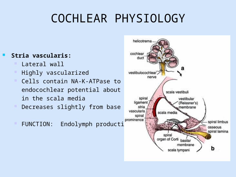
COCHLEAR PHYSIOLOGY
Stria vascularis: Lateral wall Highly vascularized Cells contain NA-K-ATPase to produce
endocochlear potential about +80mV in the scala media
Decreases slightly from base to apex
FUNCTION: Endolymph production

ORGAN OF CORTI

HAIR CELL INNERVATION 50,000 neurons innervate cochlea:
90-95% synapse directly on inner hair cells (type 1 neurons)
Predominantly afferent 15- 20 of these neurons innervate each hair
cell 5-10% synapse directly on outer hair cells
(type 2 neurons) Predominantly efferent Each type2 neuron branches to innervate 10
outer hair cells

Characteristic Inner H.C. Outer H.C.
Shape Flask Cylindrical
Number 3500 12000
Stereocilia
No.of hair cells Few Many
Arrangement 3-4 rows, slightly curved
6-7 rows, rows arranged in V or W shape
Attachement to tectorial membrane
None or loosely Long stereocilia firmly embedded
Intracellular electric potential
-40mV -70mV

COCHLEAR MECHANICS
1. Motion is a traveling wave moving longitudinally from the base to the apex
of the cochlea.2. Each point along the cochlear partition
vibrates at a frequency equal to that of the stimulus. Tonotopic organization:This means that specific areas of the basilar membrane respond to specific frequencies
3. High frequency- base of cochlea4. Low frequency- apex of cochlea
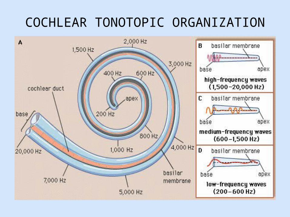
COCHLEAR TONOTOPIC ORGANIZATION

PHYSIOLOGY ORGAN OF CORTI
Stereocilia-Hair Cell complex: Deflection of Stereocilia of outer hair cells By travelling wave Opens and closes non-specific ion channels Influx of K, depolarizing of cell Ca mediated K channels -intracellular cascade Release of chemical transmitters (Glutamate) Activate afferent neurons
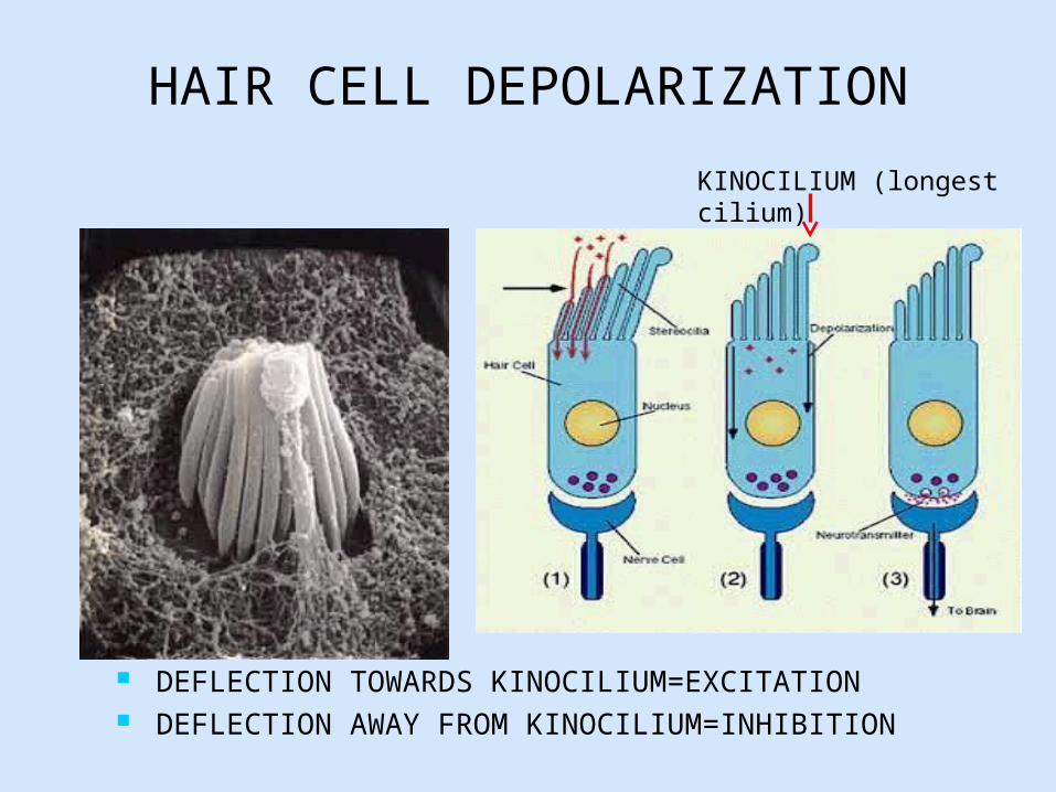
HAIR CELL DEPOLARIZATION
DEFLECTION TOWARDS KINOCILIUM=EXCITATION DEFLECTION AWAY FROM KINOCILIUM=INHIBITION
KINOCILIUM (longest cilium)

Auditory CNS Cochlea Cochlear Nerve Cochlear Nucleus (CN) Superior Olivary
Complex (SOC) Lateral Lemniscus (B) Inferior Colliculus (IC) Medial Geniculate Body
(MGB) Trapezoid body Auditory Cortex
(Temporal lobe, Brodman area 41)
Extensive crossoverTonotopicity of Cortex also.
MNEMONIC: N.N.S.L.I.M.

CONDUCTIVE HEARING LOSSSENSORINEURAL HEARING
LOSSConductive HL:Related to sound conduction travelling through air via Pinna, External Ear Canal, TM, Ossicles and Middle Ear up to the oval window.
Sensorineural HL:Related to the electrical signal that is travelling via the cochlea, nerve and onwards.


AUDIOMETRY


AIR CONDUCTION TESTING

BONE CONDUCTION TESTING

NORMAL AUDIOGRAM

CONDUCTIVE HEARING LOSS

SENSORINEURAL HEARING LOSS

TYMPANOMETRY
Not a hearing test. Objective test of
middle-ear function. It is a measure of energy transmission through the middle ear.

TYMPANOMETRY A tone of 226 Hz is
generated by the tympanometer into the ear canal, where the sound strikes the tympanic membrane, causing vibration. This is done for different air pressures.
Some of this sound is reflected back and picked up by the instrument.
Admittance is how energy is transmitted through the middle ear. The instrument measures the reflected sound and expresses it as an admittance or compliance, plotting the results on a chart known as a tympanogram.

TYMPANOGRAMS
Under normal conditions, the air pressure in the middle ear is approximately the same as ambient pressure since the eustachian tube opens periodically to ventilate the middle ear and to equalize pressure. In a healthy individual, the maximum sound is transmitted through the middle ear when the ambient air pressure in the ear canal is equal to the pressure in the middle ear.

TYMPANOGRAMS
Type A: normal ear Type As: stiff
TM/ossicles Type Ad: flaccid TM or
discontinuity of ossicles Type B: fluid in ear,
mass in ear Type C: negative
pressure means ET dysfunction.

MIDDLE EAR MUSCLES
5= STAPEDIUS MUSCLE9= TENSOR TYMPANI MUSCLE
STAPEDIUS INNERVATION: CN7TENSOR TYMPANI INNERVATION: V3

STAPEDIAL REFLEX ARC

Otoacoustic Emissions, OAEs
Sounds given off by the inner ear, 0-15dB Triggered by sound stimuli to the cochlea Usual emitted with sound pressure level at 25-30 dB Generated by the outer hair cell Can be measured by sound probe in ear canal Completely objective measure Important part of newborn hearing screening Diagnosis of auditory dysfunction in adults

Mechanism of Otoacoustic Emissions

Types of OAEs
Spontaneous otoacoustic emissions
Transient otoacoustic emissions
Distortion product otoacoustic emissions
Sustained frequency otoacoustic emissions (Evoked OAEs)


Otoacoustic


CURVED MEMBRANE EFFECT
Certain areas of TM vibrate more than others.

Ultrastructure Inner HC Outer HC
Position of nucleus Center Base
Cytoplasmic organelles
Scattered Adjacent to cell membrane
Presynaptic specializations
Large Small or absent
Glycogen content Low High
Relation to supporting cells
Completely surrounded
Only at base and surface
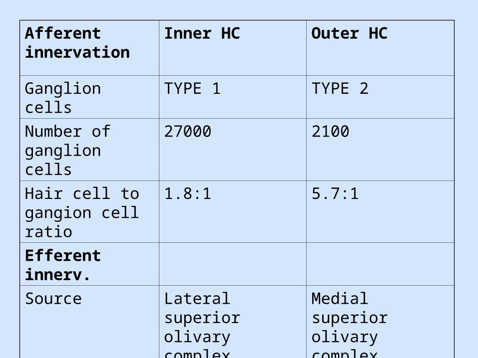
Afferent innervation
Inner HC Outer HC
Ganglion cells TYPE 1 TYPE 2
Number of ganglion cells
27000 2100
Hair cell to gangion cell ratio
1.8:1 5.7:1
Efferent innerv.
Source Lateral superior olivary complex
Medial superior olivary complex
Postsynaptic target
Afferent dendrites Base of hair cell