Embed Size (px)
Citation preview
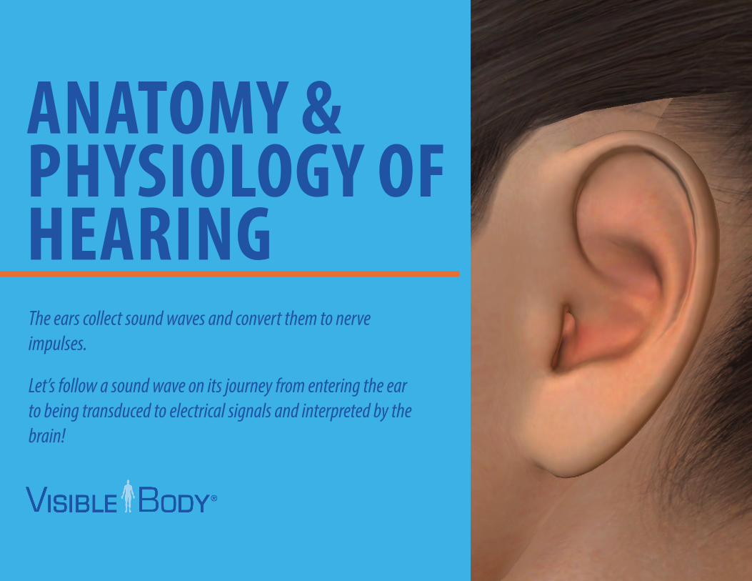
ANATOMY & PHYSIOLOGY OF HEARINGThe ears collect sound waves and convert them to nerve impulses.
Let’s follow a sound wave on its journey from entering the ear to being transduced to electrical signals and interpreted by the brain!
2
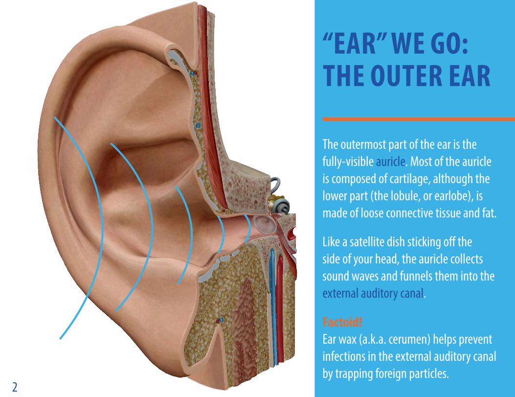
“EAR” WE GO: THE OUTER EAR
The outermost part of the ear is the fully-visible auricle. Most of the auricle is composed of cartilage, although the lower part (the lobule, or earlobe), is made of loose connective tissue and fat.
Like a satellite dish sticking off the side of your head, the auricle collects sound waves and funnels them into the external auditory canal.
Factoid! Ear wax (a.k.a. cerumen) helps prevent infections in the external auditory canal by trapping foreign particles.
3
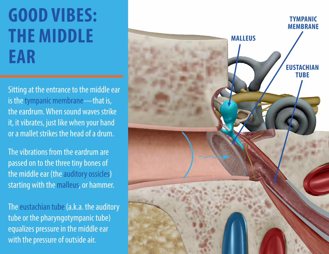
GOOD VIBES: THE MIDDLE EARSitting at the entrance to the middle ear is the tympanic membrane—that is, the eardrum. When sound waves strike it, it vibrates, just like when your hand or a mallet strikes the head of a drum.
The vibrations from the eardrum are passed on to the three tiny bones of the middle ear (the auditory ossicles) starting with the malleus, or hammer. The eustachian tube (a.k.a. the auditory tube or the pharyngotympanic tube)equalizes pressure in the middle ear with the pressure of outside air.
TYMPANIC MEMBRANE
MALLEUS
EUSTACHIAN TUBE
4
WE’VE GOT TO GO DEEPER!
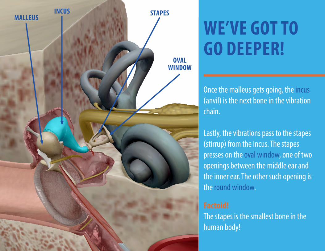
Once the malleus gets going, the incus (anvil) is the next bone in the vibration chain. Lastly, the vibrations pass to the stapes (stirrup) from the incus. The stapes presses on the oval window, one of two openings between the middle ear and the inner ear. The other such opening is the round window.
Factoid! The stapes is the smallest bone in the human body!
OVAL WINDOW
STAPESINCUSMALLEUS
5
DOUBLE LABYRINTH: THE INNER EAR
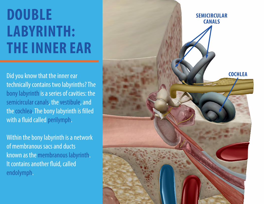
Did you know that the inner ear technically contains two labyrinths? The bony labyrinth is a series of cavities: the semicircular canals, the vestibule, and the cochlea. The bony labyrinth is filled with a fluid called perilymph. Within the bony labyrinth is a network of membranous sacs and ducts known as the membranous labyrinth. It contains another fluid, called endolymph.
SEMICIRCULAR CANALS
COCHLEA
6
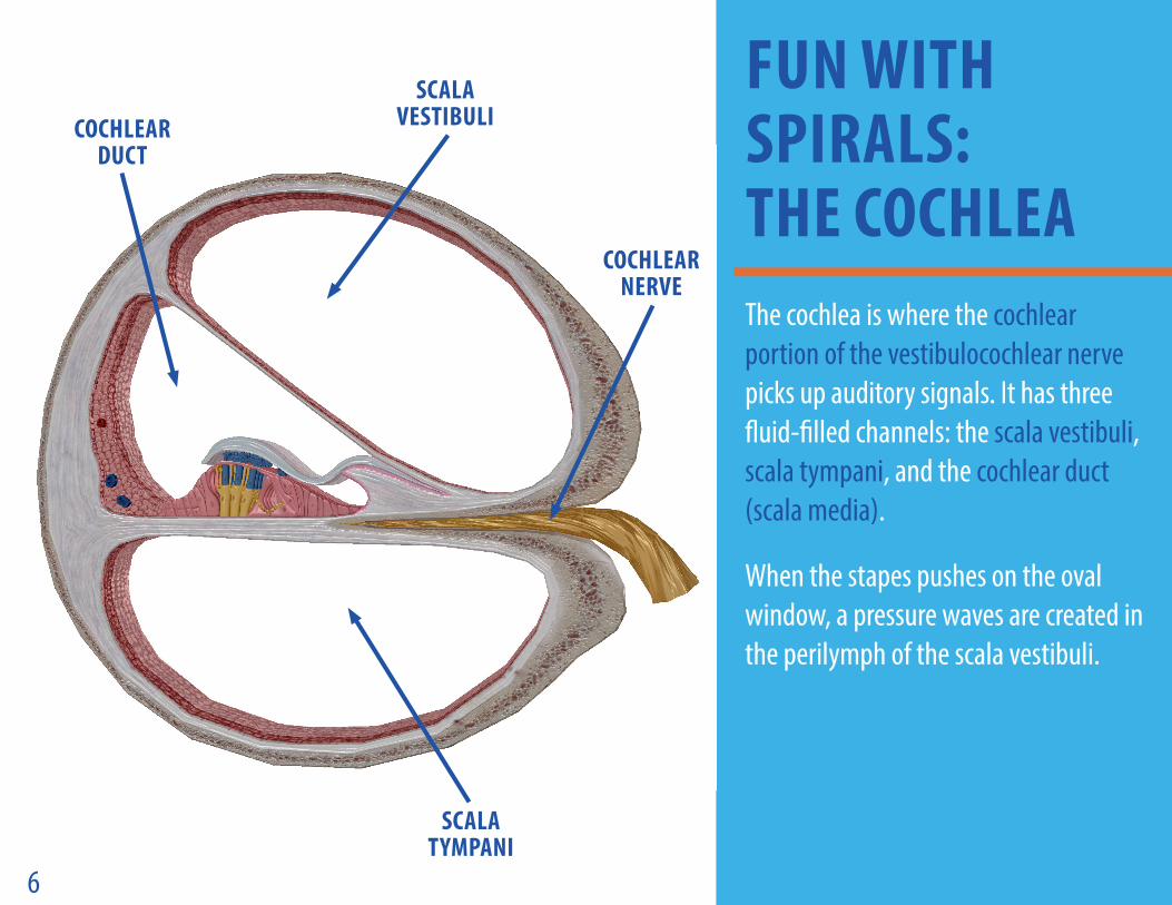
The cochlea is where the cochlear portion of the vestibulocochlear nerve picks up auditory signals. It has three fluid-filled channels: the scala vestibuli, scala tympani, and the cochlear duct (scala media).
When the stapes pushes on the oval window, a pressure waves are created in the perilymph of the scala vestibuli.
FUN WITH SPIRALS: THE COCHLEA
SCALA VESTIBULICOCHLEAR
DUCT
SCALA TYMPANI
COCHLEAR NERVE
7
A HAIRY SITUATION
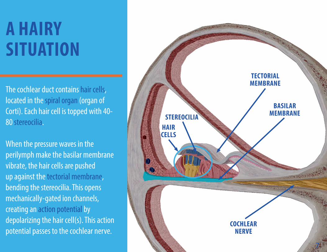
The cochlear duct contains hair cells, located in the spiral organ (organ of Corti). Each hair cell is topped with 40-80 stereocilia. When the pressure waves in the perilymph make the basilar membrane vibrate, the hair cells are pushed up against the tectorial membrane, bending the stereocilia. This opens mechanically-gated ion channels, creating an action potential by depolarizing the hair cell(s). This action potential passes to the cochlear nerve.
7
TECTORIAL MEMBRANE
BASILAR MEMBRANE
HAIR CELLS
COCHLEAR NERVE
STEREOCILIA
8
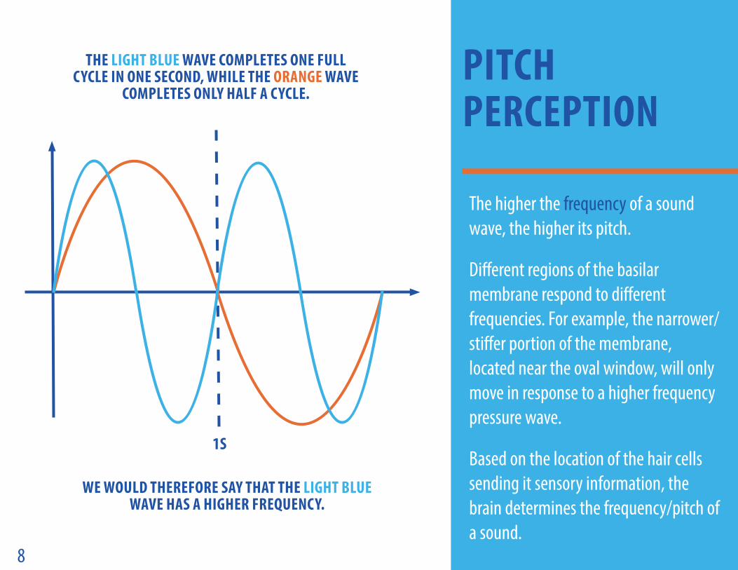
The higher the frequency of a sound wave, the higher its pitch.
Different regions of the basilar membrane respond to different frequencies. For example, the narrower/stiffer portion of the membrane, located near the oval window, will only move in response to a higher frequency pressure wave.
Based on the location of the hair cells sending it sensory information, the brain determines the frequency/pitch of a sound.
PITCH PERCEPTION
THE LIGHT BLUE WAVE COMPLETES ONE FULL CYCLE IN ONE SECOND, WHILE THE ORANGE WAVE
COMPLETES ONLY HALF A CYCLE.
WE WOULD THEREFORE SAY THAT THE LIGHT BLUE WAVE HAS A HIGHER FREQUENCY.
1S
9
HEARING AND THE BRAIN
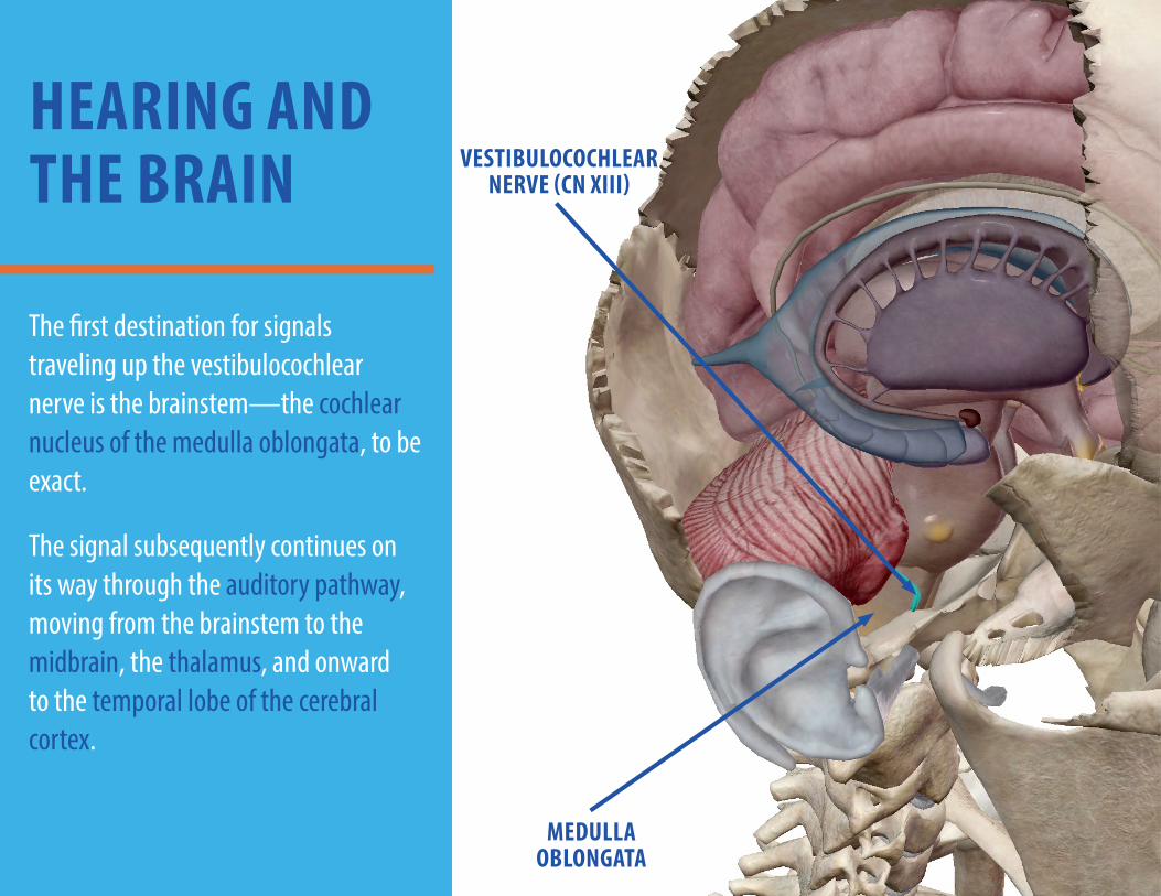
The first destination for signals traveling up the vestibulocochlear nerve is the brainstem—the cochlear nucleus of the medulla oblongata, to be exact.
The signal subsequently continues on its way through the auditory pathway, moving from the brainstem to the midbrain, the thalamus, and onward to the temporal lobe of the cerebral cortex.
VESTIBULOCOCHLEAR NERVE (CN XIII)
MEDULLA OBLONGATA
10
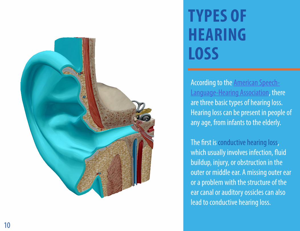
According to the American Speech-Language-Hearing Association, there are three basic types of hearing loss. Hearing loss can be present in people of any age, from infants to the elderly. The first is conductive hearing loss, which usually involves infection, fluid buildup, injury, or obstruction in the outer or middle ear. A missing outer ear or a problem with the structure of the ear canal or auditory ossicles can also lead to conductive hearing loss.
TYPES OF HEARING LOSS
11
HEARING LOSS (CONT.)
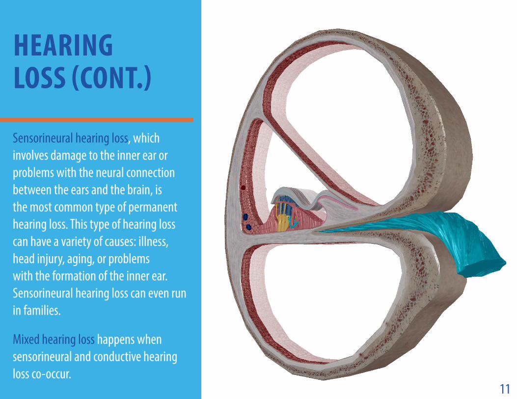
Sensorineural hearing loss, which involves damage to the inner ear or problems with the neural connection between the ears and the brain, is the most common type of permanent hearing loss. This type of hearing loss can have a variety of causes: illness, head injury, aging, or problems with the formation of the inner ear. Sensorineural hearing loss can even run in families.
Mixed hearing loss happens when sensorineural and conductive hearing loss co-occur.
12
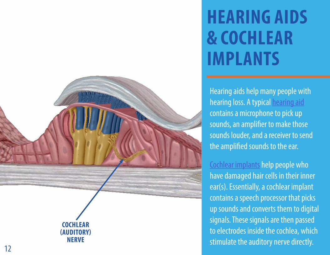
Hearing aids help many people with hearing loss. A typical hearing aid contains a microphone to pick up sounds, an amplifier to make those sounds louder, and a receiver to send the amplified sounds to the ear.
Cochlear implants help people who have damaged hair cells in their inner ear(s). Essentially, a cochlear implant contains a speech processor that picks up sounds and converts them to digital signals. These signals are then passed to electrodes inside the cochlea, which stimulate the auditory nerve directly.
HEARING AIDS & COCHLEAR IMPLANTS
COCHLEAR (AUDITORY)
NERVE
A universe of anatomical and physiological visuals and reference texts at your fingertips!
www.visiblebody.com
View a 3D Tour of all the images featured in this eBook!
If you have a mobile version of Human Anatomy Atlas 2021.1 or later: 1. Click here to view the tour.
If you have a web version of Atlas:1. Copy this link:
https://apps.visiblebody.com/share/?p=vbhaa&t=4_34154_637566144622624330_69785
2. Use the share link button in the app.3. Paste the link to view the tour.