Embed Size (px)
Citation preview
RESEARCH ARTICLE
Physiological increase of yolk testosterone
level does not affect oxidative status and
telomere length in gull hatchlings
Marco ParoliniID1*, Cristina Daniela Possenti1, Andrea Romano1,2, Manuela Caprioli1,
Diego Rubolini1, Nicola Saino1
1 Department of Environmental Science and Policy, University of Milan, Milan, Italy, 2 Department of
Ecology and Evolution, University of Lausanne, Lausanne, Switzerland
Abstract
Conditions experienced during early-life can cause the onset of oxidative stress, resulting in
pervasive effects on diverse life-history traits, including lifespan. In birds, maternally-trans-
ferred egg substances may exert positive or negative influence over the offspring phenotype.
Among these, testosterone can upregulate the bioavailability of certain antioxidants but
simultaneously promotes the production of pro-oxidants, leading to an oxidative stress situa-
tion, which is one of the main forces causing telomere attrition However, no study has investi-
gated the role of this androgen on telomere dynamics in birds and little is known about the
effects of yolk testosterone on oxidative status in early-life of these species. We physiologi-
cally increased the levels of yolk testosterone by in ovo injections in yellow-legged gull
(Larus michahellis) to evaluate the effects induced by this androgen on hatchlings plasma
total antioxidant capacity, amount of pro-oxidant molecules and telomere length at hatching.
Testosterone supplementation did not increase hatchling body growth, did not result in the
overproduction of pro-oxidant molecules nor a reduction of antioxidant capacity. Accordingly,
telomere length at hatching was not affected by testosterone treatment, although hatchlings
from the third-laid eggs showed shorter telomeres than their siblings from first- and second-
laid eggs, independently of testosterone treatment. Our results suggest that injection of
physiological levels of testosterone does not induce oxidative stress to hatchlings and, con-
sequently do not affect telomere dynamics during early post-natal periods.
Introduction
The conditions experienced during early-life can result in pervasive fitness consequences
through diverse physiological mechanisms, one of which involves telomere dynamics [1].
Telomeres are conserved repeated sequences of the TTAGGG nucleotide motif at the end of
chromosomes that protect genomic integrity [2]. Telomeres shorten throughout life, and short
telomeres at birth or rapid telomere attrition are associated with negative effects on diverse fit-
ness traits [3,4]. Telomere attrition is more pronounced during early-life [4], possibly because
PLOS ONE | https://doi.org/10.1371/journal.pone.0206503 October 26, 2018 1 / 14
a1111111111
a1111111111
a1111111111
a1111111111
a1111111111
OPEN ACCESS
Citation: Parolini M, Possenti CD, Romano A,
Caprioli M, Rubolini D, Saino N (2018)
Physiological increase of yolk testosterone level
does not affect oxidative status and telomere
length in gull hatchlings. PLoS ONE 13(10):
e0206503. https://doi.org/10.1371/journal.
pone.0206503
Editor: Camille Lebarbenchon, University of
Reunion Island, REUNION
Received: February 6, 2018
Accepted: October 15, 2018
Published: October 26, 2018
Copyright: © 2018 Parolini et al. This is an open
access article distributed under the terms of the
Creative Commons Attribution License, which
permits unrestricted use, distribution, and
reproduction in any medium, provided the original
author and source are credited.
Data Availability Statement: All relevant data are
within the manuscript and its Supporting
Information files.
Funding: The author(s) received no specific
funding for this work.
Competing interests: The authors have declared
that no competing interests exist.

intense metabolic activity underlying rapid body growth exposes the organism to the detri-
mental effects of oxidative stress [5]. In fact, oxidative stress, i.e. the imbalance of the equilib-
rium between the production of pro-oxidant molecules and antioxidant defences in favour of
the former [5], has been identified as a crucial mechanism affecting telomere length [6].
In birds, mothers transfer diverse extra-genomic substances to their eggs, which exert a pro-
found influence on offspring phenotype [7]. Whilst the allocation of exogenous antioxidants
serves to protect the offspring against detrimental effects of oxidative stress [8] and can help to
maintain telomere integrity during early-life periods [9], the transfer of steroid hormones can
impair the oxidative status and negatively affect telomere length of the progeny [10]. Testoster-
one is a maternally-transferred androgen [11,12] playing a pivotal role in the regulation of
embryo differentiation and development of diverse phenotypic traits [11]. As other androgens,
testosterone is anabolic for muscle and skeletal growth [13–15] and supports post-natal body
mass gain [16–18], although some other studies have returned none or opposite outcomes
regarding its effect on post-natal growth [19–21]. Some investigations have shown that varia-
tions in the exposure to testosterone altered the oxidative balance of birds, with contrasting
outcomes. For instance, high amounts of maternally transferred androgens, including testos-
terone, may represent a cost for offspring in terms of increased susceptibility to oxidative stress
due to the accelerated metabolism [11,12]. Enhanced developmental rate mediated by andro-
gens transferred by mothers to the eggs is associated with an increase of cell metabolism and a
concomitant overproduction of reactive oxygen species (ROS) [12], with a subsequent shift of
the balance between ROS and antioxidants [22]. Moreover, experimental findings have sup-
ported the idea that testosterone may also affect the antioxidant machinery [23,24]. A study of
zebra finch whose egg testosterone levels were artificially elevated produced male chicks, but
not female, with lower plasma antioxidant capacity [25]. Conversely, an experimental increase
of testosterone levels of wild male red grouse (Lagopus lagopus scoticus) resulted in high levels
of circulating antioxidants and high lipid peroxidation in plasma [26]. A similar manipulations
of red-legged partridge (Alectoris rufa) produced mixed responses [27,28], while testosterone
supplementation in the yolk of yellow-legged gull (Larus michahellis) eggs increased plasma
antioxidant capacity and reduced lipid peroxidation in hatchlings during early post-natal peri-
ods [7]. Considering the capability of testosterone to affect both sides of the ‘oxidative stress’
medal, this androgen might alter the individual oxidative balance and lead to an oxidative
stress situation, which can consequently change telomere dynamics. However, the effects of
testosterone on telomeres have never been reported in any bird species to date, neither under
captive or natural conditions. Thus, to shed light on the capability of testosterone to induce
oxidative stress and to elucidate its potential contribution to telomere dynamics, we assessed
the effects of a physiological increase in yolk testosterone concentration on oxidative status
markers (i.e. the plasmatic amount of pro-oxidant molecules and the total antioxidant capac-
ity) and telomere length in yellow-legged gull hatchlings. We expect that testosterone supple-
mentation would promote the oxidative cost of growth in hatchlings, boosting the production
of pro-oxidants and/or decreasing the total antioxidant capacity, and consequently reducing
telomere length. In addition, we expect that telomere length would be positively correlated
with plasma antioxidant capacity or negatively correlated with the concentration of pro-oxi-
dant molecules.
Materials and methods
Field procedures
The present study was performed on a large breeding colony (> 400 breeding pairs) of yellow-
legged gull in the Comacchio lagoon (NE Italy 44˚20’ N– 12˚11’ E). The colony was visited
No evidence for testosterone effects on oxidative status and telomere length in gulls
PLOS ONE | https://doi.org/10.1371/journal.pone.0206503 October 26, 2018 2 / 14

every second day to check for any new nests and newly laid eggs. When a new egg was found,
it was marked to monitor the progress of laying and to identify laying sequence and temporar-
ily removed from the nest for experimental manipulation. Removed egg was temporarily
replaced with a ’dummy’ egg to prevent any potential change in parental brooding behavior.
Eggs were transferred to a nearby tent for manipulation and then they were brought back
within two hours from the collection.
We aimed at increasing the concentration of testosterone by 1 standard deviation (SD) of
the concentration measured in the yolk of yellow-legged gull eggs from the same colony [29],
by the injection of a physiologically appropriate volume of a testosterone solution. As the yolk
testosterone concentration in the yellow-legged gull eggs varies according to egg size and posi-
tion in the laying sequence, we scaled the dose due to be injected accordingly. We grouped
first- (a-), second- (b-) or third- (c-) laid eggs into three classes (i.e. tertiles) of size depending
on the egg mass. Then, we calculated the standard deviation (SD) of the testosterone concen-
tration in the yolk for each tertile within each position in the laying sequence. We estimated
the yolk mass for each class size and position in laying sequence according to the following
equation: yolk mass = 0.227 (0.039 SE) egg mass + 1.815 (3.461 SE); F1,88 = 34.38, P< 0.001).
We computed the amount of testosterone due to be injected as the product of the SD
(expressed in ng/g) of testosterone concentration for each tertile and position in the laying
sequence and the estimated yolk mass. The injected testosterone doses were as follows (laying
order: class of size according to egg mass (g), amount of testosterone (T) injected (ng per
egg)): a-eggs: 84–91 g: 57 ng, 92–95 g: 59 ng, 96–108 g: 42 ng, b-eggs: 80–88 g, 74 ng, 89–92 g,
73 ng, 93–99 g: 81 ng; and c-eggs, 75–82 g, 95 ng, 82–87 g, 84 ng, 88–98 g, 76 ng. The amount
of testosterone injected in the yolk of yellow-legged gull eggs was in the same range of other
previous studies [7,21].
Testosterone was injected in the egg yolk according to a previously validated procedure
[30]. Before the injection, the egg was weighed and placed with the longitudinal axis vertical.
After eggshell disinfection, a hole close to the acute pole was drilled using a sterile pin. Injec-
tion was performed using 1-mL sterile syringe with a 0.6 × 30 mm needle, while the egg was
held firmly with its longitudinal axis vertical. The hole was sealed with a drop of epoxidic glue
and a small piece of eggshell superimposed to the hole soon after the injection. Testosterone
solutions were prepared in sterile vials dissolving the hormone in corn oil to the final concen-
tration required. Each vial contained the concentration of testosterone to be injected in egg
yolk depending on egg mass and position in the laying sequence. We adopted a within-clutch
design, whereby both control and testosterone-treated eggs were established within each
clutch. We sequentially assigned the treatment schemes to the clutches, according to the order
in which the first egg was found (nest, a-, b-, c-egg) as follows: nest 1, testosterone injection
(T), control injection (C), T; nest 2, C-T-C; nest 3, T-C-C; nest 4, C-T-T and so forth with the
following nests. Testosterone-treated eggs were injected with 30 μl of the appropriate testoster-
one solution, while control eggs were injected with the same volume of corn oil only.
After the in-ovo injection, all the nests were visited every day and eggs were monitored until
hatching. Because normally up to two days elapse between the time when the egg reaches the
pipping stage and hatching, we assigned chicks to their original egg injecting in the pipping
egg a small drop of a food dye (either blue or green). Upon the first daily visit to the nest when
any individual chick was found to have hatched, the chick was weighed (to the nearest g) and
its tarsus was measured (to the nearest 0.1 mm). Finally, a blood sample (about 70 μl) was col-
lected in heparinized capillary tubes after puncturing the hatchling ulnar vein. Blood samples
were centrifuged at 11,500 rpm for 10 min to separate red blood cells from plasma, which were
both stored at– 20 ˚C until biochemical and telomere length analyses. Molecular sexing of
chicks was performed by the amplification of a section of the CHD gene [31].
No evidence for testosterone effects on oxidative status and telomere length in gulls
PLOS ONE | https://doi.org/10.1371/journal.pone.0206503 October 26, 2018 3 / 14
Ethics statement
This study was conducted under the permission of the Parco Regionale del Delta del Po
(#252015, 20 February 2015), which allowed both the manipulation of the eggs biochemical
quality and the withdrawal of a blood sample from hatchlings. Blood samples (50–100 μl) were
collected by slightly puncturing the brachial vein with sterile needles and the puncturing site
was carefully disinfected. No obvious negative consequences of handling hatchlings were
detected.
Methods of oxidative status markers
Total antioxidant capacity (TAC) and the amount of total oxidant status (TOS) were measured
in plasma of chicks hatched from both control and T-injected eggs. TAC was measured
according to a colorimetric method developed by Erel [32]. The color of 2,2’-azinobis-(3-ethyl-
benzothiazoline-6-sulfonic acid) radical cation (ABTS�+) bleaches depending on the concen-
tration of antioxidants in the sample. The reaction is monitored spectrophotometrically and
the final absorbance is inversely related to TAC of the sample. The assay was calibrated with a
standard curve of Trolox and the results were expressed as μM Trolox equivalent. Mean TAC
intra-assay coefficient of variation (CV) was 3.0 ± 1.2% (n = 5 replicates), while the mean
inter-assay CV was 5.4 ± 3.8% (n = 3 assay plates). TOS was measured according to the colori-
metric method developed by Erel [33] in order to assess the amount of pro-oxidant molecules
in the sample. The oxidants in the plasma oxidize the ferrous ion-o-dianisidine complex to the
ferric ion, which reacting with xylenol orange gives a blue complex. Coloration (proportional
to the oxidant molecules in the plasma) was measured by a spectrophotometer at λ = 535 nm.
The assay was calibrated by using a standard curve made with hydrogen peroxide (H2O2). The
results were expressed as μM H2O2 equivalents. The mean TOS intra-assay CV was 3.2 ± 2.7%
(n = 5 replicates) and the inter-assay CV was 5.7 ± 4.1% (n = 3 assay plates).
Telomere length analysis
Telomere length (TL) analysis was performed according to the method described by Parolini
and coauthors [34]. Genomic DNA was extracted from 10–20 μl of red blood cells using 1 mL
TNSE buffer (10 mM Tris HCl, 400 mM NaCl, 100 mM EDTA and 0.6% SDS) and a standard
phenol/chloroform method. We measured the quantity and purity of the extracted genomic
DNA using a Nanophotometer (IMPLEN). Telomere length was measured by the mono-
chrome multiplex quantitative PCR method (MMQPCR) using an iQ5 real-time PCR
detection systems (BioRad). PCR reactions were prepared using 20 ng of genomic DNA as
template, Quantitative Master Mix 2X SYBR Green (Genespin), telomere and CTCF primers
at a final concentration of 1,000 nM and 500 nM each, respectively. The sequences of telomere
primers for MMQPCR were (telg 5’-ACACTAAGGTTTGGGTTTGGGTTTGGGTTTGGGTTAGTGT-3’ and telc 5’-TGTTAGGTATCCCTATCCCTATCCCTATCCCTATCCCTAACA-3’),
while the single copy sequence used as control was a fragment from the 12th exon of the swal-
low CTCF gene (CCCTC-binding factor zinc finger protein). The CTCF primers used were:
forward (5’-CCCGCGGCGGGCGGCGCGGGCTGGGCGGCTCCCAATGGAGACCTCAC-3’)
and reverse (5’-CGCCGCGGCCCGCCGCGCCCGTCCCGCCCATCACCGGTCCATCATGC-3’).
The CTCF primers are composed of a swallow genomic sequence and a GC-clamp at the 5’
end (underlined) to increase the melting temperature of the PCR product. Since the melting
temperature of PCR products of telomeres and CTCF are different, both primer pairs were
used in the same reaction. Cycling parameters for the PCR reactions were previously described
by Parolini et al. [34] and were: Stage 1: 15 min at 95 ˚C; Stage 2: 2 cycles of 15 sec at 94 ˚C, 15
sec at 49 ˚C; and Stage 3: 35 cycles of 15 sec at 94 ˚C, 10 sec at 62 ˚C, 15 sec at 74 ˚C with signal
No evidence for testosterone effects on oxidative status and telomere length in gulls
PLOS ONE | https://doi.org/10.1371/journal.pone.0206503 October 26, 2018 4 / 14
acquisition, 10 sec at 84 ˚C, 15 sec at 88 ˚C with signal acquisition. Four-fold serial dilutions
(from 10 to 100 ng) of a yellow-legged gull hatchling reference sample (DNA was extracted by
blood of a coeval chick not included in the experiment) were included in each plate to produce
a standard curve to measure reaction efficiency and quantify the amount of telomeric repeats
and single copy gene in each sample. We used the same reference sample in each plate. All
reactions were run in triplicate and six plates containing 20 samples each were performed. The
testosterone and control samples were equally distributed within each plate. Five samples were
replicated in each plate to assess repeatability of telomere measures. Telomere length was mea-
sured as the T/S ratio, corresponding to the ratio between the mean values of the amount of
telomeric repeats (T) and of a single copy gene (S), which was then related to the T/S value of
the reference sample. Thus, telomere length was expressed as relative telomere length (RTL).
The mean reaction efficiencies for both CTCF and telomere amplifications were greater than
88% and 94%, respectively. In MMQPCR performed to measure relative telomere length lower
efficiencies of amplification is expected compared to standard real time qPCR because the
primers contain mismatches relative to a perfect TTAGGG repeat. This strategy, which is used
to favor the synthesis of PCR products with homogeneous size, gives rise to lower efficiencies
during the first cycles. For the control qPCR, primers containing GC clamps/adapters are used
to increase melting temperature of the products allowing detection of both PCR reactions in
a single well. Also in this case a relatively low reaction efficiency is expected during the first
cycles. The mean intra-and inter-plate coefficient of variation (± SD) of RTL measures was
2.32 ± 2.95% and 10.76 ± 4.20%, respectively. The inter-plate coefficient of variation for RTL
was not negligible and could be due to differences in qPCR efficiencies occurring among
plates, affecting the RTL measurements, and enlarging the coefficient of variation of our mea-
surements. However, considering that the effect of T treatment is far from significance, we are
confident that inter-plate variability was not responsible for the absence of significant results.
The intra- and inter-plate repeatability of RTL measures, expressed as intra-class correlation
coefficient (ICC), was 0.68 and 0.77, respectively.
Statistical analysis
The effect of testosterone injection on oxidative status markers and RTL was analyzed in linear
mixed models (LMM), including clutch identity as a random factor. Treatment, hatchling sex
and egg-laying order were included as categorical fixed-effect factors with their two-way inter-
actions. Only non-significant (P> 0.05) interaction terms were excluded from the models in
a single step. Complete models are reported in Supporting Information (S2 Table). Moreover,
as oxidative stress is a driving force of telomere attrition, in order to investigate the possible
covariation between RTL and oxidative status markers, we re-run the same LMM including
TAC or TOS as a covariate. Complete models are reported in Supporting Information (S3
Table). Levels of TAC and TOS could not be measured in some (3–7) hatchlings because of
plasma scarceness (3 samples from control group and 4 samples for testosterone treated
group). We also excluded from the analyses a statistical outlier for RTL from an individual of
the testosterone treated group (RTL = 0.651) after performing the Grubbs’ test, also called the
ESD method (extreme studentized deviate). LMM analyses were performed by SAS 9.3 PROC
MIXED.
Results
We inoculated 201 eggs, whose hatching success significantly differed between the control
(proportion of hatched eggs = 43/97 = 0.443) and T-injected (67/104 = 0.644; χ21 = 7.387,
P = 0.006) groups. The sample of hatchlings included 110 individuals from 67 clutches (mean
No evidence for testosterone effects on oxidative status and telomere length in gulls
PLOS ONE | https://doi.org/10.1371/journal.pone.0206503 October 26, 2018 5 / 14
number of hatchlings per clutch: 1.61 (0.55 SD)), with 2 clutches only (3%) containing three
hatched chicks, 37 (55%) and 28 (42%) containing two and one chick, respectively. The sex
ratio of hatched chicks (proportion of males) did not significantly differ between experimental
groups (control eggs: 27/43 = 0.627; T-injected eggs: 28/67 = 0.418; χ21 = 3.82, P = 0.051),
although a marginally non-significant increase of females in T-treated eggs was found. At lay-
ing, egg mass did not significantly differ between the two experimental groups (F1,49.2 = 1.886,
P = 0.176; S1 Table). However, egg mass significantly declined with the position in the laying
sequence (F2,47.2 = 13.72, P < 0.01; estimated marginal means (SE): first-laid eggs: 88.46 (1.12)
g; second-laid eggs: 87.05 (1.07) g; third-laid eggs: 82.86 (1.11) g), with significant pairwise dif-
ferences between the first- and third-laid eggs, as well as between second- and third-laid eggs
(LSD test; P < 0.001 in both the cases). Testosterone supplementation did not affect the incu-
bation time (F1,59 = 2.46, P = 0.122), the body mass (F1,76 = 0.17; P = 0.683) and tarsus length
(F1,85 = 0.02, P = 0.877) of hatchlings, after controlling for sex and laying order (S1 Fig and S1
Table). Testosterone treatment did not significantly affect neither the levels of pro-oxidant
molecules nor the total antioxidant capacity in models controlling for laying order and sex
effects (Table 1 and S2 Table). However, the amount of pro-oxidants depended on laying
order, with hatchlings from second-laid eggs having smaller values than those from first-
(P = 0.020) and third-laid (P = 0.036) eggs. Testosterone treatment did not significantly
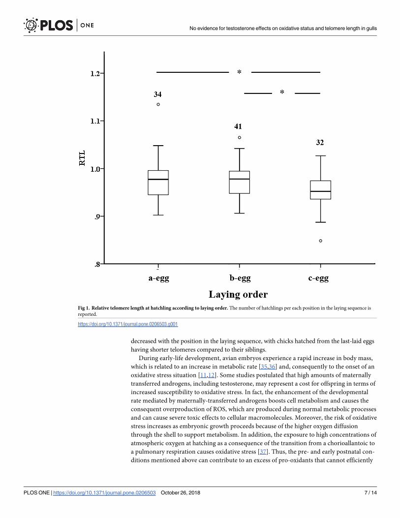
affect RTL (Table 1 and S2 Table), although it was found to vary with laying order (Fig 1), with
RTL from last-laid eggs being smaller than RTL from first- (P = 0.014) or second-laid eggs
(P = 0.017). Separate LMM of RTL including TAC or TOS as a covariate returned qualitatively
similar results compared to original models (S3 Table) and did not reveal any significant
covariation between RTL and TAC or TOS (F< 1.70 P> 0.197 in both the cases; S3 Table).
Discussion
Our results showed that an experimental increase of physiologically-relevant yolk testosterone
levels did not cause an overproduction of pro-oxidant molecules nor changes in the antioxi-
dant capacity of hatchlings. Accordingly, telomere length was not affected by the experimental
treatment. However, regardless of testosterone injection, we found that telomere length
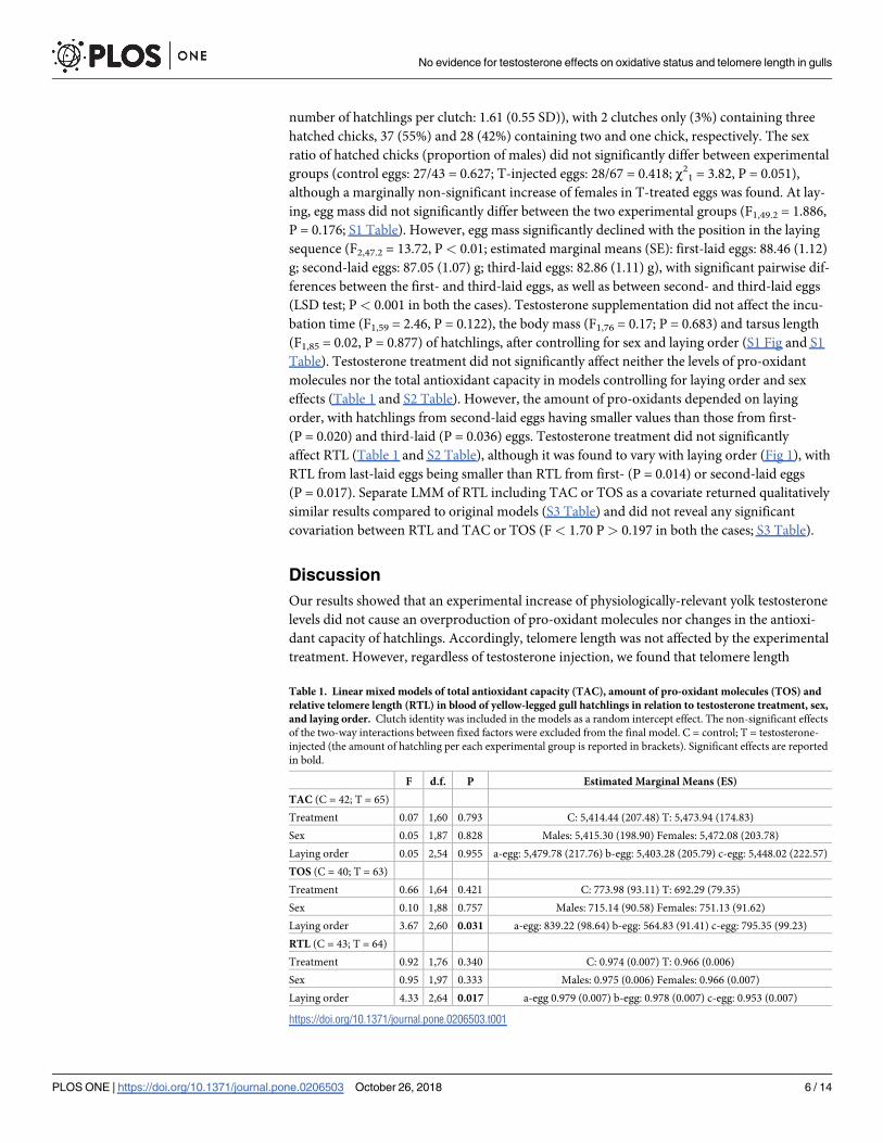
Table 1. Linear mixed models of total antioxidant capacity (TAC), amount of pro-oxidant molecules (TOS) and
relative telomere length (RTL) in blood of yellow-legged gull hatchlings in relation to testosterone treatment, sex,
and laying order. Clutch identity was included in the models as a random intercept effect. The non-significant effects
of the two-way interactions between fixed factors were excluded from the final model. C = control; T = testosterone-
injected (the amount of hatchling per each experimental group is reported in brackets). Significant effects are reported
in bold.
F d.f. P Estimated Marginal Means (ES)
TAC (C = 42; T = 65)
Treatment 0.07 1,60 0.793 C: 5,414.44 (207.48) T: 5,473.94 (174.83)
Sex 0.05 1,87 0.828 Males: 5,415.30 (198.90) Females: 5,472.08 (203.78)
Laying order 0.05 2,54 0.955 a-egg: 5,479.78 (217.76) b-egg: 5,403.28 (205.79) c-egg: 5,448.02 (222.57)
TOS (C = 40; T = 63)
Treatment 0.66 1,64 0.421 C: 773.98 (93.11) T: 692.29 (79.35)
Sex 0.10 1,88 0.757 Males: 715.14 (90.58) Females: 751.13 (91.62)
Laying order 3.67 2,60 0.031 a-egg: 839.22 (98.64) b-egg: 564.83 (91.41) c-egg: 795.35 (99.23)
RTL (C = 43; T = 64)
Treatment 0.92 1,76 0.340 C: 0.974 (0.007) T: 0.966 (0.006)
Sex 0.95 1,97 0.333 Males: 0.975 (0.006) Females: 0.966 (0.007)
Laying order 4.33 2,64 0.017 a-egg 0.979 (0.007) b-egg: 0.978 (0.007) c-egg: 0.953 (0.007)
https://doi.org/10.1371/journal.pone.0206503.t001
No evidence for testosterone effects on oxidative status and telomere length in gulls
PLOS ONE | https://doi.org/10.1371/journal.pone.0206503 October 26, 2018 6 / 14
decreased with the position in the laying sequence, with chicks hatched from the last-laid eggs
having shorter telomeres compared to their siblings.
During early-life development, avian embryos experience a rapid increase in body mass,
which is related to an increase in metabolic rate [35,36] and, consequently to the onset of an
oxidative stress situation [11,12]. Some studies postulated that high amounts of maternally
transferred androgens, including testosterone, may represent a cost for offspring in terms of
increased susceptibility to oxidative stress. In fact, the enhancement of the developmental
rate mediated by maternally-transferred androgens boosts cell metabolism and causes the
consequent overproduction of ROS, which are produced during normal metabolic processes
and can cause severe toxic effects to cellular macromolecules. Moreover, the risk of oxidative
stress increases as embryonic growth proceeds because of the higher oxygen diffusion
through the shell to support metabolism. In addition, the exposure to high concentrations of
atmospheric oxygen at hatching as a consequence of the transition from a chorioallantoic to
a pulmonary respiration causes oxidative stress [37]. Thus, the pre- and early postnatal con-
ditions mentioned above can contribute to an excess of pro-oxidants that cannot efficiently
Fig 1. Relative telomere length at hatchling according to laying order. The number of hatchlings per each position in the laying sequence is
reported.
https://doi.org/10.1371/journal.pone.0206503.g001
No evidence for testosterone effects on oxidative status and telomere length in gulls
PLOS ONE | https://doi.org/10.1371/journal.pone.0206503 October 26, 2018 7 / 14
be counteracted by antioxidant defenses and repair mechanisms. This would ultimately
leads to an imbalance of the hatchling oxidative status and, consequently, to telomere loss
[38].
Effects of testosterone on body mass and offspring sex ratio
Contrary to our expectations, testosterone supplementation did not boost somatic growth of
hatchlings. Opposite results were obtained by a previous companion study of the yellow-legged
gull investigating the effects of a physiological increase of yolk testosterone levels on the phe-
notype of developing embryos [39], showing that embryos from testosterone-treated eggs had
a larger body size and a smaller amount of residual yolk mass compared to controls [39].
These previous findings suggest that testosterone (1) enhances body size, accelerating yolk
absorption and/or (2) possesses anabolic effects that are already expressed during late embry-
onic stages [39]. Moreover, in a previous manipulative experiment of egg testosterone levels
performed on the same yellow-legged gull colony, a negative effect of testosterone was found
on body mass four days after hatching [21]. Thus, the positive effect of testosterone on pre-
natal body size seems to vanish at birth and turns into negative during post-natal growth.
These findings are consistent with a previous study of the spotless starling (Sturnus unicolor),showing stronger effects of testosterone treatment during embryo development with respect
to the nestling period [40]. Interestingly, T-treatment induced a marginally non-significant
deviation of hatchling sex ratio towards females. These findings were partly in agreement
with previous studies of birds that returned contrasting results. Whilst long-term implants of
testosterone produced more male offspring in the homing pigeon [41] and spotless starling
[42], and short-term injection increased the number of males in the zebra finch [43] and the
white leghorn chicken [44], a correlative study of the Japanese quail showed that high circulat-
ing levels of testosterone produced more female offspring [45]. Thus, these studies showed
that testosterone has the potential to mediate offspring sex ratios under natural conditions,
although the mechanism by which the adjustment in sex ratio occurred in these species
remains unclear.
Effects of testosterone on oxidative status and telomere length
Conversely to findings on embryos that showed a testosterone-induced overproduction of
pro-oxidant molecules in the brain and the liver [39], the supplementation of yolk testosterone
did not boost the production of oxidizing compounds and did not affect the total antioxidant
capacity of hatchlings. These findings are consistent with a previous study showing that the
experimental manipulation of yolk testosterone did not affect neither plasmatic ROS levels nor
antioxidant capacity in 1-day old yellow-legged gull chicks hatched from the last-laid eggs [7].
However, the same study showed a significant enhancement of antioxidant capacity in testos-
terone-treated chicks at 5 and 9 days, associated with a decrease of lipid peroxidation, suggest-
ing that yolk testosterone may induce a mobilization of antioxidants during development and/
or produce a stress during early developmental periods that could promote a compensatory
response later in life [46]. Thus, a pre-natal physiological increase of yolk testosterone did not
cause an oxidative stress situation to hatchlings, which consequently did not affect telomere
length in our study. However, since in the yellow-legged gull the consequences of testosterone
supplementation on the oxidative status emerged in late post-natal period [7], we cannot
exclude an effect on telomere length in later life stages. For instance, a positive effect of a sub-
cutaneous injection with vitamin E and methionine on telomere length was found in blue tit
nestlings only one year after the treatment [40].
No evidence for testosterone effects on oxidative status and telomere length in gulls
PLOS ONE | https://doi.org/10.1371/journal.pone.0206503 October 26, 2018 8 / 14
Variation of telomere length according to the position in the laying
sequence
Regardless of testosterone treatment, hatchling telomere length decreased with the position in
the laying sequence (Fig 1). In the yellow-legged gull there is a clear laying order in the eggs
produced within a clutch, with a decrease in egg size and changes in the egg composition (i.e.
antioxidant and hormonal content) within broods [29]. The shortening of telomere length
with laying order suggests that, at least during early-life periods, last-hatched chicks within a
clutch experienced a faster rate of cellular senescence than their siblings [47]. Because in the
yellow-legged gull the amount of maternally-transferred antioxidants decreases with the laying
order [29], shorter telomere length of last-hatched chicks may be due to the lower amount of
antioxidants compared to their siblings from first- and second-laid eggs, which do not effi-
ciently safeguard their telomere integrity. These results are consistent with previous studies of
passerine birds, showing a decrease in early postnatal telomere length between the first- and
the last-hatched nestlings [47,48]. For instance, maternally-derived antioxidants such as vita-
mins or carotenoids decline with laying order in the zebra finch [49]. As the antioxidants
play a crucial role in protecting embryos from oxidative stress during prenatal development
[50,51], it is plausible that embryos from the last-laid eggs suffer high levels of oxidative stress
during development, resulting in shorter telomeres compared to their siblings. Lastly, although
later embryos within a clutch has higher levels of maternally-transferred testosterone [29] that
might contribute to imbalance the oxidative status and affect telomere length, the present
study revealed that this androgen does not contribute to the onset of an oxidative stress situa-
tion and to the consequent telomere loss at birth.
Although our investigation did not show any significant effect of a physiological increase of
yolk testosterone levels, some previous experimental studies have demonstrated that mater-
nally-transferred androgens can affect various traits of offspring phenotype, from embryonic
stage to adulthood (reviewed in [52]). Considering the contrasting outcomes returned by
manipulative studies of testosterone levels, an unequivocal interpretation of the effect due to
maternally-transferred testosterone is difficult to be formulated [12,52–54]. The inconsistency
of results might be due to differences in the experimental approach used to modulate yolk tes-
tosterone levels, which can be performed by manipulating mothers or their eggs [54]. Whilst
the manipulation of mothers prevents risk of damaging the egg or affect embryo development,
this experimental approach returns some shortcomings [53,54] that can complicate the inter-
pretation of the results. Podmokła and coauthors [52] showed that the manipulation of andro-
gen levels in the mothers did not significantly affect physiology, reproduction, survival and
maternal traits of bird species because the female might limit the transfer of circulating steroids
to the eggs through diverse mechanisms (e.g., selective uptake of hormones into the oocyte or
by barriers to passive diffusion [55]. In addition, the maternal manipulations of testosterone
can affect not only the quantity of this focal androgen transferred to the egg, but also egg and/
or maternal quality [53,55]. For instance, androgens and antioxidants (i.e. vitamin E) are co-
adjusted within bird eggs [56]. Thus, the administration of testosterone to the mother could
result in an increased allocation of antioxidants, which might prevent or limit the direct and
indirect effects due to prenatal T exposure on oxidative stress [56], and consequently on telo-
mere dynamics. For these reasons, the injection of known amounts of hormone into the egg
appears to be more appropriate method to be used in order to investigate the effects of hor-
mones on offspring phenotype [52–54] regardless some shortcomings, namely the selection of
the appropriate dose due to be injected, the choice of the solvent to be used as vehicle of the
hormone and the timing of egg manipulation [52]. Thus, to investigate the possible effects of
testosterone on hatchling growth, oxidative status and telomere length, we strived to prevent
No evidence for testosterone effects on oxidative status and telomere length in gulls
PLOS ONE | https://doi.org/10.1371/journal.pone.0206503 October 26, 2018 9 / 14
such limitations by 1) adjusting the testosterone dose due to be injected into the yolk according
to the levels naturally occurring in the eggs of yellow-legged gull breeding in the same colony
visited in the present study [29]; 2) using a lipophilic, non-toxic [20,34,39] solvent (i.e. corn
oil) allowing to completely solubilize testosterone and to prevent differential distribution and
bioavailability of the hormone in the yolk; and 3) manipulating the levels of testosterone at the
time of laying, before the developing embryo starts producing its own hormones, in order to
mimic the natural situation in which only maternal hormones occur in the egg. Thus, the lack
of significant effects due to testosterone treatment cannot be ascribed to the experimental
design we used. However, a recent meta-analysis [52] highlighted that the dose injected into
the eggs represents the crucial factor accounting for the effects due to testosterone on offspring
phenotype. Whilst no effect of solvent used, time of manipulation and site of egg injection
was pointed out, the effect due to androgen (mainly testosterone) supplementation greatly
depended on the injected dose. In fact, several studies highlighted a significant effect on diverse
phenotypic traits after injecting a dose exceeding the natural testosterone concentration into
the yolk by a couple of standard deviations, or a pharmacological dose (>5 SD up to 2,000
SD). In contrast, other studies injecting very low doses (� 1 SD) returned no effects due to
hormonal manipulation [52], accordingly to our results. Thus, we suppose that the lack of sig-
nificant effects on offspring body growth, oxidative status and consequently telomere length
could be simply due to the low amount of testosterone we injected into the egg yolk. At the
same time, we cannot exclude that a larger supplementation of testosterone into the yolk
results in a higher growth rate and in an unbalance of offspring oxidative status due to ROS
overproduction, causing telomere shortening.
In conclusion, our study showed that a physiological increase in testosterone yolk concen-
tration has no effects on plasma oxidative status and telomere length on hatchlings. However,
considering the contrasting outcomes from previous studies investigating the effects of testos-
terone on birds, testosterone-mediated effects may enforce trade-offs in body size and oxida-
tive status at different ontogenetic stages (i.e. embryonic and postnatal periods). Thus, we
cannot exclude that testosterone may affect body growth and imbalance the oxidative equilib-
rium affecting the telomere integrity in later postnatal stages, resulting in long-term conse-
quences to the offspring.
Supporting information
S1 Data. The file summarizes all the relevant data that have been used in the statistical
analyses.
(XLSX)
S1 Fig. Mean (±SD) of body mass and tarsus length of hatchlings according to the position
in the laying sequence. Body mass was expressed in grams, while tarsus length in millimeters.
(TIF)
S1 Table. Linear mixed models of egg mass at laying and incubation time in relation to tes-
tosterone treatment, sex, and laying order. Clutch identity was included in the model as a
random intercept effect. The non-significant effects of the two-way interactions between fixed
factors were excluded from the final model. C = control; T = testosterone-injected. Significant
effects are reported in bold.
(DOCX)
S2 Table. Linear mixed models of total antioxidant capacity (TAC), amount of pro-oxidant
molecules (TOS) and relative telomere length (RTL) in blood of yellow-legged gull hatch-
lings in relation to testosterone treatment, sex, and laying order. Clutch identity was
No evidence for testosterone effects on oxidative status and telomere length in gulls
PLOS ONE | https://doi.org/10.1371/journal.pone.0206503 October 26, 2018 10 / 14
included in the model as a random intercept effect. The non-significant effects of the two-way
interactions between fixed factors were excluded from the final model. C = control; T = testos-
terone-injected. Significant effects are reported in bold.
(DOCX)
S3 Table. Linear mixed models of relative telomere length (RTL) in blood of yellow-legged
gull hatchlings in relation to testosterone treatment, sex, and laying order. Total Antioxi-
dant Capacity (TAC) and amount of pro-oxidant molecules (TOS) were singularly included
in the models as a covariate. Clutch identity was included in the model as a random intercept
effect. The non-significant effects of the two-way interactions between fixed factors were
excluded from the final model. Significant effects are reported in bold.
(DOCX)
Author Contributions
Conceptualization: Marco Parolini, Diego Rubolini, Nicola Saino.
Formal analysis: Marco Parolini.
Investigation: Marco Parolini, Cristina Daniela Possenti, Andrea Romano, Manuela Caprioli,
Diego Rubolini, Nicola Saino.
Methodology: Marco Parolini, Cristina Daniela Possenti, Manuela Caprioli.
Resources: Nicola Saino.
Supervision: Nicola Saino.
Writing – original draft: Marco Parolini.
References1. Monaghan P. Organismal stress, telomeres and life histories. J. Exp. Biol. 2014; 217: 57–66. https://
doi.org/10.1242/jeb.090043 PMID: 24353204
2. Blackburn EH. Telomere states and cell fates. Nature 2000; 408: 53–56. https://doi.org/10.1038/
35040500 PMID: 11081503
3. Boonekamp JJ, Mulder GA, Salomons HM, Dijkstra C, Verhulst S. Nestling telomere shortening, but not
telomere length, reflects developmental stress and predicts survival in wild birds. Proc. R. Soc. B 2014;
281: 20133287.
4. Herborn KA, Heidinger BJ, Boner W, Noguera JC, Adam A, et al. Stress exposure in early post-natal life
reduces telomere length: an experimental demonstration in a longlived seabird. Proc. R. Soc. B 2014;
281: 20133151.
5. Monaghan P, Ozanne SE. Somatic growth and telomere dynamics in vertebrates: relationships, mecha-
nisms and consequences. Phil. Trans. R. Soc. B 2018; 373: 20160446.
6. von Zglinicki T. Oxidative stress shortens telomeres. Trends Biochem. Sci. 2002; 27: 339–344. PMID:
12114022
7. Noguera JC, Alonso-Alvarez C, Kim SY, Morales J and Velando A. Yolk testosterone reduces oxidative
damages during postnatal development. Biol. Lett. 2011; 7: 93–95. https://doi.org/10.1098/rsbl.2010.
0421 PMID: 20659922
8. Metcalfe NB, Alonso-Alvarez C. Oxidative stress as a life-history constraint: the role of reactive oxygen
species in shaping phenotypes from conception to death. Funct. Ecol. 2010; 24: 984–996.)
9. Kim S-Y, Velando A. Antioxidants safeguard telomeres in bold chicks. Biol. Lett. 2015; 11: 20150211.
10. Haussmann MF, Longenecker AS, Marchetto NM, Juliano SA, Bowden RM. Embryonic exposure to
corticosterone modifies the juvenile stress response, oxidative stress and telomere length. Proc. R.
Soc. B 2012; 279: 1447–1456. https://doi.org/10.1098/rspb.2011.1913 PMID: 22072607
11. Groothuis TGG, Schwabl H. Hormone mediated maternal effects in birds: mechanisms matter but what
do we know of them? Phil. Trans. R. Soc. B 2008; 363: 1647–1661. https://doi.org/10.1098/rstb.2007.
0007 PMID: 18048291
No evidence for testosterone effects on oxidative status and telomere length in gulls
PLOS ONE | https://doi.org/10.1371/journal.pone.0206503 October 26, 2018 11 / 14
12. Groothuis TGG, Muller W, von Engelhardt N, Carere C, Eising CX. Maternal hormones as a tool to
adjust offspring phenotype in avian species. Neurosci. Biobehav. Rev. 2008; 29: 329–352.
13. Eising CM, Muller W, Dijkstra C and Groothuis TGG. Maternal androgens in egg yolks: relation with
sex, incubation time and embryonic growth. Gen. Comp. Endocrinol. 2003; 132: 241–247. PMID:
12812771
14. Lipar JL and Ketterson ED. Maternally derived yolk testosterone enhances the development of the
hatching muscle in the red-winged blackbird Agelaius phoeniceus. Proc. R. Soc. Lond. B Biol. Sci.
2000; 267: 2005–2010.
15. Navara KJ, Hill GE, Mendonca MT. Yolk testosterone stimulates growth and immunity in house finch
chicks. Phys. Biochem. Zool. 2006; 97: 550–555.
16. Navara KJ, Hill GE, Mendonca MT. Variable effects of yolk androgens on growth, survival, and immunity
in eastern bluebird nestlings. Physiol. Biochem. Zool. 2005; 78: 570–578. https://doi.org/10.1086/
430689 PMID: 15957111
17. Pilz KM, Quiroga M, Schwabl H, Adkins-Regan E. European starling chicks benefit from high yolk tes-
tosterone levels during a drought year. Horm. Behav. 2004; 46: 179–192. https://doi.org/10.1016/j.
yhbeh.2004.03.004 PMID: 15256308
18. Schwabl H. Maternal testosterone in the avian egg enhances postnatal growth. Comp. Biochem. Phy-
siol. 1996; 114: 271–276.
19. Podlas K, Helfenstein F, Richner H. Brood reduction via intra-clutch variation in testosterone-an experi-
mental test in the great tit. PLoS ONE 2013; 8: e56672. https://doi.org/10.1371/journal.pone.0056672
PMID: 23437207
20. Possenti CD, Romano A, Caprioli M, Rubolini D, Spiezio C, Saino N, Parolini M. Yolk testosterone
affects growth and promotes individual-level consistency in behavioral lateralization of yellow-legged
gull chicks. Horm. Behav. 2016; 80: 58–67. https://doi.org/10.1016/j.yhbeh.2016.01.007 PMID:
26836770
21. Rubolini D, Romano M, Martinelli R, Saino N. Effects of elevated yolk testosterone levels on survival,
growth and immunity of male and female yellow-legged gull chicks. Behav. Ecol. Sociobiol. 2006; 59:
344–352.
22. Alonso-Alvarez C, Bertrand S, Faivre B, Chastel O, Sorci G. Testosterone and oxidative stress: the oxi-
dation handicap hypothesis. Proc. R. Soc. Lond. B Biol. Sci. 2007; 274: 819–825.
23. von Schantz T, Bensch S, Grahn M, Hasselquist D, Wittzell H. Good genes, oxidative stress and condi-
tion-dependent sexual signals. Proc. R. Soc. Lond. B Biol. Sci. 1999; 266: 1–12.
24. Alonso-Alvarez C, Bertrand S, Faivre B, Chastel O, Sorci G. Testosterone and oxidative stress: the oxi-
dation handicap hypothesis. Proc. R. Soc. Lond. B Biol. Sci. 2007; 274: 819–825.
25. Tobler M, Sandell MI. Sex-specific effects of prenatal testosterone on nestling plasma antioxidant
capacity in the zebra finch. J. Exp. Biol. 2009; 212: 89–94. https://doi.org/10.1242/jeb.020826 PMID:
19088214
26. Mougeot F, Martınez-Padilla J, Webster LM, Blount JD, Perez-Rodrıguez L, Piertney SB. Honest sexual
signalling mediated by parasite and testosterone effects on oxidative balance. Proc. R. Soc. Lond. B
Biol. Sci. 2009; 276: 1093–1100.
27. Alonso-Alvarez C, Perez-Rodrıguez L, Mateo R, Chastel O, Vinuela J. The oxidation handicap hypothe-
sis and the carotenoid allocation trade-off. J. Evol. Biol. 2008; 21: 1789–1797. https://doi.org/10.1111/j.
1420-9101.2008.01591.x PMID: 18713241
28. Alonso-Alvarez C, Perez-Rodrıguez L, Garcia JT, Viñuela J Testosterone-mediated trade-offs in the old
age: a new approach to the immunocompetence handicap and carotenoid-based sexual signalling.
Proc. R. Soc. Lond. B Biol. Sci. 2009; 276: 2093–2101.
29. Rubolini D, Romano M, Navara KJ, Karadas F, Ambrosini R, et al. Maternal effects mediated by egg
quality in the Yellow-legged Gull Larus michahellis in relation to laying order and embryo sex. Front.
Zool. 2011; 8: 24. https://doi.org/10.1186/1742-9994-8-24 PMID: 22011400
30. Romano M, Caprioli M, Ambrosini R, Rubolini D, Fasola M, Saino N. Maternal allocation strategies and
differential effects of yolk carotenoids on the phenotype and viability of yellow-legged gull (Larus micha-
hellis) chicks in relation to sex and laying order. J. Evol. Biol. 2008; 21: 1626–1640. https://doi.org/10.
1111/j.1420-9101.2008.01599.x PMID: 18713240
31. Lee JCI, Tsai L-C, Hwa P-Y et al. A novel strategy for avian species and gender identification using the
CHD gene. Mol. Cell Probes 2010; 24: 27–31. https://doi.org/10.1016/j.mcp.2009.08.003 PMID:
19716876
32. Erel O. A novel automated direct measurement method for total antioxidant capacity using a new gener-
ation, more stable ABTS radical cation. Clin. Biochem. 2004; 37: 277–285. https://doi.org/10.1016/j.
clinbiochem.2003.11.015 PMID: 15003729
No evidence for testosterone effects on oxidative status and telomere length in gulls
PLOS ONE | https://doi.org/10.1371/journal.pone.0206503 October 26, 2018 12 / 14
33. Erel O. A new automated colorimetric method for measuring total oxidant status. Clin. Biochem. 2005;
38: 1103–1111. https://doi.org/10.1016/j.clinbiochem.2005.08.008 PMID: 16214125
34. Parolini M, Khoriauli L, Possenti CD, Colombo G, Caprioli M, Santagostino M, Nergadze SG, Milzani A,
Giulotto E, Saino N. Yolk vitamin E prevents oxidative damage in gull hatchlings. R. Soc. Open Sci.
2017; 4: 170098.
35. Hoyt D. A new model of avian embryonic metabolism. J. Exp. Zool. Suppl. 1987; 1: 127–138. PMID:
3598486
36. Vleck CM, Vleck D, Hoyt DF. Patterns of metabolism and growth in avian embryos. Am. Zool. 1980; 20:
405–416.
37. Vleck CM, Vleck D. Metabolism and energetics of avian embryos. J. Exp. Zool. Suppl. 1987; 1: 111–
125. PMID: 3298529
38. Haussmann MF, Marchetto NM. Telomeres: linking stress and survival, ecology and evolution. Curr.
Zool. 2010; 56: 714–727.
39. Parolini M, Romano A, Possenti CD, Caprioli M, Rubolini D, Saino N. Contrasting effects of increased
yolk testosterone content on development and oxidative status in gull embryos. J. Exp. Biol. 2017; 220:
625–633. https://doi.org/10.1242/jeb.145235 PMID: 28202650
40. Muriel J, Perez-Rodrıguez L, Puerta M, Gil D. Differential effects of yolk testosterone and androstene-
dione in embryo development and nestling growth in the spotless starling (Sturnus unicolor). Gen.
Comp. Endocrinol. 2013; 194: 175–182. https://doi.org/10.1016/j.ygcen.2013.09.013 PMID:
24090611
41. Goerlich VC, Dijkstra C, Schaafsma SM, Groothuis TGG. Testosterone has a long-term effect on pri-
mary sex ratio of first eggs in pigeons-in search of a mechanism. Gen. Comp. Endocrinol. 2009; 163:
184–192. https://doi.org/10.1016/j.ygcen.2009.01.004 PMID: 19344666
42. Veiga JP, Vinuela J, Cordero PJ, Aparicio JM, Polo V. Experimentally increased testosterone affects
social rank and primary sex ratio in the spotless starling. Horm. Behav. 2004; 46: 47–53. https://doi.org/
10.1016/j.yhbeh.2004.01.007 PMID: 15215041
43. Rutkowska J and Cichon M. Maternal testosterone affects the primary sex ratio and offspring survival in
zebra finches. An. Behav. 2006; 71: 1283–1288.
44. Pinson SE, Wilson JL, Navara KJ. Elevated testosterone during meiotic segregation stimulates laying
hens to produce more sons than daughters. Gen. Comp. Endocrinol. 2011; 174: 195–201. https://doi.
org/10.1016/j.ygcen.2011.08.020 PMID: 21925503
45. Correa SM, Horan CM, Johnson PA, Adkins-Regan E. Copulatory behaviors and body condition
predict post mating female hormone concentrations, fertilization success, and primary sex ratios in
Japanese quail. Horm Behav. 2011; 59:556–64. https://doi.org/10.1016/j.yhbeh.2011.02.009 PMID:
21376051
46. Dimova EG, Bryant PE, Chankova SG. ‘Adaptive response’: some underlying mechanisms and open
questions. Genet. Mol. Biol. 2008; 31: 396–408.
47. Badas EP, Martinez J, Rivero De Aguilar Cachafeiro J, Miranda F, Figuerola J, Merino S. Ageing and
reproduction: antioxidant supplementation alleviates telomere loss in wild birds. J. Evol. Biol. 2015; 28:
896–905. https://doi.org/10.1111/jeb.12615 PMID: 25758014
48. Noguera JC, Metcalfe NB, Reichert S and Monaghan P. Embryonic and postnatal telomere length
decrease with ovulation order within clutches. Sci. Rep. 2016; 25915.
49. Stier A, Massemin S, Zahn S, Tissier ML, Criscuolo F. Starting with a handicap: effects of asynchronous
hatching on growth rate, oxidative stress and telomere dynamics in free-living great tits. Oecologia
2015; 179: 999–1010 https://doi.org/10.1007/s00442-015-3429-9 PMID: 26314343
50. Royle NJ, Surai PF, Hartley IR. The effect of variation in dietary intake on maternal deposition of antioxi-
dants in zebra finch eggs. Funct. Ecol. 2003; 17: 472–481.
51. Surai PF. Natural antioxidants in avian nutrition and reproduction. ( Nottingham University Press, Not-
tingham, 2002).
52. Podmokła E, Drobniak SM and Rutkowska J. Chicken or egg? Outcomes of experimental manipulations
of maternally transmitted hormones depend on administration method–a meta-analysis. Biol. Rev.
2018; https://doi.org/10.1111/brv.12406 PMID: 29573376
53. Gil D. Hormones in avian eggs: physiology, ecology and behavior. Advances in the Study of Behavior
2008; 38: 337–398.
54. Henriksen R, Rettenbacher S, Groothuis TGG. Prenatal stress in birds: pathways, effects, function and
perspectives. Neurosci. Biobehav. Rev. 2011; 35: 1484–1501. https://doi.org/10.1016/j.neubiorev.
2011.04.010 PMID: 21536067
No evidence for testosterone effects on oxidative status and telomere length in gulls
PLOS ONE | https://doi.org/10.1371/journal.pone.0206503 October 26, 2018 13 / 14

55. Groothuis TG, Schwabl H. Hormone-mediated maternal effects in birds: mechanisms matter but what
do we know of them?. Proc. R. Soc. Lond. B Biol. Sci. 2008, 363:1647–61.
56. Giraudeau M, Ducatez S. Co-adjustment of yolk antioxidants and androgens in birds. Biol. Lett. 2016,
12: 20160676.
No evidence for testosterone effects on oxidative status and telomere length in gulls
PLOS ONE | https://doi.org/10.1371/journal.pone.0206503 October 26, 2018 14 / 14