Embed Size (px)
Citation preview

Particle-Level Visualization of Hydrodynamic and Frictional Couplings inDense Suspensions of Spherical Colloids
Taiki Yanagishima ,1 Yanyan Liu,1 Hajime Tanaka ,2,3,* and Roel P. A. Dullens 1,†
1Department of Chemistry, Physical and Theoretical Chemistry Laboratory,University of Oxford, South Parks Road, Oxford OX1 3QZ, United Kingdom
2Department of Fundamental Engineering, Institute of Industrial Science,The University of Tokyo, Komaba 4-6-1, Meguro-ku, Tokyo 153-8505, Japan3Research Center for Advanced Science and Technology, University of Tokyo,
4-6-1 Komaba, Meguro-ku, Tokyo 153-8505, Japan
(Received 1 September 2020; revised 15 March 2021; accepted 15 April 2021; published 14 June 2021)
The rotational Brownian motion of colloidal spheres in dense suspensions reflects local hydrodynamicsand contact forces, which are both key to nonlinear rheological phenomena such as shear-thickening andjamming, and transport in crowded environments, including intracellular migration and blood flow. To fullyelucidate the role of rotational dynamics experimentally, it is crucial to measure the translational androtational motion of all spheres simultaneously. Here, we directly access hydrodynamic and frictionalcoupling in colloidal suspensions up to arbitrarily high volume fractions using compositionally uniformcolloidal spheres with an off-center, fully embedded core. We reveal interparticle hydrodynamic rotation-rotation coupling in charged colloidal crystals. We also find that higher local crystallinity in denser hard-sphere crystalline sediments enhances rotational diffusivity and that nearly arrested particles exhibit astick-slip rotational motion due to frictional coupling. Our findings shed new light on the largelyunexplored, local rotational dynamics of spherical particles in dense particulate materials.
DOI: 10.1103/PhysRevX.11.021056 Subject Areas: Condensed Matter Physics, Soft Matter
I. INTRODUCTION
The dynamics of colloidal particles is key to connectingthe equilibrium phase behavior of particulate suspensionsto their atomic analogs [1,2]. The vast majority of studies ofcondensed matter phenomena only focus on translationaldegrees of freedom; examples include crystallization [3–5],melting [6,7], gelation [8,9], and the glass transition [10](see Ref. [11] for a review). However, this is only half thepicture. As noted from the outset by Perrin [12], colloidalparticles also feature rotational Brownian motion, whichincludes spherical particles that do not have an easilyvisualized orientation.For a suspension of spherical particles, rotational
Brownian motion is governed by two physical effects,hydrodynamics and contact forces, including friction.Nonlocal hydrodynamic interactions play an important rolein colloidal gelation [9,13–15], the rheology of complex
fluids [16,17], and biological systems [18–20]. Contactforces such as adhesion and friction also play a significantrole in the behavior of colloidal systems. In the self-assembly of DNA-coated colloids, irreversible adhesioncan prevent the formation of target structures, while asystem that allows colloidal “rolling” allows equilibrationto the intended state [21]. The role of interparticle frictionin rheological phenomena such as shear thickening(reviewed in Ref. [22]) is also of particular interest dueto its industrial relevance. Extensive experimental [23–28]and theoretical [29–32] work in both athermal and thermalregimes has led to a significant debate on the relative rolesof contact interactions and hydrodynamics in shear thick-ening. Notably, microscopic access to a link betweenstructure and rheology has become possible by confocalrheology [23,25,33]. Nevertheless, direct access to therotational degrees of freedom of spherical particles, whichallows access to hydrodynamic and mechanical interactionsat the particle level, has been challenging to achieve.Although orientational dynamics have been studied
extensively for anisotropic particles [34–36], there areexceedingly few studies of rotational fluctuations in densesuspensions of spheres because of the lack of a colloidalmodel system that allows both the position and orientationof all the spheres in a field of view to be imaged up toarbitrarily high volume fractions. Studies using light
*[email protected]†[email protected]
Published by the American Physical Society under the terms ofthe Creative Commons Attribution 4.0 International license.Further distribution of this work must maintain attribution tothe author(s) and the published article’s title, journal citation,and DOI.
PHYSICAL REVIEW X 11, 021056 (2021)
2160-3308=21=11(2)=021056(17) 021056-1 Published by the American Physical Society

scattering have given us valuable insights into rotationaldiffusion in suspensions [37–39], but they have beenunable to correlate dynamics with local structure to reflectstructural heterogeneity, a key strength of mesoscopiccolloidal models.Recently, there has been a clear escalation of efforts to
create a viable system. Efforts to directly image rotationaldynamics include nematic liquid crystal droplets with afrozen director [40], Janus (MOON) particles [41],anisotropic fluorescence profiles using photobleaching[42,43], and rough colloidosomes with a subpopulationof fluorescent surface probes [44], but none of these allowsconfocal microscopy studies at arbitrarily high volumefractions. A recent work encapsulated a silica core [45] in a3-trimethoxysilyl propyl methacrylate (TPM) shell to makea core-shell particle, again leaving a scattering interface thatmakes confocal microscopy in dense suspensions unfea-sible. Anisotropic polymethyl methacrylate clusters in aspherical polymethyl methacrylate shell were proposed torectify this [46], but producing monodisperse batches inbulk is not possible. As of yet, no system allows full three-dimensional (3D) characterization of rotational fluctuationsin monodisperse spheres using confocal laser scanningmicroscopy (CLSM) in arbitrarily dense systems.Here, we directly visualize hydrodynamic and contact
forces in dense colloidal suspensions by applying mono-disperse colloidal spheres with uniform composition and anonuniform fluorescence profile. The particles can be densityand index matched in a solvent mixture, allowing a single 3Dconfocal microscopy snapshot to reveal the coordinates andorientations of all observed particles up to arbitrarily highconcentrations. Using these probes, we study hydrodynamiccoupling in charged colloidal crystals, finding that the rotationof adjacent spheres exhibits transient coupling. We alsoexplore higher concentrations in a solvent with negligibleparticle charging and strong electrostatic screening; we studya dense, partially crystalline sediment, finding, for the firsttime, a positive correlation between rotational diffusivity andlocal crystallinity. Finally, we observe a stick-slip dynamics,indicative of the emergence of local contact friction.
II. COLLOIDAL SPHERES FOR TRACKING 3DROTATIONAL DYNAMICS
To facilitate the elucidation of rotational dynamics up toarbitrarily high densities, we start by producing mono-disperse, compositionally homogeneous colloidal sphereswith a core-shell structure, where the core is located justbelow the surface; the core and shell are both TPM butlabeled with different dyes (see Appendix A). Given thelack of a name for such spheres, we call these particles “off-center core under laser illumination” (OCULI) particles,and hereafter, we refer to the core as the “eye” and to thewhole particle as the “body.” Next, we coat these particleswith a nonfluorescent layer to make core-shell OCULI,particles compatible with individual particle tracking all the
way up to close packing: it is at these high volume fractionsthat hydrodynamic and contact mechanics begin to play arole. Importantly, TPM particles can also be transferred intoa density- and index-matching solvent mixture [47,48],allowing imaging deep inside dense suspensions.
A. Synthesis of OCULI and core-shell OCULI particles
A schematic of core-shell OCULI synthesis is given inFig. 1(a) together with fluorescence (FL) and bright-field(BF) microscopy images in (b) and scanning electronmicroscopy (SEM) images in (c). Details are given inSecs. VI B and VI C. Monodisperse, fluorescent TPMparticles [47] are suspended in a basic solution [Fig. 1(b1)].Prehydrolyzed TPM (hTPM) is added, allowing TPM tocondense onto the particles until it engulfs them. Interfacepinning [49] and a low wetting angle between hTPM andthe core ensures that the core is localized just inside thesurface, as shown in Fig. 1(b2). The body is labeled using aspectrally distinct dye and cross-linked. The result is an off-center core-shell particle that is compositionally isotropicexcept for a trace amount of dye, which is critical forrealizing a spherically symmetric and isotropic particle withno gravitational bias. SEM images [Figs. 1(c1)and 1(c2)]show that both the core and the OCULI particles arespherical and smooth. A two-channel 3D-CLSM image inan index-matching mixture of trichloroethylene (TCE) andtetralin is also shown in Fig. 1(d). A video is provided asSupplemental Material [78]. For this study, we chooseBDP-FL for the eye and Cyanine3 for the body. Recently,we learned that these OCULI particles were developedindependently [50] in a different context, again highlight-ing the urgency with which such probes are sought after.Fully fluorescent particles allow particle localization
when interparticle contact is rare. At higher volumefractions, when contact and hydrodynamic interactionsare expected to emerge, localization becomes prohibitivelydifficult using 3D-CLSM due to overlapping point-spreadfunctions [51]. In the same spirit as symmetric core-shellparticles [52,53], we add an extra nonfluorescent layer ofTPM to the OCULI particles following Ref. [47], asillustrated in the latter half of Fig. 1(a). OCULI particlesare exposed to Pluronic F108 before cross-linking; addinghTPMnow nucleates small surface lobes tomake raspberry-like particles [see Figs. 1(b3) and 1(c3)]. These particles arecross-linked and exposed tomore hTPM, filling the gaps andmaking the particles spherical again. Figure 1(b4) showsa bright-field image with added fluorescence excitation ofthe OCULI body inside. A confocal image is also given inFig. 1(e), suspended in an index-matching solvent mixturewith a trace amount of BDP-FL dye, like the eye. A dark,nonfluorescent layer is clearly visible at the surface, whilethe central part of the particle maintains the unique fluo-rescence profile of the OCULI particles. SEM again con-firms that the core-shell OCULI particle is also spherical and
YANAGISHIMA, LIU, TANAKA, and DULLENS PHYS. REV. X 11, 021056 (2021)
021056-2

smooth [see Fig. 1(c4)]. A detailed protocol is provided inAppendix A.
B. Tracking the rotational motionof individual particles
To study the rotational dynamics of each particleusing 3D-CLSM, we apply conventional particle-trackingmethods to obtain eye and body positions separately. Westress that this is possible even at higher concentrations dueto the separation between fluorescence profiles affordedby the core-shell OCULI. An example is given in Fig. 2; atwo-channel slice of a 3D-CLSM stack is shown in (a).Eyes were tracked using a variant on Ref. [54]: the eyesignal (c1) is smoothed before a Gaussian fit around themaximum gives a position with subpixel accuracy (c2). Thebody is located by taking the body signal (b1), band-passing it (b2), and binarizing (b3) before resmoothingusing a Gaussian kernel (b4) to reduce any bias introducedby the eye. Subpixel accuracy is achieved with a quadraticfit around the maximum. Eyes are associated with bodiesby a simple distance threshold, as shown in Fig. 2(d). Thevectors joining them are normalized to give a unit ori-entation vector uðtÞ associated with each particle. Standardmethods [54] are used to associate body positions into
trajectories over time. Particles with multiple eyes (< 0.5%of total) are removed from the analysis.
III. ROTATIONAL DYNAMICS OF ALL SPHERESIN COLLOIDAL MATERIALS
Rotational Brownian motion at high densities is chieflygoverned by nonlocal hydrodynamic and local contactinteractions. To quantitatively address these phenomenaand elucidate rotational correlations, it is a key prerequisiteto have access to both the position and orientation of all thespheres in 3D. We demonstrate this for the first time indense materials using OCULI and core-shell OCULIparticles. In particular, we elucidate hydrodynamic cou-pling in charged colloidal crystals and the relation betweenrotational diffusivity, crystallinity, and contact mechanics atthe single-particle level in dense sediments.
A. Hydrodynamic coupling in charged colloidal crystals
When charged particles are not in contact, their rotationalmotion can couple through hydrodynamic interactions.Colloidal particles in external fields are also known toassemble via hydrodynamically mediated mechanisms;e.g., polystyrene spheres in an alternating field crystallizevia collective rotational motion [55]. Rotating colloids
(a)
(c)
(b)
(d)
(e)
FIG. 1. Synthesis of OCULI and core-shell OCULI particles. (a) Schematic of how monodisperse TPM particles are embedded inprehydrolyzed TPM (hTPM) to form off-centered OCULI particles. Further coating by hTPM in a two-step procedure via intermediateraspberry particles results in “core-shell OCULI” particles. (b1) Fluorescence image of TPM “eye” particles. (b2) OCULI particles,imaged in bright field with fluorescence excitation of the eye. (b3) Raspberry OCULI particles, imaged in bright field. (b4) Core-shellOCULI particles, imaged in bright field with fluorescence excitation of the OCULI body. (c) SEM pictures of particles in (b). (d) Two-channel confocal microscopy image of OCULI particles. (e) Two-channel confocal microscopy image of core-shell OCULI particles.The solvent is also dyed with a trace amount of BDP-FL. All scale bars are 5 μm.
PARTICLE-LEVEL VISUALIZATION OF HYDRODYNAMIC AND … PHYS. REV. X 11, 021056 (2021)
021056-3

are even known to possess phases created purely byhydrodynamic interactions [56,57]. To understand howrotational motion, both passive and driven, underpinsassembly and transport, it is crucial to have a completevisualization of both particle orientations and positions.To demonstrate the utility of such a measurement, we
study 3D rotational Brownian motion in a low-densitycrystal of charged OCULI particles in a large, 3D region ofinterest. We specifically choose a solvent mixture andstabilizer concentration that leads to particle charging andmoderate electrostatic screening, a 1∶1 v/v mixture of TCEand tetralin with 0.5 wt% OLOA 1200 stabilizer. Fromprevious work [48], we estimate that the system has aDebye length of λD ≈ 170 nm. The particles are 1.90 μm indiameter with a polydispersity of 3.6% (sized with SEM);thus, as a ratio of particle size, σλ−1D ≈ 10. Despite thecrowding, the particles are sufficiently separated by electro-static repulsion to ensure that any rotational correlation isascribed to hydrodynamic interactions. The absence oflong-time translational diffusion also allows for an effectivesampling of rotational motion of pairs of particles at certaininterparticle separations. A confocal microscopy image isgiven in Fig. 3(a); the (1 1 1) plane of a face-centered cubiccrystal is parallel to the base of the sample cell [5]. Thoughthe refractive index of the particles is matched to the solventto allow 3D-CLSM imaging, the mass density is mis-matched, leading to slow sedimentation and subsequentcrystallization. The volume fraction ϕ in the crystal is about0.18, which is estimated using the SEM particle size and a3D Voronoi tessellation.We first consider single-particle rotational diffusion,
calculating the autocorrelation of the unit orientationvectors uiðtÞ of each OCULI particle i. When a sphericalparticle undergoes diffusive rotational motion, theautocorrelation function CðtÞ is expected to decayexponentially,
CðtÞ ¼ huðtÞ · uð0Þi ¼ e−t=τr ; ð1Þ
where τr ¼ 1=2Dr, with Dr the rotational diffusion con-stant. A derivation reproduced from Ref. [2] is given inAppendix B. At infinite dilution, Dr is given by Dr ¼kBT=πησ3 for nonslip boundary conditions, where kBTis the thermal energy, η is the effective viscosity ofthe surrounding medium, and σ is the diameter of theparticle. Here, CðtÞ averaged over all particles is shown inFig. 3(b), and it is well described by the exponential decayof Eq. (1). This shows that the rotational motion of theOCULI spheres, despite their proximity to other particlesin the crystal, is purely diffusive at the single-particlelevel, consistent with previous light-scattering experi-ments [37]. Given knowledge of individual CðtÞ for allthe spheres, we also look at the distribution of relaxationtimes [Fig. 3(b), inset]. While the relaxation times fromindividual particles converge around the average atτr ¼ 5.8� 0.1 s, the distribution exhibits a tail towardslonger relaxation times. The average relaxation time isalso longer than what is expected at this volume fraction[37], τrðϕ ¼ 0.18Þ ¼ 3.47 s (see Appendix E). We attrib-ute this to the gravitational compression of the crystal: thecrystal planes are compressed along the vertical direction,making interparticle separations across adjacent (1 1 1)crystal planes smaller than those within the same plane.This is also apparent from the distribution of particleseparations pðrÞ in Fig. 3(c), where a splitting is seen inthe first peak due to interlayer and intralayer particleseparations. This compaction should enhance the cou-pling between the particle-size polydispersity and therotational relaxation. The hydrodynamic friction is alsosignificantly stronger for particle pairs of larger size; thismay be the origin of the long τr tail in pðτrÞ. Note thathydrodynamic drag strongly increases with a decrease inthe interparticle distance r.
(a) (b)
(c)
(d)
FIG. 2. Orientation vector location from confocal microscopy. (a) Composite image of a single slice from a two-channel 3D-CLSMstack. (b) Particle location for the particle body. The raw data for the corresponding channel (1) are band passed (2), binarized (3), andthen resmoothed (4) before a particle location algorithm is applied [point in (4)]. (c) Particle location for the particle eye. The raw datafor the corresponding channel (1) are band passed (2) before a particle location algorithm is applied [point in (2)]. (d) Body and eyepositions are associated via a simple distance threshold (dashed circle) and joined to give a particle-specific orientation vector, u.
YANAGISHIMA, LIU, TANAKA, and DULLENS PHYS. REV. X 11, 021056 (2021)
021056-4

With access to the orientations of neighboringspheres, we now directly quantify hydrodynamic rota-tion-rotation coupling between spheres in different co-ordination shells as identified in pðrÞ [see Fig. 3(c)]. Weestimate the angular velocity vector ωi of particle i rotatingfrom uiðtÞ to uiðtþ τÞ over a time τ as ωi ¼ uiðtþ τÞ ⊗uiðtÞ. We thus define a rotation-rotation coupling constantRcðrij ¼ r; τÞ, given by (see Appendix D)
Rcðr; τÞ ¼ωi · ωj
hω2i i1=2hω2
ji1=2: ð2Þ
Note that Rc for pairs of particles will be closer to 1 whenthey rotate in the same direction and −1 when they rotate inopposite directions. Here, Rc for pairs of particles in thefirst four coordination shells is shown in Fig. 3(c) forτ ¼ 2.17 s, the time between adjacent frames. We find aweak negative coupling between particles in the firstcoordination shell, indicating that adjacent particlesare more likely to rotate in opposite directions, likemeshed gears. Since the first coordination shell is at adistance of r ¼ 3.0 μm ≈ 1.6σ, the coupling is mediatedby hydrodynamic interactions. Indeed, earlier theoreticalwork [40] showed that antisymmetric rotation-rotationcoupling between Brownian particles due to hydrody-namic effects is non-negligible for this distance. We notethat the symmetry of the nearest-neighbor particlearrangement in the crystal is incompatible with persistentrotational coupling. Yet, despite the frustration, therotational coupling is persistent enough to be detectableusing these particles, though it is clearly spatiotemporallyheterogeneous.
IV. ROTATIONAL DIFFUSIVITY AND LOCALCRYSTALLINITY IN DENSE, PARTIALLY
CRYSTALLIZED, HARD-SPHERE SUSPENSIONS
A. Spatial heterogeneity of rotational diffusivity
Pioneering work on rotational diffusion in densesuspensions [37,58] has measured and simulated howrotational diffusivity varies with volume fraction in den-sity-matched suspensions, with accurate predictions for Drusing a virial expansion. While these works highlight theultrasensitivity of Dr to short-range interactions and thepair distribution function, the effect of local structure andgravity on local rotational diffusivity remains unaddressed.Access to complete knowledge of Dr of all individualspheres puts us in a unique position to address these ideasin detail. Thus, we form a dense, partially crystallinesediment using core-shell OCULI spheres, as shown inFig. 4(a). The nonfluorescent surface layer ensures a clearseparation of fluorescence signals, facilitating accurateparticle tracking (see Sec. II B). The particles have a totaldiameter of σ ¼ 2.81 μm (SEM) with a polydispersity of2.9% and are suspended in a 3∶1 v/v mixture of TCE andtetralin. In contrast to our study of charged crystals, wechoose a species and concentration of stabilizer thatproduces hard-sphere-like interactions, 5 wt% of OLOA11000. Experimental characterization using optical trap-ping in this specific solvent mixture is also shown inAppendix F, which explicitly demonstrates the lack of anysignificant charging or long-ranged repulsion. Given neg-ligible electrostatic interactions at 100-nm separation, wemay estimate an upper limit for the Debye length such thatλD ≪ 100 nm, which would imply σλ−1D ≫ 30. Using bondorientational order parameters, we find that 34.3% ofthe sample is crystalline, corresponding to an effective
(a) (b) (c)
FIG. 3. Rotational motion in a charged colloidal crystal of OCULI particles. (a) Two-channel confocal microscopy image of OCULIparticles. The scale bar is 5 μm. (b) Average autocorrelation function of unit orientation vectors over time, and an exponential fit (redsolid line). Inset: distribution of relaxation times for single particles in a field of view (symbols) and a Gaussian fit (purple dashed line).The red vertical dashed line shows the average relaxation time found from the exponential fit to the average autocorrelation function.(c, upper) Probability distribution pðrÞ of interparticle distances in the charged crystal. (c, lower) Rotation-rotation coupling constant Rc[Eq. (2)] for different coordination shells around a central particle (separated by vertical light blue dashed lines) for τr ≈ 2 s. Error barscorrespond to the standard error in the mean value of Rc for particles in each r bin.
PARTICLE-LEVEL VISUALIZATION OF HYDRODYNAMIC AND … PHYS. REV. X 11, 021056 (2021)
021056-5

hard-sphere volume fraction of about 51% (seeAppendix G) and an effective hard-sphere size of3.00 μm (consistent with the SEM diameter swelledslightly in the solvent). This diameter is indicated as aninset in Fig. 4(a) for clarity; it should be stressed that thissystem is highly screened and not significantly charged.First, we consider the autocorrelation function CðtÞ of
uðtÞ for each particle and measure the rotational diffusivitywith an exponential fit via Eq. (1), using τr ¼ 1=2Dr.Hence, we obtain the rotational diffusivity relative to that atinfinite dilution Dr=D0 by calculating Dr=D0 ¼ τ0=τr.Here, τ0 is the rotational relaxation time at infinite dilution,calculated using the hydrodynamic diameter (seeAppendix C). This quantity is plotted as a function ofthe effective local volume fraction ϕ in Fig. 4(b). Generally,rotational diffusion slows down upon increasing thevolume fraction, consistent with earlier light scatteringand tracer-based experiments [37–39,46].
However, there are some key differences with previouswork. First, we find that Dr=D0 is smaller than in previousfindings [37,58]. Furthermore, we see that at higher localvolume fractions, 0.52 < ϕ < 0.56, there is in fact acounterintuitive and surprising plateauing of Dr=D0 withincreasing ϕ [see solid curve in Fig. 4(b)]. Previous workfound a small leveling-off of translational and rotationaldiffusivity through the freezing point with increasing ϕ[37]; for translational diffusivity, this was attributed to theincrease in free volume upon crystallization. Note thatprevious works described the dependence of the averageDron an average volume fraction; we now have access to thedependence of Dr of individual particles on their localvolume fraction ϕ. Figure 4(b) shows Dr=D0 as a functionof ϕ for crystalline particles (blue-dashed curve, q6 > 0.5)and amorphous particles (orange dashed curve, q6 < 0.5).Strikingly, the crystalline particles exhibit a distinct tran-sient increase in Dr, while Dr for the amorphous particles
(a) (b) (c)
(d)
(e)
FIG. 4. Rotational motion in a partially crystalline sediment of core-shell OCULI particles. (a) Two-channel confocal image of core-shell OCULI particles. The scale bar is 10 μm. Inset: zoomed-in portion with effective hard-sphere size indicated. (b) Rotationaldiffusivity of particles as a function of local volume fraction ϕ. Statistics are taken over three populations: crystalline particles(q6 > 0.5), amorphous particles (q6 < 0.5), and all particles. (c) Rendering of the particles in the region of interest, color coded byCð3τ0Þ values: Red is for low, and blue is for high, scaled over the entire range 0 < C < 1. (d) Unit orientation vector components ux, uy,uz as a function of time for particles undergoing diffusive [Cð3τ0Þ ≈ 0.06, left] and stick-slip [Cð3τ0Þ > 0.9, right] rotational motion, andtheir histograms. (e) Probability of a particle being rotationally arrested, parrest ¼ pðCð3τ0Þ > 0.9Þ, and the probability of two particlesbeing in contact at a particle separation, pcontact.
YANAGISHIMA, LIU, TANAKA, and DULLENS PHYS. REV. X 11, 021056 (2021)
021056-6
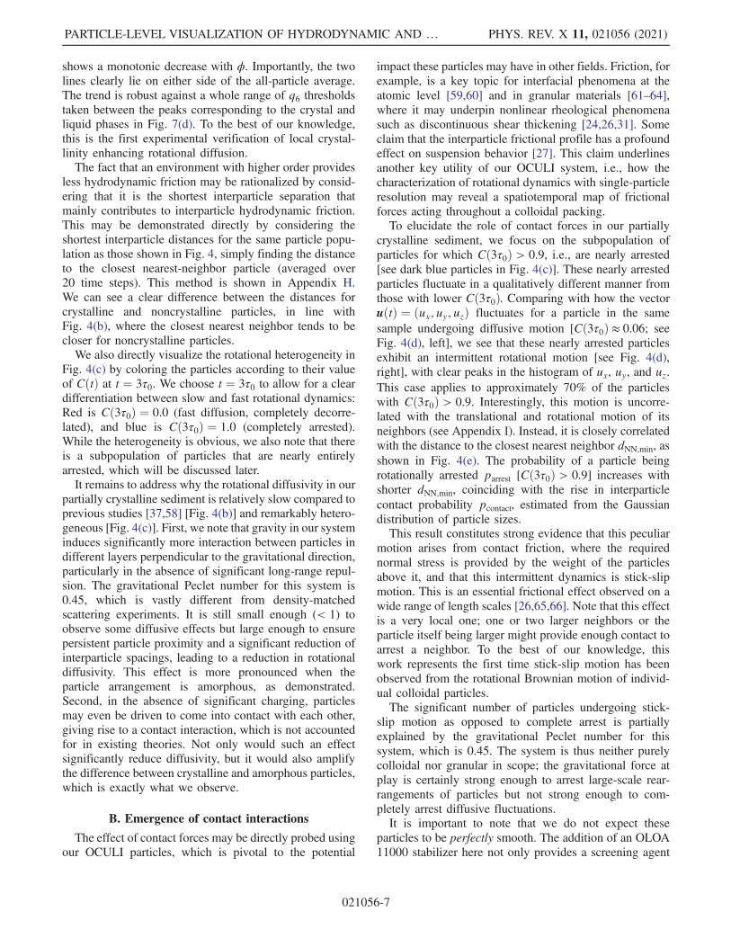
shows a monotonic decrease with ϕ. Importantly, the twolines clearly lie on either side of the all-particle average.The trend is robust against a whole range of q6 thresholdstaken between the peaks corresponding to the crystal andliquid phases in Fig. 7(d). To the best of our knowledge,this is the first experimental verification of local crystal-linity enhancing rotational diffusion.The fact that an environment with higher order provides
less hydrodynamic friction may be rationalized by consid-ering that it is the shortest interparticle separation thatmainly contributes to interparticle hydrodynamic friction.This may be demonstrated directly by considering theshortest interparticle distances for the same particle popu-lation as those shown in Fig. 4, simply finding the distanceto the closest nearest-neighbor particle (averaged over20 time steps). This method is shown in Appendix H.We can see a clear difference between the distances forcrystalline and noncrystalline particles, in line withFig. 4(b), where the closest nearest neighbor tends to becloser for noncrystalline particles.We also directly visualize the rotational heterogeneity in
Fig. 4(c) by coloring the particles according to their valueof CðtÞ at t ¼ 3τ0. We choose t ¼ 3τ0 to allow for a cleardifferentiation between slow and fast rotational dynamics:Red is Cð3τ0Þ ¼ 0.0 (fast diffusion, completely decorre-lated), and blue is Cð3τ0Þ ¼ 1.0 (completely arrested).While the heterogeneity is obvious, we also note that thereis a subpopulation of particles that are nearly entirelyarrested, which will be discussed later.It remains to address why the rotational diffusivity in our
partially crystalline sediment is relatively slow compared toprevious studies [37,58] [Fig. 4(b)] and remarkably hetero-geneous [Fig. 4(c)]. First, we note that gravity in our systeminduces significantly more interaction between particles indifferent layers perpendicular to the gravitational direction,particularly in the absence of significant long-range repul-sion. The gravitational Peclet number for this system is0.45, which is vastly different from density-matchedscattering experiments. It is still small enough (< 1) toobserve some diffusive effects but large enough to ensurepersistent particle proximity and a significant reduction ofinterparticle spacings, leading to a reduction in rotationaldiffusivity. This effect is more pronounced when theparticle arrangement is amorphous, as demonstrated.Second, in the absence of significant charging, particlesmay even be driven to come into contact with each other,giving rise to a contact interaction, which is not accountedfor in existing theories. Not only would such an effectsignificantly reduce diffusivity, but it would also amplifythe difference between crystalline and amorphous particles,which is exactly what we observe.
B. Emergence of contact interactions
The effect of contact forces may be directly probed usingour OCULI particles, which is pivotal to the potential
impact these particles may have in other fields. Friction, forexample, is a key topic for interfacial phenomena at theatomic level [59,60] and in granular materials [61–64],where it may underpin nonlinear rheological phenomenasuch as discontinuous shear thickening [24,26,31]. Someclaim that the interparticle frictional profile has a profoundeffect on suspension behavior [27]. This claim underlinesanother key utility of our OCULI system, i.e., how thecharacterization of rotational dynamics with single-particleresolution may reveal a spatiotemporal map of frictionalforces acting throughout a colloidal packing.To elucidate the role of contact forces in our partially
crystalline sediment, we focus on the subpopulation ofparticles for which Cð3τ0Þ > 0.9, i.e., are nearly arrested[see dark blue particles in Fig. 4(c)]. These nearly arrestedparticles fluctuate in a qualitatively different manner fromthose with lower Cð3τ0Þ. Comparing with how the vectoruðtÞ ¼ ðux; uy; uzÞ fluctuates for a particle in the samesample undergoing diffusive motion [Cð3τ0Þ ≈ 0.06; seeFig. 4(d), left], we see that these nearly arrested particlesexhibit an intermittent rotational motion [see Fig. 4(d),right], with clear peaks in the histogram of ux, uy, and uz.This case applies to approximately 70% of the particleswith Cð3τ0Þ > 0.9. Interestingly, this motion is uncorre-lated with the translational and rotational motion of itsneighbors (see Appendix I). Instead, it is closely correlatedwith the distance to the closest nearest neighbor dNN;min, asshown in Fig. 4(e). The probability of a particle beingrotationally arrested parrest [Cð3τ0Þ > 0.9] increases withshorter dNN;min, coinciding with the rise in interparticlecontact probability pcontact, estimated from the Gaussiandistribution of particle sizes.This result constitutes strong evidence that this peculiar
motion arises from contact friction, where the requirednormal stress is provided by the weight of the particlesabove it, and that this intermittent dynamics is stick-slipmotion. This is an essential frictional effect observed on awide range of length scales [26,65,66]. Note that this effectis a very local one; one or two larger neighbors or theparticle itself being larger might provide enough contact toarrest a neighbor. To the best of our knowledge, thiswork represents the first time stick-slip motion has beenobserved from the rotational Brownian motion of individ-ual colloidal particles.The significant number of particles undergoing stick-
slip motion as opposed to complete arrest is partiallyexplained by the gravitational Peclet number for thissystem, which is 0.45. The system is thus neither purelycolloidal nor granular in scope; the gravitational force atplay is certainly strong enough to arrest large-scale rear-rangements of particles but not strong enough to com-pletely arrest diffusive fluctuations.It is important to note that we do not expect these
particles to be perfectly smooth. The addition of an OLOA11000 stabilizer here not only provides a screening agent
PARTICLE-LEVEL VISUALIZATION OF HYDRODYNAMIC AND … PHYS. REV. X 11, 021056 (2021)
021056-7

for charge but also provides a steric repulsion by adsorbingonto the particles to form a polymer brush layer. Thecoverage is not expected to be dense in a random adsorptiveprocess [67]. Atomic force microscopy and rheologymeasurements for OLOA 11000 adsorbed onto carbonblack particles in decahydronaphthalene [68] show that thebrush thickness is around 4 nm. Given that the particlesappear smooth under scanning electron microscopy with aresolution of approximately 10 nm, we may estimate thatany roughness on the bare surface is less than 10 nm. Thus,we may conclude that the core-shell OCULI particles inthis experiment have a roughness of 4–10 nm, which meansthat particles do not necessarily need to reach surface-surface contact to experience a “contact” interaction.Given the sensitivity to contact forces, it is also important
that means are available to modify the surface. Theseparticles are composed of 3-trimethoxysilyl propyl meth-acrylate, a common bridging component between organicand inorganic materials. Thus, any of the wealth ofconjugation techniques that use silane or methacrylatechemistry may be used to modify the charge, apply apolymer brush, or change the wettability of the surface [69].The surface roughness may also be modified by growinglobes, like we have done here in the intermediate step of thecore-shell OCULI particle synthesis. This process isdescribed in more depth in Ref. [47].
V. CONCLUSIONS
We have directly visualized hydrodynamic and contactinteractions in dense suspensions of newly developedcolloidal spheres, which enable simultaneous measure-ment of both the translational and rotational motion of allindividual particles up to arbitrarily high volume frac-tions. In particular, we have measured transient rotation-rotation coupling in charged colloidal crystals and alsofound that local crystallinity can lead to an increase inrotational diffusivity in a denser, partially crystallinesediment. Finally, we have observed the onset of stick-slip rotational motion, indicating the emergence of contactfriction in dense particulate materials. Importantly, ourresults also show that these two types of friction ofhydrodynamic (fluid) and mechanical (solid) origin canbe distinguished by probing the “continuity” of therotational Brownian motion. Thus, our method allowseffective detection of solidlike contact friction at thesingle-particle level, induced by either mechanical forcesor interparticle attraction.In order to obtain a deeper understanding of self-
assembly mechanisms in flowing, driven, and nonequili-brium colloidal systems, the ability to directly imagevariations in the rotational dynamics of individual particlesis a significant development for any particulate systemwhere hydrodynamics, mechanical rigidity, and force chainnetworks play a role. Spatial mapping of rotational dif-fusivity may thus provide a unique strategy to visualize the
incidence of jamming and allow for accurate characteri-zation of how the statistics of local force chain networkschange on approaching the jamming point. However, itsgreatest potential strength is to change how we scrutinizenonlinear rheological phenomena in dense slurries andpastes. Computational work clearly shows that contactfriction has an important role to play in discontinuous shearthickening [27,70]; strategies are being developed to tuneparticle roughness to indirectly address the phenomenology[71]. The OCULI system now offers a more direct route,mapping microscopic frictional response to bulk behavior,an approach that might be applied to any number ofcomplex rheological phenomena at the interface betweencolloidal and granular matter.
VI. MATERIALS AND METHODS
A. Synthesis
Reagents, preparatory syntheses of core particles, dyes,and prehydrolyzed TPM, as well as a detailed descriptionof the synthesis of both systems described, are given inAppendix A.
B. Microscopy
Bright-fieldmicroscopywas carried out on an IX71 framewith a 100x PlanApo oil immersion objective (Olympus).Images were taken with a XIMEA USB3.0 cameraMQ042MG-CM (XIMEA). Fluorescence excitation wasachieved with a CoolLED Pe-300 white light source(CoolLED).Three-dimensional confocal laser-scanning micro-
scopy (CLSM) was carried out using the same framewith a 60x PlanApo oil immersion objective (Olympus)and a confocal scan head with a multichannel lasersource (Thorlabs). We used 532-nm and 488-nm lasersfor two-channel excitation, separated with a standardFITC/Rhodamine filter cube. Scanning in z was achievedusing an objective-mounted piezoelectric Z stage (PhysikInstrumente). We used a “fast-Z” mode, where the piezoelement would scan the objective focal plane through thesample, return to its initial position at maximum speed,and repeat. Software and a central control board providedby the manufacturer (Thorsync, Thorlabs) were used tosynchronize the components and record the state of theimaging and piezo at all times to generate accuratetimestamps for the frames.Scan parameters for the two samples were as follows:
For the charged colloidal crystal, a 128 × 128 × 251 pixelbox was scanned at 2.17-s intervals. Voxels were cubic,with 0.2-μm side length. We chose a spherical region ofinterest deep inside the interior where the crystal waspredominantly monodomain, selecting a cluster of 112particles to sample over, which was well separated from thesides; for the dense crystalline sediment, a 256 × 256 ×411 pixel box was scanned at 5.68-s intervals over 1000
YANAGISHIMA, LIU, TANAKA, and DULLENS PHYS. REV. X 11, 021056 (2021)
021056-8

frames. The voxel size is the same as above. A region ofinterest (ROI) is chosen near the base of the colloidalsediment within which we can track 99% of all particles,excluding particles at the ROI edge. We studied n ¼ 1481particles. Of these, 99.9% have complete eye trajectoriesas well.
C. Particle sizing
All TPM particles are sized using SEM. The particlesare sputter coated with platinum using an SC7620 sputtercoater (Quorum Technologies, UK) in an argon atmos-phere and imaged at a 10-kV beam energy with a JSM-6010LV scanning electron microscopy unit (JEOL,Japan).
ACKNOWLEDGMENTS
We thank Camille Boulet and Dirk Aarts for assistancewith the characterization of the organic solvent mixtures.We also thank Arran Curran for enabling the collection ofoptical trapping data. T. Y. acknowledges Grants-in-Aidfor Young Scientists (B) (No. JP15K17734) and JSPSFellows (No. JP16J06649) from the Japan Society for thePromotion of Science (JSPS); H. T. acknowledges Grants-in-Aid for Scientific Research (A) (No. JP18H03675) andSpecially Promoted Research (No. JP25000002 andNo. JP20H05619) from the JSPS; T. Y., Y. L., and R. P.A. D. acknowledge the European Research Council (ERC)(Starting Grant No. 279541-IMCOLMAT and Cons-olidator Grant No. 724834-OMCIDC).
T. Y., H. T., and R. P. A. D. conceived the project anddesigned the experiments. T. Y. performed the syntheses,experiments, and analysis. Y. L. prototyped the particlesand advised on the experimental work. T. Y. wrote themanuscript; all co-authors discussed and edited the manu-script together. H. T. and R. P. A. D. supervised theproject.
APPENDIX A: SYNTHESIS OF OCULIPARTICLES
1. Reagents
We used TPM (Sigma Aldrich), an aqueous ammoniumhydroxide solution (28 wt% solution) (Sigma Aldrich),Cyanine3 NHS Ester (Lumiprobe), BDP-FL NHS Ester(Lumiprobe), anhydrous dimethyl sulfoxide (DMSO)(Sigma Aldrich), 3-aminopropyl trimethoxysilane (APS)(Sigma Aldrich), 4-aminostyrene (Sigma Aldrich),Azobisisobutyronitrile (Sigma Aldrich), and PluronicF108 (Sigma Aldrich) as received. OLOA stabilizer wasprovided by Eric Dufresne and Azelis. Double-distilledwater was sourced from a Millipore Direct-Q 3 ultrapurewater unit.
2. Preparation of fluorescent monomer
In order to fluorescently label the TPM particles, werequire any fluorophore to bear a reactive component thatcan be incorporated into the TPM network. TPM has twovarieties of reactive moieties, three silyl groups, and amethacrylate group. Both sites are accessible before thefinal cross-linking step. We proceeded to label the BDP-FL NHS Ester with APS, and the Cyanine3 NHS Esterwith 4-aminostyrene for incorporation into the silyl andmethacrylate networks, respectively. We note that bothdyes can react with both labels. For the BDP-FL, 10 mg ofreagent was added to 5 g of chloroform and mixedthoroughly using a PTFE flea and magnetic stirrer.Once the dye was thoroughly dissolved, 10 μl of APS(approximately 5 times molar excess) was added to themixture and allowed to react overnight. For the Cyanine 3NHS Ester, the same procedure was followed using4-aminostyrene and DMSO. DMSO was not used forthe BDP-FL reaction due to adverse effects on theemission profile. After the reaction, both mixtures couldbe added directly to TPM particle syntheses.
3. Synthesis of OCULI particles(charged colloidal crystal)
We begin by synthesizing the eyes first. The exampleprotocol here describes how the eyes were made for theeyes of the OCULI particles used to make the chargedcolloidal crystal; it may be modified following previousworks [72] to change size and yield. We stirred 40 ml ofdouble-distilled water and 40 μl of 28 wt% ammoniumhydroxide solution in a 100-ml round-bottom flask for15 minutes using an oval PTFE flea. Once a uniform flowwas established, 100 μl of TPM was introduced to theflask in one smooth motion using a micropipette. It wasleft to react for approximately 90 minutes. During thistime, the TPM hydrolyzed and condensed into mono-disperse droplets. Next, 100 μl of preprepared chloro-form/BDP-FL/APS solution (from above) was added tothe mixture. Though the chloroform initially collected atthe bottom because of its density and immiscibility, thedye gradually diffused into the mixture and transferred tothe TPM phase. After 30 minutes of stirring, approxi-mately 50 mg of AIBN was added, and the mixture wasstirred for 20 minutes. It was then transferred to a 50-mlplastic tube and kept in an 80°C oven for 3 hours to cross-link the particles. The mixture was subsequently removedand allowed to cool to room temperature. The particleswere finally washed at least 5 times using centrifugation(2500 rpm for 5 minutes per spin) to remove unreactedreagents and disperse the particles in water. The finalproduct was resuspended in approximately 1.5 ml ofdouble-distilled water. These particles were approxi-mately 1.0–1.2 μm in diameter and notably monodisperse.Having obtained a suspension of eye particles, we
proceeded to coat them with a shell layer to form the
PARTICLE-LEVEL VISUALIZATION OF HYDRODYNAMIC AND … PHYS. REV. X 11, 021056 (2021)
021056-9
body of the OCULI particles. This procedure required asolution of prehydrolyzed TPM (hTPM), typically500 μl of TPM stirred in 5 ml of 0.5-mM hydrochloricacid solution. The solution was initially milky butbecame transparent after approximately 1 hour as the TPMhydrolyzed.There are two protocol variations for producing OCULI
particles. The first is a stepwise addition of hTPM,recommended for making OCULI particles that are lessthan 2 μm in size. The introduction of hTPM by hand is fastand straightforward but entails the risk of nucleating newparticles instead of growth on the eye. This is less of aproblem for fewer addition steps, i.e., smaller particles, andwas used to produce the OCULI particles in the chargedcolloidal crystal.An initial basic solution of eye particles was prepared
by taking 0.5 ml of the eye solution and adding 5 ml of14.8-mM ammonium hydroxide. It was kept in a20-ml glass bottle with a sealed lid. Once the eye particleswere well dispersed with a short burst of sonication, 1 mlof the hTPM solution was added, and the bottle quicklytumbled 2 or 3 times. Because of the pH, the hTPM wasdriven to either form new particles or wet existing ones.The key here was to ensure that there were enough eyeparticles in the solution to ensure that wetting existingparticles occurred at a faster timescale than nucleation butnot so many that multiple eyes coalesced into singleparticles.After 25 minutes, 1.5 ml of hTPM was dripped into the
bottle with gentle manual shaking. The solution was leftfor another 15 minutes. By this point, the pH droppedbecause of dilution, so 1 ml of 14.8-mM ammoniumhydroxide was added to compensate. We continued togrow the particles by adding another 1.5 ml of hTPMwhile shaking, like before. This process was repeated onemore time, and the particles were checked using standardoptical microscopy.To dye the particle body, 100 μl of Cyanine3/4-
aminostyrene/DMSO dye was added; the tube was thentumbled and left for 20 minutes. Approximately 50 mg ofAIBN was then added before the bottle was tumbled again,left for 20 minutes, then transferred to an oven at 80 °C. Theparticles were allowed to polymerize for 3 hours, with caretaken to manually tumble the bottle every hour to preventparticles from coalescing at the bottom under gravity. Afterremoval and cooling, they were washed using centrifuga-tion (1200 rpm, 5 minutes, 5 times) and redispersed indistilled water.
4. Synthesis of core-shell OCULI particles(partially crystalline sediment)
To study dense sediments and jamming, we requireparticles that are larger and do not suffer the overlapin fluorescence profiles seen in 3D-CSLM withuniformly dyed particles. Thus, we require slightly larger
OCULI and the addition of a nonfluorescent layer atthe surface, which is based on the protocol describedhere [47].The protocol below was used to create the core-shell
OCULI to observe the crystalline sediment. The eyeswere produced in a slightly different way; hTPM wasprehydrolyzed in a 1∶10 ratio using a 0.5-mM HClsolution, as above. Then, 5 ml of this hTPM solutionwas directly added to 10 ml of 14.8-mM ammoniumhydroxide solution in a glass tube before quickly tumblingand leaving stationary on the workbench. Particles couldbe seen to nucleate within 20 seconds. The solution wasleft for 20 minutes to allow all of the hTPM to coalesceinto droplets. A small PTFE magnetic flea was added, andthe mixture was stirred slowly (<300 rpm) to disperse100 μl of the labeled BDP-FL chloroform solutiondescribed above and approximately 10 mg of AIBN.After stirring for 20 minutes, the mixture was allowedto polymerize for 3 hours. It was finally redispersed indouble-distilled water up to a volume of 10 ml and keptrefrigerated until further use.Having obtained the eyes, we proceeded to make OCULI
particles. Here, we follow an alternative method to dem-onstrate how the size of OCULI may be tailored to differentapplications, which is particularly recommended for reach-ing larger particle sizes (>2 μm in diameter). We added5 ml of 14.8-mM ammonium hydroxide to a 15-ml plastictube, followed by 1 ml of the suspension of particlesprepared above. The solution was sonicated for 10 minutesto ensure that no clusters became encapsulated instead ofsingle particles, followed by 1 ml of a freshly prepared1∶10 hTPM solution before the tube was tumbled and leftstationary for 30 minutes. This suspension was then addedto 20 ml of 14.8-mM ammonium hydroxide solution in a50-ml round-bottom flask with a magnetic flea. Themixture was stirred slowly (<100 rpm) to ensure homog-enization while preventing the particles from coalescing.Then, 10 ml of hTPM solution was introduced dropwiseover 2 hours using a 10-ml plastic syringe, a PTFE tube,and a syringe pump. We note here that the preaddition of1 ml of hTPM, followed by a further dropwise addition, ledto monodisperse particles, while the direct dropwiseaddition of hTPM to cores did not. This result may bedue to an energetic barrier to prewetting the hTPM, whichis overcome for different particles at different times given alow concentration of hTPM; this issue was solved byintroducing a higher concentration of hTPM initially,followed by the gradual addition of the rest of the material.If standard OCULI particles are required, 100 μl of
dye solution and 20 mg of AIBN should be addedto the mixture before stirring for 20 minutes and thenheated in an 80 °C oven for 2 hours. However, since theobjective of this synthesis was to produce core-shellOCULI, 4 ml of 5 wt% Pluronic F108 solution wasadded, and the mixture was stirred for 20 minutes before
YANAGISHIMA, LIU, TANAKA, and DULLENS PHYS. REV. X 11, 021056 (2021)
021056-10

adding the dye/AIBN and heating for 2 hours. Thisprocess is required to modify the wetting properties ofthe particles for the next step.Once these OCULI were cross-linked, they were washed
thoroughly using centrifugation and double-distilled water(1000g spins, 10 minutes). Any smaller particles left in thesolution at this stage would lead to large secondary particlesgrowing in the following steps. The final particle solutionwas dispersed thoroughly into double-distilled water up toa total volume of 10 ml, which, again, may be refrigerateduntil further use.We proceeded to add the nonfluorescent TPM layer. We
began by nucleating droplets on the surface of the OCULIto produce “raspberry”-shaped OCULI. We added 2 ml ofthe OCULI solution above to 100 ml of 14.8-mMammonia solution in a 250-ml plastic container. We added2 ml of freshly prepared hTPM before the container wasquickly tumbled and left stationary for 45 minutes. Then,20 mg of AIBN was added before the container wastransferred to an 80 °C oven and left for 3 hours (note thatthe volume is greater, so the solution requires more time toreach the target temperature). After the added hTPMwas cross-linked, the particles were again washed thor-oughly with double-distilled water using centrifugation(1000g, 10 minutes, 5 times) until any particles nucleatedin the bulk were removed. The original OCULI werecovered in a dense layer of “lobes,” as seen in Figs. 1(b3)and 1(c3) in the main text. The final suspension wasredispersed into double-distilled water with a total volumeof 2 ml, the same volume as the solution of OCULI thatwas added.Finally, we filled in the space between the lobes to create
smooth, spherical colloidal particles. The 2 ml of raspberryOCULI were again added to 100 ml of 14.8-mM ammoniasolution in a 250-ml plastic container. This was followed by2 ml of freshly prepared hTPM solution before thecontainer was quickly tumbled and left stationary for60 minutes. The appearance of the particles was quicklyconfirmed under bright-field microscopy, with particularattention to how circular the diffraction-limited ringssurrounding the particles looked. The particles were stillrough, so another 2 ml of hTPMwas added before tumblingand waiting for another 60 minutes. This process wasrepeated as many times as required (in this case, a total of4 times, including the first addition) to obtain sphericalparticles. Under bright-field microscopy, the particles atthis point did not appear perfectly spherically symmetricdue to the index mismatch between the cross-linked lobesand the un-cross-linked TPM wetting the gaps betweenthem. The best indicator was the shape of the diffractionrings around the particle. Following the hTPM addition,20 mg of AIBN was added before the container wastransferred to an 80 °C oven and heated for 3 hours. Themixture was then recovered and allowed to cool to roomtemperature before the particles were cleaned again with
double-distilled water and centrifugation (1000g, 5 minutes,5 times). Finally, the sample was again redispersed into2 ml of double-distilled water and was then ready for use inexperiments.Note that the procedure to transfer these particles from
water to organic solvent mixtures is detailed in Ref. [48].
APPENDIX B: ROTATIONAL DIFFUSIONCONSTANT AND THE AUTOCORRELATION OF
THE UNIT VECTOR
This derivation is largely reproduced from Ref. [2]. For aspherical particle, the rotational diffusion constant Dr isdefined as the rate at which an arbitrary point fixed to thesurface diffuses while confined to the surface. Given areference axis that runs from the center of the sphere tothe point, we consider the probability that the point islocated at point Pðθ;ϕÞ in spherical coordinates at sometime t. We may first define a diffusion equation based onthis definition,
∂∂t Pðθ;ϕ; tÞ ¼ Dr∇2Pðθ;ϕ; tÞ; ðB1Þ
where Pðθ;ϕ; tÞ is the probability density function that thepoint will be located at point ðθ;ϕÞ after a time t inspherical coordinates. This formulation is particularly wellsuited for measurements using the OCULI, where the eyedefines a fixed point on the surface. The diffusion of thepoint matches the time evolution of the unit orientationvector uðtÞ. The time-averaged autocorrelation of theorientation vector u may thus be expressed as
huðt0Þ · uðt0 þ tÞi ¼ hcos θðtÞi: ðB2Þ
Here, we assume that the orientation vector is orientedalong the z-axis at t0, so the function Pðθ;ϕ; tÞ becomesindependent of ϕ. Thus, Pðθ;ϕÞ ¼ PðθÞ. The autocorre-lation hcos θi may now be expressed using Pðθ; tÞ asfollows:
hcos θi ¼Z
dθdϕPðθÞ cos θ sin θ ðB3Þ
¼ 2π
Zπ
0
dθPðθÞ cos θ sin θ: ðB4Þ
Using the diffusion equation above, the time derivative ofthis expectation value is then given by
∂∂t hcos θi ¼ 2π
Zπ
0
dθ∂PðθÞ∂t cos θ sin θ ðB5Þ
¼ 2πDr
Zπ
0
dθ(∇2PðθÞ) cos θ sin θ: ðB6Þ
PARTICLE-LEVEL VISUALIZATION OF HYDRODYNAMIC AND … PHYS. REV. X 11, 021056 (2021)
021056-11

Reexpressing the ∇2 operator in spherical coordinates, weapply two partial integrations and find
∂∂t hcos θi ¼ −2Dr
Zπ
0
dθPðθÞ sin θ cos θ ðB7Þ
¼ −2Drhcos θi; ðB8Þ
which shows that the time-averaged autocorrelation of theunit orientation vector hcos θi has an exponential decay,where
hcos θi ¼ e−2Drt: ðB9Þ
APPENDIX C: ROTATIONAL DIFFUSION INULTRADILUTE SUSPENSIONS
For the core-shell system, we considered the diffusionconstant of these particles in the ultradilute limit; anaccurate benchmark was required to quantitatively char-acterize diffusion constants as a function of localvolume fraction. To do this, we modified the densityof the solvent slightly, using a 4:1-by-volume mixture ofTCE and tetralin, to prevent sedimentation and accu-mulation of the particles at the base of the microscopycell. We assumed the slight change in the compositionof the solvent did not adversely affect the particle size.We accounted for a change in shear viscosity(1.43 mPa · s). Sampling trajectories away from surfa-ces, the autocorrelation of the particle orientations isshown in Fig. 5, yielding a relaxation time ofτr ¼ 16.9� 0.2 s. This may be converted to a diffusivityand then an effective hydrodynamic diameter using theequation Dr¼kBT=πησ3H; we found that σH ¼ 3.14 μm.This result is consistent with the size found by SEM,noting that both particle shrinkage due to drying forSEM imaging and swelling in the haloalkane solventcontribute to making the SEM value smaller. Thus, we
can also calculate an expansion coefficient linking theSEM size to a hydrodynamic diameter of 10%.
APPENDIX D: CALCULATION OFTHE ROTATIONAL COUPLING CONSTANT RC
In order to characterize the coupling between therotation of different pairs of particles, we calculate therotational coupling constant RC from an estimate ofthe angular velocity vector ω over time. Here, ω isestimated from how the unit orientation vector changesfrom uðtÞ to uðtþ ΔtÞ, where Δt is the time betweenconsecutive frames. Thus, RC may be expressed usinguðtÞ as follows:
Rcðτ; rÞ ¼ωi · ωj
hjω2i ji1=2hjω2
j ji1=2≈
h½uiðrij; τÞ ⊗ uiðrij; 0Þ� · ½ujðrij; τÞ ⊗ ujðrij; 0Þ�ihjuiðrij; τÞ ⊗ uiðrij; 0Þj2i1=2hjujðrij; τÞ ⊗ ujðrij; 0Þj2i1=2
: ðD1Þ
APPENDIX E: COMPARISON WITHDENSITY-MATCHED SCENARIOS FOR
CHARGED CRYSTALS
The single-particle rotational diffusivity seen in thecharged crystal is slower than what is expected in theultradilute limit using the particle size found fromSEM, τ0 ¼ 1=2D0 ¼ 2.26 s, which cannot be simplyaccounted for by swelling of the particle; using an
experimentally determined expansion ratio from SEM sizesto hydrodynamic diameters (see Appendix C), τ0 ¼ 3.01 s.Previous work [37,46,73] has determined analytical formsfor how rotational diffusivity changes with volumefraction. Adopting the expression used here [37] (alsotested against numerical simulations in Ref. [58]),Dr¼D0ð1−0.63ϕ−0.67ϕ2Þ, τr¼ 3.47 s, which is stillshorter than the τr ¼ 5.8� 0.1 s found in our experiment.
FIG. 5. Orientation autocorrelation CðtÞ for core-shell OCULIin the ultradilute limit. Here, CðtÞ is for the same core-shellOCULI particles used for the dense crystalline sediment. The datafit well with an exponential decay (red solid curve).
YANAGISHIMA, LIU, TANAKA, and DULLENS PHYS. REV. X 11, 021056 (2021)
021056-12

APPENDIX F: CONFIRMATION OFHARD-SPHERE-LIKE INTERPARTICLE
INTERACTIONS
To ensure that hard-sphere-like interactions wererealized in 5 wt% OLOA 11000, an optical trappingexperiment was carried out to confirm the lack of anylong-ranged repulsive interactions. For the identicalsolvent used for the semicrystalline sediment (3∶1TCE:TET, 5 wt% OLOA 11000), we applied the sameoptical trapping methodology as in Ref. [48] to directlycharacterize the interaction potential. Specifically,we trapped two core-shell OCULI particles (SEM diam-eter 4.01 μm, 3.5% polydispersity) in two weakoptical potentials, moved the potentials to differentdistances from each other (rtrap), and measured theinterparticle separation (rp) as a measure of repulsiveforce. Any long-ranged repulsive interaction will appearas a deviation in the interparticle separations from theseparation between the traps.First, we confirm that the traps are weak enough to allow
measurement of significant deviations at low energetic cost.The confining potential is found from the probabilitydistribution pðrÞ of the fluctuating interparticle separationr by a simple inversion, βUðΔrÞ ¼ −kBT log½pðΔrÞ�,where Δr ¼ r − hri, as given in the inset of Fig. 6;we confirm that the confinement is harmonic to a good
approximation (fit) and show that an interaction energy ofkBT would cause deviations in excess of 100 nm, whichis easily observable. Now, we look at how the inter-particle separations change as the traps are brought closertogether. Going from large to small separations, when adeviation is finally observed, the interparticle separationbasically remains constant (see Fig. 6). Red dashed linesshow the average interparticle separation at the threesmallest trap separations, 4.31 μm, consistent with4.01 μm under SEM swelled in the solvent.Both our own experimental data and evidence from the
literature as introduced in the main text point to negligiblesurface charge and strong screening in this dense colloidalsystem. We may safely conclude that the interparticlerepulsion in this system is hard-sphere-like. In hindsight,this conclusion also explains the agreement in our sedimentbetween the particle diameter found via SEM (2.81 μm)and the effective hard-sphere size found from phasecoexistence (3.00 μm). See Appendix G for details.
APPENDIX G: STATIC STRUCTUREIN A PARTIALLY CRYSTALLINE SEDIMENT
OF CORE-SHELL OCULI
Here, we describe the static structure of the densecrystalline sediment. We first note that there is a smallfraction of particles that fused together during thesynthesis, when raspberry particles are formed. Theseparticles are easily separable by considering the mini-mum distance to a neighbor and the arrest of rotationalmotion, as shown in Fig. 7(a). Particles where dNN <2.5 and Cð3τ0Þ > 0.9 are excluded from all analyses. Wealso consider a broad variation in density over thesample. A Voronoi tessellation allows us to calculatethe local number density n for all particles, averagingover 100 frames to lessen the effect of thermal fluctua-tions. A profile of n with height z is given as an inset inFig. 7(b). Note that there is a small reduction withheight, but this is very small, which is in agreementwith theoretical predictions for the density profile awayfrom the top sediment interface [74]. We also see that nfor particles above and below the median height havesimilar distributions [see Fig. 7(b)], with a similarlywide range.The system is partially crystalline. We may characterize
this by considering the bond-orientational order q6 [75],which is widely accepted as an appropriate local orderparameter for crystallization in hard-sphere-like systems[76,77]. To associate nearest neighbors, we use a simpledistance threshold taken from the first minimum of theradial distribution function gðrÞ in Fig. 7(c). We considerthe distribution of q6, shown in Fig. 7(d). One can clearlysee that a boundary q6 ≈ 0.5 separates crystalline andnoncrystalline parts of the sample. We may use thisthreshold and the Voronoi tessellation above to find theproportion of the sample that is crystalline. The sum of the
FIG. 6. Direct observation of lack of long-ranged inter-actions in the hard-sphere system. We show the opticaltrapping measurement of the interaction between a pair ofcore-shell OCULI particles in a pair of weak optical traps atdifferent separations. There is no deviation between interpar-ticle separation rp and trap positions rtrap until the particlesmeet, at which point the interparticle separation becomesconstant. This case is consistent with a hard-sphere-like separation. Inset: estimate of the optical confinementpotential UðΔrÞ.
PARTICLE-LEVEL VISUALIZATION OF HYDRODYNAMIC AND … PHYS. REV. X 11, 021056 (2021)
021056-13

volume associated with particles with q6 > 0.5 is 34.3% ofthe total volume of the ROI. Assuming that the sample ismonodisperse, we may use this and the average numberdensity to calculate an effective diameter for the particles,
σHS ¼�
6
ϕHSπntot
�13
: ðG1Þ
For the sample shown, σHS ¼ 3.00 μm. We use this andlocal volumes found from Voronoi tessellation to calculatelocal volume fractions ϕi in the main text of the paper.
APPENDIX H: LOCAL VOLUME FRACTIONVS CLOSEST DISTANCE TO
NEAREST-NEIGHBOR PARTICLE
To rationalize the relationship between the rotationaldiffusivity and the volume fraction, we find the distance to
(a) (b)
(d)(c)
FIG. 7. Static properties of dense, partially crystalline sediment of core-shell OCULI particles. (a) Minimum distance to neighboringparticles as a function of Cð3τ0Þ. Particles arrested by frictional interactions are indicated by the upper green dashed circle. Particlesarrested due to dimerization with other particles during the growth of the shell are shown by the lower red dashed circle. These particlesare omitted from the analysis. (b) Histogram of local volume fraction ϕ for particles below and above the median z position, as well asthe distribution for all particles in the ROI. There is a small variation, but both populations contain a wide range of ϕ. Inset: average ϕ asa function of z. There is a gentle decrease with height, as expected in a sediment, but the variation is very small. (c) Radial distributionfunction. The threshold used for detecting nearest neighbors is given as a dashed line. (d) Distribution of q6 over the whole ROI. Thethreshold used to separate crystalline and noncrystalline particles is given as a dashed line.
FIG. 8. Distance to the closest nearest neighbor dNN;minas a function of volume fraction ϕ for particles with differentcrystallinity, as indicated.
YANAGISHIMA, LIU, TANAKA, and DULLENS PHYS. REV. X 11, 021056 (2021)
021056-14

the closest nearest neighbor dmin for the same particlepopulations in each volume fraction ϕ bin, as shown inFig. 8. It is clear that particles with higher crystallinity havea greater dmin than those with lower crystallinity for thevolume fraction range where trends in Dr=D0 change.Looking closely, we note that the deviation betweencrystalline and noncrystalline particles starts at a lower ϕthan for Dr=D0. At lower ϕ, the particles are not closeenough together to strongly couple hydrodynamically;thus, the effect only arises at higher ϕ.
APPENDIX I: STICK-SLIP DYNAMICSAND DYNAMICS OF NEIGHBORING
PARTICLES
On observing intermittent rotational dynamicsof a subpopulation of particles, we considered whetherthis was simply correlated with a fluctuation in the
position or orientation of the particle itself, or itsneighbors, which is not the case. As shown inFig. 9, we can see that the intermittent rotational slipof the particle itself (a) is not accompanied by thedisplacement of the particle itself (b), nor the distanceto (c), displacement of (d), or rotation of (e) its nearestneighbors.
[1] W. C. K. Poon, Colloids as Big Atoms, Science 304, 830(2004).
[2] A. P. Philipse, Brownian Motion, Undergraduate LectureNotes in Physics (Springer International Publishing, Cham,2018).
[3] T. Palberg, Crystallization Kinetics of Colloidal ModelSuspensions: Recent Achievements and New Perspectives,J. Phys. Condens. Matter 26, 333101 (2014).
[4] J. Russo and H. Tanaka, The Microscopic Pathway toCrystallization in Supercooled Liquids, Sci. Rep. 2, 505(2012).
[5] S. Arai and H. Tanaka, Surface-Assisted Single-CrystalFormation of Charged Colloids, Nat. Phys. 13, 503 (2017).
[6] Y. L. Wu, D. Derks, A. van Blaaderen, and A. Imhof,Melting and Crystallization of Colloidal Hard-SphereSuspensions under Shear, Proc. Natl. Acad. Sci. U.S.A.106, 10564 (2009).
[7] Z. Wang, F. Wang, Y. Peng, Z. Zheng, and Y. Han, Imagingthe Homogeneous Nucleation During the Melting of Super-heated Colloidal Crystals, Science 338, 87 (2012).
[8] P. J. Lu, E. Zaccarelli, F. Ciulla, A. B. Schofield, F.Sciortino, and D. A. Weitz, Gelation of Particles withShort-Range Attraction, Nature (London) 453, 499 (2008).
[9] M. Tateno and H. Tanaka, Numerical Prediction of Colloi-dal Phase Separation by Direct Computation of Navier-Stokes Equation, npj Comput. Mater. 5, 40 (2019).
[10] G. L. Hunter and E. R. Weeks, The Physics of the ColloidalGlass Transition, Rep. Prog. Phys. 75, 066501 (2012).
[11] P. J. Lu and D. A. Weitz, Colloidal Particles: Crystals,Glasses, and Gels, Annu. Rev. Condens. Matter Phys. 4,217 (2013).
[12] J. B. Perrin, Les Atomes (Felix Alcan, Paris, 1913).[13] A. Furukawa and H. Tanaka, Key Role of Hydrodynamic
Interactions in Colloidal Gelation, Phys. Rev. Lett. 104,245702 (2010).
[14] Z. Varga, G. Wang, and J. Swan, The Hydrodynamics ofColloidal Gelation, Soft Matter 11, 9009 (2015).
[15] J. De Graaf, W. C. K. Poon, M. J. Haughey, and M. Hermes,Hydrodynamics Strongly Affect the Dynamics of ColloidalGelation but Not Gel Structure, Soft Matter 15, 10 (2019).
[16] N. J. Wagner and J. F. Brady, Shear Thickening in ColloidalDispersions, Phys. Today 62, No. 10, 27 (2009).
[17] J. J. Stickel and R. L. Powell, Fluid Mechanics andRheology of Dense Suspensions, Annu. Rev. Fluid Mech.37, 129 (2005).
[18] T. Ando and J. Skolnick, Crowding and HydrodynamicInteractions Likely Dominate In Vivo MacromolecularMotion, Proc. Natl. Acad. Sci. U.S.A. 107, 18457 (2010).
(e)
(d)
(c)
(b)
(a)
FIG. 9. Intermittent dynamics and neighboring particles. (a) x,y, and z components of the unit orientation vector of a particleundergoing stick-slip rotational motion as a function of time. Alldata below refer to the same trajectory. (b) Movement of the sameparticle over two frames, both translational Δx and orientationalΔu displacements. (c) Distance to nearest neighbors dNN as afunction of time. (d) Displacements of nearest neighbors over twoframes as a function of time. (e) Displacements in orientation Δuof nearest neighbors over two frames as a function of time. (c–e)show the mean, maximum and minimum for each statistic.
PARTICLE-LEVEL VISUALIZATION OF HYDRODYNAMIC AND … PHYS. REV. X 11, 021056 (2021)
021056-15

[19] R. Golestanian, J. M. Yeomans, and N. Uchida, Hydro-dynamic Synchronization at Low Reynolds Number, SoftMatter 7, 3074 (2011).
[20] M. A. Mourão, J. B. Hakim, and S. Schnell, Connecting theDots: The Effects of Macromolecular Crowding on CellPhysiology, Biophys. J. 107, 2761 (2014).
[21] Y. Wang, Y. Wang, X. Zheng, E. Ducrot, J. S. Yodh, M.Weck, and D. J. Pine, Crystallization of DNA-CoatedColloids, Nat. Commun. 6, 7253 (2015).
[22] E. Brown and H. M. Jaeger, Shear Thickening in Concen-trated Suspensions: Phenomenology, Mechanisms andRelations to Jamming, Rep. Prog. Phys. 77, 046602 (2014).
[23] X. Cheng, J. H. McCoy, J. N. Israelachvili, and I. Cohen,Imaging the Microscopic Structure of Shear Thinning andThickening Colloidal Suspensions, Science 333, 1276(2011).
[24] N. Y. C. Lin, B. M. Guy, M. Hermes, C. Ness, J. Sun,W. C. K. Poon, and I. Cohen, Hydrodynamic and ContactContributions to Continuous Shear Thickening in ColloidalSuspensions, Phys. Rev. Lett. 115, 228304 (2015).
[25] N. Y. C. Lin, C. Ness, M. E. Cates, J. Sun, and I. Cohen,Tunable Shear Thickening in Suspensions, Proc. Natl. Acad.Sci. U.S.A. 113, 10774 (2016).
[26] J. R. Royer, D. L. Blair, and S. D. Hudson, RheologicalSignature of Frictional Interactions in Shear ThickeningSuspensions, Phys. Rev. Lett. 116, 188301 (2016).
[27] J. Comtet, G. Chatte, A. Nigues, L. Bocquet, A. Siria, andA. Colin, Pairwise Frictional Profile between ParticlesDetermines Discontinuous Shear Thickening Transition inNon-colloidal Suspensions, Nat. Commun. 8, 15633 (2017).
[28] P. Sehgal, M. Ramaswamy, I. Cohen, and B. J. Kirby,Using Acoustic Perturbations to Dynamically Tune ShearThickening in Colloidal Suspensions, Phys. Rev. Lett. 123,128001 (2019).
[29] N. Fernandez, R. Mani, D. Rinaldi, D. Kadau, M. Mosquet,H. Lombois-Burger, J. Cayer-Barrioz, H. J. Herrmann,N. D. Spencer, and L. Isa, Microscopic Mechanism forShear Thickening of Non-Brownian Suspensions, Phys.Rev. Lett. 111, 108301 (2013).
[30] M. Wyart and M. E. Cates, Discontinuous Shear Thickeningwithout Inertia in Dense Non-Brownian Suspensions, Phys.Rev. Lett. 112, 098302 (2014).
[31] R. Mari, R. Seto, J. F. Morris, and M.M. Denn, Discon-tinuous Shear Thickening in Brownian Suspensions byDynamic Simulation, Proc. Natl. Acad. Sci. U.S.A. 112,15326 (2015).
[32] S. Jamali and J. F. Brady, Alternative Frictional Model forDiscontinuous Shear Thickening of Dense Suspensions:Hydrodynamics, Phys. Rev. Lett. 123, 138002 (2019).
[33] M. Ramaswamy, N. Y. C. Lin, B. D. Leahy, C. Ness, A. M.Fiore, J. W. Swan, and I. Cohen, How Confinement-InducedStructures Alter the Contribution of Hydrodynamic andShort-Ranged Repulsion Forces to the Viscosity ofColloidal Suspensions, Phys. Rev. X 7, 041005 (2017).
[34] Z. Dogic and S. Fraden, Ordered Phases of FilamentousViruses, Curr. Opin. Colloid Interface Sci. 11, 47 (2006).
[35] P. B. Muller, M. Rossi, A. G. Marín, R. Barnkob, P.Augustsson, T. Laurell, C. J. Kähler, and H. Bruus, Ultra-sound-Induced Acoustophoretic Motion of Microparticlesin Three Dimensions, Phys. Rev. E 88, 023006 (2013).
[36] I. I. Smalyukh, Liquid Crystal Colloids, Annu. Rev.Condens. Matter Phys. 9, 207 (2018).
[37] V. Degiorgio, R. Piazza, and R. B. Jones, RotationalDiffusion in Concentrated Colloidal Dispersions of HardSpheres, Phys. Rev. E 52, 2707 (1995).
[38] G. H. Koenderink, M. P. Lettinga, and A. P. Philipse,Rotational Dynamics of Charged Colloidal Spheres: Roleof Particle Interactions, J. Chem. Phys. 117, 7751 (2002).
[39] G. H. Koenderink, D. G. A. L. Aarts, and A. P. Philipse,Rotational Dynamics of Colloidal Tracer Spheres inSuspensions of Charged Rigid Rods, J. Chem. Phys. 119,4490 (2003).
[40] M. Reichert and H. Stark, Hydrodynamic Coupling of TwoRotating Spheres Trapped in Harmonic Potentials, Phys.Rev. E 69, 031407 (2004).
[41] C. J. Behrend, J. N. Anker, B. H. McNaughton, and R.Kopelman, Microrheology with Modulated OpticalNanoprobes (MOONs), J. Magn. Magn. Mater. 293, 663(2005).
[42] M. P. Lettinga, G. H. Koenderink, B. W.M. Kuipers, E.Bessels, and A. P. Philipse, Rotational Dynamics of Colloi-dal Spheres Probed with Fluorescence Recovery afterPhotobleaching, J. Chem. Phys. 120, 4517 (2004).
[43] J. Wenzl, R. Seto, M. Roth, H. J. Butt, and G. K.Auernhammer, Measurement of Rotation of IndividualSpherical Particles in Cohesive Granulates, Granular Mat-ter 15, 391 (2013).
[44] B. Ilhan, J. J. Schoppink, F. Mugele, and M. H. G. Duits,Spherical Probes for Simultaneous Measurement ofRotational and Translational Diffusion in 3 Dimensions,J. Colloid Interface Sci. 576, 322 (2020).
[45] B. Liu and A. Böker, Measuring Rotational Diffusion ofColloidal Spheres with Confocal Microscopy, Soft Matter12, 6033 (2016).
[46] S. Schütter, J. Roller, A. Kick, J.-M. Meijer, and A.Zumbusch, Real-Space Imaging of Translational andRotational Dynamics of Hard Spheres from the Fluid tothe Crystal, Soft Matter 13, 8240 (2017).
[47] Y. Liu, K. V. Edmond, A. Curran, C. Bryant, B. Peng,D. G. A. L. Aarts, S. Sacanna, and R. P. A. Dullens, Core–Shell Particles for Simultaneous 3D Imaging and OpticalTweezing in Dense Colloidal Materials, Adv. Mater. 28,8001 (2016).
[48] Y. Liu, T. Yanagishima, A. Curran, K. V. Edmond, S. Sacanna,and R. P. A. Dullens, Colloidal Organosilica Spheresfor Three-Dimensional Confocal Microscopy, Langmuir 35,7962 (2019).
[49] V. N. Manoharan, Pinned Down, Nat. Mater. 14, 869(2015).
[50] M. Kamp, B. de Nijs, J. J. Baumberg, and O. A. Scherman,Contact Angle as a Powerful Tool in Anisotropic ColloidSynthesis, J. Colloid Interface Sci. 581, 417 (2021).
[51] M. Leocmach and H. Tanaka, A Novel Particle TrackingMethod with Individual Particle Size Measurement and ItsApplication to Ordering in Glassy Hard Sphere Colloids,Soft Matter 9, 1447 (2013).
[52] R. P. A. Dullens, M. Claesson, D. Derks, A. Van Blaaderen,and W. K. Kegel, Monodisperse Core-Shell Poly(Methyl Methacrylate) Latex Colloids, Langmuir 19, 5963(2003).
YANAGISHIMA, LIU, TANAKA, and DULLENS PHYS. REV. X 11, 021056 (2021)
021056-16

[53] T. E. Kodger, R. E. Guerra, and J. Sprakel, Precise Colloidswith Tunable Interactions for Confocal Microscopy, Sci.Rep. 5, 14635 (2015).
[54] J. C. Crocker and D. G. Grier, Methods of Digital VideoMicroscopy for Colloidal Studies, J. Colloid Interface Sci.179, 298 (1996).
[55] J. Santana-Solano, D. T. Wu, and D.W.M. Marr, DirectMeasurement of Colloidal Particle Rotation and FieldDependence in Alternating Current ElectrohydrodynamicFlows, Langmuir 22, 5932 (2006).
[56] Y. Goto and H. Tanaka, Purely Hydrodynamic Ordering ofRotating Disks at a Finite Reynolds Number, Nat. Commun.6, 5994 (2015).
[57] K. Han, G. Kokot, S. Das, R. G. Winkler, G. Gompper, andA. Snezhko, Reconfigurable Structure and Tunable Trans-port in Synchronized Active Spinner Materials, Sci. Adv. 6,eaaz8535 (2020).
[58] M. H. J. Hagen, D. Frenkel, and C. P. Lowe, RotationalDiffusion in Dense Suspensions, Physica (Amsterdam)272A, 376 (1999).
[59] A. Socoliuc, R. Bennewitz, E. Gnecco, and E. Meyer,Transition from Stick-Slip to Continuous Sliding in AtomicFriction: Entering a New Regime of Ultralow Friction,Phys. Rev. Lett. 92, 134301 (2004).
[60] A. Vanossi, N. Manini, M. Urbakh, S. Zapperi, and E.Tosatti, Colloquium: Modeling Friction: From Nanoscale toMesoscale, Rev. Mod. Phys. 85, 529 (2013).
[61] H. M. Jaeger, C. H. Liu, S. R. Nagel, and T. A. Witten,Friction in Granular Flows, Europhys. Lett. 11, 619 (1990).
[62] A. Fall, F. Bertrand, G. Ovarlez, and D. Bonn, Yield Stressand Shear Banding in Granular Suspensions, Phys. Rev.Lett. 103, 178301 (2009).
[63] M. P. Ciamarra, R. Pastore, M. Nicodemi, and A. Coniglio,Jamming Phase Diagram for Frictional Particles, Phys.Rev. E 84, 041308 (2011).
[64] K. M. Salerno, D. S. Bolintineanu, G. S. Grest, J. B. Lech-man, S. J. Plimpton, I. Srivastava, and L. E. Silbert, Effect ofShape and Friction on the Packing and Flow of GranularMaterials, Phys. Rev. E 98, 050901(R) (2018).
[65] H. Yoshizawa and J. Israelachvili, FundamentalMechanisms of Interfacial Friction. 2. Stick-Slip Frictionof Spherical and Chain Molecules, J. Phys. Chem. 97,11300 (1993).
[66] T. Palberg and K. Streicher, Resonant Stick-Slip Motion in aColloidal Crystal, Nature (London) 367, 51 (1994).
[67] C. Ligoure and L. Leibler, Thermodynamics and Kineticsof Grafting End-Functionalized Polymers to an Interface,J. Phys. 51, 1313 (1990).
[68] S. Yasin, Ph.D. thesis, Imperial College, London (2011).[69] G. T. Hermanson, Bioconjugate Techniques, 2nd ed.
(Elsevier, New York, 2013).[70] R. Seto, R. Mari, J. F. Morris, and M.M. Denn,
Discontinuous Shear Thickening of Frictional Hard-SphereSuspensions, Phys. Rev. Lett. 111, 218301 (2013).
[71] L. C. Hsiao and S. Pradeep, Experimental Synthesis andCharacterization of Rough Particles for Colloidal andGranular Rheology, Curr. Opin. Colloid Interface Sci. 43,94 (2019).
[72] C. Van der Wel, R. K. Bhan, R. W. Verweij, H. C. Frijters, Z.Gong, A. D. Hollingsworth, S. Sacanna, and D. J.Kraft, Preparation of Colloidal Organosilica Spheresthrough Spontaneous Emulsification, Langmuir 33, 8174(2017).
[73] H. J. H. Clercx and P. P. J. M. Schram, Three ParticleHydrodynamic Interactions in Suspensions, J. Chem. Phys.96, 3137 (1992).
[74] J. K. G. Dhont, An Introduction to Dynamics of Colloids,edited by D. Mobius and R. Miller (Elsevier Science B.V.,Amsterdam, 1996).
[75] P. J. Steinhardt, D. R. Nelson, and M. Ronchetti, Bond-Orientational Order in Liquids and Glasses, Phys. Rev. B28, 784 (1983).
[76] P. N. Pusey, E. Zaccarelli, C. Valeriani, E. Sanz, W. C. K.Poon, and M. E. Cates, Hard Spheres: Crystallizationand Glass Formation, Phil. Trans. R. Soc. A 367, 4993(2009).
[77] H. Tanaka, Bond Orientational Order in Liquids: Towards aUnified Description of Water-like Anomalies, Liquid-LiquidTransition, Glass Transition, and Crystallization, Eur. Phys.J. E 35, 113 (2012).
[78] See Supplemental Material at http://link.aps.org/supplemental/10.1103/PhysRevX.11.021056 for video cap-ture of OCULI particles observed under two-channel laserscanning confocal microscopy. Both translational and rota-tional Brownian motion can be seen.
PARTICLE-LEVEL VISUALIZATION OF HYDRODYNAMIC AND … PHYS. REV. X 11, 021056 (2021)
021056-17