Embed Size (px)
Citation preview
MISCELLANEOUS PUBLICATIONS MUSEUM OF ZOOLOGY, T H E UNIVERSITY OF MICHIGAN NO. 171
Phylogenetic Interrelationships of the Stomiid Fishes
(Teleostei: Stomiiformes)
by William L. Fink
Division of Biological Sciences and
Museum of Zoology The University of Michigan
Ann Arbor, Michigan 48 109-1079
Ann Arbor MUSEUM O F ZOOLOGY, T H E UNIVERSITY O F MICHIGAN
December 3 1, 1985
MISCELLANEOUS PUBLICATIONS MUSEUM OF ZOOLOGY, UNIVERSITY OF MICHIGAN NO. 171
The publications of the Museum of Zoology, The University of Michigan, consist of two series-the Occasional Papers and the Miscellaneous Publications. Both series were founded by Dr. Bryant Walker, Mr. Bradshaw H. Swales, and Dr. W. W. Newcomb.
The Occasional Papers, initiated in 1913, serve as a medium for original studies based principally upon the collections in the Museum. They are issued separately. When a sufficient number of pages has been printed to form a volume, the Museum will supply a title page, table of contents, and an index to libraries and individuals on the mailing list for the series.
The Miscellaneous Publications, which include papers on field and museum techniques, monographic studies, and other contributions not within the scope of the Occasional Papers, were established in 1916 and are published separately. It is not intended that they be grouped into volumes. Each number has a title page and, when necessary, a table of contents.
A complete list of publications on Birds, Fishes, Insects, Mammals, Mollusks, and Reptiles and Amphibians is available. Address inquiries to the Director, Museum of Zoology, Ann Arbor, Michigan 48109-1079.

MISCELLANEOUS PUBLICATIONS MUSEUM O F ZOOI,OGY, T H E UNIVERSITY O F MICHIGAN NO. 171
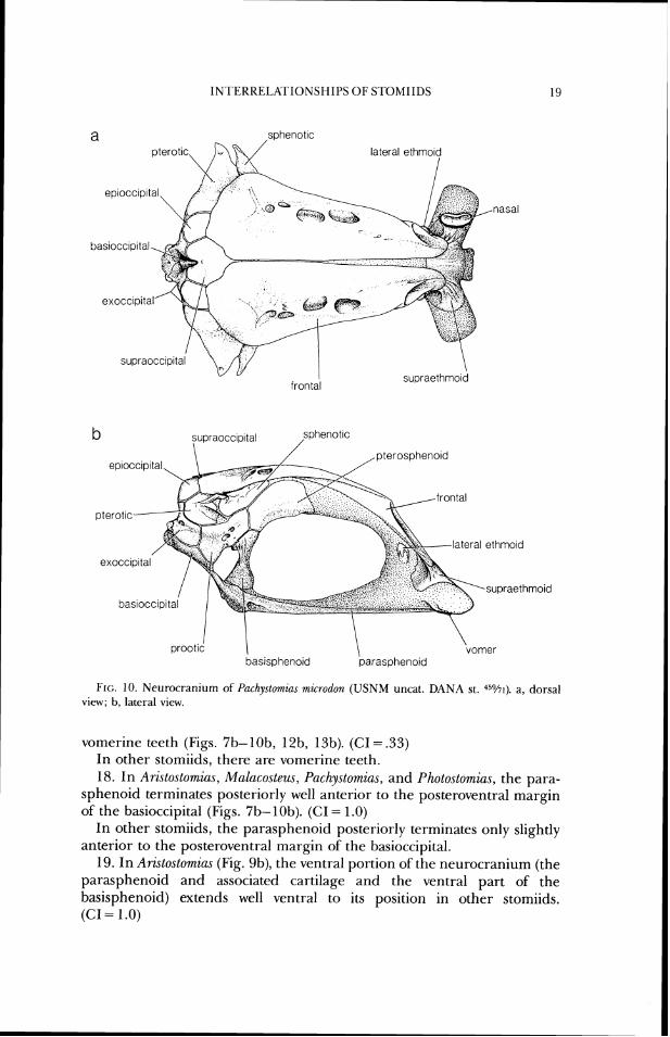
Phylogenetic Interrelationships of the Stomiid Fishes
(Teleostei: Stomiiformes)
by William L. Fink
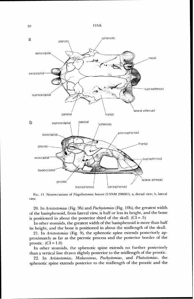
Division of Biological Sciences and
Museum of Zoology The University of Michigan
Ann Arbor, Michigan 48 109- 1079
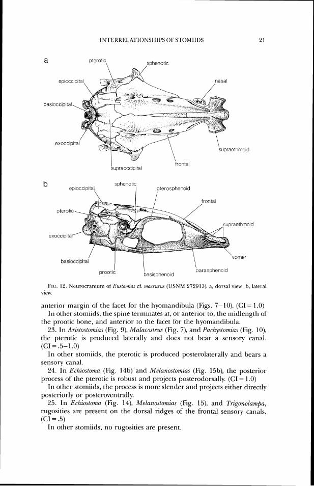
Ann Arbor MUSEUM O F ZOOLOGY, THE UNIVERSITY O F MICHIGAN
December 3 1, 1985
ABSTRACT
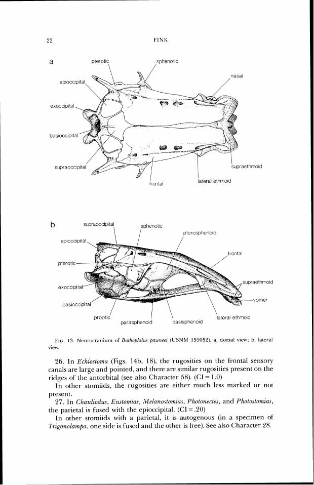
Fink, Willzanl L. 1985. Phylogenetic Interrelatiomhz of the Stomiid Fishes (Teleostei: Stomiformes). Mzsc. Publ. MW. 2001. Unzv. Michigan, 171:l-127, frontic.,fig.r. 1-70. A phylogenetic analysis of the interrelationships of the twenty-five stomiiform genera formerly classified in the superfamily Stomiatoidea is presented. Previously compris ing the families Astronesthidae, Chaul iodont idae , Idiacanthidae, Malacosteidae, Melanostomiatidae, and Stomiidae, the genera are placed in a single, expanded Stomiidae. All of the traditionally recognized genera are toutid to be monophylctic, with the exception of Stomias, which is expanded to include Macrostomiar. The character text includes detailed descriptions of stomiid morphology. Several alternative phylogenetic hypotheses are discussed. In the context of orre or more of these hypothcscs, patterris of character transformation are discussed, includitrg the evolution of the pectoral girdle and its accessory light orgaris, placement of the vertical firis toward the posterior of the body, elaboration of the anterior and posterior portions of the pelvic girdle, increase and subsequent decrcase of the number of radial elements of the pelvic girdles, and specializations of the head skeleton and anterior axial skeleton. ' f i e data matrix that was used to generate the phylogenetic hypotheses, and a brief discussion of fossils which have been assigned to the Stomiidae, are included in appendices.
Key words: Stomiiformes, Astrone.tthidar, Chauliodontidae, Idiacanthidae, Malacosteidae, Mrlanostomiidae, Stonlizdae, phylogenetics, character analysis.
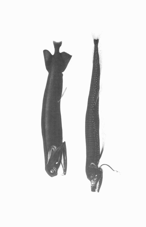
FKONTISPIECE: Malacosteus (top), Emtomias (bottom). From Zugmayer, 191 1.

CON'I'EN'I'S
PAC; E'. I N ' f K O D U C T I O N . . . . . . . . . . . . . . . . . . . . . . . . . . . . . . . . . . . . . . . . . . . . . . . . . . . . . . . . . . . . . 1 tIISI 'OKY O F CI.ASSIFIC.KI'ION 0 1 : T H E S'I'OM1II)AE . . . . . . . . . . . . . . . . . . . . . . . . . 3 MI.. '1'HOI)S . . . . . . . . . . . . . . . . . . . . . . . . . . . . . . . . . . . . . . . . . . . . . . . . . . . . . . . . . . . . . . . . . . . 8 KF.SUI.1.S . . . . . . . . . . . . . . . . . . . . . . . . . . . . . . . . . . . . . . . . . . . . . . . . . . . . . . . . . . . . . . . . . . . . . 10 (;IIARA(:TEKS . . . . . . . . . . . . . . . . . . . . . . . . . . . . . . . . . . . . . . . . . . . . . . . . . . . . . . . . . . . . . . . . 1 0
Suspcnsor~ium ;tntl laws . . . . . . . . . . . . . . . . . . . . . . . . . . . . . . . . . . . . . . . . . . . . . . . . . . . . . 27 l%~- iu~ch ia l Basket atitl Hyoid . . . . . . . . . . . . . . . . . . . . . . . . . . . . . . . . . . . . . . . . . . . . . . . . Muscles a n d IAig;iments of t h c f l e ; ~ t l . . . . . . . . . . . . . . . . . . . . . . . . . . . . . . . . . . . . . . . . . I'osccrarrial Axial Skele ton . . . . . . . . . . . . . . . . . . . . . . . . . . . . . . . . . . . . . . . . . . . . . . . . . . . Vcl.tic.11Fins . . . . . . . . . . . . . . . . . . . . . . . . . . . . . . . . . . . . . . . . . . . . . . . . . . . . . . . . . . . . . . .
. . . . . . . . . . . . . . . . . . . . . . . . . . . . . . . . . . . . . . . . . . . . . . . . . . . . . . . . . . . . . I'cctor.al (;irtllc I'clvic<;i~-dlr . . . . . . . . . . . . . . . . . . . . . . . . . . . . . . . . . . . . . . . . . . . . . . . . . . . . . . . . . . . . . . . 1. igl i tOrgans . . . . . . . . . . . . . . . . . . . . . . . . . . . . . . . . . . . . . . . . . . . . . . . . . . . . . . . . . . . . . . Misccllancous (.har.actcrs . . . . . . . . . . . . . . . . . . . . . . . . . . . . . . . . . . . . . . . . . . . . . . . . . . .
I)IS(:USSION . . . . . . . . . . . . . . . . . . . . . . . . . . . . . . . . . . . . . . . . . . . . . . . . . . . . . . . . . . . . . . . . . Sio~niicl 1ntcrt.clationsllips . . . . . . . . . . . . . . . . . . . . . . . . . . . . . . . . . . . . . . . . . . . . . . . . . . . Sf i (.hal.actcr Evolutiorr . . . . . . . . . . . . . . . . . . . . . . . . . . . . . . . . . . . . . . . . . . . . . . . . . . . . . . . . 107
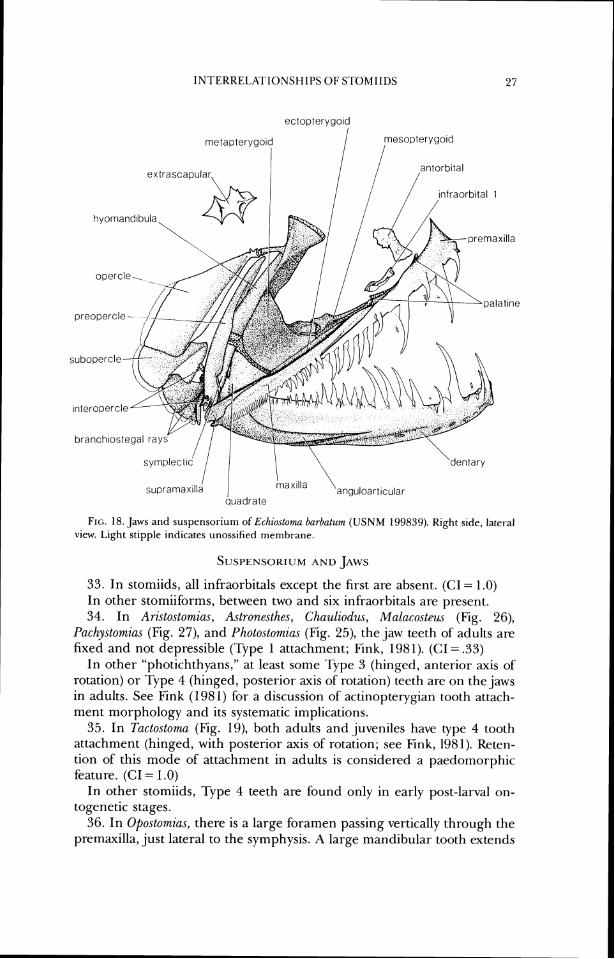
A(:KNOWLED(;MENTS . . . . . . . . . . . . . . . . . . . . . . . . . . . . . . . . . . . . . . . . . . . . . . . . . . . . . . . 117 I ~ I ' I ' E K K I ~ U K L'. (.I'f61) . . . . . . . . . . . . . . . . . . . . . . . . . . . . . . . . . . . . . . . . . . . . . . . . . . . . . . . . . 118 Al'l'kNDIX 1 . LIAlli\ MKI'KIX . . . . . . . . . . . . . . . . . . . . . . . . . . . . . . . . . . . . . . . . . . . . . . . . . 121 A1'I'ENI)IX 2 . F 0 S S I I . T E I . E O S f S I'KEVI0USI.Y REE'ERREI) -f<) ' I 'HE SI 'OMIIDAE 124 A1'I'k:NI)IX 3 . MXI'BKIAI . BXAMIN I.: D . . . . . . . . . . . . . . . . . . . . . . . . . . . . . . . . . . . . . . . . . . 125
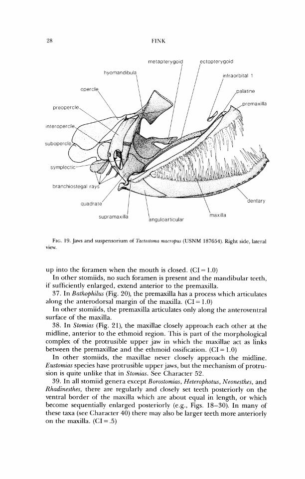
1 LLUSTRATIONS
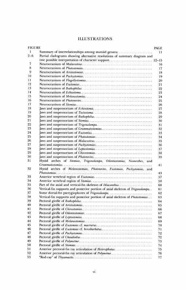
FIGURE PAC; E 1 Summary of intenelationships among stomiid genera . . . . . . . . . . . . . . . . . . . . . . I I
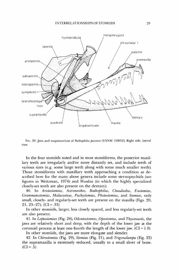
2-6 Partial clatlograms showing alternative resolutions of surnmary diagram and orre possihle interpretation of character support . . . . . . . . . . . . . . . . . . . . . . . . . . . 12-15
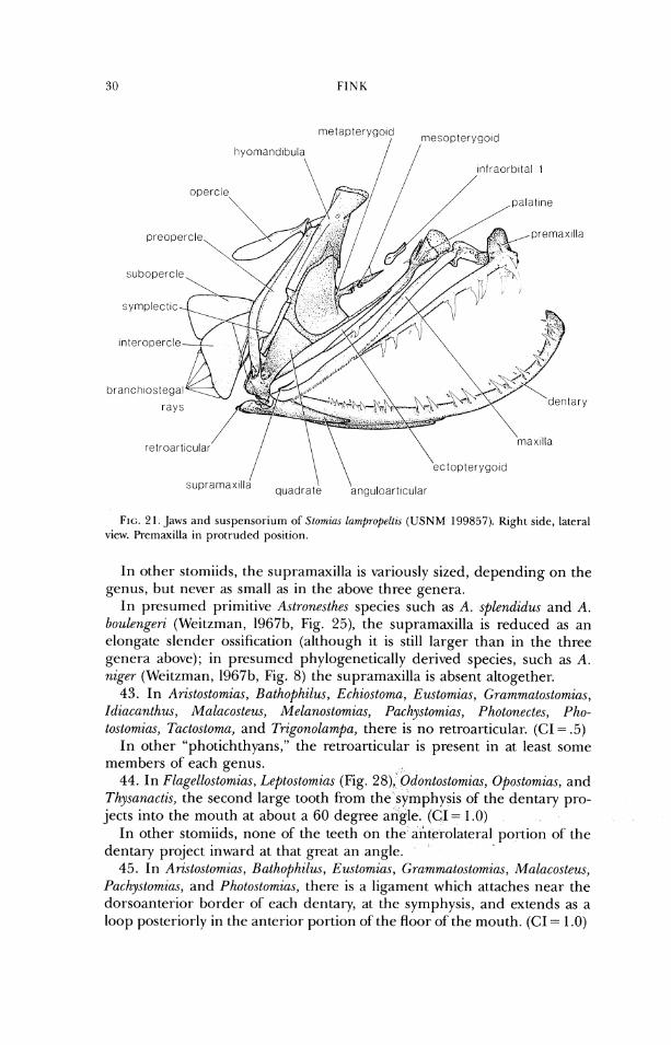
7 Ncurocratlium of Malacostru . . . . . . . . . . . . . . . . . . . . . . . . . . . . . . . . . . . . . . . . . . . . 16 8 Neurocrarliurn of Photo.stomia\ . . . . . . . . . . . . . . . . . . . . . . . . . . . . . . . . . . . . . . . . . . . . 17 9 Neu rocraniurn of Aristu.stomi(~\. . . . . . . . . . . . . . . . . . . . . . . . . . . . . . . . . . . . . . . . . . . 18
10 Neurocranium of Pacl~yystowiia.~. . . . . . . . . . . . . . . . . . . . . . . . . . . . . . . . . . . . . . . . . . . 19 1 1 Neurocrani~~m of' I'lagello.sto~nicw . . . . . . . . . . . . . . . . . . . . . . . . . . . . . . . . . . . . . . . . . . 20 12 Neurocraniurn of' Eustomia\ . . . . . . . . . . . . . . . . . . . . . . . . . . . . . . . . . . . . . . . . . . . . . . 21 13 Neurocraniurn of Ratltophzluv . . . . . . . . . . . . . . . . . . . . . . . . . . . . . . . . . . . . . . . . . . . . 22 14 Neurocranium of Ecltiostonia . . . . . . . . . . . . . . . . . . . . . . . . . . . . . . . . . . . . . . . . . . . . . 23 15 Neurocranium of Melanostomzc~s . . . . . . . . . . . . . . . . . . . . . . . . . . . . . . . . . . . . . . . . . . 24 16 Neurocraniu~n of Photonectes . . . . . . . . . . . . . . . . . . . . . . . . . . . . . . . . . . . . . . . . . . . . . 25 17 Neurocranium of Stow~ias . . . . . . . . . . . . . . . . . . . . . . . . . . . . . . . . . . . . . . . . . . . . . . . . 26 18 Jaws ant1 suspensoriurn of Echiostoma . . . . . . . . . . . . . . . . . . . . . . . . . . . . . . . . . . . . . 27 19 jaws and suspensorium of Tactoston~c~ . . . . . . . . . . . . . . . . . . . . . . . . . . . . . . . . . . . . . 28 20 Jaws and suspensor i~~rr~ of Bathophil~c . . . . . . . . . . . . . . . . . . . . . . . . . . . . . . . . . . . . 259 2 1 Jaws and suspcnsoriun~ o f Stor~~ias . . . . . . . . . . . . . . . . . . . . . . . . . . . . . . . . . . . . . . . . 30 22 Jaws and suspcnsoriurrl of Trigono1amj)a . . . . . . . . . . . . . . . . . . . . . . . . . . . . . . . . . . . 31 23 Jaws and suspensorium of Gmmmatostowiia.~ . . . . . . . . . . . . . . . . . . . . . . . . . . . . . . . . 32 24 Jaws and suspensoriurn of Ezwtow~ius . . . . . . . . . . . . . . . . . . . . . . . . . . . . . . . . . . . . . . 33 25 Jaws and suspensorium of Pholostomicw . . . . . . . . . . . . . . . . . . . . . . . . . . . . . . . . . . . . 34 26 Jaws and suspensorium of Malarostew . . . . . . . . . . . . . . . . . . . . . . . . . . . . . . . . . . . . 35 27 Jaws ancl suspensorium of P(~clty.stomia.s . . . . . . . . . . . . . . . . . . . . . . . . . . . . . . . . . . . . 36 28 Jaws and suspensoriu~n of Leptostomzrw . . . . . . . . . . . . . . . . . . . . . . . . . . . . . . . . . . . . 37 29 Jaws and suspcnsoriurn of Cttirostow~ia.~. . . . . . . . . . . . . . . . . . . . . . . . . . . . . . . . . . . 38 30 .Jaws and suspensoriurn of Photonectes . . . . . . . . . . . . . . . . . . . . . . . . . . . . . . . . . . . . . 39 3 1 H yoid arches of Stomirw. Trigonolampa. Odontostomzas. Neonr.ct/tc.c, and
Grammato.stomicw. . . . . . . . . . . . . . . . . . . . . . . . . . . . . . . . . . . . . . . . . . . . . . . . . . . . . . . . 41 32 Hyoid arches o f Mrlanostomiac. I'hotonecte.\. Rustumias. I'achy.stomin.s, and
Photoctomirw . . . . . . . . . . . . . . . . . . . . . . . . . . . . . . . . . . . . . . . . . . . . . . . . . . . . . . . . . . . . 43 33 Anterior vertebral region of Ewtowizas . . . . . . . . . . . . . . . . . . . . . . . . . . . . . . . . . . . . 57 34 Anteriol- vertebral region of Stomuw . . . . . . . . . . . . . . . . . . . . . . . . . . . . . . . . . . . . . . 58 35 Part of'the axial and vertical-fin skeleton of Idiacanthw . . . . . . . . . . . . . . . . . . . . . 60 36 Vertical-fin supports and posterior portion of axial skeleton of Trzgonolamf)~ . . 61 37 Some dorsal-fin pterygiophores of Tngonolr~mpa . . . . . . . . . . . . . . . . . . . . . . . . . . . 62
. . . Vertical-fin supports and posterior portion of axial skeleton of Photostomius 63 Pectoral girdle of Bathophil~s . . . . . . . . . . . . . . . . . . . . . . . . . . . . . . . . . . . . . . . . . . . . 64 Pectoral girclle of Arictostomiu~ . . . . . . . . . . . . . . . . . . . . . . . . . . . . . . . . . . . . . . . . . . . . 65 Pectoral girdle of Cirostomias . . . . . . . . . . . . . . . . . . . . . . . . . . . . . . . . . . . . . . . . . . . . 66 Pectoral girdle of Odontostomim . . . . . . . . . . . . . . . . . . . . . . . . . . . . . . . . . . . . . . . . . . 67 Pectoral girdle of I.eptostomic~s . . . . . . . . . . . . . . . . . . . . . . . . . . . . . . . . . . . . . . . . . . . . 68 Pectoral girdle of Melano.ctomim. . . . . . . . . . . . . . . . . . . . . . . . . . . . . . . . . . . . . . . . . . 69
. Pectoral girdle of Ewtomim cf mcntrws . . . . . . . . . . . . . . . . . . . . . . . . . . . . . . . . . . . 70 Pectoral girdle of Ewtomias cC breuibarbatw . . . . . . . . . . . . . . . . . . . . . . . . . . . . . . . . 71 Pectoral girdle of Pachy.ystomias . . . . . . . . . . . . . . . . . . . . . . . . . . . . . . . . . . . . . . . . . . . . 72 Pectoral girdle of Chauliodus . . . . . . . . . . . . . . . . . . . . . . . . . . . . . . . . . . . . . . . . . . . . . 72 Pectoral girdle of Polynietme . . . . . . . . . . . . . . . . . . . . . . . . . . . . . . . . . . . . . . . . . . . . . . 73 PectoralgirdleofStomim . . . . . . . . . . . . . . . . . . . . . . . . . . . . . . . . . . . . . . . . . . . . . . . . 74 Anterior pectoral-fin ray articulation of Heterophotw . . . . . . . . . . . . . . . . . . . . . . . 75 Anterior pectoral-fin ray articulation of Polymetme . . . . . . . . . . . . . . . . . . . . . . . . . . 76 "Rod-ray" of Thy.samctis . . . . . . . . . . . . . . . . . . . . . . . . . . . . . . . . . . . . . . . . . . . . . . . . . 77
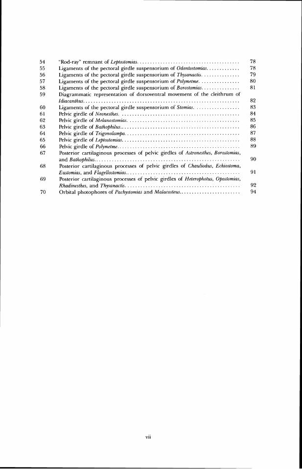
. . . . . . . . . . . . . . . . . . . . . . . . . . . . . . . . . . . . . . . "Rod-ray" remnant of Lepto.stornias. Ligaments of the pectoral girdle suspensorium of Odontostomias . . . . . . . . . . . . . Ligaments of the pectoral girdle suspensorium of Thy.sanact2s. . . . . . . . . . . . . . . Ligaments of the pectoral girdle suspensorium of Polymetme . . . . . . . . . . . . . . . . Ligaments of the pectoral girdle suspensorium of Borostomias . . . . . . . . . . . . . . . Diagrammatic representation of dorsoventral movement of the cleithrum of Idzacanthw . . . . . . . . . . . . . . . . . . . . . . . . . . . . . . . . . . . . . . . . . . . . . . . . . . . . . . . . . . . . . Ligaments of the pectoral girdle suspensorium of Stomins . . . . . . . . . . . . . . . . . . Pelvic girdle of Neonesthes . . . . . . . . . . . . . . . . . . . . . . . . . . . . . . . . . . . . . . . . . . . . . . . Rlvic girdle of Melanostomias . . . . . . . . . . . . . . . . . . . . . . . . . . . . . . . . . . . . . . . . . . . . Pelvic girdle of Bathophilw . . . . . . . . . . . . . . . . . . . . . . . . . . . . . . . . . . . . . . . . . . . . . . . Pelvic girdle of Trigonolampa . . . . . . . . . . . . . . . . . . . . . . . . . . . . . . . . . . . . . . . . . . . . . Pelvic girdle of Li?ptostomias . . . . . . . . . . . . . . . . . . . . . . . . . . . . . . . . . . . . . . . . . . . . . . Pelvic girdle of Polymetmr . . . . . . . . . . . . . . . . . . . . . . . . . . . . . . . . . . . . . . . . . . . . . . . . Posterior cartilaginous processes of pelvic girdles of Astronesthes, BorostomiaJ,
. . . . . . . . . . . . . . . . . . . . . . . . . . . . . . . . . . . . . . . . . . . . . . . . . . . . . . . . . and Bathophilw Posterior cartilaginous processes of pelvic girdles of Chauliodw, Echiostoma, E.zl\tomia.s, and Flagellostomias . . . . . . . . . . . . . . . . . . . . . . . . . . . . . . . . . . . . . . . . . . . . . Posterior cartilaginous processes of pelvic girdles of Heterophot~u, Opostomias,
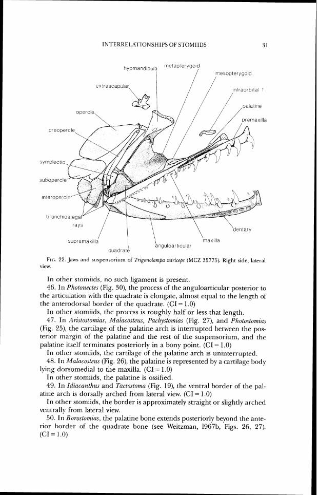
. . . . . . . . . . . . . . . . . . . . . . . . . . . . . . . . . . . . . . . . . . . . Rlladinesthe~, and Thy.sanactis. Orbital photophores of f'achystomias and Malacoslezls . . . . . . . . . . . . . . . . . . . . . . . .

IN'I'RODUCTION
This paper is the third in a series designed to outline the phylogenetic relationships in the Stomiiformes, a large group of primarily mesopelagic fishes found in all of the world's oceans. The previous works include Weitzman's (1974) revision of the Sternoptychidae and classification of stomiifor-m subgroups at the "family" level and above, and Fink and Weitzman's (1982) study of Diplophos and diagnosis of the order. Herein, I consider the relationships of the twenty-six genera of stomiiform fishes known as dragonfishes, viperfishes, snaggletooths, and loosejaws. Tradi- tionally these fishes have been placed in six families, Astronesthidae, Chau- liodontidae, Idiacanthidae, Malacosteidae, Melanostomiidae, and Stomi- idae, and most recently they were considered to comprise the superfa~nily Stornioidea (Weitzrnan, 1974). These generally elongate, darkly-pigmented fishes are characterized by numerous specializations, including presence of a mental bar-be1 associated with the hyoid apparatus, lack of gill rakers in adults, and insertion on the PO (postorbital) photophore of a portion of the adductor mandibulae muscle. By inference from their morphology and numbers, it appears that these organisms are an important component of tlie oceanic vertebrate fauna.
Several recent workers (Greenwood, et al., 1966; Morrow, 1964a; Weitzman, 1974) have distributed the genera as follows:
Astronesthes Richardson, 1844 (including Cvyptostomias Gibbs and Weitzrnan, 1965; Gibbs, pers. comm.)
Bo.rostornias Kegan, 1908 (including Diplolychnus Regan and Ti-ewavas, 1929; cf. Weitzman, 1967b)
Heterophotus Regan and Trewavas, 1929 Neonesthes Regan and Trewavas, 1929 Rhadinesthes Regan and Trewavas, 1929
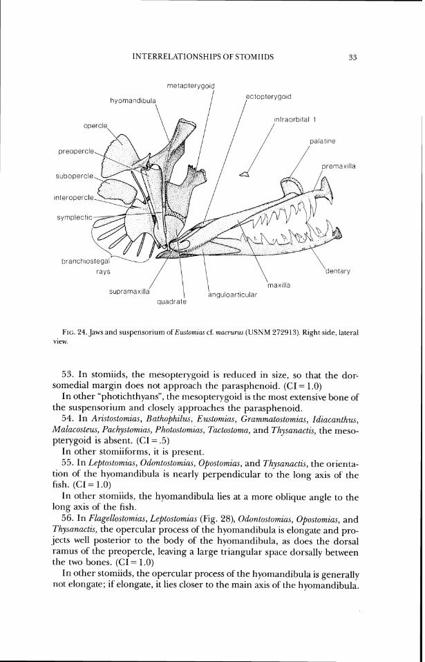
Chauliodw Bloch and Schneider, 180 1
Stomias Cuvier, 18 17 (including Macrostomias Brauer, 1902; synonymized herein, based on Fink and Fink, in press).
Bathophilw Giglioli, in Giglioli and Issel, 1884 Chirostomzas Kegan and Trewavas, 1930
Gchiostorn,~ Lowe, 1 84:3 Eustomtas Vaillant, 1888 F1agellostor~~iu.s Parr, 1927 Grurnmato.stomins Goode and Bean, 1895 Leptostomia.r Gilbert, 1905 Melanostomias Brauer, 1 902 Odontostomins Norman, 1930 0postomia.s Giinther, 1887 P(~,chystomias Gunther, 1887 Pilotonecte.~ Giinther, 1887 (including Photoneetoides Koefoed, 1956; Gibbs,
pers. comm.) Tactostoma Bolin, 1939 Thysanactis Regan and 'Irewavas, 1 930
IL)IA(:ANI I IIDAI.
Idiacanthw Peters. 1877
Ari.stostorrr.,ias Zugrnayer, 19 1 3 Ma1aco.steus Ayres, 1848 photos torn in,^ Collett, 1889 (including Ultimostomia.~ Beebe, 1933; Good-
year; 1980)
The generic synonyms listed above are changes made since the review of Gibbs ( I 964a) and Morrow and Gibbs (1 964). Hatlzysphnem Reebe (1 932) is not discussed; no specimens have been captured, and there is the strong possibility that the description is based on misobservations (see Hubbs, 1935; Morrow and Gibbs, 1964).
It is my conclusion that the family names as useti above are neither informative about relationships nor useful as indicators of gradal mor- phology and should be replaced. Use of most of these names would result in paterltly non-monophyletic taxa. For example, Idiacanthidae for Idiacunthus and Malacosteidae for Aristostomia.\, Malacosteus, and Photostomia.~, renders the large Melanostomiidae paraphyletic. Retention of the names at the Family level would require a host of' nomenclatural changes, including eleva- tion of many genera to family rank; this does not seem a useful alternative, considering the need for a relatively stable nomenclature. To preserve as much of the traditional (generic) ranking order as possible, all of' the above recognized genera are placed in the Stomiidae. A perusal of Regan and Trewavas's (1929, 1930) work will show that 1 am simply expanding their concept of the Stomiidae. One genus, Macrostomias, has been eliminated since to maintain it would render Stomias paraphyletic (Fink and Fink, in press). All other stomiid genera have been found to be monophyletic.
INTERRELATIONSHIPS OF S I'OMIIDS ?I
In order to leave this work as open to criticism as possible, I have tried to be as explicit about the characters and character polarities as I can. Addi- tionally, the data matrix upon which the computer-aided analyses were based is in Appendix 1. This will assist other workers in understanding my interpretations of the data, but they are encouraged to examine the fishes and reinterpret and recode the characters as they see fit. I have no illusions that this work is anything but a beginning in unravelling stomiid interre- lationships. Much work remains to be done at the intrageneric level, es- pecially within Eustomias and Photonectes, and I hope that the hypotheses presented herein will be an aid to an understanding of relationships within those, and other, genera.
HISTORY OF CL>ASSIFICATION OF THE STOMIIDAE
There have been few analyses of relationships of stomiiforms using ex- plicitly phylogenetic methods. In spite of this, it is useful to review past concepts of relationships in the group, however formulated, both to put the present study in historical perspective and to facilitate comparisons between my conclusions and those of earlier authors. I will concentrate here on works that dealt with storniids as recognized herein but will include com- nlents on groupings of other stomiiforms (and other teleosts) as necessary for clarity. In much of the text that follows I use taxonomic names as they were used by the authors under discussion. A history of classification of sternoptychids is available in Weitzman (1974).
Early classifications of stomiids were rather idiosyncratic, as was much of fish classification during the 19th and early 20th centuries. As new taxa were described, they were sometimes placed "near" other, already recognized, stomiiforms and sometimes not, illustrating the problems these mor- phologically diverse fishes presented for the systematic methods of the day. For example, Gill (1872) placed the three (then recognized) stomiiform fami- lies sequentially in his family list, but later (1893) placed the Ipnopidae between the Stomiatidae and Malacosteidae, also listing the Alepisauridae and Paralepidae between Idiacanthidae and Sternoptychidae.
Brauer (1906) was the first to place the stomiiform families into an ar- rangement close to that recognized in the most recent revision (Weitzman, 1974). Brauer placed the stomiiform genera in a continuous list, uninter- rupted by genera now placed in non-stomiiform families. Brauer's classifica- tion is presumably based in part on work he published in 1908 on the histology of photophores of deep-sea fishes; the diagnoses in the 1906 paper include some photophore data, but not the more complete descriptions found in the later publication. Brauer seemed to grasp the importance of photophore structure in stomiiform classification, but he did not analyze it explicitly as a systematic character. It remained for Regan and Trewavas (1929) to utilize photophore structure in diagnosing phylogenetic groups.
Regan (1923) published the first classification of stomiiforms based pri- marily on skeletal characters. He defined the suborder based on its posses- sion of photophores and divided it into two major groups, one consisting of the Gonostomatidae (in which he included some genera placed by Weitzman [1974] in the Photichthyidae) and Sternoptychidae, the other comprising the Astronesthidae, Chauliodontidae, Stomiatidae (including Idiacanthus) and Malacosteidae (Malacosteus, Photostomias, and Aristostomias). Regan considered the Gonostomatidae and Astronesthidae to be the primi- tive families of their respective subgroups. Many of the characters Regan used in his key to the families that I now place in the Stomiidae were not observed accurately or are primitive at that level, but the Chauliodontidae, Stomiatidae, and Malacosteidae were each recognized by at least one appar- ently apomorphic character.
Shortly after Regan's work, Parr (1927, 1930) proposed a classification of the stomiiforms which differed radically in concept from previous classifica- tions. In his 1927 paper, Parr suggested that the Astronesthidae, Melanostomiatidae (Stomiatidae of Regan and Trewavas without Stomias and Macrostomias), and Idiacanthidae be considered as suborder Gym- nophotodermi, of the Isospondyli, based primarily on their lack of scales and "shortness of the tail" (=shortness of the caudal peduncle). In 1930, Parr continued his reclassification, partly in response to the work of' Regan and Trewavas (1930 see below). Stomiatidae (Stomias and Macrostomias) and, provisionally, the Chauliodontidae were placed in suborder Lep- idophotodermi, and Gonostomatidae and Sternoptychidae were placed in the suborder Heterophotodermi. The Lepidophotodermi was defined by Parr's repetition of a statement of Regan and Trewavas (1930) about Stomias, i.e., that scales are present and the premaxillae are free from the maxillae; the Heterophotodermi was not redefined. Parr agreed with Regan and Trewavas (1930) in placing Idiacanthus in the Melanostomiatidae.
Parr's earlier work is the first that attempted to examine relationships among the genera in the families he investigated. He noted the difficulties encountered historically in defining genera due to discovery of new species, which often overlapped older "generic" limits, and the rarity of specimens, which did not permit detailed anatomical studies. Parr considered Astronesthes (including Borostomias) to be the most primitive genus in the suborder Gymnophotodermi, but his discussion of relationships among the melanostomiatid genera is confused and his evaluation of genera as primi- tive or advanced often seems contradictory.
In 1929 and 1930, Regan and Trewavas published their research based on the great DANA collections. The 1929 paper is a revision of the Astro- nesthidae (to which they added the new genera Heterophotus, Neonesthes, and Rhadinesthes) and the Chauliodontidae. The authors did not accept the clas- sification of Parr (1927), which was not discussed, and in fact made no major alterations in Regan's (1923) classification. Perhaps the most noteworthy aspects of the Regan and Trewavas publication are the expanded definition of the suborder (of Regan [1923]) and utilization of photophore histology in
defining groups. The histological study of Brauer (1908) is not specifically mentioned, except in reference to the "Diippelorgan," but presumably Re- gan realized the significance of Brauer's work and incorporated it into the classification. As previously noted, Brauer did not fully appreciate pho- tophore structure as a systematic character, nor evidently, did Regan when he wrote his classification of 1923.
Based on photophore structure, Kegan and Trewavas (1929) recognized two groupings within the stomioids, one consisting of the Gonostomatidae and Sternoptychidae and the other including the Astronesthidae, Chaulio- dontidae, and Stomiatidae. However, in the descriptions of the new genera mentioned above, Regan and Trewavas made no comparisons with other genera, and no interfamilial relationships were proposed or discussed.
In their 1930 paper on the families Stomiatidae and Malacosteidae, Re- gan and Trewavas again defined the various stomioid groups, b r the most part repeating what they had written in 1929. The Stomiatidae was recog- nized as including "those stomiatoids in which the dorsal and anal fins end at a short distance from the caudal, and a membrane connects the lower jaw with the hyoid arch" (this included Idiacanthm). ' f i e Malacosteidae was considered an "offshoot" of the Stomiatidae, differing primarily in lacking a membrane between the lower jaw and hyoid arch. The 1930 paper was the most important publication on stomiiforms up to that time; the authors were the first to have large series of several species available for investigation of individual variation and for skeletal comparisons. Although clearing and staining techniques were not used, and the dissections sometimes proved inadequate for accurate illustrations, Kegan and Trewavas presented generic groupings within their Stomiatidae which are close to those proposed herein. Relationships were included in an introductory section on structure and classification, where genera were listed in an order more or less reflect- ing relationships (no formal generic classification or phylogeny was given). Regan and Trewavas utilized primitive as well as specialized characters in proposing relationships, a method which led them to consider some genera, such as Chirostomias and Trigonolampa, to be related based solely on shared primitive characters.
Beebe and Crane (1939) were the first workers after Regan and Trewavas (1930) to discuss relationships within the stomioids. Their classification was esse~ltially that of Parr (1927) at the family level and above, but that of Regan and Trewavas (1930) at the generic level. Beebe and Crane reached a number of conclusions. First, they accepted division of the stomioids into three groups (Parr, 1927), but ranked them at the superfamilial rather than subordinal level, so as to leave unaltered the relative rank of the entire group in the order Isospondyli. Second, they accepted Parr's Melanostomiatidae (excluding Malacosteus, Arzstostomia.c, and Photostomiclc which they recognized as the Malacosteidae, following Regan and Trewavas [1930]). Based on Beebe (1934), they accepted Idiacanthidae (including only Idiacanthus) as separate from the Melanostomiatidae. Stomiatidae of Beebe and Crane con- sisted only of Stomias and Macrostomias. 'The authors generally agreed with
Kegan and li-ewavas (19:30) in their generic groupings, but went further than the latter authors in proposing a phylogeny, summarized in their Figure 12. From a reading of the text and an examination of the "phy- logenetic tree," it can be seen that the phylogeny proposed by Beebe and Crane is a mixture of genealogical and grade concepts. They saw groups of' genera o r fjmilies arising out of other groups of genera; for example, they regarded the members of the Astronesthidae as together possessing all the characters necessary to give rise to the Melanostomiatidae. At the conclu- sion of their discussion of generic relationships, Beebe and Crane make the first attempt to correlate the functional anatomy and relationships of' melanostoniiatids.
The next major coverage of stomiiforrns was that of Morrow (1964a-d), Morrow and Gibbs (1964) and Gibbs (1964a,b). Those publications were intended primarily for identification purposes and little discussion of rela- tionships was presented. The various families and genera were accepted for historical reasons; the only colnlnelits about acceptance of any higher cate- gory were those of Gibbs ( 1 964b) in explaining why ldiacanthus was kept in a family separate from the Melanostomiatidae.
Bassot (lY(i(i) provided stimulating new information on stomiiform pho- tophore histology. He found a basic photophore structure to be shared by all members of the order, but modified into three types, which he labeled alpha, beta, and gamma. FType alpha photophores are found in Muurolicus, Argyrofielecu.~, and Sternofityx (all now in the Sternoptychidae, sensu Weitzman, 1974). Beta photophores are found in Gono.stoma, Cyclothone, Bonapnrtia, and 1Izplopho.s (all in the Gonostomatidae of Weitz~nan, 1974). Gamma photophores are characteristic of Vinciguerria, Ichtl~yococcus (both now in Weitzman's [I9741 I'hotichthyidae), the Chauliodontidae, and the Storniatitlae (all of these taxa now in the Photichthya of Weitzman).
GI-eenwood, et al. ( I 966) disnlantled the order Isospondyli and placeti its menlbers in a numbe~- of othel- groups to reflect proposed evolutionary lineages I-ather than grade classification. 'The Stonliatoidei was placed as a suborder in the Salrr1onifo1-mes, and the families accepted were Gonostomatidae, Sternoptychidae, Astronesthidae, Melanostomiatidac, Malacosteidae, Chauliodontidae, Stomiatidae, and Idiacanthidae. No higher taxonomic groups within the suborder were proposed, and the ar- rangement of families was made without comment.
Weitzman (1967a) was concerned primarily with relationships and the origin of the stomiiforms rather than with family level classification. In addition, he includetl va1uat)le anatomical descriptions and illustrations. Weitzman (1967b) presented the first detailed discussion of the rela- tionships among the genera in the Astronesthidae and of these to other stomioids. Using osteological data almost exclusively, Weitzman found diffi- culty in considering the Astronesthidae as a group of "closely related" gen- era but made no attempt at dismantling the hmily pending more detailed analyses of' other genera. Weitzman (1967b) found that Astronesthes and Borostomia.~ appear to be closely related, that Heterophotus and Khaclinesthe.~
INTEKKELATIONSHIPS OF STOMIlDS 7
may be closely related, and that Neonesthes appears close to no other genus. In his discussion of possible relatives of the astronesthid genera among other stomioids, Weitzman proposed that all were related to Polymetme and shared a common ancestor with that genus. Weitzman also considered the Melanostomiatidae to be descendants of an astronesthid ancestor more primitive than any living species and proposed that both the Malacosteidae and Idiacanthidae arose from genera within the Melanostomiatidae. He considered the Stomiatidae and Chauliodontidae to be related and to have arisen from a very primitive astronesthid-like ancestor.
Bassot's (1970) work, although without a discussion of relationships within the stomioids, did provide further evidence regarding relationships within the suborder. The paper is an elaboration of his earlier work (Bassot, 1966); its significance is discussed by Weitzman (1974).
Weitzman (1974) is the largest recent work on relationships within the stomiids. Although he was concerned primarily with the Sternoptychidae, he also proposed a new classification of the suborder at the familial level. His most important and radical departure from previous classifications was the fission of the Gonostomatidae into two groups, each forming a primitive sister group (at the family level) in separate infraorders. This separation was done to explicitly recognize hypothesized evolutionary lineages within the larger group; thus primitive members of their lineages were placed with the more derived members ofthe lineage, rather than being left in a grade-level classification in a single, paraphyletic family. (Evidence found during the current study and in the preparation of Fink and Weitzrnan (1982) has cast doubt on the naturalness of both families and Weitzman is now working on the interrelationships of the genera.) Relevant to the issues discussed in this paper is the grouping together, as superfamily Storniatoidea, those families recognized herein as Stomiidae. Weitzman stated that "the relationships of these groups need considerable clarification and probably alteration," not- ing that "parts of the Ast~anesthidae, the Melanostomiatidae, and the Chau- liodontidae might eventually be included in an expanded Stomiatidae," the course opted for herein. He also discussed previous groupings of the in- cluded families and genera. f nk and Weitzman (1982) considered the monophyly of the stomiiforms
and their relationships with other teleosts. 'l'hey also provided a description of a morphologically primitive member of the group (Dzplophos), in part for comparison with the taxa discussed herein. Fink and Weitzman found that the Stomiiformes is monophyletic and diagnosed it with eight syn- apomorphic characters. They agreed with Kosen (1973) that the group is the sister group of the Eurypterygii and should be removed from the Prota- canthopterygii ( = Salmoniformes) and placed as a separate order within the Neoteleostei.
Fink (1984) published a brief summary of his work to that date on the stomiids. There are few differences between his conclusions and those pre- sented herein. Ahlstrom, Richards, and Weitzman (1984) reviewed the problems of relationships among the more primitive storniifbrm genera,
with special emphasis on larval traits. Their conclusions are consistent with Weitzman's dismantling of the Gonostomatidae. Kawaguchi and Moser (1984) examined larval stomiids and presented a large amount of data that may contribute to an unraveling of stomiid relationships. Although the data from their study are not included herein, it is apparent that the degree of- homoplasy found in adult stomiids should be expected in the larvae as well.
A number of fossil specimens have been refen-ed to the Stomiidae. In most cases, the placement of those specimens in the group is not support- able, and in no case does a fossil taxon alter concepts of the modern genera. For further information, see Appendix 2.
Many species oS stomiids are poorly known, often only frotn type material, and large series of most species arc not avail;~ble. For this reason it has not beer1 possible to determine the range otvariability within species ant1 genera in the skeleton of these fishes. Add to this the hct that many of the av;iilable specimens are juverriles, and it beconles clear that some of the mor- phological details described and the hypotlreses proposed in the current study may be es- pecially subject to change ;tncl reinterpt-etation. An attempt has beer] made to select for study those species in each genus which, based on outgroup cornparison, appeal- to be the most phylogenetically and morphologically prirnitive, hut this has not always been possible. Current knowledge of relationships within most genera prevents secure hypotheses of what species may be most primitive. An attempt has been rnade to secure leprcsentatives oS those groups whet-e subgenera or subgroups have been recognized historically, except when the species in a given subgroup appear to be highly spccializecl rnorphologically, rompared to members of other subgroups. Ihe re has been no attempt to study or clarify relationships below the "gcneric" level, b u ~ some comments on these lower levels are included when information was found that might guide other researchers. Material examined is listed in Appendix 3.
My discussions of' anatomy at-e in some places at variance with reports of earlier workers. In most cases, these can be attributed to availability of specimens i r ~ better cor~dition and of cleared and stained material, which greatly facilitates osteological observation. I have not attempted to go through the literature and specify what I consider errors, nlostly because it would be tedious fix both me and the reader and woulcl be of considerable length. For primitive stomiids, much of this work has been done by Weitzrnan (1967b).
Selection of characters for this study began by an examination of previously published studies, with an eye to determining what morphological structures had been used in the past, what their variation was, and what phylogenetic (or other "relationship") hypo~heses had beer1 inferred from them. I then examined the fishes in detail, both to confirm the pt-evious obscrva- tions of morphology and to search for other features. 1 soon realized that there is a lairly large degree of incongruence among many of the features, whether chosen by nryself or other workers. After this realization, structures were chosen as characters rcgarclless of how their distribution met with preconceived rlotions of relationship, no matter how far off the mark they seemed. For example, the rugose frontal bones oSL;chioslomu, Melar~ostomius, and Trigonolampn were coded as a character, even though my intuition told me that these taxa did not constitute a monophyletic group (this was eventually borne out in the parsimony analysis). Because of this character sampling technique, the data presented herein are somewhat more catholic than those often found in systematic presentations, a r ~ d they certair~ly are "noiset-", with relatively low consistency indices. An advantage of this selection procedure, however, is that it includes more oP the morphology than would otherwise be the case, and it does so with less personal bias than may be present in some other studies.
This study began as an exercise in "Hennig argumentation" involving various three taxon-
statements. As the data base became large and rather unwieldy, I turned to numerical meth- ods, all of which are based on use of parsimony in hypothesis choice. During the course of the work, several efficient computer programs havc become available, and the final analyses were done using PAUP (Phylogenetic Analysis Using Parsimony), version 2.3 on the Michigan Terminal System (MTS) Amdahl computer. Consensus trees were generated by CON'TREE, also running on MTS. Both of these plugrams were written and made available by David Swofford. Additive binary coding was used for the numerical analyses. Characters not present (either lxirnarily or secondarily) were coded as "missing" and thus had no influence on place- mcnt of those taxa on the cladogram. Character states are assigned during the optimization procedure to taxa originally coded as missing, after the tree-building procedure. The matrix is available in Appendix 1.
For each character describetl in the Characters section, the del-ived state (with the I-ange of its consistency indices, (:.I.; Kluge and Farris, 1969) and the taxa that possess it are listed first, followed by a paragraph describing the outgroup condition. Exceptions and details of anatomy arc also listed. 'l'he search for outgroup conditions included broad surveys of basal euteleosts as described and listed by Fink and Wcitznlan (1982). In the final analysis, characters were examinecl in rnernbel-s of the Photichthya (smsu Weitzrnan, 1974). Due to uncertainties about the rnonophyly and interrelationships of this large assernblage, its members arc referred to below as "photichthy;~ns", Storniid photichthyans clearly constitute a monophyletic group ant1 the other genera rnost pnhably do not, so any characters consistently plesent in the lattex- and modified in the forrrrcr should be confidently assessed as derived in the Stomiidae. In many cases, when dealing with relatively pllylogenetically-derivcd genera, the outgroup search was lilnited to other storniitls or some subgroup of stomiids, as indicatetl in the character descl-ip- tions.
When it appears pl-obable, based on other chat-acters, that a tl-ait has developed within a genus, that character is codcd as primitive rather than as diagnostic of the genus; in the tlescriptive text, these characters are indicated by the phrase "in some species. . ." For Stomin,\, evitlencc from Fink and Fink (in press) has been used to determine the primitive states of c ~ l . '11 .. nctet-s . for the genus. For chat-acters which were missing and about which polarity evidence was iunavailable, no u priori decisiorls were made about whether the absence is primary (not present in ancestors) o r secondary (present in ancestors and lost in the descendant). To have made such decisions would havc presurned a tree topology, a practice inconsistent with phy- logenetic reasoning. Examples of such characters are features of the secondary pectoral gir-dle ofI't~otostomia.s; members of that gcnus lack that part of the girdle, so when outgroups possess both pl-imitive and derived states, it is not possible to assign a state unatnbiguously to the i~lgroup taxon. With such characters, one approach would be to assume the primitive state, no evolutionary change. Another approach is to be "agnostic" about such cl~aractel-s and elinlinate them from the tree-building process. Because of my own "agnostic" leanings, in the discussion of'cliaracter evolution in the texl 1 relied on the data matrix with "missing" characters so coded. The choice of any particular equal length tree over another may cause one to accept certain cl~aractcr state I-esolutions for characters originally codcd as missing, and in this sense, to choose one tree over another is to give up some degree of agnosticism regarding character evolution. Since I am presenting all of the equal length cladograms in this study, the issue is not relevant here, b ~ ~ t it should be kept in mind when discussing character change in phylogenetic analyses.
The PAUP program has several optimization routines which allow one to make assumptions about the nature of character change. I used only the standard Farris optimization and the MINF optimization, which attempts to minimize the number of groups that are diagnosed on the basis of arbitl-a~y resolutions. Swofford (1983) should be consulted for ~LII-ther information regarding these optimi~atio~l procedures. Because Farris optimization is the method most often used in current numerical phylogenetic analyses, I have presented the data using that op- timization in the Discussion section and in the Results section documenting suppol-t for the most p;irsirnonious cladograms. I chose this approach as a conservative one. Work in progress by Swofford and Wayne Maddison will discuss the alternative optimization procedures now available.
Other than character choice, n o chat-acter weighting was employed, primarily because I krlow of rlo reasonable way to do such wcighting. Most methods proposed so f i r rilake assurnp- tiotls about evolutionary process which I am unwilling to include a ~ T Z O ~ . One altractive wcighting scheme, which weighs chal-acters based on their within-group variation (in this case it would probably have been withitr-genus variation), was not used simply because we have such skimpy knowledge about morphological variation in stomiids at any taxono~nic level.
Material from several institutions was used, including: Museum of Comparative Zoology, Harvard University (MCZ), National Museum of' Natural History, Smithsonian ltlstitution (USNM), Los Angeles County Museum (I,ACM), and Scripps Institution of' Oceanography (SIO), and specimens on loan to the USNM f'rom the DANA collections. Specimens were cleared and stained according to an enzyme procedure modified frorn 'I'dylor- (1967) and stored in glycerine or ethanol. In the figures, by cotlvention, fine stipple represents bone, large stipple, cartilage. Most drawings were outlined using a Zeiss IVB zoom microscope with camera lucida, and details were subsequently filled in using a I.eitz RS widefield stereo micro- scope. Occasionally, a Zeiss compound microscope was used for elucidation of very small detail.
Unless otherwise stated, the tel-m "cross-section" is used to signify a section through a bone, or other object, perpendicular to the long axis ot'thar object rather than to the long axis of'tlle fish body.
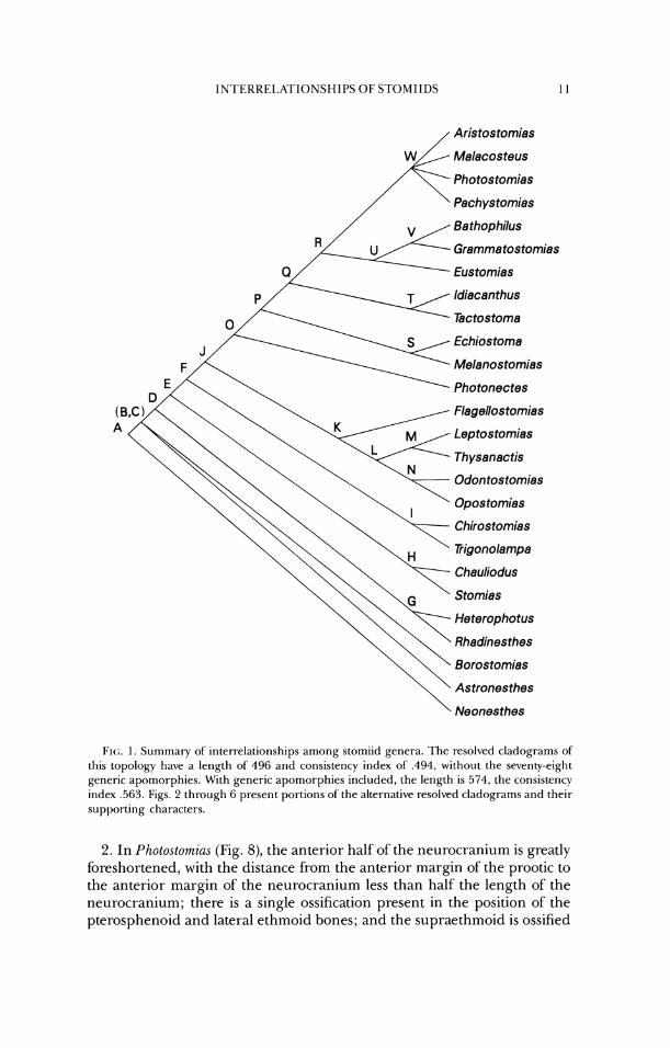
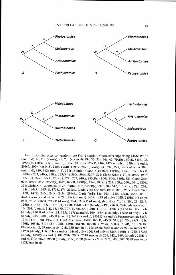
The summary cladogra~n and character distributions are presented at this point to give the reader a context in which to examine the following sections, which include presentation of the data and discussions of stomiid subgroups and character transformations. The cladogram in Fig. 1 is a strict consensus tree (Rohlf, 1982) of the six fully resolved, equally parsimonious cladograms generated from the data. Selected subunits of the phylogeny are presented in Figs. 2 through 6, and the supporting data, as numbered in the Charac- ters section, are listed in the figure captions. Alternate cladograms are con- sidered in the Discussion section.
CHARACTEKS
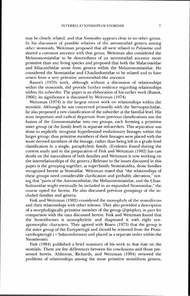
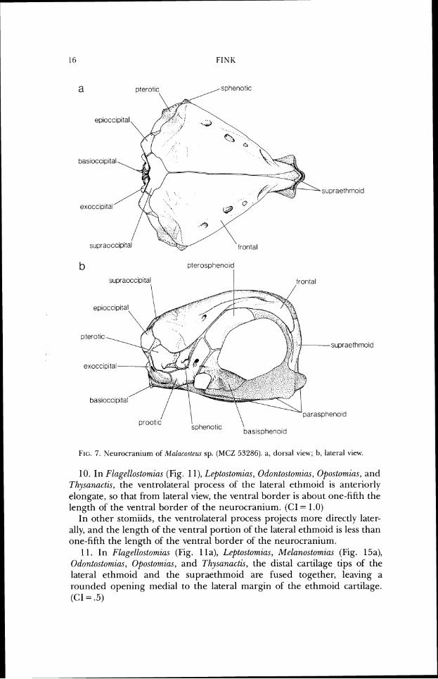
1. In MaLucoste.us (Fig. 7), the anterodorsal border of the neurocranium, from the anterior opening of the frontal sensory canal to the anteroventral margin of the supraethmoid, is highly convex from lateral view; the entire anterodorsal surface of the ethmoid is approximately vertical; and the lat- eral processes of the ethmoid cartilage are very small. (CI = 1.0)
In other stomiids, the anterodorsal border of the neurocranium is, at most, moderately convex; the anterodorsal surface of the ethmoid region slopes ventrally more gradually; and the lateral processes of' the ethmoid cartilage project further laterally.
1N~I'ERREI.ATIONSHII'S OF STOMIIDS 1 1
/ Aristostomias
Malacosteus
Photostomias
Pach ystomias
Bathophilus
Grammatostomias
0 Eustomias
ldiacanthus
Tectostoma
Echiostoma
Melanostomias
Photonectes
Flagellostomias
Leptostomias
Thysanactis
Odontostomias
Opostomias
Chirostomias
Trigonolampa
Chauliodus
Stomias
Heterophotus
Rhadinesthes
Borostomias
Astronesthes
\ Neonesthes
I'I(;. 1. Sunlrnaly o S inter-lulationships among stomiid genera. l 'he resolved cladograms of this topology have a length of 496 and consistetrcy index of' ,494, without the sevenv-eight generic aporrrol-phies. With generic apornorphies included, the length is 574, the consistency intlcx .563. Figs. 2 through 6 present portions of the alternative resolved rladograrns and their suplx)r(itlg characters.
2. In Pho~ostomias (Fig. 8), the anterior half of the neurocranium is greatly foreshortened, with the distance from the anterior margin of the prootic to the anterior margin of the neurocranium less than half the length of the neurocranium; there is a single ossification present in the position of the pterosphenoid and lateral ethmoid bones; and the supraethmoid is ossified
OTHERS ( D) OTHERS (Dl
Borostomias Borostomias
Astronesthes Astronesthes
Neonesthes Neonesthes
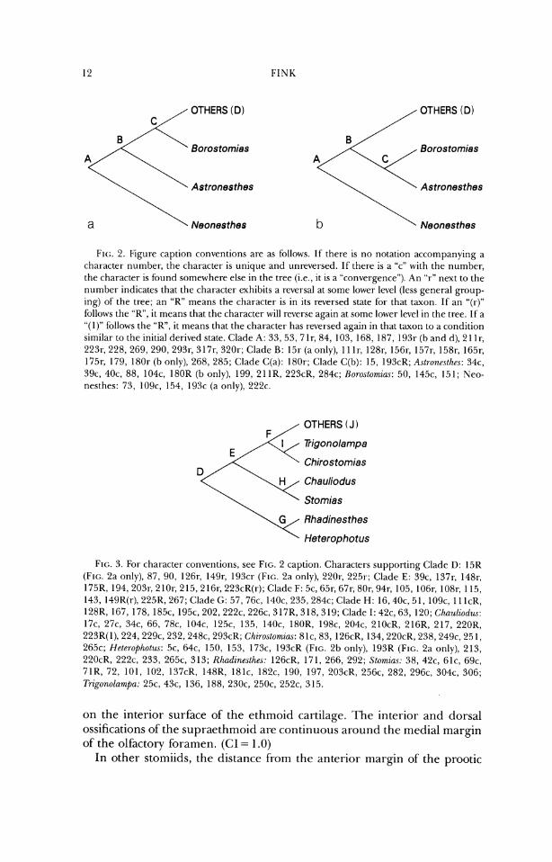
FIG. 2. Figure caption conventiot~s are as follows. If there is no notation accompanying a character number; the ch;u-acrer is unique and unreversed. If thet-e is a "c" with the nurnGer, the character is Ibund somewhere else in the tree (i.e., it is a "convergence"). An "r" next to thc number indicates that the chal-acter exhibits a reversal at sorne lower level (less general group- .. .
ing) of the tree; an "K" means the character is in its reversed state for that taxon. If' an "(r)" follows the "R", it rneans that the character will reverse again at some lower level in the tree. 11 a "(1)" lbllows the "R", it means that the chat-acter has reversed again in that taxon to a condition similar to the initial derived state. Clade A: 33, 53, 71r, 84, 103, 168, 187, 193r (b and d), 2 1 lr, 223r, 228, 269, 290, 293r, 3 17r, 3201.; Clade B: 151- (a only), 1 1 1 I; 128r, 15(ir, 157r, 1581; 165r, 1751; 179, 180r (b only), 268, 285; Cladc (:(a): 1801-; Clade (:(b): 15, 193cK; Astronnllrc~.\: 34c, 39c, 40c. 88, 104c, 180K (b only), 199, 21 IR, 223cR, 284c; No?-oslorniu.c: 50, 145c, 151; Nco- nesthes: 73, 109c, 154, 193c (a only), 2 2 2 ~ .
OTHERS (J )
Pigonolampa
Chirostomias
Chauliodus
Stomias
Rhadinesthes
Heterophotus
FIG. 3. For character convcr~tions, see FIG. 2 caption. Characters supportirig (:lade D: 15K (Frc;. 2a only), 87, 90, 126r, 149r, l93cr (FIG. 2a ot~ly), 220r. 225r; Clade E: 39c, 1371; 1481; 175K, 194,203r, 210r, 215,216r, 223cR(r); Cladc F: 5c, 65r, 671-, 801; 94r, 105, 106r, 108r, 1 15, 143, 149R(r), 225R. 267; Clatle G: 57, 76c, 140c, 235, 284c; Clade H: 16,40c, 51, 109c, 11 lcK, 128R, 167, 178, 185c, 1 9 5 ~ . 202, 222c, 226c, 317R, 318,319; Clade I: 42c, (53, 120; Chau,liod~u: 17c, 27c, 34c, 66, 78c. 104c, 125c, 135, 140c, 180R, l98c, 204c, 210cR, 216R, 217, 220K, 223R(1), 224,229c, 232, 248c, 293cR; Chzro.stomias: 8 lc, 83, 126cR, 134, 220cK, 238, 249c, 25 I , 265c; Hel~,rophotw: 5c, 64c, 150, 153, 173c, 193cK (FIG. 2b only), 193R (FIG. 2a only), 213, 220cR, 222c, 233, 2 6 5 ~ . 313; Rhadinesthes: 126cK, 171, 266, 292; S/omzu.c: 38, 42c, (ilc, 69c, 71K, 72, 101, 102, 137cR, 148K, 181c, 182c, 190, 197, 203cK, 256c, 282, 296c, 304c, 306; Trigonolarnpa: 25c, 43c, 136, 188, 230c, 250c, 252c, 315.
on the interior surface of the ethmoid cartilage. The interior and dorsal ossifications of the supraethmoid are continuous around the medial margin of the olfactory foramen. (CI = 1.0)
In other stomiids, the distance from the anterior margin of the prootic
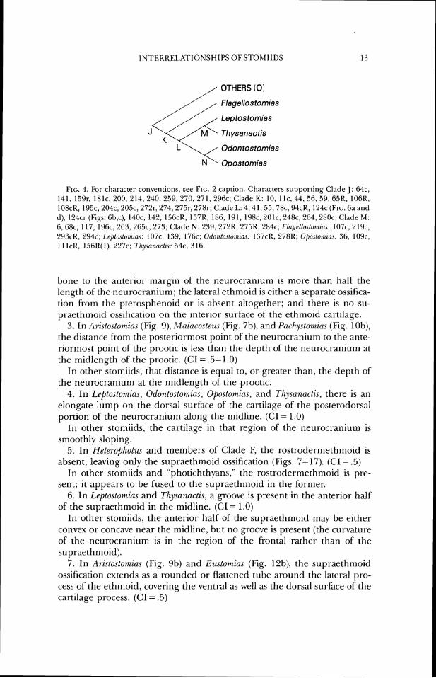
OTHERS (0)
Flagellos tomias
Leptostomias
J Thysanactis
Odontostomias
N' Opostomias
FIG. 4. For charactel- co~iventions, see FIG. 2 caption. Characters suppol-ting Clade J: 64c, 141, 15% 181c, 200, 214, 240, 259, 270, 271, 296c; (:lade K: 10, 1 I(-, 44, 56, 59, 65R, 106K, 108cR, 195c, 204c, 205c, 272r, 274,275r, 278r; (;lade L: 4 ,41 ,55 ,78c , 94cR, 124c (Flu.. 6a and d), 124cr (Figs. 6b,c), 1 4 0 ~ . 142, 156cR, 157R, 186, 191, 198c, 2 0 1 ~ . 248c, 2 6 4 , 2 8 0 ~ ; Clade M: 6, ti8c. 117, 196c, 263,265c, 273; Clade N : 239, 272R, 275K, 284c; Flugellustumias: 107c, 219c, 293cR, 2 9 4 ~ ; Le/toslomic~s: 107c, 139, 176c; Odonlostomias: 1:17cR, 278R; Opostomias: 36, 109c, I 1 lcR, 156R(I), 227c; T/zysunuc/zs: 54c, 3 16.
bone to the anterior margin of the neurocranium is more than half' the length of the neurocranium; the lateral ethmoid is either a separate ossifica- tion fiom the pterosphenoid or is absent altogether; and there is no su- praethmoid ossification on the interior surface of the ethmoid cartilage.
3. In Aristostomias (Fig. 9), Malaco.cteus (Fig. 7b), and Pachy.stomias (Fig. lob), the distance from the posteriormost point of' the neurocranium to the ante- riormost point of the prootic is less than the depth of the neurocranium at the midlength of the prootic. (CI = ,551.0)
In other stomiids, that distance is equal to, or greater than, the depth of the neurocranium at the midlength of the prootic.
4. In Leptostornias, Odontostomias, Opostomias, and Tl~ysanactis, there is an elongate lump on the dorsal surface of the cartilage of the posterodorsal portion of the neurocranium along the midline. (C1= 1.0)
In other stomiids, the cartilage in that region of the neurocranium is - - smoothly sloping.
5. In Neterophotus and members of Clade E; the rostrodermetl~moid is absent, leaving only the supraethmoid ossification (Figs. 7-17). (CI = .5)
In other stomiids and "photichthyans," the rostrodermethmoid is pre- sent; it appears to be fused to the supraethmoid in the former.
6. In L2eptostomias and Thysanactis, a groove is present in the anterior half of the supraethmoid in the midline. (CI = 1.0)
In other stomiids, the anterior half of the supraethmoid may be either convex or concave near the midline, but no groove is present (the curvature of the neurocranium is in the region of the frontal rather than of the supraethmoid).
7. In Aristostomias (Fig. 9b) and Eustomias (Fig. 12b), the supraethmoid ossification extends as a rounded or flattened tube around the lateral pro- cess of the ethmoid, covering the ventral as well as the dorsal surface of the cartilage process. (CI = .5)
FINK
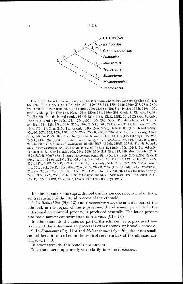
OTHERS ( W
Bathophilus
Q Grammatostomias
\'- Eustomias
ldiacanthus
Tactostoma
Echiostoma
Melanostomias
Photonectes
FIG. 5. For character conventions, see F I ~ ; . 2 caption. Characters supporting <:lade 0: 4%. 61c. 69cr, 70, 79r; 93, 112r; 1131; 1221; 123, 127r, 138, 144, 182c, 245r, 250cr, 257, 284c, 286r, 288, 289r, 291, 297r (FIG. 6a, b, and c only), 299; Clade P: 281; filer, 94cR(r), 132r, 140c, 1631; 212r; Clade Q: 12r, 17cr, 54c, 195c, 196~1; 229cr, 231, 256cr, 261; Clade R: 32c, 40c, 45,62r, 74, 75r, 85r (FIG. 6a, b, and c only), y l r , 94R(1), 113R, 122K, 158R, 161, 1621. (FIG. 6d only), 165R(r) (FIG. 6d only), 169c, 172r, 173cr, 183r, 185c, 206r, 300cr (FIG. 6d only); (:lade S: 9, 14, 24, 25c, 118c, 129, 176c, 205c, 227c, 230c, 245cR, 280c, 281; (:lade T: 49, 68c, 76c, 77, 82c, 166c, 170, 189, 242c, 245c (FIG. 6c only), 246c, 247c, 279c; Clade U: 85c (FIG. 6b and d only), 95c, 96, 107c, 152, 155r. 198cr, 205c, 253c, 256cR, 276, 297K(r) (FIG. 621, b, and c only); Clade V: 8,62R, 69cR, 92c, 97, 118c, 162c (FIG. 6a, b, and c only), 164, 165 (FIG. 6d only), 166c, 236, 245cR, 249c, 254c, 300c (FIG. ha, b, and c only), 301c; Bathophil~u: 37, 119, 155K, 262, 283, 293cR, 295c, 298, 303c, 308; Echiostwtr~u: 26, 58, 69cR, 132cK, 286cR, 297cR (FIG. 6a, b, and c only), 305c; E~~~torn ius : 7c, 13c, 27c, 2XcR, 52 ,60 , 75R, 91R, 126cR, 133c, l62cR (FIG. 6d only), 165cR (FIG. 6a, b, and c only), 192,204c, 208c, 2 19c, 22 1, 234, 243, 245c (FIG. 6c only), 250R, 287c, 289cR, 300cR (FIG. 6d only); Crummatostomia~: 89, 145c, 177, 198R, 203cR, 255,297R(l) (FIG. 6a, b, and c only), 297c (FIG. 6d only); Idiacuntl~us: 17R, 114, 130, 133c, 203cR, 218, 222c, 226c, 227c, 229R, 286cR, 297cK (FIG. 6a, b, and c only), 304c, 312c, 322, 323; Melanactomim: l lc, 27c, 28cR, 79cR, 133c, 204c, 252c, 287c, 289cR, 297c (FIG. 6d only), 300c; Pho/o~zectes: 27c, 29c, 32c, 46, 76c, 95c, 100, 1 10c, 125c, l69c, 185c, 198c, 203cR, 244, 245c (FIG. 6c only), 2 4 6 ~ . 247c, 252c, 253c, 254c, 258c, 297c (FIG. 6d only); Tuctostomn: 12cR, 35, 80cR, 81cR, 127cR, 132cR, 212R, 280c, 287c, 289cR, 297c (FIG. 6d only), 305c.
In other stomiids, the supraethnloid ossification does not extend onto the ventral surface of the lateral process of the ethmoid.
8. In Rathophilw (Fig. 13) and Grammatostomias, the anterior part of' the ethmoid, in the region of the supraethmoid and vomer, particularly the anteromedian ethmoid process, is produced ventrally. The latter process also has a narrow concavity from dorsal view. (CI = 1.0)
In other stomiids, the anterior part of the ethmoid is not produced ven- trally, and the anteromedian process is either convex or broadly concave.
9. In Echiostoma (Fig. 14b) and Melanostomias (Fig. 15b), there is a small, conical bone in a pocket on the ventrolateral surface of the ethmoid car- tilage. (CI = 1 .O)
In other stomiids, this bone is not present. It is also absent, apparently secondarily, in some Schiostoma.
1NTERKEI.KI'IONSHIPS OF STOMIIDS 15
Photostomias
Malacosteus Malacosteus
Aristostomias
a Pachystomias
Malacosteus Pachystomias
Photostomias Aristostomias
Aristostomias Malacosteus
Pachystomies Photostomias
FK:. 6. For character conventions, see FIG. 2 caption. Characters supporting Clade W: 3 r (not in d), 18, 201. (a only), 22, 23r (not in d), 29c, 30, 31r, 34c, 47, 79cR(r), 80cK. 81cR, 98, 108cR(r), 1 lOcr, 121r (b and d), 125cr (d only), 127cR, 146r, 147r (c only), 163R(r) (a only), 203cR, 207r (not in d), 222c, 225R(I), 226c, 237r (d only), 241, 260, 277, 304cr (d only), 309r (not in d), 3 10, 3 12r (not in d), 32 1 r (d only); Clade X(a): 92cr, 112R(r), 125c, 162c, 165cR, 183R(r), 237, 246cr. 258cr. 293cR(r), 300c, 304c, 320R, 321; Clade X(b): 112R(r), 124cr. 125c, 126cK(r), 1 6 2 ~ . 165cK, 173R(r), 174r, 237,246cr, 293cR(r), 300c, 304c, 320R, 321; Clade X(c): Y2cr; 124cr, 125c, 126cR(r), 162c, 1651% 173R(r), 174r, 183R(r), 237.258cr, 300c, 304c, 320R, 321; Clade X(d): 3,201; 23, 147r, 163R(r), 207,245cR(r), 297c, 309, 312,313; Clade Y(a): 20R, 1 2 4 ~ 126cR, 163R(1), 173R, 174,297cR; Clade Y(b): 85c, 92c, 121K, 183R, 258c; Clade Y(c): 112K, 147R. 245c, 246c, 247c, 293cR; Clade Y(d): 85c, 92c, 121R, 183R. 2 5 8 ~ . 320R; Arirlostornirfi (a and d): 7c, 19, 21, 112cR (d only), 146R, 147R (d only), 196R, 245R(1) (d only), 247c, 24% 256cR, 293cR (d only), 294c, 317cR (d only); (b and c): 7c, 19, 20c, 21, 124R, 126K(1), 146R, 163cR, 173R(1), 174R, 196R, 247c (b only), 2 4 9 ~ . 256cR, 294c; Malacostew: 1, 13c, 2OK (d only), 31R, 48, 67K, 79R(1), 82c, 86, 108R(1), 110R. l12R(1) (a and b), 1 lac, 124c (d only), 126cR (tl only), 131, 133c, 147c (a and b), 160, 163R(1) (d only), 173cR (d only), 174c (d only), 205c, 2 0 8 ~ . 245cR (a and b), 246R (a and b), 293R(1) (a and b); Pachystomim (a): 85cR, 121c, 147c, 159R, 245cK, 31 1; (b): 20c, 147c, 159R, 163cR, 245cK, 31 1; (c): 20c, 85cR, 121c, 150R. 163cR, 31 1; (d): 125R, 15YR, 162cR, 165cR(1), 23713, 300cR, 304R, 311, 321R; Photostomicls: 2, 3R (not in d), 12cR, 23R (not in d), 27c, 28cR, 85cR (a and c), 92K (a and c), 99, 1 12cR (d only), 1 16, 12 1 c (a and c), 124c (d only), 126cR (d only), 132cR, 149R(1), 172R, 173cR (d only), 183R(1) (a and c), 184, 201c, 206R, 207R (not in d), 209,242c, 246c (d only), 258R (a and c), 279c, 2 8 7 ~ . 293cR (d only), 295c, 297R (b and c), 301c, 302,303c, 307,309R (not in d), 312R (not in d).
b pterospheno~d
supraocc~pita( frontal
prootld I sphenotic \ baslsoheno~d
FIG:. 7 . Neurocraniunl of Mahcos te z~~ sp. (MCZ 53286). a, dorsal vicw; h, lateral vicw.
10. In Flagellostomias (Fig. 1 l) , Leptostomias, Odontostomias, Opostomias, and Tlzysanactis, the ventrolateral process of the lateral ethmoid is anteriorly elongate, so that from lateral view, the ventral border is about one-fifth the length of the ventral border of the neurocranium. (CI = 1.0)
In other stomiids, the ventrolateral process prqjects more directly later- ally, and the length of the ventral portion of the lateral ethmoid is less than one-fifth the length of the ventral border of the neurocranium.
11. In Flagellostomias (Fig. 1 la), Leptostomzas, Melanostomias (Fig. 15a), Odontostomias, Opostomias, and Thysunactis, the distal cartilage tips of the lateral ethmoid and the supraethmoid are fused together, leaving a rounded opening medial to the lateral margin of the ethmoid cartilage. (CI = .5)
supraethmoid
\ frontal
lateral ethmoid
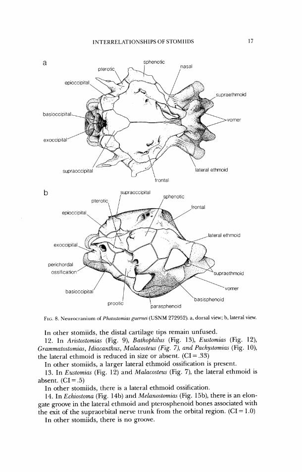
FIG. 8. Neurocranium oSPho~osLomzus guernei ( U S N M 272952). a, dorsal view; b, lateral view.
In other stomiids, the distal cartilage tips remain unfused. 12. In Ari.stostomia.s (Fig. 9), Bathophilzu (Fig. 13), Ezutomias (Fig. 12),
Grammatostomias, Idiacanthus, Malacosteu~ (Fig. 7), and Pachystomias (Fig. lo), the lateral ethmoid is reduced in size or absent. (CI = .33)
In other stomiids, a larger lateral ethmoid ossification is present. 13. In Eustomias (Fig. 12) and Malacostezu (Fig. 7), the lateral ethmoid is
absent. (CI = .5) In other stomiids, there is a lateral ethmoid ossification. 14. In Echiostoma (Fig. 14b) and Melanostomias (Fig. 15b), there is an elon-
gate groove in the lateral ethmoid and pterosphenoid bones associated with the exit of the supraorbital nerve trunk from the orbital region. (CI = 1 .O)
In other stomiids, there is no groove.
oss~f~cat~on
lateral ethmoid
\ prootic parasphenold baslspheno~d
FIG. 9. Neurocranium of An.\lO.\lOm~a.~ xenoslomn (USNM uncat. DANA st. '"''71(;). a, dorsal view; b, lateral view.
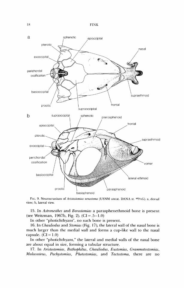
15. In Astronesthes and Horo.stomia.s a parasphenethmoid bone is present (see Weitzman, 1 Y67b, Fig. 2). (CI = .5- 1 .O)
In other "photichthyans", no such bone is present. 16. In Chauliodw and Stomius (Fig. 17), the lateral wall of the nasal bone is
much larger than the medial wall and forms a cup-like wall to the nasal capsule. (CI = 1 .O)
In other "photichthyans," the lateral and medial walls of the nasal bone are about equal in size, forming a tubular structure.
17. In Aristostornias, Rathophilw~, Chauliodus, Eustomias, Grammatostornias, Malacosteus, Pachystomias, Photostomias, and Tactostoma, there are no
1N.I'ERRELATIONSHIPS O F SI'OMIIDS
lateral ethrnold I
nasal
basioccipltal
supraocc~pltal
supraethmold frontal
pterotlc
lateral ethmold
I prootic 1 \
vomel bas~spheno~d parasphenold
FIG. 10. Neurocranium of Pachystomzns mzcrodon (USNM uncat. DANA st. 4 % ~ ) . a, dorsal vicw; b, lateral view.
vomerine teeth (Figs. 7b-lob, 12b, 13b). (CI = .33) In other stomiids, there are vomerine teeth. 18. In Aristostomzas, Malacosleus, Puchystomias, and Photostomzas, the para-
sphenoid terminates posteriorly well anterior to the posteroventral margin of the basioccipital (Figs. 7b-lob). (GI = 1.0)
In other stomiids, the parasphenoid posterior-ly terminates only slightly anterior to the posteroventral margin of the basioccipital.
19. In Arzstostomias (Fig. 9b), the ventral portion of the neurocranium (the parasphenoid and associated cartilage and the ventral part of' the basisphenoid) extends well ventral to its position in other stomiids. (CI = 1 .O)
\ lateral ethm old par~etal frontal
ateral ethmoid
bas~spheno~d parasphenold
FIG. 11 . Neurocraniurn of Fl~gelloslowtzn~ Do1c7-eez (USNM 206681). a, dorsal view; b, lateral vicw.
20. In Ari~tosto~zins (Fig. 9b) and Pachystomias (Fig. lob), the greatest width of the basisphenoid, from lateral view, is half or less its height, and the bone is positioned in about the posterior third of the skull. (CI = .5)
In other stomiids, the greatest width of the basisphenoid is more than half its height, and the bone is positioned in about the midlength of the skull.
21. In Arzstostomias (Fig. 9), the sphenotic spine extends posteriorly ap- proximately as far as the pterotic process and the posterior border of the prootic. (CI = 1 .O)
In other stomiids, the sphenotic spine extends no further posteriorly than a vertical line drawn slightly posterior to the midlength of the prootic.
22. In Aristostomius, Malacosteus, Pachystomias, and Photostomias, the sphenotic spine extends posterior to the midlength of the prootic and the
INTERRELKI'IONSHII'S O F STOMII1)S
a pterotic \ sphenotic
1 frontal supraocclp~tal
frontal
/
1 \ vomer bas~occipital
prootlc parasphenold bas~sphenold
FIG. 12. Neurocraniunl ofRwton~ia,s cf. mucrur~w (USNM 272913). a, dorsal view; b, latet-al view.
anterior margin of the hcet for the hyornandibula (Figs. 7-10). (CI= 1.0) In other stomiids, the spine terminates at, or anterior to, the midlength of
the prootic bone, and anterior to the facet tor the hyomandibula. 23. In Aristostomias (Fig. 9), Malacosteus (Fig. 7), and Pachystomiu,~ (Fig. lo),
the pterotic is produced laterally and does not bear a sensory canal. (CI = .5- 1 .O)
In other stomiids, the pterotic is produced posterolaterally and bears a sensory canal.
24. In Echiostoma (Fig. 14b) and Melanostomias (Fig. 15b), the posterior process of the pterotic is robust and projects posterodorsally. (CI = 1.0)
In other stomiids, the process is more slender and projects either directly posteriorly or posteroventrally.
25. In Echiostoma (Fig. 14), Melanostomias (Fig. 15), and Trigonolampa, rugosities are present on the dorsal ridges of the frontal sensory canals. (CI = .5)
In other stomiids, no rugosities are present.
I frontal lateral ethmo~d
/ I \ \ prootic lateral ethrno~d
parasphenold bas~spheno~d
FIG. 13. Nenr.ocranium of Hntl~ophzl~~\ pnioneei (IJSNM 1590.52). a, dorsal view; b, lateral view.
26. In Echiostoma (Figs. 14b, 18), the rugosities on the frontal sensory canals are large and pointed, and there are similar rugosities present on the ridges of the antorbital (see also Character 58). (CI = 1.0)
In other stomiids, the rugosities are either much less marked or not present.
27. In Chauliodus, Eu~tomia.r, Melanostomins, Photonectes, and Photostomins, the parietal is fused with the epioccipital. (CI = .20)
In other- stomiids with a parietal, it is autogenous (in a specimen of Trzgonolampa, one side is fused and the other is free). See also Character 28.
INTERKELRI'IONSHIPS OF STOMII1)S
lateral ethmoid
frontal
lateral ethrnoid
ethmoid bone
basisphenoid parasphenold "Omer
FIG. 14. Neurocranium of Echzostoma bnrbatum (USNM 199839). a, dorsal view; b, lateral view.
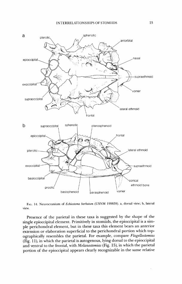
Presence of the parietal in these taxa is suggested by the shape of the single epioccipital element. Primitively in stomiids, the epioccipital is a sim- ple perichondral element, but in these taxa this element bears an anterior extension or elaboration superficial to the perichondral portion which top- ographically resembles the parietal. For example, compare Flagellostomias (Fig. 1 l), in which the parietal is autogenous, lying dorsal to the epioccipital and ventral to the frontal, with Melanostomias (Fig. 15), in which the parietal portion of the epioccipital appears clearly recognizable in the same relative
frontal lateral ethmo~d
lateral ethmoid
ethmoid bone baslsuheno~d
FIG. 15. Nen~.ocranium of Melano.\lomias ter~lac1~1al11.c ( U S N M 199848). a , clor.sal view; h, lateral view.
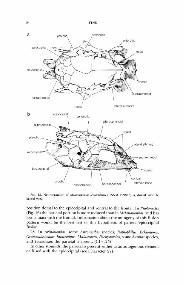
position dorsal to the epioccipital and ventral to the frontal. In Photonectes (Fig. 16) the parietal portion is more reduced than in Melano.stomia.s, and has lost contact with the frontal. Information about the ontogeriy of this fusion pattern would be the best test of this hypothesis of parietallepioccipital fusion.
28. In Aristostornias, some Astronesthes species, Hathophilus, Echiostoma, Grammatostomias, Idiacanthus, Mal(icosteus, Pachystomias, some Stomias species, and Tactostoma, the parietal is absent. (CI = .25)
In other stomiids, the parietal is present, either as an autogenous element or fused with the epioccipital (see Character 27).
INTEKKELATIONSHIPS OF STOMIIDS
frontal lateral ethmold
parasphenold bas~sphenoid
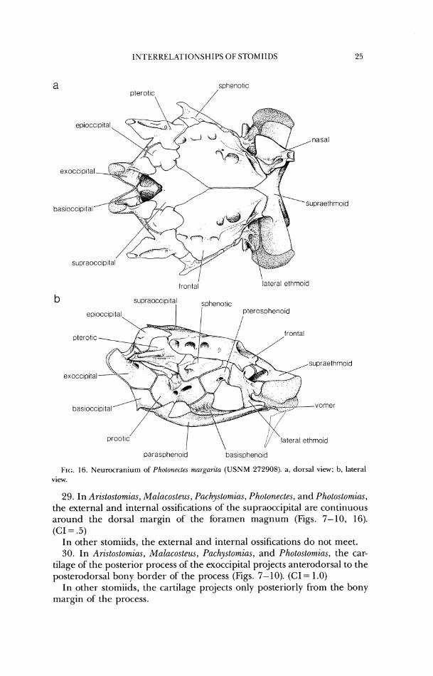
F I ~ ; . 16. Neurocran iu~n of Photonectes margarita ( U S N M 272908). a , dorsal view; b, lateral view.
29. In Ari.stostomzus, Malacostew, Pachystomias, Photonectes, and Photostomias, the external and internal ossifications of the supraoccipital are continuous around the dorsal margin of the foramen magnum (Figs. 7-10, 16). (CI = .5)
In other stomiids, the external and internal ossifications do not meet. 30. In Ari.~tostomias, Malacosteus, Pachystomias, and Photostomias, the car-
tilage of the posterior process of the exoccipital projects anterodorsal to the posterodorsal bony border of the process (Figs. 7-10). (CI = 1.0)
In other stomiids, the cartilage projects only posteriorly from the bony margin of the process.
a sphenot~c
rostroderrnethrnotd
lateral ethrno~d frontal
b
prootlc lateral ethrnoid bastspheno~d parasphenold
F I ~ : . 17. Neurocranium of S1orr~za.s Iamnpi-opcltzs (USNM 199857). a, dorsal view; b, lateral view.
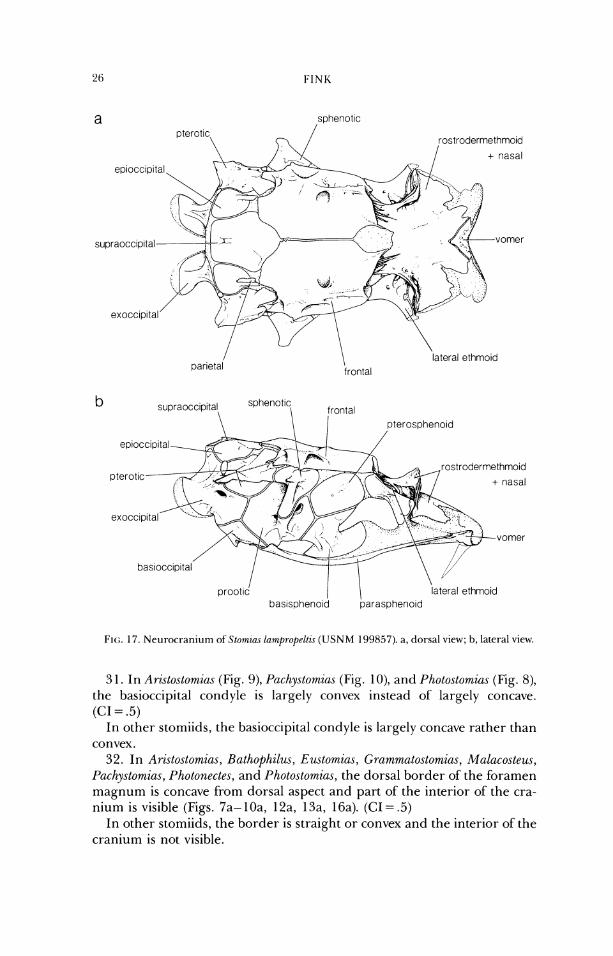
3 1. In Arzstostomias (Fig. 9), P(~chy.rtomia.r (Fig. 1 O), and Photostomias (Fig. 8), the basioccipital condyle is largely convex instead of largely concave. (CI = .5)
In other stomiids, the basioccipital condyle is largely concave rather than convex.
32. In Aristostom%as, Bathophilus, E~utomias, (;rammatostomias, Mulacosteus, Pachystornias, Photonectes, and Photo.stomia.s, the dorsal border of'the foramen magnum is concave from dorsal aspect and part of the interior of' the cra- nium is visible (Figs. 7a- 10a, 12a, 13a, 16a). (CI = .5)
In other stomiids, the border is straight or convex and the interior of the cranium is not visible.
INTEKKELA'FIONSHIl'S OF STOMIIDS
ectopterygoid
extrascapular, I / ,antortxiaI
'dentary
supramax~lla max~lla anguloartlcular
quadrate
FIG. 18. Jaws and suspensorium of Echiostoma barbatum ( U S N M 199839). Right side, lateral view. Light stipple indicates unossified rnembrane.
33. In stomiids, all infraorbitals except the first are absent. (CI = 1.0) In other stomiiforms, between two and six infraorbitals are present. 34. In Aristostomias, A.strone.rthes, Chauliodus, Malacostew (Fig. 26),
Pachylystomias (Fig. 27), and Photostomias (Fig. 25), the jaw teeth of adults are fixed and not depressible (Type 1 attachment; Fink, 1981). (CI = .33)
In other "photichthyans," at least some Type 3 (hinged, anterior axis of rotation) or Type 4 (hinged, posterior axis of rotation) teeth are on thejaws in adults. See Fink (1981) for a discussion of actinopterygian tooth attach- ment morphology and its systematic implications.
35. In Tactostoma (Fig. 19), both adults and juveniles have type 4 tooth attachment (hinged, with posterior axis of rotation; see Fink, 1981). Reten- tion of this mode of attachment in adults is considered a paedomorphic feature. (CI = 1 .O)
In other stomiids, Type 4 teeth are found only in early post-larval on- togenetic stages.
36. In Opostomins, there is a large foramen passing vertically through the premaxilla, just lateral to the symphysis. A large mandibular tooth extends
branchlostegal ray
FIG. 19. Jaws and suspensoriurn of T(~rlo.tloma macroptu (USNM 187654). Right side, lateral view.
up into the foramen when the mouth is closed. (CI = 1.0) In other stomiids, no such foramen is present and the mandibular teeth,
if sufficiently enlarged, extend anterior to the premaxilla. 37. In Bathophilus (Fig. 20), the premaxilla has a process which articulates
along the anterodorsal margin of the maxilla. (CI = 1.0) In other stomiids, the premaxilla articulates only along the anteroventral
surface of the maxilla. 38. In Stomias (Fig. 21), the maxillae closely approach each other at the
midline, anterior to the ethmoid region. This is part of the morphological complex of the protrusible upper jaw in which the maxillae act as links between the premaxillae and the ethmoid ossification. (CI = 1.0)
In other stomiids, the maxillae never closely approach the midline. Eustomias species have protrusible upper jaws, but the mechanisrn of protru- sion is quite unlike that in Stomins. See Character 52.
39. In all stomiid genera except Borostomiu.~, Heterophotus, Neonesthes, and Khadinesthes, there are regularly and closely set teeth posteriorly on the ventral border of the maxilla which are about equal in length, or- which become sequentially enlarged posteriorly (e.g., Figs. 18-30). In many of these taxa (see Character 40) there may also be larger teeth more anteriorly on the maxilla. (CI = .5)
quadrate 1 \ rnax~lla
anguloart~cular
FI(;. 20. Jaws and s~~spensorium of Rathophilw pawner2 (USNM 159052). Right side, lateral vicw.
In the four stomiids noted and in most stomiibr~ns, the posterior maxil- lary teeth are irregularly and/or more distantly set, and include teeth of various sizes (e.g. some large teeth along with some much smaller teeth). Those stomiiforms with maxillary teeth approaching a condition as de- scribed here for the many above genera include some sternoptychids (see figures in Weitzman, 1974) and Woodsia (in which the highly specialized closely-set teeth are also present on the dentary).
40. In Aristostomias, Astronesthes, Bathophilus, Chaulioctus, Ewtomius, Grarnmatostomias, Malacosteus, Pachystomias, Photostomia.~, and Stomias, only small, closely- and regularly-set teeth are present on the maxilla (Figs. 20, 2 1, 23-27). (CI = .33)
In other stomiids, larger, less closely spaced, and less regularly-set teeth are also present.
4 1. In Leptostomias (Fig. 28), Odontostomias, Opostomias, and Thysanactu, the jaws are relatively short and deep, with the depth of the lower jaw at the coronoid process at least one-fourth the length of the lower jaw. (CI = 1 .O)
In other stomiids, the jaws are more elongate and slender. 42. In Chirostomias (Fig. 29), Stomim (Fig. 21), and Trigonolampa (Fig. 22)
the supramaxilla is extremely reduced, usually to a small sliver of bone. (CI = .5)
FINK
ectopterygoid
s u p r a m a x " i a "I
FIG. 2 I. Jaws and susperlsoriurrl of Stomir~s larr~propelti~s (USNM 199857). Right side, lateral view. Pre~naxilla in protruded position.
I11 other stomiids, the supramaxilla is variously sized, depending on the genus, but never as small as in the above three genera.
In presumed primitive Astrone.sthes species such as A. splendidus and A. boulengerz (Weitzman, 1967b, Fig. 25), the supramaxilla is reduced as an elongate slender ossification (although it is still larger than in the three genera above); in presumed phylogenetically derived species, such as A. niger (Weitzman, 1967t1, Fig. 8) the supramaxilla is absent altogether.
43. In Arislostomias, Bathophilus, Echiostoma, Ewtomias, ~~rammatostomias, Idiacanthus, Malacosteus, Melanostomias, Pachystomias, Photonectes, Pho- tostornias, Tactostoma, and Trzgonolarnpa, there is no retroarticular. (CI = .5)
In other "photichthyans," the retroarticular is present in at least some members of each genus.
44. In Flagellostomias, L~ptostomias (Fig. 28),' Odontostomiu.~, Opostomia.~, and Thysanactis, the second large tooth from the symphysis of the dentary pro- jects into the mouth at about a 60 degree angle. (C1= 1.0)
In other stomiids, none of the teeth on the anterolateral portion of' the dentary project inward at that great an angle.
45. In Aristostomias, Bathophilus, Euslomias, Grammatostomias, Malacostew, Pachystomias, and Photostomias, there is a ligament which attaches near the dorsoanterior border of each dentary, at the symphysis, and extends as a loop posteriorly in the anterior portion of'the floor of the mouth. (CI = 1.0)
INI'ERREI~ATIONSHII'S O F STOMIIDS 3 1
c opercle
\
quadrate
FIG. 22. Jaws and suspcnsoriurn of 7hgonolamf)a miriceps ( M C Z 35775). Right side, lateral view.
In other stomiids, no such ligament is present. 46. In Pholonectes (Fig. 30), the process of the anguloarticular posterior to
the articulation with the quadrate is elongate, almost equal to the length of the anterodorsal border of the quadrate. (CI = 1 .O)
In other stomiids, the process is roughly half or less that length. 47. In Aristostomias, Malacosteus, Pachystomias (Fig. 27), and Photostomias
(Fig. 25), the cartilage of the palatine arch is interrupted between the pos- terior margin of the palatine and the rest of the suspensorium, and the palatine itself terminates posteriorly in a bony point. (CI = 1.0)
In other stomiids, the cartilage of the palatine arch is uninterrupted. 48. In Malacosteu,~ (Fig. 26), the palatine is represented by a cartilage body
lying dorsomedial to the maxilla. (CI = 1 .O) In other stomiids, the palatine is ossified. 49. In Idiacanthus and Tactostoma (Fig. 19), the ventral border of the pal-
atine arch is dorsally arched from lateral view. (CI = 1.0) In other stomiids, the border is approximately straight or slightly arched
ventrally from lateral view. 50. In Borostomias, the palatine bone extends posteriorly beyond the ante-
rior border of the quadrate bone (see Weitzman, 1967b, Figs. 26, 27). (CI = 1 .0)
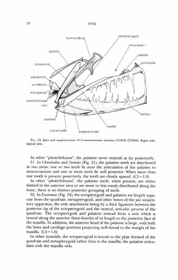
FIG. 23. ,Jaws and suspensor-iurn of GrammaLortomzu.\ denlatu-r ( U S N M 272903). Right side, lateral view.
In other "photichthyans", the palatine never extends as far posteriorly. 51. In Chauliodzu and Stomins (Fig. 2 1), the palatine teeth are distributed
in two areas: one or two teeth lie near the articulation of' the palatine to neurocranium and one or more teeth lie well posterior. When more than one tooth is present posteriorly, the teeth are closely spaced. (CI = 1.0)
In other "photichthyans", the palatine teeth, when present, are either limited to the anterior area or are more or less evenly distributed along the bone; there is no distinct posterior grouping of teeth.
52. In Eustomias (Fig. 24), the ectopterygoid and palatine are largely sepa- rate from the quadrate, metapterygoid, and other bones of the jaw suspen- sory apparatus, the only attachment being by a thick ligament between the posterior tip of the ectopterygoid and the ventral, articular process of the quadrate. 'The ectopterygoid and palatine instead b r m a unit which is bound along the anterior three-fourths of its length to the posterior face of the maxilla. In addition, the anterior head of the palatine is large, with both the bony and cartilage portions projecting well dorsal to the margin of the maxilla. (CI = 1 .O)
In other stomiids, the ectopterygoid is bound to the plate formed of the quadrate and metapterygoid rather than to the maxilla; the palatine articu- lates with the maxilla only.
lntraorbltal 1
quadrate
FIG. 24. Jaws and suspellsorium of E?lstomzas cf. nlucrurlw. (USNM 272'313). Right side, lateral view.
53. In stomiids, the mesopterygoid is reduced in size, so that the dor- sornedial margin does not approach [he parasphenoid. (CI = 1.0)
In other "photichthyans", the mesopterygoid is the most extensive bone of the suspensorium and closely approaches the parasphenoid.
54. In Aristostomias, Bathophilus, Eudomias, Grammatostomias, Idiacanthus, Malacosteus, Pachystomias, Photoslomias, Tacto.storna, and Thysannctis, the meso- ptcrygoid is absent. ((3 = .5)
In other stomiiforms, it is present. 55. In Leptostomias, Odontostomias, Opostomias, and Tt~ysunactis, the orienta-
tion of the hyomandibula is nearly perpendicular to the long axis of the fish. (CI = 1.0)
In other stomiids, the hyomandibula lies at a more oblique angle to the long axis of the fish.
56. In Flagellostomias, Leptostornias (Fig. 28), Odontostomias, Oposlomias, and Thysunactis, the opercular process of the hyomandibula is elongate and pro- jects well posterior to the body of the hyomandibula, as does the dorsal I-amus of the preopercle, leaving a large triangular space dorsally between the two bones. (CI = 1 .O)
In other stomiids, the opercular process of the hyomandibula is generally not elongate; if elongate, it lies closer to the main axis of the hyomandibula.
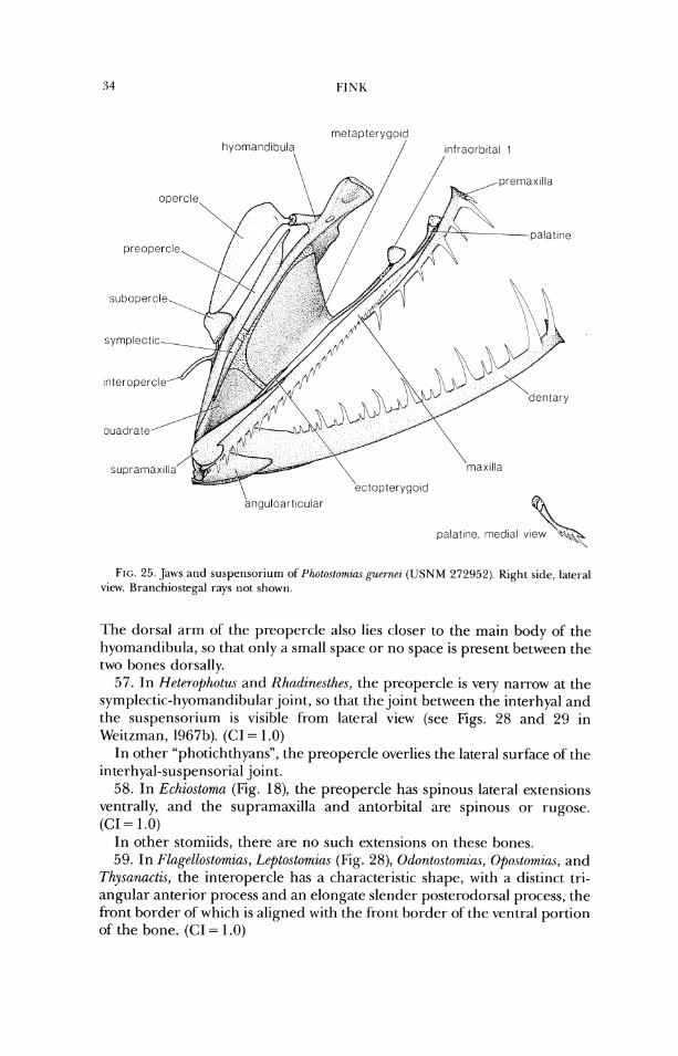
FIG. 25. Jaws and suspcnsoriurn of Photostomiu.\ p~<,rnrz ( U S N M 272052). Right side, lateral view. Bra~ichiostegal rays not shown.
dorsal arm of the preopercle also lies closer to the main body of' the hyomandibula, so that only a small space or no space is present between the two bones dorsally.
57. In Heterophotus and Khndinrsthes, the preopercle is very narrow at the symplectic-hyomandibular joint, so that the joint between the interhyal and the suspensorium is visible from lateral view (see Figs. 28 and 29 in Weitzman, 1967b). (CI = 1 .O)
In other "photichthyans", the preopercle overlies the lateral surface of the interhyal-suspensorial joint.
58. In Echiosloma (Fig. 18), the preopercle has spinous lateral extensions ventrally, and the supramaxilla and antorbital are spirlous or rugose. (CI = 1 .O)
In other stomiids, there are no such extensions on these bones. 59. In Flagellostomias, Lcp1oslomia.s (Fig. 28), Odonto.stomias, Oposlomias, and
Thysanactis, the interopercle has a characteristic shape, with a distinct tri- angular anterior process and an elongate slender posterodorsal process, the front border of which is aligned with the front border of'the ventral portion of the bone. (CI = 1.0)
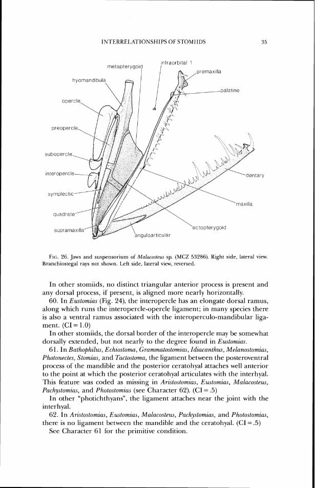
INTEKRELATIONSHIPS OF STOMIIDS
FIG. 26. Jaws and susperisorium of'Ma1r~cosleu.s sp. (MCZ 53286). Right side, lateral view. Branchiostcgal rays not shown. 1,eSt side, lateral view, reversed.
In other stomiids, no distinct triangular anterior process is present and any dorsal process, if present, is aligned more nearly horizontally.
60. In Eustomias (Fig. 24), the interopercle has an elongate dorsal ramus, along which runs the interopercle-opercle ligament; in many species there is also a ventral ramus associated with the interoperculo-mandibular liga- ment. (CI = 1 .O)
In other stomiids, the dorsal border of'the interopercle may be somewhat dorsally extended, but not nearly to the degree found in Eustomias.
61. In Bathophilus, Echiostoma, Gmmmatostomias, Idiacanthus, Melanostomias, Photonectes, Stomias, and Tactostorna, the ligament between the posteroventral process of the mandible and the posterior ceratohyal attaches well anterior to the point at which the posterior ceratohyal articulates with the interhyal. This feature was coded as missing in Aristostomias, Eustomias, Malacosteus, Pachystomias, and Photostomias (see Character 62). (CI = .5)
In other "photichthyans", the ligament attaches near the joint with the interhyal.
62. In Aristostomias, Euctomius, Malacosteus, Pachystomias, and Photostomias, there is no ligament between the mandible and the ceratohyal. (CI = .5 )
See Character 61 for the primitive condition.
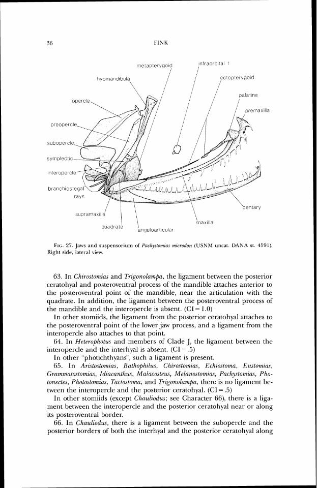
Frc;. 27. ,Jaws a n d suspensor-iurri o f I'c~chys~ornzus mzcrodon (USNM uncat. DANA st. 4.591). Right side, lateral view.
63. In Chiro.stomias and Trigonolurnpa, the ligament between the posterior ceratohyal and posteroventral process of the mandible attaches anterior to the posteroventral point of the mandible, near the articulatiorl with the quadrate. In addition, the liganlent between the posteroventral process of the mandible and the interopercle is absent. (CI = 1.0)
In other stomiids, the ligament from the posterior cera~ohyal attaches to he posteroventral point of the lower jaw process, and a ligament from the interopercle also attaches to that point.
64. In Hete~ophotus and members of Clade J , the ligament between the interopercle and the interhyal is absent. ((71 = .5)
In other "photichthyans", such a ligament is present. 65. In Ari.stostomias, Rathophilu.~, Chirostomias, Echio.ttoma, Eustomias,
Grammatostomias, Idiacanthus, Mnlacosteus, Melanostomias, Pnchystomias, Pho- tonectes, Photostomius, Tactostorna, and Trigonolumpa, there is no ligament be- tween the interopercle and the posterior ceratohyal. (CI = .5)
In other stomiids (except Chauliodus; see Character 66), there is a liga- ment between the interopercle and the posterior ceratohyal near or along its posteroventral border.
66. In Chauliodus, there is a ligament between the subopercle and the posterior borders of both the interhyal and the posterior ceratohyal along
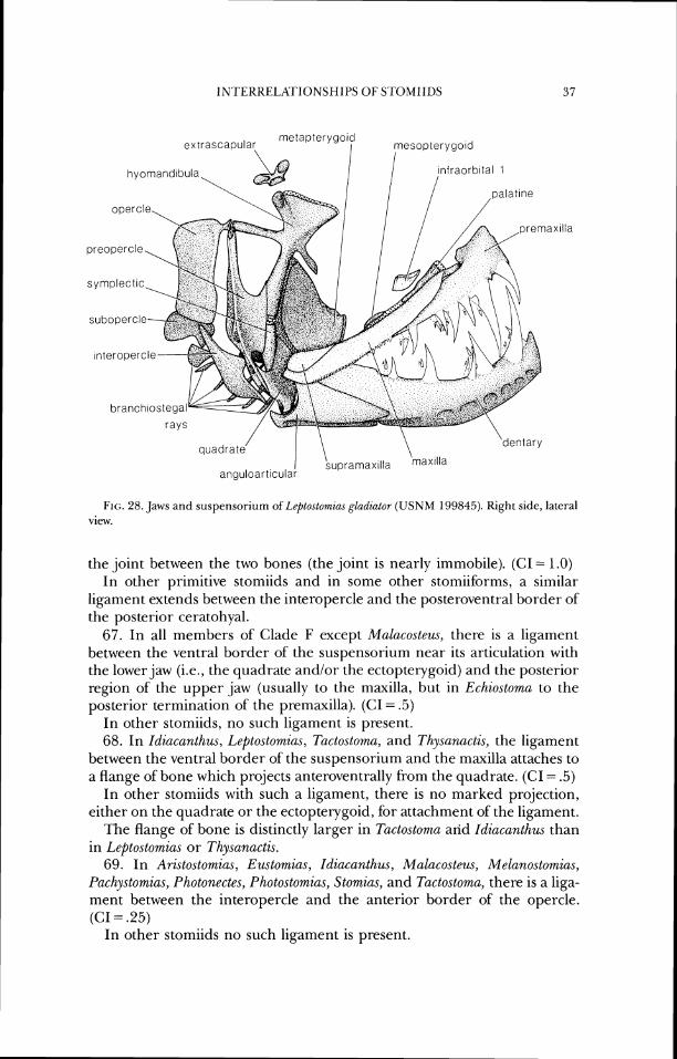
FIG;. 28. Jaws and suspcnsoriurn of Lrptostomia.c gladiator ( U S N M 199845). Right side, lateral vicw.
the joint between the two bones (the joint is nearly immobile). (C1= 1.0) In other primitive stomiids and in some other stomiiforms, a similar
ligament extends between the interopercle and the posteroventral border of the posterior ceratohyal.
67. In all members of Clade F except Malacosteus, there is a ligament between the ventral border of the suspensorium near its articulation with the lower jaw (i.e., the quadrate and/or the ectopterygoid) and the posterior region of the upper ,jaw (usually to the maxilla, but in Echiosloma to the posterior termination of the premaxilla). (CI = .5)
In other stomiids, no such ligament is present. 68. In Idiacanthus, Leptostomias, Tactostoma, and Thysunactis, the ligament
between the ventral border of the suspensorium and the maxilla attaches to a flange of bone which projects anteroventrally from the quadrate. (C1= .5)
In other stomiids with such a ligament, there is no marked projection, either on the quadrate or the ectopterygoid, for attachment of the ligament.
The flange of bone is distinctly larger in Tactostoma arid Idiacanthus than in Leptostomia.r or Thysanactzs.
69. In Arislostomias, Eustomias, Idiacanthus, Malacosteus, Melanostomia.~, Pac/~ystn.mias, Phatanectes, Pholostomias, Stumias, and Tactostoma, there is a liga- ment between the interopercle and the anterior border of the opercle. (CI = .25)
In other stomiids no such ligament is present.
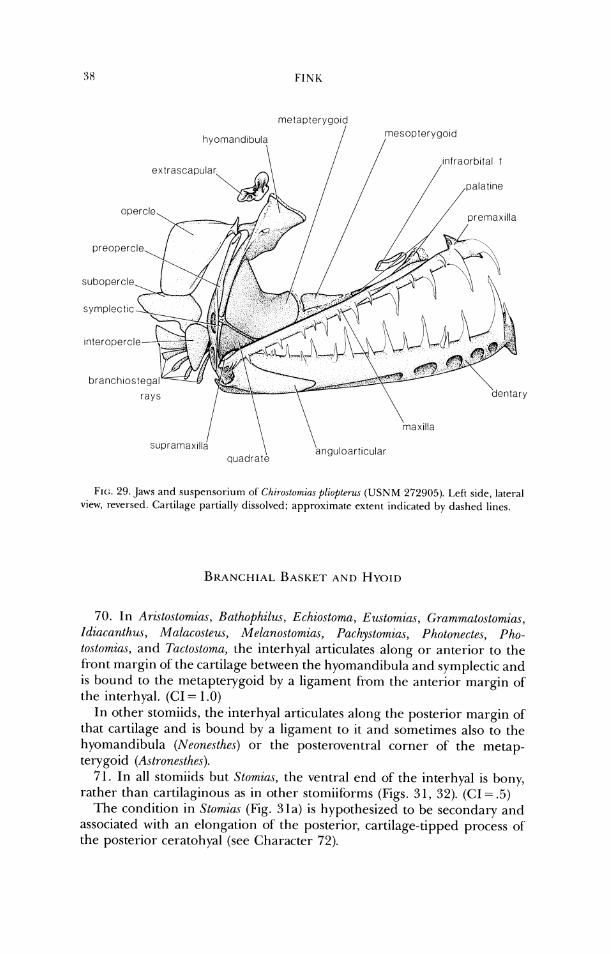
metapteryg07 mesopterygord
hyomand~bul? /
1;1(:. 21). Jaws and suspensoriurn of C/li?-ostomim pliopt~ra\ (USNM 272905). 1.eti side, late1-al view, reversed. Cartilage partially dissol\~ed; approxirrlatr extenc indicated by dashed lines.
70. I n A~sto.stomias, Bathophilus, Echios~oma, Eu~.stomtu.s, C~rnm~mato.slom,ias, I(iiucanlhu,v, Malacost~us, Melanostomic~s, Puclzystomia.~, Pholonectes, Pho- ~ostornia.~, and T(~ctoslorna, the interhyal articulates along o r anterior to the front margin of the cartilage between the hyomandibula and symplectic and is bound to the metapterygoid by a ligament from the anterior margin of' the interhyal. (CI = 1.0)
In other stomiids, the interhyal articulates along the posterior margin of' that cartilage and is bound by a ligament to it and sometimes also to the hyomandibula (Neonesthes) or the posteroventral corner of' the metap- terygoid (Astronestl~es).
71. In all stomiids but Stomins, the ventral end of the interhyal is bony, rather than cartilaginous as in other stomiiforrns (Figs. 31, 32). (CI = .5)
'I'he condition in Stomias (Fig. 31a) is hypothesized to be secondary and associated with an elongation of the posterior, cartilage-tipped process of the posterior ceratohyal (see Character 72).
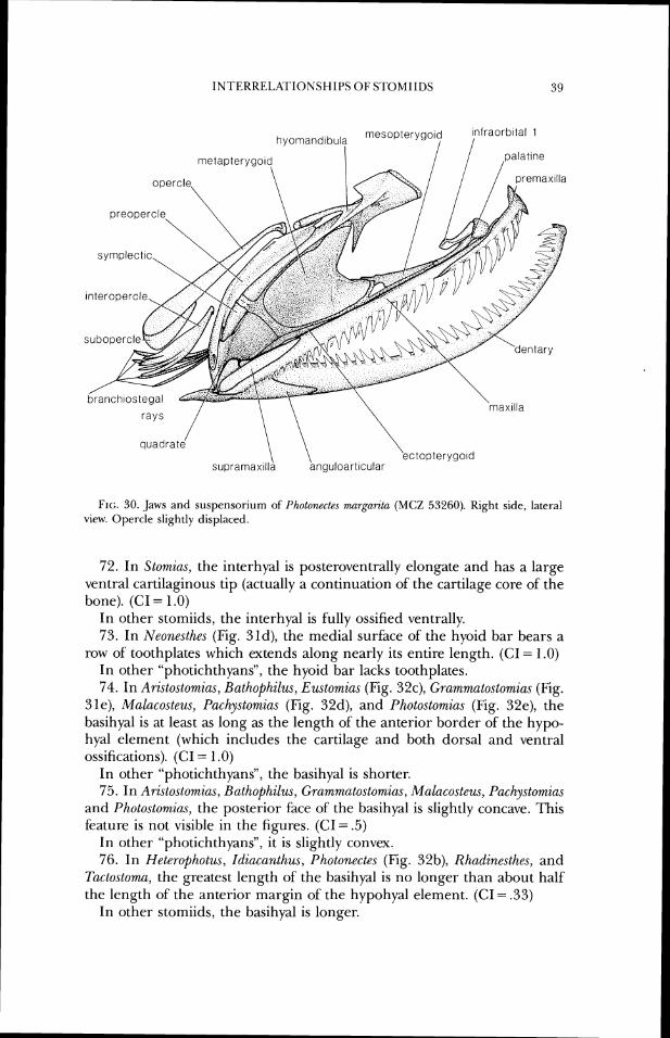
I N T E K U E L A T I O N S H I P S O F S'TOMIIDS 39
FIG. 30. ,Jaws and susperlsorium of Pholonectes marganta ( M C Z ,53260). Kight side, lateral view. Opercle slightly displaccd.
72. In Stomias, the interhyal is posteroventrally elongate and has a large ventral cartilaginous tip (actually a continuatiorl of the cartilage core of the bone). (CI = 1.0)
In other stomiids, the interhyal is f~rlly ossified ventrally. 73. In Neonesthes (Fig. 31d), the medial surface of the hyoid bar bears a
row of toothplates which extends along nearly its entire length. (CI = 1.0) In other "photichthyans", the hyoid bar lacks toothplales. 74. In Arzstostomias, Bathophilus, Eustomias (Fig. 32c), ~~rammatostomia.s (Fig.
31e), Malacosteus, Pacl~ystonzzas (Fig. 32d), and Photostomzus (Fig. 32e), the basihyal is at least as long as the length of the anterior border of the hypo- hyal element (which includes the cartilage and both dorsal and ventral ossifications). (CI = 1 .O)
In other "photichthyans", the basihyal is shorter. 75. In ArOto.storni(is, Bathophilus, Grammato.stomias, Malacosteus, I'achystomias
and Photostomia.~, the posterior face of the basihyal is slightly concave. This feature is not visible in the figures. (CI = .5)
In other "photichthyans", it is slightly convex. 76. In Heterophotus, Idiacanthus, Pholonectes (Fig. 32b), Rhadinesthes, and
Tactostoma, the greatest length of the basihyal is no longer than about half lhe length of the anterior margin of the hypohyal element. (CI = .33)
In other storniids, the basihyal is longer.
77. In Idiucc~ntlzus and 7hctostoma, the basihyal is reduced to a thin, cylin- drical element no broader distally than proxilnally and there is no ligament between its distal tip and the hypohyal element. (CI = 1 .O)
In other stomiids, the basihyal is a larger element, broader distally than proximally, and the ligament is present.
78. In Chauliodus, Leptostowzias, Odontostomias (Fig. 31c), Opostomias, and Thysanactis, there is a process pr-oxinially on the posterior face of the basihyal for attachment of a ligament to each dorsal hypohyal. (CI = .5)
In other stomiids, the process is absent, although the ligaments usually are present.
79. In Bathophilu.~, Echiostoma, Eustomir~s, Grammatostomias, IdWct~,nthus, Malacosteus, Photonectes, and Tuctostoma, the urohyal is no longer than ap- proximately the length of basibranchial 2. This is hypothesized to be a synapomorphy for a group conlprised of the above genera plus Arislostomias, Pachystomia.~, and Photostornias. 'Ille presence of a urohyal about one-half (Photostomia.~) or one and one-third (the other two) the length of basibranchial 2 is interpreted as a reversal. (CI = .25)
In other "photichthyans", the urohyal is longer than the length of basibranchial 2.
80. In Bathofihilus, Chirostomias, Bchiostorna, Eu.stornias, El(~gellostomia.s, Grammatostomias, Idiacanthus, Lefitostomias, Melanostornias, Odontostomic~s, Opostornias, Photonectes, Thysanacti.s, and Trzgor~olumpa, thc width of the urohyal anteriorly is at least slightly greater than the width of the cartilage between basibranchials 2 and 3 at its widest point. (CI = .33)
In other "photichthyans", the width of the urohyal anteriorly is equal to or less than the width o f the cartilage at its widest point.
8 1. In Rathophilus, Chirostomias, Echiostoma, Ezrstomius, Gramrna~o.stomia.\, Idic~canthus, and Melanostomias, the width of the urohyal anteriorly is roughly twice the width of the cartilage between basibranchials 2 and 3 at its widest point. (CI = .25)
In other stomiids, the width of the urohyal anteriorly is not as great. (Because all components of the gill arches in these fishes change in pro-
portion to each other, it is difficult to find a constant against which to measure the width of the urohyal. In Idiacanthus, the urohyal is actually broader than twice the width of the basibranchial 2-3 region because the latter region is unusually narrow. In Batl~ophilus, the latter region is un- usually broad relative to the length of the basibranchial series, so that by this measure the urohyal is not quite twice the width.)
82. In Idiacanlhus, Malaco.ste~ls, and Tactostorna, the posterior process of the urohyal is nearly absent. (CI = .5)
In other stomiids, the posterior process is distinctly longer. 83. In Chirostomias, the urohyal anteriorly has a pair of ventral processes
longer than the anterior margin of the basihyal element. (C1= 1 .O) In other stomiids, any ventral processes are not as elongate. 84. In stomiids, the depth of the urohyal posteriorly is less than the depth
of the basibranchial series. (CI = 1.0)
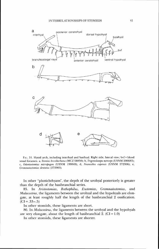
a posterior ceratohyal
branch~ostegal ray
1;1(:. 31. Hyoicl a~-ch, including interhyal and basihyal. Right side, lateral view; bvf= blood vessel bramen. a, Slomlns hrc~vzbu~dat~~s (MCZ 58850); 11, fiigonolamnpa mirzcrps ( U S N M 206683); c, Odon,loslomias niicl-opogon ( U S N M 199849); d , Neonesthes ruprruzs ( U S N M 272906); e, C;rc~r~~rtialostomiu., d r r~ t (~ t w (272903).
In other "photichthyans", the depth of the urohyal posteriorly is greater than the depth of the basibrancllial series.
85. In Aristostomias, Bathophzlus, Euslomias, Grammatostomias, and Mnlacosteus, the ligaments between the urohyal and the hypohyals are elon- gate, at least roughly half the length of' the basibranchial 2 ossification. (CI = .33-.5)
In other stomiids, these ligaments are short. 86. In Malacosteus, the ligaments between the urohyal and the hypohyals
are very elongate, about the length of basibranchial 2. (CI = 1.0) In other stomiids, these ligaments are shorter.
87. In members of Clade D, the blood vessel which passes through the hypohyal element penetrates the lateral face of the element in the ventral hypohyal (Figs. 3 la-3 lc, 3 le). (C1= 1 .O)
In other "photichthyans", this blood vessel penetrates the lateral face in the cartilage near the dorsal border of the hypohyal element (Fig. 31d).
88. In Astronesthes, the blood vessel which passes through the hypohyal element exits the dorsal hypohyal on the lateral face of the bone, then reenters the lateral fBce in the ventral hypohyal. (CI = 1.0)
In other "photichthyans", and in Astron,esthes niger (a phylogenetically de- rived species in the genus), this lateral exit and the second entrance are not present.
89. In Grammatostomias (Fig. 3 1 e), the foramen for passage of the blood vessel through the hypohyal element is located at about the midlength of the element (near the ventral border). (GI = 1 .O)
In other stomiids with a foramen near the ventral border of the hypohyal element, the foramen is near the anteroventral corner of the element.
90. In members of Clade D, the ligament between the basihyal and the hypohyal attaches on the anterior half of the hypohyal element. ((;I = 1 .O)
In other "photichthyans", such as Photichtl~y~s and Wr~odsia, as well as Diplophos, the ligament attaches on the posterior half of the hypohyal ele- ment. In many primitive stomiiforms, the basihyal is reduced and closely bound to the front of the hypohyal element.
9 1. In Ari.~tostomias, Rathophilus, Grammatostomias (Fig. 3 1 e), Malacoste~w., Pacl~ystomias (Fig. 32d), and Photostomias (Fig. 32e), the ligament between the basihyal and the hypohyal element attaches to the latter ventral to its mid- height. (CI = .5)
In other stomiids, the ligament attaches more dorsally. 92. In Arzstostomia.~, Bathophilus, Grammato.stom.ias (Fig. Sle), and
Mal~costeus, the ligament from the anterior of the basihyal attaches adjacent to the ventral border of the ventral hypohyal. (CI = .33-.5)
In other "photichthyans", the ligament (when present) attaches more dor- sally, either near the dorsal margin of the ventral hypohyal or on the dorsal hypohyal.
93. In Aristostomia.s, Bathopkilus, Echiostomu, E~~stornia.~, Grammato.stomias, Idiacanth,m, Malacosleus, Mel(~nostomias, Pachystomias, Photonectes, Pho- tostomias, and Tactostoma, the front margin of the hypohyal element is ori- ented at roughly a 60 degree angle to the dorsal margin of basibranchial 1. In most cases, this results in the anterior margin of the urohyal being at about the level of a vertical drawn from the anterior margin of basibranchial 2, although the anterior margin of the urohyal is further anterior in those genera with an elongate basibranchial 1 (Ar&tostomias, Pachy.stomias, Pho- tonectes, and Photostomir~s; see Character 1 10). (CI = 1.0)
In other stomiids, the front margin of the hypohyal element is at roughly a 90 degree angle to the dorsal margin of the first basibranchial, and the anterior margin of the urohyal is ventral to the first basibranchial.
94. In Aristostomias, Rathophilus, Chirostomias, Eustomias (Fig. 32c),
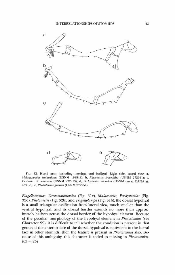
INTERRELATIONSHIPS OF S7DMIIDS
171(:. 32. Hyoid arch, including interllyal and basihyal. Right side, lateral view. a, Mrlarrostomias tentaculat~ls (USNM 199848); h, Photonectes leucospilzlr (USNM 27291 1); c, Ewtomzas cf: macrul-us (USNM 272913); d, Pachystomim microdon (USNM uncat. DANA st. 4591-6); e, Photostomias p r r r ~ e i (USNM 272952).
Flagellostomias, Gmmmatostomias (Fig. 31e), Malacostem, Pachystomias (Fig. 32d), Photonectes (Fig. 32b), and Trigonolampa (Fig. 3 lb), the dorsal hypohyal is a small triangular ossification from lateral view, much smaller than the ventral hypohyal, and its dorsal border extends no more than approx- imately halfway across the dorsal border of the hypohyal element. Because of the peculiar morphology of the hypohyal element in Photostomias (see Character 99), it is difficult to tell whether the condition is present in that genus; if the anterior face of the dorsal hypohyal is equivalent to the lateral face in other stomiids, then the feature is present in Photostomias also. Be- cause of this ambiguity, this character is coded as missing in Photostomias. (GI = .25)
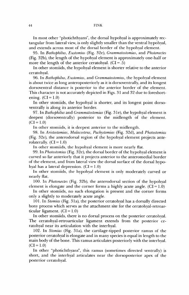
I11 most other "photichthyans", the dorsal hypohyal is approximately rec- tangular from lateral view, is only slightly smaller than the ventral hypohyal, and extends across most of the dorsal border of the hypohyal element.
95. 111 Bathophilus, Eustomias (Fig. 32e), Grammatoslomias, and Photonectes (Fig. 32b), the length of the hypohyal element is approximately one-half or more the length of the anterior ceratohyal. (CI = .5)
In other storniids, the hypohyal element is shorter relative to the anterior ceratohyal.
96. In Bathophilw, Eustomias, and Grammatostomias, the hypohyal element is about twice as long anteroposteriorly as it is dorsoventrally, and its longest dorsoventral distance is posterior to the anterior border of the element. 'I-his character is not accurately depicted in Figs. 3 1 and 32 due to foreshort- ening. (CI = 1 .O)
In other stomiids, the hypohyal is shorter, and its longest point dorso- ventrally is along its anterior border.
97. In Bathophilus and Gram.matostomia,s (Fig. 3 le), the hypohyal element is deepest (dorsoventrally) posterior to the midlength of the element. (CI = 1.0)
In other stomiids, it is deepest anterior to the midlength. 98. In Aristostowzias, Malacosteus, Pachystomias (Fig. 32d), and Photostom.ias
(Fig. 32e), the anterodorsal region of the hypohyal element projects ante- rolaterally. (CI = 1 .O)
In other stomiids, the hypohyal elenient is more nearly flat. 99. In Photostomias (Fig. 32e), the dorsal border ofthe hypohyal element is
curved so far anteriorly that it projects anterior to the anteromedial border of'the element, and from lateral view the dorsal surfice of the dorsal hypo- hyal has a lateral depression. (CI = 1.0)
In other stomiids, the hypohyal element is only moderately curved or nearly flat.
100. In Photonectes (Fig. 32b), the anterodorsal section of the hypohyal element is elongate and the corner forms a highly acute angle. (CI = 1.0)
In other stomiids, no such elongation is present and the corner forms only a slightly to moderately acute angle.
10 1. In Stomias (Fig. 3 la), the posterior ceratohyal has a dorsally directed bony process which serves as the attachment site for the ceratohyal-retroar- ticular ligament. (CI = 1 .O)
In other stomiids, there is no dorsal process on the posterior ceratohyal. The ceratohyal-retroarticular ligament extends from the posterior ce- ratohyal near its articulation with the interhyal.
102. In Stomias (Fig. 31a), the cartilage-tipped posterior ramus of the posterior ceratohyal is elongate and in many species is equal in length to the main body of the bone. This ramus articulates posteriorly with the interhyal. (CI = 1.0)
In other "photichthyans", this ramus (sometimes directed ventrally) is short, and the interhyal articulates near the dorsoposterior apex of the posterior ceratohyal.
INTEKKE1,XI'IONSHIPS O F STOMIIDS 45
103. In stomiids, gill rakers are absent in adults. See Gibbs and Weitzman (1965) for a brief discussion of gill-raker ontogeny in certain stomiids. (CI = 1 .O)
In other "photichthyans", gill rakers are present. 104. In Astronesthes and Chauliodus, the basibranchials are very narrow
relative to their length; the cartilage between basibranchials 2 and 3 at its widest point is less than one-fourth the length of basibranchial2 (the length being measured between the articulations of hypobranchials 1 and 2). (CI = .5)
In other stomiids and in Woodsia among "photichthyans", the basibranchials are not as narrow. This feature in Astronesthes and Chauliodus is a reversal relative to other "photichthyans", which have very narrow basibranchials.
105. In members of Clade F, there is no more than one pair of toothplates associated with any basibranchial ossification. (CI = 1.0)
In other "photichthyans", there are either more than one pair of tooth- plates associated with some basibranchials, or no toothplates at all (see Char- acter 109).
106. In Chirostomias, Trigonolampa, and members of Clade 0, the tooth- plates associated with the basibranchials are large relative to those associated with the hypobranchials, ceratobranchials, and epibranchials. (CI = .5)
In other stomiids, the toothplates are about equal in size. 107. In Bathophilus, Eustomias, Flagellostomins, Crammatostomias,
Leptostomias, and some Melano.stomias, there is only one tooth per toothplate on the bilateral pairs of basibranchial toothplates. (CI = .33)
In other stomiids with bilateral pairs of toothplates, there are usually more teeth per toothplate (Idiacanthus and some Echiostoma may have only one tooth per toothplate on basibranchial 2, and some Malacosteus specimens have only one tooth per toothplate on one side of basibranchial
1 ). 108. In Bathophilus, Chirostomias, Echiostoma, Eustomias, Grammatostomia.s,
Idiacanthus, Malacosteus, Melanostomias, Photonectes, Tactostoma, and Trigonolampa, the teeth associated with the basibranchials are large. (CI = .25)
In other stomiids, the teeth are moderate in size, small, or absent. 109. In Chauliodus, Neonesthes, Opostomias, and Stomias, there are no teeth
or toothplates associated with any basibranchials. (CI = .33) In other stomiids, toothplates are associated with at least two, and primi-
tively three, of the basibranchials. 110. In Aristostomias, Pachystomias, Photonectes, and Photostomias,
basibranchial 1 is slender and elongate, more slender anteriorly, and the ossification surrounds the ventral cartilage margin of the copula except for a small cartilage process at the anteroventral border of the element. (CI = .33)
In almost all other stomiids, basibranchial 1 is about as deep as it is long, and the cartilage of the copula extends ventral to the margin of the ossifica-
tion (in Flagellostomias, there is an intermediate condition in which the basibranchial is slightly longer than it is deep and the ventral cartilage is interrupted by bone at approximately its midlength).
11 1. In members of Clade (B,C), with the exceptions noted below, there are one or more toothplates associated with basibranchial 1 . The absence of such toothplates in Opostomias and in the group Chauliodus plus Stomias (Character 109) is considered due to reversals. (CI = .33)
In Neonmthes and in most "photichthyans" (all but Photichthys), such tooth- plates are absent.
112. In Bathophilus, Echiostoma, Eustomia.~, Grc~mmatostomic~s, Idiacanthus, Malacosteus, Melan.ostomias, Pachystomias, Photonectes, and Tactostoma, the toothplate (Idiacanthus) or toothplates (the other taxa) associated with basibranchial 1 are positioned at or adjacent to the anterodorsal margin of the basibranchial. (CI = .33-.5)
In other stomiids, the toothplates are positioned more posteriorly on the basibranchial.
1 13. In Echiostoma, Idiacanthus, Melanostomias, some Photonectes species, and Tactostoma, the bilateral toothplates associated with the first basibranchial project anterior to the basibranchial-basihyal articulation. In Idiacanthus, the projection is slight; in the others, it is well developed. '.This character is coded as missing in Photonectes because I am unable to estimate whether species with the trait are phylogenetically primitive. (CI = .5)
In other stomiids, the tooth plates do not project anterior to the articula- tion.
1 14. In Idiacantlzus, the pair of toothplates associated with basibranchial 1 are fused in the midline. (CI = 1 .O)
In other stomiids with such toothplates, no fusion is present. 115. In members of Clade F, the toothplates associated with
basibranchials 1 and 2 are limited to two per basibranchial, arranged in a closely associated bilateral pair. 'This is hypothesized to be a synapomorphy for a group comprised of the above genera plus Opostomias, which has lost all basibranchial toothplates (and is coded as missing). (CI = 1 .O)
In other stomiids with basibranchial toothplates, there may be either one or two toothplates laterally on each basibranchial, the number may vary between one side and the other, and the plates are not arranged in closely bilateral pairs.
116. In Photostomias, there is a bilateral pair of toothplates situated with their midlengths between basibranchials 1 and 2 and another with their midlengths between basibranchials 3 and 4 (see also Character 120). (CI = 1 .O)
In other stomiids with paired basibranchial toothplates, any equivalent toothplates are positioned on basibranchials 1 and 3.
117. In Leptostomias and Thysanactis, the lateral margins of the cartilage core in basibranchial 2 projects posterolaterally from the midlength of the basibranchial at roughly a 60 degree angle from the longitudinal axis of the basibranchials. (CI = 1 .O)
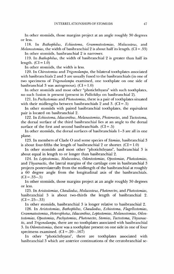
1NTERREI.ATIONSNIPS O F STOMIIDS 47
111 other stomiids, those margins project at an angle roughly 30 degrees or less.
118. In Bathophilus, Echiostoma, Grammatostomias, Malacosteus, and Melanostomias, the width of basibranchial 2 is about half its length. (CI = 3 3 )
In other stomiids, basibranchial 2 is narrower. 119. In Bath,ophilus, the width of basibranchial 2 is greater than half its
length. (C1= 1 .O) In other stomiids, the width is less. 120. In Chirostomius and Trigonolampa, the bilateral toothplates associated
with basibranchials 2 and 3 are usually fused to the basibranchials (in one of two specimens of Trigonolampa examined, one toothplate on one side of basibranchial 3 was autogenous). (C1= 1.0)
In other stomiids and most other "photichthyans" with such toothplates, no such fusion is present (present in Pollichthys on basibranchial 2).
12 1. In Pachystomias and Photostomias, there is a pair of toothplates situated with their midlengths between basibranchials 2 and 3. (CI = .5)
In other stomiids with paired basibranchial toothplates, the equivalent pair is located on basibranchial 2.
122. In Echiostoma, Idiacanthu~, Melanostomias, Photonectes, and Tactostoma, the dorsal surface of the third basibranchial lies at an angle to the dorsal surface of the first and second basibranchials. (CI = .5)
In other stomiids, the dorsal surfaces of basibranchials 1-3 are all in one plane.
123. In members of Clade 0 and some species of Stomias, basibranchial 3 is about four-fifths the length of basibranchial 2 or shorter. (CI = 1.0)
In other stomiids and most other "photichthyans", basibranchial 3 is about equal in length to or longer than basibranchial 2.
124. In Leptostomias, Malacosteus, Odontostomias, Opostomias, Photostomias, and Thysanactis, the lateral margins of the cartilage core in basibranchial 3 projects posterolaterally from the midlength of the basibranchial at roughly a 60 degree angle from the longitudinal axis of the basibranchials. (CI = 33-.5)
In other stomiids, those margins project at an angle roughly 30 degrees or less.
125. In Aristostomias, Chauliodus, Malacosteus, Photonectes, and Photostomias, basibranchial 3 is about two-thirds the length of basibranchial 2. (CI = .25-.33)
In other stomiids, basibranchial 3 is longer relative to basibranchial 2. 126. In Aristostomias, Bathophilus, Chauliodus, Echiostoma, Flagellostomias,
Grammatostomias, Heterophotw, Idiacanthus, Leptostomias, Melanostomius, Odon- tostomias, Opostomias, Pachystomias, Photonectes, Stomias, Tactostoma, Thysanac- tis, and Trigonolampa, there are no toothplates associated with basibranchial 3. In Odontostomias, there was a toothplate present on one side in one of four specimens examined. (CI = .20-. 167)
In other "photichthyans", there are toothplates associated with basibranchial 3 which are anterior continuations of the ceratobranchial se-
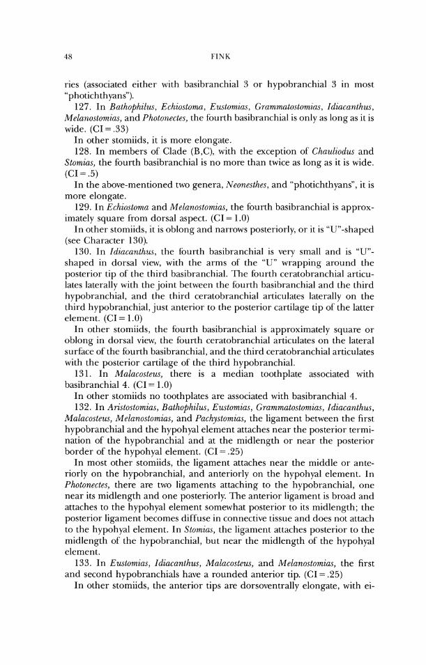
ries (associated either with basibranchial 3 or hypobranchial 3 in most "photichthyans").
127. In Bathophilus, Echiostoma, Eustomias, Grammatostomius, Idiacanthus, Melanostomias, and Photonectes, the fourth basibranchial is only as long as it is wide. (CI = .33)
In other stomiids, it is Inore elongate. 128. In members of Clade (B,C), with the exception of' Chauliodus arid
Stomias, the fourth basibranchial is no more than twice as long as it is wide. (CI = .5)
In the above-mentioned two genera, Neonesthe.~, and "photichthyans", it is more elongate.
129. In Echiostoma and Melanostomias, the fourth basibranchial is approx- imately square from dorsal aspect. (CI = 1.0)
In other stomiids, it is oblong and narrows posteriorly, or it is "Un-shaped (see Character 130).
130. In Idiacanthus, the fourth basibranchial is very small and is "Un- shaped in dorsal view, with the arms of the "U" wrapping around the posterior tip of the third basibrarichial. The fourth ceratobranchial articu- lates laterally with the joint between the fi)urth basibranchial and the third hypobranchial, and the third ceratobranchial articulates laterally on the third hypobranchial, just anterior to the posterior cartilage tip of the latter element. (CI = 1.0)
In other stomiids, the fourth basibranchial is approximately square or oblong in dorsal view, the fourth ceratobranchial articulates on the lateral surface of the fourth basibranchial, and the third ceratobranchial articulates with the posterior cartilage of the third hypobranchial.
13 1. In Malacosteus, there is a median toothplate associated with basibranchial 4. (<:I = 1.0)
In other stomiids no toothplates are associated with basibranchial 4. 132. In Ari.ctostomias, Bathophilws, Eustomias, ~;mmmatostomias, Idiacanthus,
Malacosteus, Melanostomias, and IJachy.stomias, the ligament between the first hypobranchial and the hypohyal element attaches near the posterior termi- nation of the hypobranchial and at the midlength or near the posterior border of the hypohyal element. (CI = .25)
In most other stomiids, the ligament attaches near the middle or ante- riorly on the hypobranchial, and anteriorly on the hypohyal element. In Photonectes, there are two ligaments attaching to the hypobranchial, one near its midlength and one posteriorly. The anterior ligament is broad and attaches to the hypohyal element somewhat posterior to its midlength; the posterior ligament becomes diffuse in connective tissue and does not attach to the hypohyal element. In Stomias, the ligament attaches posterior to the midlength of the hypobranchial, but near the midlength of the hypohyal element.
133. In Eustomias, Idiacanthus, Malacosteus, and Melanostomias, the first and second hypobranchials have a rounded anterior tip. (CI = .25)
In other stomiids, the anterior tips are dorsoventrally elongate, with ei-
ther a single, elongate tip or a bifurcated tip. 134. In Chirostornius, the anterior tips of the first and second hypo-
branchials are bifurcated, articulating dorsally and ventrally with the basibranchial cartilage. (CI = 1 .O)
In most other stomiids, the anterior tips of the first and second hypo- branchial are single. In Bathophilus and Flagellostomias, there is a bifurcation present only on the second hypobranchial; in Tactostoma, a bifurcation is present only on the first hypobranchial.
135. In Chauliodus, hypobranchial 2 is less than half the length of hypo- branchial 1 . (CI = 1.0)
In other storniids, hypobranchial 2 is longer in proportion to hypo- branchial 1.
136. In Tr~gonolumpn, the third hypobranchials are fused to the third basibranchial. (CI = 1 .O)
In other stomiids, no such fusion is present. 137. In Chaulzociu~ and members of Clade F (but see below), there are no
toothplates associated with hypobranchial 3. This feature is hypothesized to have arisen independently in Chauliodus (associated with other toothplate reduction) and in the members of Clade E In Odontostomius, a toothplate was present on one side of two specimens examined, and on each side in two other specimens; this condition in the genus is considered a reversal. (CI = .33)
In other "photichthyans", tooth plates (andlor gill-raker homologues) are associated with hypobranchial 3.
138. In members of Clade 0, the proximal ends of ceratobranchials 1 and 2 are enlarged in a roughly vertrolateral direction, providing an increased area of attachment for the obliqui ventrales muscles. In most cases, this results in the anterior termination of the ceratobranchials being distinctly larger than the posterior termination of the hypobranchials (in Idiacanthus, hypobranchial 1 is enlarged at its posterior end so that it extends further ventrally than the ceratobranchial). (CI = 1.0)
In other stomiids, no enlarged attachment site is present. 139. In Lepto.stomius, the toothplates along the posterior edge of ce-
ratobranchial 4 are reduced to tiny discs of bone and lack teeth. (GI = 1.0) In Odontostomias, Opostomias, and Thysanactis, the toothplates are larger
and almost invariably bear one tooth each. 140. In Arzstostomim, Rathophilus, Chauliodus, Echiostoma, Eustomias,
Grammatostomias, Heterophotus, Idiacanthzls, Leptostomias, Malacosteus, Melanostomias, Odontostomias, Opo,stomias, I'achystomias, Photonectes, Pho- tostomias, Rhadinesthes, Tuctostoma, and Thysanactis, there are no teeth at- tached to ceratobranchial 5. (CI = 25)
In other "photichthyans" (except Ichthyococcus), there are teeth attached to ceratobranchial 5.
141. In Flagellostomias, there is only a single tooth attached to the fifth ceratobranchial. (CI = 1 .O)
In other stomiids, there are either no teeth or more than one. This char-
acter is coded as niissing ill all taxa listed as having the derived state of Character 140.
142. In Leptostomius and 'I'hysanaclis, only one toothplate is associated with the anterior border of the fifth ceratobranchial. (CI = 1.0)
In other stomiids with such toothplates, more than one is present. This character is coded as missing in those stomiids without such toothplates (see Character 140).
143. In members of (:lade F, the main, cartilage-filled body of the third pharyngobranchial has a distinctly concave medial border, viewed from dor- sal aspect. (CI = 1 .O)
In other stomiids, the medial border of the third pharyngobranchial is only slightly concave, if at all.
144. In members of Clade 0, the cartilage portion of the third pharyn- gobranchial is more curved in a horizontal plane than it is in other stomiids. (GI = 1.0)
145. In Borostomias, Gmmmutostornias, and some Photonectes species, the short process on the third pharyngobranchial for articulation with the sec- ond epibranchial is absent. (<:I = .5)
In other stomiids, there is a short process on the third pharyngobranchial for articulation with the second epibranchial.
146. In Malacosteus, Puchyslomias, and Photostomias, the third pharyn- gobranchial is very short relative to its width and has the medial border deeply concave. (CI = .5)
In other stomiids, the third pharyngobranchial is more elongate relative to its width and is either less curved or straight.
147. In Malacosteus and Pachystomias, the toothplate portion of' the third pharyngobranchial overlaps the toothplate of' the fourth pharyn- gobranchial. (CI = .5)
In other stomiids, the toothplates do not overlap or the toothplate ofthe fourth pharyngobranchial overlaps the ossification of the third pharyn- gobranchial.
148. In C h a u l i o d ~ ~ and members of' Clade F, the fourth pharyn- gobranchial is fused to the third pharyngobranchial. (CI = .5)
In other stomiids, the third and fourth pharyngobranchials are separate. 149. In Chauliodus, Heterophotus, Pholostomias, Rhadin.esthes, and Stomiu.~,
the toothplate of the fourth pharyngobranchial tapers posteriorly to a point. (CI = 3 3 )
In other "photichthyans", the toothplate posterior border is broadly rounded.
150. In Heterophotus, there is a small, separate cartilage articulating be- tween the anterior tip of the second epibranchial and the short, ante- rolateral process of the third pharyngobranchial. (CI = 1.0)
In other stomiids, no such cartilage is present; the epibranchial articu- lates directly with the short anterolateral process of the third pharyn- gobranchial.
15 1. In Borostomias, the second epibranchial does not articulate with the
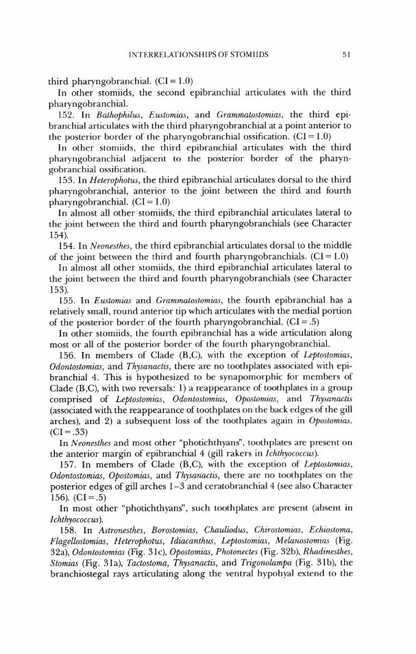
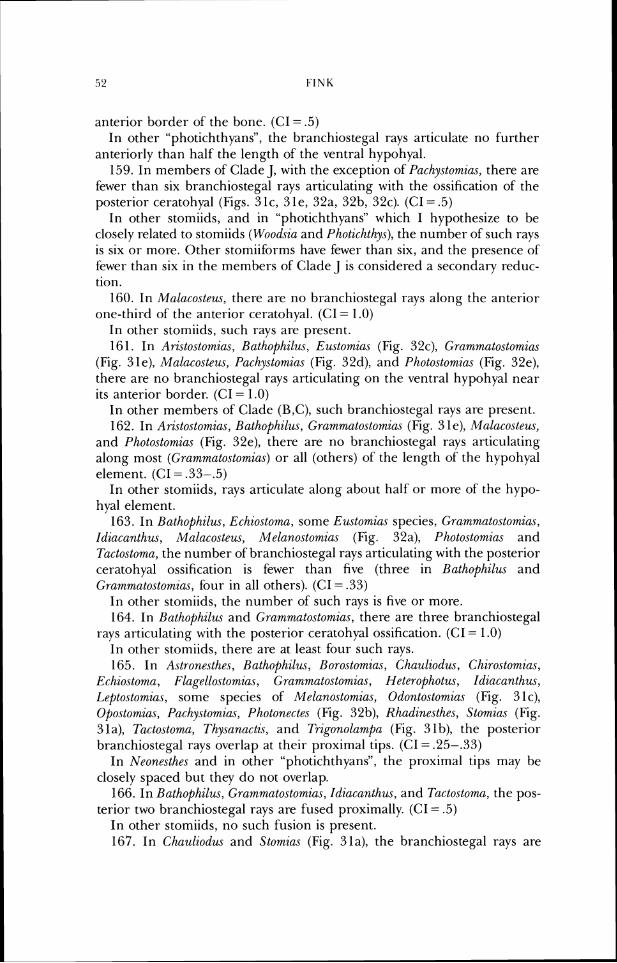
anterior border of the bone. (CI = .5) In other "photichthyans", the branchiostegal rays articulate no further
anteriorly than half the length of the ventral hypohyal. 159. In members of Clade J , with the exception of Pachystornic~.~, there are
fewer than six branchiostegal rays articulating with the ossification of the posterior ceratohyal (Figs. 31c, 31e, 32a, 32b, 32c). (CI= .5)
In other stomiids, arld in "photichthyans" which I hypothesize to be closely related to stomiids (Woodsia and Photicht/~y~s), the number of such rays is six or more. Other stomiiforms have fewer than six, and the preserlce of fewer than six in the members of Clade .J is considered a secondary reduc- tion.
160. In Mnlacosteus, there are no branchiostegal rays along the anterior one-third of the anterior ceratohyal. (CI = 1.0)
In other stomiids, such rays are present. 16 1. In Ari.sto.stomias, Rathophilus, Ewtomias (Fig. 32c), Grc~mmatostomias
(Fig. 3 le), Mnlacosteus, Pachystomias (Fig. 32d), and photos tom la.^ (Fig. 32e), there are no branchiostegal rays articulating on the ventral hypohyal near its anterior border. (C1= 1.0)
In other members of Clade (B,C), such branchiostegal rays are present. 162. In Aristo.stomias, Bathophilus, Gmmmato.stomia.s (Fig. 3 le), MaL(~costeu.s,
and Photostomia,~ (Fig. 32e), there are no branchiostegal rays articulating along most (~;mmmato.sto,mia.s) or all (others) of the length of the hypohyal element. (<:I = .33-.5)
In other stomiids, rays articulate along about half or more of the hypo- hyal element.
163. In Bnthophilus, Echio.stoma, some Eustomia.r species, (;ru~mmatostomia.s, Idiucanthus, Malacosteus, Melano.stornia.s (Fig. 32a), Photostomias and Tactostoma, the number of branchiostegal rays articulating with the posterior ceratohyal ossification is fewer than five (three in Rathophilus and Grammutostomia.~, four in all others). (CI = .33)
In other stomiids, the number of such rays is five or more. 164. In Bathophilus and Gmmmato.stomia.s, there are three branchiostegal
rays articulating with the posterior ceratohyal ossification. (<:I = 1.0) In other stomiids, there arc- at least four such rays. 165. In Astronesthes, Bathophilus, Borostomias, Chauliodw-, Chirostomia.~,
Echiostoma, Flagellostomias, C;mmmatostomias, Heterophotus, Idiacanthus, Leptostomias, some species of Melanostomias, Odontostomias (Fig. 31c), Opostornias, Pachystomias, Photonectes (Fig. 32b), Rhadine.sthes, Stomias (Fig. 3 la), Tactostomn, Thysanactis, and Trigonolampa (Fig. 3 1 b), the posterior branchiostegal rays overlap at their proximal tips. (CI = .25-33)
In Neonesthes and in other "photichthyans", the proximal tips may be closely spaced but they do not overlap.
166. In Bathophilus, Grammatostomias, Idiacanthus, and Tactostomu, the pos- terior two branchiostegal rays are fused proximally. (CI = .5)
In other stomiids, no such fusion is present. 167. In Chauliodus and Stomias (Fig. 31a), the branchiostegal rays are
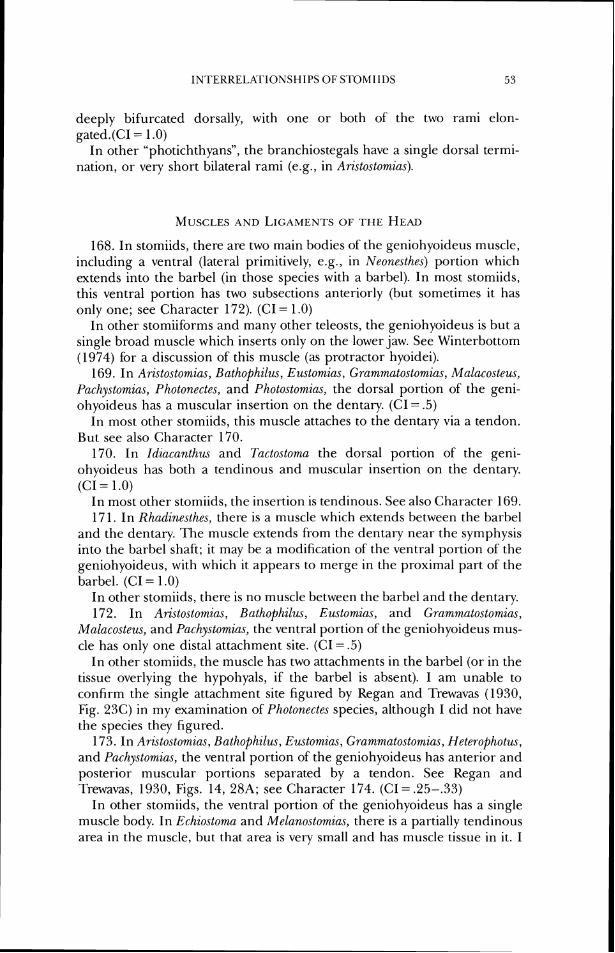
INTEKKELATIONSHII'S OF STOMI 11)s 53
deeply bifurcated dorsally, with one or both of the two rami elon- gated.(CI = 1.0)
In other "photichthyans", the branchiostegals have a single dorsal termi- nation, or very short bilateral rami (e.g., in Arzsto.stomias).
168. In stomiids, there are two main bodies of the geniohyoideus muscle, including a ventral (lateral primitively, e.g., in Neonesthes) portion which extends into the barbel (in those species with a barbel). In most stomiids, this ventral portion has two subsections anteriorly (but sometimes it has only one; see Character 172). (CI- 1.0)
In other stomiiforms and many other teleosts, the gerliohyoideus is but a single broad muscle which inserts only on the lower jaw. See Winterbottom (1974) for a discussion of this muscle (as protractor hyoidei).
169. In Aristostomias, Bathophilus, Eustomias, Gmmmatostomias, Malacostew., Pachystomias, Photonectes, and Photo.stomias, the dorsal portion of the geni- ohyoideus has a muscular insertion on the dentary. ((:I = .5)
In most other stomiids, this muscle attaches to the dentary via a tendon. But see also Character 170.
170. In Idiacanthus and Tactostoma the dorsal portion of the geni- ohyoideus has both a tendinous and muscular insertion on the dentary. (CI = 1 .0)
In most other stomiids, the insertion is tendinous. See also Character 169. 17 1. In Rhadinesthes, there is a muscle which extends between the barbel
and the dentary. The muscle extends from the dentary near the symphysis into the barbel shaft; it may be a modification of the ventral portion of the geniohyoideus, with which it appears to merge in the proximal part of the barbel. (CI = 1.0)
In other stomiids, there is no muscle between the barbel and the dentary. 172. In Aristostomias, Bathophilus, Eustomias, and Grammatostomim,
Mc~lacosteu.r, and Puchystomias, the ventral portion of the geniohyoideus mus- cle has only one distal attachment site. (CI = .5)
In other stomiids, the muscle has two attachments in the barbel (or in the tissue overlying the hypohyals, if the barbel is absent). I am unable to confirm the single attachment site figured by Kegan and Trewavas (1930, Fig. 23C) in my examination of Photonectes species, although I did not have the species they figured.
173. In Aristostomias, Bathophilus, Eustomius, Grammc~tostomias, Heterophotw., and Pachystomias, the ventral portion of the geniohyoidetrs has anterior and posterior muscular portions separated by a tendon. See Regan and Trewavas, 1930, Figs. 14, 28A; see Character 174. (CI= .25-.33)
In other stomiids, the ventral portion of the geniohyoideus has a single muscle body. In Echiostom and Melanostomias, there is a partially tendinous area in the muscle, but that area is very small and has muscle tissue in it. I
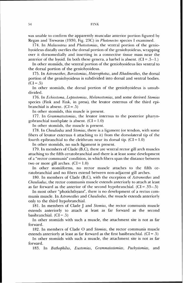
was unable to confirm the apparently muscular anterior portion figured by Kegan and Trewavas (1930, Fig. 23C) in Photonertes species I examined.
174. In Malacosteus and Photostomias, the ventral portion of the genio- hyoideus distally overlies the dorsal portion of the geniohyoideus, wrapping over it dorsomedially and inserting in a connective tissue mass near the anterior of the hyoid. In both these genera, a barbel is absent. (GI = .5- 1 .)
In other stomiids, the ventral portion of the geniohyoideus lies ventral LO the dorsal portion of the geniohyoideus.
175. In Astronesthes, Borostomia.~, Heterophotus, and Rhadinesthes, the dorsal portion of the geniohyoideus is subdivided into dorsal and ventral bodies. (CI = .5)
In other stomiids, the dorsal portion of the geniohyoideus is unsub- divided.
176. In Echzostoma, Leptostomias, Melano.stomius, and some derived Slomia.~ species (Fink and Fink, in press), the levator externus of the third epi- branchial is absent. (CI = .5)
In other stomiids, this muscle is present. 177. In Grammatostomias, the levator internus to the posterior pharyn-
gobranchial toothplate is absent. (CI = 1 .O) In other stomiids, the muscle is present. 178. In Chauliodus and Stomias, there is a ligament (or tendon, with some
fibers of levator externus 4 attaching to it) from the dorsolateral tip of the fourth epibranchial to the cleithrum near its dorsal tip. (CI = 1.0)
I11 other stomiids, no such ligament is present. 179. In members of Clade (B,C), there are ventral rector gill arch muscles
attaching to the fifth ceratobranchial and there is at least some development o fa "rector communis" condition, in which fibers span the distance between two or inore gill arches. (CI = 1.0)
In other stomiiforms, no rector rnuscle attaches to the fifth ce- ratobranchial and no fibers extend between non-adjacent gill arches.
180. In members of Clade (H,C), with the exception of Astrone.stlzes and Chuuliodu.~, the rector communis muscle extends anteriorly to attach at least as far forward as the anterior of the second hypobranchial. (CI = 3 3 - 5 )
In most other "photichthyans", there is no development of a rectus com- mur~is muscle. In Aslronesthes and Chauliodus, the muscle extends anteriorly only to the third hypobranchial.
181. In members of Clade J and Stomias, the rector comlnunis muscle extends anteriorly to attach at least as far forward as the second basibranchial. (CI = .5)
In other stomiids with such a muscle, the attachment site is not as far forward.
182. In inembers of Clade 0 and Stomim, the rector communis muscle extends anteriorly at least as far forward as the first basibranchial. (CI = .5)
In other stomiids with such a muscle, the attachment site is not as far forward.
183. In Bathophilus, Eustomias, Grammato,stomias, Pachy.vtomias, and
Photostomias, the rector communis muscle attaches anteriorly, at least in large part, to the ventral hypohyal (some attachment to basibranchial 1 may also be present; see Character 182). (CI = .33-.5)
In other stomiids with such a muscle, the attachment site does not include the hypohyal.
184. In Photostomias, the rector communis muscle attaches anteriorly solely to the ventral hypohyal. (CI = 1.0)
In other stomiids which have attachment of the muscle to the ventral hypohyal, some other attachment is also present.
185. In Aristostomias, Rathophilus, Chaulzodus, Eustomias, Gmmmatostomias, Malacoste~s, Puchystomias, Photonectes, Photostomia.~, and Stomias, the obliquus superioris muscle extends anteriorly to or beyond the level of the adductor arcus palatini. The muscle inserts on the parasphenoid or the vomer. In some genera, such as Ari.sto.stomias and Photonectes, some fibers may insert in a raphe with the adductor arcus palatini. (CI = .33)
In other stomiids, the obliquus superioris muscle reaches anteriorly only to the basioccipital bone, well posterior to the adductol- arcus palatini.
186. In Leptostomias, Odontostomias, Opostomias, some apparently derived species of Photonectes, and Thysanactis, the adductor arcus palatini extends over the entire floor of the orbit. (CI = 1.0)
In other stomiids, the muscle lies primarily in the posterior half of the orbit floor.
187. In stomiids, a portion ofthe actductor mandibulae inserts on the PO photophore. This muscle rotates the organ so that the luminescent portion is rolled inwardly and the darkly pigmented surface is placed laterally, allowing the fish to "turn off' the photophore. In some derived species of Astronesthes, Eustomias, and Stomias, the photophor-e is small and the muscle may be absent. (CI = 1 .O)
In other stomiiforms, no adductor musculature inserts on the PO or any other photophore.
188. In Trigonolampa, the portion of the adductor mandibulae inserting on the PO photophore forms the posterior muscular border of the orbit and has its origin on the sphenolic and pterolic bones. (CI = 1 .O)
In other stomiids, the muscle lies posterior to the border of the eye and has its origin either as a fascia in the A,A, musculature, or on the hyoman- dibula, or rarely, on the pterotic. See also Character 190.
189. In Idiacanthus and Tactostoma, the dorsal section of' the medial divi- sion of the adductor mandibulae has its origin anterior or anteromedial to the insertion of the levator arcus palatini. (CI = 1 .O)
In other stomiids, this section has its insertion lateral to the levator arcus palatini.
190. In Stomias, the A, portion ofthe adductor mandibulae has its origin on the sphenotic spine anteriorly, and is joined along its anterodorsal border by a I-aphe to the levator arcus palatini. (CI = 1.0)
In other stomiids, the A, has its origin posterior to the sphenotic spine and is not joined to the levator arcus palatini.
19 1. In Le~to.stomia.s, Odontostomias, Opostomias, and Thy.sanactis, the ante- rior few vertebral centra (3-6) are very narrow, with intercentral notochord spaces wider than the width of the centra. (CI = 1.0)
In most other storniids, the anterior centra are broadel- than the inter- central spaces (many anterior ccntra are missing altogether in Chnu1iodu.r and Eustomias).
192. In Eustomia.~ (Fig. 33), the notochord is greatly curved. This is part of a highly specialized morphology involving reduction of vertebral centra and hypertrophy of the neural arches. (CI = 1.0)
In other stomiids, the anterior vertebral region specializations do not include curvature of the notocho1-d.
193. In stomiids, with the exception of Astronesthes, Borostomia.~, and Heterophotus, supraneurals are absent for the posterior half of the distance between the posterior margin of the neurocranium and the dorsal-fin ori- gin. (CI = .33)
In the three above-mentioned genera and other "photichthyans", su- praneurals are present for most or all of that distance.
194. In the members of Clade E (Fig. 34), most or all of the supraneurals are absent; the few remaining (if any) are at the anterior of' the vertebral column. (CI = I .O)
In other "photichthyans", supr-aneurals are present along alnlost half to all the distance between the posterior margin of the neurocranium and the dorsal-fin origin.
195. In A?-zsto.stomias, Rutl~ophzl~u, Chauliodus, Eustomic~s, Flagellostomiccs, Grammatostomias, Idiacanthus, L~~to.stomias, Malaco.steus, Odonto.stomins, Opostomias, Pachystomias, Photo.stomia.s, Slomias, Tactostoma, and 'I'hy.sanacti.s, no more than one supraneural (that associated with the first neural arch) is present. (CI = .33)
In other "photichthyans", more than one supraneural is present. 196. In Bathophilus, Exstomins, ~;rammatostornia.s, Idiacccnthu.~, Le~~tostorn,ia..v,
Malacostew, Pachystowrias, Photostomia.~, Tacto.stoma, and Thysc~7~actis, all su- praneurals are absent. (C1= .33)
In other stomiids, at least one supraneural is present. 197. In Stomia.~ (Fig. 34), the anterior supraneural is proximally greatly
expanded in an anteroposterior plane. (CI = 1.0) In other "photichthyans", the anterior supraneural is riot greatly ex-
panded proximally (although it may be slightly larger in circumference) relative to more posterior supraneurals (Fink and Weitzman, 1982, Fig. 5).
198. In Bathophilus, Chauliodus, Eustomias (Fig. 33), Lepto.stomin.s, Odontostomias, Opostomias, Photonectes, and Thysanactis, I he anterior accessory neural arch is present as a rounded cartilage. ([:I = .20)
Such a cartilage is present on one side of a Hetrrophotxs ophwtoma specimen (USNM DANA st. 375 1); on the other side, the neural arch of the anterior vertebra has a large anterior lobe, indicating that presence of' such a
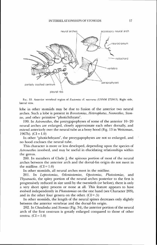
neural arches accessorv neural arch
urocranlum
part~ally oss~ f~ed centr
pleural r ~ b s
Flc;. 33. Anrer-ior ver~cht.al I-egion of Eu.\Lomins cf: rnacrurlL\ ( U S N M 272913). Right side, lateral view.
lobe in other stomiids may be due to fusion of the anterior two neural arches. Such a lobe is present in Boro.stomia.s, Heteropholus, Neonesthes, Stom- ias, and other primitive "photichthyans".
199. In Aslronesthes, the prezygapophyses of sorne of the anterior 10-20 neural arches are enlarged, closely approximate each other dorsally, and extend anteriorly over the neural tube as a bony hood (Fig. 13 in Weitzman, 1967b). (CI = 1.0)
In other "photichthyans", the prezygapophyses are not so enlarged, and no hood encloses the neural tube.
This character is more or less developed, depending upon the species of Aslronesthes involved, and may be useful in elucidating relationships within the genus.
200. In members of Clade J, the spinous portion of most of the neural arches between the anterior arch and the dorsal-fin origin do not meet in the midline. (CI = 1.0)
In other stomiids, all neural arches meet in the midline. 201. In Leptostomic~s, Odontostomias, Opostomias, Pholo.slomia.c, and
Thy.sanactis, the spiny portion of the neural arches posterior to the first is progressively reduced in size until by the twentieth (or before), there is only a very short spiny process or none at all. This feature appears to have evolved independently in Photostomias on the one hand (see Character 'Log), and in the other four genera on the other. (CI = .5)
In other stomiids, the length of the neural spines decreases only slightly between the anterior vertebrae and the dorsal-fin origin.
202. In Chauliodus and S1omia.s (Fig. 34), the anterior portion of the neural arch of the first centrum is greatly enlarged compared to those of other centra. (CI = 1 .O)
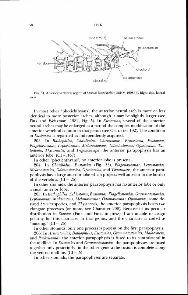
supraneural neural arches
eurocranlurn
vertebra
parapophys~s / ' pleural r ~ b
FIG. 34. Atlterior vertebral region of Stomlac lamnproj)eltz\ ( U S N M 199857). Right sidc, lateral view.
In most other "photichthyaris", the anterior neural arch is more or less identical to more posterior arches, although it may be slightly larger (see Fink and Weitzman, 1982, Fig. 5). In Ewtomias, several of' the anterior neural arches may be enlarged as a part of the complex modification of the anterior vertebral column in that genus (see Character 192). ' f i e condition in Eustomias is regarded as independently acquired.
203. In Batl~ofhilus, Chaulzodus, Chirostomnias, Echio,stoma, Eustomiccs, Flagellostomias, Leftostomia.~, Melanostomia.s, Odonto.stomias, Opostomias, TUC- tostoma, Thysanactis, and Trigonolamnpa, the anterior parapophysis has an anterior lobe. ((:I = .167)
In other "photichthyans", no anterior lobe is present. 204. In Chaulzodus, E~~stomia.~ (Fig. 33), Flagellostomia,s, I,epto.stomia~,
Melanostomia.~, Odontostomias, Opostomic~.s, and Thysanactis, the anterior para- pophysis has a large anterior lobe which projects well anterior to the border of the vertebra. (CI = .25)
In other- stomiids, the anterior parapophysis has no anteriol- lobe or only a small anterior lobe.
205. In Bathophilus, Echiostoma, Eustomias, Flagello.stomias, (;ram~m~atostomias, Leptostomia.~, Malacoste~~s, MM~nostomias, Odontostorn~a.~, Opo.stomias, some de- rived Stwmias species, and Thysanactzs, the anterior parapophysis bears two elongate processes (or more, see Character 208). Because of its peculiar distribution in Stomias (Fink and Fink, in press), I am unable to assign polarity for this character in that genus, and the character is coded as "missing." (CI = .25)
In other stomiids, only one process is present on the first parapophysis. 206. In Ari.stostomius, Bathophilus, Eustomias, Grammatostomias, Malacoste~~s,
and Pa,chystomias, the anterior parapophysis is fused to its contralateral in the midline. In Eustomias and Grammatostomias, the parapophyses are fused together only posteriorly; in the other genera the fusion is complete along the ventral midline. (C1= .5)
In other stomiids, the parapophyses are separate.
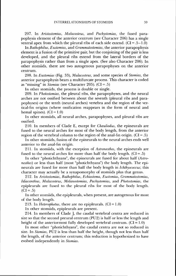
1NTERREI.ATIONSHIPS OF STOMIIDS 59
207. In Aristostorn2as, Malacostew, and Pachy.rtomias, the fused para- pophysis element of the anterior centrum (see Character 206) has a single ventral apex from which the pleural ribs of each side extend. (CI = .5-1.0)
In Bathophilus, Eustomias, and Grammatostomias, the anterior parapophysis element is a fusion of the primitive pair, but the conjoining of the pair is less developed, and the pleural ribs extend from the lateral borders of the parapophysis rather than from a single apex. (See also Character 206). In other stomiids, there are two autogenous parapophyses on the anterior centrum.
208. In Eustomias (Fig. 33), Malacosteus, and some species of Stomias, the anterior parapophysis bears a multifurcate process. -1Iis character is coded as "missing" in Stomim (see Character 205). (CI = .5)
In other stomiids, the process is double or single. 209. In Photostomias, the pleural ribs, the parapophyses, and the neural
arches are not ossified between about the seventh (pleural ribs and para- pophyses) or the tenth (neural arches) vertebra and the region of the ver- tical-fin origins (where ossification reappears in the form of' neural and hemal spines). (CI = 1 .O)
In other stomiids, all neural arches, parapophyses, and pleural ribs are ossified.
210. In members of Clade E, except for Chauliodus, the epineurals are fused to the neural arches for most of the body length, from the anterior region of'the vertebral column to the region of the anal-fin origin. (CI = .5)
In other stomiids, fusion of the epineurals to the neural arches ends well anterior to the anal-fin origin.
2 11. In stomiids, with the exception of Astronesthe.~, the epineurals are fused to the neural arches for more than half the body length. (CI = .5)
In other "photichthyans", the epineurals are fused for about half (Astro- nesthes) or less than half (most "photichthyans") the body length. The epi- neurals are fused for more than half the body length in Ichthyococcus; this character may actually be a synapomorphy of stomiids plus that genus.
212. In Aristostomias, Rathophilus, Echiostoma, Eustomim, Grammatostomias, Idiacanthus, Malacosteus, Melanostomias, Pachystomic~s, and Photostomias, the epipleurals are fused to the pleural ribs for most of the body length. (CI = .5)
In other stomiids, the epipleurals, when present, are autogenous for most of the body length.
2 13. In Heterophotus, there are no epipleurals. (CI = 1 .O) In other stomiids, epipleurals are present. 214. In members of Clade J, the caudal vertebral centra are reduced in
size so that the second preural centrum (PU2) is half or less the length and height of the anteriormost fully developed vertebral centrum. (CI = 1.0)
In most other "photichthyans", the caudal centra are not so reduced in size. In Stomim, PU2 is less than half the height, though not less than half the length, of the anterior centrum; this reduction is hypothesized to have evolved independently in Stomias.
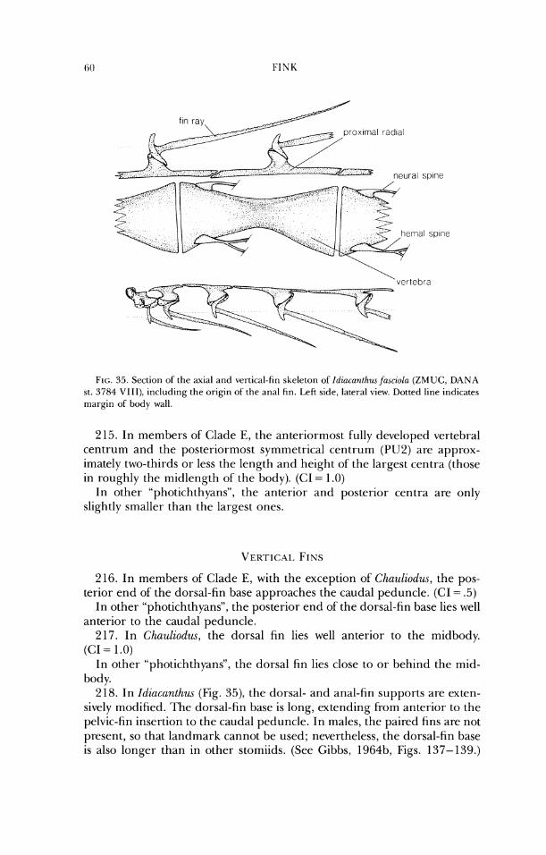
f ~ n ray prox~rrial radldl
- , c.. 2
,
FIG. 35. Sccr io~~ of the axial ~ I I I ~ vertical-fin skeleton of' Idincnntt~~w fasczoln (ZMUC, DANA st. 3784 VIII) , including the origin of the atla1 tin. Left side, latel-al view. Dotted line indicates margin of bocly wall.
2 15. In members of Clade E, the anteriormost fully developed vertebral centrum and the posteriormost symmetrical centrum (PU2) are approx- imately two-thirds or less the length and height of' the largest centra (those in roughly the midlength of the body). (CI = 1.0)
I n other "photichthyans", the anterior and posterior centra are only slightly smaller than the largest ones.
216. In members of' Clade E, with the exception of' Chauliorlus, the pos- terior end of' the dorsal-fin base approaches the caudal peduncle. (CI = .5)
In other "photichthyans", the posterior end of the dorsal-fin base lies well anterior to the caudal peduncle.
217. In Chauliodw., the dorsal fin lies well anterior to the midbody. (CI = 1.0)
In other "photichthyans", the dorsal fin lies close to or behind the mid- body.
218. In Idiacunthw. (Fig. 35), the dorsal- and anal-fin supports are exten- sively modified. l'he dorsal-fin base is long, extending from anterior to the pelvic-fin insertion to the caudal peduncle. In males, the paired fins are not present, so that landmark cannot be used; nevertheless, the dorsal-fin base is also longer than in other stomiids. (See Gibbs, 1964b, Figs. 137-139.)
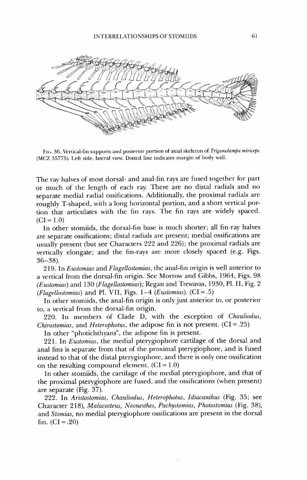
IN~I'E'.KREI.RI'IONSH IPS O F ST0MIIL)S
Flc:. 36. Vertical-fin supports a n d posterior portion of axial skeleton of ?i-igonolam/)n razriceps (MCZ 35775). I.cSt side, lateral view. Dotted line indicates margin of body wall.
The ray halves of most dorsal- and anal-fin rays are fused together for part or much of the length of each ray. There are no distal radials and no separate medial radial ossifications. Additionally, the proximal radials are roughly T-shaped, with a long horizontal portion, and a short vertical por- tion that articulates with the fin rays. The fin rays are widely spaced. (C1= 1.0)
In other stomiids, the dorsal-fin base is much shorter; all fin-ray halves are separate ossifications; distal radials are present; medial ossifications are usually present (but see Characters 222 and 226); the proximal radials are vertically elongate; and the fin-rays are more closely spaced (e.g. Figs. 36-38).
219. In Eustomias and Flagellostomias, the anal-fin origin is well anterior to a vertical from the dorsal-fin origin. See Morrow and Gibbs, 1964, Figs. 98 (Eustomia.~) and 130 (Flage1lostomia.s); Kegan and Trewavas, 1930, PI. 11, Fig. 2 (Flagellostornias) and PI. VII, Figs. 1-4 (Euslomias). (CI = .5)
In other stomiids, the anal-fin origin is only just anterior to, or posterior to, a vertical from the dorsal-fin origin.
220. In members of Clade D, with the exception of Chauliodus, Chirostomias, and Heteropholus, the adipose fin is not present. (CI = .25)
In other "photichthyans", the adipose fin is present. 221. In Eustornias, the medial pterygiophore cartilage of the dorsal and
anal fins is separate from that of the proximal pterygiophore, and is fused instead to that of the distal pterygiophore, and there is only one ossification on the resulting compound element. (CI = 1.0)
In other stomiids, the cartilage of the medial pterygiophore, and that of the proximal pterygiophore are fused, and the ossifications (when present) are separate (Fig. 37).
222. In Ari.rto.stomias, Chauliodus, Heterophotus, Idiacanthus (Fig. 35; see Character 2 18), Malacosteus, Neonesthes, Pachystomias, Photostomias (Fig. 38), and Stomias, no medial pterygiophore ossifications are present in the dorsal fin. (CI = .PO)
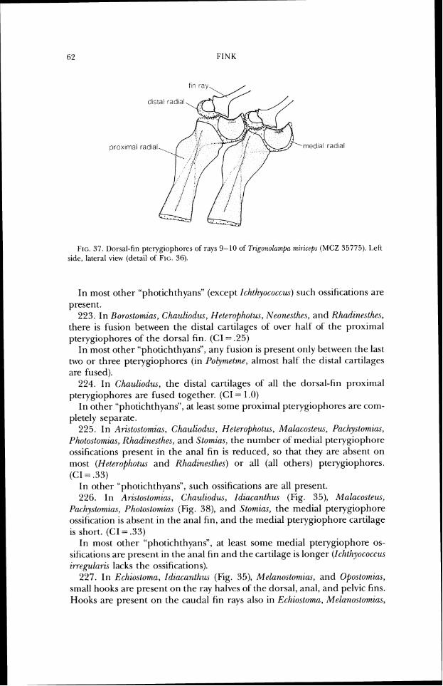
FIG. 37. Dorsal-fin ptetygiophot-es of rays 9-10 of Trigonolant~~ri miricrf), ( M C Z 35775). I.cli sidc, lalet-al view (dctail of' Flc:. 36).
In most other "photichthyanS' (except Ichthyococcus) such ossific-ntions are present.
c c 2.23. ' In Uoro.stomia.s, Chauliodus, Heterophot~u, Neonesth~s, and Rhadin~.sthes, there is fusion between the distal cartilages of over half of' the proximal pte~ygiophores of the dorsal fin. (C1= .25)
In most other "photichthyans", any fusion is present only between the last two or three pterygiophores (in Polymetme, almost half' the distal cartilages are fused).
224. In Chnuliodus, the distal cartilages of all the dorsal-fin proximal pterygiophores are fused together. (CI = 1.0)
In other "photichthyans", at least some proximal pterygiophores are con)- pletely separate.
225. In Aristostomias, Chauliodus, Heleropholus, Malr~costeus, Pachy.stomia.s, Photo.stomia.s, Khadinesthes, and Stomias, the number of medial pterygiophore ossifications present in the anal fin is reduced, so that they are absent on most (Heterophotus and Rhadinesthe.~) or all (all others) pterygiophores. (CI = .33)
In other "photichthyans", such ossifications are all present. 226. In Aristostomias, Chauliodm, Idiacanth~u (Fig. 35), Malacosteus,
Puchystomir~.s, Photostomiu.r (Fig. 38), and Stomias, the medial pterygiophore ossification is absent in the anal fin, and the medial pterygiophore cartilage is short. (<:I = .33)
In most other "photichthyans", at least some medial pterygiophore os- sifications are present in the anal fin and the cartilage is longer (Ichthyococcus irregul(~ric lacks the ossifications).
227. In Echiostoma, Idiacanthzcs (Fig. 35), Melanostomias, and Opostomins, small hooks are present on the ray halves of the dorsal, anal, and pelvic fins. Hooks are present on the caudal fin rays also in Echiostoma, Melanoslomiac,
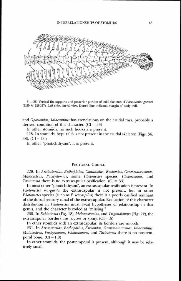
FIG. 38. Vertical-fin supports and posterior portion of axial skeleton of Photostomias guernci (USNM 225027). Left side, lateral view. Dotted line indicates margin of body wall.
and Opostomias; Idiacanthus has crenelations on the caudal rays, probably a derived condition of this character. (GI = .33)
In other stomiids, no such hooks are present. 228. In stomiids, hypural6 is not present in the caudal skeleton (Figs. 36,
38). (CI = 1.0) In other "photichthyans", it is present.
229. In Aristostomius, Balhophilu~, Chauliodw, Eustomias, Grammatostomias, Malucosteus, Pachystomias, some Photonectes species, Photostomias, and Tactostoma there is no extrascapular ossification. (CI = .33)
In most other "photichthyans", an extrascapular ossification is present. In Photonectes margarita the extrascapular is not present, but in other Photonectes species (such as P. leucospilus) there is a poorly ossified remnant of the dorsal sensory canal of the extrascapular. Evaluation of this character distribution in Photonectes must await hypotheses of relationship in that genus, and the character is coded as "missing."
230. In Echiostoma (Fig. 18), Melanostomias, and Trigonolampa (Fig. 22), the extrascapular borders are rugose or spiny. (CI = .5)
In other stomiids with an extrascapular, its borders are smooth. 231. In Aristostomias, Bathophilus, Eustomias, Grammatostomias, Idiacanthus,
Malacosteus, Pachystom.ia,s, Photostomias, and Tactostoma there is no posttem- poral bone. (CI = 1 .O)
In other stomiids, the posttemporal is present, although it may be rela- tively small.
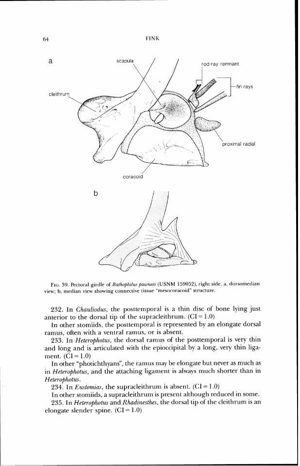
FIG. 39. Pectoral girdle of Halhof)/lilzw pau~n~ri (USNM 159052), right side. a , dorsomcdian view; b, nledian view sllowing connective tissuc "n~csocoraroitl" structure.
232. In Chuuliodzus, the posttemporal is a thin disc of bone lying just anterior to the dorsal tip of the supracleithrum. (CI = 1.0)
In other stomiids, the posttemporal is represented by an elongate dorsal ramus, often with a ventral ramus, o r is absent.
233. In Heterophotus, the dorsal rarnus of the posttemporal is very thin and long and is articulated with the epioccipital by a long, very chill liga- ment. (CI = 1.0)
In other "photichthyans", the ramus may be elongate but never as much as in Hetcrophotus, and the attaching ligament is always much shorter than in Heterophotus.
234. In E~~~tornias , the supracleithrum is absent. ((:I = 1.0) In other stomiids, a supracleithrum is present although reduced in some. 235. In Hcterophotus and Rhudinesthes, the dorsal tip of the cleithrum is an
elongate slender spine. (CI = 1.0)
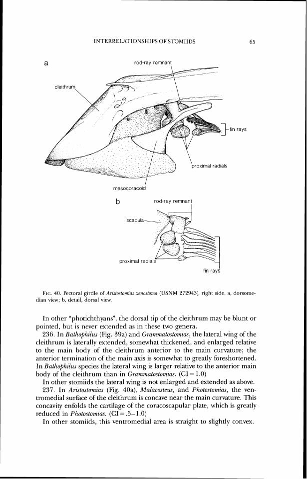
a rod-ray remnant
- f ~ n rays
mesocoraco~d
b rod-ray remnant I
fin rays
FIG. 40. Pectoral girdle of Aristostomim xenostoma (USNM 272043), right side. a, dorsorne- dian view; b, detail, dorsal view.
I11 other "photichthyans", the dorsal tip of the cleithrum may be blunt or pointed, but is never extended as in these two genera.
236. In Bathophilus (Fig. 39a) and Grammatostomias, the lateral wing of the cleithrum is laterally extended, somewhat thickened, and enlarged relative to the main body of the cleithrum anterior to the main curvature; the anterior termination of the main axis is somewhat to greatly foreshortened. In Bathophilus species the lateral wing is larger relative to the anterior main body of the cleithrum than in Grammatostomias. (CI = 1.0)
In other stomiids the lateral wing is not enlarged and extended as above. 237. In Aristostomias (Fig. 40a), Malacosteus, and Photostomias, the ven-
tromedial surface of the cleithrum is concave near the main curvature. This concavity enfolds the cartilage of the coracoscapular plate, which is greatly reduced in Photostomias. (CI = .5- 1 .O)
In other stomiids, this ventromedial area is straight to slightly convex.
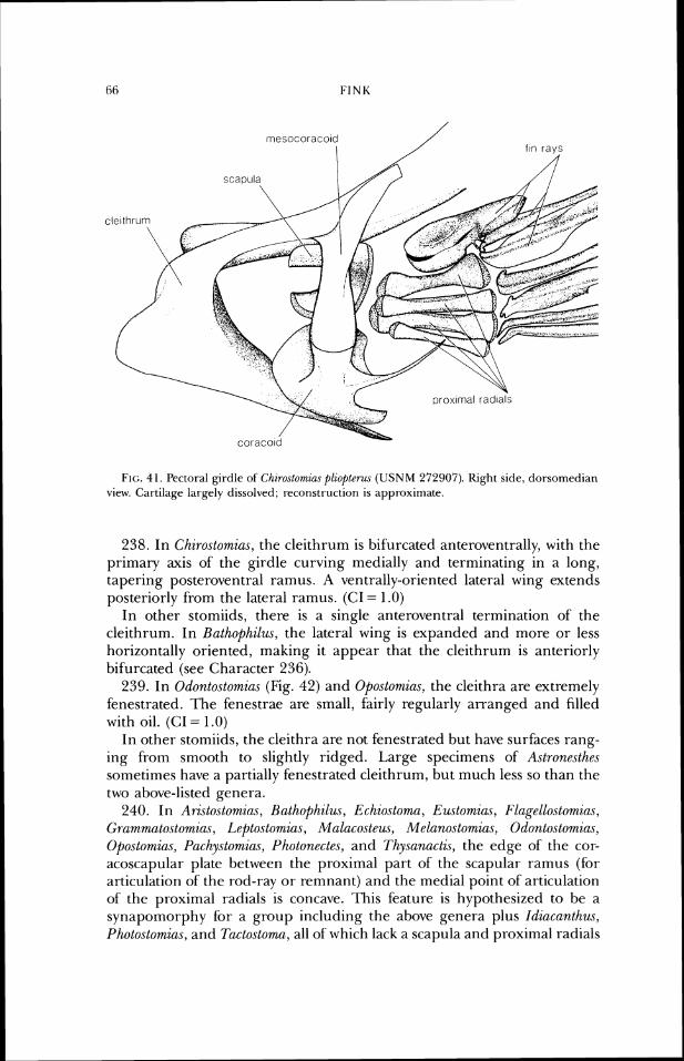
rnesocoraco~d I f ~ n ravs
FIG. 41. l'ectot-al girdle of Chirostomias pliopter~ls (USNM 272907). Right side, dorsomcdian view. Cartilage largely dissolved; reconstruction is approximate.
238. In Chirostomias, the cleithrum is bifurcated anteroventrally, with the primary axis of the girdle curving medially and terminating in a long, tapering posteroventral ramus. A ventrally-oriented lateral wing extends posteriorly from the lateral ramus. (CI = 1 .O)
In other stomiids, there is a single anteroventral termination of the cleithrum. In Bathophilus, the lateral wing is expanded and more or less horizontally oriented, making it appear that the cleithrum is anteriorly bifurcated (see Character 236).
239. In Odontostomias (Fig. 42) and Opostomias, the cleithra are extremely fenestrated. The fenestrae are small, fairly regiilarly arranged and filled with oil. (CI = 1.0)
In other stomiids, the cleithra are not fenestrated but have sul-faces rang- ing from smooth to slightly ridged. Large specimens of Astronesth.es sonletimes have a partially fenestrated cleithrum, but much less so than the two above-listed genera.
240. In Aristostomia.~, Bathophilus, Echiostoma, Emtomias, Flagellostomias, Gramrnatostomias, Lepto.stomias, Malacosteus, Melanostomias, Odontostomias, Opostomias, Pachystomias, Photon,ectes, and Thysunactis, the edge of the cor- acoscapular plate between the proximal part of the scapular ramus (for articulation of the rod-ray or remnant) and the medial point of articulation of the proximal radials is concave. This feature is hypothesized to be a synapomorphy for a group including the above genera plus Idic~canth~u, Photostomias, and Tactostoma, all of which lack a scapula and proximal radials
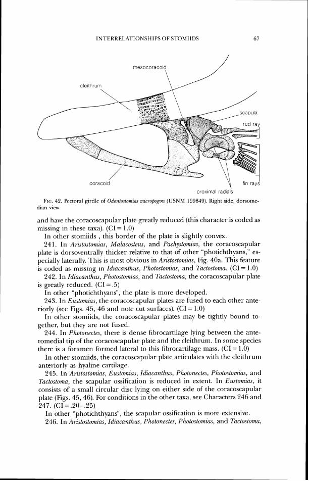
INTERRELATIONSHIPS OF S T O M IlDS (5 7
rnesocoracol
c le~ thrum
prox~mal rad~als
Flc:. 42. Pectoral girdle o f Odontostomins micropogon (USNM 199849). Right side, dorsome- tlian view.
and have the coracoscapular plate greatly reduced (this character is coded as missing in these taxa). (CI = 1 .O)
In other stomiids , this border of the plate is slightly convex. 24 1. In Aristo.stomias, Malacosteus, and Pachystomius, the coracoscapular
plate is dorsoventrally thicker relative to that of other "photichthyans," es- pecially laterally. This is most obvious in Aristostomias, Fig. 40a. This feature is coded as missing in Idiacanthus, Photostomias, and Tactostoma. (CI = 1.0)
242. In Idiacanthus, Photostomias, and Tactostoma, the coracoscapular plate is greatly reduced. (CI = .5)
I n other "photichthyans", the plate is more developed. 243. In Eustomias, the coracoscapular plates are fused to each other ante-
riorly (see Figs. 45, 46 and note cut surfaces). (CI = 1.0) In other stomiids, the coracoscapular plates may be tightly bound to-
gether, but they are not fused. 244. In Photonecles, there is dense fibrocartilage lying between the ante-
romedial tip of the coracoscapular plate and the cleithrum. In some species there is a foramen formed lateral to this fibrocartilage mass. (CI = 1.0)
In other stomiids, the coracoscapular plate articulates with the cleithrum anteriorly as hyaline cartilage.
245. In Aristostomias, Ewtomias, Idiacanthw, Photonectes, Photostomius, and Tactostoma, the scapular ossification is reduced in extent. In Eustomias, it consists of a small circular disc lying on either side of the coracoscapular plate (Figs. 45,46). For conditions in the other taxa, see Characters 246 and 247. (CI = .20-.25)
In other "photichthyans", the scapular ossification is more extensive. 246. In Aristostomias, Idiacanthus, Photonectes, Photostomias, and Tactostoma,
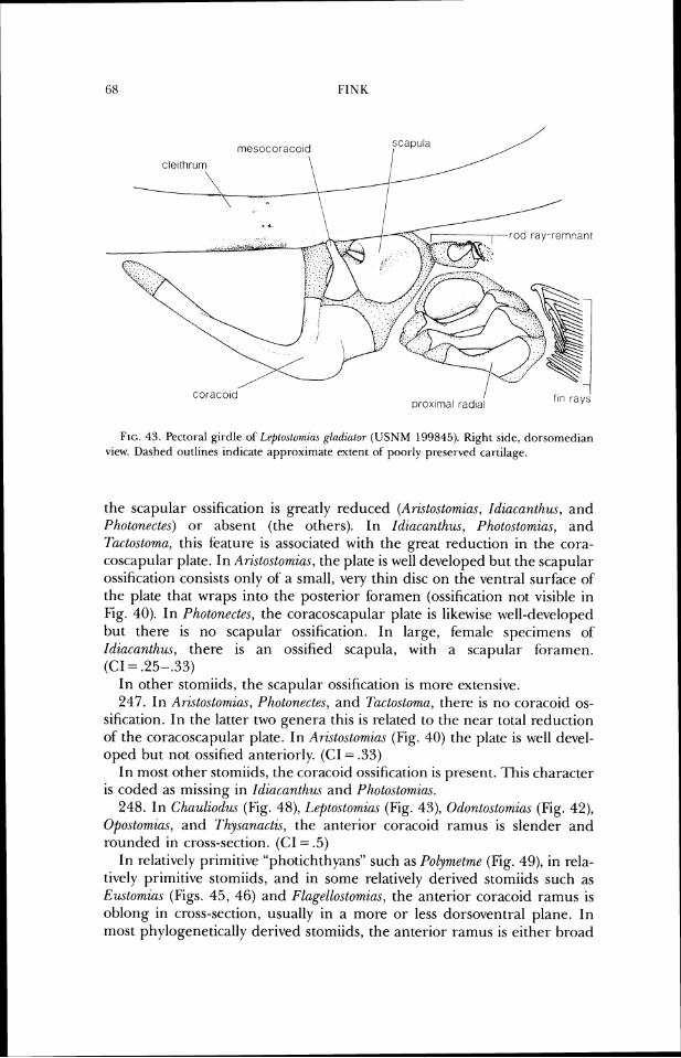
FINK
mesocoracoid scapula I
FIG. 43. Pectot-al girdle of L~~ptostomiar glndzator (USNM 199845). Right side, dorsonieclian vicw. Dashed outlines indicate appn~ximate extent of poorly preserved cartilage.
the scapular ossification is greatly reduced (Ari.stostomias, Idiacuntlzu.~, and Photonectes) or absent (the others). In Idiacunthus, Photo.storniu.s, and Tactostomu, this feature is associated with the great reduction in the cora- coscapular plate. In Arzstostomia.~, the plate is well developed but the scapular ossification consists only of a small, very thin disc on the ventral surface of the plate that wraps into the posterior foramen (ossification not visible in Fig. 40). In Photonectes, the coracoscapular plate is likewise well-developed but there is no scapular ossification. in large, female specimens of' Idiacanthus, there is an ossified scapula, with a scapular foramen. (CI = .25-.33)
In other stomiids, the scapular ossification is more extensive. 247. In Aristostomias, Photonecte.~, and Tactostoma, there is no coracoid os-
sification. In the latter two genera this is related to the near total reduction of the coracoscapular plate. In An.sto.stomias (Fig. 40) the plate is well devel- oped but not ossified anteriorly. (CI = .33)
In most other stomiids, the coracoid ossification is present. This character is coded as missing in Idiacanthus and Photo.stomim.
248. In Chauliodus (Fig. 48), Leptostomius (Fig. 43), 0dontostomia.s (Fig. 42), Opostomias, and Thysanactzs, the anterior coracoid ramus is slender and rounded in cross-section. (CI = .5)
In relatively primitive "photichthyans" such as Polym,etme (Fig. 49), in rela- tively primitive stomiids, and in some relatively derived stomiids such as Ewtomias (Figs. 45, 46) and Flagellostomias, the anterior coracoid ramus is oblong in cross-section, usually in a more or less dorsoventral plane. in most phylogenetically derived stomiids, the anterior ramus is either broad
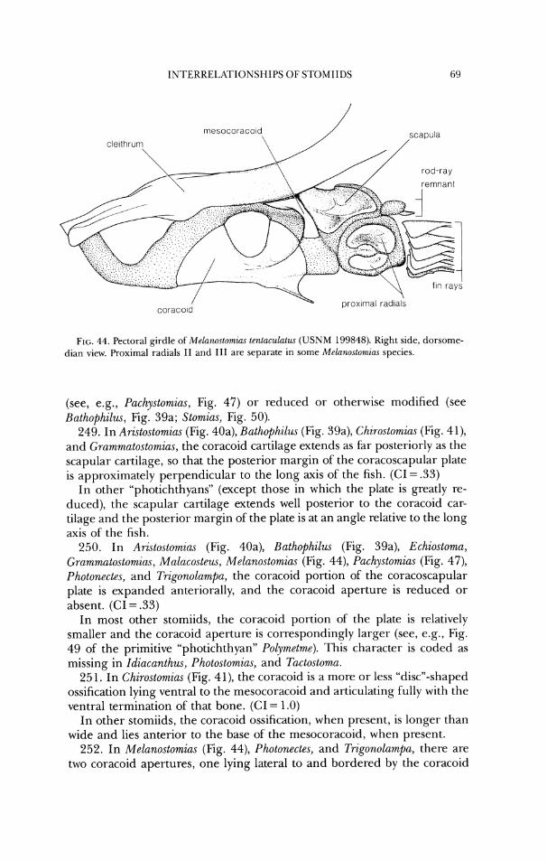
IN'I 'ERRELKTIONSHIPS OF STOMIIDS
Flc;. 44. Pectoral girdle of' Melunoslomzas tentacvlulw ( U S N M 199848). Kight side, dorsome- dian view. Proximal radials I 1 and 111 are separate i r ~ sorrie Mel(~nostomias spccies.
(see, e.g., Pachystomiu.~, Fig. 47) or reduced or otherwise modified (see Bathophilus, Fig. 39a; Stomias, Fig. 50).
249. In Arzstostomias (Fig. 40a), Bathophilus (Fig. 39a), Chirostomius (Fig. 41), and Grarnmatostomias, the coracoid cartilage extends as fBr posteriorly as the scapular cartilage, so that the posterior margin of the coracoscapular plate is approximately perpendicular to the long axis of the fish. (C1= .33)
In other "photichthyans" (except those in which the plate is greatly re- duced), the scapular cartilage extends well posterior to the coracoid car- tilage and the posterior margin of the plate is at an angle relative to the long axis of the fish.
250. In Aristostomias (Fig. 40a), Buthophilwr (Fig. 39a), Echiostoma, Grammatostomia.~, Malacosteus, Melanostomias (Fig. 44), Pachystomias (Fig. 47), Photonectes, and Trigonolampa, the coracoid portion of the coracoscapular plate is expanded anteriorally, and the coracoid aperture is reduced or absent. (CI = .33)
In most other stomiids, the coracoid portion of the plate is relatively smaller and the coracoid aperture is correspondingly larger (see, e.g., Fig. 49 of' the primitive "photichthyan" Polymetme). This character is coded as missing in Idiacunthus, Photostomia.\, and Tactostomu.
25 1. In Chirostomias (Fig. 4 l), the coracoid is a more or less "discn-shaped ossification lying ventral to the mesocoracoid and articulating fully with the ventral termination of that bone. (CI = 1.0)
In other stomiids, the coracoid ossification, when present, is longer than wide and lies anterior to the base of the mesocoracoid, when present.
252. In Melanostomias (Fig. 44), Photonectes, and Trigonolampa, there are two coracoid apertures, one lying lateral to and bordered by the coracoid
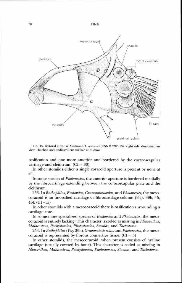
FIG. 45. Pectoral girdle of Emtomias cf. mucrurus (USNM 292913). Right side, dorsorncdian view. Hatched area indicates cut surface at miclline.
ossification and one more anterior and bordered by the coracoscapular cartilage and cleithrum. (CI = .33)
In other stomiids either a single coracoid aperture is present or none at all.
In some species of Photonectes, the anterior aperture is bordered medially by the fibrocartilage extending between the coracoscapular plate and the cleithrum.
253. In Bathophilus, Eustomias, Grammatostomias, and Photonectes, the meso- coracoid is an unossified cartilage or fibrocartilage column (Figs. 39b, 45, 46). (CI = .5)
In other stomiids with a mesocoracoid there is ossification surrounding a cartilage core.
In some more specialized species of Eustomias and Photonectes, the meso- coracoid is entirely lacking. This character is coded as missing in Idiacanthus, Malacosteus, Pachystomias, Photostomias, Stomias, and Tactostoma.
254. In Bathophilus (Fig. 39b), Grammatostomias, and Photonectes, the meso- coracoid is represented by fibrous connective tissue. (CI = .5)
In other stomiids, the mesocoracoid, when present consists of hyaline cartilage (usually covered by bone). This character is coded as missing in Idiacanthus, Malacostem, Pachystomias, Photostomias, Stomias, and Tactostoma.
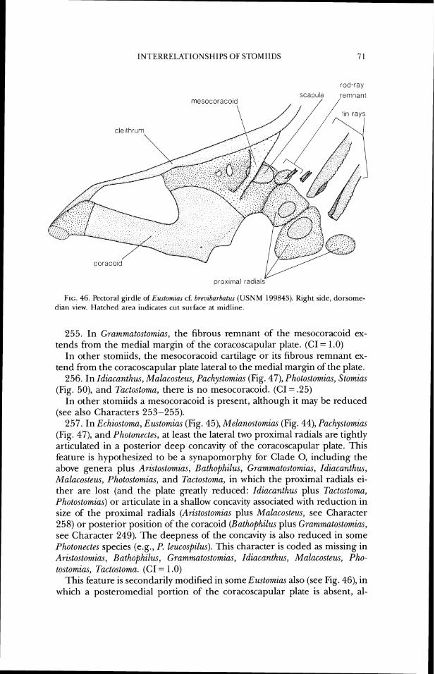
INTERRELATIONSHIPS OF STOMIIDS
rod-ray
FIG. 46. Pectoral girdle of Emlomius cf. brevibarbatus ( U S N M 199843). Right side, dorsome- dian view. Hatched area indicates cut surface at midline.
255. In Grammatostom.ias, the fibrous remnant of the mesocoracoid ex- tends from the medial margin of the coracoscapular plate. (CI = 1.0)
In other stomiids, the mesocoracoid cartilage or its fibrous remnant ex- tend from the coracoscapular plate lateral to the medial margin of the plate.
256. In Idiacanthus, Malacosteus, Pachystomias (Fig. 47), Photostomias, Stomias (Fig. 50), and Tactostoma, there is no mesocoracoid. (CI = .25)
In other stomiids a mesocoracoid is present, although it may be reduced (see also Characters 253-255).
257. In Echiostoma, Eustomias (Fig. 45), Melanostomias (Fig. 44), Pachystomias (Fig. 47), and Photonectes, at least the lateral two proximal radials are tightly articulated in a posterior deep concavity of the coracoscapular plate. This feature is hypothesized to be a synapomorphy for Clade 0, including the above genera plus Aristostomias, Rathophilus, Grammatostomias, Idiacanthus, Malacosteus, Photostomias, and Tactostoma, in which the proximal radials ei- ther are lost (and the plate greatly reduced: Idiacanthus plus Tactostoma, Photostomias) or articulate in a shallow concavity associated with reduction in size of the proximal radials (Aristostomias plus Malacosteus, see Character 258) or posterior position of the coracoid (Bathophilus plus Grammatostomias, see Character 249). The deepness of the concavity is also reduced in some Photonectes species (e.g., P. leucospilus). This character is coded as missing in Aristostomias, Bathophilus, Grammatostomias, Idiacanthus, Malacosteus, Pho- tostomias, Tactosto,ma. (CI = I .O)
This feature is secondarily modified in some Eustomias also (see Fig. 46), in which a posteromedial portion of the coracoscapular plate is absent, al-
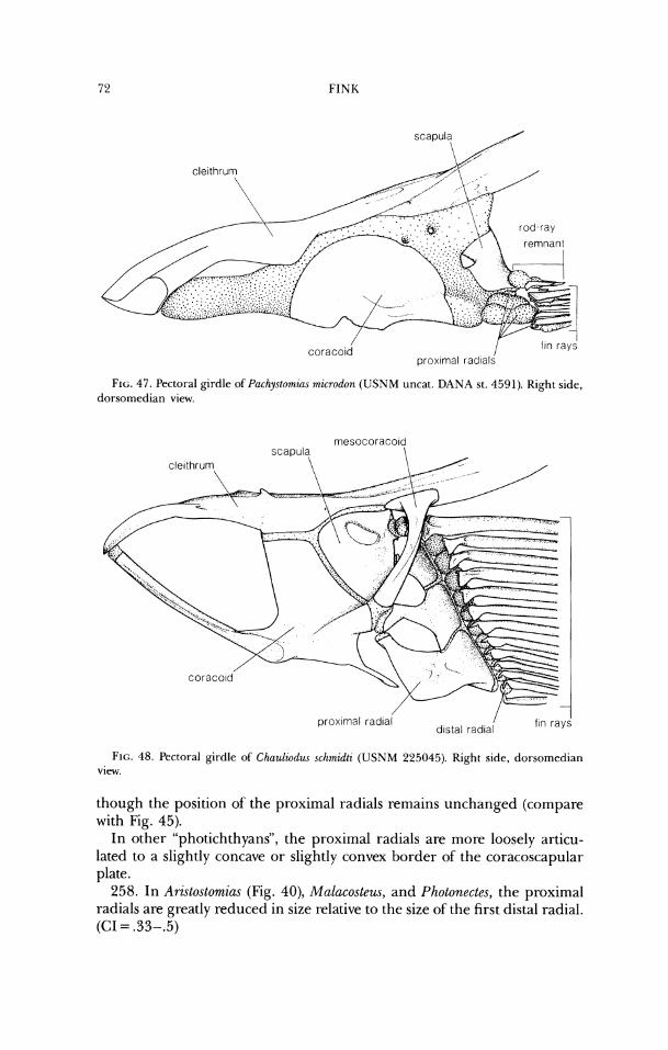
FIG. 47. l'ectoral girdle oPI'achyslomias microdon (USNM uncat. D A N A st. 459 1). Right side, dorsomedian view.
FIG. 48. Pectoral girdle of C h a u l i o d ~ ~ ~chmidti (USNM 225045). Right side, dorsomedian view.
though the position of the proximal radials remains unchanged (compare with Fig. 45).
In o&er "photichthyans". the proximal radials are more loosely articu- lated to a slightly concave or slightly convex border of the coracoscapular plate.
258. In Aristostomias (Fig. 40), Malacostew, and Photonectes, the proximal radials are greatly reduced in size relative to the size of the first distal radial. (CI = .33-.5)
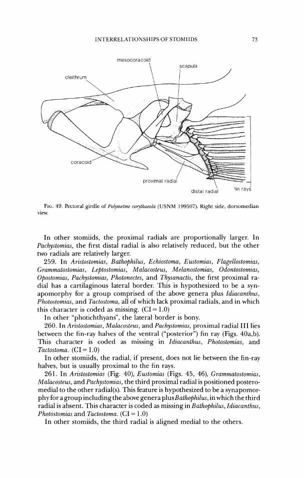
IN'I 'ERRELATIONSHIPS O F S'I'OMIIDS
FIG. 49. Pectoral girdle of Polymrtme corythaeola ( U S N M 199507). Right side, dorsomedian view.
In other stomiids, the proximal radials are proportionally larger. In Pac/~ystomias, the first distal radial is also relatively reduced, but the other two radials are relatively larger.
259. In Aristostomias , Bathophilus, Echiostoma, Eustomim, Flagellostomias, Gramw~utostomia~, Leptostow~ias, Mulacosteus, Melanostomias, Odontostomias, Opostornias, Pachystomias, Photonectes, and ThysanactG, the first proximal ra- dial has a cartilaginous lateral border. This is hypothesized to be a syn- apomorphy for a group comprised of the above genera plus Idiacanthw, Photostomias, and Tactostoma, all of which lack proximal radials, and in which this character is coded as missing. (CI = 1.0)
In other "photichthyans", the lateral border is bony. 260. In Ari~tostow~ias, Malacosteus, and Pachystomias, proximal radial I11 lies
between the fin-ray halves of the ventral ("posterior") fin ray (Figs. 40a,b). This character is coded as missing in Idiacanthus, Photostomias, and Tactostoma. (CI = 1 .O)
In other stomiids, the radial, if present, does not lie between the fin-ray halves, but is usually proximal to the fin rays.
261. In Aristostomias (Fig. 40), Eustomias (Figs. 45, 46), Grammatostomias, Malacosteus, and Pachystomias, the third proximal radial is positioned postero- medial to the other radial(s). This feature is hypothesized to be a synapomor- phy for a group including the above genera plus Bathophilus, in which the third radial is absent. This character is coded as missing in Bathophilus, Idiacanthus, Photostomias and Tactostoma. (CI = 1 .O)
In other stomiids, the third radial is aligned medial to the others.
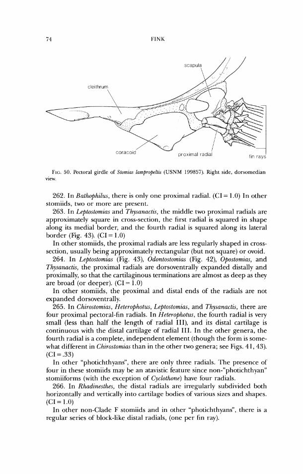
coraco~d' nrn
FIG. 50. Pectoral girdle of Slornza.7 la7npropeltw ( U S N M 199857). Right aide, dorsomcdiarr view.
262. In Hathophilus, there is only one proximal radial. (CI = 1.0) In other stomiids, two or more are present.
263. In Leptostomias and Thysanactis, the middle two proximal radials are approximately square in cross-section, the first radial is squared in shape along its medial border, and the fourth radial is squared along its lateral border (Fig. 43). (CI = 1.0)
In other stomiids, the proximal radials are less regularly shaped in cross- section, usually being approximately rectangular (but not square) or ovoid.
264. In Leptostomias (Fig. 43), Odontostomias (Fig. 42), Opostomias, and Thysanactis, the proximal radials are dorsoventrally expanded distally and proximally, so that the cartilaginous terminations are almost as deep as they are broad (or deeper). (CI = 1 .O)
In other stomiids, the proximal and distal ends of the radials are not expanded dorsoventrally.
265. In Chirostomias, Heterophotus, Leptostomias, and Thysanactis, there are four proximal pectoral-fin radials. In Heterophotus, the fourth radial is very small (less than half the length of radial 111), and its distal cartilage is continuous with the distal cartilage of radial 111. In the other genera, the fourth radial is a complete, independent element (though the form is some- what different in Chirostomim than in the other two genera; see Figs. 4 1,43). (CI = .33)
In other "photichthyans", there are only three radials. 'Ile presence of four in these stomiids may be an atavistic feature since non-"photichthyan" stomiiforms (with the exception of Cyclothone) have four radials.
266. In Rhadinesthes, the distal radials are irregularly subdivided both horizontally and vertically into cartilage bodies of various sizes and shapes. (CI = 1.0)
In other non-Clade F stomiids and in other "photichthyans", there is a regular series of block-like distal radials, (one per fin ray).
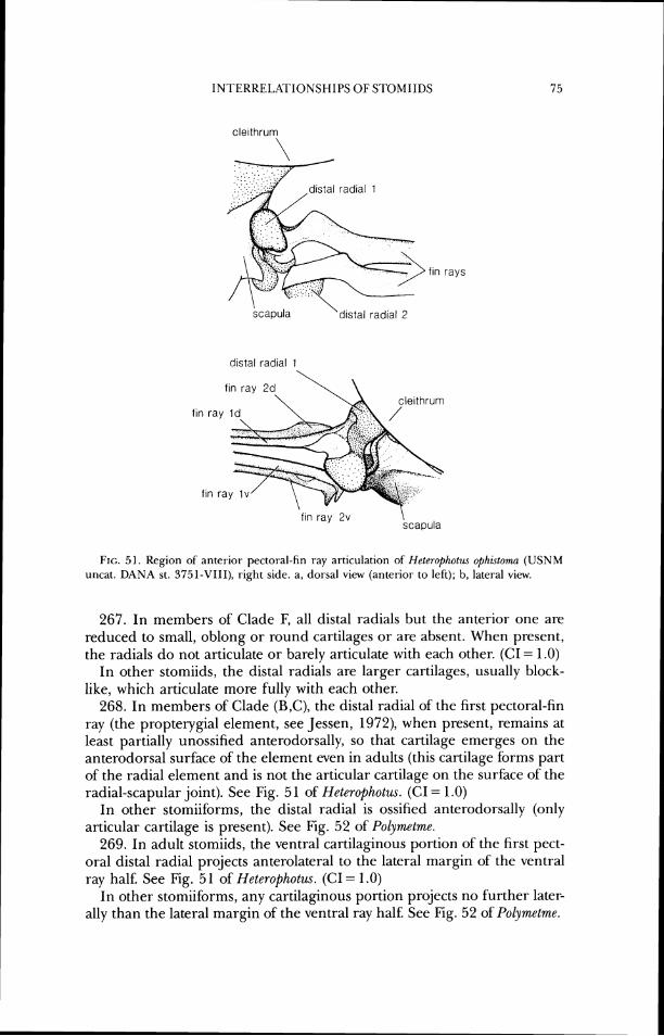
INTERRELATIONSI-IIPS OF STOMIIDS
scapula \ distal radial 2
f ~ n ray id. fay
f ~ n ray 7m I V
f ~ n ray 2v \ scapula
FIG. 51. Region of anterior pectoral-fin ray articulation of Heterophotzrs ophwloma (USNM uncat. DANA st. 375 1-VIII), right side. a, dorsal view (anteriol- to left); b, lateral view.
267. In members of Clade F, all distal radials but the anterior one are reduced to small, oblong or round cartilages or are absent. When present, the radials do not articulate or barely articulate with each other. (CI = 1 .O)
In other stomiids, the distal radials are larger cartilages, usually block- like, which articulate more fully with each other.
268. In members of Clade (B,C), the distal radial of the first pectoral-fin ray (the propterygial element, see Jessen, 1972), when present, remains at least partially unossified anterodorsally, so that cartilage emerges on the anterodorsal surface of the element even in adults (this cartilage forms part of the radial element and is not the articular cartilage on the surface of the radial-scapular joint). See Fig. 5 1 of Heterophotus. (CI = 1 .O)
In other stomiiforms, the distal radial is ossified anterodorsally (only articular cartilage is present). See Fig. 52 of Polymetme.
269. In adult stomiids, the ventral cartilaginous portion of the first pect- oral distal radial projects anterolateral to the lateral margin of the ventral ray half. See Fig. 5 1 of Heterophotus. (CI = 1 .O)
In other stomiiforms, any cartilaginous portion projects no further later- ally than the lateral margin of the ventral ray half. See Fig. 52 of Polymetme.
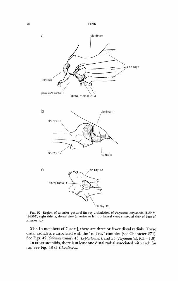
f ~ n rays
f ~ n ray Id
, f ~ n ray I v I
scapula
distal radial 1
\ fin ray Iv
FIG. 52. Region of anterior pectoral-fin ray articulation of Polymetme corythaeola (USNM 199507), right side. a, dorsal view (anterior to left); b, lateral view; c, medial view of base of anterior ray.
270. In members of Clade J, there are three or fewer distal radials. These distal radials are associated with the "rod-ray" complex (see Character 27 1). See Figs. 42 (Odontostomias), 43 (Leptostomias), and 53 (Thysunactis). (CI = 1 .O)
In other stomiids, there is at least one distal radial associated with each fin ray. See Fig. 48 of Chauliodw.
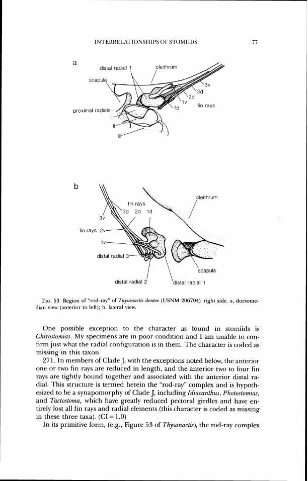
INTERRELATIONSHIPS OF S'IDMIIDS
a
proxin
b
fin rays
scapula
distal radial 2 distal radial 1
FIG. 53. Region of "rod-ray" of Thysamcl?s dentex (USNM 206704), right side. a, dorsorne- dian view (anterior to left); b, lateral view.
One possible exception to the character as found in stomiids is Chirostomias. My specimens are in poor condition and I am unable to con- firm just what the radial configuration is in them. The character is coded as missing in this taxon.
27 1. In members of Clade J, with the exceptions noted below, the anterior one or two fin rays are reduced in length, and the anterior two to four fin rays are tightly bound together and associated with the anterior distal ra- dial. This structure is termed herein the "rod-ray" complex and is hypoth- esized to be a synapomorphy of Clade J, including Idiacanthus, Photostomias, and Tactostoma, which have greatly reduced pectoral girdles and have en- tirely lost all fin rays and radial elements (this character is coded as missing in these three taxa). (GI = 1.0)
In its primitive form, (e.g., Figure 53 of ThysanactG), the rod-ray complex
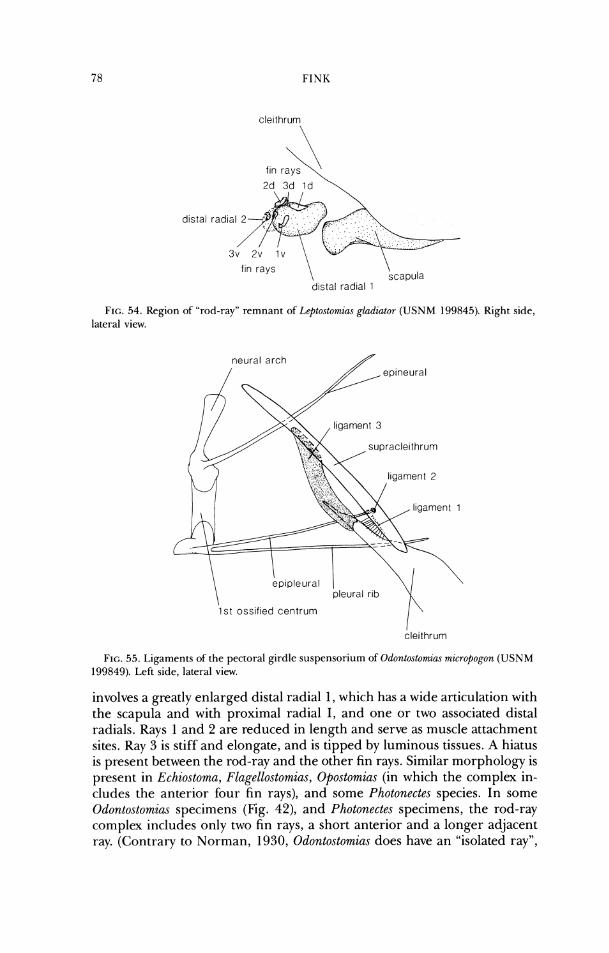
FINK
f ~ n rays 'II,
fln rays \ \ scapula
dlstal radlal 1
FIG. 54. Region of "rod-ray" remnant of Leptostomias gladiator (USNM 199845). Right side, lateral view.
neural arch ,/'
1st osslfled centrum I' FIG. 55. 1,igaments of the pectoral girdle suspensorium of Odontostomia~ micropogon (USNM
199849). [>eft side, lateral view.
involves a greatly enlarged distal radial 1, which has a wide articulation with the scapula and with proximal radial I , and one or two associated distal radials. Rays 1 and 2 are reduced in length and serve as muscle attachment sites. Ray 3 is stiff and elongate, and is tipped by luminous tissues. A hiatus is present between the rod-ray and the other fin rays. Similar morphology is present in Echiostoma, Flagellostomias, Opostomias (in which the complex in- cludes the anterior four fin rays), and some Photonectes species. In some Odontostomias specimens (Fig. 42), and Photonectes specimens, the rod-ray complex includes only two fin rays, a short anterior and a longer adjacent ray. (Contrary to Norman, 1930, Odontostomias does have an "isolated ray",
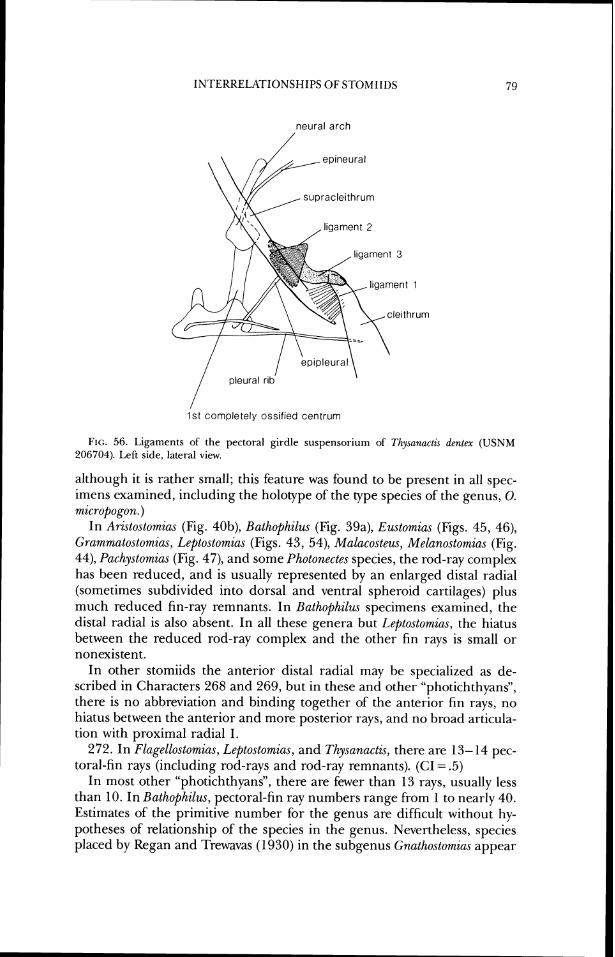
INTERRELATIONSHIPS OF STOMIIDS
neural arch
epineural
supracleithrum
,ligament 2
I
pleural r ~ b \
/ 1st completely ossif~ed centrum
FIG. 56. Ligaments of the pectoral girdle suspensorium of Thysanactu dentex (USNM 206704). Left side, lateral view.
although it is rather small; this feature was found to be present in all spec- imens examined, including the holotype of the type species of the genus, 0. micropogon.)
In Aristostomias (Fig. 40b), Bathophilus (Fig. 39a), Emtomias (Figs. 45, 46), Grammatostomias, Leptostomias (Figs. 43, 54), Malacosteus, Melanostomias (Fig. 44), Pachystomias (Fig. 47), and some Photonectes species, the rod-ray complex has been reduced, and is usually represented by an enlarged distal radial (sometimes subdivided into dorsal and ventral spheroid cartilages) plus much reduced fin-ray remnants. In Bathophilzls specimens examined, the distal radial is also absent. In all these genera but Leptostomius, the hiatus between the reduced rod-ray complex and the other fin rays is small or nonexistent.
In other stomiids the anterior distal radial may be specialized as de- scribed in Characters 268 and 269, but in these and other "photichthyans", there is no abbreviation and binding together of the anterior fin rays, no hiatus between the anterior and more posterior rays, and no broad articula- tion with proximal radial I.
272. In Flagellostomias, Leptostomias, and Thysanactls, there are 13- 14 pec- toral-fin rays (including rod-rays and rod-ray remnants). (CI = .5)
In most other "photichthyans", there are fewer than 13 rays, usually less than 10. In Bathophilus, pectoral-fin ray numbers range from 1 to nearly 40. Estimates of the primitive number for the genus are difficult without hy- potheses of relationship of the species in the genus. Nevertheless, species placed by Regan and Trewavas (1930) in the subgenus Gnathostomim appear
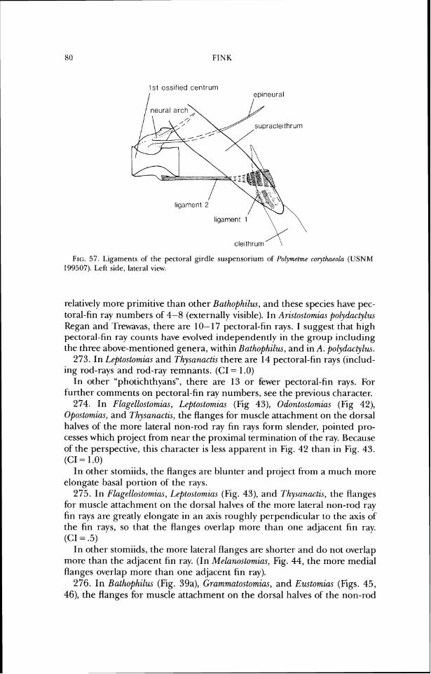
1st ossified centrum
I ep~neural I
FIG. 57 . Ligaments of the pectoral girdle suspensorium of I'olymetmr corythneolr~ (USNM 199507). Left side, lateral view.
relatively more primitive than other Bathophilus, and these species have pec- toral-fin ray numbers of 4-8 (externally visible). In Aristostomias polydactylus Kegan and 'IFewavas, there are 10- 17 pectoral-fin rays. I suggest that high pectoral-fin ray counts have evolved independently in the group including the three above-mentioned genera, within Bathophilus, and in A. polydactylus.
273. In Leptostomias and Thysanactis there are 14 pectoral-fin rays (includ- ing rod-rays and rod-ray remnants. (CI = 1 .O)
In other "photichthyans", there are 13 or fewer pectoral-fin rays. For further comments on pectoral-fin ray numbers, see the previous character.
274. In Flagellostomias, Leptostomias (Fig 43), Odontostomias (Fig 42), Opostomias, and Thysanactis, the flanges for muscle attachment on the dorsal halves of the more lateral non-rod ray fin rays form slender, pointed pro- cesses which project from near the proximal termination of the ray. Because of the perspective, this character is less apparent in Fig. 42 than in Fig. 43. (CI = 1 .O)
In other stomiids, the flanges are blunter and project from a much more elongate basal portion of the rays.
275. In Flagellostomias, Leptostomias (Fig. 43), and Thysanactis, the flanges for muscle attachment on the dorsal halves of the more lateral non-rod ray fin rays are greatly elongate in an axis roughly perpendicular to the axis of the fin rays, so that the flanges overlap more than one adjacent fin ray. (CI = .5)
In other stomiids, the more lateral flanges are shorter and do not overlap more than the adjacent fin ray. (In Melanostomias, Fig. 44, the more medial flanges overlap more than one adjacent fin ray).
276. In Bathophilus (Fig. 39a), Grammatostomias, and Eustomim (Figs. 45, 46), the flanges for muscle attachment on the dorsal halves of the non-rod
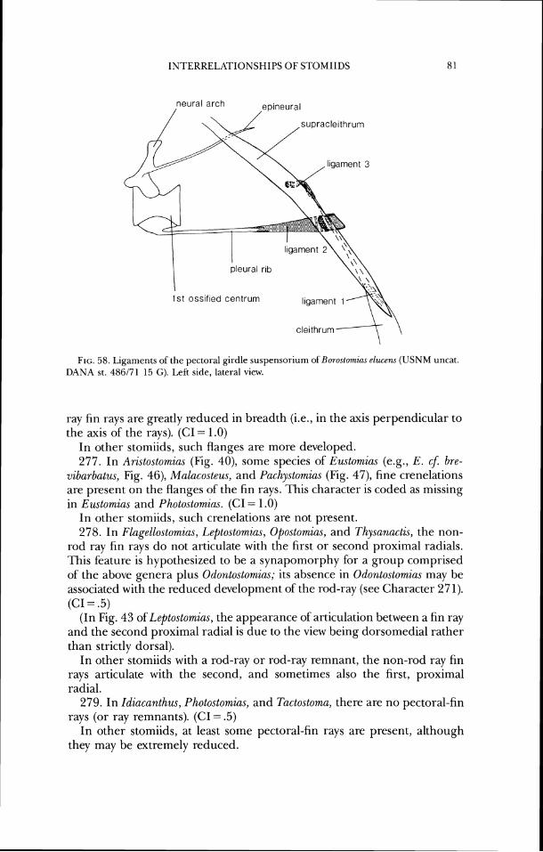
INTERRELXrIONSHIPS O F STOMIIDS
neural arch /
epineural
I 1 st ossified centrurn llgarnent 1
FIG. 58. Ligaments of the pectoral girdle suspensoriunl of Borostomicu elucen.~ (USNM uncat. DANA st. 486171 15 G). Left side, lateral view.
ray fin rays are greatly reduced in breadth (i.e., in the axis perpendicular to the axis of the rays). (CI = 1.0)
In other stomiids, such flanges are more developed. 277. In Aristostomias (Fig. 40), some species of E~stomias (e.g., E. cJ: bre-
vibarbatus, Fig. 46), Malacosteus, and Pachystomias (Fig. 47), fine crenelations are present on the flanges of the fin rays. This character is coded as missing in Eustomias and Photostomias. (CI = 1.0)
In other stomiids, such crenelations are not present. 278. In Flagellostomias, Leptostomias, Opostomius, and Thysanactis, the non-
rod ray fin rays do not articulate with the first or second proximal radials. This feature is hypothesized to be a synapomorphy for a group comprised of the above genera plus Odontostomias; its absence in Odonlostomias may be associated with the reduced development of the rod-ray (see Character 27 1). (CI = .5)
(In Fig. 43 of Leptostomias, the appearance of articulation between a fin ray and the second proximal radial is due to the view being dorsomedial rather than strictly dorsal).
In other stomiids with a rod-ray or rod-ray remnant, the non-rod ray fin rays articulate with the second, and sometimes also the first, proximal radial.
279. In Idiacanthus, Photostomias, and Tactostoma, there are no pectoral-fin rays (or ray remnants). (CI = .5)
In other stomiids, at least some pectoral-fin rays are present, although they may be extremely reduced.
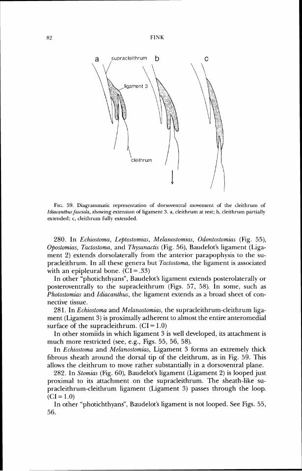
FIG. 59. Diagrammatic representation of dorsovetltral movement of the cleithrum of Idzaconthus fasciola, showing extension of ligament 3. a, cleithruin at rest; b, cleithrum partially extended; c, cleithrum fully extended.
280. In Echiostoma, Leptostomias, Melanostomias, Odontostomias (Fig. 55), Opostomias, Tactostoma, and Thysunactis (Fig. 56), Baudelot's ligament (Liga- ment 2) extends dorsolaterally from the anterior parapophysis to the su- pracleithrum. In all these genera but Tactostoma, the ligament is associated with an epipleural bone. (CI = .33)
In other "photichthyans", Baudelot's ligament extends posterolaterally or posteroventrally to the supracleithrum (Figs. 57, 58). In some, such as Photostomias and Idiacanthm, the ligament extends as a broad sheet of con- nective tissue.
28 1. In Echiostonza and Melanostomias, the supracleithrum-cleithrum liga- ment (Ligament 3) is proximally adherent to almost the entire anteromedial surface of the supracleithrum. (CI = 1.0)
In other stomiids in which ligament 3 is well developed, its attachment is much more restricted (see, e.g., Figs. 55, 56, 58).
In Echiostoma and Melanostomias, Ligament 3 forms an extremely thick fibrous sheath around the dorsal tip of the cleithrum, as in Fig. 59. This allows the cleithrum to move rather substantially in a dorsoventral plane.
282. In Stomias (Fig. 60), Baudelot's ligament (Ligament 2) is looped just proximal to its attachment on the supracleithrum. The sheath-like su- pracleithrum-cleithrum ligament (Ligament 3) passes through the loop. (CI = 1.0)
In other "photichthyans", Baudelot's ligament is not looped. See Figs. 55, 56.
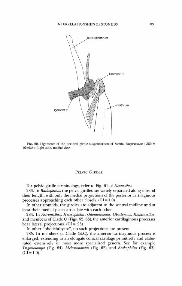
ligament
FIG. 60. Ligaments of the pectoral girdle suspensorium of Stomiac longzbarbatw (USNM 225035). Right side, medial view.
For pelvic girdle terminology, refer to Fig. 61 of Neonesthes. 283. In Bathophilus, the pelvic girdles are widely separated along most of
their length, with only the medial projections of the posterior cartilaginous processes approaching each other closely. (CI = 1 .O)
In other stomiids, the girdles are adjacent to the ventral midline and at least their medial plates articulate with each other.
284. In Astronesthes, Heterophotus, Odontostomias, Opostomias, Rhadinesthes, and members of Clade 0 (Figs. 62, 63), the anterior cartilaginous processes bear lateral projections. (CI = .25)
In other "photichthyans", no such projections are present 285. In members of Clade (B,C), the anterior cartilaginous process is
enlarged, extending as an elongate conical cartilage primitively and elabo- rated extensively in most more specialized genera. See for example T?-igonolampa (Fig. 64), Melanostomias (Fig. 62), and Bathophilus (Fig. 63). (CI = 1.0)
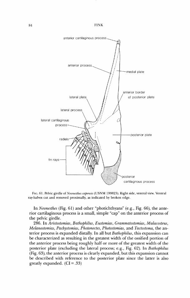
later
anterlor cart~lag~nous process
anterlor process
antenor border
of posterlor plate
cartllagrnous process
FIG. 61. Pelvic girdle of Neone.slhes cap~risi (USNM 199823). Right side, vctltral view. Venrl-al ray-halves cut and removed proximally, as indicated by broken cdgc.
In Neonesthes (Fig. 61) and other "photichthyans" (e.g., Fig. 66), the ante- rior cartilaginous process is a small, simple "cap" on the anterior process of' the pelvic girdle.
286. In Aristostomias, Bathophilus, Eustomias, Grammutostomias, Malacosteus, Melanostomias, Pachystomias, Photonecles, Photostomias, and Tactostornu, the an- terior process is expanded distally. In all but Bathophilus, this expansion can be characterized as resulting in the greatest width of the ossified portion of the anterior process being roughly half or more of the greatest width of the posterior plate (excluding the lateral process; e.g., Fig. 62). In Bathophilus (Fig. 63), the anterior process is clearly expanded, but this expansion cannot be described with reference to the posterior plate since the latter is also greatly expanded. (CI = .33)
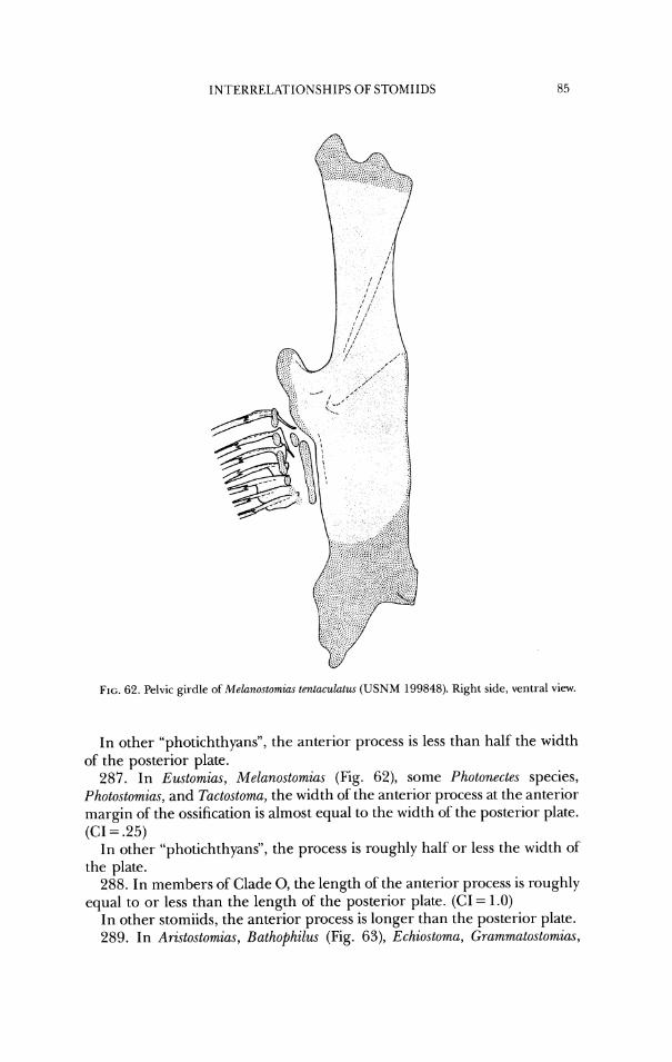
INTERRELATIONSHIPS OF STOMIIDS
FIG. 62. Pelvic girdle of Melanostomias tentaculatzcs ( U S N M 199848). Right side, ventral view.
In other "photichthyans", the anterior process is less than half the width of the posterior plate.
287. In Eustomias, Melanostomias (Fig. 62), some Photonectes species, Photostomias, and Tactostoma, the width of the anterior process at the anterior margin of the ossification is almost equal to the width of the posterior plate. (CI = .25)
In other "photichthyans", the process is roughly half or less the width of the plate.
288. In members of Clade 0, the length of the anterior process is roughly equal to or less than the length of the posterior plate. (CI = 1.0)
In other stomiids, the anterior process is longer than the posterior plate. 289. In Aristostomias, Bathophilus (Fig. 63), Echiostoma, Grammatostomim,
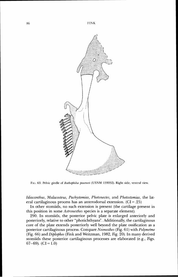
FIG. 63. Pelvic girdle of Balhophzlra pawneei (USNM 159052). Right sicle, ventral view.
Idiacanthus, Malacosteus, Pachystomias, Photonectes, and Photostomias, the lat- eral cartilaginous process has an anterodorsal extension. (CI = .25)
In other stomiids, no such extension is present (the cartilage present in this position in some Astronesthes species is a separate element).
290. In stomiids, the posterior pelvic plate is enlarged anteriorly and posteriorly, relative to other "photichthyans". Additionally, the cartilaginous core of the plate extends posteriorly well beyond the plate ossification as a posterior cartilaginous process. Compare Neonesthes (Fig. 61) with Polymetm~ (Fig. 66) and Diplophos (Fink and Weitzman, 1982, Fig. 20). In many derived stomiids these posterior cartilaginous processes are elaborated (e.g., Figs. 67-69). (Cl = 1 .O)
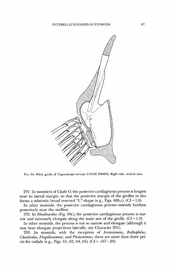
INTERRELATIONSHIPS OF STOMIIDS
FIG. 64. Pelvic girdle of Trigonolampa miricrps ( U S N M 206683). Right side, ventral view
29 1. In members of Clade 0, the posterior cartilaginous process is longest near its lateral margin, so that the posterior margin of the girdles in situ forms a relatively broad inverted "U"-shape (e.g., Figs. 68b,c). (CI= 1.0)
In other stomiids, the posterior cartilaginous process extends furthest posteriorly near the midline.
292. In Rhadinesthes (Fig. 69c), the posterior cartilaginous process is nar- row and extremely elongate along the main axis of the girdle. (CI = 1.0)
In other stomiids, the process is not so narrow and elongate (although it may bear elongate projections laterally; see Character 291).
293. In stomiids, with the exception of Aristostomias, Bathophilus, Chauliodus, Flagellostomias, and Photostomias, there are more than three pel- vic-fin radials (e.g., Figs. 6 1, 62, 64, 65). (CI =. 167-.20)
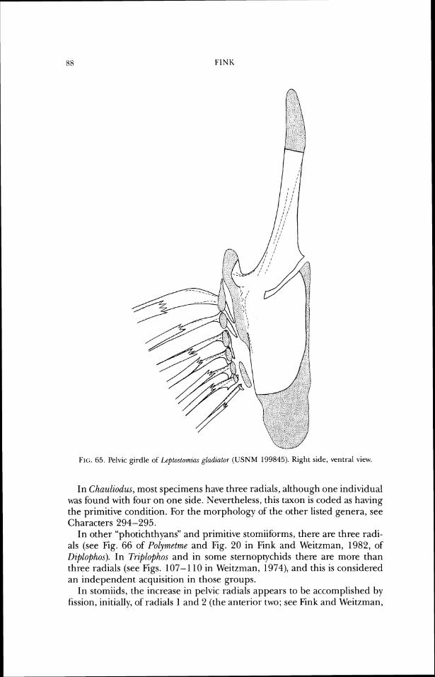
FIG. 65. Pelvic girdle of Leploslomzc~s gl(~dialor ( U S N M 199845). Kight side, ventral view.
In Chauliodus, most specimens have three radials, although one individual was found with four on one side. Nevertheless, this taxon is coded as having the primitive condition. For the morphology of the other listed genera, see Characters 294-295.
In other "photichthyans" and primitive stomiiforms, there are three radi- als (see Fig. 66 of Polymetme and Fig. 20 in Fink and Weitzman, 1982, of Diplophos). In Triplophos and in some sternoptychids there are more than three radials (see Figs. 107- 1 10 in Weitzman, 1974), and this is considered an independent acquisition in those groups.
In stomiids, the increase in pelvic radials appears to be accomplished by fission, initially, of radials 1 and 2 (the anterior two; see Fink and Weitzman,
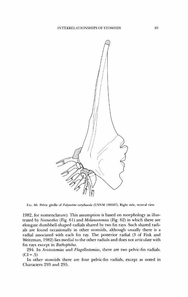
IN'I'ERRELRI'IONSHIPS 01.' STOMIIDS
FIG. 66. l'elvic girdle of Po'olymetme corythaeola ( U S N M 199507). Right side, ventral view.
1982, for nomenclature). This assumption is based on morphology as illus- trated by Neonesthes (Fig. 61) and Melanoslomias (Fig. 62) in which there are elongate dumbbell-shaped radials shared by two fin rays. Such shared radi- als are found occasionally in other stomiids, although usually there is a radial associated with each fin ray. The posterior radial (3 of Fink and Weitzman, 1982) lies medial to the other radials and does not articulate with fin rays except in Bathophilus.
294. In Aristostomias and Flagellostomias, there are two pelvic-fin radials. (CI = .5)
In other stomiids there are four pelvic-fin radials, except as noted in Characters 293 and 295.
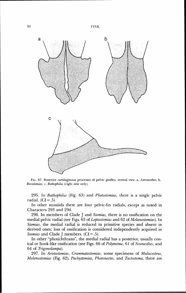
FIG. 67. Posterior cartilaginous processes of pelvic girdles, ventral view. a, A.\lronrsthes; b, Burustomias; c, Bathophil~~r (right side only).
295. In Bathophilzw. (Fig. 63) and Photostomias, there is a single pelvic radial. (CI = .5)
In other stomiids there are four pelvic-fin radials, except as noted in Characters 293 and 294.
296. In members of Clade J and Stomias, there is no ossification on the medial pelvic radial (see Figs. 65 of Leptostomias and 62 of Melanostomias). In Stomias, the medial radial is reduced in primitive species and absent in derived ones; loss of ossification is considered independently acquired in Stomias and Clade J members. (CI = .5)
In other "photichthyans", the medial radial has a posterior, usually con- ical or hook-like ossification (see Figs. 66 of Polymetme, 61 of Neonesthes, and 64 of Trzgonolampa).
297. In Aristostomias, Grammatostomias, some specimens of Malacostezw., Melanostomias (Fig. 62), Paclzystomias, Photonectes, and Tactostoma, there are
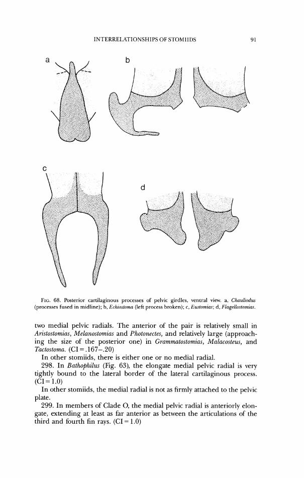
INTERRE1,ATIONSHIPS OF STOMIIDS
FIG. 68. Posterior cartilaginous processes of pelvic girdles, ventral view. a, Chauliodu (processes fused in midline); b, Echiostoma (left process broken); c, E~~tomias; d , Flagellostomias.
two medial pelvic radials. The anterior of the pair is relatively small in Aristostomias, Melanostomias and Photonectes, and relatively large (approach- ing the size of the posterior one) in Grammatostomias, Malacosteus, and Tactostoma. (CI = .167-.20)
In other stomiids, there is either one or no medial radial. 298. In Bathophilus (Fig. 63), the elongate medial pelvic radial is very
tightly bound to the lateral border of the lateral cartilaginous process. (CI = 1 .O)
In other stomiids, the medial radial is not as firmly attached to the pelvic plate.
299. In members of Clade 0, the medial pelvic radial is anteriorly elon- gate, extending at least as far anterior as between the articulations of the third and fourth fin rays. (CI = 1.0)
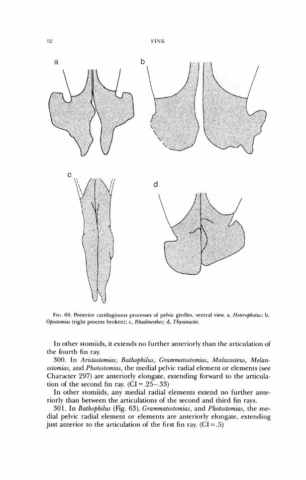
FIG. 69. Posterior cartilagirious processes of pelvic girdles, ventral view. a, Hel~rophotzcs; b, O~uslumius (right process broken); c , Rhudineslhes; d, Tllysanactis.
In other stomiids, it extends no further anteriorly than the articulation of the fourth fin ray.
300. In Aristostomias, Bathophilus, Grammatostomias, Malacosteus, Melan- ostomias, and Photostomias, the medial pelvic radial element or elements (see Character 297) are anteriorly elongate, extending forward to the articula- tion of the second fin ray. (CI = .25-.33)
In other stomiids, any medial radial elements extend no further ante- riorly than between the articulations of the second and third fin rays.
301. In Bathophilus (Fig. 63), Grammatostomias, and Photostomias, the me- dial pelvic radial element or elements are anteriorly elongate, extending just anterior to the articulation of the first fin ray. (CI = .5)
In other stomiids, any medial radial elements extend no further ante- riorly than the articulation of the second fin ray.
302. In Photostomias, the medial pelvic radial is abbreviated posteriorly, extending no further back than the articulation of the fourth fin ray. The lateral process of the pelvic girdle is also abbreviated posteriorly, and the fin rays are bunched anteriorly, articulating in the region of the short lateral process (the fin rays are also reduced in number, see Character 304). (CI = 1 .O)
In other stomiids, the medial radial extends posterior to the articulation of the last fin ray, the lateral process is more elongate, and the rays are not bunched together.
303. In Bathophilus and Photostomias, there are no pelvic-fin distal radials. (CI = .5)
In other stomiids, there is at least one distal pelvic radial. 304. In Aristostomias, some Bathophilus species, Idiacanthus, Malacosteus,
Photostomias, and Stomias, the number of pelvic-fin rays is six or fewer (four or five in Stornias, four to six in some Bathophilus, six in all others). (CI = .25-.33)
In other stomiids, there are at least seven fin rays. 305. In some Bathophilus species, Echiostoma, some Eustomias species, some
Opostornias species (micrzpnus, Gibbs, pers. comm.), and ?;zctostoma, the number of pelvic-fin rays is consistently more than seven (8-26 in Bath,o$hilus, eight in Echiostoma, 7-9 in Opostomias, 8-10 in Tactostoma). (CI = .5)
In other stomiids, with the exceptions noted in Character 304, there are seven rays.
306. In Stomias, there are 4 or 5 pelvic-fin rays. (CI = 1.0) In other stomiids and "photichthyans" there are 6 or more pelvic-fin rays. 307. In Photostomias, the halves of each pelvic-fin ray are fused together
along their anterior margins for most of their length, both halves of the anterior ray have an expanded flange with a wavy posterior edge for about half its length, and the anterior two rays are tightly bound together for most of their lengths. (CI = 1.0)
No such modifications are present in other stomiids. 308. In Bathoph,ilus, there are no pelvic-fin interradial membranes.
((:I = 1 .O) In other stomiids, such membranes are present.
309. In Aristostornias, Ma,lacosteus, and Pachystomicls, there is a large ac- cessory light organ (accessory orbital photophore I [AOPI]) anteroventral or ventral to the eye, which extends medially well into the orbital cavity (Fig. 70). (CI = .5- 1 .O)
In other stomiids, there are usually only two light organs associated with the eye, the "suborbital organ" and the "postorbital organ". See Discussion
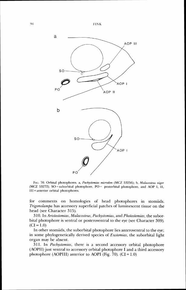
FIG. 70. Orbital photophores. a, Pachylystomzas wnlirrodon (MCZ 53256); b, Malucos te~~~ rrzg~r (MCZ 53272). SO=suborbital photophorc, PO= postorbital pho~ophore, and AOP I , 11, 11 1 = aritcrior orbital photophores.
for comments on homologies of head photophores in stomiids. Trigonolampa has accessory superficial patches of luminescent tissue on the head (see Character 3 15).
3 10. In Arzstostomias, Malacosteus, Pachyslomzas, and I'hotostomias, the subor- bital photophore is ventral or posteroventral to the eye (see Character 309). (CI = 1 .O)
In other stomiids, the suborbital photophore lies anteroventral to the eye; in some phylogenetically derived species of Eustomias, the suborbital light organ may be absent.
31 1. In Pachystomzas, there is a second accessory orbital photophore (AOPII) just ventral to accessory orbital photophore I and a third accessory photophore (AOPIII) anterior to AOPI (Fig. 70). (CI = 1.0)
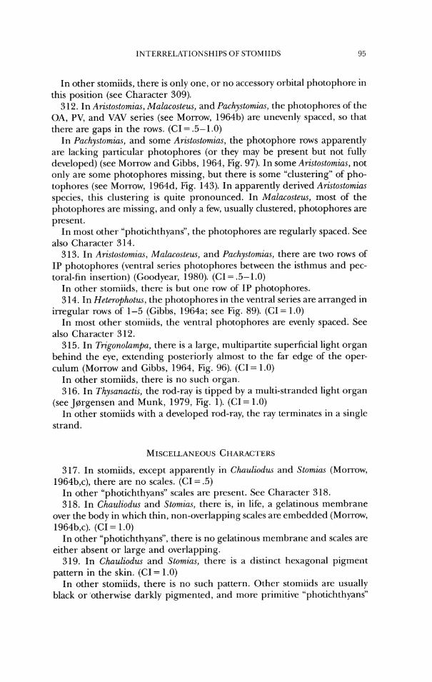
IN I'EKKELATIONSHIPS OF STOMIIDS 1) 5
In other stomiids, there is only one, or no accessory orbital photophore in this position (see Character 309).
3 12. In Aristostomias, Malacostew, and Pachystomias, the photophores of the OA, PV, and VAV series (see Morrow, 1964b) are unevenly spaced, so that there are gaps in the rows. (CI = .5- 1 .O)
In Pachystomias, and some Aristostomias, the photophore rows apparently are lacking particular photophores (or they may be present but not fully developed) (see Morrow and Gibbs, 1964, Fig. 97). In some Aristostomias, not only are some photophores missing, but there is some "clustering" of pho- tophores (see Morrow, 1964d, Fig. 143). In apparently derived ArGtostomias species, this clustering is quite pronounced. In Malacostew, most of the photophores are missing, and only a few, usually clustered, photophores are present.
In most other "photichthyans", the photophores are regularly spaced. See also Character 314.
3 13. In Aristostomias, Malacostem, and Pachystomias, there are two rows of IP photophores (ventral series photophores between the isthmus and pec- toral-fin insertion) (Goodyear, 1980). (CI = .5- 1 .O)
In other stomiids, there is but one row of IP photophores. 3 14. In Heterophotus, the photophores in the ventral series are arranged in
irregular rows of 1-5 (Gibbs, 1964a; see Fig. 89). (C1= 1.0) In most other stomiids, the ventral photophores are evenly spaced. See
also Character 3 12. 315. In Trigonolampa, there is a large, multipartite superficial light organ
behind the eye, extending posteriorly almost to the far edge of the oper- culum (Morrow and Gibbs, 1964, Fig. 96). (CI = 1.0)
In other stomiids, there is no such organ. 3 16. In Thysanactis, the rod-ray is tipped by a multi-stranded light organ
(see JGrgensen and Munk, 1979, Fig. 1). (CI = 1.0) In other stomiids with a developed rod-ray, the ray terminates in a single
strand.
317. In stomiids, except apparently in Chauliodus and Stomias (Morrow, 1964b,c), there are no scales. (GI= .5)
In other "photichthyans" scales are present. See Character 318. 318. In Chauliodus and Stomias, there is, in life, a gelatinous membrane
over the body in which thin, non-overlapping scales are embedded (Morrow, 1964b,c). (GI = 1 .O)
In other "photichthyans", there is no gelatinous membrane and scales are either absent or large and overlapping.
319. In Chauliodus and Stomias, there is a distinct hexagonal pigment pattern in the skin. (CI = 1.0)
In other stomiids, there is no such pattern. Other stomiids are usually black or otherwise darkly pigmented, and more primitive "photichthyans"
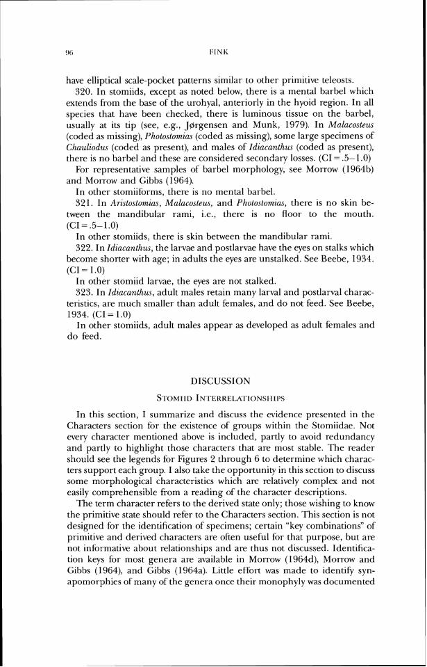
have elliptical scale-pocket patterns similar to other primitive teleosts. 320. In stomiids, except as noted below, there is a mental barbel which
extends from the base of the urohyal, anteriorly in the hyoid region. In all species that have been checked, there is luminous tissue on the barbel, usually at its tip (see, e.g., Jgrgensen and Munk, 1979). In Malacosteus (coded as missing), Photostomic~s (coded as missing), some large specimens of Chaulzodus (coded as present), and males of Idiacanthus (coded as present), there is no barbel and these are considered secondary losses. (CI = .5- 1.0)
For representative samples of barbel morphology, see Morrow (1964b) and Morrow and Gibbs (1964).
In other stomiiforms, there is no mental barbel. 32 1. In Aristostomias, Malacosteus, and Photostomias, there is no skin be-
tween the mandibular rami, i.e., there is no floor to the mouth. (C1= .5- 1 .O)
In other stomiids, there is skin between the mandibular rarni. 322. In Idzacanthus, the larvae and postlarvae have the eyes on stalks which
become shorter with age; in adults the eyes are unstalked. See Beebe, 1934. (CI = 1.0)
In other stomiid larvae, the eyes are not stalked. 323. In Idincanlhw, adult males retain many larval and postlarval charac-
teristics, are much smaller than adult females, and do not feed. See Ueebe, 1934. (CI = 1 .O)
In other stomiids, adult males appear as developed as adult females and do feed.
DISCUSSION
In this section, I summarize and discuss the evidence presented in the Characters section for the existence of groups within the Stomiidae. Not evely character mentioned above is included, partly to avoid redundancy and partly to highlight those characters that are most stable. The reader should see the legends for Figures 2 through 6 to determine which charac- ters support each group. I also take the opportunity in this section to discuss some morphological characteristics which are relatively complex and not easily comprehensible from a reading of the character descriptions.
The term character refers to the derived state only; those wishing to know the primitive state should refer to the Characters section. This section is not designed for the identification of specimens; certain "key combinations" of primitive and derived characters are often useful for that purpose, but are not informative about relationships and are thus not discussed. Identifica- tion keys for most genera are available in Morrow (1964d), Morrow and Gibbs (1964), and Cibbs (1964a). Little effort was made to identify syn- apomorphies of many of the genera once their monophyly was documented
to my satisfiction; further investigations will be needed to describe more fully the features of most genera, especially since many have not been shown previously to be monophyletic.
As outlined in the Introduction, the taxa I recognize as comprising the Stomiidae historically have been relegated to several families, depending on various authors' preferences; these are Astronesthidae, Chauliodontidae, Idiacanthidae, Malacosteidae, Melanostomiidae, and Stomiidae. Inasmuch as the hypothesis of relationships generated by this study demonstrates that some of these groups are unnatural or that their continued recognition at family rank would require a radical reclassification of the entire group, the most conservative taxonomic approach appears to be to expand the Stom- iidae of Regan and Trewavas (1930) to include all of the stomiiforms with barbels.
'fie Stomiidae is documented as monophyletic based on a number of characters, including presence of a single infraorbital bone (Character 33), lack of gill rakers in adults (Character 103), presence of two main bodies of the geniohyoideus muscle (Character 168), a portion of the adductor man- dibulae inserting on the postorbital photophore (Character 187), lack of hypural 6 (Character 228), and presence of'a mental barbel associated with the hyoid apparatus (Character 320). This family includes some of the most morphologically specialized of teleosts, such as Aristostomias, Emtomias, and Malacosteus. Also of interest is the persistence of several morphologically and phylogenetically primitive representatives, such as Astronesthes and Khadinesthes, which present a rich picture of the origins of such novelties as pectoral-fin specializations and changes in placement of the vertical fins. Some of these evolutionary changes are mentioned below in the context of various subgroups, and others are discussed in the more general summary which follows the subgroup descriptions.
Neonesthes is the sister group to all other stomiids. It is diagnosable by presence of a row of toothplates along the entire medial length of the hyoid bar (Character 73).
Regan and Trewavas (1929) originally described Neonesthes, placing it in the Astronesthidae. Gibbs (1964a) figured and described individuals of N. capensis, and Weitzman (1967b) described much of its osteology.
All other stomiids are united by such traits as presence of ventral rector gill-arch muscles attaching to the fifth ceratobranchial (Character 179), morphology of the first distal radial of the pectoral fin, including lack of complete ossification (Character 268), and enlargement of the anterior car- tilaginous process of the pelvic girdle (Character 285). All of these charac- teristics are further elaborated within the group.
At this node of the cladogram there are two alternate hypotheses which are equally supported by the data at hand: Astronesthes as the sister group to either Borostomias, or to all remaining (i.e., non-Neonesthes) stomiids. For this reason, in the summary cladogram the unresolved node is noted as (B,C), representing the alternative hypotheses represented in Fig. 2.
Astronesthes is one of the most perplexing of the stomiid clades. The
morphology includes traits which appear independently in more advanced groups, and there are character transformations within the genus which mimic similar transformations in stomiids as a whole. Astronesthes is diag- nosable by the form of the blood-vessel passage in the hypohyal element (Character 88), and the way in which the prezygapophyses of the anterior 10-20 neural arches are enlarged, closely approximated serially, and ex- tend over the neural tube as a bony hood (Character 199).
Other traits of Astronesthes include "serraen-like teeth on the maxilla, in combination with the dorsal fin near the dorsal midpoint of the body. The former is also found in the members of Clade E, and the latter is primitive for stomiids.
Within Astronesthes, there are morphological changes that are similar to those in other stomiids. For example, the parietal bones are absent in some more derived species (such as A. niger, see Weitzman, 1967b, Fig. 3), as they are in Arzstostomias, Echiostoma, and some Stomias species (see Character 28). Body elongation, typical of many stomiids, occurs in some species, such as A. psychrolutes ((Gibbs and Weitzman). The posterior pelvic plate ranges from a relatively simple projection to a large, single fused plate-like cartilage (Weitzman, 1967b, Fig. 18). The general phenetic "gestalt" of Astronesthes, particularly of the head, is suggestive of a phylogenetically more derived taxon than detailed comparisons show. In the context of all the characters, these similarities are shown to be convergently acquired, which m i ~ h t make
- . A
further work on the genus easier and more interesting. The characters found have not allowed a resolution of the relationships of
Astronesthes, although its general placement near the base of the cladogram is amply demonstrable. But whether Astronesthes is the sister group of all stomiids but Neone.sthe.s (Fig. 2a), or whether it is most closely related to Borostomias (Fig. 2b) is not clear. As is often so in primitive members of a large group, there are few specializations which are informative about their interrelationships. The evidence for Astronesthes and Borostomias being sister taxa is presence of a parasphenethmoid bone (Character 15), and su- praneural placement (reversal of Character 193 to the primitive condition), a reversal which occurs elsewhere in the tree. The evidence that Astronesthes is the sister group ofBorostomias plus the other stomiids is based on presence in the latter taxa of the rectus communis muscles extending anteriorly as far as the second hypobranchial (Character 180; this trait is not present in Chaulzodus). Based on the unique parasphenethmoid, my inclination is to consider Astronesthes and Borostornias as sister taxa, but the alternate hypoth- esis is equally supported in the context of the entire stomiid lineage. Thus these taxa are left in an unresolved trichotomy in the summary cladogram.
Borostomias itself is diagnosable by virtue of such traits as extent of the palatine bone posteriorly beyond the anterior border of the quadrate (Char- acter 50) and lack of articulation between the second epibranchial and the third pharyngobranchial (Character 15 1).
The remaining stomiids (Clade D) are considered monophyletic based on several characters, including position of a blood vessel in the ventral hypo-
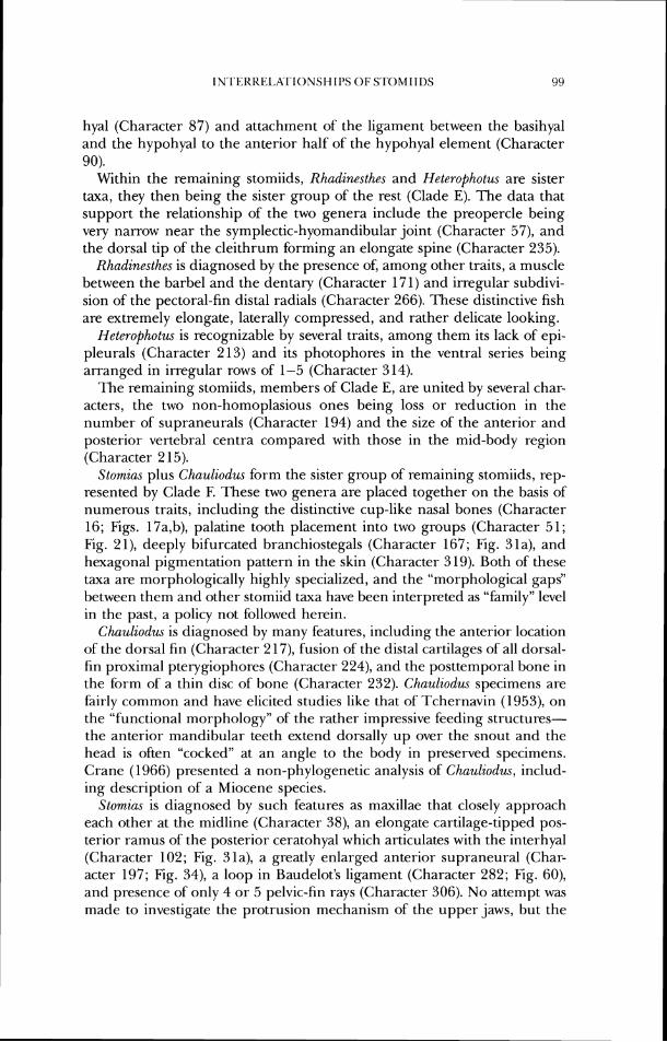
IN~I ' I<KKEL~Kl~IONSI-I IPS 01; S.I'OMI IDS 9:)
hyal (Character 87) and attachment of the ligament between the basihyal and the hypohyal to the anterior half of the hypohyal element (Character 90).
Within the remaining stomiids, Rhadinesthes and Heterophotus are sister taxa, they then being the sister group of the rest (Clade E). The data that support the relationship of the two genera include the preopercle being very narrow near the symplectic-hyomandibular joint (Character 57), and the dorsal tip of the cleithrum forming an elongate spine (Character 235).
Rhadinesthes is diagnosed by the presence of, among other traits, a muscle between the barbel and the dentary (Character 171) and irregular subdivi- sion of the pectoral-fin distal radials (Character 266). These distinctive fish are extremely elongate, laterally compressed, and rather delicate looking.
Heterophotus is recognizable by several traits, among them its lack of epi- pleurals (Character 213) and its photophores in the ventral series being arranged in irregular rows of 1-5 (Character 3 14).
The remaining stomiids, members of Clade E, are united by several char- acters, the two non-homoplasious ones being loss or reduction in the number of supraneurals (Character 194) and the size of the anterior and posterior vertebral centra compared with those in the mid-body region (Character 2 15).
Stomias plus Chauliodus form the sister group of remaining stomiids, rep- resented by Clade F. These two genera are placed together on the basis of numerous traits, including the distinctive cup-like nasal bones (Character 16; Figs. 17a,b), palatine tooth placement into two groups (Character 51; Fig. 21), deeply bifurcated branchiostegals (Character 167; Fig. 31a), and hexagonal pigmentation pattern in the skin (Character 319). Both of these taxa are morphologically highly specialized, and the "morphological gaps" between them and other stomiid taxa have been interpreted as "family" level in the past, a policy not followed herein.
Chauliodus is diagnosed by many features, including the anterior location of the dorsal fin (Character 2 17), fusion of the distal cartilages of all dorsal- fin proximal pterygiophores (Character 224), and the posttemporal bone in the form of a thin disc of bone (Character 232). Chauliodus specimens are fairly common and have elicited studies like that of Tchernavin (1953), on the "functional morphology" of the rather impressive feeding structures- the anterior mandibular teeth extend dorsally up over the snout and the head is often "cocked" at an angle to the body in preserved specimens. Crane (1966) presented a non-phylogenetic analysis of Chauliodus, includ- ing description of a Miocene species.
Stomias is diagnosed by such features as maxillae that closely approach each other at the midline (Character 38), an elongate cartilage-tipped pos- terior ramus of the posterior ceratohyal which articulates with the interhyal (Character 102; Fig. 31a), a greatly enlarged anterior supraneural (Char- acter 197; Fig. 34), a loop in Baudelot's ligament (Character 282; Fig. 60), and presence of only 4 or 5 pelvic-fin rays (Character 306). No attempt was made to investigate the protrusion mechanism of the upper jaws, but the
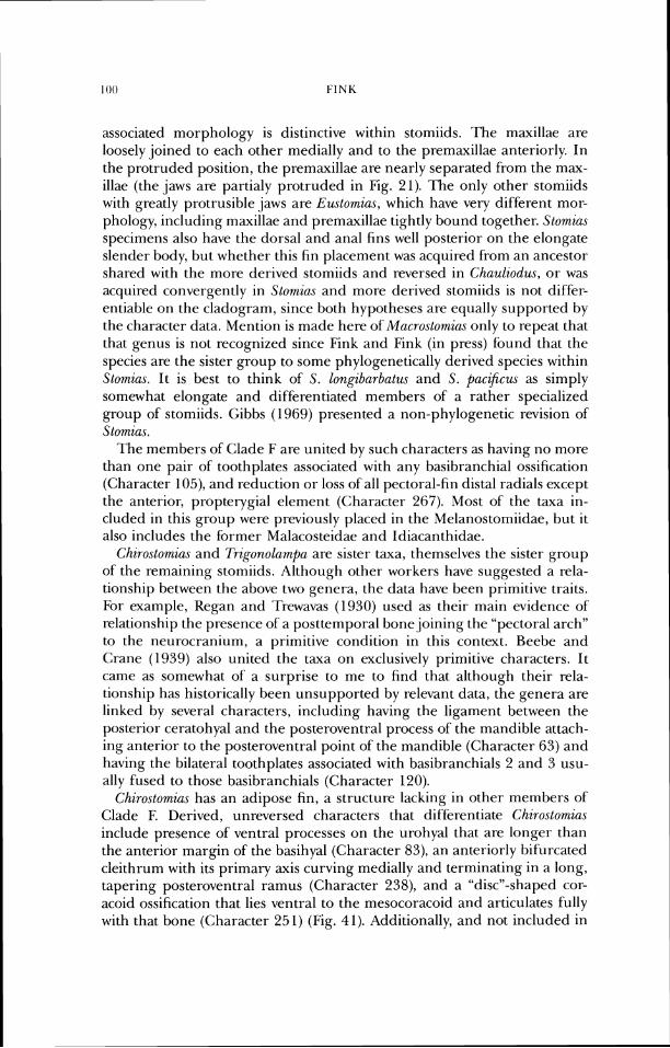
associated morphology is distinctive within stomiids. The maxillae are loosely joined to each other medially and to the premaxillae anteriorly. In the protruded position, the premaxillae are nearly separated from the max- illae (the jaws are partialy protruded in Fig. 21). The only other stomiids with greatly protrusible jaws are Eustomias, which have very different mor- phology, including maxillae and premaxillae tightly bound together. Stornias specinlens also have the dorsal and anal fins well posterior on the elongate slender body, but whether this fin placement was acquired fiom an ancestor shared with the more derived stomiids and reversed in Chauliodus, or was acquired convergently in Slomias and more derived stomiids is not diffcr- entiable on the cladogram, since both hypotheses are equally supported by the character data. Mention is made here of Mncroslomia.~ only to repeat that that genus is not recognized since Fink and Fink (in press) found that the species are the sister group to some phylogenetically derived species within Storkus. It is best to think of S. lonpburbatus and S. paczJlcus as simply somewhat elongate and differentiated members of a rather specialized group of' stomiids. <;il-,bs (1969) presented a non-phylogenetic revision of Stomius.
The members of <;lade F are united by such characters as having no more than one pair of toothplates associated with any basibranchial ossification (Character 105), and reduction or loss of all pectoral-fin distal radials except the anterior, propterygial element (Character 267). Most of the taxa in- cluded in this group were previously placed in the Melanostorniidae, but it also includes the brmer Malacosteidae and Idiacanthidae.
Chiroslomic~s and Trig~r~olamf~a are sister taxa, themselves the sister group of the remaining storniids. Although other workers have suggested a rela- tionship between the above two genera, the data have been primitive traits. For example, Kegan and ?Trewavas (1930) used as their main evidence of' relationship the presence of a posttemporal bone joining the "pectoral arch" to the neurocranium, a primitive condition in this context. Beebe and Crane (1939) also united the taxa on exclusively primitive characters. It came as somewhat of a surprise to me to find that although their rela- tionship has historically been unsupported by relevant data, the genera are linked by several characters, including having the ligament between the posterior ceratohyal and the posteroventral process of the mandible attach- ing anterior to the posteroventral point of the mandible (Character 63) and having the bilateral toothplates associated with basibranchials 2 and 3 usu- ally fused to those basibranchials (Character 120).
Chim.stomic~s has an adipose fin, a structure lacking in other members of Clade F. Derived, unreversed characters that differentiate Chiroslomia.~ include presence of' ventral processes on the urohyal that are longer than the anterior margin of the basihyal (Character 83), an anteriorly bifurcated cleithrum with its primary axis curving medially and terminating in a long, tapering posteroventral ramus (Character 238), and a "discn-shaped cor- acoid ossification that lies ventral to the mesocoracoid and articulates fully with that bone (Character 251) (Fig. 41). Additionally, and not included in
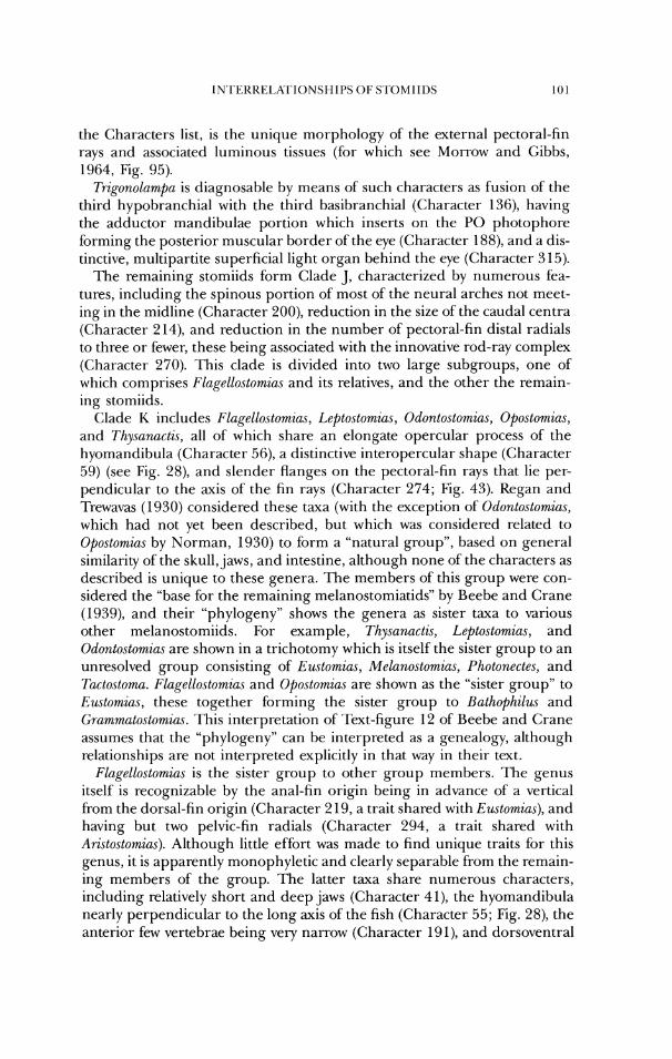
the Characters list, is the unique morphology of the external pectoral-fin rays and associated luminous tissues (for which see Morrow and Gibbs, 1964, Fig. 95).
7i-igonolarnpu is diagnosable by means of such characters as fusion of' the third hypobranchial with the third basibranchial (Character 136), having the adductor mandibulae portion which inserts on the PO photophore forrning the posterior muscular border of the eye (Character 188), and a dis- tinctive, rnult.ipartite superficial light organ behind the eye (Character 315).
The remaining stomiids form Clade J , characterized by numerous fea- turcs, including the spirlous portion of most of the neural arches not meet- ing in the midline (Character 200), reduction in the size of the caudal centra (Character 2 14), and reduction in the number of pectoral-fin distal radials to three or fewer, these being associated with the innovative rod-ray complex (Character 270). This clade is divided into two large subgroups, one of which comprises FLage1lo.stomiu.s and its relatives, anti the other the remain- ing stomiids.
Clade K includes Flagello.slomias, Leptostomiu.~, Odonto.slomia.s, Opostomias, and Thysanactis, all of which share an elongate opercular process of the hyomandibula (Character 56), a distinctive interopercular shape (Character 59) (see Fig. 28), arld slender flanges on the pectoral-tin rays that lie per- pendicular to the axis of the fin rays (Character 274; Fig. 43). Kegan and l'rewavas (1930) considered these taxa (with the exception of Odontostomias, which had not yet been described, but which was considered related to Ofiostomias hy Norman, 1930) to form a "natural group", based on general similarity of'the skull, jaws, ant1 intestine, although none of the characters as described is unique to these genera. 'Ille members ofthis group were con- sidered the "base fi)r the remaining melanostomiatids" by Beebe and Crane (1939), and their "phylogeny" shows the genera as sister taxa to various other nlelanostomiids. For example, Thysunucti.~, Lepto.stomia.s, and Odonlostomias are shown in a trichotomy which is itself the sister group to an unresolved group consisting of Eustomius, Melanostomias, Pholonectes, and ?i~cto.stomu. E1ugello.slomiu.s and Opostomias are shown as the "sister group" to Eustomias, these together forming the sister group to Ruthophilus and Grammutostomius. This interpretation of -1ext-figure 12 of' Beebe and Crane assumes that the "phylogeny" can be interpreted as a genealogy, although relationships arc not interpreted explicitly in that way in their text.
FlugeLLostomiu,~ is the sister group to other group members. The genus itscll' is recognizable by the anal-fin origin being in advance of a vertical frorrl the dorsal-fin origin (Character 2 19, a trait shared with Eustomias), and having but two pelvic-tin radials (Character 294, a trait shared with Aristo.rtwmia.r). Although little effort was made to find unique traits for this genus, it is apparently monophyletic and clearly separable from the remain- ing members of the group. The latter taxa share numerous characters, including relatively short and deep jaws (Character 41), the hyomandibula nearly perpendicular to the long axis of the fish (Character 55; Fig. 28), the anterior few vertebrae being very narrow (Character 191), and dorsoventral
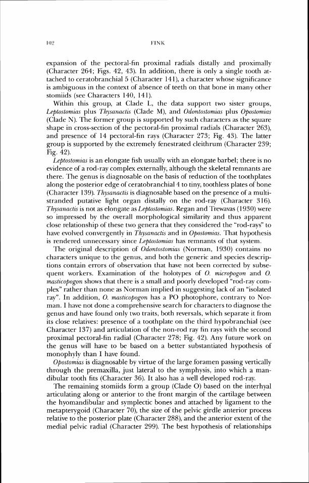
expansion of the pectoral-fin proximal radials distally and proximally (Character 264; Figs. 42, 43). In addition, there is only a single tooth at- tached to ceratobranchial 5 (Character 141), a character whose significance is ambiguous in the context of absence of teeth on that bone in many other stomiids (see Characters 140, 14 1).
Within this group, at Clade L, the data support two sister groups, Leptostomias plus Thysanactis (Clade M), and 0dontostomia.c plus Opostomias (Clade N). The former group is supported by such characters as the square shape in cross-section of the pectoral-fin proximal radials (Character 263), and presence of 14 pectoral-fin rays (Character 273; Fig. 43). 'I'he latter group is supported by the extremely fenestrated cleithrum (Character 239; Fig. 42).
Lepto.stomia.s is an elongate fish usually with an elongate barbel; there is no evidence of a rod-ray complex externally, although the skeletal remnants are there. The genus is diagnosable on the basis of reduction ofthe toothplates along the posterior edge of ceratobranchial4 to tiny, toothless plates of bone (Character 139). Thysanactzs is diagnosable based on the presence of a multi- stranded putative light organ distally on the rod-ray (Character 316). Thysanactis is not as elongate as Le$)tostomius. Regan and 'I'rewavas ( I 930) were so impressed by the overall morphological similarity and thus apparent close relationship of these two genera that they considered the "rod-rays" to have evolved convergently in Thysnnacti.~ and in Opostomius. That hypothesis is rendered unnecessary since I,epto.stomiu.s has remnants of' that system.
The original description of 0donto.stomias (Norman, 1930) contains no characters unique to the genus, and both the generic and species descrip- tions contain errors of observation that have not been corrected by subse- quent workers. Examination of the holotypes of 0. micropogon and 0. masticopogon shows that there is a small and poorly developed "rod-ray com- plex" rather than none as Norman implied in suggesting lack of an "isolated ray". In addition, 0. masticopopn has a PO photophore, contrary to Nor- man. I have not done a comprehensive search for characters to diagnose the genus and have found only two traits, both reversals, which separate it froni its close relatives: presence of a toothplate on the third hypobranchial (see Character 137) and articulation of the non-rod ray fin rays with the second proximal pectoral-fin radial (Character 278; Fig. 42). Any future work on the genus will have to be based on a better substantiated hypothesis of monophyly than I have found.
Opostomias is diagnosable by virtue of the large foramen passing vertically through the premaxilla, just lateral to the symphysis, into which a man- dibular tooth fits (Character 36). It also has a well developed rod-ray.
The remaining stomiids form a group (Clade 0) based on the interhyal articulating along or anterior to the front margin of the cartilage between the hyomandibular and symplectic bones and attached by ligament to the metapterygoid (Character 70), the size of the pelvic girdle anterior process relative to the posterior plate (Character 288), and the anterior extent of the medial pelvic radial (Character 299). The best hypothesis of relationships
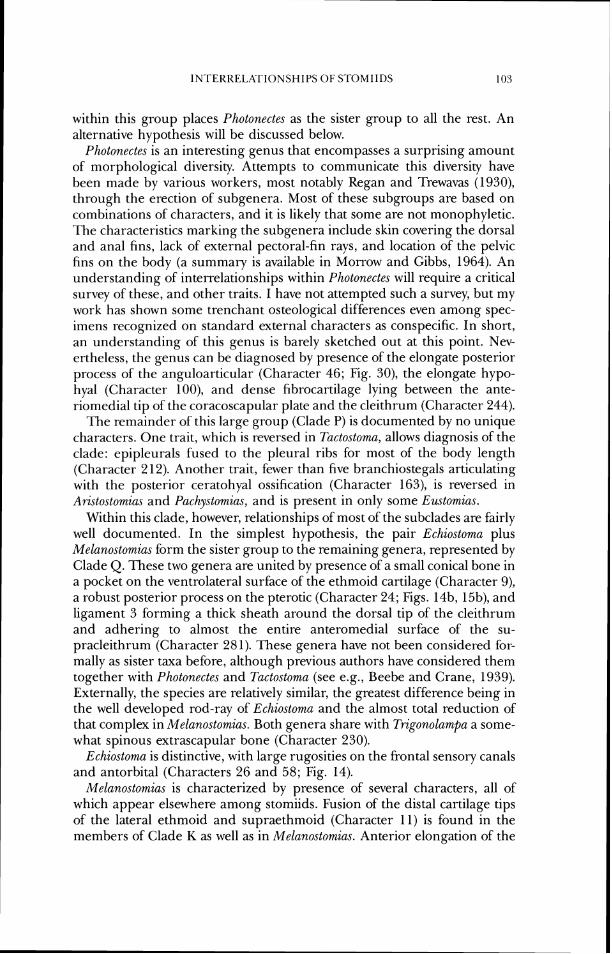
within this group places Photonectes as the sister group to all the rest. An alternative hypothesis will be discussed below.
Photonectes is an interesting genus that encompasses a surprising amount of morphological diversity. Attempts to communicate this diversity have been made by various workers, most notably Regan and Trewavas (1930), through the erection of subgenera. Most of these subgroups are based on combinations of characters, and it is likely that some are not monophyletic. The characteristics marking the subgenera include skin covering the dorsal and anal fins, lack of external pectoral-fin rays, and location of the pelvic fins on the body (a summary is available in Morrow and Gibbs, 1964). An understanding of interrelationships within Photonectes will require a critical survey of these, and other traits. I have not attempted such a survey, but my work has shown some trenchant osteological differences even among spec- imens recognized on standard external characters as conspecific. In short, an understanding of this genus is barely sketched out at this point. Nev- ertheless, the genus can be diagnosed by presence ofthe elongate posterior process of the anguloarticular (Character 46; Fig. 30), the elongate hypo- hyal (Character loo), and dense fibrocartilage lying between the ante- riomedial tip of the coracoscapular plate and the cleithrum (Character 244).
The remainder of this large group (Clade P) is documented by no unique characters. One trait, which is reversed in Tactostoma, allows diagnosis of the clade: epipleurals fused to the pleural ribs for most of the body length (Character 2 12). Another trait, fewer than five branchiostegals articulating with the posterior ceratohyal ossification (Character 163), is reversed in Aristostomias and Pachystomias, and is present in only some Eustornias.
Within this clade, however, relationships of most of the subclades are fairly well documented. In the simplest hypothesis, the pair Echiostoma plus Melanostomias form the sister group to the remaining genera, represented by Clade Q. These two genera are united by presence of a small conical bone in a pocket on the ventrolateral surface of the ethmoid cartilage (Character 9), a robust posterior process on the pterotic (Character 24; Figs. 14b, 15b), and ligament 3 forming a thick sheath around the dorsal tip of the cleithrum and adhering to almost the entire anteromedial surface of the su- pracleithrum (Character 28 1). These genera have not been considered for- mally as sister taxa before, although previous authors have considered them together with Photonectes and Tactostomn (see e.g., Beebe and Crane, 1939). Externally, the species are relatively similar, the greatest difference being in the well developed rod-ray of Echiostoma and the almost total reduction of that complex in Melanostomias. Both genera share with Trzgonolampa a some- what spinous extrascapular bone (Character 230).
Echiostoma is distinctive, with large rugosities on the frontal sensory canals and antorbital (Characters 26 and 58; Fig. 14).
Melanostomias is characterized by presence of several characters, all of which appear elsewhere among stomiids. Fusion of the distal cartilage tips of the lateral ethmoid and supraethmoid (Character 11) is found in the members of Clade K as well as in Melanostomias. Anterior elongation of the
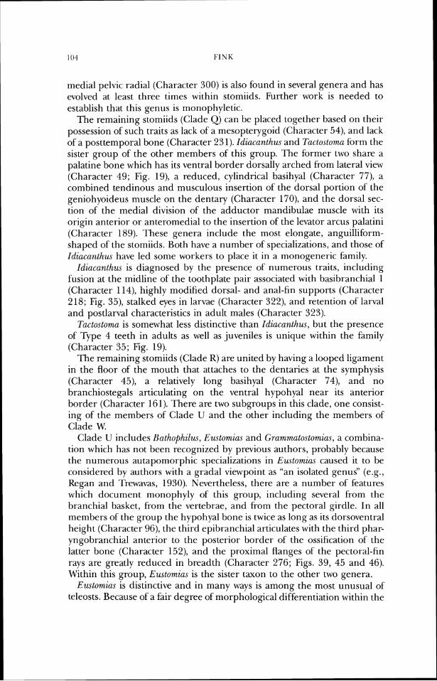
medial pelvic radial (Character 300) is also found in several genera and has evolved at least three times within stomiids. Further work is needed to establish that this genus is monophyletic.
The remaining stomiids (Clade (2) can be placed together based on their possession of such traits as lack of a mesopterygoid (Character 54), and lack of a posttemporal bone (Character 23 1). 1diacanthu.s and Tactostoma form the sister group of the other members of this group. The former two share a palatine bone which has its veritral border dorsally arched from lateral view (Character 49; Fig. 19), a reduced, cylindrical basihyal (Character 77), a combined tendinous and musculous insertion of the dorsal portion of the geniohyoideus muscle on the deritary (Character 170), and the dorsal sec- tion of the medial division of the adductor mandibulae muscle with its origin anterior or anteromedial to the insertion of the levator arcus palatirli (Character 189). l'hese genera include the most elongate, anguil1ifi)rm- shaped of the stomiids. Both have a number of specializations, and those of' Idiacanthus have led some workers to place it in a monogeneric family.
1diacanthu.s is diagnosed by the presence of' numerous traits, including fusion at the midline of the toothplate pair associated with basibranchial 1 (Character 114), highly modified dorsal- and anal-fin supports (Character 218; Fig. 35), stalked eyes in larvae (Character 322), and retention of larval and postlarval characteristics in adult males (Character 323).
Tactostoma is somewhat less distinctive than Idiacanthus, but the presence of Type 4 teeth in adults as well as juveniles is unique within the family (Character 35; Fig. 19).
The remaining stomiids (Clade R) are united by having a looped ligament in the floor of the mouth that attaches to the deritaries at the symphysis (Character 45), a relatively long basihyal (Character 74), and no branchiostegals articulating on the ventral hypohyal near its anterior border (Character 161). 'There are two subgroups in this clade, one consist- ing of the members of Clade U and the other including the members of' Clade W.
Clade U includes Hathophilus, Eustomias and Grammatostornia.~, a combiria- tion which has not been recognized by previous authors, probably because the numerous autapomorphic specializations in Eustomias caused it to be considered by authors with a gradal viewpoint as "an isolated genus" (e.g., Regan and Trewavas, 1930). Nevertheless, there are a number of features which document monophyly of this group, including several from the branchial basket, from the vertebrae, and from the pectoral girdle. In all members of the group the hypohyal bone is twice as long as its dorsoventral height (Character 96), the third epibranchial articulates with the third phar- yngobranchial anterior to the posterior border of the ossification of the latter bone (Character 152), and the proximal flanges of the pectoral-fin rays are greatly reduced in breadth (Character 276; Figs. 39, 45 and 46). Within this group, Eustomias is the sister taxon to the other two genera.
Eustomias is distinctive and in many ways is among the most unusual of teleosts. Because of a fair degree of morphological differentiation within the
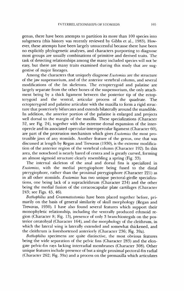
genus, there have been attempts to partition its more than 100 species into subgenera (this history was recently reviewed by Gibbs et al., 1983). How- ever, these attempts have been largely unsuccessful because there have been no explicitly phylogenetic analyses, and characters purporting to diagnose most groups are usually combinations of primitive and derived traits. '13e task of detecting relationships among the many included species will not be easy, but there are many traits examined during this study that are sug- gestive of major lineages.
Among the characters that uniquely diagnose Eustornia.s are the structure of the jaw suspensorium, and of the anterior vertebral column, and several modifications of the fin skeletons. 'Ike ectopterygoid and palatine are largely separate from the other bones of the suspensorium, the only attach- ment being by a thick ligament between the posterior tip of the ectop- terygoid and the ventral, articular process of the quadrate. The ectopterygoid and palatine articulate with the maxilla to form a rigid struc- ture that posteriorly bifurcates and extends bilaterally around the mandible. In addition, the anterior portion of the palatine is enlarged and projects well dorsal to the margin of the maxilla. These specializations (character 52, see Fig. 24), together with the extreme dorsal expansion of the inter- opercle and its associated opercular-interopercular ligament (Character 60), are part of the protrusion mechanism which gives Eustomiu.s the most pro- trusible jaws of any stomiids. Another feature of the genus, one that was discussed at length by Regan and ?Trewavas (1930), is the extreme modifica- tion of the anterior region of the vertebral column (Character 192). In this area, the notochord is nearly bared ofcentra and is greatly curved, forming an almost sigmoid structure clearly resembling a spring (Fig. 33).
The internal skeleton of the anal and dorsal fins is specialized in Eustornias, with the medial pterygiophore being fused to the distal pterygiophore, rather than the proximal pterygiophore (Character 22 1) as in all other stomiids. Eustomias has two unique pectoral-girdle specializa- tions, one being lack of a supracleithrum (Character 234) and the other being the medial fusion of the coracoscapular plate cartilages (Character 243; see Figs. 45, 46).
Bathophilus and Grarnmatostomias have been placed together before, pri- marily on the basis of general similarity of skull morphology (Regan and Trewavas, 1930). I have also found several features which support their monophyletic relationship, including the ventrally produced ethmoid re- gion (Character 8; Fig. 13), presence of only 3 branchiostegals on the pos- terior ceratohyal (Character 164), and the morphology of the cleithrum, in which the lateral wing is laterally extended and somewhat thickened, and the cleithrum is foreshortened anteriorly (Character 236; Fig. 39a).
Rathophilus specimens are quite distinctive, the most obvious features being the wide separation of the pelvic fins (Character 283) and the elon- gate pelvic-fin rays lacking interradial membranes (Character 308). Other unique features include presence of but a single proximal pectoral-fin radial (Character 262; Fig. 39a) and a process on the premaxilla which articulates
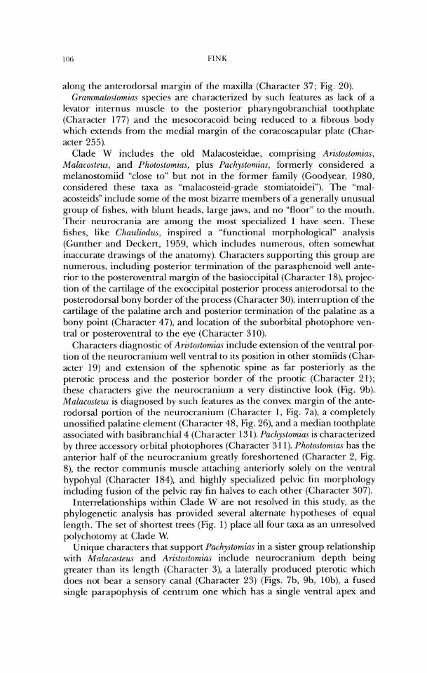
along the anterodorsal rnargin of the maxilla (Character 37; Fig. 20). C;rammato.stomias species are characterized by such features as lack of a
levator internus muscle to the posterior pharyngobranchial toothplate (Character 177) and the mesocoracoid being reduced to a fibrous body which extends from the medial margin of the coracoscapular plate (Char- acter 255).
(;lade W includes the old Malacosteidae, comprising Ari.stostomias, Malacosteus, and Photostomias, plus Pachystomias, formerly considered a melanostomiid "close to" but not in the former family (Goodyear, 1980, considered these taxa as "malacosteid-grade stomiatoidei"). The "mal- acosteids" include some of the most bizarre members of a generally unusual group of fishes, with blunt heads, largejaws, and no "floor" to the mouth. Their- neurocrania are among the most specialized I have seen. These fishes, like Chauliodws, inspired a "functional morphological" analysis (Gunther and Deckert, 1959, which includes numerous, often somewhat inaccurate drawings of the anatomy). Characters supporting this group are nurnerous, including posterior termination of the parasphenoid well ante- rior to the posteroventral margin of the basioccipital (Character 18), prqjec- tion of the cartilage of the exoccipital posterior process anterodorsal to the posterodorsal bony border of the process (Character 30), interruption of the cartilage ofthe palatine arch and posterior termination of the palatine as a bony point (Character 47), and location of the suborbital photophore ven- tral or posteroventral to the eye (Character 3 10).
Characters diagnostic of Aristostomias include extension of the ventral por- tion of the neurocranium well ventral to its position in other stomiids (Char- acter 19) and extension of the sphenotic spine as tar posteriorly as the pterotic process and the posterior border of the prootic (Character 21); these characters give the neurocranium a very distinctive look (Fig. 9b). Malacosteus is diagnosed by such features as the convex margin of the ante- rodorsal portion of the neurocranium (Character 1 , Fig. 7a), a completely unossified palatine element (Character 48, Fig. 26), and a median toothplate associated with basibranchial4 (Character 13 1). I'achystomias is characterized by three accessory orbital photophores (Character 3 1 1). I'hotostomias has the anterior half of the neurocranium greatly fi~reshortened (Character 2, Fig. 8), the rector communis muscle attaching anteriorly solely on the ventral hypohyal (Character 184), and highly specialized pelvic fin morphology including fusion of the pelvic ray fin halves to each other (Character 307).
Interrelationships within Clade W are not resolved in this study, as the phylogenetic analysis has provided several alternate hypotheses of equal length. The set of shortest trees (Fig. 1) place all four taxa as an unresolved polychotomy at Clade W.
Unique characters that support Pachystomias in a sister group relationship with Malacosteus and Aristostomias include neurocranium depth being greater than its length (Character 3), a laterally produced pterotic which does not bear a sensory canal (Character 23) (Figs. 7b, 9b, lob), a fused single parapophysis of centrum one which has a single ventral apex and
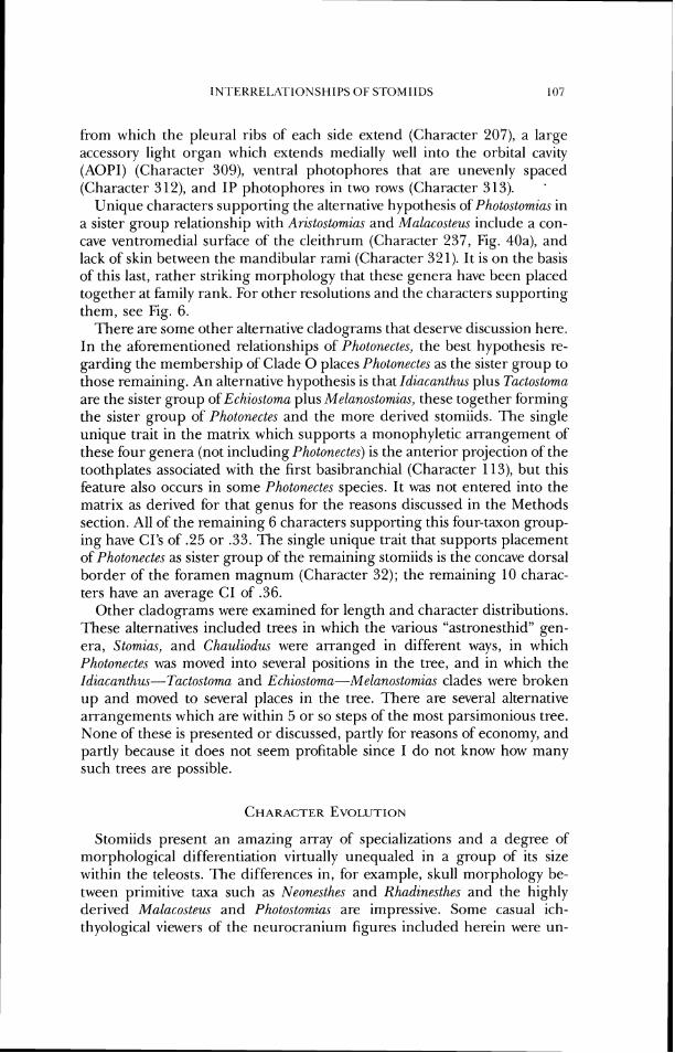
from which the pleural ribs of each side extend (Character 207), a large accessory light organ which extends medially well into the orbital cavity (AOPI) (Character 309), ventral photophores that are unevenly spaced (Character 3 12), and IP photophores in two rows (Character 3 13). '
Unique characters supporting the alternative hypothesis of Photostomias in a sister group relationship with Ari~stostomias and Malacosteus include a con- cave ventromedial surface of the cleithrum (Character 237, Fig. 40a), and lack of skin between the mandibular rami (Character 321). It is on the basis of this last, rather striking morphology that these genera have been placed together at family rank. For other resolutions and the characters supporting them, see Fig. 6.
There are some other alternative cladograms that deserve discussion here. In the aforementioned relationships of'Photon,ecte.~, the best hypothesis re- garding the membership of Clade 0 places Photonectes as the sister group to those remaining. An alternative hypothesis is that Idiacanth~~s plus Tactostoma are the sister group of Echiostoma plus Melanostomias, these together forming the sister group of Photonectes and the more derived stomiids. The single unique trait in the matrix which supports a monophyletic arrangement of these four genera (not including Photonectes) is the anterior projection of the toothplates associated with the first basibranchial (Character 113), but this feature also occurs in some Photonectes species. It was not entered into the matrix as derived for that genus for the reasons discussed in the Methods section. All of the remaining 6 characters supporting this four-taxon group- ing have CIS of .25 or .33. The single unique trait that supports placement of Photonecle.~ as sister group of the remaining stomiids is the concave dorsal border of the foramen magnum (Character 32); the remaining 10 charac- ters have an average CI of 36 .
Other cladograms were examined for length and character distributions. These alternatives included trees in which the various "astronesthid" gen- era, Stomias, and Chauliod~~s were arranged in different ways, in which Photonectes was moved into several positions in the tree, and in which the Idiacnnthus-Tactostoma and Echiostoma-Melanostomias clades were broken up and moved to several places in the tree. There are several alternative arrangements which are within 5 or so steps of the most parsimonious tree. None of these is presented or discussed, partly for reasons of economy, and partly because it does not seem profitable since I do not know how many such trees are possible.
Stomiids present an amazing array of specializations and a degree of morphological differentiation virtually unequaled in a group of its size within the teleosts. The differences in, for example, skull morphology be- tween primitive taxa such as Neonesthes and Rhadznesthes and the highly derived Malacosteus and Photostomias are impressive. Some casual ich- thyological viewers of the neurocranium figures included herein were un-
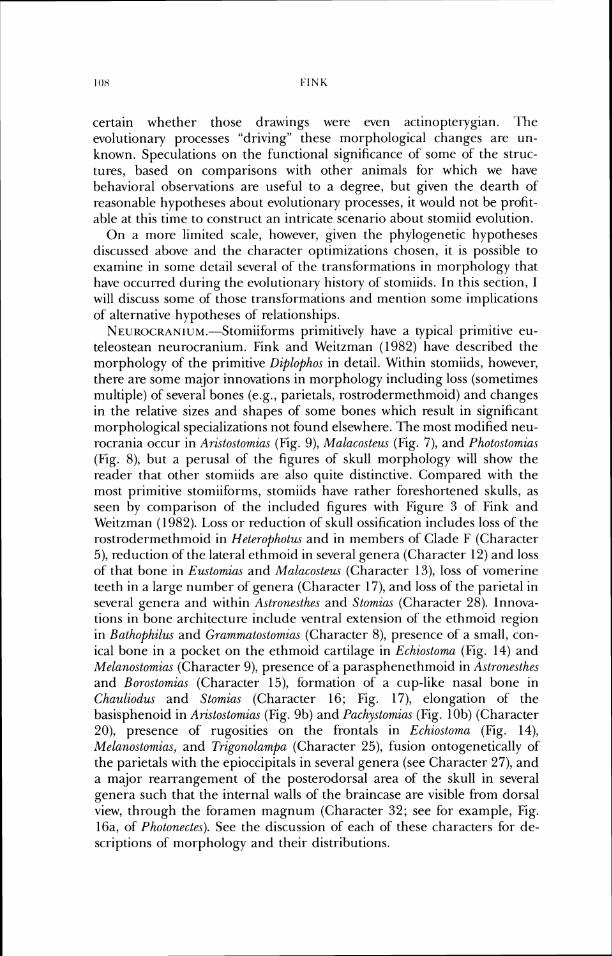
certain whether those drawings were even actinoprerygian. ~ l h e evolutionary processes "driving" these morphological changes are un- known. Speculations on the functional significance of sorlie of the struc- tures, based on comparisons with other animals for which we have behavior-a1 observations are useful to a degree, but given the dearth of reasonable hypotheses about evolutionary processes, it would not be profit- able at this time to construct an intricate scenario about stomiicl evolution.
On a more limited scale, however, given the phylogenetic hypotheses discussed above and the character optimizations chosen, it is possible to examine in some detail several of' the transformations in morphology that have occurred during the evolutionary history of stomiids. In this section, I will discuss some of' those transformations and mention some implications of alternative hypotheses of relationships. NE~R0~R~~1~~.-St0miifbrms primitively have a typical primitive eu-
teleostean neurocraniunl. Fink arid Weitzman (1982) have described the morphology of the primitive Diplopho.~ in detail. Within stomiids, however, there are some major innovations in morphology including loss (sometimes multiple) of several bones (e.g., parietals, rostrodermethmoid) and changes in the relative sizes and shapes of' some bones which result in significant morphological specializations not found elsewhere. The most modified neu- rocrania occur in Ari~stostomicls (Fig. 9), Malacosteus (Fig. 7), and Photostomius (Fig. 8), but a perusal of the figures of skull morphology will show the reader that other stomiids are also quite distinctive. Compared with the most primitive stomiiforms, stomiids have rather foreshortened skulls, as seen by comparison of the included figures with Figure 3 of Fink and Weitzman (1982). Loss or reduction of skull ossification includes loss of the rostrodermethmoid in Heterophotus and in members of Clade F (('1 . idracter - 5), reduction of the lateral ethrrioid in several genera (Character 12) and loss of that bone in Eustomias and Malcccoste~~s (Character 13), loss of vomerine teeth in a large number of' genera (Character 17), and loss of the parietal in several genera and within Astronesthes and Stomias (Character 28). Innova- tions in bone architecture include ventral extension of the ethmoid region in Bathophilus and Grarnmato.stomia.s (Character 8), presence of a small, con- ical bone in a pocket on the ethmoid cartilage in Echiostoma (Fig. 14) and Melanostomias (Character t)), presence of a parasphenethmoid in Astronesthes and Borostomias (Character 15), fixmation of a cup-like nasal bone in Chauliodus and Stomias (Character 16; Fig. 17), elongation of the basisphenoid in Arzstostomzas (Fig. 9b) and Pachystomias (Fig. 10b) (Character 20), presence of rugosities on the frontals in Echiostomcr. (Fig. 14), Melanostomias, and Trigonolarnpa (Character 25), fusion ontogenetically of the parietals with the epioccipitals in several genera (see Character 27), and a major rearrangement of the posterodorsal area of the skull in several genera such that the internal walls of the braincase are visible from dorsal view, through the foramen magnum (Character 32; see for example, Fig. 16a, of Photonectes). See the discussion of each of these characters for de- scriptions of morphology and their distributions.
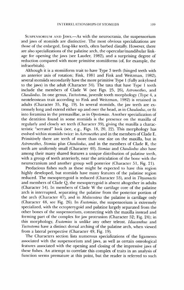
SUSI~L'.NSOKIUM A N D .JAWS.-AS with the neurocrania, the suspensorium and jaws of stomiids are distinctive. 'Ihe most obvious specializations are tliosc of the enlarged, fang-like teeth, often barbed distally. However, there are also specializations ofthe palatine arch, the opercular/mandibular link- age for opening the jaws (see Lauder, 1982), and a surprising degree of reduction compared with more primitive stomiiforms (of, for example, the in ti-aorbitals).
Although it is a stomiiform trait to have Type 3 teeth (hinged teeth with an anterior axis of rotation; Fink, 1981 and Fink and Weitzman, 1982), several stomiids secondarily have the more primitive 'Type 1 (fully ankylosed lo the jaws) in the adult (Character 34). The taxa that have q p e 1 teeth include the members of Clade W (see Figs. 25, 26), Astronesthes, and Chauliodus. In one genus, Tactostoma, juvenile tooth morphology (Type 4, a neoteleostean trait according to Fink and Weitzman, 1982) is retained in adults (Character 35, Fig. 19). In several stomiids, the jaw teeth are ex- tremely long and extend either up and over the head, as in Chauliodus, or fit into foramina in the premaxillae, as in Opostomius. Another specialization of the dentition found in some stomiids is the presence on the maxilla of regularly and closely set teeth (Character 39), giving the maxilla a charac- teristic "serrated" look (see, e.g., Figs. 18, 20, 22). This morphology has evolved within stomiids twice: in Astronesthw and in the members of Clade F,. Primitively there are teeth of more than one size on the maxilla, but in Astronesthes, Stomias plus Chauliodus, and in the members of Clade R, the teeth are uniformly small (Character 40). Stomias and Chauliodus also have among their many shared features a unique distribution of palatine teeth, with a group of teeth anteriorly, near the articulation of the bone with the neurocranium arid another group well posterior (Character 5 1 , Fig. 2 1).
Predacious fishes such as these might be expected to have this region highly developed, but stomiids have many features of the palatine region reduced. The mesopterygoid is reduced (Character 53), and in Thysanac~is and members of Clade Q, the mesopterygoid is absent altogether in adults (Character 54). In members of Clade W the cartilage core of the palatine arch is interrupted, separating the palatine from the posterior portion of the arch (Character 47), and in Malacosteus the palatine is cartilage only (Character 48, see Fig. 26). In Eustornias, the suspensorium is extremely specialized, with the ectopterygoid and palatine largely separated from the other bones of the suspensorium, connecting with the maxilla instead and forming part of the complex for jaw protrusion (Character 52; Fig. 24); in this morphology, Eustomias is unlike any other teleost. Idiacanthzu and Tactostornn have a distinct dorsal arching of the palatine arch, when viewed from a lateral perspective (Character 49, Fig. 19).
The Characters section lists numerous specializations of the ligaments associated with the suspensorium and jaws, as well as certain osteological features associated with the opening and closing of the impressive jaws of these fishes. An attempt to correlate this complex of traits in an analysis of function seems premature at this point, but the reader is referred to such
attempts for the "malacosteids" (Gunther and Deckert, 1959) and Chauliodus ('Tchernavin (1953).
BIZAN(;I-IIAI. BASKET A N D HYOID.-These two intimately connected mor- phological structures are complex and details of their features should be sought in the Characters section. Many of the characters used in the analysis of relationships involve the shapes and interconnections via ligaments of the many bony and cartilaginous elements of these structures. That these areas are complex should not be surprising since much ofthe head morphology in these fishes is also specialized, apparently for the acquisition of food, and the hyoid and branchial basket are functionally associated with mouth opening and food processing, respectively. A few of the more significant specializations are briefly mentioned below.
Stoiniids lack gill rakers as adults (Character 103), although they may be present in juveniles. In Neonesthes the medial surface of the hyoid bar bears a row of toothplates (Character 73). Stornias has an interesting specialization of'the hyoid in that the posterior ceratohyal has an elongate posterior ramus which articulates with an elongated interhyal (Fig. 31a); a remnant of' the primitive interhyal-ceratohyal articulation point is often retained in a more anterior position on the dorsal border of the ceratohyal, and this indicates that in Stomias the articulation has shifted well posteriorly.
MUSCLES AND I,I(;AMENTS 01: 1.1-IE HEW.-Numerous specializations arc present in this morphological complex, again probably correlated with the general specialization in this region of the body. Most of the differentiation has to do with increases in muscle complexity, changes in insertions and origins of muscles, as well as changes in tendons and ligaments. An example of these structural changes may be seen in the geniohyoideus muscle (see Characters 168-175). In stomiids the muscle primitively is divided into two bodies (in contrast to the broad single muscle of other stomiiforms), and the ventral portion is often subdivided; one part of the ventral portion extends into the barbel. Primitively, the muscle has a tendinous inscrtion on the dentary, but in the inembers of Clade K and Photonectes, the dorsal portion has a muscular insertion, and in Idiacunthus and Tuctostorna the insertion is both muscular and tendinous. Additionally, the ventral portion of the mus- cle may have orie (derived) or two (primitive) distal attachment sites. This part of the muscle may also be further subdivided by a tendon, forming two muscle bodies. In Malacosteus and Photostomias, the ventral portion of the geniohyoideus distally overlies the dorsal portion and wraps over it dor- somedially. In a few stomiids, thc dorsal portion of the geniohyoideus is subdivided into dorsal and ventral bodies.
POSICKANIAI. AXIAL SKELE. I .ON. -M~~~ of the specializations occurring in stomiids represent "reduction," with loss of bones or reduction in ossifica- tion, compared with outgroups. Such specializations include a reduced number of supraneurals (Characters 193- 196), decrease in the size of the spiny portions of the neural arches (Character 201), and reduction in size of the caudal vertebral centra (Character 214; compare the relative sizes of these centra in Fig. 36 of Trzgonolampa and Fig. 38 of Photostomias). However,
IN'I'LKIII.:I.XI'IONSI LIPS O F S'T0MIIL)S I l l
a surprising number of features of this part of the body involve increases in complexity. These include the specialized hood-like prezygapophyses of Astronesthw (Character 199), the greatly enlarged anterior neural arch of Chuuliodus and Stomzas (Character 202; Fig. 34), and the various specializa- tions and elaborations of the anterior parapophyses in numerous genera (Characters 203-208).
Also among the several interesting features of the axial skeleton are the reduction of centra in the anterior of the body in Leptostomias, Odontostomias, Opostomia.c, and Thysunactis (Character 191), and the striking multi-curved notochord of Eustomias, with its various centra and neural arches (Character 192, see Fig. 36), which resembles a spring mechanism.
VERTICAL F ~ ~ s . - o n e striking aspect of the morphology of most stom- iids is vertical-fin position. Most members of the group have the dorsal, anal, and caudal fins together at the posterior of the body. Several phy- logenetically primitive members have the anal and dorsal fins, especially the latter, in the plesiomor-ph position near the midbody. On the hypothesis of relationships I have generated, the dorsal fin has moved posteriorly on the body at least three times in stomiids, and possibly four. Some species of A.ctronesthes have the dorsal fin somewhat posteriorly placed, although this has not been quantified; that position is clearly derived within the genus. Hcterophotw has the dorsal-fin origin somewhat posteriorly placed, appar- ently an autapomorphy of the genus. In the members of Clade E, dorsal-fin position is well posterior in all members except Chaulzodus, where the fin is well anterior on the body. Within this group, it is equally parsimonious to assume that: posterior dorsal-fin position evolved once in members of' Clade E and the anterior dorsal-fin position of Chauliodus evolved as a reversal from the posterior state, or the posterior dorsal-fin position evolved inde- pendently in Stomzas and in the members of Clade F, and the anterior position in Chau,liodus evolved from the primitive midbody position.
Chauliodus is interesting for two reasons related to fin morphology. First, the adipose dorsal fin is rather large and lies dorsal to the posteriorly placed anal fin. 'Ihus, i t appears that the posterior-fin placement of other members of Clade E is achieved by Chauliodus as well, but with the adipose fin rather than the dorsal fin. Although any functional explanatioris of this posterior- fin placement would be speculative, presumably the combined fin area well posterior on the body generates a large amount of burst-speed thrust for prey capture (Webb, 1975). Second, the second dorsal-fin ray, which is long and hlamentous, is supposed to be tipped with luminous tissues. Tcher- navin (1953) and others have interpreted this structure to be a "lure" as is found in anglerfishes.
PEC:TORAL GIRDLE.-A complex series of morphological trar~sformations appears to have taken place in the evolution of the pectoral fin and girdle in stomiids. Some of these transformations seem to be correlated with reduc- lion of the pectoral fins as swimming organs and associatiorl of some fin rays with light organs, but they cannot be totally accounted for by such explana- tions.

The most conlplex set of transfi~rmations involves some reduction in tin size accompanied by elongation and greater mobility ofthe anterior fin rays, with luminous tissues in association with these rays (the "rod-ray" complex), then subsequent loss of these rod-rays, and finally some neomorph reac- quisition of light organs on other rays of the pectoral fins.
The pectoral-fin rays in many stomiids are an integral part of a presum- ably light-organ bearing complex. In those primitive stomiids where the primary function of the fin is locomotor and it lacks organized luminous bodies, the fin rays, with the exception ofthe articulation of the first ray with the scapula, resemble those of Polymetrne (Fig. 49) in general morphology. In these primitive members of the group, the rays are relatively strong and are branched. In more specialized stomiids, with the exception of the rod-rays, the relative size of the fin rays is less than in primitive genera and the rays are unbranched. It is not unusual for the ray halves to be veiy loosely bound together.
Associated with the alterations in fin-ray morphology is an alteration in distal radial morphology, involving specialization of the anterior one to three radials and loss of the others. In stomiids, the anterior distal radial has its cartilaginous core extending anterolateral to the lateral margin of the ventral ray-half in adults (see Character 269). In stomiids, except for Neonesthes, the cartilaginous core also extends dorsally (Character 268). Primitively, in stomiids other than Neon~sthes, the anterior distal radial is somewhat to markedly bilobed ("dumb-bell") in shape and is in the position occupied in outgroups by the cartilage-lined articulation surface on the dorsal 11alf of the first fin ray. Compare Fig. 52 of Polymetme and Fig. 5 1 of Heterophotus for examples of these in which the anterior pectoral-fin ray articulates with the scapula. In Astrone.vthes, the anterior radial is "dumb- bell" shaped only in juveniles, becoming more ossified and rather more primitive in appearance in adults. In Borostomias, the anterior distal radial directly touches the scapular surface and displaces the dorsal ray-half dor- sally. Further, a column of the cartilage of the distal radial extends dorsally above the horizontal plane of the scapula and serves as an attachment site for muscles. Bone of the dorsal ray-half reinforces this cartilage. In Chauliod~~s, Heterophotus, Stornias, and lk~gonolarr~pa, the anterior distal radial clearly has a "dumb-bell" morphology. Rhadinesthes has the anterior distal radial somewhat cuboid in shape, but it retains the concave proximal sur- face. While no function can be ascribed with certainty to this bilobed radial shape, it appears to lend greater mobility to the anterior pectoral-fin ray than is present in non-stomiids. It may be significant that Trigonolampa has presumed luminous tissues in the anterior region of the pectoral fins, and movement of the fins would cause the lights to move as well. In Chirostomias (Fig. 41), pectoral radiaVfin morphology is uniquely specialized, and no specimens available to me had cartilage fully intact, so I am unable to say what its distal radial morphology is like.
In the remaining stomiids, there is primitively a "rod-ray" complex involv- ing the anterior one to three distal radials and anterior two to four fin rays.
IN~I'ERKEI,Rl'IONSI IIPS 01; S'I<)MI 11)s 113
As described under Character 27 1, the first and sometimes second fin rays are reduced in length, and the anterior two to four rays are tightly bound together and associated with the anterior radial(s). There is a wide articula- tion of the distal radial(s) with proximal radial 1, the scapula, and in some cases with some of the more posterior distal radials. This morphology is well illustrated in Fig. 53 of Tizysanactzs. The anterior, shorter rays act as muscle attachment sites, and the more elongate rays, bound together as a stiff rod, carry presumed luminous tissues either at their tips (as in 2'hysanactis), or along their length (as in Echiostoma). In addition, there is no interradial membrane connecting the rod-ray to the more posterior fin rays. The latter lack distal radials (Character 271) and are generally somewhat reduced.
Speculations about the function of these highly mobile, sometimes rather elaborate luminous structures have included their being tactile, serving as sexual or prey attractants, and so on, but until we are able to observe the animals in captivity or in nature for long periods of time, it seems fruitless to continue this tradition of'conjecture. Whatever their function, and whatever advantage they may have conferred, according to my hypothesis of rela- tionship the majority of this group of stomiids has lost the external mani- festations of the rod-ray. There are small internal remnants ofthe system in many, including Aristoslomias (Fig. 40b), Bathophilus (Fig. 39a), Eustomias (Figs. 45, 46), Leptostomias (Figs. 43, 54), Grammatostomias, Malacosteus, Melanostomias (Fig. 44), Pachystomias (Fig. 47), and some Photonectes. 'The primary pectoral girdle is virtually absent in Idiacanthm, Photostomias, and Tactostoma, but presumably their ancestors also had remnants of the rod-ray complex. According to the hypothesis of' choice, the rod ray has been lost externally at least four times during the course of stomiid evolution. 1,eptostomias has remnants while its sister group, Thysunactis, and their sister lineages have the external rays. The external system has been lost within Photo,nectes, as some species have a rod-ray, and also in Melanostomias, as its sister taxon Echostornu has a well developed ray system. The remaining stoniiids which have lost the rod-ray form a monophyletic group; loss in a single common ancestor could account for its absence in all of them.
Interestingly, in a number of genera with remnants of the rod-ray, the remaining pectoral-fin rays are specialized in various ways, often as bearers of luminous tissues. Examples include Aristostomias, Bathophilus, some spe- cies of Eustomia.~, Grammato.stomia.s, Malacosteus, Melanostomias, Pachystomias, and some species of Photonectes. For instance, in Aristostomias the pectoral-fin rays have luminous tissues at their bases, and the anterior two rays are elongate and surrounded by luminous tissues fi-om base to tip. All the fin rays also have well developed, serrated flanges proximally for muscle attach- ments. 'Thc obvious inference is that a remote ancestor of Aristostomias (and a more inclusive group) had a functional rod-ray complex, lost it, and subse- quenlly, the immediate ancestor of the Aristostomias clade evolved a func- tionally similar structure from the remaining fin rays. Much the same course of events appears to have occurred in the history of Grammatostomias, which also has a rod-ray remnant combined with modified, posterior fin
rays which bear light organs. Additional evidence of the function of the lulninous pectoral fins of' Grammatostomius can be seen in the presence of a groove in the skin into which the fin can be placed, thereby ef-fectively halting light emission.
Some species of' Photonectes have evidently also reevolved a light-bearing ray complex. In P. leucospilus, there is a rod-ray remnant; the remaining, externally visible rays are bound together into a unit by interradial mem- brane and have luminous material on the anterior and posterior edges. The entire fin unit pivots around the broadly convex, posterior border of the scapula. In some other members of the genus (e.g., specimens identified as I-'. margarita), the girdle morphology is quite different and an original rod- ray is present, though possibly without luminous function (I could find no associated luminous tissues on the rod-ray, but Beebe and Crane, 1939, reported finding a bulbous terminal tip).
Many species of Eustomias have specialized fin rays of unknown function. The primitive condition within Eustomias appears to be presence of a rod- ray remnant, with the remaining fin rays relatively numerous and well devel- oped (e.g., Fig. 45). In some presumably more derived species, shown in Fig. 46, only one or two elongate rays are present externally, with no interradial membrane, and apparently no luminescent tissues.
Another set of pectoral-girdle transformations involving several apparent reversals concerns the number of' pectoral girdle proximal radials. As diag- nosed by Weitzman (1974), members of the Photichthya have three prox- imal radials, in contrast to other stomiiforms and primitive teleosts generally, which have four. However, I have found four to be present in Chirostomins, Heterophotus, Leptostomias, and Thysanactis (Character 265). 'These are interpreted as neomorphic structures which have appeared inde- pendently in each of the first two genera and once in an ancestor of the latter two genera. I am unable to determine the homology of' any of the radials of Chirostomia., relative to other stomiids, primarily because of their relative placement to each other and similarity in size and shape. ' f i e fourth radial of Heterophotus is very small and continuous with the third radial distally, and is presumably derived ontogenetically from 111. I suspect that radial IV of Leptostomias plus Thysanactis is derived by fission from radial 111.
Other changes in pectoral girdle evolution include reduction of' the pri- mary girdle, with loss of ossification of the scapula (e.g. Characters 245, 246), coracoid (Characters 247-251), and mesocoracoid (Characters 253-256). The morphology of the coracoscapular plate in each stomiid genus is distinctive, and any adult or juvenile specimen can be identified to genus simply by examination of' that plate, as may be seen by a perusal ofthe figures of pectoral girdles. I know of no other large teleost group with such morphological diversity in the pectoral girdle. Loss or reduction of the extrascapular (Character 229) and posttemporal (Character 231), are other minor but noteworthy events which mark large groups of stomiids.
Also of some functional interest is the complex of ligaments connecting the pectoral girdle to the head. 'lllese structures are involved in the rather
extensive movements the pectoral girdle must pass through as large prey items are swallowed, and they presumably help guide and limit the move- ment of the skeletal parts. An example is Ligament 3 in Echiostoma and Melanostomias, which forms a thick sheath between the supracleithrun~ and cleithrum. Another interesting morphology is that of Stomias, in which Baudelot's ligament is looped around the sheath-like Ligament 3. The func- tional significance of this loop, if any, is not currently ascertainable.
The pectoral girdle morphology of Eustomias is noteworthy in that the coracoscapular plates are fully fused medially.
PELVIC GIRDLE.-Also striking for their morphological diversity are the pelvic girdles of stomiids. While not quite as distinctive as pectoral girdle morphology, pelvic girdle morphology alone allows identification of quite a number of genera. ?'he main trend in the evolution of these structures is elaboration of the anterior and posterior cartilaginous processes, expansion of the anterior process, and increase (and then reduction) of the car- tilaginous radial elements. See Fig. 61 of Neoneshhes for the terminology of pelvic girdle morphology and Fig. 66 of Polymetme for plesiomorph stomi- iform girdle morphology.
In all stomiids except Neonesthes, the anterior cartilaginous process is enlarged (Character 2115). There are lateral projections, in some cases rather elaborate, extending from this process in several lineages, including Astronesthes, the Odontostomias plus Opostomias sister group, and in the mono- phyletic group including Tuctostoma through Aristostomias in the preferred cladogram (Character 284; Fig. 63 of Bathophilus). In this latter group, there is also, with the exception of Idiacanthus and Echiostoma, a distal expansion of the anterior process (Character 286; Figure 62 of Melanostomias) so that in most taxa the process anteriorly is about half the width of the posterior plate.
In stomiids, the posterior pelvic plate is enlarged relative to other stomi- iforms, and the cartilaginous core of the plate extends posteriorly beyond the plate ossification (Character 290). Compare Fig. 66 of Polymetme with Figs. 61 of Neonesthes and 62 of Melanostomias. In many taxa, these posterior cartilaginous extensions are very elaborate, as can be seen by examination of Figs. 67-69. The anterior and posterior cartilaginous processes in stomiids seem to serve to anchor the pelvic girdle in the body; the posterior pro- cesses also serve as attachment sites for muscles that insert on the pelvic-fin rays.
Another major theme in pelvic girdle evolution in stomiids is the pro- liferation of pelvic-fin radials, as described under Character 293. Indeed, it is a stomiid trait to have more than three such radials, although there has been secondary reduction in some lineages and similar proliferation in some other stomiiform subgroups. The original source for the increased number of radials in stomiids seems to be fission of the anterior two radials (note the bilobed shape of these elements in Neonesthes, Fig. 61). Subsequent subdivisions result in every fin ray having its own radial in most stomiids; this is a morphology unreported in other teleosts. As far as I am aware, the
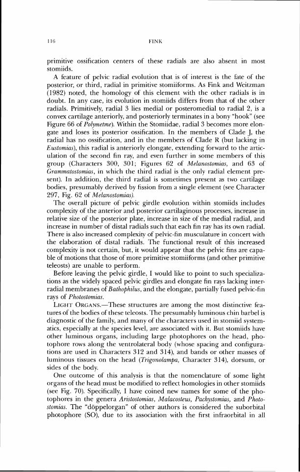
primitive ossification centers of these radials are also absent in most stomiids.
A feature of pelvic radial evolution that is of interest is the fate of the posterior, or third, radial in primitive stomiiforms. As Fink and Weitzman (1982) noted, the homology of this element with the other radials is in doubt. In any case, its evolution in stomiids differs from that of the other radials. Primitively, radial 3 lies medial or posteronledial to radial 2, is a convex cartilage anteriorly, and posteriorly terminates in a bony "hook" (see Figure 66 of Polymetme). Within the Stomiidae, radial 3 becomes more elon- gate and loses its posterior ossification. In the members of Clade J , the radial has no ossification, and in the members of' Clade K (but lacking in Eustomias), this radial is anteriorly elongate, extending fi)rward to the artic- ulation of the second fin ray, and even further in some members of this group (Characters 300, 301; Figures 62 of Melanostomias, and 63 of Grammatostomias, in which the third radial is the only radial element pre- sent). In addition, the third radial is sometimes present as two cartilage bodies, presumably derived by fission from a single element (see Character 297, Fig. 62 of Melanostomia.~).
l'he overall picture of pelvic girdle evolution within stomiids irlcludes complexity of the anterior and posterior cartilaginous processes, increase in relative size of the posterior plate, increase in size of the medial radial, and increase in number of distal radials such that each tin ray has its own radial. There is also increased complexi~y of pelvic-fin musculature in concert with the elaboration of distal radials. I h e functional result of this increased conlplexity is not certain, but, it would appear that the pelvic fins are capa- ble of motions that those of more primitive stomiiforms (and other primitive teleosts) are unable to perform.
Before leaving the pelvic girdle, I would like to point to such specializa- tions as the widely spaced pelvic girdles and elongate fin rays lackin& r lnter- '
radial membranes of Bathophilus, and the elongate, partially fused pelvic-fin rays of Photostomias.
L,IGI-~T O ~ c ~ ~ s . - - T h e s e structures are among the most distinctive fea- tures of the bodies of these teleosts. The presumably luminous chin barbel is diagnostic of the family, and many of the characters used in stomiid system- atics, especially at the species level, are associated with it. But stomiids have other luminous organs, including large photophores on the head, pho- tophore rows along the ventrolateral body (whose spacing and configura- tions are used in Characters 312 and 314), and bands or other masses of luminous tissues on the head (Ti-igonolampu, Character 314), dorsum, or sides of the body.
One outcome of this analysis is that the nomenclature of some light organs of the head must be modified to reflect homologies in other stomiids (see Fig. 70). Specifically, I have coined new names for some of the pho- tophores in the genera Aristostomias, Malacosteus, Puchystomias, and Photo- stomias. The "diippelorgan" of other authors is considered the suborbital photophore (SO), due to its association with the first infraorbital in all
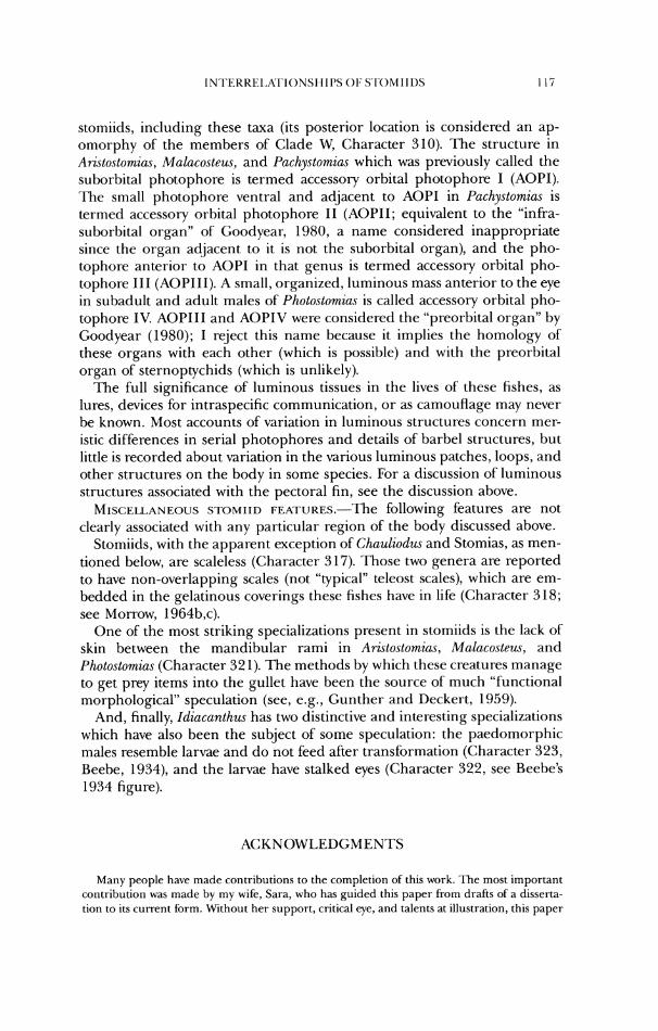
stomiids, including these taxa (its posterior location is considered an ap- omorphy of the members of Clade W, Character 310). The structure in Ari.stostomiu.s, Malacosteus, and Pachystomias which was previously called the suborbital photophore is termed accessory orbital photophore I (AOPI). 171e small photophore ventral and adjacent to AOPI in Pachystomias is termed accessory orbital photophore 11 (AOPII; equivalent to the "infra- suborbital organ" of Goodyear, 1980, a name considered inappropriate since the organ adjacent to it is not the suborbital organ), and the pho- tophore anterior to AOPI in that genus is termed accessory orbital pho- tophore 111 (AOPIII). A small, organized, luminous mass anterior to the eye in subadult and adult males of Photostomias is called accessory orbital pho- tophore IV. AOPIII and AOPIV were considered the "preorbital organ" by Goodyear (1980); I reject this name because it implies the homology of these organs with each other (which is possible) and with the preorbital organ of sternoptychids (which is unlikely).
The full significance of luminous tissues in the lives of these fishes, as lures, devices for intraspecific communication, or as camouflage may never be known. Most accounts of variation in luminous structures concern mer- istic tlif'f'er-ences in serial photophores and details of barbel structures, but little is recorded about variation in the various luminous patches, loops, and other structures on the body in some species. For a discussion of luminous structures associated with the pectoral fin, see the discussion above.
MIS(:ELLANF,C)US SI'OMIID FEA.I.UKES.-T~~ following features are not clearly associated with any particular region of the body discussed above.
Stomiids, with the apparent exception of Chauliodus and Stomias, as men- tioned below, are scaleless (Character 3 17). Those two genera are reported to have non-overlapping scales (not "typical" teleost scales), which are em- bedded in the gelatinous coverings these fishes have in life (Character 318; see Morrow, 1964b,c).
One of the most striking specializations present in stomiids is the lack of skin between the mandibular rami in Aristostomia~, Malacosteus, and Photostomias (Character 32 1). The methods by which these creatures manage to get prey items into the gullet have been the source of much "functional rnor-phological" speculation (see, e.g., Gunther and Deckert, 1959).
And, finally, Idiacanthus has two distinctive and interesting specializations which have also been the subject of some speculation: the paedomorphic males resemble larvae and do not feed after transformation (Character 323, Beebe, 1934), and the larvae have stalked eyes (Character 322, see Beebe's 1934 figure).
ACKNOWLEDGMENTS
Many people have made contributions to the completion of this work. The most important contribution was made by my w~fe, Sara, who has guided this paper from drafts of a disserta- lion to its current for~n. Without her support, critical eye, and talents at illustration, this paper
would have never come to be. Stan Weitzman srarted me on this project, and his advice and great knowledge of stomiiform fishes has been much appreciated over these past years.
Early versions of the paper (in dissertation form for George Washington University) were commented on by David L,. Atkins, Daniel M. Cohen, Robert R. Gibbs, Randall C. Packer, and Stanley H. Weitzman. Stan Weitzman, David L. Swofford, and especially Bob Gibbs are thanked for their helpful reviews of the final draft. Dan Brooks, Steve Farris, Julian Humph- ries, Michael Miyamoto, Richard Pimentel, and David Swofford all helped in various ways with the numerical analyses. Gordon Howes graciously hand carried type material from the British Museum. Karsten Hartel, Susan Jewett, Douglas Nelson, and Alexandra Snyder all provided technical assistance. To all these people I owe an appreciative "thank you." Sara Fink drew Figures 7-66.
This work was supported by a Sniithsonian Insti~ution Predoctoral Fellowship, by NSF Grant DEB79-04729, and by the Milton Fund of Harvard University.
LITERATURE CITED
Ahlstrom, E. I-I., W. J. Richards, and S.H. Weitzman. 1984. Families Gonostornatidae, Sternop- tychidae, and associated stomiiform groups: development and relationships, pp. 184-198. In: "Ontogcny and Systematics of Fishes," Amer. Soc. Ichthyol. and Herp., Spec. h b l . No. 1.
Ararnbourg, C. 1943. Note preliminairc sur quelques Poissons fossiles nouveaux. I. Bull. Soc. Geol. France, ser. 5, vol. 8:281-285, 1 pl.
. 1954. Les poissons cretaces de ~ e b e l 'rselfat (Moroc). Ed. Serv. Geol. du Moroc, Notes et Memoires, (1 18), 188 pp., 20 pl.
-. 1967. Les poissons oligocenes dc 1'Iran. Notes et Memoires sur le Moyen-Orient. Vol. VIII, Mus. Nat. d'Hist. Nat. Paris, 210 pp., 17 pls.
Ayrcs, W. 0 . 1848. Description of a new genus of fishes, Mnlacosteu~. Proc. Boston Soc. Nat. Hist., 3:69-70.
Bertin, L. and C. Arambourg. 1958. Super-ordre des 7eleosteens (Teleostei), pp. 2204-2500. In: Traite de Zoologie, P.-P. Grasse (ed.), 13.
Bassot, J. M. 1966. On the comparative morphology of some luminous organs, pp.557-610. In: Bioluminescence in Progress, ed. by H. Johnson and Y. Haneda, Princeton University Press.
1970. Structure and evolution of the light organs of stomiatoid fish, in I h e World Ocean, Joint Oceanography Assenibly, 'Tokyo, 13-24 September 1970. Contribution in Bio- logical Oceanography, mimeographed, 4 pages
- -
Beebe, W. 1932. Nineteen new species and four post-larval deep-sea fish. Zoologica, 13(4):47-107, figs. 8-31.
. 1933. Deep-sea stomiatoid fishes. One new genus and eight new species. Copeia, 1933(4): 160-175.
-. 1934. Deep-sea fishes of the Bermuda Expeditions. Family Idiacanthidae. Zoologica, 16(4):149-241.
----- and J. Crane. 1939. Deep-sea fishes of the Bermuda Oceanographic Expeditions. Family Melanostorniatidae. Zoologica, 24(6):65-238.
Bloch, M. E. and J. G. Schneider. 1801. Systema Icthyologicae Iconibus. CX illustratum, Berlin.
Bolin, R. L. 1939. A new stomiatoid fish from California. Copeia, 1939(1):39-41. Brauer, A. 1902. Diagnosen von neuen tiefseefischen, welche von der Valdivia-expedetion
gesammelt sind. Zool. Anz., 25(668):277-298. . 1906. Die Tiefsee-Fische. I. Systematischer Teil, in Wissenschaftliche Ergebnisse der
Deutschen Tiefsee-Expedition auf dem Dampfer Valdivia 1898-1899. Jena, Verlag von Gustav Fescher, vol. 15, no. 1.
. 1908. Die Tiefsee-Fische. 11. Anatomischer Teil. In: ibid., vol. 15, no. 2.
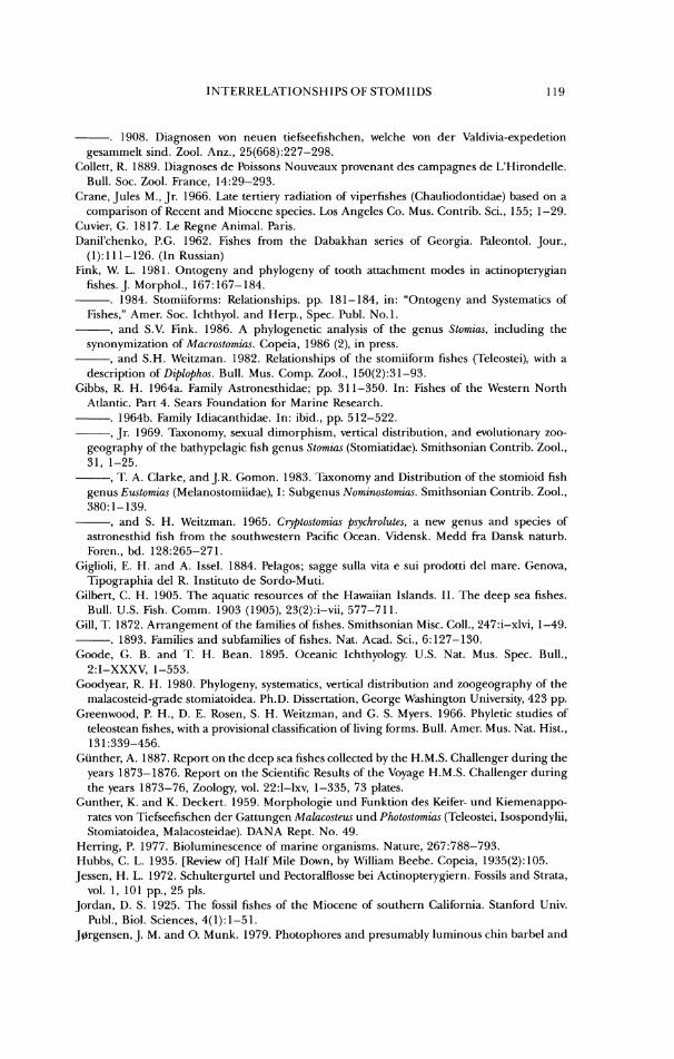
IN1'EKKEI,ATIONSMIPS OF S IOMIIDS 110
-. 1908. Diagnosen von neuen tiefseefishchen, welche von der Valdivia-expedetion gesammelt sind. Zool. Anz., 25(668):227-298.
<:ollett, R. 1889. Diagnoses de Poissons Nouveaux provenant des campagnes de L'Hirondelle. Bull. Soc. Zool. France, 14:29-293.
Crane, Jules M., Jr. 1966. Late tertiery radiation of viperfishes (Chauliodontidae) based on a comparison of Recent and Miocene species. Los Angeles Co. Mus. Contrib. Sci., 155; 1-29.
Cuvier, G. 181 7. Le Regne Animal. Paris. Danil'chenko, P.G. 1962. Fishes from the Dabakhan series of Georgia. Paleontol. Jour.,
(I): 1 1 1-126. (In Russian) Fink, W. L. 1981. Ontogeny and phylogeny of tooth attachment modes in actinopterygian
fishes. 1. Morphol., 167: 167- 184. 1984. Stomiiforms: Relationships. pp. 181-184, in: "Ontogeny and Systematics of
Fishes," Amcn Soc. Ichthyol. and Herp., Spec. Publ. No.1. ------, arid S.V. Fink. 1986. A phylogerietic analysis of the genus Stomias, including the
synonymization of Macrostomias. Copeia, 1986 (2). in press. ----, and S.H. Weitzman. 1982. Relationships of the stomiiform fishes (Teleostei), with a
description or Dzplophos. Bull. Mus. Comp. Zool., 150(2):31-93. Gibhs, R. M. 1964a. Family Astronesthidae; pp. 31 1-350. In: Fishes of the Western North
Atlantic. Part 4. Sears Foundation for Marine Research. -. 1964b. Family Idiacanthidae. In: ibid., pp. 512-522. ----, Jr. 1969. 'Taxonomy, sexual dimorphism, vertical distribution, and evolutionary zoo-
geography of the bathypelagic fish genus Stomias (Stomiatidae). Smithsonian Contrib. Zool., 31, 1-25.
-, 71 A. Clarke, and J.R. Gomon. 1983. Taxonomy and Distribut~on of the stomioid fish genus I:'u.~tomzas (Melanostomiidae), I: Subgcnus Nomzno.~tomzas. Sm~thsonian Contrib. Zool., 380: 1 - 139.
-, and S. H. Weitzman. 1965. Cryptostomias psychrolutes, a new genus and species of astronesthid fish from the southwestern Pacific Ocean. Vidensk. Medd fra Dansk naturb. Foren., bd. 128:265-271.
Giglioli, E. H. and A. Issel. 1884. Pelagos; sagge sulla vita e sui prodotti del mare. Genova, 'Tipographia del R. Institute de Sordo-Muti.
Gilbert, C. H. 1905. The aquatic resources of the Hawaiian Islands. 11. The deep sea fishes. Bull. U.S. Fish. Comm. 1903 (1905), 23(2):i-vii, 577-711.
Gill, 71 1872. Arrangement of the families of fishes. Smithsonian Misc. Coll., 247:i-xlvi, 1-49. -. 1893. Families and subfamilies of fishes. Nat. Acad. Sci., 6:127-130. Goodc, G. B. and T. H. Bean. 1895. Oceanic Ichthyology. U.S. Nat. Mus. Spec. Bull.,
2:I-XXXV, 1-553. Goodyear, K. H. 1980. Phylogeny, systematics, vertical distribution and zoogeography of the
malacosteid-grade stomiatoidea. Ph.D. Dissertation, George Washington University, 423 pp. Greenwood, P. H., D. E. Rosen, S. H. Weitzman, and G. S. Myers. 1966. Phylctic studies of
teleostean fishes, with a provisional classification of living forms. Bull. Amer. Mus. Nat. Hist., 13 1 :339-456.
Giinther, A. 1887. Report on the deep sea fishes collected by the H.M.S. Challenger during the years 1873-1876. Report on the Scientific Results of the Voyage H.M.S. Challenger during the years 1873-76, Zoology, vol. 22:I-lxv, 1-335, 73 plates.
Gunther, K. and K. Deckert. 1959. Morphologie und Funktion des Keifer- und Kiemenappo- rates von Tiefseefischen der Gattungen Malacostew und Photostomias (Teleostei, Isospondylii, Stomiatoidea, Malacosteidae). DANA Rept. No. 49.
Herring, P. 1977. Bioluminescence of marine organisms. Nature, 267:788-793. Hubbs, C. L,. 1935. [Review of] Half Mile Down, by William Beebe. Copeia, 1935(2):105. Jessen, H. L. 1972. Schultergurtel und Pectoralflosse bei Actinopterygiern. Fossils and Strata,
vol. 1, 101 pp., 25 pls. Jordan, D. S. 1925. The fossil fishes of the Miocene of southern California. Stanford Univ.
Publ., Biol. Sciences, 4(1): 1-5 1. Jorgensen, J. M. and 0. Munk. 1979. Photophores and presumably luminous chin barbel and
pectoral fin I-ay filaments of Thysan~r lw d m k x (Pisces: Stomiatoidea). Acta. zool. (Stockh.), 60:33-42.
Kawaguchi, K. arid H. G. Moser. 1984. Stomiatoidea: Development, pp. 169-181. In: "On- togeny and Systetnatics of Fishes," Amer. Soc. Ichthyol. and Herp., Spec. Publ. No 1.
Kluge, A. G. and J. S. Farris. 1969. Quantitative phyletics and the evolution of Anurans. Syst. Zool., 18(1): 1-32.
Koefocd, E. 1956. Isospondyli. I. Gymnophotodermi and Lepidophotodermi from the "Michael Sars" North Atlantic Deep-Sea Expedition 19 10. Rept. Sci. Results "Michael Sars" Deep-Sea Exped. 1910, 4: 1-21.
I.auder, G. V. 1982. Patterns of evolution in the feeding mechanism of actinopterygian hshes. Amer. Zool., 22:275-285.
Lowc, R. 'r, 1843. Notices of fishes newly observed or discovered in Madeira during the years 1840-1841 and 1842. Proc. Zool. Soc. London, Part XI, pp. 81-95.
Morrow, J. E. 1964a. General discussion and key to families, subordel- Stomiatoidea, pp. 71-76. In: Fishes of the Western North Atlantic. Part 4. Sears I'oundation for Mai-inc Research.
. l964b. Family Chauliodontidae. In: ibid., pp. 274-289. Morrow, J. E. 1964~ . Family Stomiatidac. In: ibid., pp. 290-310. -. 1964d. Family Malacosteidae. In: ibid., pp. 523-549. Morrow, J. E. and K. H. Gibbs. 1964. Family Melanostomiatidae, In: ibid., pp. 351-51 1. Norman, ,I. R. 1930. Oceanic fishes and flatlishes collected in 1924-1927. Discovery Reports,
2:261-370. Part-, A. E. 1927. ' 1%~ storniatoid fishes of the suborder Gymnophotodermi (Astronesthidae,
Melanostomiatidae, Idiacanthidae) with a complete review of the species. Bull. Bingharn Oceanogr. Coll. 3, art. 2, 123 pp., 62 figs.
. 1930. A note on the classification of the stomiatoid fishes. Copeia, 1930(4):136. Peters, W. C. H. 1876 (1877). Uebersicht der Wahrend der von 1874 bis 1876-Uebersand~en
Fische. Monatschr. Konigl. Pr. Akad. Wiss. Berlin, pp. 831-854. Regan, <:. T. 1908. The Percy Sladen Trust Expedition to the Indian Ocean in 1905. No. 14.
Report on the Marine fishes Collected by M.J. Stanley Gardiner in the Indian Ocean. Trans. Linn. Soc. London. 2nd Ser., Zool. vol. XII, pt. 3, pp. 217-255, pls. 23-32.
. 1923. The classification of the stomiatoid fishcs. Ann. and Mag. Nat. Hist., ser. 9, vol. 11, pp. 612-614.
-, and E. Trewavas. 1929. The fishcs of the families Astronesthidae and Chauliodontidae. Danish Dana Expedition in the North Atlantic and Gulf of Panama, 1920-22, Keport No. 5, pp. 1-39, 7 pls.
. 1930. The fishes of the families Stomiatidae and Malacosteidae. Ibid., Report No. 6 , pp. 1-143, 14 plates.
Richardson, J . 1844. Ichthyology. In: The zoology of the voyage of the H.M.S. Sulphur, under the command of Captain Sir Edward Belcher ... during the years 1836-42, ed. by K.B. Hinds. London, Smith, Elder and Co.
Rohlf, F. J. 1982. Consensus indices for comparing classifications. Math. Biosci., 59:131-144. Rosen, D. E. 1973. Interrelationships of higher eutelcostean fishes, pp. 397-513. In: Interre-
lationships of Fishes, ed. P.H. Greenwood, R.S. Miles, and <:olin Patterson. Zool. J . Linn. Soc. (Suppl. No. I), 53.
Swofford. D. L. 1983. Phylogenetic Analysis Using Parsimony (PAUP), User's Manual, mimeo,
50 PP. Tiylor, W. R. 1967. An enzyme method of clearing and staining small vertebrates. Proc. U.S.
Nat. Mus., 122(3596):1-17. Tchernavin, V. V. 1953. The feeding mechanisms of a deep sea tish, Chuulzod~c~ sloani. British
Museum (Natural History), London, 101 pp., 36 figs., 10 pls. Vaillant, L. 1888. Expeditions scientifiques du Travailleur et du Talisman, pendant les Annees
1880, 1881, 1882, 1883. Poissons, Paris, pp. 1-406, 28 pls. Webb, P. W. 1975. Hydrodynamics and energetics of fish propulsion. Bull. Fish. Res. Bd.
Canada, Bull. 190, 158 pp.
Weitzman, S. H. 1967a. The origin of'the stomiatoid fishes with comments on the classification of salmoniform fishes. Copeia, 1967(3):507-540.
-. 1967b. The osteology and relationships of the Astronesthidae, a family of oceanic Fishes. Dana Report, 7 1 : 1-54, figs. 1-3 1.
1974. Osteology and evolutionary relationships of' the Sternoptychidae, with a new classification of stomiatoid f'amilies. Bull. Amer. Mus. Nat. Hist., 153, art. 3:329-478.
Winterbottom, R. 1974. A descriptive synonymy of the striated muscles of the l'eleostei. Proc. Acad. Nat. Sri. Phila., 125(12):225-317, figs. 1-56.
Woodward, A. S. 1901. Catalogue of the fossil fishes in the British Museum (Natural History), IV, 636 pp., 19 pls.
Zugmayer, E. 1913. Diagnoses des Stomiatides nouveaux provenant des campagnes du yacht "Hirondelle 11" (191 1 et 1912). Bullctin de L'Institut Oceanographique, Monaco, no. 2.53, 7
Accc?{)ted for PuDlicalion , ] m e 28, 1985
APPENDIX 1
Primitive states are coded as 0, derived states as 1. Characters coded as missing are noted b y question marks. See Methods section for discussion. 000000000000000000000000000000000000000000000000000000000000000000 000000000000000000000000000000000000000000000000000000000000000000 000000000000000000000000000000000000000000000000000000000000000000 000000000000000000000000000000000000000000000000000000000000000000 00000000000000000000000000000000000000000000000000000000000 o u t g r oup 001010100001000011111110000111111100001100101010000011000000?10110 101110011000000001101001111100010000101100011000100000001011010001 00001101??11000100000001101011000001100110000011110010100000111000 0100000110011101110001010011011010000010011000111011000000?1111000 00111110000010000001110111100101101100010000110110001001100 a r i s t o s t 000000000000001000000000000000001100001100000000000010000000000000 000010000000000001000100000000000000110000001000000000000000010000
000000000000000000000001110000001001000000100010000000100000000000 100000000000000000000000000001000000000000000000000000000000000000
00011000000000000001100001001000000000000000000000001001000 a s t r o n e s 000010010001000010000000000100011000101100101000000011000000100110 100110011000111001101001111111100000101111001100100110001001110001 00001101??11000100010001101011111101100110000011111010100000111101 0100101100011101110001000000011010000100010000000011001100?010?100 001111100001000000111101111000110111101??001000000001001000 b a t h o p h i 000000000000001000000000000000001000000000000000010010000000000000 000010000000000001000000000000000000100000001000000000000000010000 000000000000100000100001110000001001000000100011000000100000000000 000000000000100000000000100001000000000000000000000000000000000000 00011000000000000000100001001000000000000000000000001001000 boros tom 000000000000000110000000001000001100001100000000001010000000000001 000010000001000001001001000000000000110000100000000000000011000000
00101001?'!0000011000000111000000l011000000000110000010100000111001 00011100000010001010000lllll01100100000000000000010000000000000000 00011000000000000000100001000000000000000000000000000111000 chaul iod 000010000000000000000000000000001000001001000000000010000000001010
10001000000001101100l00l000l00000000l01101001000100001000000010000 01001000001000010000000111000000100l000000000011000000100000110000 000010000001100011000000000001000000000l00000000001010000000000000 10111?00000000000000100001001000000000000000000000001001000 chirosto 000010001000010000000001ll0l0000l000001000100000000010000100100110 100110000000111001001001001000000000101101001110100100011001111000 00001101??11000100000001111000101001000000010011110000100000110000 010010100001110111000l00000011010000000001000000000100000010100000 00111110000000011001100111101001001000001000000000001001000
echiosto 000010100001100010000000001000011000001100101000000111000001?10110 101110010000111001101001001111000000l0l111001100100000001000110001 10001101??11000100010011101010?00001100110000011111010100001111101 0100111101011101110011l000000110100l000001001010000000100010101000 001111100001?000000111l10110100l00l00000?000000000001001000 eustomia 000010000110000000000000000000001000001000010000000010010010000100
100010000000010001001001000100000000101010001000100000000001010000 00001000101000010000000l111000001001000000000011100000100000111000 010011100001100111001100000001000000000001000000000000000000100000
00111111011001000000100001000101000000000000000000001001000 flagel lo 00001001000100001000000000010001100000l100101000000011000000100110 100110011000111001101011111111100000101l11001100100100001001110001 00001101??11100100010011101011111101100110001011111010100000111100 0100001100011101110001000000011010000l00010000000011001110?0101000 00111110000100000001110ll1l0100ll0lll0000000000000001001000 gr ammat o 000010000000000000000000000000001000000000000000000010001000000100 000010000100000001001001000000000000100000001000000000000001010000
00000001??00000011001001ll0000001001000010100011000000100000000000 00000000000010100000000ll0l001000010l00000000000000000000000000000
10011000000000000001l0000l00l000000000000000000001001001000 heteroph 000010000001000000000000000100001000001000100000100011000000100110 11111000011011110100100100l00000000010l101001111100000011001110101 10001101??1100010000000111100010l101010000000011110000101000111100 010000000001110111010l0l0001l100l00000000??10011?00?00???1?0???000 001111?000000010000110011110100l0010000l0000000000001001011 idiacant 000111000110000000000000000000001000001010010000000010110010000100
11001000000101000100l00l000000000000l01010001000101000000101010000 00001011?11000010000000001100000l00l000000010011100001100010111101 011011100001100111000l0000000l000000000001000000010000000000100011 1011111111100101000010000100l001000000000000000000001001000
leptosto 101010000001100011000110000111011100001100101011000011000000?10110 0011100110001001011110011l1100010000l0l101001100100100001110010011 10001101??11011100000001101111100001100l01000011110010100000111100
010000111101110111000101001101101000001001100000000100???1?1111000 00111110000010000001110111101001?0110001000011011000100?100 malacost 000010001010010000000001101000001000001000100000000010000000100110
1011100000000110010010010010000000001011?1001110100100011001111001 10001101??1100010000000111100010?001000000010011110000100000110000 010011100001110111000100000011010000000001000000000101000010100000 00111110000000011001111101101001101100000000000000001001000
melanost 000000000000000000000000000000001000000000000000000010000000000000 000010100000000001000000000000000000100000100000000000000000000000
000000000000000000000100000000000001000000000000000000100000100000 000000000000100000000001100001000000000000000000000000000000000000
00001000000000000000000001001000000000000000000000001001000 neones th 000110000110000000000000000000001000001010010000000010110010000100 100010000001010001001001000000000000101000001000100000000101010000
00000001??10000100000000011000001001000000000011100001100010111001 011011100001100111000100000001000000000011000000010000000000100001
00111110010000010001100001001001000000000000000000001001000 odontost 000110000110000000000000000000001001001010010000000010110010000100
100010000001010001001001000000000000101000100000?00000000101010000 00001001??10000100000001011000001001000000000011100001100010111001 011011100001100111000100000011000000000011000000010000000000100001 00111110010001010001100001001001000000000000000000001001000
opostomi 001010000001000011010110000111111100001100101010000011000000?10110 101110011000000001001001101100010000101100011100100000101001010001 00001101??11011100000001100010001001100110000011111010100000111100 010000011001110111000101001101101000000001100000000100???110111000 00111110000010000001110111101001101000000000111110001001000 pachysto 000010000000000000000000001010011000001000100100000010000000100110 1011100001001100010010010011100001001011010111?0100000011011110000 00001100??11?0010000000111100000100110000000001111001?100000110001 010000000001100111000100000001?00000000001000111100101110011100000 0011111000000000000111?111101001101000000000000000001001000 photonec 010010000000000011000100001011111100001100101010000011000000?10110 101110011000000001001001101?00011000101100011000110000101110010000 00001101??11010110000001101011100001100001000011111110100000111100 01100000001111011100010100110110100000100??10011?00?00???1?0???000 001111?00000?010000111111110001100111111001001000000100?100 photosto 000000000000000000000000000000001000000000000000000010001000000000 000010000100000001001001000000000000100000001000000000000000010000
00000001??00000010000001110000001001001000100011000000100000100000 000000000000100000000100101001000000100000000000000000000000000000 01011000000000000001100001011000000000000000000000001001000 rhadines 000000000000000100000000000000001000011101000000001010000000100000 001001000000000001001001000000000011100000100000000000000001000000 000000000000000010000001110000001011000000000111110010100100111010
000100?00?01100011000101001101000000000000000000000000???100000000
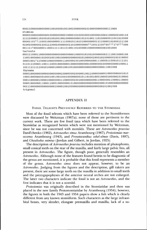
00011000000000000100100001001001000000010100000000000111000 stomias 000010000000000010000000000100001010001000100000100011000000100110
111110000110100101001001001000000000101101001110100000011001010000 00001101~?11000100000001111000101101010000000011110000101000111100 01001000000110011100010000000110100000000??10011100?00???1?0???000 001111?0000000110001111101101001101000001000000000001001000 tactosto 000111000110000000000000000000001000001010010000000011110010000100
110010000001010001001001000000000000101000001000101000000101010000 00001001?110000100000000011000001001000000000011100001100010111101 011011100001100111000100000001000000000001000000010000000000100011
10111111111001010000100001001001000000000000000000011001000
thysana 000010000000000000000000100000001000001001100000000010000000001010
100010000000010001001001000100000000101101001000100001000001010000
000110000010000100000001110000001001000000000011000000110000110000 000010000001100011000100000001010000000000000000000101000000000000
00111000000000000000100001001000000000000000000000101001000
trigonol
APPENDIX I1
Most of the fossil teleosts which have been referred to the Stomiiformes were discussed by Weitzman (1967a); none of those are pertinent to the current work. There are flve fossil taxa which have been referred to the Stomiidae as recognized herein which were not mentioned by Weitzman, since he was not concerned with sto~niids. These are Astronesthes pmevius Danil'chenko (1962), Astrone.sthe.s simus Arambourg (1967), Protostomias vmur- occanus Arambourg (1943), and Pronotacanthus sahel-almae (Davis, 1887), and Chaulzodus exirnius (Jordan and Gilbert, in Jordan, 1925).
'l'he tlescription of Astronesthe.~ praevius includes mention of photophores, small conical teeth on the rear ofthe maxilla, and fairly large pelvic tins, all present in Astronesthes. The figure, though poor, generally resembles an Astronesthes. Although none of the features found herein to be diagnostic of the genus are mentioned, it is probable that this fossil represents a member of the genus. Astronesthe.~ .sirnus does not appear, however, to be an Astronesthes. Judging from the figures and the description, gill rakers are present, there are some large teeth on the maxilla in addition to small teeth and the prezygapophyses of the anterior neural arches are not enlarged. The latter two characters indicate the fossil is not an Astronest/zes, and the first indicates that it is not a stomiid.
Protostomias was originally described in the Stomiatidae and then was placed in the new family Protostomiatidae by Arambourg (1954); however, the figures in both the 1943 and 1954 papers show a fish which is clearly different from any known stomiiform. Such characters as the large infraor- bital bones, very slender, elongate premaxilla and maxilla, lack of a su-
pramaxilla, and greatly enlarged coronoid process of the mandible clearly indicate that Protostomia.~ is not a member of the Stomiidae, and that it is probably not a stomiiform at all. The condition of the specimen and the poor quality of the photographs makes any confident assessment of the reconstructions difficult. Although Arambourg makes reference (1954:95) to the teeth (found only on the dentary) and states that they have a large "cavite pulpaire comme celles des Stomiatidae actuels," it is not possible to determine whether there are Type 3 teeth as would be expected in a stomi- id. Examinatio~l of the specimen will be necessary to determine its placement.
Arambourg (1943) mentioned in passing that Pronotacanthw Woodward is also probably a stomiid, a suggestion that was formalized by Bertin and Arambourg (1958), without comment. Pronotacanthus was erected by Wood- ward (1901) for Ann.pilla sahel-almae Davis, which Woodward considered a notacarlthid. There is a clear phenetic similarity between Pro~ostomias and l'ronotacanthw and they may be related (or the same); however, as far as can be determined from Woodward's description (pp. 169- 17 1) and figure (Plate XV), Pronotucanth~~ also lacks any features which would place it in the Stomiidae (or Stomiiformes).
Chauliodw eximius has been discussed by Crane (1966), who reviewed its taxonomic history and provided photographs to confirm that the specimens are indeed Chauliodus. 1 have exami~ed some fiagmentary material at the MCX.
APPENDIX 111
The fbllowing list includes specimens which are not already listed in pa- pers by Fink (lS81), Fink and Weitzman (1982), or Weitzman (1967b, 1974). 'The genera and species are listed alphabetically, followed by museum number, number of specimens in parentheses, size in standard length, and whether one or more of the specimens was dissected (d), or cleared and staiilcd (c). If more than one specimen was cleared, the number is given.
Ari,sto.~~ornias lunifer, M<:Z 53267 (I), 40 mrn. Aristostomias tzttmanni, MCZ 5881 1 (I), 180 mm. Arlslostomias x~nostomo, USNM 272943 (l) , c . Aristostomias sp., MCZ 58795 (I), 135 mm. Aristostomias sp., MCZ 58791 (3). 39-66 mm. Ari.\tostomzcl sp,. MCZ 58789 (2), 41-100 mm. Astror~csthes cnulophoncs, MCZ 58806 (I), 95 mm, d. A~trorlesthes s i m i l ~ ~ , MCZ 35590 (I), 105 mm. Astron~sthcs sp., MCZ 58817 (I), 163 mm, d. Ualhophilus ater, MCZ 52027 (I), 150 mm. Bnthophilus Drevir, USNM 206701 ( I ) , c. bat hop hi la^ k i n g , MCZ 49146 (l) , 98 mm. Batllophiltu melallicus, MCZ 42285 (3), 53-57 mm, c. Bathophilus pawneei, M<:Z 42279 ( I ) , 77 mm.
Hnt/noph~lu\ sp., MC% 58797 (I) , 94 rnrrr. Honaparttu p(,daliota, MCZ 58794 (7), 43-61 mtn, d. Honr~partia pedulzota, MCZ 58798 (I) , 74 mrn. Bonai~arlia pedallota, MCZ 58818 (4), 42-65 rnm, d. Bonai~art~a pedaliota, MCZ 58851 (I) , 64 mni, c. Boroslomia~ antarcticw., MCZ 42877 ( l ) , 220 nrrn, d. Rorostorrnias ~111cen.\, MCZ 58786 (2), 74-20 1 mnr, d. Horostomzas c,lvcrns, USNM uncat. WH. STA. 486171 (I), c. Roro.stornia.s sp., MCZ 58814 (2), 107-233 tnrn, d . Cl~aulzodus darnae, MCZ 58784 (?I), 86- 128 mtn. C1znuliodu.s mcrcouni, LACM 37 12 1- 1 (2), 53- 1 18 mm, c. Clzuulioclw sloan~, MCZ 58788 (9). 152-200 nun, d. Chtru.sfomia.s p l iop t~ru , MCZ 42274 (I), 1 13 nirn, d. Chirostum2us pliopterw, USNM 272907 (I) , c. CII~I-ostomias plzopterus, USNM 272905 (I), c. Cyclothonc sp., MCZ 58808 (approx. 150), c 20-c 60 mnr, d. L)if)lol~ho.\ rnader~nsis, MCZ 61476 (I) , 96 mm. I:'chlo.stoma barbatum, MCZ 42203 ( l ) , 290 mm. Ecl~iostonlct Ourbaturn, MCZ 58819 (3), 161-236 mrn, d, 1 c-.
Ecltiostorna Oarbuturn, MCZ 58852 ( 1 ) . 166 rnm, c. E~wlonnzc~s b$li.\, USNM uncat. 71-3-2 (I), c. Ewtumtus bingltanti, USNM irncat. MMS 47, S I A 66 (I) , c. I:'~~stomias cf breviharbatu~, MCZ 47789 ( I ) , 116 tnrrr. 1:'clstornias rnacruru.\, USNM 2729 13 ( I ) , c. Ewtomia.\ oh.scura, MCZ 58790 (I), 158 rrrnr, d. Flagrlloslomias bounvi, MCZ 58802 (I) , 298 rnni, d. Flrgdllo.\tomi(~.s hovr(v,i, MCZ 588 13 ( 1 ), 120 mni. I;l(zg(~llo.stomras boureei, USN M 20668 1 ( 1 ), c. (;ramwtcntosto~rnias dentattu, USNM 272909 (I) , 156 mrn, d . (;tnmmc~tustomzcls denLat1~5, USNM 272903 (I) , c. (;rc~mirialoslo~~~ttrs/lagcllibarba, USNM 273256 (I) , 190 nrrn. H~t~roj)hotx\ ophistorr~a, MC:Z 421 16 (I) , 203 mm, d. Idlncc~nthxs fasc~olu, MCZ 42390 (3), 167-260 rnm. Idiacnnthto f(nsczola, MCZ 58796 (I) , 273 rnm. Idiacat~tlt~~s fasciola, MCZ 58815 (I). 295 rnnr. L(./loslomicis glnd~crtor, MCZ 58793 (I) , 162 mm, d . M(tlacotlcc~s rniger, MC:Z 53272 ( l ) , 150 nirrr, (I. Malacoslrtw n i p , MCZ 58804 (3) , 105- 183 nrrrr, ti. Mnlac.osto~~.s nlg-clr, MCZ 58824 ( I ) , 69 mtn. Mulaco.ste~~.s iiigc~r, MCZ 58825 ( I ) , 42 nrtn. Mnlacaste~w nigrr, MCZ 58826 (3), c 40-c 57 mm. Mnrirolicil-\ muellen, MCZ 58809 (?!I), 46-6 l 111111, d. Mplanostornias bzsrrzat~w, M(:Z 4201 3 ( 1 ), 175 tntt~, c. M~~lrmo.\tomius cf: bi.\eriatw, USNM 2721) 1 1, c. M~~lr~noslo~)~ras sp., MC% 56955 (I), 257 Iritn, d. Nconn1lnc.s captrn.s~s, MCZ 58800 (I) , l 16 rnm, d. N(,onr.sth(,s cctpeiuis, MCZ 58803 (2), 127- 133 111111, d. Nr'onc,.\lh(,s capensts, MCZ 525849 ( I ) , 45 mm, c. Nro~tc~cl1~c.j cap(vn:,i~s, USN M 2721106 ( 1 ), c. Odonto,stontzcts micropogon, M<:Z 58810 ( I ) , 204 tnrn, d , par-tial c-.
Oj)oslo?r~~u.\ micrlpnu.~, MC:Z 58845 ( I ) , 6ti rrrnr. Opostomzas micl-ij~nzcc, MCZ 58846 ( I ) , (i6 trlrn. Opostownias micripn~u, MCZ 58847 (I) , (5 l rnni, d. Opo.\lortnicw mlcrzpnus, MCZ 58848 (2), 63-68 rrrrrr. I'cncl~ystor~~ms microdon, MCZ 58801 ( l ) , 78 tirtn.
Pachystomia.\ mzcrodon, MCZ 53256 (I), 188 mm. Pachystomius microdon, USNM uncat. Sta . 4591 (I), c. Pholonectes braueri, MCZ 54284 (4), 120-143 mm, d. Photonectesfimbr~a, MCZ 55080 (l) , 132 mrn, d. Photonecte.s graczlis, MCZ 58805 (I), 143 mm, d. Photonectrs leucospzl~~i, USNM 27291 1 ( I ) , c. Pl~otoneclrs marganta, MCZ 61477 (I), 64 mm, c. l'l~olonrctes margarita, MCZ 53260 (I), 3001nm, c. l'l~otonectes margarita, MCZ 53258 (I), 26 mm, c. Photonectes margalzta, USNM 272908 (I) , c. l'hotonc~c(cs rnirahills?, MCZ 55079 (I), 133 mm, d. l'hotonectes sp., MCZ 58792 (I), 205 mm. Photonecte.\ sp., MCZ 58807 (l) , 230 mm, d. l'l~otoston~ias guernci, MCZ 42394 (17), 79-103 mm. Photostwmius <pernri, MCZ 58816 (4), 61-165 mm, d. Photo.stotrlia.s gucrnei, USNM 272952 (I) , c. l'l~otostomias guernci, USNM 272904 ( I ) , c. Polymrlme corythaeola, USNM 188236 (I), c. I'olymelme corythaeola, USNM uncat. #3598 (I) , c. Rhadir~estl~es deczmzw., USNM 2729 10 (I) , 335 mm, d. Slomias alfinis, MCZ 57655 (1). 108 mm, c. Stornias a f i r ~ k , USNM uncat. Oregon 11 1 12 15 (2), 103-1 8 1 mm, d. Stomias aflinis, USNM uncat. Oregon 4441 (l) , 161 mm. Stomins alj~nis, USNM uncat. Oregon 10832 (I), 106 mm. Stomias affinis, USNM uncat. Oregon 10794 (I), 130 mm. Stomias aJfinzs, USNM uncat. Oregon 10834 (3), 107-146 mm. Stomias atnuenter, SIO 63-44 1 (8), ?133-209 mm, d, c. Stomias atiiuentr~; MCZ 58844 ( I ) , 190 mlrt, c. Stomia.~ boa boa, MCZ 58820 (16), 31-154 mm, d, 4 c. Stomzcu Ooa Don, MCZ 58821 (3), 88-1 10 mm, d, c. Slo~n,ias Ooa frrox, MCZ 58822 (lo), 90-175 rnm, d. Stomins boa frrox, MCZ 58823 (17), 86-230 mm, d, 3 c. Slo~rrias brc~~~iDarDat~(s, MCZ 58828 (I), 86 rnm. Stomia.\ t~reuiburbatzt.$, MCZ 58829 (2), 61-61 mrn. Stomias brevibarhatuc, MCZ 58827 (I), 75.1 mrn. Slomias brruibarDafu.~, M(:Z 58850 (I), 144 mm, c. Stornzas colr~brinzu, MCZ 58834 (2 I ) , 57-242 mrn, d, c. Stomzas (Lanae, MCZ 58785 (I) , 39 mrn. Slomia.\ (Lunar, USNM 201830 (I), 158 nlln, d. Stonlzas &nag, USNM uncat. ".I'eritun 71-6-16 (I), mm, c. ??? Stomia.~ gmcilis, USNM 221032 (2), 223- mm, d, c. ???? Slomius lan~j~roj)elti~s, USNM 205500 (2), 178-220 rnm. Stomias la~r~j)ropelti.c, USNM 207524 (4), 94-105 rnrn. Slom,ias lont+barDatto, MCZ 58787 (2), 252-290 nun, d. Slomiu.\ lor~gi/~a7-/)at1ls, MCZ 58812 (I), 279 mm. S1omza.s n r b u l o s ~ ~ ~ , MC:Z 58832 (I) , 156 mrn, d. Slomzas nebulos~u, MCZ 58835 (8), 90-136 mm, c. Sloirtias pac$cu.\, UMMZ 213426 (I), 340 mm. c. Tuctostoma macropz~\, L.ACM 33732-1 1 (3), 140-155 mm, d. 7 . 1 I~ysannctis dent(!x, USNM 206704 (I), c. 7i-igonolumpa mirireps, MCZ 45959 ( l ) , 62 mm, d. Triplophos hemz~~gi , USNM 20384? (2), 146-162 mm, d. Vincig~rria lurrtza, USNM uncat. H6212 (9), c. Wood~ia nonsucl~ae, USNM uncat "ti-om LaJolla" (1 neurocranium, branchial basket), c Wood.sia nonsuchae, USNM 208539 (I), 50 mm, c.