Embed Size (px)
Citation preview
Journal of Applied Pharmaceutical Science Vol. 3 (09), pp. 129-141, September, 2013 Available online at http://www.japsonline.com DOI: 10.7324/JAPS.2013.3923 ISSN 2231-3354
Photoprotection of natural flavonoids Nisakorn Saewan* and Ampa Jimtaisong School of Cosmetic Science, Mae Fah Luang University, Chiang Rai, 57100, Thailand.
ARTICLE INFO
ABSTRACT
Article history: Received on: 28/07/2013 Revised on: 07/08/2013 Accepted on: 25/08/2013 Available online: 30/09/2013
Overexposure to ultraviolet (UV) radiation can cause a number of skin disorders such as erythema, edema, hyperpigmentation, immunosuppression, photoaging, and skin cancer. Since the level of UV radiation is increasing as a result of depletion of the stratospheric ozone and climate change, the protection of human skin against the harmful effects of UV radiation is an urgent need. Topical application of sunscreens is a strategy to protect the deleterious effect of UV radiation on the skin. Sunscreens today contain one or several synthetic UV filter molecules which protect the UV radiation exposed on the epidermis. These molecules are broadly divided into physical and chemical agents. Physical sunscreens reflect and scatter UVB, UVA and visible radiation. Chemical sunscreens act by absorbing ultraviolet radiation and re-emitting chemical energy as heat or light. Several synthetic UV filter molecules are available in the market but they have limited use because these active molecules may create adverse effects on human skin. Some information on possible photon induced reaction such as photoirritation, photosensitization and contact dermatitis by sunscreen products containing synthetic organic sunscreen agents. To overcome these side effects, naturally occurring compounds have gained considerable attention as photoprotective agents. Flavonoids, a group of natural occurring compounds, act as catalysts in the light phase of photosynthesis and as stress protectants in plant cells by scavenging reactive oxygen species (ROS). Natural flavonoids have the potential photoprotection because of their UV absorbing, their ability to act as direct and indirect antioxidants as well as anti-inflammatory and immunomodulatory agents which provide exciting platforms for the development of photoprotection. This review summarizes the structure and potential photoprotection activity of several natural flavonoids.
Key words: Flavonoids, photoprotection, UV radiation.
INTRODUCTION
Sun light composed of various wavelengths ranging from ultraviolet light through infrared to visible light. Among all, ultraviolet light is the most harmful to the skin (Svobodová et al., 2003). The amount of ultraviolet (UV) radiation reaching the Earth's surface has markedly increased in recent years (McKenzie et al., 2003). These phenomena related to the continuously growing of human skin disorders due to overexposure to UV radiation (Svobodová et al., 2003). The ultraviolet radiation (UVR) is divided into three categories dependent on wavelengths: short wavelengths UVC (200-280 nm), medium wavelengths UVB (280-320 nm), and long wavelengths UVA (320-400 nm) (Afaq et al., 2002; Duthie et al., 1999). Effects of UV radiation on the skin
UVC is extremely damaging to the skin, even very short exposure. Fortunately, UVC radiation is completely absorbed by molecular oxygen and ozone in the Earth’s atmosphere (Afaq et al., 2002). . * Corresponding Author E-mail [email protected]
UVB radiation, known as burning rays, makes up 4-5% of UV light. UVB can penetrate the epidermis layer of the skin (160–180 nm) and considerate as the most active constituent of solar light.
UVB is 1000 times more capable of causing sunburn than UVA. It induces direct and indirect adverse biological effects, including the formation of pyrimidine photoproducts (Katiyar 2005), isomerization of trans- to cis-urocanic acid (Capote et al., 2006), induction of ornithine decarboxylase activity (Ahmad and Gilliam, 2001), stimulation of DNA synthesis (Andley et al., 1996), free radical production (Aitken et al., 2007), photoaging (Park et al., 2010) and photo-carcinogenesis (Gruijl, 2000). It is considered to be responsible for inducing skin cancer (squamous and basal cell carcinoma) and immunosuppression (Adhami et al., 2008; Afaq and Mukhtar, 2001). UVA radiation, known as aging rays, represents more than ninety percent of UV radiation that reaches the Earth’s surface (Svobodová et al., 2003). It can penetrate deeper into the epidermis and dermis of the skin (around 1 mm) and advance the generation of reactive oxygen species (ROS) (Wondrak et al., 2006). Compared to UVB, UVA is about 1000 times more .
130 Saewan and Jimtaisong / Journal of Applied Pharmaceutical Science 3 (09); 2013: 129-141
effective in the production of an immediate tanning effect which is caused by darkening of the melanin in the epidermis (Brenner and Hearing, 2008). Chronic UVA exposure can damage underlying structures in the dermis and cause premature photoaging of the skin (Ichihashi et al., 2009). It mainly causes skin sagging rather than wrinkle (Krutmann, 2001). UVA can produce structural damage to the DNA, impair the immune system, and lead to cancer. It has been linked to 67 % of malignant melanoma (Afaq et al., 2002; Adhami et al., 2008; Afaq and Mukhtar, 2002).
To exert biological effects, cellular chromophores must be absorbed UVR and transform the energy into a biochemical signal. Subsequent photobiochemical reactions induce biological responses resulting in harmful effects in the skin occurrence. The harmful effects of UVR in the skin can be divided into acute (sunburn or erythema, phototoxic reactions, photoallergy and photosensitivity) and chronic (photoaging, skin cancer and immunesuppression) (Adhami et al., 2008). The cellular chromophores for UVB radiations are nucleic acids, amino acids, such as tryptophan and tyrosine, quinines, flavins, porphyrins, and urocanic acid whereas cellular chromophore absorbing UVA radiations is only trans-urocanic acid (Goihman-Yahr, 1996). The main lesions induced by UVB are cyclobutane-pyrimidine dimers (CPDs) and 6-4 pyrimidine-pyrimidone (6-4PP) photoproducts. The DNA photoproduct blocks the RNA transcription leading to the activation of p53 gene that induces apoptosis of irradiated keratinocytes. Repair of photolesions is the primary response to DNA photodamage in surviving cells. However, if the damage persists into the S phase of the cell cycle, other repair mechanisms might lead to mutagenesis resulting mainly in characteristic cytosine (C) to thymine (T) substitution. When such mutations occur in the p53 gene, keratinocytes lose their ability to undergo the apoptotic process following high-dose UV exposure that results in the development of skin cancer (Clydesdale et al., 2001; Trautinger, 2001).
The exposure of skin to UV radiation results in the generation of ROS and reactive nitrogen species (RNS). The predominant ROS produced upon UV radiation are hydroxyl radical, superoxide anion and peroxyl radical and their active precursors namely singlet oxygen, hydrogenperoxide and ozone (Inal et al., 2001; Thiele and Elsner, 2001). Nitric oxide and nitric dioxide are RNS produced by UV radiation. ROS are constantly generated in keratinocytes and fibroblasts, and are rapidly removed by nonenzymic and enzymic antioxidant substances to prevent the living system from the harmful effects of free radicals. These maintain a proxidant/antioxidant balance results in the stabilization of cell structure. Excess of free radicals results in a cascade of events mediating progressive deterioration of cellular structure and function lead to a loss of cellular integrity by modification of DNA and also to abnormal expression of cellular genes. UV-generated ROS affect mitogen-activated protein kinase (MAPK) pathway. The nuclear factor kappa B (NF- κB) and activator protein 1 (AP1) families of transcription factor are then activated. Both may contribute to the induction of heme oxygenase-1 (HO-1) and matrix metalloproteinases (MMPs) in the
skin. Increased level of HO-1 may elevate cellular levels of iron that can promote further ROS generation. MMPs induction leads to enhanced degradation of extracellular matrix proteins that favor wrinkle formation and metastases. ROS damage cell membranes by peroxidation of fatty acids within the phospholipid structure of the membrane. During this process, lipid peroxide radicals, lipid hydroperoxides and other fragmentation products, that are themselves active oxidizing agents, are formed. The lipid peroxides are comparatively longer-lived species and can initiate the chain reactions causing disruption of cell membrane functions (Thiele and Elsner 2001).
Moreover, exposure to UV radiation causes inflammation as well as local and systemic immunosuppression in both animals and humans ( Kripke, 1984; Halliday, 2005; Katiyar and Elmets 2001). Alterations in the morphology and function of antigen-presenting Langerhans cells, release of immunosuppressive cytokines, and enhanced prostaglandin synthesis have all been reported (Simon et al., 1992; Vermeer and Streilein 1990; Chung et al., 1986). Upregulation of TNF-alpha is a key early response to UVB by keratinocytes, and represents an important component of the inflammatory cascade in skin. UVB irradiation induces TNF-alpha expression in both keratinocytes and fibroblasts, with TNF-alpha mRNA induction seen as early as 1.5 h after UVB (Bashir et al., 2009). The immediate reaction also includes epidermal keratinocyte release of pro-inflammatory cytokines, interleukin-1 (IL-1) (Chung et al., 1996; Kondo et al., 1994; Feldmeyer et al., 2007) and then trigger the synthesis of other proinflammatory cytokines (Schwarz et al., 1986; Chung et al., 1996) which has numerous targets, such as other secreted cytokines, chemokines, growth factors, and their receptors, with broad implications to inflammatory responses (Yano et al., 2008). Thus, release of IL-6 and IL-8 induces further inflammation (Kondo et al., 1993). This release is believed to be due to DNA lesions characteristic to UVR, cyclobutane pyrimidine dimers (CPDs) and 6-4 photoproducts (Setlow and Carrier, 1966; Mitchell and Nairn, 1989; Mitchell, 1988). This cytokine UVB radiation induces not only IL-1 and IL-6, and TNF-alpha, but also IL-10 and IL-12, UVA radiation, however, induces only IL-10, mainly produced by dermal CD11b + macrophages and neutrophils that infiltrate epidermis after intense UV (Ichihashi et al., 2009). UV radiation exerts its immunosuppressive effects in several different ways by (i) inhibiting the function of antigen-presenting cells, (ii) inducing T cells with suppressor activity, and (iii) causing the release of immunosuppressive cytokines (Schwarz et al., 2002). IL-10 appears to be a key mediator of UV radiation-induced immunosuppression (Loser et al., 2007). In contrast to the effects of IL-10, interleukin-12 (IL-12) prevents UVB radiation-induced local immunosuppression (Schwarz et al., 1996). Importantly, IL-12 was found to accelerate the removal of UV-induced DNA lesions in keratinocytes by inducing nucleotide-excision repair, strongly suggesting that it plays a protective role in photocarcinogenesis of the skin (Schwarz et al., 2002). Erythema is one of the clinically detectable consequences of UV radiation damage to the skin in humans. It occurs within 24 h after exposure
Saewan and Jimtaisong / Journal of Applied Pharmaceutical Science 3 (09); 2013: 129-141 131
to UV radiation (Andersen et al., 1991; Andersen et al., 1992) and can be visually assessed and further objectively quantified using reflectance spectroscopy (Andersen et al., 1990; Wagner et al., 2002). Furthermore, erythema is considered to be a biomarker of skin photodamage that can ultimately lead to skin cancer (O'Donovan et al., 2005; Young et al., 2000). Photoprotection
The first defense against the sun is production of melanin which absorbs dangerous UV rays and thus protects skin cells from the detrimental effects of UV exposure (Pathak, 1995). In certain circumstances, the amount of melanin produced is not sufficient enough to protect the skin. Several strategies are applicable for protecting of skin against adverse effects of UV exposure. Sun avoidance during the peak UV radiation hours and the use of photoprotective clothing, wide-brimmed hats, and sunglasses, complemented with the use of broadspectrum sunscreens are principles of photoprotection (González, 2008). Sunscreen products contain sunscreen agents that can absorb or reflect UV radiation at the skin surface and thus protect the skin from the harmful effects of solar UV radiation. Based on their mechanism of action, sunscreen agents can be classified into two broad categories: physical and chemical sunscreens (Lowe et al., 1997). Physical sunscreens contain inert particles such as zinc oxide or titanium dioxide that reflect photons of both UVA and UVB away from the skin (Lowe et al., 1997). These particles also reflect visible photons therefore they are often visible on the skin surface lead to cosmetically undesirable for customers. Their advantage is that they are chemically inert and hence do not cause allergic sensitization (More, 2007). Chemical sunscreens are generally organic aromatic compounds conjugated with carbonyl group (Rai, 2012). This chromosphere allows the molecule to absorb ultraviolet rays and release lower energy rays thereby preventing the skin from damaging effects of UVR (Rai, 2012). Typical example for chemical sunscreen includes oxybenzone, sulisobenzone, octyl methoxy cinnamate, etc. Chemical sunscreens are more cosmetic appeal since they are invisible on the skin surface (Rai, 2012). However, UV absorption may activate organic sunscreens and they may consequently interact with cutaneous molecules, causing adverse skin reactions. Some of chemical sunscreen namely aminobenzoic acid and its esters, cinnamates and oxybenzone can cause contact dermatitis or photosensitivity reactions (Dromgoole and Maibach, 1990). Thus, the photoprotection from naturally occurring substances has gained considerable attention in recent years due to their wide range of biological activities. Natural Flavonoids as Photoprotection
Flavonoids are a class of secondary plant phenolics with significant antioxidant and chelating properties. These substances were known for their beneficial effects on health long before these natural products were isolated. More than 4000 varieties of flavonoids have been identified, many of which are responsible for the attractive colors of flowers, fruit, and leaves (Brouillard,
1988). Apart from various vegetables and fruits, flavonoids are found in seeds, nuts, grains, spices, and different medicinal plants as well in beverages, such as wine (particularly red wine), tea, and (at lower levels) beer. In plant, flavonoids play different roles in the ecology. The protective effects of flavonoids in biological systems are ascribed to their capacity to transfer electrons free radicals, chelate metal catalysts (Ferrali et al., 1997), activate antioxidant enzymes (Elliott et al., 1992), reduce alpha-tocopherol radicals (Hirano et al., 2001), and inhibit oxidases (Cos et al., 1998). Furthermore, flavonoids protect plants from solar UV radiation and scavenge UV generated ROS (Shirley, 1996). Therefore, flavonoids have 3 different photoprotection effects including UV absorption, direct and indirect antioxidant properties, and modulate several signaling pathways.
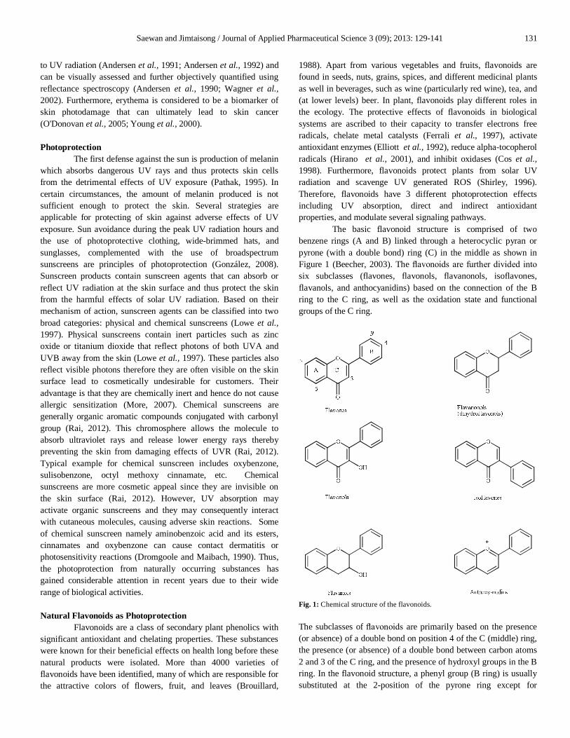
The basic flavonoid structure is comprised of two benzene rings (A and B) linked through a heterocyclic pyran or pyrone (with a double bond) ring (C) in the middle as shown in Figure 1 (Beecher, 2003). The flavonoids are further divided into six subclasses (flavones, flavonols, flavanonols, isoflavones, flavanols, and anthocyanidins) based on the connection of the B ring to the C ring, as well as the oxidation state and functional groups of the C ring.
Fig. 1: Chemical structure of the flavonoids. The subclasses of flavonoids are primarily based on the presence (or absence) of a double bond on position 4 of the C (middle) ring, the presence (or absence) of a double bond between carbon atoms 2 and 3 of the C ring, and the presence of hydroxyl groups in the B ring. In the flavonoid structure, a phenyl group (B ring) is usually substituted at the 2-position of the pyrone ring except for
132 Saewan and Jimtaisong / Journal of Applied Pharmaceutical Science 3 (09); 2013: 129-141
isoflavones the substitution is at the 3-position. Within each subclass, individual compounds differ in the pattern of substitution of the A and B rings (Beecher, 2003). Flavones
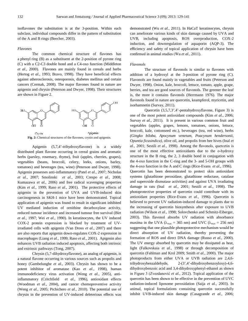
The common chemical structure of flavones has a phenyl ring (B) as a substituent at the 2-position of pyrone ring (C) with a C2-C3 double bond and a C4-oxo function (Middleton et al., 2000). Flavones are mainly found in cereals and herbs (Hertog et al., 1993; Bravo, 1998). They have beneficial effects against atherosclerosis, osteoporosis, diabetes mellitus and certain cancers (Cermak, 2008). The major flavones found in nature are apigenin and chrysin (Peterson and Dwyer, 1998). Their structures are shown in Figure 2.
Fig. 2: Chemical structures of the flavones, crysin and apigenin.
Apigenin (5,7,4’-trihydroxyflavone) is a widely distributed plant flavone occurring in cereal grains and aromatic herbs (parsley, rosemary, thyme), fruit (apples, cherries, grapes), vegetables (beans, broccoli, celery, leeks, onions, barley, tomatoes) and beverages (tea, wine) (Peterson and Dwyer, 1998). Apigenin possesses anti-inflammatory (Patel et al., 2007; Nicholas et al., 2007; Smolinski et al., 2003; Crespo et al., 2008; Kumazawa et al., 2006) and free radical scavenging properties (Kim et al., 1999; Raso et al., 2001). The protective effects of apigenin in the prevention of UVA and UVB-induced skin carcinogenesis in SKH-1 mice have been demonstrated. Topical application of apigenin was found to result in significant inhibited UV mediated induction of ornithine decarboxylase activity, reduced tumour incidence and increased tumour free survival (Birt et al., 1997; Wei et al., 1990). In keratinocytes, the UV induced COX-2 protein expression is blocked by treatment of UV-irradiated cells with apigenin (Van Dross et al., 2007) and there are also reports that apigenin down-regulates COX-2 expression in macrophages (Liang et al., 1999; Raso et al., 2001). Apigenin also enhances UVB radiation induced apoptosis, affecting both intrinsic and extrinsic pathways (Tong, 2007).
Chrysin (5,7-dihydroxyflavone), an analog of apigenin, is a natural flavone occurring in various sources such as propolis and honey (Gambelunghe et al., 2003). Chrysin has shown to be a potent inhibitor of aromatase (Kao et al., 1998), human immunodeficiency virus activation (Weng et al., 2005), anti-inflammatory (Critchfield et al., 1996), antioxidant effects (Woodman et al., 2004), and cancer chemopreventive activity (Weng et al., 2005; Pichichero et al., 2010). The potential use of chrysin in the prevention of UV-induced deleterious effects was
demonstrated (Wu et al., 2011). In HaCaT keratinocytes, chrysin can ameliorate various kinds of skin damage caused by UVA and UVB, including apoptosis, ROS overproduction, COX-2 induction, and downregulation of aquaporin (AQP-3). The efficiency and safety of topical application of chrysin have been confirmed in animal studies (Wu et al., 2011). Flavonols
The structure of flavonols is similar to flavones with addition of a hydroxyl at the 3-position of pyrone ring (C). Flavonols are found mainly in vagetables and fruits (Peterson and Dwyer, 1998). Onion, kale, broccoli, lettuce, tomato, apple, grape, berries, and tea are good sources of flavonols. The greener the leaf is, the more it contains flavonols (Herrmann 1976). The major flavonols found in nature are quercetin, keampferol, myricetin, and isorhamnetin (Survay, 2011).
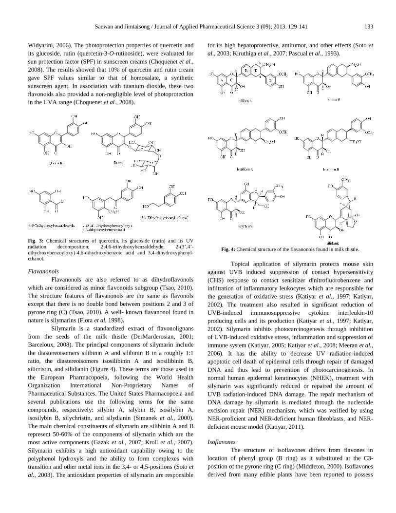
Quercetin (3,5,7,3’,4’-pentahydroxyflavone, Figure 3) is one of the most potent antioxidant compounds (Kim et al., 2006; Survay et al., 2011). It is present in various common fruit and vegetables (apples, grapes, lemons, tomatoes, onions, lettuce, broccoli, kale, cottonseed etc.), beverages (tea, red wine), herbs (Gingko biloba, Apocynum venetum, Poacynum hendersonii, Opuntia ficusindica), olive oil, and propolis from bee hives (Inal et al., 2001; Sestili et al., 1998). Among the flavonols, quercetin is one of the most effective antioxidants due to the o-hydroxy structure in the B ring, the 2, 3 double bond in conjugation with the 4-oxo function in the C-ring and the 3- and 5-OH groups with the 4-oxo function in the A and C rings (Rice-Evans et al., 1996). Quercetin has been demonstrated to protect skin antioxidant systems (glutathione peroxidase, glutathione reductase, catalase and superoxide dismutase activities) and against UVA irradiating damage in rats (Inal et al., 2001; Sestili et al., 1998). The photoprotective properties of quercetin could contribute with its antioxidant properties (Rice-Evans et al., 1996). Quercetin is believed to prevent UV radiation-induced damage to plants due to the increasing of quercetin biosynthesis after exposure to UVB radiation (Wilson et al., 1998; Solovchenko and Schmitz-Eiberger, 2003). This flavonol absorbs UV radiation with absorbance maxima in the UVA (max = 365 nm) and UVC (max = 256 nm) suggesting that one plausible photoprotective mechanism would be direct absorption of UV radiation, thereby preventing the formation of ROS and direct DNA damage (Russo et al., 2000). The UV energy absorbed by quercetin may be dissipated as heat, light (Falkovskaia et al., 1998) or through decomposition of quercetin (Fahlman and Krol 2009; Smith et al., 2000). The major photoproducts from either UVA or UVB radiation are 2,4,6-trihydroxybenzaldehyde, 2-(3’,4’-dihydroxybenzoyloxy)-4,6-dihydroxybenzoic acid and 3,4-dihydroxyphenyl-ethanol as shown in Figure 3 (Zvezdanović et al., 2012). Topical application of the quercetin has been shown to be effective in the prevention of UVC radiation-induced liposome peroxidation (Saija et al., 2003). In animal, topical formulations containing quercetin successfully inhibit UVB-induced skin damage (Casagrande et al., 2006;
Saewan and Jimtaisong / Journal of Applied Pharmaceutical Science 3 (09); 2013: 129-141 133
Widyarini, 2006). The photoprotection properties of quercetin and its glucoside, rutin (quercetin-3-O-rutinoside), were evaluated for sun protection factor (SPF) in sunscreen creams (Choquenet et al., 2008). The results showed that 10% of quercetin and rutin cream gave SPF values similar to that of homosalate, a synthetic sunscreen agent. In association with titanium dioxide, these two flavonoids also provided a non-negligible level of photoprotection in the UVA range (Choquenet et al., 2008).
Fig. 3: Chemical structures of quercetin, its glucoside (rutin) and its UV radiation decomposition; 2,4,6-trihydroxybenzaldehyde, 2-(3’,4’-dihydroxybenzoyloxy)-4,6-dihydroxybenzoic acid and 3,4-dihydroxyphenyl-ethanol. Flavanonols
Flavanonols are also referred to as dihydroflavonols which are considered as minor flavonoids subgroup (Tsao, 2010). The structure features of flavanonols are the same as flavonols except that there is no double bond between positions 2 and 3 of pyrone ring (C) (Tsao, 2010). A well- known flavanonol found in nature is silymarins (Flora et al, 1998).
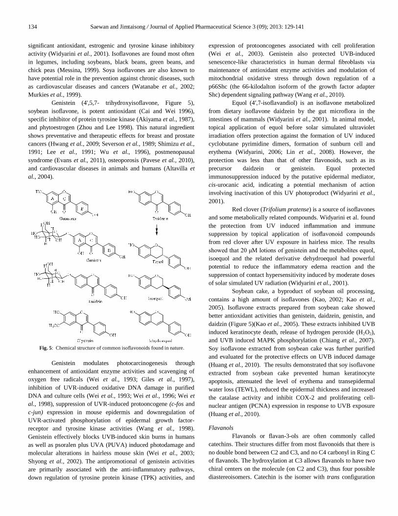
Silymarin is a standardized extract of flavonolignans from the seeds of the milk thistle (DerMarderosian, 2001; Barceloux, 2008). The principal components of silymarin include the diastereoisomers silibinin A and silibinin B in a roughly 1:1 ratio, the diastereoisomers isosilibinin A and isosilibinin B, silicristin, and silidianin (Figure 4). These terms are those used in the European Pharmacopoeia, following the World Health Organization International Non-Proprietary Names of Pharmaceutical Substances. The United States Pharmacopoeia and several publications use the following terms for the same compounds, respectively: silybin A, silybin B, isosilybin A, isosilybin B, silychristin, and silydianin (Simanek et al., 2000). The main chemical constituents of silymarin are silibinin A and B represent 50-60% of the components of silymarin which are the most active components (Gazak et al., 2007; Kroll et al., 2007). Silymarin exhibits a high antioxidant capability owing to the polyphenol hydroxyls and the ability to form complexes with transition and other metal ions in the 3,4- or 4,5-positions (Soto et al., 2003). The antioxidant properties of silymarin are responsible
for its high hepatoprotective, antitumor, and other effects (Soto et al., 2003; Kiruthiga et al., 2007; Pascual et al., 1993).
Fig. 4: Chemical structure of the flavanonols found in milk thistle.
Topical application of silymarin protects mouse skin against UVB induced suppression of contact hypersensitivity (CHS) response to contact sensitizer dinitrofluorobenzene and infiltration of inflammatory leukocytes which are responsible for the generation of oxidative stress (Katiyar et al., 1997; Katiyar, 2002). The treatment also resulted in significant reduction of UVB-induced immunosuppressive cytokine interleukin-10 producing cells and its production (Katiyar et al., 1997; Katiyar, 2002). Silymarin inhibits photocarcinogenesis through inhibition of UVB-induced oxidative stress, inflammation and suppression of immune system (Katiyar, 2005; Katiyar et al., 2008; Meeran et al., 2006). It has the ability to decrease UV radiation-induced apoptotic cell death of epidermal cells through repair of damaged DNA and thus lead to prevention of photocarcinogenesis. In normal human epidermal keratinocytes (NHEK), treatment with silymarin was significantly reduced or repaired the amount of UVB radiation-induced DNA damage. The repair mechanism of DNA damage by silymarin is mediated through the nucleotide excision repair (NER) mechanism, which was verified by using NER-proficient and NER-deficient human fibroblasts, and NER-deficient mouse model (Katiyar, 2011). Isoflavones
The structure of isoflavones differs from flavones in location of phenyl group (B ring) as it substituted at the C3-position of the pyrone ring (C ring) (Middleton, 2000). Isoflavones derived from many edible plants have been reported to possess
134 Saewan and Jimtaisong / Journal of Applied Pharmaceutical Science 3 (09); 2013: 129-141
significant antioxidant, estrogenic and tyrosine kinase inhibitory activity (Widyarini et al., 2001). Isoflavones are found most often in legumes, including soybeans, black beans, green beans, and chick peas (Messina, 1999). Soya isoflavones are also known to have potential role in the prevention against chronic diseases, such as cardiovascular diseases and cancers (Watanabe et al., 2002; Murkies et al., 1999).
Genistein (4',5,7- trihydroxyisoflavone, Figure 5), soybean isoflavone, is potent antioxidant (Cai and Wei 1996), specific inhibitor of protein tyrosine kinase (Akiyama et al., 1987), and phytoestrogen (Zhou and Lee 1998). This natural ingredient shows preventative and therapeutic effects for breast and prostate cancers (Hwang et al., 2009; Severson et al., 1989; Shimizu et al., 1991; Lee et al., 1991; Wu et al., 1996), postmenopausal syndrome (Evans et al., 2011), osteoporosis (Pavese et al., 2010), and cardiovascular diseases in animals and humans (Altavilla et al., 2004).
Fig. 5: Chemical structure of common isoflavonoids found in nature.
Genistein modulates photocarcinogenesis through enhancement of antioxidant enzyme activities and scavenging of oxygen free radicals (Wei et al., 1993; Giles et al., 1997), inhibition of UVR-induced oxidative DNA damage in purified DNA and culture cells (Wei et al., 1993; Wei et al., 1996; Wei et al., 1998), suppression of UVR-induced protooncogene (c-fos and c-jun) expression in mouse epidermis and downregulation of UVR-activated phosphorylation of epidermal growth factor-receptor and tyrosine kinase activities (Wang et al., 1998). Genistein effectively blocks UVB-induced skin burns in humans as well as psoralen plus UVA (PUVA) induced photodamage and molecular alterations in hairless mouse skin (Wei et al., 2003; Shyong et al., 2002). The antipromotional of genistein activities are primarily associated with the anti-inflammatory pathways, down regulation of tyrosine protein kinase (TPK) activities, and
expression of protooncogenes associated with cell proliferation (Wei et al., 2003). Genistein also protected UVB-induced senescence-like characteristics in human dermal fibroblasts via maintenance of antioxidant enzyme activities and modulation of mitochondrial oxidative stress through down regulation of a p66Shc (the 66-kilodalton isoform of the growth factor adapter Shc) dependent signaling pathway (Wang et al., 2010).
Equol (4′,7-isoflavandiol) is an isoflavone metabolized from dietary isoflavone daidzein by the gut microflora in the intestines of mammals (Widyarini et al., 2001). In animal model, topical application of equol before solar simulated ultraviolet irradiation offers protection against the formation of UV induced cyclobutane pyrimidine dimers, formation of sunburn cell and erythema (Widyarini, 2006; Lin et al., 2008). However, the protection was less than that of other flavonoids, such as its precursor daidzein or genistein. Equol protected immunosuppression induced by the putative epidermal mediator, cis-urocanic acid, indicating a potential mechanism of action involving inactivation of this UV photoproduct (Widyarini et al., 2001).
Red clover (Trifolium pratense) is a source of isoflavones and some metabolically related compounds. Widyarini et al. found the protection from UV induced inflammation and immune suppression by topical application of isoflavonoid compounds from red clover after UV exposure in hairless mice. The results showed that 20 μM lotions of genistein and the metabolites equol, isoequol and the related derivative dehydroequol had powerful potential to reduce the inflammatory edema reaction and the suppression of contact hypersensitivity induced by moderate doses of solar simulated UV radiation (Widyarini et al., 2001).
Soybean cake, a byproduct of soybean oil processing, contains a high amount of isoflavones (Kao, 2002; Kao et al., 2005). Isoflavone extracts prepared from soybean cake showed better antioxidant activities than genistein, daidzein, genistin, and daidzin (Figure 5)(Kao et al., 2005). These extracts inhibited UVB induced keratinocyte death, release of hydrogen peroxide (H2O2), and UVB induced MAPK phosphorylation (Chiang et al., 2007). Soy isoflavone extracted from soybean cake was further purified and evaluated for the protective effects on UVB induced damage (Huang et al., 2010). The results demonstrated that soy isoflavone extracted from soybean cake prevented human keratinocyte apoptosis, attenuated the level of erythema and transepidermal water loss (TEWL), reduced the epidermal thickness and increased the catalase activity and inhibit COX-2 and proliferating cell-nuclear antigen (PCNA) expression in response to UVB exposure (Huang et al., 2010). Flavanols
Flavanols or flavan-3-ols are often commonly called catechins. Their structures differ from most flavonoids that there is no double bond between C2 and C3, and no C4 carbonyl in Ring C of flavanols. The hydroxylation at C3 allows flavanols to have two chiral centers on the molecule (on C2 and C3), thus four possible diastereoisomers. Catechin is the isomer with trans configuration
Saewan and Jimtaisong / Journal of Applied Pharmaceutical Science 3 (09); 2013: 129-141 135
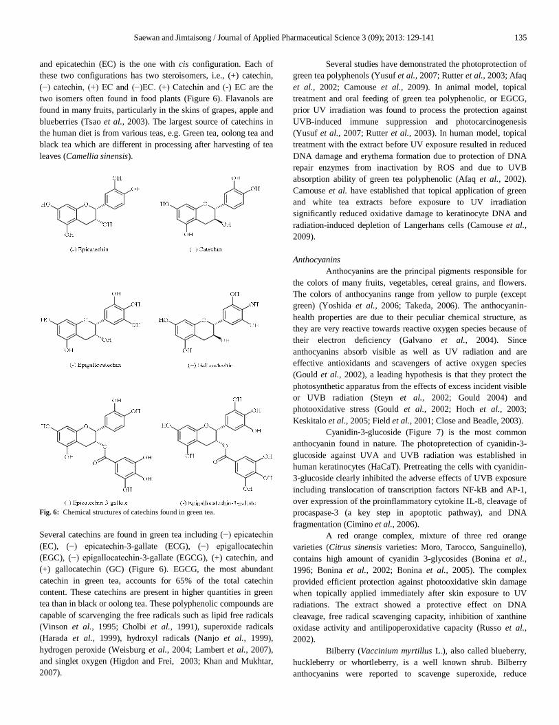
and epicatechin (EC) is the one with cis configuration. Each of these two configurations has two steroisomers, i.e., (+) catechin, (−) catechin, (+) EC and (−)EC. (+) Catechin and (-) EC are the two isomers often found in food plants (Figure 6). Flavanols are found in many fruits, particularly in the skins of grapes, apple and blueberries (Tsao et al., 2003). The largest source of catechins in the human diet is from various teas, e.g. Green tea, oolong tea and black tea which are different in processing after harvesting of tea leaves (Camellia sinensis).
Fig. 6: Chemical structures of catechins found in green tea. Several catechins are found in green tea including (−) epicatechin (EC), (−) epicatechin-3-gallate (ECG), (−) epigallocatechin (EGC), (−) epigallocatechin-3-gallate (EGCG), (+) catechin, and (+) gallocatechin (GC) (Figure 6). EGCG, the most abundant catechin in green tea, accounts for 65% of the total catechin content. These catechins are present in higher quantities in green tea than in black or oolong tea. These polyphenolic compounds are capable of scarvenging the free radicals such as lipid free radicals (Vinson et al., 1995; Cholbi et al., 1991), superoxide radicals (Harada et al., 1999), hydroxyl radicals (Nanjo et al., 1999), hydrogen peroxide (Weisburg et al., 2004; Lambert et al., 2007), and singlet oxygen (Higdon and Frei, 2003; Khan and Mukhtar, 2007).
Several studies have demonstrated the photoprotection of green tea polyphenols (Yusuf et al., 2007; Rutter et al., 2003; Afaq et al., 2002; Camouse et al., 2009). In animal model, topical treatment and oral feeding of green tea polyphenolic, or EGCG, prior UV irradiation was found to process the protection against UVB-induced immune suppression and photocarcinogenesis (Yusuf et al., 2007; Rutter et al., 2003). In human model, topical treatment with the extract before UV exposure resulted in reduced DNA damage and erythema formation due to protection of DNA repair enzymes from inactivation by ROS and due to UVB absorption ability of green tea polyphenolic (Afaq et al., 2002). Camouse et al. have established that topical application of green and white tea extracts before exposure to UV irradiation significantly reduced oxidative damage to keratinocyte DNA and radiation-induced depletion of Langerhans cells (Camouse et al., 2009). Anthocyanins
Anthocyanins are the principal pigments responsible for the colors of many fruits, vegetables, cereal grains, and flowers. The colors of anthocyanins range from yellow to purple (except green) (Yoshida et al., 2006; Takeda, 2006). The anthocyanin-health properties are due to their peculiar chemical structure, as they are very reactive towards reactive oxygen species because of their electron deficiency (Galvano et al., 2004). Since anthocyanins absorb visible as well as UV radiation and are effective antioxidants and scavengers of active oxygen species (Gould et al., 2002), a leading hypothesis is that they protect the photosynthetic apparatus from the effects of excess incident visible or UVB radiation (Steyn et al., 2002; Gould 2004) and photooxidative stress (Gould et al., 2002; Hoch et al., 2003; Keskitalo et al., 2005; Field et al., 2001; Close and Beadle, 2003).
Cyanidin-3-glucoside (Figure 7) is the most common anthocyanin found in nature. The photopretection of cyanidin-3-glucoside against UVA and UVB radiation was established in human keratinocytes (HaCaT). Pretreating the cells with cyanidin-3-glucoside clearly inhibited the adverse effects of UVB exposure including translocation of transcription factors NF-kB and AP-1, over expression of the proinflammatory cytokine IL-8, cleavage of procaspase-3 (a key step in apoptotic pathway), and DNA fragmentation (Cimino et al., 2006).
A red orange complex, mixture of three red orange varieties (Citrus sinensis varieties: Moro, Tarocco, Sanguinello), contains high amount of cyanidin 3-glycosides (Bonina et al., 1996; Bonina et al., 2002; Bonina et al., 2005). The complex provided efficient protection against photooxidative skin damage when topically applied immediately after skin exposure to UV radiations. The extract showed a protective effect on DNA cleavage, free radical scavenging capacity, inhibition of xanthine oxidase activity and antilipoperoxidative capacity (Russo et al., 2002).
Bilberry (Vaccinium myrtillus L.), also called blueberry, huckleberry or whortleberry, is a well known shrub. Bilberry anthocyanins were reported to scavenge superoxide, reduce

136 Saewan and Jimtaisong / Journal of Applied Pharmaceutical Science 3 (09); 2013: 129-141
hydrogen peroxide induced radicals, and inhibit lipid peroxidation in vitro (Mart ı́n-Arag´on et al., 1996) and in vivo (Milbury et al., 2002). Pretreatment (1 h) or posttreatment (4 h) of HaCaT keratinocytes with bilberry fruit extract which consist of 25% anthocyanins resulted in reduction of UVA stimulated ROS formation, attenuation of UVA caused peroxidation of membrane lipids and depletion of intracellular GSH (Svobodov´a et al., 2008).
Fig. 7: Chemical structures of the common anthocyanins found in nature.
Bog blueberry (Vaccinium uliginosum L.) extract consists of cyanidin-3-glucoside, petunidin-3-glucoside, malvidin-3-glucoside, and delphinidin-3-glucoside as the major anthocyanins (Figure 7). Bog blueberry anthocyanins showed the protected against UVB-induced skin photoaging by blocking collagen destruction and inflammatory responses via transcriptional mechanisms of NF-κB and MAPK signaling (Bae et al., 2009).
Strawberry (Fragaria x ananassa) is one of the most commonly consumed berries worldwide as an important dietary source of bioactive compounds, most of which are natural antioxidants that contribute to the high nutritional quality of the fruit. The high variety and content of polyphenolic constituent of strawberries are mainly represented by anthocyanins and hydrolyzable tannins. Anthocyanins are the quantitatively most important in strawberry (Tulipani et al., 2008; Giampieri et al., 2012a; Aaby et al., 2007). Pelargonidin is the major representative aglycone, although also some cyanidin derivatives are generally observed (Figure 8). The strawberry extract showed strong anti-inflammatory (Heim et al., 2012), antioxidant (Wang and Lin, 2000), antimutagenic (Okuda et al., 1989), anticarcinogenic (Wedge et al., 2001). The photoprotective properties of strawberry have been demonstrated in human dermal fibroblasts (Giampieri et al., 2012b). A photoprotective activity was demonstrated by
increasing cellular viability, and diminishing DNA damage on UVA-induced skin damage. Its photoprotective potential may be based on the high level of anthocyanins which is potentially able to block collagen destruction and inflammatory responses via transcriptional mechanisms of NF-κB and MAPK signaling (Giampieri et al., 2012b).
Fig. 8: Chemical structures of anthocyanin found in strawberry. CONCLUSION
Exposure to ultraviolet radiation is associated with a variety of harmful effects ranging from photoaging to skin cancer. Many synthetic sunscreen agents are available in the market with certain limitation according to their potential for generating photoreaction adducts and may consequently create adverse effects on human skin. Natural compounds may work in various ways such as stimulate the immune response, induce gene suppression, block oxidative damage to DNA, etc. Natural flavonoids are one candidate for prevention of the adverse effects of UV radiation due to their UV absorbing property, antioxidant properties, and modulate several signaling pathways. However, the development of new substances must demonstrate for their safety in addition to their photoprotective effect. REFERENCES
Aaby K, Ekeberg D, Skrede G. Characterization of phenolic compounds in strawberry (Fragaria × ananassa) fruits by different HPLC detectors and contribution of individual compounds to total antioxidant capacity. J Agric Food Chem, 2007; 55:4395−4406.
Adhami VM, Syed DN, Khan N, Afaq F. Phytochemicals for prevention of solar ultraviolet radiationinduced damages. Photochem Photobiol, 2008; 84:489-500.
Afaq F, Adhami VM, Ahmad N, Mukhtar H. Botanical antioxidants for chemoprevention of photocarcinogenesis. Front Biosci, 2002; 7:784–792.
Afaq F, Mukhtar H. Effects of solar radiation on cutaneous detoxification pathways. J Photochem Photobiol B, 2001; 63:61–69.
Saewan and Jimtaisong / Journal of Applied Pharmaceutical Science 3 (09); 2013: 129-141 137
Afaq F, Mukhtar H. Photochemoprevention by botanical antioxidants. Skin Pharmacol Appl Skin Physiol, 2002; 15:297–306.
Ahmad N, Gilliam AC. A definitive role of ornithine decarboxylase in photocarcinogenesis. American Journal of Pathology, 2001; 159:885-892.
Aitken GR, Henderson JR, Chang SC, McNeil CJ, Birch-Machin MA. Direct monitoring of UV-induced free radical generation in HaCaT keratinocytes. Clin Exp Dermatol, 2007; 32:722-727.
Akiyama T, Ishida J, Nakagawa S, Ogawara H, Watanabe S, Itoh N, Shibuya M, Fukami Y. Genistein, a specific inhibitor of tyrosine-specific protein kinases. J Biol Chem, 1987; 262:5592–5595.
Altavilla D, Crisafulli, A, Marini H, Esposito M, D'Anna R, Corrado F, Bitto A, Squadrito F. Cardiovascular effects of the phytoestrogen genistein.Curr Med Chem Cardiovasc Hematol Agents, 2004; 2:179-186.
Andersen PH, Abrams K, Bjerring P, Maibach H. A time-correlation study of ultraviolet B-induced erythema measured by reflectance spectroscopy and laser Doppler flowmetry. Photodermatol Photoimmunol Photomed, 1991; 8:123–128.
Andersen PH, Abrams K, Maibach H. Ultraviolet B dose-dependent inflammation in humans: a reflectance spectroscopic and laser Doppler flowmetric study using topical pharmacologic antagonists on irradiated skin. Photodermatol Photoimmunol Photomed, 1992; 9:17–23.
Andersen PH, Bjerring P. Spectral reflectance of human skin in vivo. Photodermatol Photoimmunol Photomed, 1990; 7:5–12.
Andley UP, Becker B, Hebert JS, Reddan JK, Morrison AR, Pentland AP. Enhanced prostaglandin synthesis after ultraviolet-B exposure modulates DNA synthesis of lens epithelial cells and lowers Intraocular pressure in vivo. Invest Ophthalmol Vis Sci, 1996; 37:142-153.
Bae JY, Lim SS, Kim SJ, Choi JS, Park J, Ju SM, Han SJ, Kang IJ, Kang YH. Bog blueberry anthocyanins alleviate photoaging in ultraviolet-B irradiation-induced human dermal fibroblasts. Mol Nutr Food Res, 2009; 53:726−738.
Barceloux DG. 2008. Medical Toxicology of Natural Substances. New Jersey: John Wiley & Sons, Inc.
Bashir MM, Sharma MR, Werth VP. TNF-alpha production in the skin Arch Dermatol Res, 2009; 301:87-91.
Beecher GR. Overview of dietary flavonoids: nomenclature, occurrence and intake. J Nutr, 2003; 133:3248S-3254S.
Birt DF, Mitchell D, Gold B, Pour P, Pinch HC. Inhibition of ultraviolet light induced skin carcinogenesis in SKH-1 mice by apigenin, a plant flavonoid. Anticancer Res, 1997; 17: 85–92.
Bonina F, Lanza M, Montenegro L, Puglisi C, Tomaino A and Trombetta D. Flavonoids as potential protective agents against photo-oxidative skin damage. Int J Pharm, 1996; 145:87-91.
Bonina FP, Leotta C, Scalia G, Puglia C, Trombetta D, Tringali G, Roccazzello AM, Rapisarda P, Saija A. Evaluation of oxidative stress in diabetic patients after supplementation with standardised red orange extract. Diabetes Nutr Metab, 2002; 15:14–19.
Bonina FP, Puglia C, Cimino F, Trombetta D, Tringali G, Roccazzelloc RM, Insirelloc E, Rapisardad P, Saija A. Oxidative stress in handball players: effect of supplementation with a red orange extract. Nutr Res, 2005; 25:917–924.
Bravo L. Polyphenols: chemistry, dietary sources, metabolism, and nutritional significance. Nutr Rev, 1998; 56:317–333.
Brenner M, Hearing VJ. The protective role of melanin against UV damage in human skin. Photochem Photobiol, 2008; 84: 539–549.
Brouillard R, Cheminant A. 1998. Flavonoids and plant color. In: Cody V, Middleton E and Harborne JB eds. plant flavonoids in biology and medicine: biochemical, cellular and medicinal properties. New York: Alan R. Liss, Inc. 93–106.
Cai Q, Wei H. Effect of dietary genistein on antioxidant enzyme activities in SENCAR mice. Nutr Cancer, 1996; 25:1–7.
Camouse MM, Domingo DS, Swain FR, Conrad EP, Matsui MS, Maes D, Declercq L, Cooper KD, Stevens SR, Baron ED. Topical application of green and white tea extracts provides protection from solar-simulated ultraviolet light in human skin. Exp Dermatol, 2009; 18:522-526.
Capote R, Alonso-Lebrero JL, Garcia F, Brieva A, Pivel JP, Gonzalez S. Polypodium leucotomos extract inhibits trans-urocanic acid photoisomerization and photodecomposition. J. Photochem Photobiol B, 2006; 82: 173–179.
Casagrande R, Georgetti SR, Verri WA Jr, Dorta DJ, dos Santos AC, Fonseca MJ. Protective effect of topical formulations containing quercetin against UVB-induced oxidative stress in hairless mice. J. Photochem Photobiol B, 2006; 84:21-27.
Cermak R. Effect of dietary flavonoids on pathways involved in drug metabolism. Expert Opin Drug Metab Toxicol, 2008; 4:17–35.
Chiang HS, Wu WB, Fang JY, Chen BH, Kao TH, Chen YT, Huang CC, Hung CF. UVB-protective effects of isoflavone extracts from soybean cake in human keratinocytes. Int J Mol Sci, 2007; 8:651-661.
Cholbi MR, Paya M, Alcaraz MJ. Inhibitory effects of phenolic compounds on CCl4-induced microssomal lipid peroxidation. Experientia, 1991; 47: 195-199.
Choquenet B, Couteau C, Paparis E, Coiffard IJ. Quercetin and rutin as potential sunscreen agents: Determination of efficacy by an in vitro method. J Nat Prod, 2008; 71:1117–1118.
Chung HT, BurnhamDK, Robertson B, Roberts LK, Daynes RA. Involvement of prostaglandins in the immune alterations caused by the exposure of mice to ultraviolet radiation. J Immunol, 1986; 137: 2478–2484.
Chung JH, Youn SH, Koh WS, Eun HC, Cho KH, Park KC, Youn JI. Ultraviolet B irradiation-enhanced interleukin (IL)-6 production and mRNA expression are mediated by IL-1α in cultured human keratinocytes. J Invest Dermatol, 1996; 106: 715–720.
Cimino F, Ambra R, Canali R, Saija A, Virgili F. Effect of cyanidin-3-O-glucoside on UVB-induced response in human keratinocytes. J. Agric. Food Chem, 2006; 54:4041−4047.
Close, DC, Beadle CL. The ecophysiology of foliar anthocyanin. Botanical Review, 2003; 69:149-161.
Clydesdale GJ, Dandie GW, Muller HK. Ultraviolet light induced injury: Immunological and inflammatory effests. Immunol Cell Biol, 2001; 79:547–568.
Cos P, Ying L, Calomme M, Hu JP, Cimanga K, Van Poel B, Pieters L, Vlietnck AJ, Vanden Berghe D. Structure-activity relationship and classification of flavonoids as inhibitors of xanthine oxidase and superoxide scavengers, J Nat Prod, 1998; 61:71–76.
Crespo I, Garcia-Mediavilla MV, Almar M, Gonzalez P, Tunon MJ, Sanchez-Campos S, Gonzalez-Gallego J. Differential effects of dietary flavonoids on reactive oxygen and nitrogen species generation and changes in antioxidant enzyme expression induced by proinflammatory cytokines in chang liver cells. Food Chem Toxicol, 2008; 46:1555-1569.
Critchfield JW, Butera ST, Folks TM. Inhibition of HIV activation in latently infected cells by flavonoid compounds. AIDS Res. Hum. Retroviruses, 1996; 12:39-46.
DerMarderosian A. The Reviews of natural products. 1st ed. Facts and Comparisons: St. Louis, Missouri, 2001.
Dromgoole SH, Maibach HI. Sun-screening intolerance: Contact and photocontact sensitization and contact urticaria. J. Am. Acad. Dermatol, 1990; 22:1068-1078.
Duthie MS, Kimber I, Norval M. The effects of ultraviolet radiation on the human immune system. Br. J. Dermatol, 1999; 140:995–1009.
Elliott AJ, Scheiber SA, Thomas C, Pardini RS. Inhibition of glutathione reductase by flavonoids, Biochem Pharmacol, 1992; 44:1603–1608.
Evans M, Elliott JG, Sharma P, Berman R, Guthrie N. The effect of synthetic genistein on menopause symptom management in healthy postmenopausal women: a multi-center, randomized, placebo-controlled study. Maturitas, 2011; 68:189-96.
Fahlman BM, Krol ES. UVA and UVB radiation-induced oxidation products of quercetin. Journal of Photochemistry and Photobiology B: Biology, 2009; 97:123–131.
Falkovskaia E, Sengupta PK, Kasha M. Photophysical induction of dual fluorescence of quercetin and related hydroxyflavones upon
138 Saewan and Jimtaisong / Journal of Applied Pharmaceutical Science 3 (09); 2013: 129-141
intermolecular H-bonding to solvent matrix, Chem. Phys. Lett, 1998; 297:109–114.
Feild TS, Lee DW, Holbrook NM. Why leaves turn red in autumn. The role of anthocyanins in senescing leaves of red-osier dogwood. Plant Physiology, 2001; 127:566-574.
Feldmeyer L, Keller M, Niklaus G, Hohl D, Werner S, Beer HD. The inflammasome mediates UVB-induced activation and secretion of interleukin-1β by keratinocytes. Curr Biol, 2007; 17: 1140–1145.
Ferrali M, Signorini C, Caciotti B, Sugherini L, Ciccoli L, Giachetti D, Comporti M. Protection against oxidative damage of erythrocyte membranes by the flavonoid quercetin and its relation to iron chelating activity. Febs Lett, 1997; 416:123–129.
Flora K, Hahn M, Rosen H, Benner K. Milk thistle (Silybum marianum) for the therapy of liver disease. Am J Gastroenterol, 1998; 93:139-143.
Galvano F, Fauci LL, Lazzarino G, Fogliano V. Cyanidins:metabolism and biological properties. J Nutr Biotechem, 2004; 15:2-11.
Gambelunghe C, Rossi R, Sommavilla M, Ferranti C, Rossi R, Ciculi C, Gizzi S, Micheletti A, Rufini S. Effects of chrysin on urinary testosterone levels in human males. J Med Food, 2003; 6:387-390.
Gazak R, Walterova D, Kren V. Silybin and silymarin: new and emerging applications in medicine. Curr Med Chem, 2007; 14:315-338.
Giampieri F, Alvarez-Suarez J, Tulipani S, Gonzàles-Paramàs AM, Santos-Buelga C, Bompadre S, Quiles J, Mezzetti B, Battino M. Photoprotective potential of strawberry (Fragaria × ananassa) extract against UVA irradiation damage on human fibroblasts. J. Agric. Food Chem, 2012b; 60: 2322−2327.
Giampieri F, Tulipani S, Alvarez-Suarez JM, Quiles JL, Mezzetti B, Battino M. The strawberry: Composition, nutritional quality, and impact on human health. Nutrition, 2012a; 28:9−19.
Giles D, Wei H. Effect of structurally-related flavones/isoflavone on hydrogen peroxide production and oxidative DNA damage in a phorbol ester-stimulated HL-60 cells. Nutr Cancer, 1997; 29:77-82.
Goihman-Yahr M. Skin aging and photoaging: An Outlook Clin Dermatol, 1996; 14: 153–160.
González S, Fernández-Lorente M, Gilaberte-Calzada Y. The latest on skin photoprotection. Clinics in Dermatology, 2008; 26:614-626.
Gould KS, McKelvie J, Markham KR. Do anthocyanins function as antioxidants in leaves? Imaging of H2O2 in red and green leaves after mechanical injury. Plant Cell Environ, 2002; 25:1261-1269.
Gould KS. Nature's Swiss Army Knife: The diverse protective roles of anthocyanins in leaves. J. Biomed. Biotechnol, 2004; 5: 314–320.
Gruijl FR. Photocarcinogenesis: UVA vs UVB. Methods Enzymol, 2000; 319:359-366.
Halliday GM. Inflammation, gene mutation and photoimmunosuppression in response to UVR-induced oxidative damage contributes to photocarcinogenesis. Mutat Res, 2005; 571:107–120.
Harada M, Kan Y, Naoki H, Fukui Y, Kageyama N, Nakai M, Miki W, Kiso Y. Identification of the major antioxidative metabolites in biological fluids of the rat with ingested (+)-catechin and (-)-epicatechin. Biosci Biotechnol Biochem, 1999; 63:973-977.
Heim KC, Angers P, Le´onhart S, Ritz BW. Anti-inflammatory and neuroactive properties of selected fruit extracts. J Med Food, 2012; 15:1–4.
Herrmann K. Flavonols and flavones in food plants: A review. J Food Technol, 1976; 11:433–448.
Hertog MGL, Hollman PCH, Putte B. Content of potentially anticarcinogenic flavonoids of tea infusions, wines, and fruit juices. J Agric Food Chem, 1993; 41:1242–1246.
Higdon JV, Frei B. Tea catechins and polyphenols: health effects, metabolism, and antioxidant functions. Crit Rev Food Sci Nutr, 2003; 43:89-143.
Hirano R, Sasamoto W, Matsumoto A, Itakura H, Igarashi O, Kondo K. Antioxidant ability of various flavonoids against DPPH radicals and LDL oxidation, J Nutr Sci Vitaminol (Tokyo), 2001; 47:357–362.
Hoch WA, Singsaas EL, McCown BH. Resorption protection. Anthocyanins facilitate nutrient recovery in autumn by shielding leaves from potentially damaging light levels. Plant Physiol, 2003; 133:1296-1305.
Huang CC, Hsu BY, Wu NL, Tsui WH,Lin TJ, Su CC, Hung CF. Anti-photoaging effects of soy isoflavone extract (aglycone and acetylglucoside form) from soybean cake. Int J Mol Sci, 2010; 12:4782-4795.
Hwang YW, Kim SY, Jee SH, Kim YN, Nam CM. Soy food consumption and risk of prostate cancer: A meta-analysis of observational studies. Nutrition and Cancer, 2009; 61: 598–606.
Ichihashi M, Ando H, Yoshida M, Niki Y, Matsui M. Photoaging of the skin. Anti-Aging Medicine, 2009; 6:46-59.
Inal ME, Kahramant A, Kkent T. Beneficial effects of quercetin on oxidative stress induced by ultraviolet A. Clin Exp Dermatol, 2001; 26: 536–539.
Kao TH, Chen BH. An improved method for determination of isoflavones in soybean powder by liquid chromatography. Chromatographia, 2002; 56:423-430.
Kao TH, Lu YF, Chen BH. Preparative column chromatography of four groups of isoflavones from soybean cake. Eur. Food Res. Technol, 2005; 221:459-465.
Kao Y-C, Zhou C, Sherman M, Laughton CA, Chen S. Molecular basis of the inhibition of human aromatase (estrogen synthetase) by flavone and isoflavone phytoestrogens: a site-directed mutagenesis study. Environ. Health Perspect, 1998; 106:85–92.
Katiyar S. Silymarin and skin cancer prevention: anti-inflammatory, antioxidant and immunomodulatory effects (Review). Int J Oncol, 2005; 26:169-76.
Katiyar SK, Elmets CA. Green tea polyphenolic antioxidants and skin photoprotection (Review). Int J Oncol, 2001; 18:1307–1313.
Katiyar SK, Korman NJ, Mukhtar H, Agarwal R. Protective effects of silymarin against photocarcinogenesis in a mouse skin model. J Natl Cancer Inst, 1997; 89:556–566.
Katiyar SK, Mantena SK, Meeran SM. Silymarin Protects epidermal keratinocytes from ultraviolet radiation-induced apoptosis and DNA damage by nucleotide excision repair mechanism. PLoS ONE, 2011; 6: e21410.
Katiyar SK, Meleth S, Sharma SD. Silymarin, a flavonoid from milk thistle (Silybum marianum L.), inhibits UV-induced oxidative stress through targeting infiltrating CD11b+ cells in mouse skin. Photochem Photobiol, 2008; 84:266–271.
Katiyar SK. Treatment of silymarin, a plant flavonoid, prevents ultraviolet light-induced immune suppression and oxidative stress in mouse skin. Int J Oncol, 2002; 21:1213-1222.
Keskitalo J, Bergquist G, Gardeström P, Jansson S. A cellular timetable of autumnal senescence in aspen. Plant Physiology, 2005; 139:1635-1648.
Khan N, Mukhtar H. Tea polyphenols for health promotion. Life Sci, 2007; 81:519-533.
Kim H, Jeong K, Jung S. Molecular Ddynamics simulations on the coplanarity of quercetin backbone for the antioxidant activity of quercetin-3-monoglycoside. Bull. Korean Chem Soc, 2006; 27: 325-328.
Kim HK, Cheon BS, Kim YH, Kim SY, Kim HP. Effects of naturally occurring flavonoids on nitric oxide production in the macrophage cell line RAW 264.7 and their structure activity relationships. Biochem. Pharmacol, 1999; 58:759-765.
Kiruthiga PV, Shafreen RB, Pandian SK, Arun S, Govindu S. Devi, K.P. Protective effect of silymarin on erythrocyte haemolysate against benzo(a)pyrene and exogenous reactive oxygen species (H2O2) induced oxidative stress. Chemosphere, 2007; 68: 1511-1518.
Kondo S, Kono T, Sauder DN, McKenzie RC. IL-8 gene expression and production in human keratinocytes and their modulation by UVB. J Invest Dermatol, 1993; 101: 690–694.
Kondo S, Sauder DN, Kono T, Galley KA, McKenzie RC. Differential modulation of interleukin-1 alpha (IL-1 alpha) and interleukin-1 beta (IL-1 beta) in human epidermal keratinocytes by UVB. Exp Dermatol, 1994; 3:29-39.
Saewan and Jimtaisong / Journal of Applied Pharmaceutical Science 3 (09); 2013: 129-141 139
Kripke ML. Immunological unresponsiveness induced by ultraviolet radiation. Immunol Rev, 1984; 80: 87–102.
Kroll DJ, Shaw HS, Oberlies NH. Milk thistle nomenclature: why it matters in cancer research and pharmacokinetic studies. Integr Cancer Ther, 2007; 6:110-119.
Krutmann J. The role of UV rays in skin aging. Eur J Dermatol, 2001; 11:170-171.
Kumazawa Y, Kawaguchi K, Takimoto H. Immunomodulating effects of flavonoids on acute and chronic inflammatory responses caused by tumor necrosis factor alpha. Curr Pharm Des, 2006; 12:4271-4279.
Lambert JD, Sang S, Yang CS. Possible controversy over dietary polyphenols: benefits vs risks. Chem Res Toxicol, 2007; 20:583-585.
Lee HP, Gourley L, Duffy SW, Esteve J, Lee J, Day NE. Dietary effects on breast-cancer risk in Singapore. Lancet, 1991; 337: 1197–1200.
Liang YC, Huang YT, Tsai SH, Lin-Shiau SY, Chen CF, Lin JK. Suppression of inducible cyclooxygenase and inducible nitric oxide synthase by apigenin and related flavonoids in mouse macrophages. Carcinogenesis, 1999; 20:1945–1952.
Lin J, Tournas J, Burch J, Monteiro-Riviere N, Zielinski J. Topical isoflavones provide effective photoprotection to skin. Photodermatol Photoimmunol Photomed, 2008; 24:61-66.
Loser K, Apelt J, Voskort M, Mohaupt M, Balkow S, Schwarz T et al. IL-10 controls ultraviolet-induced carcinogenesis in mice. J Immunol, 2007; 179: 365–371.
Lowe NJ, Shauth NA, Patahk MA. 1997. Sunscreens: Development, evaluation and regulatory aspects. New York: Marcel Dekker
Mart´ın-Arag´on SB, Basabe B, Bened JM, Viklat AM. Antioxidant action of Vaccinium myrtillus L. Phototerapy Res, 1998; 12:S104–S106.
McKenzie RL, Björn LO, Bais A, Ilyasd M. Changes in biologically active ultraviolet radiation reaching the Earth’s surface. Photochem Photobiol Sci, 2003; 2:5–15.
Meeran SM, Katiyar S, Elmets CA, Katiyar SK. Silymarin inhibits UV radiation-induced immunosuppression through augmentation of interleukin-12 in mice. Mol Cancer Ther, 2006; 5:1660–1668.
Messina MJ. 1999. Legumes and soybeans: overview of their nutritional profiles and health effects. Am J Clin Nutr, 1999;70(suppl):439S–50S.
Middleton EJR, Kandaswami C, Theoharides TC. The effects of plant flavonoids on mammalian cells:implications for inflammation, heart disease, and cancer. Pharmacol Rev, 2000; 52:673–751.
Milbury BE, Gao G, Prior RL, Blumberg J. Bioavailablility of elderberry anthocyanins. Mech Ageing Dev, 2002; 123:997–1006.
Mitchell DL : The relative cytotoxicity of (6-4) photoproducts and cyclobutane dimers in mammalian cells. Photochem Photobiol 48 :51-57, 1988
Mitchell DL, Nairn RS. The biology of the (6-4) photoproduct. Photochem Photobiol, 1989; 49:805-819.
More BD. Physical sunscreens: on the comeback trail. Indian J Dermatol Venereol Leprol, 2007; 73:80-85.
Murkies AL, Dalais FS, Briganti EM, Healy DL, Urger HG, Wahlqvist ML, Davis SR. Phytoestrogen and androgen levels in Australian. The 3rd International Symposium on the Role of Soy in Preventing and Treating Chronic Diseases, Washington, DC. 1999. p. 38.
Nanjo F, Mori M, Goto K, Hara Y. Radical scavenging activity of tea catechins and their related compounds. Biosci Biotechnol Biochem, 1999; 63:1621.
Nicholas C, Batra S, Vargo MA, Voss OH, Gavrilin MA, Wewers MD, Guttridge DC, Grotewold E, Doseff AI. Apigenin blocks lipopolysaccharide-induced lethality in vivo and proinflammatory cytokines expression by inactivating NF-κB through the suppression of p65 phosphorylation. J Immunol, 2007; 179:7121-7127.
O'Donovan P, Perrett CM, Zhang X, Montaner B, Xu YZ, Harwood CA et al. Azathioprine and UVA light generate mutagenic oxidative DNA damage. Science, 2005; 309: 1871–1874.
Okuda T, Yoshida T, Hatano T. Ellagitannins as active constituents of medical plants. Planta Med, 1989; 55:117-22.
Park HM, Moon E, Kim AJ, Kim MH, Lee S, Lee JB, Park YK, Jung HS, Kim YB, Kim SY. Extract of Punica granatum inhibits skin photoaging induced by UVB irradiation. Int J Dermatol, 2010; 49:276-82.
Pascual C, Gonzalez R, Armesto J, Muriel P. Effect of silymarin and silybinin on oxygen radicals. Drug Development Research, 1993; 29: 73-77.
Patel D, Shukla S, Gupta S. Apigenin and cancer chemoprevention: progress, potential and promise (review). Int J Oncol, 2007; 30:233-245.
Pathak M. A. 1995 Functions of melanin and protection by melanin. In Melanin: Its Role in Human Photoprotection (Zeise, L.,Chedekel, M. R., and Fitzpatrick, T. B., eds) pp. 125–134,Valdemar Publishing Company, Overland Park
Pavese JM, Farmer RL, Bergan RC. Inhibition of cancer cell invasion and metastasis by genistein. Cancer Metastasis Rev 2010; 29:465–482.
Peterson J, Dwyer J. Flavonoids: dietary occurrence and biochemical activity. Nutr Res. 1998; 18:1995–2018.
Pichichero E, Cicconi R, Mattei M, Gallinella-Muzi M, Canini A. Acacia honey and chrysin reduces proliferation of melanoma cells through alteration in cell cycle progression. Int J Oncol 2010; 37: 973-981.
Rai R, Shanmuga SC, Srinivas CR. Update on photoprotection. Indian J Dermatol. 2012; 57: 335–342.
Raso GM, Meli R, Di Carlo G, Pacilio M, Di Carlo R. Inhibition of inducible nitric oxide synthase and cyclooxygenase-2 expression by flavonoids in macrophage J774A.1. Life Sci, 2001; 68:921-931.
Rice-Evans CA, Miller NJ, Paganga G. Structure-antioxidant activity relationships of flavonoids and phenolic acids, Free Radical Biol. Med, 1996; 20:933–956.
Russo A, Acquaviva R, Campisi A, Sorrenti V, Di Giacomo C, Virgata G, Barcellona ML, Vanella A. Bioflavonoids as antiradicals, antioxidants and DNA cleavage protectors. Cell. Biol. Toxicol., 2000; 16: 91-98.
Russo A, Bonina F, Acquaviva R, Campisi A, Galvano F, Ragusa N, Vanella A. Red orange extract: effect on DNA cleavage. Journal of Food Science, 2002; 67:2814–2818.
Rutter K, Sell D, Fraser N, Obrenovich M, Zito M, Starke-Reed P, Monnier VM. Green tea extract suppresses the age-related increase in collagen crosslinking and fluorescent products in C57BL/6 mice. Int J Vitam Nutr Res, 2003; 73:453-460.
Saija A, Tomaino A, Trombetta D, Pellegrino ML, Tita B, Messina C, Bonina FP, Rocco C, Nicolosi G, Castelli F. In vitro antioxidant and photoprotective properties and interaction with model membranes of three new quercetin esters. Eur J Pharm Biopharm, 2003; 56:167–174.
Schwarz A, Grabbe S, Aragane Y, Sandkuhl K, Riemann H, Luger TA, Kubin M, Trinchieri G, Schwarz T. Interleukin-12 prevents ultraviolet B-induced local immunosuppression and overcomes UVB-induced tolerance. J Invest Dermatol, 1996; 106: 1187–1191.
Schwarz A, Ständer S, Berneburg M, Böhm M, Kulms D, van Steeg H, Grosse-Heitmeyer K, Krutmann J, Schwarz T. Interleukin-12 suppresses ultraviolet radiation-induced apoptosis by inducing DNA repair. Nat Cell Biol, 2002; 4:26–31.
Schwarz T, Luger TA. Effect of UV irradiation on epidermal cell cytokine production. J Photochem Photobiol B, 1989; 4:1–13.
Sestili P, Guidarelli A, Dacha M, Cantoni O. Quercetin prevents DNA single strand breakage and cytotoxicity by tertbutylhydropreroxide: free radical scavenging versus iron chelating mechanisms. Free Radic Biol Med, 1998; 25:196–200.
Setlow RB, Carrier WL. Pyrimidine dimers in ultraviolet-irradiated DNA’s. J Mol Biol, 1966; 17:237-254.
Severson RK, Nomura AM, Grove JS, Stemmermann GN. A prospective study of demographics, diet, and prostate cancer among men of Japanese ancestry in Hawaii.Cancer Research, 1989; 49:1857–1860.
Shimizu H, Ross RK, Bernstein L, Yatani R, Henderson BE, Mack TM. Cancers of the prostate and breast among Japanese and white
140 Saewan and Jimtaisong / Journal of Applied Pharmaceutical Science 3 (09); 2013: 129-141
immigrants in Los Angeles county. British Journal of Cancer, 1991; 63:963–966.
Shirley BW. Flavonoid biosynthesis: ‘new’ functions for an ‘old’ pathway Trends Plant Sci, 1996; 31:377-382.
Shyong EQ, Lu Y, Lazinsky A, Saladi RN, Phelps RG, Austin LM, Lebwohl M, Wei H. Effects of the isoflavone 4′,5,7-trihydroxyisoflavone (genistein) on psoralen plus ultraviolet A radiation (PUVA)-induced photodamage. Carcinogenesis, 2002; 23: 317-321.
Simanek V, Kren V, Ulrichova J, Vicar J, Cvak L. Silymarin: What is in the name...? An appeal for a change of editorial policy. Hepatology, 2000; 32:442-444.
Simon JC, Krutmann J, Elmets CA, Bergstresser PR, Cruz Jr. PD. Ultraviolet B-irradiated antigen-presenting cells display altered accessory signaling for T-cell activation: relevance to immune responses initiated in skin. J Invest Dermatol, 1992; 98: 66S–69S.
Smith GJ, Thomsen SJ, Markham KR, Andary C, Cardon D. The photostabilities of naturally occurring 5-hydroxyflavones, flavonols, their glycosides and their aluminium complexes, J. Photochem Photobiol, 2000; 136:87–91.
Smolinski AT, Pestka JJ. Modulation of lipopolysaccharide-induced proinflammatory cytokine production in vitro and in vivo by the herbal constituents apigenin (chamomile), ginsenoside rb(1) (ginseng) and parthenolide (feverfew). Food Chem Toxicol 2003; 41:1381-1390.
Solovchenko A, Schmitz-Eiberger M. Significance of skin flavonoids for UV-B protection in apple fruits, J. Exp. Bot, 2003; 54:1977–1984.
Soto C, Recoba R, Barron H, Alvarez C, Favari L. Silymarin increases antioxidant enzymes in alloxan-induced diabetes in rat pancreas. Comp Biochem Physiol C Toxicol Pharmacol, 2003; 136: 205-212.
Steyn WJ, Wand SJE, Holcroft DM, Jacobs G. Anthocyanins in vegetative tissues: a proposed unified function in photoprotection. New Phytol, 2002; 155:349-361.
Survay NS, Upadhyaya CP, Kumar B, Young KE, Yoon DY, Park SW. New genera of flavonols and flavonol derivatives as therapeutic molecules. J. Korean Soc. Appl Biol Chem, 2011; 54:1-18.
Svobodová A, Psotová J, Walterová D. Natural phenolics in the prevention of UV- induced skin damage. Biomed Papers, 2003; 147:137–145.
Svobodová A, Rambousková J, Walterová D, Vostalová J. Bilberry extract reduces UVA-induced oxidative stress in HaCaT keratinocytes: A pilot study. Biofactors, 2008; 33:249–266.
Takeda K. Blue metal complex pigments involved in blue flower color. Proc Jpn Acad: Ser B, 2006; 82:142–154.
Thiele J, Elsner P. Oxidants and antioxidants in cutaneous biology. Basel: Karger, 2001.
Tong X, Van Dross R, Abu-Yousif A, Morrison A, Pelling J. Apigenin prevents UVB-induced cyclooxygenase 2 expression: coupled mRNA stabilization and translational inhibition. Mol Cell Biol, 2007; 27:283-296.
Trautinger F. Mechanisms of photodamage of the skin and its functional consequences for skin agening. Clin Exp Dermatol, 2001; 26:573–577.
Tsao R, Yang R, Young JC, Zhu H. Polyphenolic profiles in eight apple cultivars using high-performance liquid chromatography (HPLC). J Agric Food Chem, 2003; 51:6347-6353.
Tsao, R. Chemistry and biochemistry of dietary polyphenols. Nutrients, 2010; 2:1231–1246.
Tulipani S, Mezzetti B, Capocasa F, Bompadre S, Beekwilder J, Ric de Vos CH, Capanoglu E, Bovy A, Battino M. Antioxidants, phenolic compounds, and nutritional quality of different strawberry genotypes. J Agric Food Chem, 2008; 56:696−704.
Van Dross R, Hong X, Essengue S, Fischer SM, Pelling JC. Modulation of UVB-induced and basal cyclooxygenase-2 (COX-2) expression by apigenin in mouse keratinocytes: role of USF transcription factors. Mol. Carcinog, 2007; 46:303-14
Vermeer M, Streilein JW. Ultraviolet B light-induced alterations in epidermal Langerhans cells are mediated in part by tumor necrosis factor-alpha. Photodermatol Photoimmunol Photomed, 1990; 7:258–265.
Vinson JA, Hao Y, Su X, Zubik L, Bose P. Phenol antioxidant quantity and quality in foods: fruits. J Agric Food Chem, 2001; 49:5315–5321.
Wagner JK, Jovel C, Norton HL, Parra EJ, Shriver MD. Comparing quantitative measures of erythema, pigmentation and skin response using reflectometry. Pigment Cell Res, 2002; 15: 379–384.
Wang SY, Lin HS. Antioxidant activity in fruits and leaves of blackberry, raspberry, and strawberry varies with cultivar and developmental stage. J Agric Food Chem, 2000; 48: 140−146.
Wang Y, Yaping E, Zhang X, Lebwohl M, DeLeo V, Wei H. Inhibition of ultraviolet B (UVB)-induced c-fos and c-jun expression in vivo by a protein tyrosine kinase inhibitor genistein. Carcinogenesis, 1998; 19:649-654.
Wang YN, Wu W, Chen HC, Fang H. Genistein protects against UVB-induced senescence-like characteristics in human dermal fibroblast by p66Shc down-regulation. J Dermatol Sci, 2010; 58:19–27.
Watanabe S, Uesugi S, Kikuchi Y. Isoflavones for prevention of cancer, cardiovascular diseases, gynecological problems and possible immune potentiation. Biomed Pharmacother, 2002; 56:302–312.
Wedge DE, Meepagala KM, Magee JB, Smith SH, Huang G, Larcom L. Anticarcinogenic activity of strawberry, blueberry, and raspberry Eextracts to breast and cervical cancer cells. J Med Food, 2001; 4:49-51.
Wei H, Cai Q, Rahn R, Zhang X, Wang Y, Lebwohl M. DNA structural integrity and base composition affect ultraviolet light-induced oxidative DNA damage. Biochemistry, 1998; 37:6485-9490.
Wei H, Cai Q, Rahn R. Inhibition of UV light- and Fenton reaction-induced oxidative DNA damage by the soybean isoflavone genistein. Carcinogenesis, 1996; 17:73-77.
Wei H, Saladi R, Lu Y, et al. Isoflavone genistein: photoprotection and clinical implications in dermatology. J Nutr, 2003; 133: 11S-19S.
Wei H, Tye L, Bresnick E, Birt DF. Inhibitory effect of apigenin, a plant flavonoid, on epidermal ornithine decarboxylase and skin tumor promotion in mice. Cancer Res, 1990; 50:499–502.
Wei H, Wei L, Frenkel K, Bowen R, Barnes S. Inhibition of tumor promoter-induced hydrogen peroxide production in vitro and in vivo by genistein. Nutr Cancer, 1993; 20:1-12.
Weisburg JH, Weissman DB, Sedaghat T, Babich H. In vitro cytotoxicity of epigallocatechin gallate and tea extracts to cancerous and normal cells from the human oral cavity. Basic Clin Pharmacol Toxicol, 2004; 95:191-200.
Weng MS, Ho YS and Lin JK. Chrysin induces G1 phase cell cycle arrest in C6 glioma cells through inducing p21Waf1/Cip1 expression: involvement of p38 mitogenactivated protein kinase. Biochem Pharmacol, 2005; 69: 1815-1827.
Widyarini S, Spinks N, Husband AJ, Reeve VE. Isoflavonoid compounds from red clover (Trifolium pratense) protect from inflammation and immune suppression induced by UV radiation. Photochem Photobiol, 2001; 74:465-470.
Widyarini S. Protective effect of the isoflavone equol against DNA damage induced by ultraviolet radiation to hairless mouse skin. J Vet Sci, 2006; 7:217-223.
Wilson KE, Wilson MI, Greenberg BM. Identification of the flavonoid glycosides that accumulate in Brassica napus L. cv. Topas specifically in response to ultraviolet B radiation, Photochem Photobiol, 1998; 67:547–553.
Wondrak GT, Jacobson MK, Jacobson EL. Endogenous UVA-photosensitizers: mediators of skin photodamage and novel targets for skin photoprotection. Photochem Photobiol Sci, 2006; 5: 215–237.
Woodman OL, Chan E. Vascular and anti-oxidant actions of flavonols and flavones. Clin Exp Pharmacol Physiol, 2004; 31:786-790.
Wu AH, Ziegler RG, Horn-Ross PL, Nomura AM, West DW, Kolonel LN, Rosenthal JF, Hoover RN, Pike MC. Tofu and risk of breast cancer in Asian-Americans. Cancer Epidemiology, Biomarkers & Prevention, 1996; 5:901–906.
Wu NL, Fang JY, Chen M, Wu CJ, Huang CC, Hung CF. Chrysin protects epidermal keratinocytes from UVA- and UVB-induced damage. J Agric Food Chem, 2011; 59:391-400.
Saewan and Jimtaisong / Journal of Applied Pharmaceutical Science 3 (09); 2013: 129-141 141
Yano S, Banno T, Walsh R, Blumenberg M. Transcriptional responses of human epidermal keratinocytes to cytokine interleukin-1. J Cell Physiol, 2008; 214:1–13.
Yoshida K, Kitahara S, Ito D, Kondo T. Ferric ions involved in the flower color development of the Himalayan blue poppy, Meconopsis grandis. Phytochemistry, 2006; 67:992-998.
Young AR, Sheehan JM, Chadwick CA, Potten CS. Protection by ultraviolet A and B sunscreens against in situ dipyrimidine photolesions in human epidermis is comparable to protection against sunburn. J Invest Dermatol, 2000; 115:37–41.
Yusuf N, Irby C, Katiyar S, Elmets C. Photoprotective effects of green tea polyphenols. Photodermatol Photoimmunol Photomed, 2007; 23:48-56.
Zhou Y, Lee AS. Mechanism for the suppression of the mammalian stress response by genistein, an anticancer phytoestrogen from soy. J Natl Cancer Inst, 1998; 90:381-388.
Zvezdanović JB, Stanojević JS, Marković DZ, Cvetković DJ. Irreversible UV-induced quercetin and rutin degradation in solution studied by UV spectrophotometry and HPLC chromatography. J Serb Chem Soc, 2012; 77:297–312.
How to cite this article:
Nisakorn Saewan and Ampa Jimtaisong. Photoprotection of natural flavonoids. J App Pharm Sci. 2013; 3 (09): 129-141.