Embed Size (px)
Citation preview
Vo
lum
e 1
5 .
Issu
e 4
Win
ter
20
02
. IS
SN 1
04
4-5
53
6
PrefaceFor an emeritus, recently resigned from his duties as a co-editor of the Advances in Photochemistry,
it is a privilege to be asked to contribute to The Spectrum a report on photochemistry done at theUniversity of Siegen after it was founded in 1972. Previous research of the author, not dealt withhere, had developed along two very different lines: photochemistry of heterogeneous systems, mainlysolutions,1 and multiphoton ionization of metal organic compounds, mainly π-complexes, underthe conditions of mass spectrometry.2
In 1978 Karl Heinz Drexhage, then at Kodak (Rochester), accepted a call to the University ofSiegen. As a leading authority in the field of laser dyes, Drexhage established here a very successfulgroup preparing the dyes and investigating their properties for a wealth of applications. Amongother achievements he proved the long time doubted feasibility of anti-Stokes cooling.3 He alsodevised a novel (patented) way of color copying. Only recent work of his group devoted to fluores-cence markers is referred to in the present contribution. Two separate chapters are devoted to achieve-ments of former students of Drexhage. One refers to Erwin Thiel and his group working on inge-nious photochemical measuring techniques. The other draws attention to a high-tech spin-offcompany managed by Christoph Zander at the University of Siegen. ATTO-TEC Siegen4 producesprobes and devices for bioanalytical assays of utmost efficiency.
After the author retired in 1995 his successor Alfred Meixner carried on photochemistry in Siegenon completely new grounds.5 In this report the wide scope of his approach to the photochemistry ofsingle molecules can only be touched on briefly.
Dyes for Biochemical Applications (Drexhage)The availability of dyes with high chemical stability and high quantum yields of fluorescence,
particularly in the red and near infrared spectral regions, has been a challenge for the developmentof dye lasers for decades. In recent years additional attention was paid to these substances as fluo-rescent markers for medical diagnostics and biochemical investigations, such as rapid DNA se-quencing.6 In 1999 the high demand for these applications lead to the foundation of ATTO-TECSiegen, one of the leading companies in this field (vide infra). Established techniques for labeling ofbiological compounds are based on rhodamines, cyanines and oxazines. Recently amidopyryliumand carbopyronin dyes7 were prepared and tested as markers, Figure 1. They fluoresce in the redspectral region, sufficiently apart from the blue and green regions where biological material oftenexhibits fluorescence of its own. An additional advantage of long wavelength absorbing and fluo-rescing dyes is the availability of inexpensive and effective excitation sources, for example,laser diodes.
Both classes of compounds are related to rhodamine and cyanine dyes, but their absorption andfluorescence is shifted considerably towards longer wavelengths. While the mesomeric structuresof the carbopyronins are symmetric, those of amidopyrylium dyes are not. Absorption and fluores-cence bands of the latter are broader and exhibit larger Stokes shifts. As typical examples of the
Continued on page 3
Photochemistry in Siegen
Günther von Bünau, University of Siegen
The
Spec
tru
m©
Cen
ter
for
Ph
oto
chem
ical
Sci
ence
s .
Bo
wlin
g G
reen
Sta
te U
niv
ersi
ty .
Bo
wlin
g G
reen
. O
hio
. 4
34
03
The Spectrum Page 2
From the Executive Director
D. C. Neckers, Executive Director, Center for Photochemical Sciences, Bowling Green State University
A year ago the United States was reeling from the effects of the September 11, 2001 attacks. We remain vigilant inAmerica and this has a number of implications. Americans find air travel less than pleasant; foreign visitors findobtaining visas more difficult; universities now refuse research contracts because the federal government insists onrestrictions and confidentiality; and our current President is beating the drum for a war with Iraq.
The business climate in America, and probably elsewhere, is similarly problematic. There’s discussion of deflationrather than inflation and salary reductions rather than raises. Layoffs have become more common even among pro-fessionals. Several of our former students are either uneasy about current jobs, or have been forced to find newemployment. The boom of the communications revolution of several years ago has become one big fat bust. Suddenlyall those undergraduates who were recently so interested in “computer science” have been struggling to think ofanother major.
In spite of all this, (I may be the odd man out), I’m exceedingly optimistic about the immediate future. Scientistsparticularly are really needed right now. The economies of our states, particularly those in the Midwest, are in need ofnew businesses and business opportunities. Technology initiatives, like Ohio’s Wright Center and the Third Frontierprograms, are specifically targeted at our university research laboratories, researchers and programs. Ohio’s ThirdFrontier program will place several large research centers around the state where collaborative groups of scientistswill attack major problems like “alternative sources of energy”, “fuel cells”, or “polymeric materials”. Photoscientistsand our Center for Photochemical Sciences will be right in the middle of such initiatives.
In Ohio we are also talking seriously about the first, in the United States, statewide broadband network. When it isin place, scientists will be able to use the ultrafast spectrometers of the new Laboratory for Kinetic Spectrometry atBowling Green from anywhere in the state. All an attendant will need do is appropriately place a sample, and theexperimenter at a distance can control the spectrometer, accumulate data, and analyze the results. Small industrieswill be able to use our atomic force microscopes to analyze surfaces, interrogate the integrity of coatings, and under-stand surface oxidation and corrosion on their samples at their desks or in their offices or labs. Again, all an operatorwill do is place the sample and find the image. Clearly, Center for Photochemical Sciences’ photoscientists will beamong several in Ohio making their techniques “operator friendly from a distance”.
At a meeting at which I spoke in Osaka last month, I asked another speaker a question about the origin of a wordhe used frequently in his talk. Neither he nor anyone in the audience knew the answer to my question, but within fiveminutes someone “attending the talk” on the Internet sent me the origins and derivations of the word.
With all of this communications power, and all of the vexing problems of health and welfare that need be solved,how can anyone be anything but extraordinarily optimistic? I am, and I hope most of you are as well.
In This Issue
Photochemistry in Siegen .................................................................................................................................................... 1From the Executive Director ................................................................................................................................................. 2Intramolecular Charge-Transfer Studied by T ime-Resolved Vibrational Spectroscopies ............................................ 8Polymer and Protein Folding Dynamics Revealed By Fluorescence and Infrared DetectedTemperature Jump Relaxation ........................................................................................................................................... 13Center for Photochemical Sciences Publications ........................................................................................................... 19
Volume 15 Issue 4 Winter 2002
Page 3 The Spectrum
Volume 15 Issue 4 Winter 2002
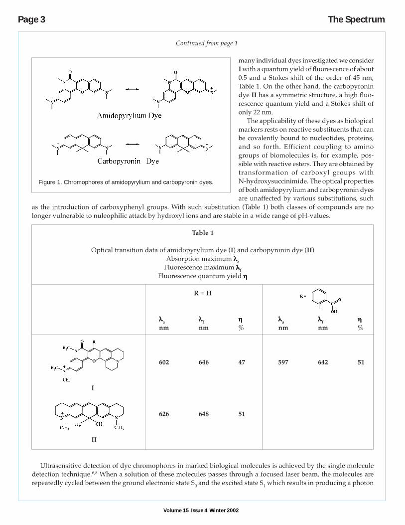
Figure 1. Chromophores of amidopyrylium and carbopyronin dyes.
Continued from page 1
many individual dyes investigated we considerI with a quantum yield of fluorescence of about0.5 and a Stokes shift of the order of 45 nm,Table 1. On the other hand, the carbopyronindye II has a symmetric structure, a high fluo-rescence quantum yield and a Stokes shift ofonly 22 nm.
The applicability of these dyes as biologicalmarkers rests on reactive substituents that canbe covalently bound to nucleotides, proteins,and so forth. Efficient coupling to aminogroups of biomolecules is, for example, pos-sible with reactive esters. They are obtained bytransformation of carboxyl groups withN-hydroxysuccinimide. The optical propertiesof both amidopyrylium and carbopyronin dyesare unaffected by various substitutions, such
as the introduction of carboxyphenyl groups. With such substitution (Table 1) both classes of compounds are nolonger vulnerable to nuleophilic attack by hydroxyl ions and are stable in a wide range of pH-values.
Table 1
Optical transition data of amidopyrylium dye (I) and carbopyronin dye (II)Absorption maximum λλλλλa
Fluorescence maximum λλλλλfFluorescence quantum yield ηηηηη
R = H
λλλλλa λλλλλf ηηηηη λλλλλa λλλλλf ηηηηηnm nm % nm nm %
602 646 47 597 642 51
I
626 648 51
II
Ultrasensitive detection of dye chromophores in marked biological molecules is achieved by the single moleculedetection technique.6,8 When a solution of these molecules passes through a focused laser beam, the molecules arerepeatedly cycled between the ground electronic state S0 and the excited state S1 which results in producing a photon
The Spectrum Page 4
Volume 15 Issue 4 Winter 2002
burst. While this can be sensitively detected by a number of techniques, careful elimination of scattered light from thebackground and of luminescence from impurities in the solvent is a prerequisite. Rayleigh scattering and reflectedlight can be suppressed by suitable optical filters. Raman scattering can not, but since it is proportional to the detec-tion volume it can be minimized by a confocal microscope set-up with a detection volume of only a few femtoliters.Signal to noise ratios of more than 100 are readily obtained. Excitation was carried out using a pulsed diode laser at640 nm and pulses of less than 400 ps duration at a repetition frequency of 56 MHz. Time-resolved data were acquiredby time-correlated single photon counting and data processing by a personal computer. Up to three mononucleotidescould be separately identified on the basis of the fluorescence life times of the attached dye molecules.
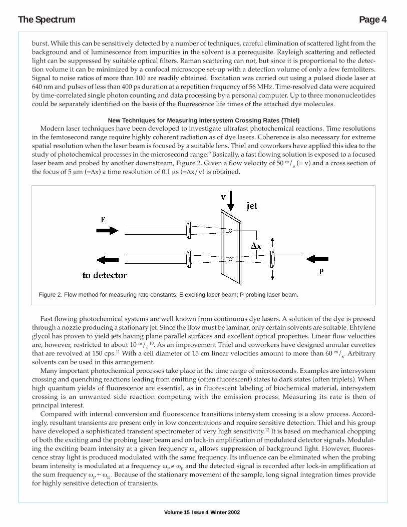
New Techniques for Measuring Intersystem Crossing Rates (Thiel)Modern laser techniques have been developed to investigate ultrafast photochemical reactions. Time resolutions
in the femtosecond range require highly coherent radiation as of dye lasers. Coherence is also necessary for extremespatial resolution when the laser beam is focused by a suitable lens. Thiel and coworkers have applied this idea to thestudy of photochemical processes in the microsecond range.9 Basically, a fast flowing solution is exposed to a focusedlaser beam and probed by another downstream, Figure 2. Given a flow velocity of 50 m/s (= v) and a cross section ofthe focus of 5 µm (=∆x) a time resolution of 0.1 µs (=∆x/v) is obtained.
Fast flowing photochemical systems are well known from continuous dye lasers. A solution of the dye is pressedthrough a nozzle producing a stationary jet. Since the flow must be laminar, only certain solvents are suitable. Ehtyleneglycol has proven to yield jets having plane parallel surfaces and excellent optical properties. Linear flow velocitiesare, however, restricted to about 10 m/s
10. As an improvement Thiel and coworkers have designed annular cuvettesthat are revolved at 150 cps.11 With a cell diameter of 15 cm linear velocities amount to more than 60 m/s. Arbitrarysolvents can be used in this arrangement.
Many important photochemical processes take place in the time range of microseconds. Examples are intersystemcrossing and quenching reactions leading from emitting (often fluorescent) states to dark states (often triplets). Whenhigh quantum yields of fluorescence are essential, as in fluorescent labeling of biochemical material, intersystemcrossing is an unwanted side reaction competing with the emission process. Measuring its rate is then ofprincipal interest.
Compared with internal conversion and fluorescence transitions intersystem crossing is a slow process. Accord-ingly, resultant transients are present only in low concentrations and require sensitive detection. Thiel and his grouphave developed a sophisticated transient spectrometer of very high sensitivity.12 It is based on mechanical choppingof both the exciting and the probing laser beam and on lock-in amplification of modulated detector signals. Modulat-ing the exciting beam intensity at a given frequency ωE allows suppression of background light. However, fluores-cence stray light is produced modulated with the same frequency. Its influence can be eliminated when the probingbeam intensity is modulated at a frequency ωP ≠ ωE and the detected signal is recorded after lock-in amplification atthe sum frequency ωP + ωE . Because of the stationary movement of the sample, long signal integration times providefor highly sensitive detection of transients.
Figure 2. Flow method for measuring rate constants. E exciting laser beam; P probing laser beam.
Page 5 The Spectrum
Volume 15 Issue 4 Winter 2002
Alternatively, transients were investigatedby measuring fluorescence intensities of themoving sample instead of absorptions.11 Thisapproach is based on the depletion of theground state paralleling the population of tran-sient states. A laser beam is split up intobranches of equal power passing at separatepoints through the sample as in Figure 2. Be-cause of ground state bleaching fluorescenceintensity is lower at a focus downstream andrelated uniquely to the ground state concen-tration. By chopping the two beams at differ-ent frequencies and lock-in amplification of theresultant photo current relative changes of thefluorescence emission of the order of 10-5 canbe detected.
The techniques were applied to dyes of in-terest as biological markers. Rates of intersys-tem crossing to the triplet state and from there
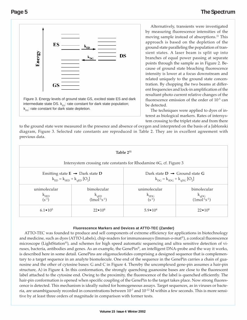
to the ground state were measured in the presence and absence of oxygen and interpreted on the basis of a Jablonskidiagram, Figure 3. Selected rate constants are reproduced in Table 2. They are in excellent agreement withprevious data.
Table 211
Intersystem crossing rate constants for Rhodamine 6G, cf. Figure 3
Emitting state E → → → → → Dark state D Dark state D →→→→→ Ground state GkED = k0ED + kqED [O2] kDG = k0DG + kqDG [O2]
unimolecular bimolecular unimolecular bimoleculark0ED kqED k0DG kqDG(s-1) (lmol-1s-1) (s-1) (1mol-1s-1)
6.1•105 22•108 5.9•104 22•108
Fluorescence Markers and Devices at ATTO-TEC (Zander)ATTO-TEC was founded to produce and sell components of extreme efficiency for applications in biotechnology
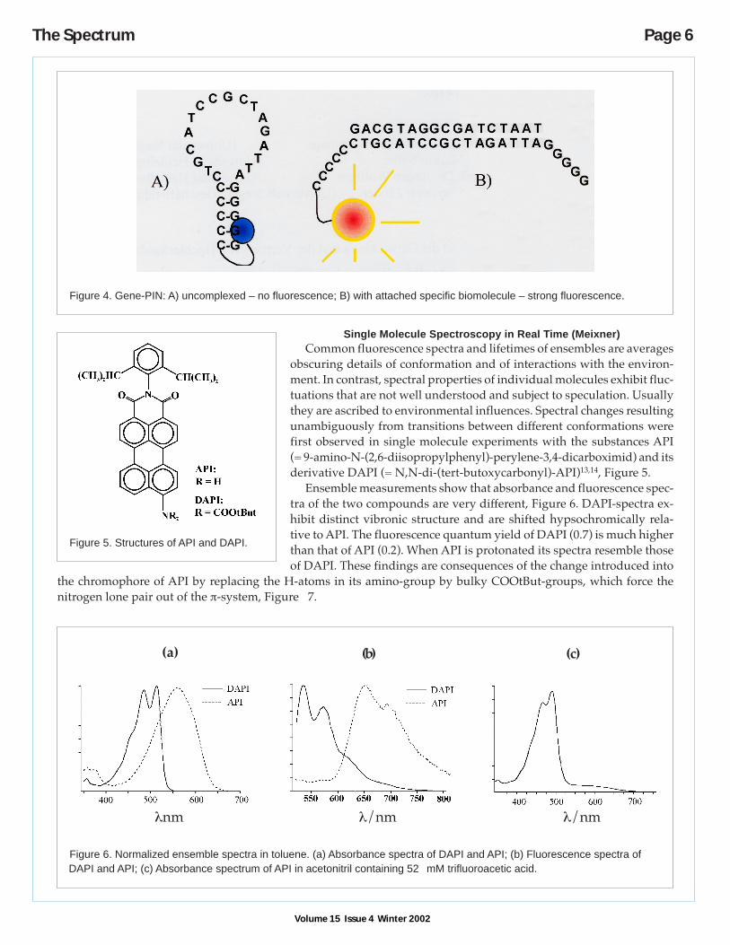
and medicine, such as dyes (ATTO-Labels), chip-readers for immunoassays (Immun-o-mat®), a confocal fluorescencemicroscope (LightStation®), and schemes for high speed automatic sequencing and ultra sensitive detection of vi-ruses, bacteria, antibodies and genes. As an example, the GenePin®, an intelligent DNA-probe and the way it works,is described here in some detail. GenePins are oligonucleotides comprising a designed sequence that is complemen-tary to a target sequence in an analyte biomolecule. One end of the sequence in the GenePin carries a chain of gua-nosine and the other of cytosine bases: G and C in Figure 4. Thereby the uncomplexed gene-pin assumes a hair-pinstructure, A) in Figure 4. In this conformation, the strongly quenching guanosine bases are close to the fluorescentlabel attached to the cytosine end. Owing to the proximity, the fluorescence of the label is quenched efficiently. Thehair-pin conformation is opened when specific coupling of the GenePin to the target takes place. Now strong fluores-cence is detected. This mechanism is ideally suited for homogeneous assays. Target sequences, as in viruses or bacte-ria, are unambiguously recorded in concentrations between 10-7 and 10-12 M within a few seconds. This is more sensi-tive by at least three orders of magnitude in comparison with former tests.
Figure 3. Energy levels of ground state GS, excited state ES and darkintermediate state DS. kED: rate constant for dark state population;kDG: rate constant for dark state depletion.
The Spectrum Page 6
Volume 15 Issue 4 Winter 2002
Single Molecule Spectroscopy in Real Time (Meixner)Common fluorescence spectra and lifetimes of ensembles are averages
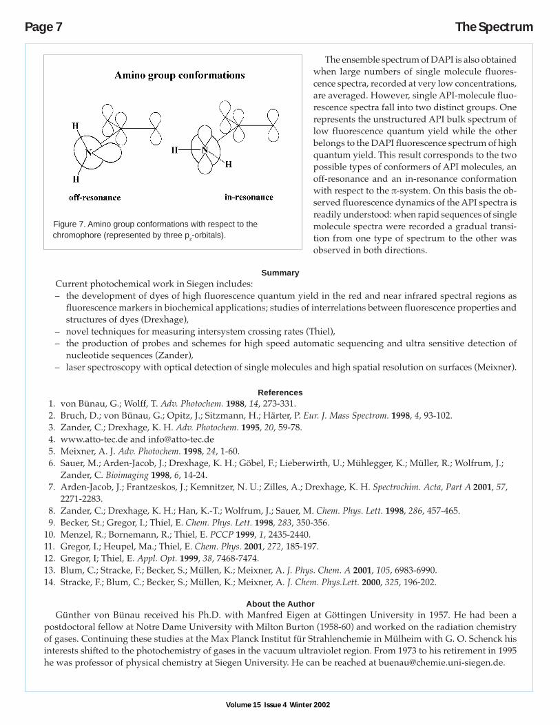
obscuring details of conformation and of interactions with the environ-ment. In contrast, spectral properties of individual molecules exhibit fluc-tuations that are not well understood and subject to speculation. Usuallythey are ascribed to environmental influences. Spectral changes resultingunambiguously from transitions between different conformations werefirst observed in single molecule experiments with the substances API(= 9-amino-N-(2,6-diisopropylphenyl)-perylene-3,4-dicarboximid) and itsderivative DAPI (= N,N-di-(tert-butoxycarbonyl)-API)13,14, Figure 5.
Ensemble measurements show that absorbance and fluorescence spec-tra of the two compounds are very different, Figure 6. DAPI-spectra ex-hibit distinct vibronic structure and are shifted hypsochromically rela-tive to API. The fluorescence quantum yield of DAPI (0.7) is much higherthan that of API (0.2). When API is protonated its spectra resemble thoseof DAPI. These findings are consequences of the change introduced into
the chromophore of API by replacing the H-atoms in its amino-group by bulky COOtBut-groups, which force thenitrogen lone pair out of the π-system, Figure 7.
Figure 4. Gene-PIN: A) uncomplexed – no fluorescence; B) with attached specific biomolecule – strong fluorescence.
Figure 5. Structures of API and DAPI.
Figure 6. Normalized ensemble spectra in toluene. (a) Absorbance spectra of DAPI and API; (b) Fluorescence spectra ofDAPI and API; (c) Absorbance spectrum of API in acetonitril containing 52 mM trifluoroacetic acid.
(a) (b) (c)
λnm λ/nm λ/nm
Page 7 The Spectrum
The ensemble spectrum of DAPI is also obtainedwhen large numbers of single molecule fluores-cence spectra, recorded at very low concentrations,are averaged. However, single API-molecule fluo-rescence spectra fall into two distinct groups. Onerepresents the unstructured API bulk spectrum oflow fluorescence quantum yield while the otherbelongs to the DAPI fluorescence spectrum of highquantum yield. This result corresponds to the twopossible types of conformers of API molecules, anoff-resonance and an in-resonance conformationwith respect to the π-system. On this basis the ob-served fluorescence dynamics of the API spectra isreadily understood: when rapid sequences of singlemolecule spectra were recorded a gradual transi-tion from one type of spectrum to the other wasobserved in both directions.
SummaryCurrent photochemical work in Siegen includes:– the development of dyes of high fluorescence quantum yield in the red and near infrared spectral regions as
fluorescence markers in biochemical applications; studies of interrelations between fluorescence properties andstructures of dyes (Drexhage),
– novel techniques for measuring intersystem crossing rates (Thiel),– the production of probes and schemes for high speed automatic sequencing and ultra sensitive detection of
nucleotide sequences (Zander),– laser spectroscopy with optical detection of single molecules and high spatial resolution on surfaces (Meixner).
References1. von Bünau, G.; Wolff, T. Adv. Photochem. 1988, 14, 273-331.2. Bruch, D.; von Bünau, G.; Opitz, J.; Sitzmann, H.; Härter, P. Eur. J. Mass Spectrom. 1998, 4, 93-102.3. Zander, C.; Drexhage, K. H. Adv. Photochem. 1995, 20, 59-78.4. www.atto-tec.de and [email protected]. Meixner, A. J. Adv. Photochem. 1998, 24, 1-60.6. Sauer, M.; Arden-Jacob, J.; Drexhage, K. H.; Göbel, F.; Lieberwirth, U.; Mühlegger, K.; Müller, R.; Wolfrum, J.;
Zander, C. Bioimaging 1998, 6, 14-24.7. Arden-Jacob, J.; Frantzeskos, J.; Kemnitzer, N. U.; Zilles, A.; Drexhage, K. H. Spectrochim. Acta, Part A 2001, 57,
2271-2283.8. Zander, C.; Drexhage, K. H.; Han, K.-T.; Wolfrum, J.; Sauer, M. Chem. Phys. Lett. 1998, 286, 457-465.9. Becker, St.; Gregor, I.; Thiel, E. Chem. Phys. Lett. 1998, 283, 350-356.
10. Menzel, R.; Bornemann, R.; Thiel, E. PCCP 1999, 1, 2435-2440.11. Gregor, I.; Heupel, Ma.; Thiel, E. Chem. Phys. 2001, 272, 185-197.12. Gregor, I; Thiel, E. Appl. Opt. 1999, 38, 7468-7474.13. Blum, C.; Stracke, F.; Becker, S.; Müllen, K.; Meixner, A. J. Phys. Chem. A 2001, 105, 6983-6990.14. Stracke, F.; Blum, C.; Becker, S.; Müllen, K.; Meixner, A. J. Chem. Phys.Lett. 2000, 325, 196-202.
About the AuthorGünther von Bünau received his Ph.D. with Manfred Eigen at Göttingen University in 1957. He had been a
postdoctoral fellow at Notre Dame University with Milton Burton (1958-60) and worked on the radiation chemistryof gases. Continuing these studies at the Max Planck Institut für Strahlenchemie in Mülheim with G. O. Schenck hisinterests shifted to the photochemistry of gases in the vacuum ultraviolet region. From 1973 to his retirement in 1995he was professor of physical chemistry at Siegen University. He can be reached at [email protected].
Figure 7. Amino group conformations with respect to thechromophore (represented by three pz-orbitals).
Volume 15 Issue 4 Winter 2002
Volume 15 Issue 4 Winter 2002
The Spectrum Page 8
IntroductionIn August 2002, the death occurred of George Porter, the co-inventor of flash photolysis, which earned for him and
Ronald Norrish a share in the 1967 Nobel Prize. The technique has been of immense benefit to the photochemistrycommunity over the past fifty years or so, and in tribute to George Porter, what will be described in this article is adevelopment which records the vibrational spectra and kinetics of intermediates in photochemical reactions ratherthan the electronic spectra conventionally recorded. Electronic spectra of complex polyatomic molecules are usuallybroad and featureless, and thus do not convey much structural information about the intermediate. Vibrational spec-tra can in principle yield quality structural information, as will be demonstrated with regard to the molecule,4,4’dimethyl amino benzonitrile (DMABN), a prototype for molecules which exhibit emission from an intramolecularcharge-transfer state in polar solvents.
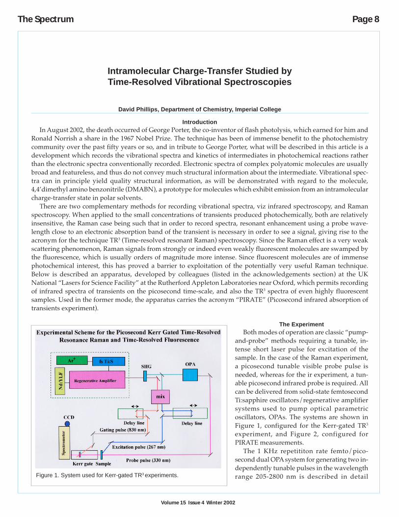
There are two complementary methods for recording vibrational spectra, viz infrared spectroscopy, and Ramanspectroscopy. When applied to the small concentrations of transients produced photochemically, both are relativelyinsensitive, the Raman case being such that in order to record spectra, resonant enhancement using a probe wave-length close to an electronic absorption band of the transient is necessary in order to see a signal, giving rise to theacronym for the technique TR3 (Time-resolved resonant Raman) spectroscopy. Since the Raman effect is a very weakscattering phenomenon, Raman signals from strongly or indeed even weakly fluorescent molecules are swamped bythe fluorescence, which is usually orders of magnitude more intense. Since fluorescent molecules are of immensephotochemical interest, this has proved a barrier to exploitation of the potentially very useful Raman technique.Below is described an apparatus, developed by colleagues (listed in the acknowledgements section) at the UKNational “Lasers for Science Facility” at the Rutherford Appleton Laboratories near Oxford, which permits recordingof infrared spectra of transients on the picosecond time-scale, and also the TR3 spectra of even highly fluorescentsamples. Used in the former mode, the apparatus carries the acronym “PIRATE” (Picosecond infrared absorption oftransients experiment).
The ExperimentBoth modes of operation are classic “pump-
and-probe” methods requiring a tunable, in-tense short laser pulse for excitation of thesample. In the case of the Raman experiment,a picosecond tunable visible probe pulse isneeded, whereas for the ir experiment, a tun-able picosecond infrared probe is required. Allcan be delivered from solid-state femtosecondTi:sapphire oscillators/regenerative amplifiersystems used to pump optical parametricoscillators, OPAs. The systems are shown inFigure 1, configured for the Kerr-gated TR3
experiment, and Figure 2, configured forPIRATE measurements.
The 1 KHz repetititon rate femto/pico-second dual OPA system for generating two in-dependently tunable pulses in the wavelengthrange 205-2800 nm is described in detail
Intramolecular Charge-Transfer Studied byTime-Resolved Vibrational Spectroscopies
David Phillips, Department of Chemistry, Imperial College
Figure 1. System used for Kerr-gated TR3 experiments.

Volume 15 Issue 4 Winter 2002
Page 9 The Spectrum
elsewhere,1 but is centred around a Ti:S regen-erative amplifier (Spectraphysics Spitfire/Mer-lin) operating at 1 KHz, and ca 800 nm, anddelivering from 0.7 to 2.5 mJ per pulse. The re-generative amplifier is seeded from a mode-locked Ti:S laser (Spectraphysics Tsunami).Two modes of operation are possible with thissystem: (1) picosecond, in which spectral fil-tering of the Ti:S oscillator produces 800 nmoutput of ca 1-2 ps FWHM pulse duration, and15-7 cm-1 bandwidth respectively, and (2) us-ing a femtosecond stretcher/compressor toprovide an output of 150 fs and ca 100 cm-1
bandwidth. The 800 nm output is frequencydoubled in a BBO crystal, and split to pro-duce two OPAs, which when combined withupconversion, (including the idler tunability),give the 205-2800 nm spectra; range.
For the TR3 measurements, a high perfor-mance Kerr-gate designed for the suppressionof fluorescence in both time-resolved andsteady state resonance Raman spectroscopy isutilised.2 The gate opens for ca 4 ps , through-put in the open state is ~40%, and the extinc-tion ratio in the closed state is 10-5. The useablespectral range is 300-700 nm with a single setof polarisers. The effectiveness of the device isshown in Figure 3, where the Raman spectrumrecorded for the highly fluorescent dye Cou-marin C480 is shown without the gate, using asubtraction technique, and with the gate. Veryhigh quality time-resolved fluorescence mea-surements can be made on this apparatus by“inverting” the modus operandi, that is, sup-pressing the pump and Raman signals and per-mitting the fluorescence to be transmitted byrotation of one of the polarisers. High qualitytime-resolved electronic absorption measure-ments can also be made on this system.
For the time-resolved infrared (PIRATE) ex-periments, the 150 fs, ca 150 cm-1 FWHM mid-infrared probe and reference pulses are gener-ated by difference frequency mixing of near irpulses in Type I AgGaS2, and are dispersedover two 64-element linear infrared array de-tectors. This gives a sensitivity of delta OD ~10-4 to 10-5 with 1 minute acquisition time.3
The two techniques of TR3 and TRIR aretruly complementary, as will be shown by ref-erence to the specific example below. The TR3
experiment has poorer spectral resolution, butbroader spectral coverage than the TRIR, which
Figure 2. System used for “PIRATE” experiments.
Figure 3. Suppression of fluorescence in TR3 spectroscopy.Spectra of acetonitrile contaminated with Coumarin 480.(a) without gate; (b) without gate, fluorescence subtracted;(c) Kerr-gated Raman spectrum.
Volume 15 Issue 4 Winter 2002
The Spectrum Page 10
currently is limited to the range 100-200 cm-1 on eachscan, whereas the TR3 experiment can give the wholespectrum on one accumulation. TRIR also has bettertime-resolution, but is limited at present to the spectralregion above 1000 cm-1. However, since not all vibra-tional motions are both ir and Raman active, both tech-niques are required.
Application to Charge-Transfer inDimethylaminobenzonitrile (DMABN)
(Structure shown in Figure 5)DMABN has been the subject of very many studies
in the forty years or so since Lippert4 first reported the“dual” fluorescence of the molecule in polar solvents,yet controversy still surrounds the structure of the in-tramolecular charge-transfer (ICT) state which is pro-duced upon excitation of the molecule in polar solventsand there has not been general agreement between dif-ferent time-resolved studies, usually on fluorescence,regarding the kinetics of conversion between initially-produced locally excited state and the ICT state, andtheir decays. Many mechanisms, both monomole-cular5-10 and arguments proposing specific solute-solvent interactions11-15 have been proposed.
There are two main contenders as models for thestructural changes accompanying the ICT reaction. Inthe “twisted” ICT model (TICT), the ICT state is formedby twisting the dimethylamino group from its planarconfiguration in the locally excited state (as in theground state), into a geometry perpendicular to theplane of the benzonitrile moiety; this is assumed to beaccompanied by full electron transfer from the lone-pair of the amino N-atom to the in-plane π orbital ofthe benzonitrile group.16 The 90 degree twisted confor-mation would lead to electronic decoupling of theamino and benzonitrile groups, and consequent lossof conjugation between the donor (amino) andacceptor(benzonitrile) groups. In the “planar” (PICT)model,17 the geometry change is supposed as a changefrom a pyramidal structure of the amino group in theLE state towards a planar configuration in the ICT state,giving a quinoidal planar resonance structure in whichthe donor and acceptor groups are strongly coupled.
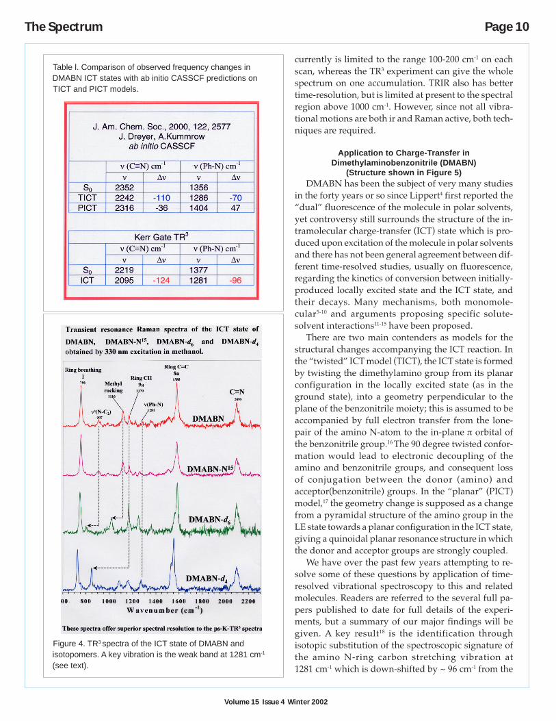
We have over the past few years attempting to re-solve some of these questions by application of time-resolved vibrational spectroscopy to this and relatedmolecules. Readers are referred to the several full pa-pers published to date for full details of the experi-ments, but a summary of our major findings will begiven. A key result18 is the identification throughisotopic substitution of the spectroscopic signature ofthe amino N-ring carbon stretching vibration at1281 cm-1 which is down-shifted by ~ 96 cm-1 from the
Figure 4. TR3 spectra of the ICT state of DMABN andisotopomers. A key vibration is the weak band at 1281 cm-1
(see text).
Table l. Comparison of observed frequency changes inDMABN ICT states with ab initio CASSCF predictions onTICT and PICT models.
Volume 15 Issue 4 Winter 2002
Page 11 The Spectrum
corresponding band in the locally excited state.See Table 1 and Figure 4.
This result clearly favours the TICT model,in which a weakening of the bond through lossof conjugation would be expected, over thePICT model, in which a strengthening of thebond would occur, with consequent upshift ofthe frequency, as proposed in theoreticalCASSCF work.19 However, it must be statedthat the vibrational analysis of ground-stateDMABN shows that the weak mode corre-sponding to the 1281 cm-1 ICT mode is a com-plex vibration, which has contributions fromnumerous other local modes, and this complex-ity weakens the conclusions based upon thefrequency shift of this mode. Although we re-main favourably disposed towards this conclu-sion, others are opposed,20 and the proof ofeither TICT or PICT models remains elusiveuntil further structural studies on these short-lived transients can be performed.
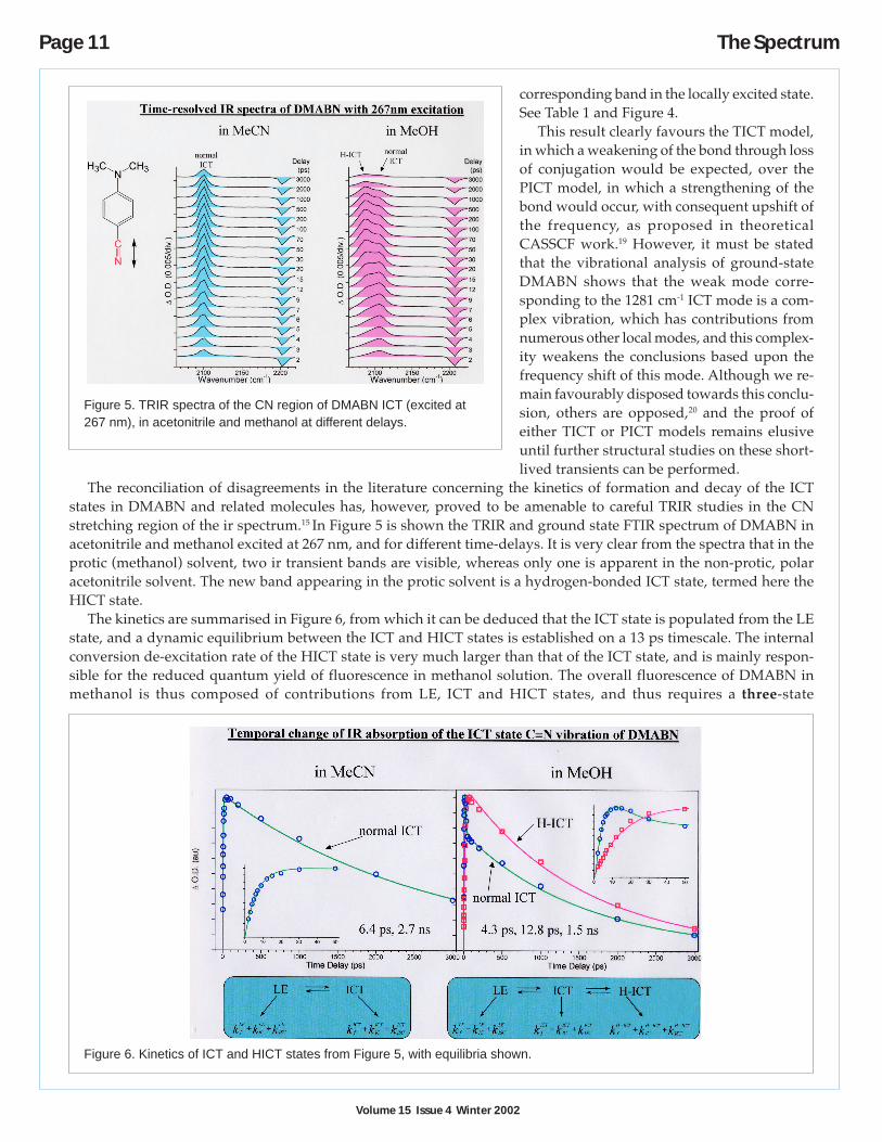
The reconciliation of disagreements in the literature concerning the kinetics of formation and decay of the ICTstates in DMABN and related molecules has, however, proved to be amenable to careful TRIR studies in the CNstretching region of the ir spectrum.15 In Figure 5 is shown the TRIR and ground state FTIR spectrum of DMABN inacetonitrile and methanol excited at 267 nm, and for different time-delays. It is very clear from the spectra that in theprotic (methanol) solvent, two ir transient bands are visible, whereas only one is apparent in the non-protic, polaracetonitrile solvent. The new band appearing in the protic solvent is a hydrogen-bonded ICT state, termed here theHICT state.
The kinetics are summarised in Figure 6, from which it can be deduced that the ICT state is populated from the LEstate, and a dynamic equilibrium between the ICT and HICT states is established on a 13 ps timescale. The internalconversion de-excitation rate of the HICT state is very much larger than that of the ICT state, and is mainly respon-sible for the reduced quantum yield of fluorescence in methanol solution. The overall fluorescence of DMABN inmethanol is thus composed of contributions from LE, ICT and HICT states, and thus requires a three-state
Figure 5. TRIR spectra of the CN region of DMABN ICT (excited at267 nm), in acetonitrile and methanol at different delays.
Figure 6. Kinetics of ICT and HICT states from Figure 5, with equilibria shown.

Volume 15 Issue 4 Winter 2002
The Spectrum Page 12
mechanism for the dual fluorescence of DMABN and related molecules in protic solvents. Such a mechanism has notbeen proposed before, but can be shown to be compatible with results here and in the literature. The spectroscopicidentification of this new hydrogen-bonded ICT state is a graphic illustration of the power of the high-resolution,high-sensitivity TRIR technique, which, when used in conjunction with the complementary TR3 method, should becapable of solving many other photochemical and photophysical problems.
References1. Towrie, M.; Parker, A. W.; Shaikh, W.; Matousek, P. Meas. Sci. Technol. 1998, 9, 816.2. Matousek, P.; Towrie, M.; Ma, C.; Kwok, W. M.; Phillips, D.; Toner, W. T.; Parker, A. W. J. Raman Spectrosc. 2001,
32, 983.3. Towrie, M.; Grills, D. C.; Dyer, J.; Weinstein, J. A.; Matousek, P.; Barton, R.; Birley, P. D.; Subramanian, N.;
Kwok, W. M.; Ma, C.; Phillips, D.; Parker, A. W.; George, M. W. Development of a broadband picosecondtime-resolved infrared spectrometer. Appl. Spectrosc. 2003, in press.
4. Lippert, E.; Luder, W.; Moll, F.; Nagele, W.; Boos, H.; Prigge, H.; Seibold-Blankenstein, I. Angew. Chem. 1961, 73,695.
5. Rettig, W.; Bliss, B.; Dirnberger, K. Chem. Phys. Lett. 1999, 305, 8.6. Rettig, W.; Lutze, S. Chem. Phys. Lett. 2001, 341, 263.7. Zachariasse, K. Chem. Phys. Lett. 2000, 320, 8.8. Kwok, W. M.; Ma, C.; Phillips, D.; Matousek, P.; Parker, A. W.; Towrie, M. J. Phys. Chem. A 2000, 104, 4819.9. Hicks, J. M.; Vandersall, M. T.; Sitzmann, E. V.; Eisenthal, K. B. Chem. Phys. Lett. 1987, 135, 413.
10. Changenet, P.; Plaza, P.; Martin, M. M.; Meyer, Y. H. J. Phys. Chem. A 1997, 101, 8186.11. Pilloud, D.; Suppan, P.; Haelst, L. V. Chem. Phys. Lett. 1987, 137, 130.12. Visser, R. J.; Varma, C. A. G. O.; Konijnberg, J.; Bergwerf, P. J. Chem. Soc., Faraday Trans. 2 1989, 79, 347.13. Cazeau-Dubroca, C.; Lyazidi, S. A.; Cambou, P.; Peirigua, A.; Cazeau, P.; Pesquer, M. J. Phys. Chem. 1989, 93,
2347.14. Khalil, O. S.; Hofeldt, R.; McGlynn, S. P. J. Lumin. 1973, 6, 229.15. Kwok, W. M.; George, M. W.; Grills, D. C.; Ma, C.; Matousek, P.; Parker, A. W.; Phillips, D.; Toner, W. T.;
Towrie, M. Direct observation of a hydrogen-bonded charge-transfer state of 4-dimethyl amino benzonitrile,(DMABN) in methanol by time-resolved infrared spectroscopy. Angew. Chem., Int. Ed. 2003, in press.
16. Rotkiewicz, K.; Grellmann, K. H.; Grabowski, Z. R. Chem. Phys. Lett. 1973, 19, 315.17. Zachariasse, K. A.; Grobys, M.; Vonderhaar, T.; Hebecker, A.; Iichev, Y. V.; Jiang, Y.-B.; Morawski, O.; Kuhnle, W.
J. Photochem. Photobiol., A 1996, 102, 59.18. Kwok, W. M.; Ma, C.; Matousek, P.; Parker, A. W.; Phillips, D.; Toner, W. T.; Towrie, M; Umapathy, S.
J. Phys. Chem. A 2001, 105, 984.19. Dreyer, J.; Kummrow, A. J. Amer. Chem. Soc. 2000, 122, 2577.20. Okamoto, H.; Kinoshita, M.; Kohtani, S.; Nakagaki, R.; Zachariasse, K. A. Bull. Chem. Soc. Jpn. 2002, 75, 957.
AcknowledgementsThe work described here has all been carried out by my colleagues at Rutherford Appleton Laboratories (Tony
Parker, Pavel Matousek, and Mike Towrie), The University of Oxford (Bill Toner), the University of Nottingham(Mike George and David Grills), and Imperial College London (Wai-Ming Kwok and Chensheng Ma), under theterms of research grants from the Engineering and Physical Sciences Research Council, UK.
About the AuthorDavid Phillips was awarded his Ph.D. in physical chemistry in 1964 at the University of Birmingham, UK. He
traveled on a Fulbright Fellowship to the University of Texas at Austin to carry out a postdoctoral fellowship with W.Albert Noyes Jr. (1964-66), then was Royal Society/Academy of Sciences of USSR Exchange Fellow in the Institute ofChemical Physics, Moscow, USSR during 1966-67. He was on the Chemistry staff of the University of Southampton,UK from 1967-79, when he then became Wolfson Professor of Natural Philosophy and Deputy Director of the RoyalInstitution of Great Britain, London, at the time when Lord Porter, then Sir George Porter, was Director. In 1989Phillips moved to Imperial College, London, as Professor of Physical Chemistry, then Head of Department from 1992-2002, and is now Hofmann Professor of Chemistry, and Dean of the Faculties of Life Sciences and Physical Sciences.His address is Department of Chemistry, Imperial College, London SW72AZ, UK; email: [email protected].
Volume 15 Issue 4 Winter 2002
Page 13 The Spectrum
Large biomolecules such as proteins have the capacity of self-assembly. Assembly can be intermolecular, forexample, when many proteins aggregate into an ordered photosynthetic light harvesting antenna complex, or it canbe intramolecular, for example, when a single protein acquires its highly packed three-dimensional fold.1 In recentyears, materials and synthetic chemists have also succeeded at creating oligomers capable of folding into veryspecific structures.
By what dynamical processes does such assembly occur? A chemist with a background in small molecules may betempted to treat the folding of a single protein as an activated unimolecular reaction U → F with one or more steps ofsingle exponential kinetics along a single reaction coordinate. A polymer chemist may envision a collapse not gov-erned by a single rate process, and not describable by a single reaction coordinate. The assembly dynamics of naturaland highly ordered artificial polymers lies between these extremes. The formation process often can be described byone or a few exponential functions, and it is slow enough that the picture of an activated rate process is a reasonableapproximation. Yet closer examination reveals subtle discrepancies: unusual non-Arrhenius behavior of the rate con-stant, or different time scales for the kinetics when examined by different spectroscopic probes.
The dynamics corresponding to large-scale structural changes generally ranges from nanoseconds to milliseconds.The lower limit is governed by the time required by macromolecular segments to diffuse through the solvent: a 1000amu (1kDa) α-helix simply does not move over multiple Ångstroms in less than nanoseconds.2 The upper limit can bemuch longer than milliseconds, but optimally designed small polymers (<15,000 amu or 15 kDa) generally do notrequire much more time to settle into a folded structure, or to assemble at sufficiently high monomer concentrations.
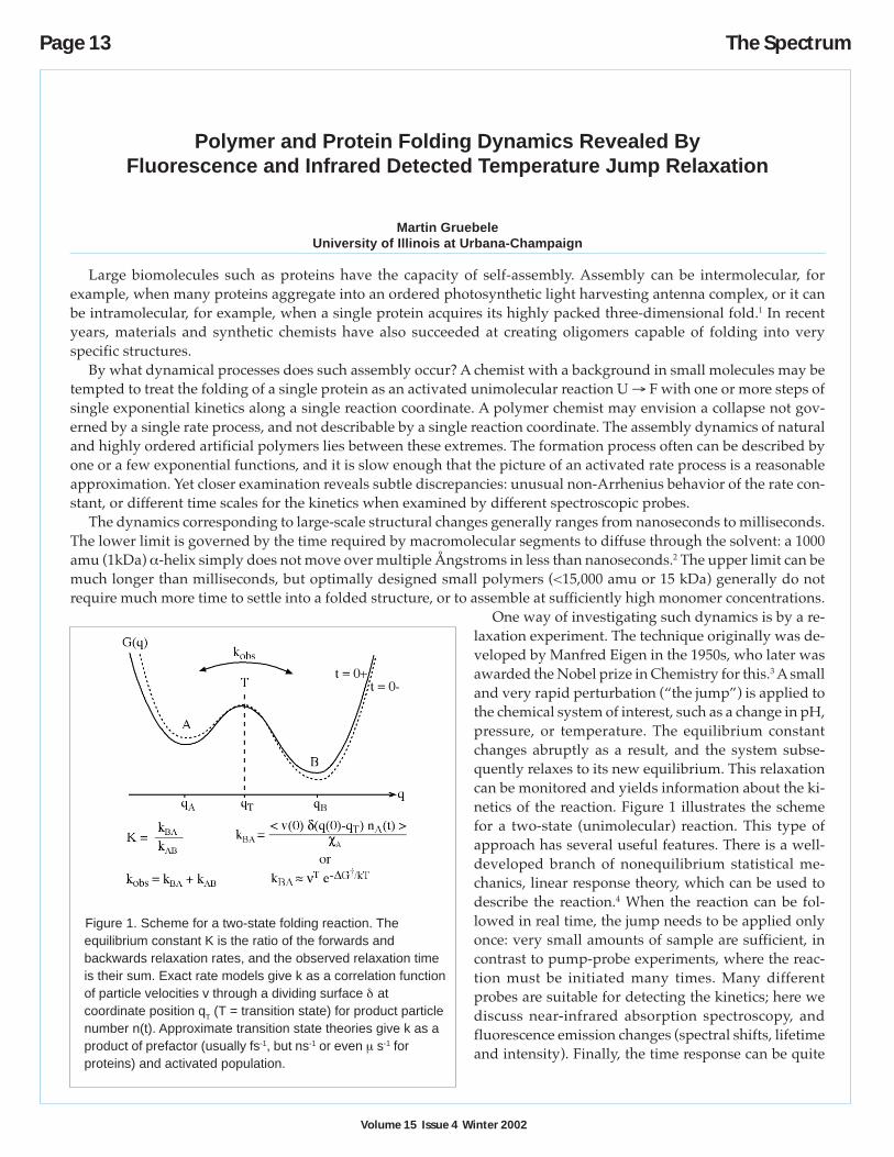
One way of investigating such dynamics is by a re-laxation experiment. The technique originally was de-veloped by Manfred Eigen in the 1950s, who later wasawarded the Nobel prize in Chemistry for this.3 A smalland very rapid perturbation (“the jump”) is applied tothe chemical system of interest, such as a change in pH,pressure, or temperature. The equilibrium constantchanges abruptly as a result, and the system subse-quently relaxes to its new equilibrium. This relaxationcan be monitored and yields information about the ki-netics of the reaction. Figure 1 illustrates the schemefor a two-state (unimolecular) reaction. This type ofapproach has several useful features. There is a well-developed branch of nonequilibrium statistical me-chanics, linear response theory, which can be used todescribe the reaction.4 When the reaction can be fol-lowed in real time, the jump needs to be applied onlyonce: very small amounts of sample are sufficient, incontrast to pump-probe experiments, where the reac-tion must be initiated many times. Many differentprobes are suitable for detecting the kinetics; here wediscuss near-infrared absorption spectroscopy, andfluorescence emission changes (spectral shifts, lifetimeand intensity). Finally, the time response can be quite
Polymer and Protein Folding Dynamics Revealed ByFluorescence and Infrared Detected Temperature Jump Relaxation
Martin GruebeleUniversity of Illinois at Urbana-Champaign
Figure 1. Scheme for a two-state folding reaction. Theequilibrium constant K is the ratio of the forwards andbackwards relaxation rates, and the observed relaxation timeis their sum. Exact rate models give k as a correlation functionof particle velocities v through a dividing surface δ atcoordinate position qT (T = transition state) for product particlenumber n(t). Approximate transition state theories give k as aproduct of prefactor (usually fs-1, but ns-1 or even µ s-1 forproteins) and activated population.
Volume 15 Issue 4 Winter 2002
The Spectrum Page 14
fast. For example, water, a common medium for biopolymer reactions, can be heated by an infrared pulse and locallyequilibrated in a few picoseconds, long before the polymer chain can respond.
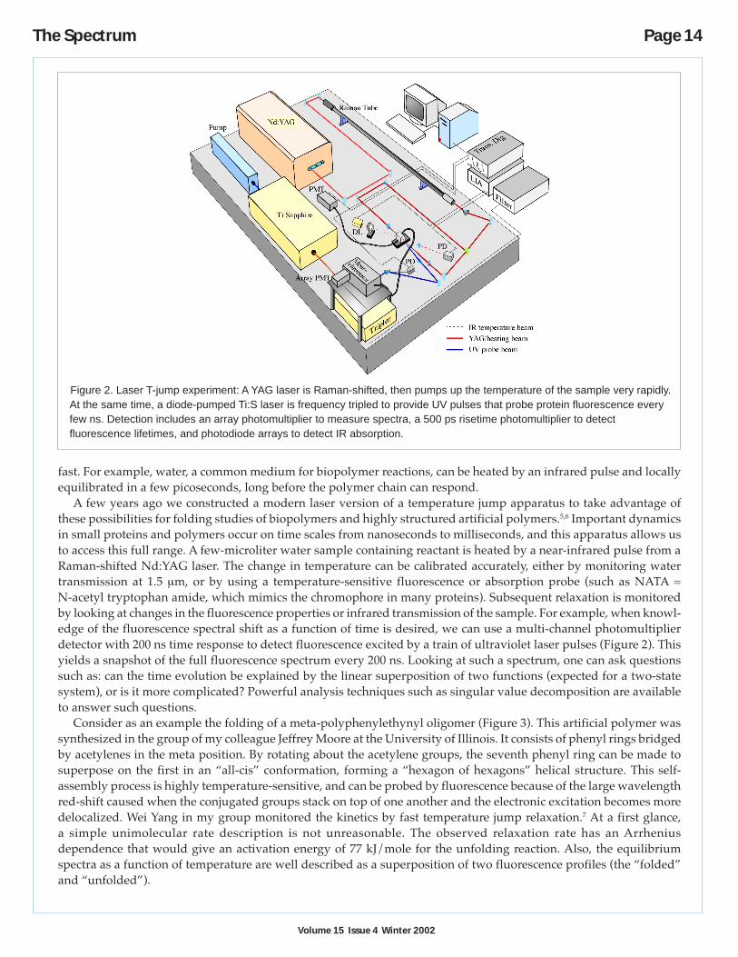
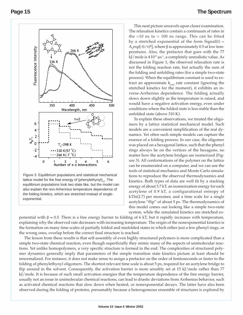
A few years ago we constructed a modern laser version of a temperature jump apparatus to take advantage ofthese possibilities for folding studies of biopolymers and highly structured artificial polymers.5,6 Important dynamicsin small proteins and polymers occur on time scales from nanoseconds to milliseconds, and this apparatus allows usto access this full range. A few-microliter water sample containing reactant is heated by a near-infrared pulse from aRaman-shifted Nd:YAG laser. The change in temperature can be calibrated accurately, either by monitoring watertransmission at 1.5 µm, or by using a temperature-sensitive fluorescence or absorption probe (such as NATA =N-acetyl tryptophan amide, which mimics the chromophore in many proteins). Subsequent relaxation is monitoredby looking at changes in the fluorescence properties or infrared transmission of the sample. For example, when knowl-edge of the fluorescence spectral shift as a function of time is desired, we can use a multi-channel photomultiplierdetector with 200 ns time response to detect fluorescence excited by a train of ultraviolet laser pulses (Figure 2). Thisyields a snapshot of the full fluorescence spectrum every 200 ns. Looking at such a spectrum, one can ask questionssuch as: can the time evolution be explained by the linear superposition of two functions (expected for a two-statesystem), or is it more complicated? Powerful analysis techniques such as singular value decomposition are availableto answer such questions.
Consider as an example the folding of a meta-polyphenylethynyl oligomer (Figure 3). This artificial polymer wassynthesized in the group of my colleague Jeffrey Moore at the University of Illinois. It consists of phenyl rings bridgedby acetylenes in the meta position. By rotating about the acetylene groups, the seventh phenyl ring can be made tosuperpose on the first in an “all-cis” conformation, forming a “hexagon of hexagons” helical structure. This self-assembly process is highly temperature-sensitive, and can be probed by fluorescence because of the large wavelengthred-shift caused when the conjugated groups stack on top of one another and the electronic excitation becomes moredelocalized. Wei Yang in my group monitored the kinetics by fast temperature jump relaxation.7 At a first glance,a simple unimolecular rate description is not unreasonable. The observed relaxation rate has an Arrheniusdependence that would give an activation energy of 77 kJ/mole for the unfolding reaction. Also, the equilibriumspectra as a function of temperature are well described as a superposition of two fluorescence profiles (the “folded”and “unfolded”).
Figure 2. Laser T-jump experiment: A YAG laser is Raman-shifted, then pumps up the temperature of the sample very rapidly.At the same time, a diode-pumped Ti:S laser is frequency tripled to provide UV pulses that probe protein fluorescence everyfew ns. Detection includes an array photomultiplier to measure spectra, a 500 ps risetime photomultiplier to detectfluorescence lifetimes, and photodiode arrays to detect IR absorption.
Volume 15 Issue 4 Winter 2002
Page 15 The Spectrum
This neat picture unravels upon closer examination.The relaxation kinetics contain a continuum of rates inthe <10 ns to > 100 ns range. This can be fittedby a stretched exponential of the form Signal(t) =A0exp[-(t/τ)β], where β is approximately 0.5 at low tem-peratures. Also, the prefactor that goes with the 77kJ/mole is 4.1013 µs-1, a completely unrealistic value. Asdiscussed in Figure 1, the observed relaxation rate isnot the folding reaction rate, but actually the sum ofthe folding and unfolding rates (for a simple two-stateprocess). When the equilibrium constant is used to ex-tract an approximate kfold rate constant (ignoring thestretched kinetics for the moment), it exhibits an in-verse-Arrhenius dependence. The folding actuallyslows down slightly as the temperature is raised, andwould have a negative activation energy, even underconditions where the folded state is less stable than theunfolded state (above 310 K).
To explain these observations, we treated the oligo-mers by a lattice statistical mechanical model. Suchmodels are a convenient simplification of the real dy-namics. Yet often such simple models can capture theessence of a folding process. In our case, the oligomerwas placed on a hexagonal lattice, such that the phenylrings always lie on the vertices of the hexagons, nomatter how the acetylene bridges are isomerized (Fig-ure 3). All conformations of the polymer on the latticecan be enumerated on a computer, and we can use thetools of statistical mechanics and Monte Carlo simula-tions to reproduce the observed thermodynamics andkinetics. Both types of data are well fit by a stackingenergy of about 3.7 kT, an isomerization energy for eachacetylene of 0.9 kT, a configurational entropy ofkTln(2.7) per monomer, and a time scale for a singleacetylene “flip” of about 5 ps. The thermodynamics ofthis model comes out looking like a simple two-statesystem, while the simulated kinetics are stretched ex-
ponential with β ≈ 0.5. There is a free energy barrier to folding of 6 kT, but it rapidly increases with temperature,explaining why the observed rate decreases with increasing temperature. The origin of the nonexponential kinetics isthe formation on many time scales of partially folded and misfolded states in which either just a few phenyl rings, orthe wrong ones, overlap before the correct final structure is reached.
The lesson from these results is that self-assembly of even highly structured polymers is more complicated than asimple two-state chemical reaction, even though superficially they mimic many of the aspects of unimolecular reac-tions. Yet unlike homopolymers, a very specific structure is formed in the end. The complexities of structured poly-mer dynamics generally imply that parameters of the simple transition state kinetics picture at least should berenormalized. For instance, it does not make sense to assign a prefactor on the order of femtoseconds or faster to thefolding of phenylethynyl oligomers. The shortest relevant time scale is about 5 ps, required for an acetylene bridge toflip around in the solvent. Consequently, the activation barrier is more sensibly set at 15 kJ/mole rather than 77kJ/mole. It is because of such small activation energies that the temperature dependence of the free energy barrier,usually not an issue in unimolecular chemical reactions, can lead to drastic deviations from Arrhenius behavior, suchas activated chemical reactions that slow down when heated, or nonexponential decays. The latter have also beenobserved during the folding of proteins, presumably because a heterogeneous ensemble of structures is explored by
Figure 3. Equilibrium populations and statistical mechanicallattice model for the free energy of (phenylethynyl)12. Theequilibrium populations look two state like, but the model canalso explain the non-Arhennius temperature dependence ofthe folding kinetics, which are stretched instead of single-exponential.
Volume 15 Issue 4 Winter 2002
The Spectrum Page 16
very fast-folding proteins en route to the folded state. The behavior of the phenylethynylene has also been analyzedsuccessfully by full-atom molecular dynamics simulations, confirming the picture from our simple lattice model.
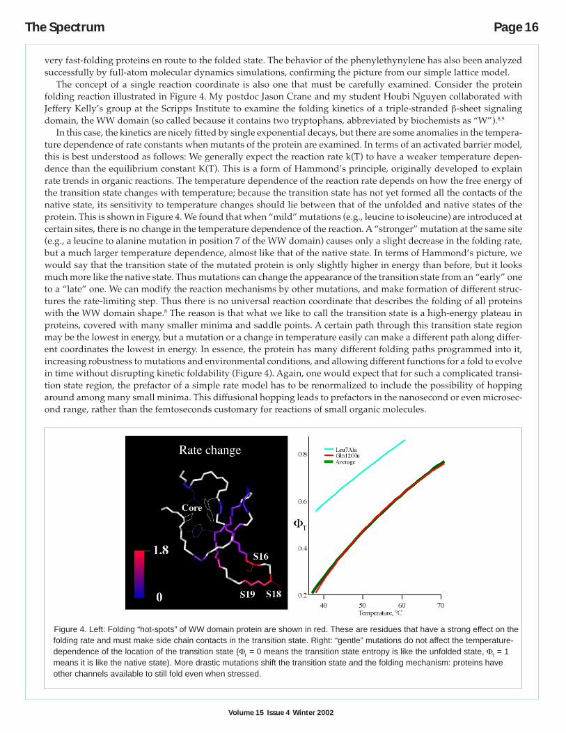
The concept of a single reaction coordinate is also one that must be carefully examined. Consider the proteinfolding reaction illustrated in Figure 4. My postdoc Jason Crane and my student Houbi Nguyen collaborated withJeffery Kelly’s group at the Scripps Institute to examine the folding kinetics of a triple-stranded β-sheet signalingdomain, the WW domain (so called because it contains two tryptophans, abbreviated by biochemists as “W”).8,9
In this case, the kinetics are nicely fitted by single exponential decays, but there are some anomalies in the tempera-ture dependence of rate constants when mutants of the protein are examined. In terms of an activated barrier model,this is best understood as follows: We generally expect the reaction rate k(T) to have a weaker temperature depen-dence than the equilibrium constant K(T). This is a form of Hammond’s principle, originally developed to explainrate trends in organic reactions. The temperature dependence of the reaction rate depends on how the free energy ofthe transition state changes with temperature; because the transition state has not yet formed all the contacts of thenative state, its sensitivity to temperature changes should lie between that of the unfolded and native states of theprotein. This is shown in Figure 4. We found that when “mild” mutations (e.g., leucine to isoleucine) are introduced atcertain sites, there is no change in the temperature dependence of the reaction. A “stronger” mutation at the same site(e.g., a leucine to alanine mutation in position 7 of the WW domain) causes only a slight decrease in the folding rate,but a much larger temperature dependence, almost like that of the native state. In terms of Hammond’s picture, wewould say that the transition state of the mutated protein is only slightly higher in energy than before, but it looksmuch more like the native state. Thus mutations can change the appearance of the transition state from an “early” oneto a “late” one. We can modify the reaction mechanisms by other mutations, and make formation of different struc-tures the rate-limiting step. Thus there is no universal reaction coordinate that describes the folding of all proteinswith the WW domain shape.8 The reason is that what we like to call the transition state is a high-energy plateau inproteins, covered with many smaller minima and saddle points. A certain path through this transition state regionmay be the lowest in energy, but a mutation or a change in temperature easily can make a different path along differ-ent coordinates the lowest in energy. In essence, the protein has many different folding paths programmed into it,increasing robustness to mutations and environmental conditions, and allowing different functions for a fold to evolvein time without disrupting kinetic foldability (Figure 4). Again, one would expect that for such a complicated transi-tion state region, the prefactor of a simple rate model has to be renormalized to include the possibility of hoppingaround among many small minima. This diffusional hopping leads to prefactors in the nanosecond or even microsec-ond range, rather than the femtoseconds customary for reactions of small organic molecules.
Figure 4. Left: Folding “hot-spots” of WW domain protein are shown in red. These are residues that have a strong effect on thefolding rate and must make side chain contacts in the transition state. Right: “gentle” mutations do not affect the temperature-dependence of the location of the transition state (ΦT = 0 means the transition state entropy is like the unfolded state, ΦT = 1means it is like the native state). More drastic mutations shift the transition state and the folding mechanism: proteins haveother channels available to still fold even when stressed.
Volume 15 Issue 4 Winter 2002
Page 17 The Spectrum
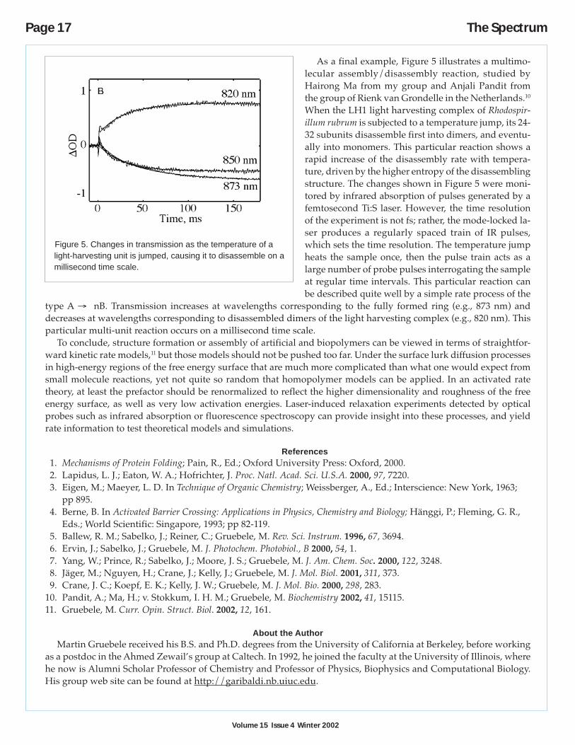
As a final example, Figure 5 illustrates a multimo-lecular assembly/disassembly reaction, studied byHairong Ma from my group and Anjali Pandit fromthe group of Rienk van Grondelle in the Netherlands.10
When the LH1 light harvesting complex of Rhodospir-illum rubrum is subjected to a temperature jump, its 24-32 subunits disassemble first into dimers, and eventu-ally into monomers. This particular reaction shows arapid increase of the disassembly rate with tempera-ture, driven by the higher entropy of the disassemblingstructure. The changes shown in Figure 5 were moni-tored by infrared absorption of pulses generated by afemtosecond Ti:S laser. However, the time resolutionof the experiment is not fs; rather, the mode-locked la-ser produces a regularly spaced train of IR pulses,which sets the time resolution. The temperature jumpheats the sample once, then the pulse train acts as alarge number of probe pulses interrogating the sampleat regular time intervals. This particular reaction canbe described quite well by a simple rate process of the
type A → nB. Transmission increases at wavelengths corresponding to the fully formed ring (e.g., 873 nm) anddecreases at wavelengths corresponding to disassembled dimers of the light harvesting complex (e.g., 820 nm). Thisparticular multi-unit reaction occurs on a millisecond time scale.
To conclude, structure formation or assembly of artificial and biopolymers can be viewed in terms of straightfor-ward kinetic rate models,11 but those models should not be pushed too far. Under the surface lurk diffusion processesin high-energy regions of the free energy surface that are much more complicated than what one would expect fromsmall molecule reactions, yet not quite so random that homopolymer models can be applied. In an activated ratetheory, at least the prefactor should be renormalized to reflect the higher dimensionality and roughness of the freeenergy surface, as well as very low activation energies. Laser-induced relaxation experiments detected by opticalprobes such as infrared absorption or fluorescence spectroscopy can provide insight into these processes, and yieldrate information to test theoretical models and simulations.
References1. Mechanisms of Protein Folding; Pain, R., Ed.; Oxford University Press: Oxford, 2000.2. Lapidus, L. J.; Eaton, W. A.; Hofrichter, J. Proc. Natl. Acad. Sci. U.S.A. 2000, 97, 7220.3. Eigen, M.; Maeyer, L. D. In Technique of Organic Chemistry; Weissberger, A., Ed.; Interscience: New York, 1963;
pp 895.4. Berne, B. In Activated Barrier Crossing: Applications in Physics, Chemistry and Biology; Hänggi, P.; Fleming, G. R.,
Eds.; World Scientific: Singapore, 1993; pp 82-119.5. Ballew, R. M.; Sabelko, J.; Reiner, C.; Gruebele, M. Rev. Sci. Instrum. 1996, 67, 3694.6. Ervin, J.; Sabelko, J.; Gruebele, M. J. Photochem. Photobiol., B 2000, 54, 1.7. Yang, W.; Prince, R.; Sabelko, J.; Moore, J. S.; Gruebele, M. J. Am. Chem. Soc. 2000, 122, 3248.8. Jäger, M.; Nguyen, H.; Crane, J.; Kelly, J.; Gruebele, M. J. Mol. Biol. 2001, 311, 373.9. Crane, J. C.; Koepf, E. K.; Kelly, J. W.; Gruebele, M. J. Mol. Bio. 2000, 298, 283.
10. Pandit, A.; Ma, H.; v. Stokkum, I. H. M.; Gruebele, M. Biochemistry 2002, 41, 15115.11. Gruebele, M. Curr. Opin. Struct. Biol. 2002, 12, 161.
About the AuthorMartin Gruebele received his B.S. and Ph.D. degrees from the University of California at Berkeley, before working
as a postdoc in the Ahmed Zewail’s group at Caltech. In 1992, he joined the faculty at the University of Illinois, wherehe now is Alumni Scholar Professor of Chemistry and Professor of Physics, Biophysics and Computational Biology.His group web site can be found at http://garibaldi.nb.uiuc.edu.
Figure 5. Changes in transmission as the temperature of alight-harvesting unit is jumped, causing it to disassemble on amillisecond time scale.

The Spectrum Page 18
Copyright 2002 by the Center for Photochemical SciencesThe Spectrum is a quarterly publication of the Center forPhotochemical Sciences, Bowling Green State University,Bowling Green, OH 43403.Phone 419.372.2033 Fax 419.372.0366Email [email protected] http://www.bgsu.edu/departments/photochem/
Executive Director: D. C. NeckersPrincipal Faculty: P. Anzenbacher, G. S. Bullerjahn,
J. R. Cable, F. N. Castellano,M. E. Geusz, D. C. Neckers,M. Y. Ogawa, V. V. Popik,M. A. J. Rodgers, D. L. Snavely,B. R. Ullrich
The Spectrum Editor: Pat GreenProduction Editor: Alita Frater
COPYRIGHT PERMISSION
A person may make a single copy of any or all articles in this issuefor personal use. Copying beyond that permitted by the U.S.Copyright law is allowed provided that the appropriate per copyfee is paid through the Copyright Clearance Center, Inc., 27Congress St., Salem, MA 01970. For reprint permission, please writeto the Center for Photochemical Sciences.
EDITORIAL POLICY
The Spectrum reserves the right to review and edit all submissions.The Spectrum is not responsible for contents of articles.
Articles submitted to The Spectrum will appear at the discretion ofthe editorial staff as space is available.
Advertising Special in The Spectrum
The Spectrum is distributed worldwide and on the WEB at no cost as a part of the outreach of the Center forPhotochemical Sciences. In an effort to defray the increasing costs, we are now offering space for advertisementson a limited basis. Following are the prices for single issues and a special for four issues.*
Single Issue Four Issue Special
Full page – black and white $1200 Full page – black and white $4000
Full page – color $1800 Full page – color $6000
Half page – black and white $600 Half page – black and white $1800
Half page – color $900 Half page – color $3000
To reserve space e-mail [email protected]
* The deadline for the four-issue special is March 1, 2003. Deadlines for individual issues are March 1,June 1, September 1 and December 1.
The Spectrum on the World-Wide Web
The Spectrum is available on the Center’s Web site: http://www.bgsu.edu/departments/photochem/. You can accessvia Acrobat Reader. There are instructions for downloading a free copy of Acrobat Reader from the Adobe Web site.
If you plan to access The Spectrum electronically, please send an e-mail to: [email protected]. Wewill remove you from our paper mailing list. Please browse our Web site for up-to-date information about the Centerand its programs.
Page 19 The Spectrum
Center for Photochemical Sciences Publications
427. Hoostal, M. J.; Bullerjahn, G. S.; McKay, R. M. L. Molecular assessment of the potential for in situbioremediation of PCBs from aquatic sediments. Hydrobiologia 2002, 469, 59-65.
433. Gu, H.; Ren, K.; Martin, D.; Marino, T.; Neckers, D. C. Cationic UV cured coatings containing epoxidizedsoybean oil initiated by new onium salts containing tetrakis(pentafluorophenyl)gallate anion. J. Coatings Tech.2002, 74, 49-52.
437. Ren, K.; Malpert, J. H.; Li, H.; Gu, H.; Neckers, D. C. Studies of weakly coordinating anions paired withiodonium cations. Macromolecules 2002, 35, 1632-1637.
442. Ren, K.; Malpert, J. H.; Gu, H.; Li, H.; Neckers, D. C. Synthesis, properties and photolysis of new iodoniumtetrakis(pentafluorophenyl)gallate photoinitiators and comparison with their indate and aluminate analogs.Tetrahedron 2002, 58, 5267-5273.
445. Ren, K.; Serguievski, P.; Gu, H.; Grinevich, O.; Malpert, J. H.; Neckers, D. C. Relative photoactivities ofiodonium tetrakis(pentafluoropheny)gallates measured by fluorescence probe techniques. Macromolecules2002, 35, 898-904.
452. Durham, K. A.; Porta, D.; Twiss, M.; McKay, R. M. L.; Bullerjahn, G. S. Construction and characterization ofan iron-dependent Synechococcus sp. PCC7942 bioreporter. FEMS Microbiol. Lett. 2002, 209, 215-221.
456. Kaafarani, B.; Gu, H.; Pinkerton, A.; Neckers, D. C. The crystal and molecular structures of1-naphthylphenyliodonium tetrafluoroborate and 1-naphthylphenyliodonium tetrakis(pentafluorophenyl)gallate. J. Chem. Soc., Dalton Trans. 2002, 11, 2318-2321.
457. Fedorova, A.; Ogawa, M. Y. Site-specific modification of de novo designed coiled-coil polypeptides withinorganic redox complexes. Bioconjugate Chem. 2002, 13, 150-154.
458. Schroeder, R.; Graupner, W.; Scherf, U.; Ullrich, B. Intrachain exciton quenching analysis in conjugatedpolymers by two-photon spectroscopy. J. Chem. Phys. 2002, 116, 3449-3454.
459. Myshkin, E.; Leontis, N. B.; Bullerjahn, G. S. Computational simulation of the docking of Prochlorothrixplastocyanin to Photosystem I: modeling the electron transfer complex. Biophys. J. 2002, 82, 3305-3313.
460. Komarova, E.; Ren, K. Neckers, D. C. Influence of microencapsulation on stability and reactivity of2,4,6-triphenylpyrylium tetrakis(pentafluorophenyl)gallate as a cationic photoinitiator. Langmuir 2002, 18,4195-4197.
461. Anzenbacher, P., Jr.; Marquez, M.; Aldakov, D. Toward anion sensing by conductive polymers.Polym. Mater. Sci. Eng. 2002, 86, 23-24.
462. Tyson, D. S.; Bignozzi, C. A.; Castellano, F. N. Metal-organic approach to binary optical memory.J. Am. Chem. Soc. 2002, 124, 4562-4563.
463. Anzenbacher, P., Jr.; Tyson, D. S.; Jursikova, K.; Castellano, F. N. Luminescence lifetime-based sensor forcyanide and related anions. J. Am. Chem. Soc. 2002, 124, 6232-6233.
464. Kubat, P.; Lang, K.; Kral, V.; Anzenbacher, Jr., P. Preprogramming of porphyrin-nucleic acid assemblies viavariation of the alkyl/aryl substituents of phosphonium tetratolylporphyrins. J. Phys. Chem. B 2002, 106,6784-6792.
The Spectrum Page 20
465. Durham, K. A.; Bullerjahn, G. S. Immunocytochemical localization of the stress-induced DpsA protein in thecyanobacterium Synechococcus sp. strain PCC7942. J. Basic Microbiol. 2002, 42, 367-372.
467. Shah, B. K.; Neckers, D. C. Triplet energy distribution in photoinitiators containing two dissociable groups. J.Org. Chem. 2002, 67, 6117-6123.
468. Kaafarani, B. R.; Wex, B.; Krause Bauer, J. A.; Neckers, D. C. Photocyclization of a naphthyl substituted Y-enyne. Tetrahedron Lett. 2002, 43, 8227-8230.
469. Tyson, D. S.; Luman, C. R.; Castellano, F. N. Photodriven electron and energy transfer from a light-harvestingmetallodendrimer. Inorg. Chem. 2002, 41, 3578-3586.
470. Kaafarani, B. R.; Wex, B.; Strehmel, B.; Neckers, D. C. Structural concept for fluorinated Y-enynes withsolvatochromic properties. Photochem. Photobiol. Sci. 2002, 1, 942-950.
471. Merzlikine, A. G.; Voskresensky, S. V.; Danilov, E. O.; Rodgers, M. A. J.; Neckers, D. C. Observations ofdifferent triplet conformations in time-resolved infrared spectra of alkyl phenylglyoxylates. J. Am. Chem. Soc.2002, 124, 14532-14533.
472. Komarova, E.; Ren, K.; Neckers, D. C. Driving force and kinetic studies of iodide ion uptake bytriphenylpryrylium gallate microcapsules. Langmuir 2002, 18, 7753-7755.
474. Crowley, P. B.; Vintonenko, N.; Bullerjahn, G. S.; Ubbink, M. Plastocyanin - cytochrome f interactions:hydrophobic patch mutants studied by NMR. Biochemistry 2002, 41, 15698-15705.
475. Schroeder, R.; Ullrich, B. Absorption and subsequent emission saturation of two-photon excited materials:theory and experiment. Opt. Lett. 2002, 27, 1285-1287.
476. Schroeder, R.; Ullrich, B. Photovoltaic hybrid device with broad tunable spectral response achieved byorganic/inorganic thin film heteropairing. Appl. Phys. Lett. 2002, 81, 556-558.
477. Ullrich, B.; Schroeder, R. Bulk emission and interface probing of thin film CdS by two-photon spectroscopy.Chem. Phys. 2002, 279, 249-253.
478. Ullrich, B. Hybrid bistable device realization based on lock-in technique. Semicond. Sci. Technol. 2002, 17,L33-L35.
479. Ullrich, B.; Schroeder, R.; Sakai, H.; Zhang, A.; Cheng, S. Z. D. Two-photon-excited green emission and itsdichroic shift of oriented thin-film CdS on glass formed by laser deposition. Appl. Phys. Lett. 2002, 80, 356-358.
For reprints of any of these publications, please write or e-mail the Center for Photochemical Sciences and refer to thereprint by number.
Correction
An incorrect e-mail address was listed for Dr. Steven A. Fleming, author of “Photocyclization” on page 14 of thefall 2002 issue of The Spectrum. Dr. Fleming can be reached by e-mail at [email protected].